Review

Therapeutic Applications of Native and Engineered Saccharomyces Yeasts

Department of Bio and Fermentation Convergence Technology, Kookmin University, Seoul 02707, Republic of Korea
Fermentation 2024, 10(1), 51; https://doi.org/10.3390/fermentation10010051
Submission received: 18 December 2023 / Revised: 6 January 2024 / Accepted: 8 January 2024 / Published: 10 January 2024

Abstract

:
Saccharomyces cerevisiae var. boulardii (Sb) is currently receiving significant attention as a synthetic probiotic platform due to its ease of manipulation and inherent effectiveness in promoting digestive health. A comprehensive exploration of Sb and other S. cerevisiae strains (Sc) would shed light on the refinement and expansion of their therapeutic applications. This review aims to provide a thorough overview of Saccharomyces yeasts from their native health benefits to recent breakthroughs in the engineering of Saccharomyces yeasts as synthetic therapeutic platforms. Molecular typing and phenotypic assessments have uncovered notable distinctions, including the superior thermotolerance and acid tolerance exhibited by Sb, which are crucial attributes for probiotic functions. Moreover, parabiotic and prebiotic functionalities originating from yeast cell wall oligosaccharides have emerged as pivotal factors influencing the health benefits associated with Sb and Sc. Consequently, it has become imperative to select an appropriate yeast strain based on a comprehensive understanding of its actual action in the gastrointestinal tract and the origins of the targeted advantages. Overall, this review underscores the significance of unbiased and detailed comparative studies for the judicious selection of strains.

1. Introduction

Saccharomyces cerevisiae var. boulardii (Sb) was obtained from tea made with peels of tropical fruits and described by Henri Boulard in 1920. Sb is recognized as nonpathogenic, is generally regarded as safe (GRAS), and has been employed for managing various gastrointestinal disorders [1]. Recent molecular typing technologies and phylogenetic analyses categorized Sb into the same species sharing very similar karyotypes with brewer’s yeast S. cerevisiae (Sc) but a different strain [2]. Early studies have reported Sb as a distinct yeast species from Sc, considering the differences between the two yeasts on a number of key physiologic and metabolic traits [3,4]. First of all, better thermotolerance and acid tolerance have been considered to represent the phenotypic distinction of Sb as a probiotic yeast strain because they permit better viability through the host digestive tract, while the bile salt tolerance of Sb is weaker than Sc [5,6]. In addition, galactose utilization by Sb is significantly inefficient compared to Sc [3,7], albeit the culture pattern on galactose is slightly varied among Sb strains [8]. The truncation of PGM2 encoding phosphoglucomutase, which likely led to its loss of function, was the major cause of impaired galactose utilization by Sb. Intriguingly, recovery of the full length of PGM2 resulted in a detriment to the growth rate on glucose, the universal carbon source for Saccharomyces, at human body temperature, connoting that phosphoglucomutase could play a pivotal role in the thermotolerance of Sb [7]. It is also known that Sb cannot produce ascospores, which wild-type Sc produces [3]. In addition, the Sb cell wall composition has more mannan but less glucan compared to that of Sc. Transmission electron microscopy also demonstrated that Sb carries a thicker and coarser mannan layer and thinner glucan layer on its cell wall than Sc [9,10].
Sb is the only commercialized probiotic yeast to date and has been prescribed in the past 40 years as an effective prophylactic or therapeutic avenue in a wide range of gastrointestinal disorders including infectious diseases [1,4,11]. Sb has been believed to encompass pathogen exclusion, enhancement of gut barrier function, immune modulation, and trophic effects. Although most of these efficacies have been validated in animal models or humans through placebo-controlled clinical trials [12], the intrinsic mechanisms behind the efficacies are not entirely understood yet [1,12]. Also, investigations on Sb have predominantly aimed at uncovering potential mechanisms behind its beneficial properties and exploring its applications as a probiotic strain only [4].
Due to its recognition as a eukaryotic host system with robust viability at human body temperature and the ease with which it undergoes genetic transformation, Sb emerges as a synthetic probiotic chassis with the capacity to deliver therapeutic molecules within the host intestinal environment as well [13]. Early Sb engineering studies had faced significant inefficiencies, primarily due to the absence of auxotrophic mutants [14], concerns surrounding the use of genetic markers for drug resistance [15], and the low efficiencies of classic genome editing systems, such as UV random mutagenesis and the Cre-loxP system [16,17,18], before CRISPR-mediated genome editing arose in the yeast engineering field. This review first introduces the native health benefits of Sb and illustrates its potential as a synthetic probiotic or parabiotic chassis with examples of recent advances in Sb engineering with therapeutic purposes. This review also looks into Sc engineering cases together, considering its genetic similarity to Sb and corresponding potential as a therapeutic microbe. Also, existent controversies and limitations of the therapeutic applications of the two Saccharomyces yeasts are discussed.

2. Health Benefits of Sb and Sc, and Their Modes of Action

Previous studies have identified diverse functionalities of Sb against the host and pathogens including control of the balance of intestinal microbes, disruption of the colonization and infection of pathogens on the mucosa, local and systemic immune response adjustment, and stabilization of the gastrointestinal barrier function. It has been reported that lyophilized Sb products carry a higher number of viable cells and outperform heat-killed Sb products regarding the pharmacokinetics and probiotic stability at room temperature [1], but the efficacy difference between the two product types may vary depending on whether the mechanism of action is probiotic or parabiotic. In the case of Sc, a few health benefits of its intake have been reported but mostly from a nutritional perspective [19]. Despite the considerable genetic similarity, the efficacies of Sc as a prophylactic or therapeutic avenue against gastrointestinal disorders have not been studied as thoroughly as those of Sb. Considering their genotypic and phenotypic similarities, however, Sc may also provide some of the reported benefits of Sb. The following subsections introduce detailed examples of the probiotic and non-probiotic mechanisms of the health benefits of the Saccharomyces yeasts (described in Figure 1 and Table 1).

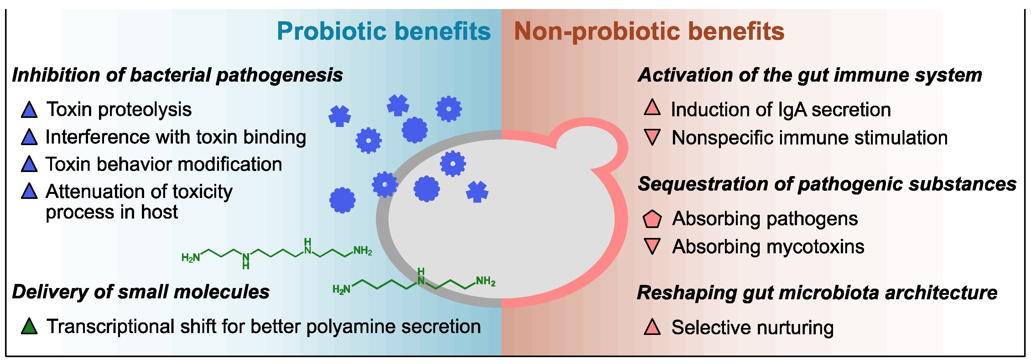
2.1. Innate Probiotic Benefits

The inhibitory activity against the pathogenic mechanisms of varied bacterial toxins has been thoroughly investigated as a representative probiotic capability of Sb. For instance, colitis associated with C. difficile infection has been a major target ailment of the probiotic application of Sb; the protective effect of Sb administration against Clostridioides difficile infection has been proven not only in animal models but also in placebo-controlled clinical trials [20,21,22,36,37,38]. A gnotobiotic murine model demonstrated that the protective effect was associated with the viability of administered Sb as well as its dose [22]. In vivo investigation using a rat model and in vitro assessment employing human colonic cells substantiated that a 54 kDa serine protease secreted by Sb possesses the capacity to attenuate the pathogenicity of C. difficile by proteolyzing its two exotoxins, toxins A and B (TcdA and TcdB) [23,24]. In addition, the serine protease inhibited the binding of TcdA to its receptor on the brush border epithelium in rats [39]. Similarly, Sb exhibits a prophylactic effect on gastrointestinal anthrax by inactivating the lethal toxin from Bacillus anthracis, the causative pathogen of anthrax [40]. As its major virulence factor, B. anthracis synthesizes the lethal toxin consisting of protective antigens and the lethal factor. In vitro tests using human intestinal epithelial cells determined two mechanisms of Sb inactivating the lethal toxin, namely absorbing the protective antigens on its cell wall and inducing its cleavage [25]. However, the molecules exerting the binding and proteolytic actions against the B. anthracis lethal toxin have not been demonstrated from Sb yet.
In addition, the inhibition of bacterial endotoxin by Sb was also demonstrated with Escherichia coli O55:B5 as a model pathogen in a rat model. The key element of the inhibitory activity was a 63 kDa protein phosphatase catalyzing the dephosphorylation of two phosphorylation sites of the lipopolysaccharide of E. coli O55:B5. In vivo tests revealed that the intraperitoneal injection of intact E. coli O55:B5 lipopolysaccharide into rats resulted in 100 ng/mL of circulating tumor necrosis factor-α, along with inflammatory lesions and apoptotic bodies in the liver and heart after 9 h. In contrast, rats injected with dephosphorylated lipopolysaccharide had 40 ng/mL of tumor necrosis factor-α without any observable organic lesions [26].
Sb also attenuates the morphological damage caused by Vibrio cholerae. It was demonstrated in multiple rat model studies that Sb decreased cholera toxin-induced fluid and sodium secretion [41]. Cholera toxin increases cyclic adenosine monophosphate levels by activating adenylate cyclase. The elevation of cyclic adenosine monophosphate levels prompts the secretion of chloride and bicarbonate in crypt cells while inhibiting chloride absorption in villi [42]. In a rat intestinal cell model, the inhibitory effect of Sb on cyclic adenosine monophosphate was abolished when Sb was heat-inactivated. A 120 kDa protein identified from an Sb-conditioned medium has been proposed as the factor mediating the protective efficacy of Sb toward V. cholerae. The 120 kDa protein neutralized the cholera toxin-induced secretion by not exerting proteolytic or protein modification activities on cholera toxin but reducing cyclic adenosine monophosphate levels [27].
While these specific 54 kDa, 63 kDa, and 120 kDa proteins have been proposed to play pivotal roles in the probiotic activities of Sb, genes encoding those proteins have not been identified in the Sb genome [2,43]. Accordingly, their existence in genomes of Sc or other Saccharomyces species has also not been confirmed yet.
Another probiotic capability of Sb is the in situ delivery of advantageous small molecules. In a simulated gastrointestinal tract environment, Sb and Sc showed different transcriptional patterns of genes encoding enzymes involved in the production and secretion of polyamines, such as spermidine and spermine. Specifically, Sb exhibited higher expression levels of the synthetic pathway of ornithine, the precursor of spermidine and spermine, and the polyamine exporter Tpo2p compared to Sc. On the other hand, Sb down-regulated the expression of the ornithine catabolic pathway, the polyamine importer Tpo1p, and the positive regulator of spermine uptake Ptk1p [2,44]. In a rat model featuring a 60% proximal small bowel resection, an elevation in mucosal polyamine concentrations attributable to the influence of Sb was discerned [28]. Polyamines promote the expression of digestive enzymes and nutrient transporters in gut epithelial cells, maintain the integrity of the gut epithelium, and regulate macrophage differentiation for anti-inflammatory effects [2,28,45,46].

2.2. Innate Non-Probiotic Benefits

Saccharomyces yeast cell biomass is reported to interact with the host via cell wall oligosaccharides, such as mannan and glucan, regardless of cell viability. The administration of cell wall polysaccharide fractions of Sb or its whole cells triggers the gut mucosal immune system by stimulating enterocytes and gastrointestinal-associated immune cells via β-glucan and mannose receptors in various animal models [29,47,48,49,50]. In vivo (mice) and in vitro (human colonic cells) assays demonstrated that the induction by Sb cell wall components leads to immunomodulatory responses including the secretion of immunoglobulins, which protects intestinal epithelium from pathogenic bacteria and their toxins [20,51,52]. In addition, the cell wall mannoprotein and β-glucan of Sc were also documented as nonspecific immune stimulators demonstrating interactions with macrophages, neutrophils, and eosinophils in an in vitro evaluation employing murine cell lines [30].
Also, in vitro assays have demonstrated that the mannan oligosaccharide on the surface of both Sc and Sb is a biomaterial that traps enteric pathogens carrying mannose-specific adhesins or receptors, such as Salmonella enterica Typhimurium and Escherichia coli O157, and form yeast–bacteria clusters [9,31,32,53]. Importantly, the binding affinity between representative Saccharomyces strains and gut commensal bacteria has not been reported except for the Sc UFMG 905 strain and Bacteroides fragilis [32]. The trapping capability of Sc and Sb is independent of their viability but prominent in the stationary phase compared to other growth phases [31,32,53]. As the adhesive interaction is dependent on the mannan and mannan-specific adhesion factors, the presence of other sugars and bile salts can interfere with the trapping mechanism [32,54]. The adhesive interaction between the pathogenic bacteria and the Saccharomyces yeast surface can contribute to the therapeutic efficacy of Sb against enteric diseases, as the rivalry between yeast cell wall mannan and oligomannoside chains on enterocytes reduces the colonization and infection chances of the pathogenic bacteria [32,55,56]. Because Saccharomyces yeasts stay in the host gut transiently, yeast cells pass through the host gut, capturing pathogenic bacteria and ultimately diminishing the intestinal population of the pathogens [12,50]. Nevertheless, the in vivo substantiation of the parabiotic protective efficacy of Sb predicated on adhesive interactions with pathogens remains unestablished.
Yeast cell wall polysaccharides absorb not only pathogenic bacteria but also mycotoxins. Aflatoxin B1 is a representative mycotoxin, demonstrating a binding affinity with the majority of Sc strains. In poultry farming, Sc has therefore been utilized as a performance-promoting ingredient with an ameliorating effect against aflatoxin B1 [33]. An in vitro binding test manifested the dose-dependent binding of the Sc cell wall fraction and aflatoxin B1, and the binding affinity was affected by the cell wall mannan condition [57]. On the other hand, thermolyzed Sc and pure mannan oligosaccharide could not successfully attenuate liver damage by aflatoxins, while dehydrated active Sc maintained efficacy against aflatoxins during an in vivo bioassay with rats [34]. Together, these results suggest that the aflatoxin-absorbing capacity of Sc is a parabiotic property but thermosensitive and probably requires all cell wall components [58]. The in vitro binding assay utilizing Sc cell wall materials indicated a notable binding affinity between zearalenone and fumonisin B1 with Sc cell wall polysaccharides, while deoxynivalenol did not exhibit a noticeable binding affinity [57]. However, there is currently no substantiation of the mycotoxin-absorbing www in human subjects.
Furthermore, the administration of cell wall mannan can reshape the architecture of gut microbiota as a selective carbon source. Sb administration increased relative abundances of Bacteroidetes but decreased those of Firmicutes in the mouse gut at the phylum level, and the genus Bacteroides was one of the major momenta of the increase in the Bacteroidetes phylum [35,59]. This taxonomic reconstruction of gut microbiota is connected to the efficacy of Sb administration in multiple disorders including obesity, inflammation, skin dryness, and infectious diseases [9,35,59]. In vitro competition between Bacteroides thetaiotaomicron and C. difficile for quenched Saccharomyces yeast cells demonstrated that the selective nurturing effect is a non-probiotic characteristic of yeast biomass [9]. Bacteroides is a representative genus that efficiently metabolizes various polysaccharides, including yeast cell wall mannan, via a large number of carbohydrate-active enzymes [60]. In particular, B. thetaiotaomicron, one of the dominant members of the commensal gut microbiota, is well known for its capacity to utilize Saccharomyces cell wall mannan through a selfish mechanism. B. thetaiotaomicron does not break down extracellular mannan into small oligosaccharides or mannose monomers. Instead, it produces complex mannan chunks that are not readily usable by many bacteria in the gut [60,61]. B. thetaiotaomicron imports the complex mannan chunks into its periplasmic space through the sus-like transport system and then digests them further to mannan monomers. The selfish mechanism has also been overserved in Bacteroides ovatus, another example of commensal Bacteroides [60,62]. The administration of Saccharomyces cell wall mannan enhanced the relative abundances of both B. thetaiotaomicron and B. ovatus in a human feces fermentation system, and a positive correlation was noted in the relative ratio of B. thetaiotaomicron and B. ovatus. This indicates a coordinated utilization of Saccharomyces cell wall mannan by the two Bacteroides species [62].

3. Engineering of Saccharomyces Yeasts as Therapeutic Avenues

Saccharomyces yeasts have multiple advantages as synthetic probiotic or parabiotic chassis. First, engineered Saccharomyces can be used as complementary therapy together with established avenues for controlling bacterial pathogens, such as bacteriophages and antibiotics targeting pathogenic bacteria, as yeast is tolerant of them. Also, yeast is a better host system than bacteria for synthesizing proteins activated via post-translational modifications. Moreover, Saccharomyces yeasts exhibit a transient stay in the host gut, which is desirable where the impact on the native gut microbiome must be minimized [63]. In addition, numerous genetic tools have been actively developed for Sc, the representative eukaryote model organism. Thus, Sb is also amenable to engineering by sharing the same genetic tools [8,14,64]. Still, compared to Sc, the development of genetically engineered Sb has been limitedly reported to date. There are even fewer reported cases of application and functional validation of engineered Sc strains as synthetic probiotic chassis, probably because they have been considered an inferior platform compared to Sb regarding probiotic capabilities [1,4].
The report by Liu et al. on the construction of Sb auxotrophic mutant strains is a representative and comprehensive example presenting the potentiality of synthetic yeast probiotics [14]. The auxotrophic Sb was developed without antibiotic markers using a CRISPR/Cas9-based system originally optimized for Sc genome editing [14,65]. Auxotrophic mutations capacitate yeast genetic modifications without pricy and toxic selection pressures, which preclude large-scale processes and have unintended effects on cellular functions, such as antibiotics [66]. The potential of the auxotrophic mutant Sb as an engineering chassis was demonstrated by overexpressing a heterologous gene, validating a localization signal tag that has been used for Sc engineering, and building a new metabolic pathway assimilating a new carbon source. In addition, CRISPR/Cas9-based genomic integration of the human lysozyme secretion cassette rendered the Sb culture supernatant capable of lysing bacteria [14]. CRISPR/Cas9-based genomic integration is a more appropriate approach than conventional yeast cloning methodologies for building synthetic probiotics whose biological functions must be stably and accurately manifested in the host gut environment without unnecessary heterologous genetic elements including antibiotic resistance markers [67]. This study successfully proved the potential of CRISPR-based molecular tools and metabolic engineering strategies for developing yeast-based avenues for controlling digestive conditions. The following subsections introduce and discuss more recent advances in the engineering of Saccharomyces yeasts for probiotic or prebiotic purposes (Table 2 summarizes the cases).

3.1. In Situ Delivery of Therapeutic Proteins

Microbial delivery of therapeutic enzymes, antibodies, and cytokines to the host digestive system is an attractive approach because of its cost-efficiency [73,74]. Genotypic and phenotypic characteristics of Saccharomyces must be considered carefully to develop live vectors using the yeasts for the delivery of functional therapeutic proteins. For instance, codon optimization considering the codon bias of Saccharomyces is a critical prerequisite to maximize the efficiency of the production and secretion of functional proteins by Saccharomyces [66,75]. The secretion signal is another major factor affecting protein delivery efficiency. Previous studies have reported a few secretion signal candidates exhibiting better secretion efficiencies than the traditional α-mating factor secretion signal in Saccharomyces, such as secretion signals derived from chicken lysozyme and Sed1p [14,68,76].
Sb has been utilized for reshaping the microbial taxonomic structure in the host digestive system. Kim et al. increased the copy number of human lysozyme secretion cassettes to enhance the bacteria-lyzing capability of the above-mentioned engineered Sb secreting human lysozyme [14]. The cassettes were integrated via CRISPR/Cas9 into intergenic sites that were previously proven to be safe sites in Sc for inserting a large genetic element without perturbing the phenotype [77]. Two copies of the cassette significantly enhanced the lyzing capability compared to that of the parent strain, but triple-copy integration did not enhance the capability further. Administration of the engineered Sb secreting human lysozyme resulted in differential architectures of the murine microbiome compared to the wild-type control, such as a lower taxonomic α-diversity and a lower Firmicutes/Bacteroidetes ratio, and accordingly caused distinctive metabolomic patterns between the test and control groups [68].
Saccharomyces yeasts also have been exploited to control the immunological and biochemical environment in the host gut to control infectious diseases and inflammation. A recent study proved the capability of Saccharomyces as a live vector for delivering antibody fragments that restrain intestinal infectious diseases using Clostridiodes difficile infection (CDI) as a model disease and an antibody fragment (ABAB) neutralizing the major virulence factors of C. difficile, namely the TcdA and TcdB toxins [66,78]. Sc was first harnessed to confirm the toxin-neutralizing functionality of ABABs being secreted from yeast and to screen the best secretion signal for ABABs. The selected construction containing minimal α-mating factor signals was introduced into Sb via an auxotrophic plasmid after codon optimization to maximize secretion efficiency without using antibiotic resistance genes; codon optimization for Saccharomyces yeast enhanced functional ABAB secretion from Sb four-fold. The final engineered Sb strain successfully delivered ABABs in the mouse intestine after antibiotic treatment. Also, the administration of the engineered Sb before the C. difficile spore challenge protected the host mice from death [66]. Similarly, Sb has been a successful host system for the in situ delivery of anti-inflammatory proteins as well. As an example, engineered Sb secreting atrial natriuretic peptide ameliorated the health conditions of a dextran sulfate sodium salt-induced murine colitis model, such as body weight, disease activity index, and survival rate [69]. It is notable that the atrial natriuretic peptide-secreting efficiency and stability of the engineered Sb were secured through multi-copy chromosomal integration on the long terminal repeats of Ty retrotransposons using EasyCloneMulti, which was originally developed for and validated in Sc [79]. As another example, Scott et al. demonstrated the efficacy of the in situ delivery of apyrase, an extracellular ATP-degrading enzyme encoded by Solanum tuberosum RROP1, via an engineered Sc in inflammatory bowel disease. Extracellular adenosine triphosphate is generated by both activated immune cells and commensal gut bacteria and promotes intestinal inflammation and pathology via purinergic signaling. Administrating apyrase-secreting Sc suppressed gut inflammation in murine hosts of inflammatory bowel disease, and accordingly colitis-associated fibrosis and dysbiosis as well, with a similar or higher therapeutic efficacy compared to conventional therapies [63].

3.2. In Situ Delivery of Small Molecules

Saccharomyces yeasts have advantages for synthesizing non-native small molecules, such as terpenes and terpenoids, with the most significant being the compatibility for expressing related enzymes from other eukaryotic origins [64,80]. To assess its potential for the in situ biosynthesis and release of small molecules, Durmusoglu et al. built synthetic metabolic pathways that enable the production of a wide array of model small molecules from vitamin precursors (i.e., β-carotene) to pharmaceuticals (i.e., violacein) and introduced them into Sb. To finely control the pathway expression levels, the influence of various genetic elements such as promoters, terminators, selective markers, and copy numbers on target protein expressions was assessed in advance. They also revealed that Sb effectively colonized in the gnotobiotic mouse gut for over 30 days [64], which was significantly longer than the residency time of 1–2 days in both untreated and antibiotic-treated mice, probably due to competing for niche spaces with commensal microbes [81]. Leveraging these findings and engineered Sb, in vivo production of 194 μg of β-carotene, a notable 56-fold higher β-carotene quantity compared to that presented in the administered Sb, was achieved for 14 days in the gnotobiotic mouse gut [64]. This result corroborated the feasibility of in situ small molecule biosynthesis and release via synthetic Sb strains.

3.3. Biosensing and Expression Systems in Synthetic Probiotic Yeasts

A tunable expression system in synthetic probiotics is essential to achieve an appropriate in situ delivery of therapeutics whose overdose leads to adverse effects. The sensor module is a crucial requirement for the tunable expression system to be self-modulated by designated environmental factors in the host gut. The above-mentioned study by Scott et al. about in situ apyrase delivery via the administration of engineered Sc built a self-tunable system linking extracellular adenosine triphosphate and apyrase secretion levels [63]. The sensor module was developed via the directed evolution of the human P2Y2 receptor, a G protein-coupled receptor that senses both extracellular adenosine triphosphate and extracellular uridine triphosphate [82], using error-prone polymerase chain reaction for enhancing its sensitivity and specificity toward extracellular adenosine triphosphate only when expressed in Sc. The responding element was built based on RROP1 encoding an apyrase with an N-terminal MFα1 signal peptide sequence and the mating-responsive FUS1 promoter [83]. The administration of the resulting engineered Sc indeed secreted functional apyrase in an extracellular adenosine triphosphate-dependent manner and ameliorated intestinal inflammation. Importantly, the efficacy of the engineered Sc was validated without the undesirable side effects linked to fibrosis and dysregulation of the microbiome, which constitutive apyrase delivery or conventional therapies could induce, owing to the extracellular adenosine triphosphate-responsive secretion system [63].
The engineering of transcriptional control is a critical prerequisite to enable probiotic yeast to predictably control innate benefits or drawbacks and execute introduced functions. Zalatan et al. constructed a CRISPR-based synthetic transcriptional program, which consists of nuclease-null Cas9 (dCas9), scaffold RNA (scRNA) carrying a domain recruiting the designated RNA-binding protein (RBP), and an RBP–activator fusion protein, and demonstrated its functionality in Sc [84]. This CRISPR-based synthetic transcription mechanism was optimized and validated in Sb as well to build a scalable and tunable synthetic transactivation system for the purpose of probiotic engineering [8]. Promoters for this system were designed by combining a scRNA target sequence and the core region of the GAL7 promoter [85]. Thus, the resulting transactivation system was easily expanded by introducing a new target sequence to the promoter and corresponding scRNA. More importantly, the resulting transactivations could be orthogonal to native transcriptions, including the galactose metabolic pathway, and to each other due to the complementarity between the promoter and scRNA and the binding specificity between scRNA and RBP. Furthermore, the system could become tunable by the level of nutrients via inducible promoters for the expression of RBP–activator fusion proteins, for instance, macronutrient-inducible promoter (GAL1 promoter by galactose) and micronutrient-inducible promoter (CUP1 promoter by copper) [8].

3.4. Control of the Viability and Activity of Synthetic Yeasts

The transitory stay of Saccharomyces yeasts in the mammalian host gut [1,50] is not disadvantageous for engineering with prebiotic or parabiotic purposes and may even be advantageous regarding the biosafety of genetically modified yeasts in the host gut [9,63]. However, the short duration in the host gut physically limits the efficiency of the in situ production and delivery of therapeutic molecules, such as proteins and metabolites, regardless of expression or secretion efficiencies [68]. Meanwhile, several studies demonstrated the colonization of Sb in a gnotobiotic antibiotic-disturbed host gut, suggesting that one of the major causes of the transient stay of Saccharomyces is its weak nutritional competitiveness in the host gut [22,64,86]. In the meantime, L-fucose is one of the major monosaccharides comprising the mucin oligosaccharide in the gut. Accordingly, multiple gut microbes can utilize L-fucose as a carbon source in the host gut [87,88]. Kim et al. introduced the L-fucose assimilation pathway into Sb to improve its viability and metabolic activities in the gut. The overexpression of E. coli fucose mutarotase, fucose isomerase, fuculose kinase, fuculose 1-phosphate aldoase, and the native hexose transporter showing the highest fucose transport efficiency (HXT4) in Sb enabled its utilization of L-fucose under oxygen-limited culture conditions. However, the impact of L-fucose-utilizing capacity on the viability and metabolic activities of engineered Sb was not validated in an animal model [70]. Chávez-Falcón et al. validated encapsulation as an avenue to enhance the bioavailability of Sb after gastrointestinal digestion. Sb encapsulated in alginate with 5% agavin and 3.75% whey protein exhibited a notable 88.5% cell survival following simulated gastrointestinal digestion; this combination increased the survival of Sb compared to encapsulation with alginate or whey protein independently [71].
On the other hand, consideration of biocontainment strategies is a crucial step in the development of engineered probiotics to mitigate the potential for the genetically modified probiotics to spread beyond the targeted individual, and this is also true for the engineering of Sb or Sc. To address the risk of engineered Sb proliferating outside of the host, Hedin et al. built a robust biocontainment system by combining cold-sensitive and auxotrophic fitness control layers. Specifically, thiamine auxotrophy and elevated sensitivity to low-temperature were accomplished by disrupting THI6 and BTS1 encoding thiamine-phosphate diphosphorylase and geranylgeranyl diphosphate synthase, respectively [72]; BTS1 knockout was previously reported to induce a growth defect at temperatures lower than 25 °C [89]. The biocontained Sb displayed constrained growth when thiamine levels did not exceed 1 ng/mL and at temperatures lower than 20 °C [72].

3.5. Engineering of Yeast Cell Wall Polysaccharides as Parabiotic and Prebiotic Biomaterials

Saccharomyces yeasts, not only Sc but also Sb, pass through the gastrointestinal tract of mammalian hosts quicker than bacterial probiotics [1,50]. Considering their transient stay in the host gut, the parabiotic and prebiotic properties of the yeast biomass must be considered crucial targets of Saccharomyces yeasts for their engineering with therapeutic purposes. Cell wall mannan and glucan polysaccharides are the most representative sources of varied intrinsic health benefits of the yeasts, as discussed in Section 2.2. GDP-mannose and UDP-glucose are sugar moiety donors for the biosynthesis of cell wall mannan and glucan and can be generated from the intermediates of upper glycolysis, fructose 6-phosphate and glucose 6-phosphate, respectively [90]. Theoretically, the oversupply of these nucleotide sugars can enhance the content of corresponding polysaccharides on the yeast cell wall. However, simple overexpression of metabolic pathways toward the nucleotide sugars in Sc and Sb could not enhance their cell wall polysaccharide contents [9]. This is mainly because metabolic fluxes in the nucleotide sugar synthetic pathway cannot overcome the strong and rigid fluxes through glycolysis and ethanol fermentation on fermentable sugars [91]. Disruption of the allosteric upregulation mechanism on phosphofructokinase by fructose 2,6-bisphosphate is an effective approach to overcome the innate metabolic limitation of Saccharomyces yeast [92,93]. Indeed, the deletion of PFK26 and PFK27 encoding isozymes catalyzing fructose 2,6-bisphosphate synthesis significantly increased both the intracellular level of UDP-glucose and cell wall glucan content of Sb. In particular, the augmentation in cell wall glucan content resulting from the double deletion was notably more significant than the increase in UDP-glucose levels. This implies that the primary bottleneck for cell wall glucan overproduction in Sb is the supply of the UDP-glucose precursor, glucose 6-phosphate, rather than the UDP-glucose synthetic pathway or the pathway converting UDP-glucose to cell wall glucan [9].
On the other hand, the cell wall mannan content of Sb was not enhanced by the PFK26 and PFK27 double deletion, suggesting extra metabolic limiting steps before and after GDP-mannose biosynthesis. Intracellular GDP-mannose levels could be significantly increased by the combination of the double deletion and the overexpression of the GDP-mannose synthetic pathway. Furthermore, additional overexpression of the cell wall mannoprotein Sed1p and mannan elaboration pathways in the endoplasmic reticulum and Golgi complex successfully increased the cell wall mannan content of Sb [9]. The higher cell wall mannan content enhanced cell wall mannan-derived protective functionalities of Sb correspondingly, such as an adhesive capacity against S. enterica Typhimurium and a selective nurturing effect on B. thetaiotaomicron against C. difficile [9,32,61]. It is notable that the selective nurturing effect of Sb administration conflicts with previous studies demonstrating viability-associated protective efficacies of Sb against C. difficile infection [23,39]. Still, it is compatible with multiple previous reports about parabiotic and prebiotic effects of Sb and its cell wall mannan, such as the selfish mannan assimilation by B. thetaiotaomicron and the increase in relative Bacteroides abundances in murine gut microbiota [35,61]. Further investigations, including in vivo tests employing appropriate animal models and yeast strains displaying varying levels of cell wall polysaccharides, are required to demonstrate the precise contribution of cell wall mannan to the protective efficacy of Sb against C. difficile infection.
Intriguingly, in the case of Sc, identical engineering increased intracellular UDP-glucose and GDP-mannose levels but could not notably change the levels of either cell wall mannan or glucan [9]. This indicates that Sb has better capacities to biosynthesize and display oligosaccharides on the cell wall than Sc, but the incompetency of Pgm2p (phosphoglucomutase) on glucose 6-phosphate limited the cell wall glucan levels of Sb before the modulation of glycolysis fluxes [7,9]. However, the biochemical mechanism behind the better biosynthesis and display of cell wall polysaccharides by Sb is still unknown.

4. Discussion

4.1. Controversies about the Potential of Sb as a Probiotic Chassis

Better proliferation at human body temperature and acid tolerance at the pH level of gastric fluid have been regarded as the key phenotypic advantages of Sb as a probiotic yeast or a synthetic probiotic chassis. However, it was recently reported that Sc S288C (MATα SUC2 gal2 mal2 mel flo1 flo8-1 hap1 ho bio1 bio6), a well-characterized laboratory Sc strain, exhibits better proliferation at body temperature and higher survival rates in a simulated gut fluid environment than Sb ATCC MYA-796 [9]. Despite its representativeness as a laboratory Sc strain, S288C had not been employed for phenotypic comparison between Sb and Sc before the study. Instead, other laboratory Sc strains with mutations affecting built-in stress response mechanisms have been employed for phenotypic comparison with Sb in other previous studies. For instance, Pais et al. compared Sc BY4741 (MATa his3Δ1 leu2Δ met15Δ ura3Δ) [2], Fietto et al. compared Sc W303 (MATa leu2–3,112 ura3–1 trp1–1 his3–11,15 adn2–1 can1–100 GAL SUC) [6], and Liu et al. compared Sc BY4742 (MATα his3Δ1 leu2Δ lys2Δ ura3Δ) [7] with wild-type Sb regarding the growth at human body temperature. These Sc strains are amino acid auxotrophic mutants, and amino acid auxotrophy affects diverse stress-response mechanisms of Saccharomyces yeasts [94,95,96,97]. Similarly, Edwards-Ingram et al. employed Sc BY3 (MATa ura3-52), another amino acid auxotrophic strain, and Sc Σ1278b to compare the survival rates and acid tolerances of Sb and Sc [5]; Σ1278b is also a widely used laboratory Sc strain like S288C but innately defective in the induction of stress-responsive genes [98]. These previous phenotypic comparisons between wild-type Sb and mutant Sc strains carrying impaired stress response mechanisms might not be fair enough to conclude that Sb is a better probiotic chassis than Sc. They simultaneously emphasize that the protective functionalities of Sb cell biomass should be considered critical determinants of the benefits and engineering targets of Sb as a therapeutic agent, although these potentialities have not been significantly considered yet.

4.2. Concerns about the Safety and Tractability of Sb

In spite of their reported health benefits and potential as chassis for probiotic and prebiotic engineering, the Saccharomyces strains have inherent concerns and limitations that must be kept in mind. First, while Sb is typically classified as a nonpathogenic yeast strain, its administration has been associated with fungemia [99,100,101,102,103,104]. Saccharomyces fungemia is a rare disease and is primarily observed in immunocompromised patients subjected to elevated doses of probiotic interventions containing Sb. Nevertheless, an outbreak case was reported in individuals cohabiting with patients receiving Sb-containing probiotic regimens [99]. It is noteworthy that even among immunocompetent individuals, fungemia induced by Sb is not an impossible scenario. This particular circumstance assumes significance due to its potential to impact morbidity and mortality, especially in cohabiting immunocompromised individuals, thereby contributing to a fungemia outbreak [99,104]. Second, Sb exhibits significantly lower transformation efficiency compared to Sc, probably due to the discrepancy in the cell wall polysaccharide structures between the two yeasts, which makes the construction of synthetic Sb more laborious. [9,66,105]. Lastly, both Sb and Sc have a peculiar metabolic characteristic, namely the Crabtree effect, which represses metabolic pathways other than the glycolytic and ethanol-fermenting pathways on fermentable sugars, including glucose [91]. The exclusive transcriptional pattern arising from the Crabtree effect may impede the exertion of probiotic and non-probiotic benefits of these Saccharomyces yeasts that require metabolic activities other than ethanol fermentation. Further biochemical investigations and synthetic biological conceptions for solving or bypassing the above-mentioned issues would be necessary to make probiotic applications of Sb and Sc safer and more effective and extend their application scope.

5. Conclusions

The non-pathogenic, commercialized probiotic yeast Sb is being utilized extensively for varied gastrointestinal disorders, while the potential of other Sc strains as probiotics has been underestimated. These Saccharomyces yeast strains are genetically amenable and have unique advantages as synthetic probiotic chassis over bacterial probiotics, such as tolerances to antibiotics and bacteriophages and better posttranslational modification capability. There are multiple synthetic probiotic and parabiotic engineering cases of Saccharomyces yeasts that have been accomplished for exerting new functionalities, overcoming intrinsic limitations, and enhancing their own strengths. Still, controversies and concerns about their mode of action, safety, and genetic tractability must be accurately understood and addressed to maximize their effectiveness.

Funding

This work was supported by the National Research Foundation of Korea (NRF) grant funded by the Korean government (MSIT) (RS-2023-00253771) and the Biomaterials Specialized Graduate Program through the Korea Environmental Industry & Technology Institute (KEITI) funded by the Ministry of Environment (MOE).

Institutional Review Board Statement

Not applicable.

Informed Consent Statement

Not applicable.

Data Availability Statement

Data sharing not applicable.

Conflicts of Interest

The authors declare no conflicts of interest.

References

  1. Kelesidis, T.; Pothoulakis, C. Efficacy and safety of the probiotic Saccharomyces boulardii for the prevention and therapy of gastrointestinal disorders. Ther. Adv. Gastroenterol. 2012, 5, 111–125. [Google Scholar] [CrossRef] [PubMed]
  2. Pais, P.; Oliveira, J.; Almeida, V.; Yilmaz, M.; Monteiro, P.T.; Teixeira, M.C. Transcriptome-wide differences between Saccharomyces cerevisiae and Saccharomyces cerevisiae var. boulardii: Clues on host survival and probiotic activity based on promoter sequence variability. Genomics 2021, 113, 530–539. [Google Scholar] [CrossRef] [PubMed]
  3. McFarland, L.V. Saccharomyces boulardii is not Saccharomyces cerevisiae. Clin. Infect. Dis. 1996, 22, 200–201. [Google Scholar] [CrossRef]
  4. Pais, P.; Almeida, V.; Yılmaz, M.; Teixeira, M.C. Saccharomyces boulardii: What Makes It Tick as Successful Probiotic? J. Fungi 2020, 6, 78. [Google Scholar] [CrossRef] [PubMed]
  5. Edwards-Ingram, L.; Gitsham, P.; Burton, N.; Warhurst, G.; Clarke, I.; Hoyle, D.; Oliver, S.G.; Stateva, L. Genotypic and physiological characterization of Saccharomyces boulardii, the probiotic strain of Saccharomyces cerevisiae. Appl. Environ. Microbiol. 2007, 73, 2458–2467. [Google Scholar] [CrossRef]
  6. Fietto, J.L.R.; Araújo, R.S.; Valadão, F.N.; Fietto, L.G.; Brandão, R.L.; Neves, M.J.; Gomes, F.C.O.; Nicoli, J.R.; Castro, I.M. Molecular and physiological comparisons between Saccharomyces cerevisiae and Saccharomyces boulardii. Can. J. Microbiol. 2004, 50, 615–621. [Google Scholar] [CrossRef] [PubMed]
  7. Liu, J.-J.; Zhang, G.-C.; Kong, I.I.; Yun, E.J.; Zheng, J.-Q.; Kweon, D.-H.; Jin, Y.-S. A Mutation in PGM2 Causing Inefficient Galactose Metabolism in the Probiotic Yeast Saccharomyces boulardii. Appl. Environ. Microbiol. 2018, 84, 2280–2287. [Google Scholar] [CrossRef]
  8. Kwak, S.; Mahmud, B.; Dantas, G. A Tunable and Expandable Transactivation System in Probiotic Yeast Saccharomyces boulardii. ACS Synth. Biol. 2021, 11, 508–514. [Google Scholar] [CrossRef]
  9. Kwak, S.; Robinson, S.J.; Lee, J.W.; Lim, H.; Wallace, C.L.; Jin, Y.-S. Dissection and enhancement of prebiotic properties of yeast cell wall oligosaccharides through metabolic engineering. Biomaterials 2022, 282, 121379. [Google Scholar] [CrossRef]
  10. Hudson, L.E.; McDermott, C.D.; Stewart, T.P.; Hudson, W.H.; Rios, D.; Fasken, M.B.; Corbett, A.H.; Lamb, T.J. Characterization of the Probiotic Yeast Saccharomyces boulardii in the Healthy Mucosal Immune System. PLoS ONE 2016, 11, e0153351. [Google Scholar] [CrossRef]
  11. Plaza-Diaz, J.; Ruiz-Ojeda, F.J.; Gil-Campos, M.; Gil, A. Mechanisms of Action of Probiotics. Adv. Nutr. 2019, 10, S49–S66. [Google Scholar] [CrossRef] [PubMed]
  12. Czerucka, D.; Piche, T.; Rampal, P. Review article: Yeast as probiotics—Saccharomyces boulardii, Aliment. Pharmacol. Ther. 2007, 26, 767–778. [Google Scholar] [CrossRef]
  13. Nielsen, J. Yeast Systems Biology: Model Organism and Cell Factory. Biotechnol. J. 2019, 14, e1800421. [Google Scholar] [CrossRef] [PubMed]
  14. Liu, J.-J.; Kong, I.I.; Zhang, G.-C.; Jayakody, L.N.; Kim, H.; Xia, P.-F.; Kwak, S.; Sung, B.H.; Sohn, J.-H.; Walukiewicz, H.E.; et al. Metabolic Engineering of Probiotic Saccharomyces boulardii. Appl. Environ. Microbiol. 2016, 82, 2280–2287. [Google Scholar] [CrossRef] [PubMed]
  15. Schwartz, D.J.; Langdon, A.E.; Dantas, G. Understanding the impact of antibiotic perturbation on the human microbiome. Genome Med. 2020, 12, 82. [Google Scholar] [CrossRef] [PubMed]
  16. Hudson, L.E.; Fasken, M.B.; McDermott, C.D.; McBride, S.M.; Kuiper, E.G.; Guiliano, D.B.; Corbett, A.H.; Lamb, T.J. Functional heterologous protein expression by genetically engineered probiotic yeast Saccharomyces boulardii. PLoS ONE 2014, 9, e112660. [Google Scholar] [CrossRef] [PubMed]
  17. Hamedi, H.; Misaghi, A.; Modarressi, M.H.; Salehi, T.Z.; Khorasanizadeh, D.; Khalaj, V. Generation of a Uracil Auxotroph Strain of the Probiotic Yeast Saccharomyces boulardii as a Host for the Recombinant Protein Production. Avicenna J. Med. Biotechnol. 2013, 5, 29–34. [Google Scholar] [PubMed]
  18. Wang, L.; Sun, H.; Zhang, J.; Liu, Q.; Wang, T.; Chen, P.; Li, H.; Xiao, Y.; Wang, F.; Zhao, X. Establishment and application of target gene disruption system in Saccharomyces boulardii. Biotechnol. Bioprocess Eng. 2015, 20, 26–36. [Google Scholar] [CrossRef]
  19. Moslehi-Jenabian, S.; Pedersen, L.L.; Jespersen, L. Beneficial effects of probiotic and food borne yeasts on human health. Nutrients 2010, 2, 449–473. [Google Scholar] [CrossRef]
  20. McFarland, L.V.; Surawicz, C.M.; Greenberg, R.N.; Fekety, R.; Elmer, G.W.; Moyer, K.A.; Melcher, S.A.; Bowen, K.E.; Cox, J.L.; Noorani, Z. A randomized placebo-controlled trial of Saccharomyces boulardii in combination with standard antibiotics for Clostridium difficile disease. JAMA 1994, 271, 1913–1918. [Google Scholar] [CrossRef]
  21. Surawicz, C.M.; McFarland, L.V.; Greenberg, R.N.; Rubin, M.; Fekety, R.; Mulligan, M.E.; Garcia, R.J.; Brandmarker, S.; Bowen, K.; Borjal, D.; et al. The search for a better treatment for recurrent Clostridium difficile disease: Use of high-dose vancomycin combined with Saccharomyces boulardii. Clin. Infect. Dis. 2000, 31, 1012–1017. [Google Scholar] [CrossRef]
  22. Elmer, G.W.; Corthier, G. Modulation of Clostridium difficile induced mortality as a function of the dose and the viability of the Saccharomyces boulardii used as a preventative agent in gnotobiotic mice. Can. J. Microbiol. 1991, 37, 315–317. [Google Scholar] [CrossRef] [PubMed]
  23. Castagliuolo, I.; LaMont, J.T.; Nikulasson, S.T.; Pothoulakis, C. Saccharomyces boulardii protease inhibits Clostridium difficile toxin A effects in the rat ileum. Infect. Immun. 1996, 64, 5225–5232. [Google Scholar] [CrossRef] [PubMed]
  24. Castagliuolo, I.; Riegler, M.F.; Valenick, L.; LaMont, J.T.; Pothoulakis, C. Saccharomyces boulardii protease inhibits the effects of Clostridium difficile toxins A and B in human colonic mucosa. Infect. Immun. 1999, 67, 302–307. [Google Scholar] [CrossRef]
  25. Pontier-Bres, R.; Rampal, P.; Peyron, J.-F.; Munro, P.; Lemichez, E.; Czerucka, D. The Saccharomyces boulardii CNCM I-745 strain shows protective effects against the B. anthracis LT toxin. Toxins 2015, 7, 4455–4467. [Google Scholar] [CrossRef] [PubMed]
  26. Buts, J.-P.; Dekeyser, N.; Stilmant, C.; Delem, E.; Smets, F.; Sokal, E. Saccharomyces boulardii produces in rat small intestine a novel protein phosphatase that inhibits Escherichia coli endotoxin by dephosphorylation. Pediatr. Res. 2006, 60, 24–29. [Google Scholar] [CrossRef]
  27. Czerucka, D.; Roux, I.; Rampal, P. Saccharomyces boulardii inhibits secretagogue-mediated adenosine 3′,5′-cyclic monophosphate induction in intestinal cells. Gastroenterology 1994, 106, 65–72. [Google Scholar] [CrossRef]
  28. Buts, J.P.; De Keyser, N.; Marandi, S.; Hermans, D.; Sokal, E.M.; Chae, Y.H.; Lambotte, L.; Chanteux, H.; Tulkens, P.M. Saccharomyces boulardii upgrades cellular adaptation after proximal enterectomy in rats. Gut 1999, 45, 89–96. [Google Scholar] [CrossRef]
  29. Buts, J.P.; Bernasconi, P.; Vaerman, J.P.; Dive, C. Stimulation of secretory IgA and secretory component of immunoglobulins in small intestine of rats treated with Saccharomyces boulardii. Dig. Dis. Sci. 1990, 35, 251–256. [Google Scholar] [CrossRef]
  30. Ha, C.H.; Yun, C.W.; Paik, H.D.; Kim, S.W.; Kang, C.W.; Hwang, H.J.; Chang, H.I. Preparation and analysis of yeast cell wall mannoproteins, immune enhancing materials, from cell wall mutant Saccharomyces cerevisiae. J. Microbiol. Biotechnol. 2006, 16, 247–255. [Google Scholar]
  31. Gedek, B.R. Adherence of Escherichia coli serogroup O 157 and the Salmonella typhimurium mutant DT 104 to the surface of Saccharomyces boulardii. Mycoses 1999, 42, 261–264. [Google Scholar] [CrossRef]
  32. Tiago, F.C.P.; Martins, F.S.; Souza, E.L.S.; Pimenta, P.F.P.; Araujo, H.R.C.; Castro, I.M.; Brandão, R.L.; Nicoli, J.R. Adhesion to the yeast cell surface as a mechanism for trapping pathogenic bacteria by Saccharomyces probiotics. J. Med. Microbiol. 2012, 61, 1194–1207. [Google Scholar] [CrossRef]
  33. Pizzolitto, R.P.; Armando, M.R.; Salvano, M.A.; Dalcero, A.M.; Rosa, C.A. Evaluation of Saccharomyces cerevisiae as an antiaflatoxicogenic agent in broiler feedstuffs. Poult. Sci. 2013, 92, 1655–1663. [Google Scholar] [CrossRef]
  34. Baptista, A.S.; Horii, J.; Calori-Domingues, M.A.; da Glória, E.M.; Salgado, J.M.; Vizioli, M.R. The capacity of mannooligosaccharides thermolysed yeast and active yeast to attenuate aflatoxicosis. World J. Microbiol. Biotechnol. 2004, 20, 475–481. [Google Scholar] [CrossRef]
  35. Everard, A.; Matamoros, S.; Geurts, L.; Delzenne, N.M.; Cani, P.D. Saccharomyces boulardii administration changes gut microbiota and reduces hepatic steatosis, low-grade inflammation, and fat mass in obese and type 2 diabetic db/db mice. MBio 2014, 5, e01011–e01014. [Google Scholar] [CrossRef]
  36. Toothaker, R.D.; Elmer, G.W. Prevention of clindamycin-induced mortality in hamsters by Saccharomyces boulardii. Antimicrob. Agents Chemother. 1984, 26, 552–556. [Google Scholar] [CrossRef]
  37. Corthier, G.; Dubos, F.; Ducluzeau, R. Prevention of Clostridium difficile induced mortality in gnotobiotic mice by Saccharomyces boulardii. Can. J. Microbiol. 1986, 32, 894–896. [Google Scholar] [CrossRef]
  38. Castex, F.; Corthier, G.; Jouvert, S.; Elmer, G.W.; Lucas, F.; Bastide, M. Prevention of Clostridium difficile-induced experimental pseudomembranous colitis by Saccharomyces boulardii: A scanning electron microscopic and microbiological study. J. Gen. Microbiol. 1990, 136, 1085–1089. [Google Scholar] [CrossRef]
  39. Pothoulakis, C.; Kelly, C.P.; Joshi, M.A.; Gao, N.; O’Keane, C.J.; Castagliuolo, I.; Lamont, J.T. Saccharomyces boulardii inhibits Clostridium difficile toxin A binding and enterotoxicity in rat ileum. Gastroenterology 1993, 104, 1108–1115. [Google Scholar] [CrossRef]
  40. Beatty, M.E.; Ashford, D.A.; Griffin, P.M.; Tauxe, R.V.; Sobel, J. Gastrointestinal anthrax: Review of the literature. Arch. Intern. Med. 2003, 163, 2527–2531. [Google Scholar] [CrossRef]
  41. Czerucka, D.; Rampal, P. Experimental effects of Saccharomyces boulardii on diarrheal pathogens. Microbes Infect. 2002, 4, 733–739. [Google Scholar] [CrossRef]
  42. Conner, J.G.; Teschler, J.K.; Jones, C.J.; Yildiz, F.H. Staying Alive: Vibrio cholerae’s Cycle of Environmental Survival, Transmission, and Dissemination. Microbiol. Spectr. 2016, 4, 593–633. [Google Scholar] [CrossRef]
  43. Khatri, I.; Akhtar, A.; Kaur, K.; Tomar, R.; Prasad, G.S.; Ramya, T.N.C.; Subramanian, S. Gleaning evolutionary insights from the genome sequence of a probiotic yeast Saccharomyces boulardii. Gut Pathog. 2013, 5, 30. [Google Scholar] [CrossRef]
  44. Buts, J.P.; De Keyser, N.; De Raedemaeker, L. Saccharomyces boulardii enhances rat intestinal enzyme expression by endoluminal release of polyamines. Pediatr. Res. 1994, 36, 522–527. [Google Scholar] [CrossRef]
  45. Rao, J.N.; Xiao, L.; Wang, J.-Y. Polyamines in Gut Epithelial Renewal and Barrier Function. Physiology 2020, 35, 328–337. [Google Scholar] [CrossRef]
  46. Ray, R.M.; McCormack, S.A.; Covington, C.; Viar, M.J.; Zheng, Y.; Johnson, L.R. The requirement for polyamines for intestinal epithelial cell migration is mediated through Rac1. J. Biol. Chem. 2003, 278, 13039–13046. [Google Scholar] [CrossRef]
  47. Gómez-Verduzco, G.; Cortes-Cuevas, A.; López-Coello, C.; Avila-González, E.; Nava, G.M. Dietary supplementation of mannan-oligosaccharide enhances neonatal immune responses in chickens during natural exposure to Eimeria spp. Acta Vet. Scand. 2009, 51, 11. [Google Scholar] [CrossRef]
  48. Kudoh, K.; Shimizu, J.; Ishiyama, A.; Wada, M.; Takita, T.; Kanke, Y.; Innami, S. Secretion and excretion of immunoglobulin A to cecum and feces differ with type of indigestible saccharides. J. Nutr. Sci. Vitaminol. 1999, 45, 173–181. [Google Scholar] [CrossRef]
  49. Swanson, K.S.; Grieshop, C.M.; Flickinger, E.A.; Bauer, L.L.; Healy, H.-P.; Dawson, K.A.; Merchen, N.R.; Fahey, G.C. Supplemental fructooligosaccharides and mannanoligosaccharides influence immune function, ileal and total tract nutrient digestibilities, microbial populations and concentrations of protein catabolites in the large bowel of dogs. J. Nutr. 2002, 132, 980–989. [Google Scholar] [CrossRef]
  50. Gut, A.M.; Vasiljevic, T.; Yeager, T.; Donkor, O.N. Salmonella infection—Prevention and treatment by antibiotics and probiotic yeasts: A review. Microbiology 2018, 164, 1327–1344. [Google Scholar] [CrossRef]
  51. Qamar, A.; Aboudola, S.; Warny, M.; Michetti, P.; Pothoulakis, C.; LaMont, J.T.; Kelly, C.P. Saccharomyces boulardii stimulates intestinal immunoglobulin A immune response to Clostridium difficile toxin A in mice. Infect. Immun. 2001, 69, 2762–2765. [Google Scholar] [CrossRef]
  52. Pontier-Bres, R.; Munro, P.; Boyer, L.; Anty, R.; Imbert, V.; Terciolo, C.; André, F.; Rampal, P.; Lemichez, E.; Peyron, J.-F.; et al. Saccharomyces boulardii modifies Salmonella typhimurium traffic and host immune responses along the intestinal tract. PLoS ONE 2014, 9, e103069. [Google Scholar] [CrossRef]
  53. Posadas, G.A.; Broadway, P.R.; Thornton, J.A.; Carroll, J.A.; Lawrence, A.; Corley, J.R.; Thompson, A.; Donaldson, J.R. Yeast pro- and paraprobiotics have the capability to bind pathogenic bacteria associated with animal disease. Transl. Anim. Sci. 2017, 1, 60–68. [Google Scholar] [CrossRef]
  54. Martins, F.S.; Dalmasso, G.; Arantes, R.M.E.; Doye, A.; Lemichez, E.; Lagadec, P.; Imbert, V.; Peyron, J.-F.; Rampal, P.; Nicoli, J.R.; et al. Interaction of Saccharomyces boulardii with Salmonella enterica serovar Typhimurium protects mice and modifies T84 cell response to the infection. PLoS ONE 2010, 5, e8925. [Google Scholar] [CrossRef]
  55. Fàbrega, A.; Vila, J. Salmonella enterica serovar Typhimurium skills to succeed in the host: Virulence and regulation. Clin. Microbiol. Rev. 2013, 26, 308–341. [Google Scholar] [CrossRef]
  56. Sharon, N.; Eshdat, Y.; Silverblatt, F.J.; Ofek, I. Bacterial adherence to cell surface sugars. Ciba Found. Symp. 1981, 80, 119–141. [Google Scholar]
  57. Shetty, P.H.; Jespersen, L. Saccharomyces cerevisiae and lactic acid bacteria as potential mycotoxin decontaminating agents. Trends Food Sci. Technol. 2006, 17, 48–55. [Google Scholar] [CrossRef]
  58. Hassan, Z.U.; Al Thani, R.; Atia, F.A.; Alsafran, M.; Migheli, Q.; Jaoua, S. Application of yeasts and yeast derivatives for the biological control of toxigenic fungi and their toxic metabolites. Environ. Technol. Innov. 2021, 22, 101447. [Google Scholar] [CrossRef]
  59. Tanihiro, R.; Sakano, K.; Oba, S.; Nakamura, C.; Ohki, K.; Hirota, T.; Sugiyama, H.; Ebihara, S.; Nakamura, Y. Effects of Yeast Mannan Which Promotes Beneficial Bacteroides on the Intestinal Environment and Skin Condition: A Randomized, Double-Blind, Placebo-Controlled Study. Nutrients 2020, 12, E3673. [Google Scholar] [CrossRef] [PubMed]
  60. Abbott, D.W.; Martens, E.C.; Gilbert, H.J.; Cuskin, F.; Lowe, E.C. Coevolution of yeast mannan digestion: Convergence of the civilized human diet, distal gut microbiome, and host immunity. Gut Microbes 2015, 6, 334–339. [Google Scholar] [CrossRef]
  61. Cuskin, F.; Lowe, E.C.; Temple, M.J.; Zhu, Y.; Cameron, E.A.; Pudlo, N.A.; Porter, N.T.; Urs, K.; Thompson, A.J.; Cartmell, A.; et al. Human gut Bacteroidetes can utilize yeast mannan through a selfish mechanism. Nature 2015, 517, 165–169. [Google Scholar] [CrossRef]
  62. Oba, S.; Sunagawa, T.; Tanihiro, R.; Awashima, K.; Sugiyama, H.; Odani, T.; Nakamura, Y.; Kondo, A.; Sasaki, D.; Sasaki, K. Prebiotic effects of yeast mannan, which selectively promotes Bacteroides thetaiotaomicron and Bacteroides ovatus in a human colonic microbiota model. Sci. Rep. 2020, 10, 17351. [Google Scholar] [CrossRef]
  63. Scott, B.M.; Gutiérrez-Vázquez, C.; Sanmarco, L.M.; da Silva Pereira, J.A.; Li, Z.; Plasencia, A.; Hewson, P.; Cox, L.M.; O’Brien, M.; Chen, S.K.; et al. Self-tunable engineered yeast probiotics for the treatment of inflammatory bowel disease. Nat. Med. 2021, 27, 1212–1222. [Google Scholar] [CrossRef]
  64. Durmusoglu, D.; Al’Abri, I.S.; Collins, S.P.; Cheng, J.; Eroglu, A.; Beisel, C.L.; Crook, N. In Situ Biomanufacturing of Small Molecules in the Mammalian Gut by Probiotic Saccharomyces boulardii. ACS Synth. Biol. 2021, 10, 1039–1052. [Google Scholar] [CrossRef]
  65. Zhang, G.-C.; Kong, I.I.; Kim, H.; Liu, J.-J.; Cate, J.H.D.; Jin, Y.-S. Construction of a quadruple auxotrophic mutant of an industrial polyploid Saccharomyces cerevisiae strain by using RNA-guided Cas9 nuclease. Appl. Environ. Microbiol. 2014, 80, 7694–7701. [Google Scholar] [CrossRef]
  66. Chen, K.; Zhu, Y.; Zhang, Y.; Hamza, T.; Yu, H.; Fleur, A.S.; Galen, J.; Yang, Z.; Feng, H. A probiotic yeast-based immunotherapy against Clostridioides difficile infection. Sci. Transl. Med. 2020, 12, eaax4905. [Google Scholar] [CrossRef]
  67. Mugwanda, K.; Hamese, S.; Van Zyl, W.F.; Prinsloo, E.; Plessis, M.D.; Dicks, L.M.T.; Thimiri Govinda Raj, D.B. Recent advances in genetic tools for engineering probiotic lactic acid bacteria. Biosci. Rep. 2023, 43, BSR20211299. [Google Scholar] [CrossRef]
  68. Kim, J.; Atkinson, C.; Miller, M.J.; Kim, K.H.; Jin, Y.-S. Microbiome Engineering Using Probiotic Yeast: Saccharomyces boulardii and the Secreted Human Lysozyme Lead to Changes in the Gut Microbiome and Metabolome of Mice. Microbiol. Spectr. 2023, 11, e0078023. [Google Scholar] [CrossRef]
  69. Liu, C.-H.; Chang, J.-H.; Chang, Y.-C.; Mou, K.Y. Treatment of murine colitis by Saccharomyces boulardii secreting atrial natriuretic peptide. J. Mol. Med. 2020, 98, 1675–1687. [Google Scholar] [CrossRef] [PubMed]
  70. Kim, J.; Cheong, Y.E.; Yu, S.; Jin, Y.-S.; Kim, K.H. Strain engineering and metabolic flux analysis of a probiotic yeast Saccharomyces boulardii for metabolizing L-fucose, a mammalian mucin component. Microb. Cell Fact. 2022, 21, 204. [Google Scholar] [CrossRef] [PubMed]
  71. Chávez-Falcón, M.S.; Buitrago-Arias, C.; Avila-Reyes, S.V.; Solorza-Feria, J.; Arenas-Ocampo, M.L.; Camacho-Díaz, B.H.; Jiménez-Aparicio, A.R. Kinetics and Mechanisms of Saccharomyces boulardii Release from Optimized Whey Protein-Agavin-Alginate Beads under Simulated Gastrointestinal Conditions. Bioengineering 2022, 9, 460. [Google Scholar] [CrossRef]
  72. Hedin, K.A.; Kruse, V.; Vazquez-Uribe, R.; Sommer, M.O.A. Biocontainment strategies for in vivo applications of Saccharomyces boulardii. Front. Bioeng. Biotechnol. 2023, 11, 1136095. [Google Scholar] [CrossRef] [PubMed]
  73. Alexander, L.M.; van Pijkeren, J.-P. Modes of therapeutic delivery in synthetic microbiology. Trends Microbiol. 2023, 31, 197–211. [Google Scholar] [CrossRef] [PubMed]
  74. Nielsen, J. Production of biopharmaceutical proteins by yeast: Advances through metabolic engineering. Bioengineered 2023, 4, 207–211. [Google Scholar] [CrossRef]
  75. Yang, S.; Song, L.; Wang, J.; Zhao, J.; Tang, H.; Bao, X. Engineering Saccharomyces cerevisiae for efficient production of recombinant proteins. Eng. Microbiol. 2023, 4, 100122. [Google Scholar] [CrossRef]
  76. Jin, Y.; Yu, S.; Liu, J.-J.; Yun, E.J.; Lee, J.W.; Jin, Y.-S.; Kim, K.H. Production of neoagarooligosaccharides by probiotic yeast Saccharomyces cerevisiae var. boulardii engineered as a microbial cell factory. Microb. Cell Factories 2021, 20, 160. [Google Scholar] [CrossRef] [PubMed]
  77. Kwak, S.; Kim, S.R.; Xu, H.; Zhang, G.-C.; Lane, S.; Kim, H.; Jin, Y.-S. Enhanced isoprenoid production from xylose by engineered Saccharomyces cerevisiae. Biotechnol. Bioeng. 2017, 114, 2581–2591. [Google Scholar] [CrossRef]
  78. Vedantam, G.; Clark, A.; Chu, M.; McQuade, R.; Mallozzi, M.; Viswanathan, V.K. Clostridium difficile infection: Toxins and non-toxin virulence factors, and their contributions to disease establishment and host response. Gut Microbes 2012, 3, 121–134. [Google Scholar] [CrossRef]
  79. Maury, J.; Germann, S.M.; Jacobsen, S.A.B.; Jensen, N.B.; Kildegaard, K.R.; Herrgård, M.J.; Schneider, K.; Koza, A.; Forster, J.; Nielsen, J.; et al. EasyCloneMulti: A Set of Vectors for Simultaneous and Multiple Genomic Integrations in Saccharomyces cerevisiae. PLoS ONE 2016, 11, e0150394. [Google Scholar] [CrossRef]
  80. Paramasivan, K.; Mutturi, S. Progress in terpene synthesis strategies through engineering of Saccharomyces cerevisiae. Crit. Rev. Biotechnol. 2017, 37, 974–989. [Google Scholar] [CrossRef]
  81. Bauer, M.A.; Kainz, K.; Carmona-Gutierrez, D.; Madeo, F. Microbial wars: Competition in ecological niches and within the microbiome. Microb. Cell 2018, 5, 215–219. [Google Scholar] [CrossRef] [PubMed]
  82. Xu, P.; Feng, X.; Luan, H.; Wang, J.; Ge, R.; Li, Z.; Bian, J. Current knowledge on the nucleotide agonists for the P2Y2 receptor. Bioorganic Med. Chem. 2018, 26, 366–375. [Google Scholar] [CrossRef] [PubMed]
  83. Hagen, D.C.; McCaffrey, G.; Sprague, G.F. Pheromone response elements are necessary and sufficient for basal and pheromone-induced transcription of the FUS1 gene of Saccharomyces cerevisiae. Mol. Cell. Biol. 1991, 11, 2952–2961. [Google Scholar] [PubMed]
  84. Zalatan, J.G.; Lee, M.E.; Almeida, R.; Gilbert, L.A.; Whitehead, E.H.; La Russa, M.; Tsai, J.C.; Weissman, J.S.; Dueber, J.E.; Qi, L.S.; et al. Engineering complex synthetic transcriptional programs with CRISPR RNA scaffolds. Cell 2015, 160, 339–350. [Google Scholar] [CrossRef]
  85. Greger, I.H.; Aranda, A.; Proudfoot, N. Balancing transcriptional interference and initiation on the GAL7 promoter of Saccharomyces cerevisiae. Proc. Natl. Acad. Sci. USA 2000, 97, 8415–8420. [Google Scholar] [CrossRef]
  86. Klein, S.M.; Elmer, G.W.; McFarland, L.V.; Surawicz, C.M.; Levy, R.H. Recovery and elimination of the biotherapeutic agent, Saccharomyces boulardii, in healthy human volunteers. Pharm. Res. 1993, 10, 1615–1619. [Google Scholar] [CrossRef]
  87. Pacheco, A.R.; Curtis, M.M.; Ritchie, J.M.; Munera, D.; Waldor, M.K.; Moreira, C.G.; Sperandio, V. Fucose sensing regulates bacterial intestinal colonization. Nature 2012, 492, 113–117. [Google Scholar] [CrossRef]
  88. Sicard, J.-F.; Le Bihan, G.; Vogeleer, P.; Jacques, M.; Harel, J. Interactions of Intestinal Bacteria with Components of the Intestinal Mucus. Front. Cell. Infect. Microbiol. 2017, 7, 387. [Google Scholar] [CrossRef]
  89. Jiang, Y.; Proteau, P.; Poulter, D.; Ferro-Novick, S. BTS1 encodes a geranylgeranyl diphosphate synthase in Saccharomyces cerevisiae. J. Biol. Chem. 1995, 270, 21793–21799. [Google Scholar] [CrossRef]
  90. Orlean, P. Architecture and biosynthesis of the Saccharomyces cerevisiae cell wall. Genetics 2012, 192, 775–818. [Google Scholar] [CrossRef]
  91. Pfeiffer, T.; Morley, A. An evolutionary perspective on the Crabtree effect. Front. Mol. Biosci. 2014, 1, 17. [Google Scholar] [CrossRef] [PubMed]
  92. Boles, E.; Göhlmann, H.W.; Zimmermann, F.K. Cloning of a second gene encoding 6-phosphofructo-2-kinase in yeast, and characterization of mutant strains without fructose-2,6-bisphosphate. Mol. Microbiol. 1996, 20, 65–76. [Google Scholar] [CrossRef] [PubMed]
  93. Kwak, S.; Yun, E.J.; Lane, S.; Oh, E.J.; Kim, K.H.; Jin, Y.-S. Redirection of the glycolytic flux enhances isoprenoid production in Saccharomyces cerevisiae. Biotechnol. J. 2019, 15, e1900173. [Google Scholar] [CrossRef] [PubMed]
  94. Swinnen, S.; Goovaerts, A.; Schaerlaekens, K.; Dumortier, F.; Verdyck, P.; Souvereyns, K.; Van Zeebroeck, G.; Foulquié-Moreno, M.R.; Thevelein, J.M. Auxotrophic Mutations Reduce Tolerance of Saccharomyces cerevisiae to Very High Levels of Ethanol Stress. Eukaryot. Cell 2015, 14, 884–897. [Google Scholar] [CrossRef] [PubMed]
  95. O’Doherty, P.J.; Lyons, V.; Tun, N.M.; Rogers, P.J.; Bailey, T.D.; Wu, M.J. Transcriptomic and biochemical evidence for the role of lysine biosynthesis against linoleic acid hydroperoxide-induced stress in Saccharomyces cerevisiae. Free Radic. Res. 2014, 48, 1454–1461. [Google Scholar] [CrossRef] [PubMed]
  96. Petti, A.A.; Crutchfield, C.A.; Rabinowitz, J.D.; Botstein, D. Survival of starving yeast is correlated with oxidative stress response and nonrespiratory mitochondrial function. Proc. Natl. Acad. Sci. USA 2011, 108, E1089–E1098. [Google Scholar] [CrossRef] [PubMed]
  97. Bauer, B.E.; Rossington, D.; Mollapour, M.; Mamnun, Y.; Kuchler, K.; Piper, P.W. Weak organic acid stress inhibits aromatic amino acid uptake by yeast, causing a strong influence of amino acid auxotrophies on the phenotypes of membrane transporter mutants. Eur. J. Biochem. 2003, 270, 3189–3195. [Google Scholar] [CrossRef]
  98. Stanhill, A.; Schick, N.; Engelberg, D. The yeast ras/cyclic AMP pathway induces invasive growth by suppressing the cellular stress response. Mol. Cell. Biol. 1999, 19, 7529–7538. [Google Scholar] [CrossRef]
  99. Cassone, M.; Serra, P.; Mondello, F.; Girolamo, A.; Scafetti, S.; Pistella, E.; Venditti, M. Outbreak of Saccharomyces cerevisiae subtype boulardii fungemia in patients neighboring those treated with a probiotic preparation of the organism. J. Clin. Microbiol. 2003, 41, 5340–5343. [Google Scholar] [CrossRef]
  100. Thygesen, J.B.; Glerup, H.; Tarp, B. Saccharomyces boulardii fungemia caused by treatment with a probioticum. BMJ Case Rep. 2012, 2012, bcr0620114412. [Google Scholar] [CrossRef]
  101. Roy, U.; Jessani, L.G.; Rudramurthy, S.M.; Gopalakrishnan, R.; Dutta, S.; Chakravarty, C.; Jillwin, J.; Chakrabarti, A. Seven cases of Saccharomyces fungaemia related to use of probiotics. Mycoses 2017, 60, 375–380. [Google Scholar] [CrossRef] [PubMed]
  102. Niault, M.; Thomas, F.; Prost, J.; Ansari, F.H.; Kalfon, P. Fungemia due to Saccharomyces species in a patient treated with enteral Saccharomyces boulardii. Clin. Infect. Dis. 1999, 28, 930. [Google Scholar] [CrossRef] [PubMed]
  103. Cherifi, S.; Robberecht, J.; Miendje, Y. Saccharomyces cerevisiae fungemia in an elderly patient with Clostridium difficile colitis. Acta Clin. Belg. 2004, 59, 223–224. [Google Scholar] [CrossRef] [PubMed]
  104. Lherm, T.; Monet, C.; Nougière, B.; Soulier, M.; Larbi, D.; Le Gall, C.; Caen, D. Malbrunot. Seven cases of fungemia with Saccharomyces boulardii in critically ill patients. Intensive Care Med. 2002, 28, 797–801. [Google Scholar] [CrossRef]
  105. Kapteyn, J.C.; Riet, B.T.; Vink, E.; Blad, S.; De Nobel, H.; Van Den Ende, H.; Klis, F.M. Low external pH induces HOG1-dependent changes in the organization of the Saccharomyces cerevisiae cell wall. Mol. Microbiol. 2001, 39, 469–479. [Google Scholar] [CrossRef]
Figure 1. Overview of innate health benefits of Sb and Sc. Up-pointing triangle, benefits demonstrated only in Sb to date; down-pointing triangle, benefits demonstrated only in Sc to date; pentagon, benefits demonstrated in both Sc and Sb. Blue, benefits associated with secreted proteins; green, benefits associated with small molecules; pink, benefits associated with cell wall polysaccharides.
Fermentation 10 00051 g001
Table 1. Studies demonstrating key health benefits of Sb and Sc and their mechanisms.
Health BenefitStudy Design and MethodologyOutcomeRef.
Protection against C. difficile infectionRandomized placebo-controlled clinical trial, the combination of Sb and antibioticsLower relative risk of recurrent C. difficile infection in Sb recipients than placebo[20,21]
In vivo (mice), Sb administrationDose- and viability-dependent prophylactic effect of Sb decreasing lethality[22]
In vivo (rats), Sb administration54 kDa protease digested TcdA and inhibited its binding to rat ileal brush border [23]
In vitro (human colonic mucosa), functional validation of 54 kDa protease of Sb Attenuation of toxin-induced electrophysiologic and cytotoxic effects [24]
Potential protection from anthrax In vitro, biochemical assay of B. anthracis lethal toxin and Sb cellsTrapping and proteolysis of protective antigens of lethal toxin by Sb[25]
Inactivation of E. coli endotoxinIsolation of phosphatases from rat small intestines after Sb administration Dephosphorylation and inhibition of E. coli O55:B5 LPS toxicity by 63 kDa protein[26]
Protection against cholera pathogenesisIn vitro (rat small intestine epithelial and human colon cells), Sb or Sb product treatmentModulation of cAMP levels by 120 kDa protein in Sb-conditioned medium [27]
Recovery from proximal enterectomyIn vivo (60% proximal enterectomy rats), Sb administrationImprovement of functional adaptation of remnant ileum via polyamine metabolites[28]
Activation of host immune systemIn vivo (rats), Sb administration Enhanced secretory IgA in the duodenal fluid of rats after Sb administration[29]
In vitro (murine macrophage and fibroblast cells), Sc cell wall fraction treatmentNonspecific immune stimulation (higher NO secretion and macrophage activity) [30]
Absorbing enteric pathogensIn vitro, binding assays of Sb and enteric pathogensAdhesion and sedimentation with S. enterica Typhimurium and enterohemorrhagic E. coli [31]
In vivo (gnotobiotic mice), evaluation of Sb–pathogen adhesion Adhesion between Sb and S. enterica Typhimurium on intestinal epithelium[32]
Absorbing mycotoxinsIn vivo (broiler chicks), Sc administration after aflatoxicosis Positive protection effect of Sc administration on liver weight, histopathology, and growth[33]
In vivo (rats), MOS, thermolyzed Sc, and dehydrated Sc treatment after aflatoxicosisAttenuation of the toxicity and liver damage only by dehydrated Sc administration[34]
Obesity and type 2 diabetesIn vivo (obese and type 2 diabetic mice), Sb administrationReduction of fat mass, hepatic steatosis, and inflammation with shift in host gut microbiome [35]
LPS, lipopolysaccharide; cAMP, cyclic adenosine monophosphate; MOS, manno-oligosaccharide.
Table 2. Examples of Sb and Sc engineering as therapeutic chassis described in this review.
StrategyPurposeStrainRef.
In situ delivery of therapeutic proteins
Secretion of human lysozymeReshaping the taxonomic architecture of
the host gut microbiome
Sb[68]
Secretion of the antibody fragment-neutralizing
TcdA and TcdB
Performing yeast-based immunotherapy
for C. difficile infection
Sb[66]
Multi-copy genomic integration of
atrial natriuretic peptide secretion cassettes
Alleviating colitis in the mammalian host gut Sb[69]
Secretion of apyrase degrading extracellular ATPControlling the inflammatory mechanism
induced by extracellular ATP
Sc[63]
In situ delivery of small molecules
Optimization and assembly of
genetic elements for multiple gene expressions
In situ biomanufacturing and delivery of
β-carotene and violacein
Sb[64]
Biosensing and expression systems
Engineering human P2Y2 receptorAchieving extracellular ATP-specific
apyrase secretion system
Sc[63]
dCas9-scRNA-based synthetic transactivationAchieving nutrient-dependent
synthetic signaling mechanisms
Sb[8]
Control of the viability and activity
Introduction of heterogenous L-fucose assimilation pathwayImproving competence in the mammalian host gutSb[70]
Whey protein–agavin–alginate encapsulationEnhancing Sb viability
after the gastrointestinal digestion
Sb[71]
Knock-out of THI6 and BTS1Building multi-layered biocontainment
via cold-sensitive thiamine auxotroph
Sb[72]
Cell wall oligosaccharide engineering
Modulation of glycolysis and sugar nucleotide
synthetic pathways
Enhancing cell wall oligosaccharide contents
and related prebiotic and parabiotic effects
Sb, Sc[9]
Disclaimer/Publisher’s Note: The statements, opinions and data contained in all publications are solely those of the individual author(s) and contributor(s) and not of MDPI and/or the editor(s). MDPI and/or the editor(s) disclaim responsibility for any injury to people or property resulting from any ideas, methods, instructions or products referred to in the content.

