Article

Differential Cytotoxic Effects of Graphene Oxide and Its Functionalized Derivatives on Colon 26 Carcinoma Cells: Implications for Cancer Therapeutic Applications


by
Solange Amigues
1,
Natalia Krasteva
2,
Kamelia Hristova-Panusheva
2,
Milena Keremidarska-Markova
3,
Giorgio Speranza
4 and
Firas Awaja
1,*
1
Department of Medicine, University of Galway, H91TK33 Galway, Ireland
2
Institute of Biophysics and Biomedical Engineering, Bulgarian Academy of Sciences, Room 303, Bl. 21, Acad. Georgi Bonchev Str., 1113 Sofia, Bulgaria
3
Faculty of Biology, Sofia University, St. Kliment Ohridski, 1164 Sofia, Bulgaria
4
Fondazione Bruno Kessler, 18 Sommarive Str., 38123 Trento, Italy
*
Author to whom correspondence should be addressed.
Submission received: 9 November 2025 / Revised: 7 December 2025 / Accepted: 31 December 2025 / Published: 6 January 2026
(This article belongs to the Topic Application of Graphene-Based Materials, 2nd Edition)

Abstract

Graphene oxide (GO)-based nanomaterials hold significant potential for targeted cancer therapy owing to their tunable physicochemical properties and surface versatility. In this study, we systematically evaluated the cytotoxicity of pristine GO (graphene oxide) and its surface-functionalized derivatives, GO-CH4 (methyl), GO-NH2 (amine), and GO-O2 (carboxyl), against murine Colon 26 carcinoma cells. Cell morphology, adhesion, and proliferation were assessed after three days of exposure using fluorescein diacetate (FDA) live/dead staining and the WST-1 mitochondrial activity assay. Distinct material-dependent biological responses were observed: GO-CH4 (methyl) and GO-O2 (carboxyl) exhibited pronounced cytotoxicity, reducing cell adhesion and proliferation by more than 50% relative to controls, whereas GO-NH2 (amine) induced only moderate effects. Pristine GO (graphene oxide) showed minimal impact on cell viability and morphology, consistent with its limited cellular internalization. These results demonstrate that surface functionalization critically governs GO (graphene oxide) biocompatibility and cytotoxicity, underscoring its potential as a tunable platform for developing graphene-based cancer therapeutics, implant coatings, and biointerfaces with controlled cellular responses.

1. Introduction

Cancer remains a leading cause of mortality worldwide, with complex pathophysiology that often renders conventional therapies (surgery, chemotherapy, radiotherapy) insufficient due to issues such as multidrug resistance, limited tumour selectivity, off-target toxicity and poor bioavailability of therapeutic agents [1,2]. In recent years, nanomaterials have emerged as powerful adjuncts or alternatives in oncology, offering targeted delivery, enhanced contrast/imaging, and new mechanisms such as photothermal or photodynamic therapy [3,4]. Among such nanomaterials, two-dimensional (2D) carbon-based sheets, most notably Graphene, Graphene Oxide (GO) and Reduced Graphene Oxide (rGO), have attracted intense interest due to their remarkable physical, chemical and biological properties.
Although GO contains intrinsic oxygen-based groups, these are randomly distributed and chemically unstable. Plasma treatment enables targeted enrichment of specific functional groups with controlled density and surface accessibility, producing tailored biological responses.

1.1. Graphene and Graphene Oxide: Structure and Physics

Graphene is a single atomic layer of carbon atoms arranged in a hexagonal honeycomb lattice, with each carbon atom sp2-hybridized and contributing to a delocalized π-electron network [3]. This arrangement yields exceptional properties: extraordinarily high electrical conductivity, excellent thermal conductivity, extreme mechanical strength and high surface area. When deliberately oxidized, graphene oxide introduces a variety of oxygen-containing functional groups (epoxides, hydroxyls, carbonyls, carboxyls) and structural defects, transforming the material’s electronic, chemical and dispersion behaviour [2,5]. These functional groups disrupt the continuous sp2 carbon lattice but create reactive sites for further modification and impart amphiphilic behaviour (hydrophobic graphene patches and hydrophilic oxidized domains) [2]. In rGO (reduced graphene oxide) the oxygen content is reduced (via chemical or thermal treatment), partially restoring conjugation and electrical/thermal conductivity while preserving many of the sheet-like features and high surface area [6]. From a physics standpoint, the oxidation state, defect density, and functionalization modulate key parameters such as electronic band structure, π–π stacking capacity, hydrophobic/hydrophilic balance, surface charge, ripple morphology and mechanical stability [5,7].
The presence of ripples, wrinkles or internal layers (in multilayer stacks) in graphene/GO (graphene oxide) sheets is not merely a morphological curiosity: they influence surface area, edge-to-basal plane ratio, local curvature, and thus adsorption, interaction with biomolecules or membranes, and mechanical stresses [5]. These microstructural features, combined with chemical functionality, provide a unique platform for biomedical applications.

1.2. Surface Functional Groups and Biological Interaction

The functional groups on GO (graphene oxide) and rGO (reduced graphene oxide) serve multiple roles in biological contexts. Carboxyl (–COOH), hydroxyl (–OH) and epoxy (–O–) groups enhance dispersion in aqueous media, permit conjugation with biomolecules (proteins, DNA/RNA, targeting ligands) and enable further surface tailoring [2,8]. The oxidized domains generate local charges, potential for hydrogen-bonding, π–π interactions (especially with aromatic drug molecules), and also payload attachment via electrostatic/π–π stacking. In therapeutic contexts, such surface groups are integral to drug/gene delivery, photothermal/photodynamic functions, and modulation of cellular uptake or cytotoxicity [1,6].
Conversely, the degree of oxidation, the nature and density of functional groups, lateral size, sheet thickness and aggregation tendency critically influence biological behaviour—including cellular uptake, internalization mechanism, membrane interaction, generation of reactive oxygen species (ROS), immune response, and cytotoxicity [2]. For example, increased epoxide/hydroxyl loading can lead to structural rippling and reduction in mechanical rigidity [5]. which may translate to altered interactions with cell membranes. The richer the functionalization, the greater the number of binding or reactive sites, but also the higher the potential for undesired biological responses such as oxidative stress or membrane damage.

1.3. Graphene-Based Materials in Cancer Therapy

Graphene-based materials (GBMs) have been increasingly explored in oncological applications. Early work demonstrated that PEGylated GO (graphene oxide) can act as nanocarriers for anticancer drugs (e.g., doxorubicin), utilizing large specific surface area, π–π stacking, hydrophobic interactions and the enhanced permeability and retention (EPR) effect to accumulate in tumour tissues [2,8]. Subsequent research expanded into photothermal therapy (PTT), photodynamic therapy (PDT), combined chemo-/photo-therapeutics and biosensing/diagnosis [1,3]. For instance, GO (graphene oxide) and derivatives have been shown to convert near-infrared (NIR) light into heat (photothermal) and/or generate ROS under irradiation, enabling targeted tumour ablation [1,2].
One review [2] highlights the versatility of graphene oxide-based multifunctional nanomaterials in cancer treatment—including cargo delivery, phototherapy and imaging—and stresses that “surface properties, photothermal property, and pH sensitivity” are key determinants of performance. Similarly, another review [3] emphasizes that the sheet-like morphology, high surface area and functionalization flexibility of 2D graphene-based materials confer advantages in both therapeutic and diagnostic (‘theranostic’) modalities.
Moreover, recent systematic analyses [9] have shown that GO/rGO (graphene oxide/reduced graphene oxide) platforms are actively being investigated for bone cancer and other solid tumour types, indicating a broadening of therapeutic horizons. Nonetheless, despite these advances, translation to clinical applications remains limited—in large part due to challenges in biocompatibility, reproducible large-scale production, surface safety and unravelling the precise mechanisms of cytotoxicity and tumour specificity.

1.4. Rationale for Current Study

Despite the promise of GO/rGO (graphene oxide/reduced graphene oxide) in cancer therapy, a persistent gap lies in understanding how specific surface functionalizations (hydrophobic groups, amine groups, acidic groups, etc.) influence the cellular interactions, cytotoxicity and therapeutic potential of GO (graphene oxide) derivatives—especially in a cancer cell context. Many studies treat GO (graphene oxide) as a relatively generic platform, but the oxidation level, functional group type and distribution are rarely systematically correlated with biological outcomes such as adhesion, proliferation, apoptosis, membrane integrity or uptake.
In this work, we focus on the well-characterized Colon 26 carcinoma cell line and compare the cytotoxic and morphological responses to GO (graphene oxide) and three derivative forms: GO-CH4 (methyl) (hydrophobic functionalization), GO-NH2 (amine) (amine functionalization) and GO-O2 (carboxyl) (acidic/oxidized surface functionalization). By correlating functional group chemistry with cell morphology, adhesion, mitochondrial activity and proliferation, we aim to reveal how surface modification governs GO cytocompatibility and cytotoxicity, and by extension, its suitability for potential cancer-therapeutic or implant-coating applications.
Our approach also pays particular attention to the physics of GO (graphene oxide) sheets (internal layering, ripple formation, surface area, functional group density) as determinants of the biological interaction. Specifically, we explore how increased internal layers and rippling can enhance surface area and provide enhanced interaction with cell membranes, and how functional group chemistry modulates membrane interaction, ROS generation and ultimately cellular fate.
By doing so, we aim not only to deliver new experimental data, but also to contribute to the mechanistic understanding of graphene oxide-based nanomaterials in oncology, thereby helping to inform safer and more effective design of graphene-derived platforms for cancer therapies and associated biomedical uses.

2. Materials and Methods

2.1. Materials

The material used in this study involves pristine graphene oxide (GO) which from San Sebastian, Spain, graphene derivatives with different groups attached, including GO-CH4 (methyl), GO-NH2 (amine), and GO-O2 (carboxyl). The GO sheets were made through Hummer’s method [10]. The control was glass coverslips, and the cell line was Murine Colon 26 carcinoma cells (ATCC® CRL-2638™, Manassas, VA, USA) [11].

2.2. Plasma Treatments

In this experimental study, graphene oxide_paper foils were plasma-treated to functionalize their surfaces with specific chemical groups exhibiting well-defined polarity. The treatments were carried out in a low-pressure RF-Inductively Coupled Plasma (ICP) reactor (CCR Technology GmbH, Troisdorf, Germany). Uniformity of plasma functionalization was confirmed by sampling three independent XPS measurement points across each substrate (centre and two edge positions). No statistically meaningful variation (<2%) in elemental composition was detected, indicating homogeneous plasma exposure.
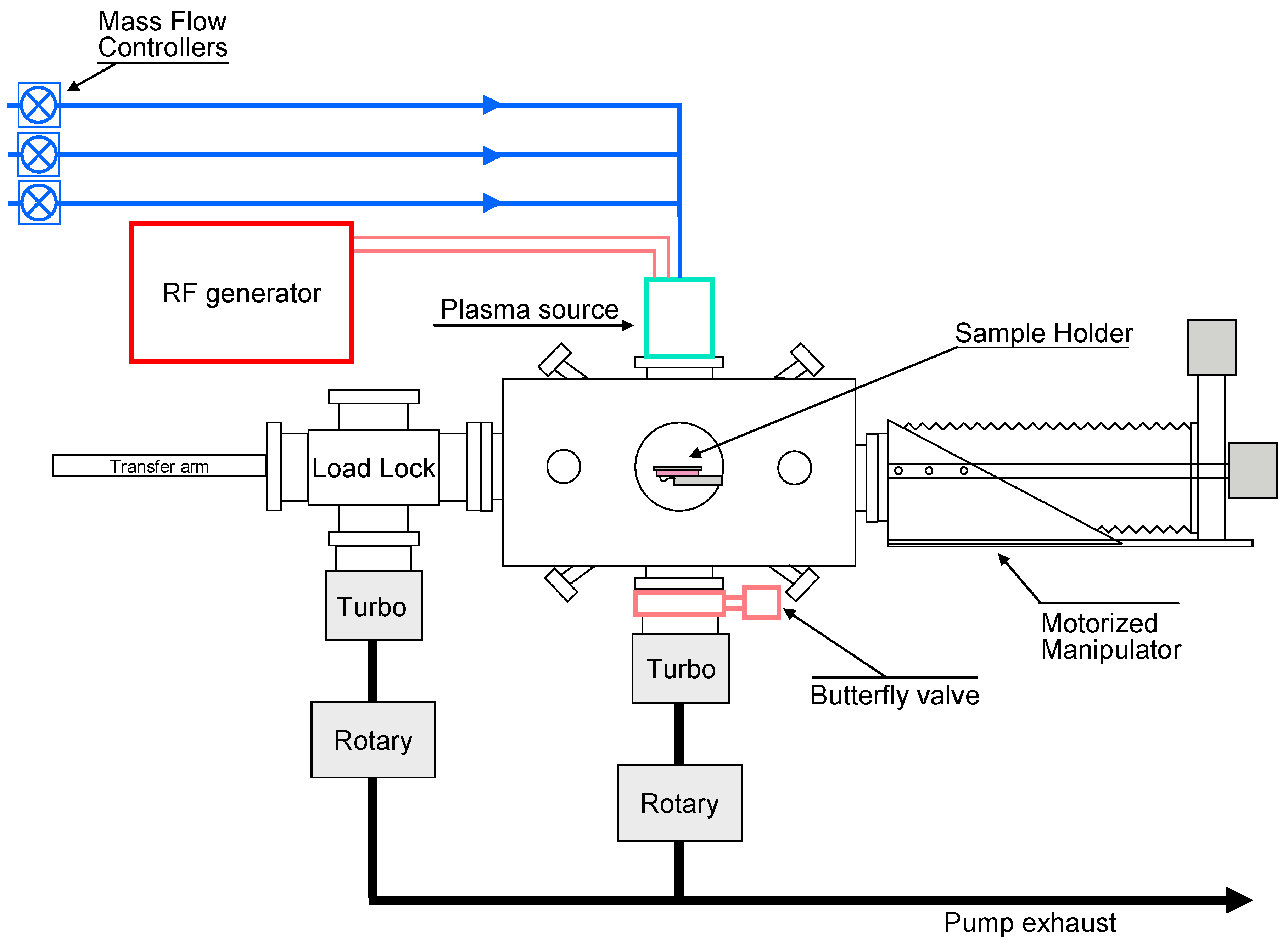
The plasma source has a diameter of 200 mm, which makes it possible to treat large surfaces while maintaining acceptable variations in the amount of grafted elements across the foil. Furthermore, we observe that the ICP reactor is a low-pressure cold plasma enabling surface treatments at RT temperature. Pure CH4, as well as mixtures of CH4 with O2 or NH3 in different proportions, was used to impart neutral, negative, and positive polarities to the substrate surfaces. A schematic of the system is shown in Figure 1.
For all plasma treatments, the RF power was kept constant at 250 W. The plasma source operated at an RF frequency of 13.56 MHz under inductively coupled excitation. To enhance plasma generation efficiency, electron cyclotron resonance was applied, enabling the production of high ion currents while maintaining low and controllable ion energy. Additionally, an integrated RF-matching network ensured efficient power transfer. The system allowed full customization of key experimental parameters, including RF power, precursor composition, precursor flow rate, and pressure. These parameters are essential for achieving effective surface functionalization under fully controlled and reproducible conditions.
When using CH4 + O2 or CH4 + NH3 mixtures, the total flow rate was kept constant at 80 sccm in the former case and 50 sccm in the latter. The operating parameters for all plasma treatments are summarized in Table 1.

2.3. X-Ray Photoelectron Spectroscopy (XPS) Analysis

The effect of plasma treatments was analyzed using X-ray Photoelectron Spectroscopy (XPS), a non-destructive technique employed to investigate the surface chemistry of pristine and plasma-treated graphene oxide_paper substrates. Wide-scan spectra, used to identify the chemical elements present, were acquired with an Axis DLD Ultra spectrometer (Kratos, Manchester, UK) at a pass energy of 160 eV. High-resolution spectra were recorded at a pass energy of 20 eV, providing an energy resolution of 0.3 eV. Peak binding energies were referenced to NIST spectral standards and fitted using constrained FWHM ranges consistent with carbon-based materials. All measurements and plasma handling were performed at controlled room temperature (22–24 °C), within the stability range reported for GO-based materials.
Elemental composition values obtained from XPS are semi-quantitative and represent peak-fitted spectral data. The reported data are based on representative spectra collected from each sample. Spot-to-spot variations remained within 3%, which is consistent with commonly accepted analytical precision for XPS measurements. Therefore, standard deviation values are not included.
Spectral processing was performed with a custom-developed software package (RxpsG.2.3-2) [12]. Core-level fitting was carried out using Gaussian components for peak decomposition, and chemical bond assignments were made based on reference manuals and established databases [13,14].

2.4. Biological Experiments

Murine Colon 26 carcinoma cells were cultured in RPMI-1640 medium supplemented with 10% fetal bovine serum (FBS) and maintained at 37 °C in a humidified atmosphere containing 5% CO2. The cultures were then exposed to graphene oxide (GO) and its derivatives at a concentration of 50 µg/mL for five days (15–20 January 2018). Following exposure, cell morphology and viability were assessed using fluorescein diacetate (FDA) staining for live/dead visualization.
Cell proliferation and cytotoxicity were quantified based on mitochondrial activity using the WST-1 assay, with absorbance measured at 450 nm. Cell adhesion was evaluated by counting the number of attached cells from FDA-stained images, analyzing five randomly selected fields per sample, including the untreated control.
All data are presented as mean ± standard deviation (n = 3). Statistical significance was determined using one-way ANOVA followed by Tukey’s post hoc test, with p < 0.05 considered statistically significant.

2.5. Cell Culture

Murine colon carcinoma cell line Colon 26 (CLS, Mannheim, Germany) was used in this study. Only cancer-derived Colon-26 cells were included, as the study specifically targeted tumour-selective cytotoxicity. Future comparative work will incorporate normal epithelial colon cells to assess selectivity between malignant and healthy tissues. Cells were maintained in RPMI-1640 medium (Sigma-Aldrich, Taufkirchen, Germany) supplemented with L-glutamine and 10% fetal bovine serum (FBS, Invitrogen, Carlsbad, Canada) at 37 °C in a humidified atmosphere containing 5% CO2. The culture medium was replaced every two to three days. Once the cells reached approximately 80% confluence, they were detached using 0.1% trypsin/EDTA solution and seeded onto the modified GO materials at a density of 3 × 10⁴ cells/mL for morphology and proliferation experiments.

2.6. Cell Morphology

To evaluate overall cell morphology, GO samples and plain cover glass (CG, control) were placed in 12-well plates (CoStar Group, Richmond, VA, USA). Colon 26 cells were seeded at a concentration of 3 × 10⁴ cells/mL. After 24 h of incubation, viable cells were stained with fluorescein diacetate (FDA) by adding 5 μL/mL from a 5 mg/mL stock solution prepared in acetone. FDA penetrates cell membranes and is hydrolyzed by nonspecific esterases in living cells, producing fluorescein that accumulates intracellularly and enables direct visualization under fluorescence microscopy. Representative images of adherent cells were captured using a fluorescence microscope (Zeiss Axiovert 25, Jena, Germany; 10× magnification) equipped with a digital camera. At least three representative images were obtained for each sample and subsequently analyzed for quantification of adhered cells using ImageJ software(Version 1.54).

2.7. Cell Proliferation Assay

The cytotoxic effects of modified GO materials on Colon 26 cells were evaluated using the Cell Proliferation Reagent WST-1 (Roche). This nonradioactive, colorimetric assay is based on the bioreduction of a water-soluble tetrazolium salt (WST-1) into a soluble formazan product by cellular enzymes, primarily at the cell surface. This reaction depends largely on the glycolytic production of NAD(P)H in metabolically active cells; thus, the amount of formazan dye formed directly correlates with the number of viable cells.
For the assay, cells were seeded on the samples in 12-well plates at a density of 3 × 10⁴ cells/mL in 2 mL of RPMI medium supplemented with 10% FBS. At 24, 48, and 72 h post-seeding, the culture medium was discarded, cells were washed once with PBS, and the WST-1 assay was performed according to the manufacturer’s instructions. Absorbance of the formazan product was measured at 450 nm using a spectrophotometer. Cells cultured on plain cover glass (without GO materials) served as controls.

3. Results

3.1. Material Characterization

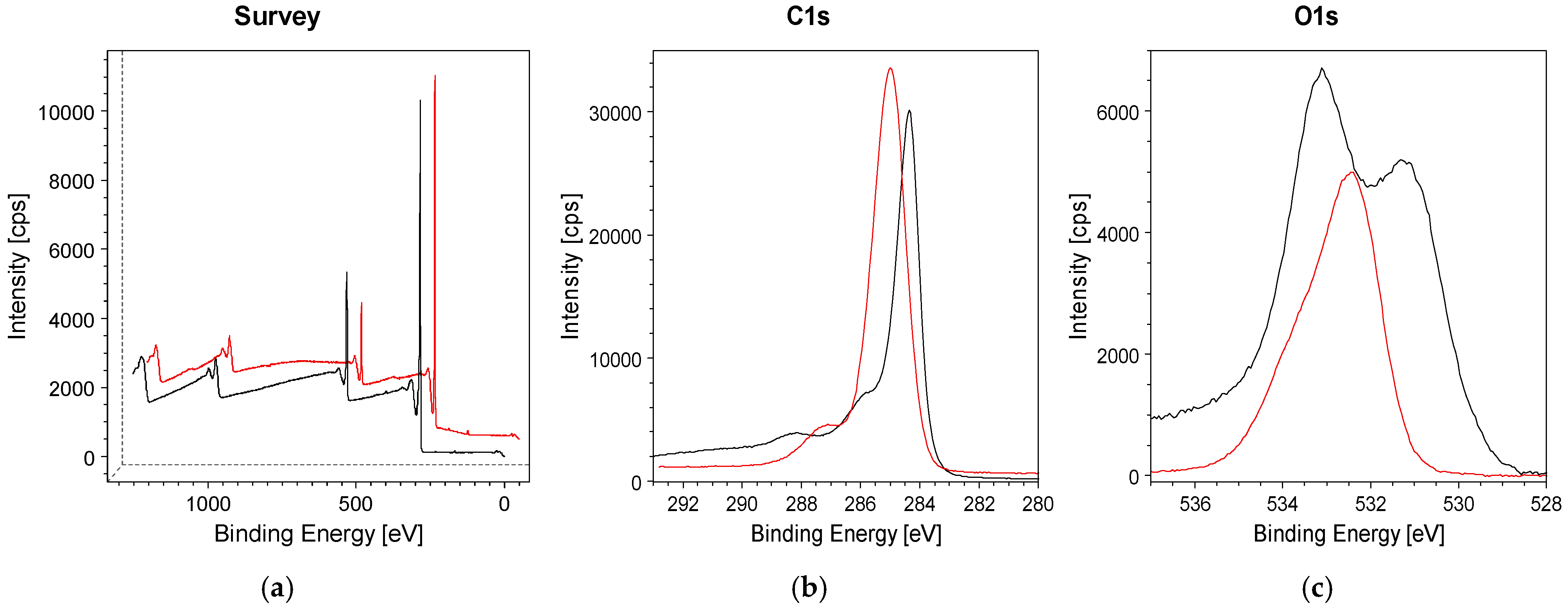
The effects of plasma treatments were investigated by XPS. Figure 2 shows the spectra of pristine GO_paper (black) and GO_paper treated in a pure methane plasma (sample S1 red).
The wide-scan spectra in Figure 2a are dominated by two main peaks: C 1s at a binding energy (BE) of ~284.4 eV and O 1s at ~532 eV. Additional features at higher binding energies correspond to Auger transitions of the same elements. The plasma treatment forms a nanoscale surface modification layer (<5 nm), which remains within the XPS sampling depth and does not produce a bulk film.
The high-resolution C 1s spectrum of pristine graphene oxide_paper (Figure 2b, black line) displays a main peak at ~284.4 eV, attributed to carbon atoms in a graphite-like structure. This component arises from the partial reduction of GO (graphene oxide) during the synthesis of graphene oxide_paper, which includes a thermal treatment step to remove residual water. Nonetheless, carbon–oxygen bonds remain present in the material, as indicated by features at ~286.7 eV, 287.9 eV, and 289.0 eV, assigned to ether-like, O–C–O, and carboxyl groups, respectively.
In contrast, the C 1s spectrum of plasma-treated graphene oxide_paper (sample S1, red line) exhibits a significant spectral change. The C 1s peak shifts to a higher binding energy (~285 eV) compared to the pristine sample. This position is typically associated with hydrocarbon species. In this case, the spectral change can be attributed to the deposition of a hydrogenated amorphous carbon (a-C:H) film formed during CH4 plasma treatment. No evidence of structural degradation was observed based on C 1s peak symmetry and microscopic inspection. The C and O atomic concentrations derived from these spectra are summarized in Table 2.
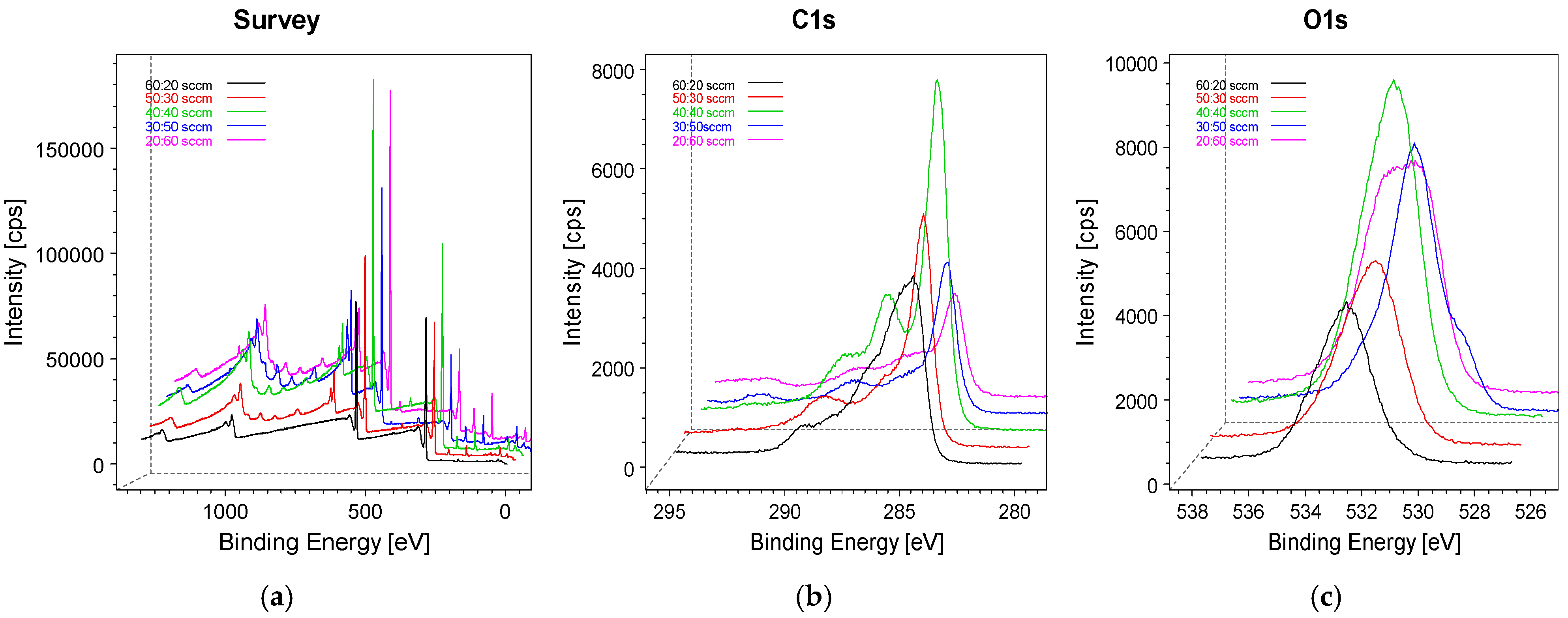
We now turn to samples S2–S6, corresponding to graphene oxide_paper treated in CH4 + O2 plasmas with varying oxygen content. Unlike pure CH4 (methyl) plasma, the presence of oxygen leads to an increase in oxygen incorporation. The resulting effects are shown in Figure 3. As expected, the wide-scan spectra in Figure 3a reveal a progressive increase in the oxygen-to-carbon ratio from sample S2 to S6. The high-resolution spectra in Figure 3b,c highlight pronounced changes in both the C 1s and O 1s core lines as plasma conditions vary.
Specifically, the C 1s spectra show enhanced intensity in the 287–289.5 eV range, corresponding to carbonyl, O–C–O, and carboxyl functional groups. This increase is mirrored by a stronger high-BE component in the O 1s spectra. Together, these changes reflect the higher oxygen content in the plasma-treated graphene oxide_paper and indicate a progressive increase in surface polarity with increasing O2 (oxygen) flow rate. The elemental abundances obtained from XPS analysis are summarized in Table 2.
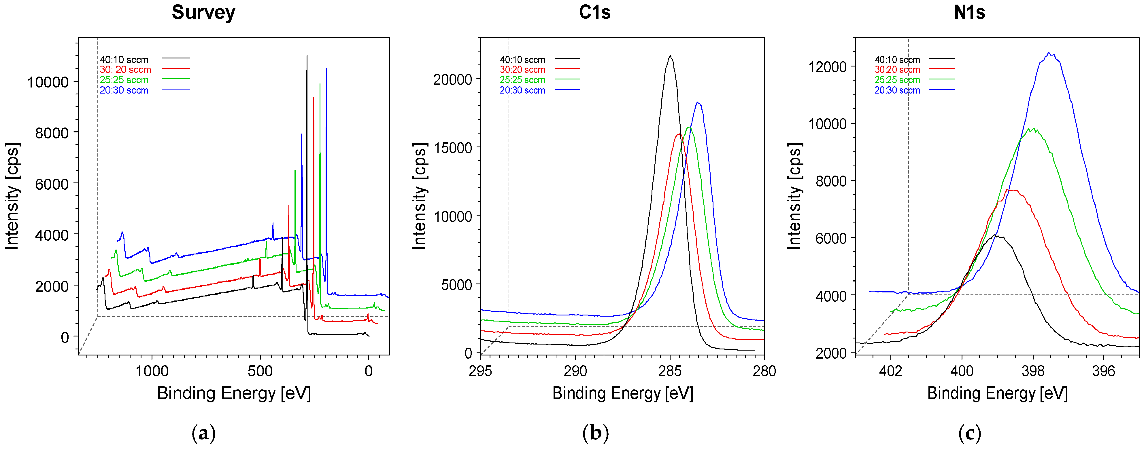
Finally, Figure 4 presents the surface composition of samples S7–S10, which were treated in CH4 + NH3 plasmas with varying proportions. As shown in the wide-scan spectra, increasing the NH3 flow rate results in a progressive increase in the N 1s peak intensity at ~400 eV. This increase is accompanied by a decrease in the C 1s intensity, consistent with the reduced CH4 flow rate in the reactor feed.
The wide spectra also display a minor peak at ~530 eV, attributable to oxygen. This feature likely originates from residual H2O molecules released from the reactor walls, which generate O-based radicals subsequently incorporated into the graphene oxide_paper surface.
The C 1s spectrum (Figure 4b) consists of a broad, featureless peak, which was deconvoluted into three components corresponding to CHx, C–N, and C–O bonds. The high-resolution N 1s spectrum (Figure 4c) was decomposed into two main components, assigned to C–N bonds and protonated amine (C–NH+) species.
The elemental abundances derived from XPS analysis are summarized in Table 2.

3.2. Overall Cell Morphology (FDA Staining)

Following FDA staining, cellular morphology was examined under microscopy for the control group and cells cultured on GO (graphene oxide) and its functionalized derivatives (GO-CH4, GO-NH2, and GO-O2). Control cells displayed a healthy, spindle-shaped morphology and formed a confluent monolayer characteristic of viable epithelial cultures. Cells grown on pristine GO (graphene oxide) exhibited minor membrane irregularities but maintained overall adhesion. In contrast, cultures on GO-CH4 (methyl) showed pronounced cytoplasmic shrinkage, cellular rounding, and evident detachment from the substrate. Cells exposed to GO-NH2 (amine) demonstrated partial loss of adhesion accompanied by moderate membrane blebbing. The GO-O2 (oxide) group displayed the most severe response, characterized by extensive cell fragmentation and the presence of numerous apoptotic bodies.

3.3. Overall Cell Morphology on Functionalized GO (Graphene Oxide) Surfaces/Sheets

Fluorescence microscopy was used to assess the morphology of Colon 26 cells cultured on pristine and functionalized GO (graphene oxide) sheets (Figure 5) and the results were in accordance with the literature. In the control group, cells exhibited typical fibroblast-like morphology with an elongated spindle shape, extensive cytoplasmic extensions, and a confluent monolayer. Cells grown on non-modified GO (graphene oxide) displayed minor membrane irregularities but retained some adhesion, although overall density was reduced [15]. In contrast, cells on GO-CH4 (methyl) showed severe cytoplasmic shrinkage, detachment, and a rounded morphology, indicative of compromised viability [16]. GO-NH2 (amine) partially supported adhesion, with moderate membrane blebbing observed, reflecting improved but not complete restoration of normal morphology [17]. Finally, cells on GO-O2 (carboxyl) exhibited extensive fragmentation and formation of apoptotic bodies, demonstrating pronounced cytotoxicity [18].
These observations correlate with the proliferation data and suggest that the ability of a surface to support the transition from exploratory cell behaviour to fully adherent spreading is a key determinant of biocompatibility [15]. Overall, surface chemistry strongly influenced cell morphology, with GO-NH2 (amine) showing the highest compatibility, while pristine GO (graphene oxide) exhibited the least supportive properties for Colon 26 cell attachment and proliferation.

3.4. Cell Adhesion on Functionalized Graphene Oxide Substrates

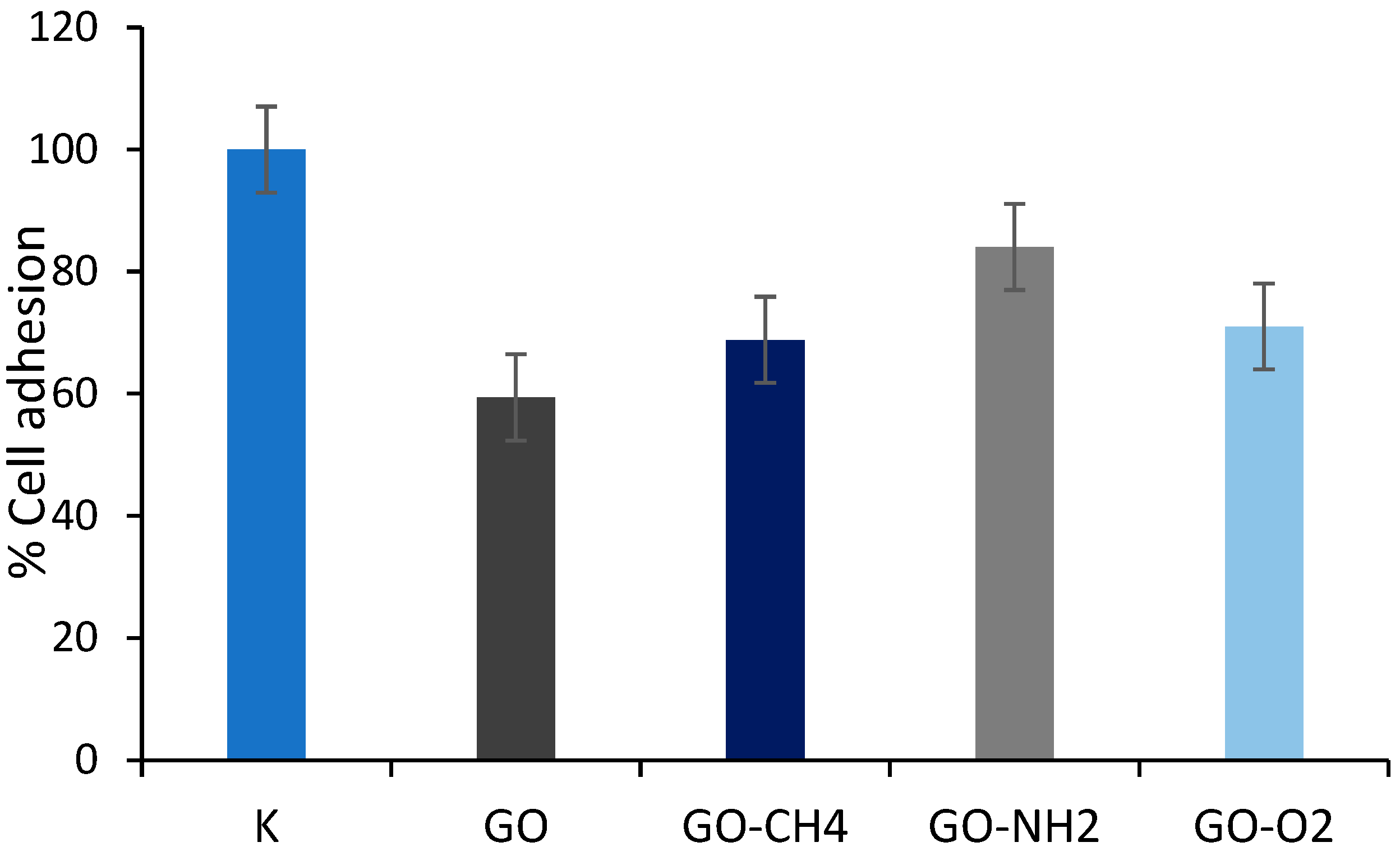
The adhesion of Colon26 cells on graphene oxide (GO) substrates with varying surface chemistries was quantified after 24 h, and expressed as a percentage relative to the control (Figure 6). Cell adhesion was significantly influenced by the specific functionalization of the GO (graphene oxide) sheets. Unmodified GO (graphene oxide) supported the lowest level of cell adhesion, with only 59.4% of the cells adhering relative to the control tissue culture plastic (set at 100%). This indicates that the base GO (graphene oxide) material is not highly conducive to cell attachment. Functionalization of the GO (graphene oxide) sheets markedly altered their cell-adhesive properties. The introduction of methyl groups (GO-CH4) improved adhesion to 68.8% of the control. A similar, though slightly higher, adhesion level was observed for the GO-O2 (carboxyl) sample, at 71.0%. Notably, amine-functionalization yielded the most significant enhancement in biocompatibility. The GO-NH2 (amine) substrate supported robust cell adhesion, reaching 84.1% of the control value. This represents a substantial recovery towards the adhesion levels observed on the standard control surface and identifies the amine group as a key modifier for promoting Colon26 cell attachment.
Together, these findings demonstrate that surface chemistry profoundly affects cellular attachment, with pristine GO (graphene oxide) maintaining relatively low adhesion, whereas oxygen, hydrocarbon and amine functionalization significantly improve it, as amine groups provide the most favourable functionalization for supporting adhesion among the modified surfaces.

3.5. Cell Proliferation on Functionalized Graphene Oxide Substrates

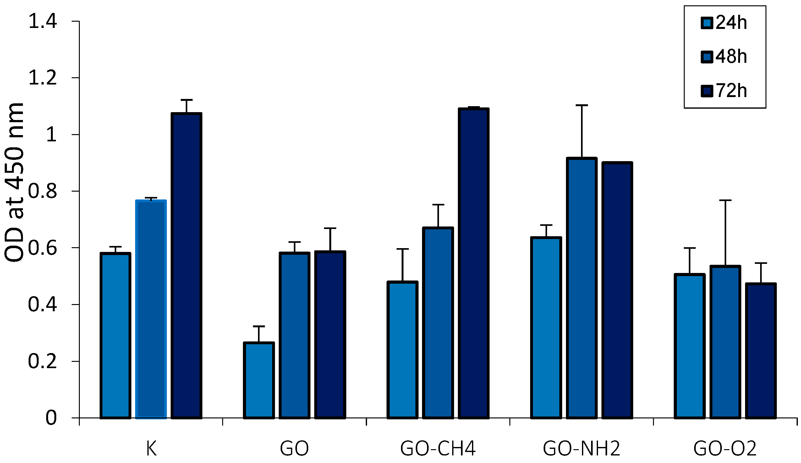
The proliferation of Colon 26 cells on graphene oxide (GO) substrates with different surface functional groups was assessed over a 72 h period by measuring optical density (OD) at 450 nm, a proxy for cell viability and number. As illustrated in Figure 7, the control group exhibited robust, time-dependent cell proliferation, with the OD value increasing steadily from 24 to 72 h.
In contrast, all graphene-based substrates initially showed significantly suppressed cell proliferation at the 24 h time point compared to the control. The unmodified GO (graphene oxide) surface consistently supported the lowest cell viability across all time points, indicating that it is not only anti-adhesive but also strongly inhibitory to cell growth. The impact of surface functionalization on cell proliferation was pronounced. The GO-CH4 (methyl) and GO-O2 (carboxyl) samples showed intermediate performance, maintaining viability levels slightly above those of unmodified GO (graphene oxide) but still substantially below the control throughout the experiment. Notably, the amine-functionalized GO-NH2 (amine) substrate demonstrated the most favourable profile for cell proliferation among the test materials. While its initial viability at 24 h was still lower than the control, the cells on GO-NH2 (amine) showed a clear and steady increase in OD over time [17]. By the 72 h time point, the proliferation on GO-NH2 (amine) had recovered significantly, approaching the levels observed in the control group. This trend suggests that the GO-NH2 (amine) surface supports continued cell growth and is the most biocompatible of the modified GO substrates.
While all GO (graphene oxide) materials initially inhibit Colon 26 cell proliferation, surface modification, particularly with amine groups, can mitigate this inhibitory effect and support long-term cell growth.

4. Discussion

The results of this study demonstrate that plasma-induced surface functionalization of graphene oxide (GO) substantially influences its interaction with cancer cells, with each modified surface exhibiting a distinct biological response profile. The observed effects can be directly linked to changes in surface chemistry, polarity, and defect structure introduced by controlled plasma processing [19].
XPS analysis confirmed successful incorporation of defined functional groups, including hydrocarbon-rich, oxygen-enriched, and nitrogen-containing domains. These modifications introduce variations in surface charge distribution and electron density, which in turn influence biomolecule adsorption, cell membrane interactions, and oxidative stress responses. Oxygen-rich GO-O2 (carboxyl) samples exhibited the highest proportion of carbonyl and carboxyl functionalities, which correlate with strong oxidative reactivity and increased formation of reactive oxygen species (ROS). This likely contributed to reduced cell adhesion, extensive apoptotic morphology, and impaired mitochondrial activity observed across biological assays.
In contrast, the nitrogen-modified GO-NH2 (amine) surfaces displayed a markedly different phenotype. Amine incorporation introduces protonatable nitrogen species that shift the net surface charge toward partial positivity and increase chemical basicity. These features affect protein corona formation and electrostatic interactions with negatively charged cellular membranes. As a result, GO-NH2 (amine) supported superior early cell attachment and spreading compared to pristine or oxygen-functionalized GO (graphene oxide), although viability remained lower than control surfaces. This suggests that amine functionality enhances compatibility with initial cell–material interface formation while still generating moderate downstream metabolic stress [20].
Methane-functionalized GO-CH4 (methyl) exhibited distinct biological effects, likely due to its increased hydrophobic character and formation of hydrogenated amorphous carbon-like surface regions. Hydrophobic domains facilitate lipid-facing interactions that may encourage direct membrane insertion or localized mechanical disruption. This mechanism aligns with the observed rounding and detachment of cells in microscopy images and reduced proliferation signals captured in viability assays.
Pristine GO (graphene oxide) showed the lowest overall adhesion and proliferation, reflecting its intermediate polarity and tendency to agglomerate in biological media. These structural and chemical features reduce available contact area and weaken cell anchoring compared to plasma-functionalized variants. The comparative hierarchy of biological response—GO-NH2 (amine) (most compatible) > GO-CH4 (methyl) ≈ GO-O2 (carboxyl) (most cytotoxic) > pristine GO (graphene oxide)—emphasizes that even subtle chemical modifications profoundly alter cancer cell behaviour.
Taken together, these findings point to defect chemistry and controlled dopant incorporation as key determinants of bioresponse. Oxygen-based defects promote oxidative stress pathways, nitrogen dopants modulate electrostatic and protein–surface interactions, and hydrocarbon functionalization enhances membrane affinity and mechanical perturbation [20] While the present study focused exclusively on cancer cells, such mechanistic diversity suggests opportunities to tune GO (graphene oxide) toward selective therapeutic or biomedical interface roles [21].
A limitation of the present work is the absence of comparative testing using non-malignant colon epithelial cells, which would allow evaluation of tumour selectivity. Additionally, although surface changes were quantified chemically, nanoscale morphological effects could also contribute to cell behaviour and merit further characterization. Despite these constraints, the consistent correlation between functional group identity and biological response provides a strong indication that plasma-based surface engineering is an effective tool for tailoring GO (graphene oxide) biological activity [22] Complementary bulk characterization techniques such as FTIR or NMR may further validate bonding motifs; however, since biological response depends on surface chemistry, XPS alone is suitable for capturing the relevant surface-level modifications.

5. Future Perspectives

Future investigations should include:
  • In vivo biocompatibility studies to assess inflammatory and immune responses.
  • Dose–response analysis to define safe therapeutic windows.
  • Proteomic and transcriptomic profiling to elucidate the molecular pathways underlying observed cytotoxic effects.
  • Exploration of combinatorial functionalization strategies (e.g., dual NH2 (amine)/PEG modification) to balance cytotoxicity and targeting efficiency.
Integrating these approaches will advance the rational design of graphene oxide-based nanomaterials for cancer therapeutics and regenerative medicine.

6. Conclusions

This study demonstrates that plasma functionalization of graphene oxide enables controlled modulation of its surface chemistry and consequently its interaction with cancer cells. The introduction of defined functional groups results in distinct cytotoxicity profiles: oxygen-enriched GO-O2 (carboxyl) induces strong oxidative stress and cell death, hydrophobic GO-CH4 (methyl) disrupts membrane integrity and attachment, while nitrogen-containing GO-NH2 (amine) improves early adhesion and spreading but maintains moderate cytotoxic influence. Pristine GO (graphene oxide) exhibited the weakest interaction with cells, reflecting limited reactivity and surface engagement.
These findings establish a clear relationship between defect structure, dopant type, and biological response, indicating that GO (graphene oxide) cytotoxicity can be rationally tuned rather than treated as an intrinsic property of the material [18]. This tunability positions plasma-functionalized GO (graphene oxide) as a promising platform for cancer-targeting materials, implant surface treatments requiring controlled cell inhibition, or future multifunctional therapeutic carriers.
Future work should incorporate healthy epithelial cells to assess selectivity and further explore oxidative stress pathways, protein corona evolution, and long-term cellular adaptation. These results contribute to a growing understanding of how nanoscale carbon surface design influences biological outcomes and support the strategic application of defect-engineered GO (graphene oxide) in cancer-focused biomedical technologies.

Author Contributions

S.A. contributed to the manuscript through critical review and editing. N.K., K.H.-P. and M.K.-M. conducted the biological experiments and related analyses. G.S. performed the XPS measurements and data interpretation. F.A. conceived the original idea, carried out the materials characterization, and led the writing and revision of the manuscript. All authors have read and agreed to the published version of the manuscript.

Funding

This research received no external funding.

Institutional Review Board Statement

An ethical statement is not needed as the cell line is commercially available.

Data Availability Statement

Data available upon request. The original contributions presented in the study are included in the article, further inquiries can be directed to the corresponding author.

Conflicts of Interest

The authors declare no conflicts of interest.

References

  1. Dilenko, H.; Bartoň Tománková, K.; Válková, L.; Hošíková, B.; Kolaříková, M.; Malina, L.; Bajgar, R.; Kolářová, H. Graphene-based photodynamic therapy and overcoming cancer resistance: A review. Int. J. Nanomed. 2024, 19, 4127–4146. [Google Scholar] [CrossRef] [PubMed]
  2. Liu, L.; Ma, Q.; Cao, J.; Gao, Y.; Han, S.; Liang, Y.; Zhang, T.; Song, Y.; Sun, Y. Recent progress of graphene oxide-based multifunctional nanomaterials for cancer treatment. Cancer Nanotechnol. 2021, 12, 18. [Google Scholar] [CrossRef]
  3. Asadi, M.; Ghorbani, S.H.; Mahdavian, L.; Aghamohammadi, M. Graphene-based hybrid composites for cancer diagnostic and therapy. J. Transl. Med. 2024, 22, 10. [Google Scholar] [CrossRef] [PubMed]
  4. Wang, Y.; Li, J.; Li, X.; Shi, J.; Jiang, Z.; Zhang, C.Y. Graphene-based nanomaterials for cancer therapy and anti-infections: Current status and future perspective. Bioact. Mater. 2022, 14, 335–349. [Google Scholar] [CrossRef] [PubMed]
  5. Tavakol, M.; Montazeri, A.; Aboutalebi, S.H.; Asgari, R. Mechanical properties of graphene oxide: The impact of functional groups. arXiv 2020. [Google Scholar] [CrossRef]
  6. Shafiee, A.; Iravani, S.; Varma, R.S. Graphene and graphene oxide with anticancer applications: Challenges and future perspectives. Mater. Today Chem. 2022, 3, e118. [Google Scholar] [CrossRef] [PubMed]
  7. Wang, S.; Luo, X. A first-principles study of graphene and graphene oxide as drug carriers: Tamoxifen adsorption behaviour. ACS Omega 2025, in press. [Google Scholar] [CrossRef]
  8. Li, R.; Wang, Y.; Du, J.; Wang, X.; Duan, A.; Gao, R.; Liu, J.; Li, B. Graphene oxide loaded with tumor-targeted peptide and anticancer drug for synergistic cancer therapy. Sci. Rep. 2021, 11, 81218. [Google Scholar] [CrossRef]
  9. Barba-Rosado, L.V.; Carrascal-Hernández, D.C.; Insuasty, D.; Grande-Tovar, C.D. Graphene Oxide (GO) for the Treatment of Bone Cancer: A Systematic Review and Bibliometric Analysis. Nanomaterials 2024, 14, 186. [Google Scholar] [CrossRef] [PubMed]
  10. Zaaba, N.I.; Foo, K.L.; Hashim, U.; Tan, S.J.; Liu, W.W.; Voon, C.H. Synthesis of Graphene Oxide using Modified Hummers Method: Solvent Influence. Procedia Eng. 2017, 184, 469–477. [Google Scholar] [CrossRef]
  11. CT26.WT—CRL-2638|ATCC. Available online: https://www.atcc.org/products/crl-2638 (accessed on 19 November 2025).
  12. Speranza, G.; Canteri, R. RxpsG a new open project for Photoelectron and Electron Spectroscopy data processing. SoftwareX 2019, 10, 100282. [Google Scholar] [CrossRef]
  13. Beamson, G.; Briggs, D. High Resolution XPS of Organic Polymers: The ScientaESCA300 Database; Surface Spectra: Chichester, UK, 1992. [Google Scholar]
  14. Naumkin, A.V.; Kraut-Vass, A.; Gaarenstroom, S.W.; Powell, C.J. NIST X-Ray Photoelectron Spectroscopy, Database, Version 5.0; National Institute of Standards and Technology: Gaithersburg, MD, USA, 2023. [CrossRef]
  15. Wang, K.; Ruan, J.; Song, H.; Zhang, J.; Wo, Y.; Guo, S.; Cui, D. Biocompatibility of graphene oxide. Nanoscale Res. Lett. 2011, 6, 8. [Google Scholar] [CrossRef] [PubMed]
  16. Rahimi, S.; Chen, Y.; Zareian, M.; Pandit, S.; Mijakovic, I. Cellular and subcellular interactions of graphene-based materials with cancerous and non-cancerous cells. Adv. Drug Deliv. Rev. 2022, 189, 114467. [Google Scholar] [CrossRef] [PubMed]
  17. Rive, C.; Reina, G.; Wagle, P.; Treossi, E.; Palermo, V.; Bianco, A.; Delogu, L.G.; Rieckher, M.; Schumacher, B. Improved biocompatibility of amino-functionalized graphene oxide in Caenorhabditis elegans. Small 2019, 15, e1902699. [Google Scholar] [CrossRef] [PubMed]
  18. Ou, L.; Song, B.; Liang, H.; Liu, J.; Feng, X.; Deng, B.; Sun, T.; Shao, L. Toxicity of graphene-family nanoparticles: A general review of the origins and mechanisms. Part. Fibre Toxicol. 2016, 13, 57. [Google Scholar] [CrossRef] [PubMed]
  19. Stehle, Y.Y.; Barnum, T.J.; Schujman, S.; Vlassiouk, I.V.; Cortez, R. Plasma-Induced Tailoring of Graphene Oxide Surfaces for Electrochemical Applications: Functionalization and Etching. ACS Appl. Electron. Mater. 2025, 7, 6635–6645. [Google Scholar] [CrossRef] [PubMed]
  20. Rong, L.; Fu, Y.; Li, Q.; Yang, X.; Li, Y.; Yan, L.; Wang, L.; Wu, W. Effects of the surface charge of graphene oxide derivatives on ocular compatibility. Nanomaterials 2022, 12, 735. [Google Scholar] [CrossRef] [PubMed]
  21. Krętowski, R.; Szynaka, B.; Borzym-Kluczyk, M.; Tyszka, N.; Jabłońska-Trypuć, A.; Gil, M.; Cechowska-Pasko, M. Effectiveness of Graphene Oxide (GO) in Activating the Mitochondrial Pathway of Oxidative Stress-Induced Apoptosis in Breast Cancer Cells. Cells 2025, 14, 1717. [Google Scholar] [CrossRef] [PubMed]
  22. Golda-Cepa, M.; Kumar, D.; Bialoruski, M.; Lasota, S.; Madeja, Z.; Piskorz, W.; Kotarba, A. Functionalization of graphenic surfaces by oxygen plasma toward enhanced wettability and cell adhesion: Experiments corroborated by molecular modelling. J. Mater. Chem. B 2023, 11, 4946–4957. [Google Scholar] [CrossRef] [PubMed]
Figure 1. Schematics of the ICP reactor used for functionalizing the GO_paper foils.
Carbon 12 00003 g001
Figure 2. Black pristine GO sheet, red GO sheet treated in 80 sccm CH4 plasma: (a) Wide spectra; (b) High-resolution C 1s spectra; (c) High-resolution O1s spectra.
Carbon 12 00003 g002
Figure 3. GO (graphene oxide) paper (a) treated in a plasma of a mixture of CH4 (methyl) (b) + O2 (oxygen) (c) in the following different proportions: black 60:20 sccm, red 50:30 sccm, green 40:40 sccm, blue 30:50 sccm and pink 20:60 sccm.
Carbon 12 00003 g003
Figure 4. GO (graphene oxide) paper (a) treated in a plasma of a mixture of CH4 (b) + NH3 (c) in the following different proportions: black 40:10 sccm, red 30:20 sccm, green 25:25 sccm, blue 20:30 sccm.
Carbon 12 00003 g004
Figure 5. Morphology of Colon 26 cells cultured on Control and graphene oxide-modified surfaces. Control cells exhibited elongated, well-spread morphology with high density. On non-modified GO (graphene oxide), cells appeared sparse, rounded, and poorly attached. GO-CH4 (methyl) surfaces supported slightly improved adhesion but with limited spreading. GO-NH2 (amine) functionalization promoted the best adhesion and elongated morphology, closely resembling control. GO-O2 (carboxy) surfaces showed moderate adhesion with mixed cell shapes.
Carbon 12 00003 g005
Figure 6. Quantification of Colon 26 cell attachment/adhesion on control group (K), GO (graphene oxide) and functionalized GO (graphene oxide) surfaces (CH4, NH2, and O2). Standard deviation values were rounded to one decimal place during plotting, which caused small numerical differences (<0.5%) to appear visually identical on the chart.
Carbon 12 00003 g006
Figure 7. Quantification of Colon 26 cell attachment on control group (K) GO (graphene oxide) and functionalized GO (graphene oxide) surfaces (CH4, NH2, and O2). The standard deviation is symmetrical around the mean, so equivalent negative-direction error values exist but were hidden due to the plotting configuration.
Carbon 12 00003 g007
Table 1. Experimental parameters used for the plasma treatments of the GO paper substrates.
SampleCH4 (sccm)O2 (sccm)NH3 (sccm)
S180////
S26020//
S35030//
S44040//
S53050//
S62060//
S740//10
S830//20
S925//25
S1020//30
Table 2. Element concentrations (%) Estimated on the pristine and plasma treated GO (graphene oxide) paper samples. While the element concentrations are reported on a semi-quantitative basis, the calculated values are precise, with no associated measurement errors in the reported data.
SampleC (%)O (%)N (%)
GO83.816.2//
S193.16.9//
S277.122.9//
S368.931.1//
S467.832.2//
S550.249.8//
S643.256.8//
S788.21.710.1
S879.82.817.4
S976.92.420.7
S1073.42.224.4
Disclaimer/Publisher’s Note: The statements, opinions and data contained in all publications are solely those of the individual author(s) and contributor(s) and not of MDPI and/or the editor(s). MDPI and/or the editor(s) disclaim responsibility for any injury to people or property resulting from any ideas, methods, instructions or products referred to in the content.