


Abstract
Diabetic wounds remain a major clinical challenge owing to persistent dysregulation of the wound microenvironment, which substantially limits the effectiveness of conventional therapies. In recent years, multifunctional hydrogels have emerged as promising platforms for diabetic wound management, attributed to their excellent biocompatibility, tunable physicochemical properties, and unique capacity to actively remodel pathological microenvironments through integrated therapeutic functions. This comprehensive narrative review provides an in-depth synthesis of the pathogenesis and current therapeutic strategies for diabetic wounds, with a particular focus on recent advances in multifunctional hydrogels, as well as their classification, design principles, mechanisms of action, and translational potential. Furthermore, emerging directions are discussed as promising approaches for next-generation therapies, including intelligent closed-loop systems, interdisciplinary technological convergence, and the integration of bioactive components derived from traditional Chinese medicine. Collectively, these advances are poised to facilitate the transition from passive wound coverage to active microenvironment remodeling, paving the way for precision and personalized diabetic wound care.
1. Introduction
Diabetes is a prevalent chronic metabolic disorder that poses a substantial global health burden. In 2024, the number of adults with diabetes worldwide exceeded 500 million, and this figure is projected to rise to approximately 900 million by 2050 [1]. Among diabetic patients, the incidence of diabetic wounds reaches up to 25%, with a five-year recurrence rate of 66% [2]. Under persistent hyperglycemia, diabetic wound pathogenesis involves multiple interconnected factors, including vascular dysfunction and peripheral neuropathy, microenvironmental disruption, cellular dysfunction, growth factor imbalance, and infection [3,4]. The interplay of these factors impedes normal wound healing, ultimately resulting in chronic wounds characterized by delayed or impaired healing [5]. Diabetic foot ulcers, one of the most prevalent forms of diabetic wounds, represent a major public health concern due to their high incidence, recurrence rate, healing difficulty, and risk of amputation [6,7,8,9,10,11].
A variety of clinical strategies are currently employed for diabetic wound management, including surgical debridement, skin grafting, stem cell therapy, negative pressure wound therapy, hyperbaric oxygen therapy, and wound care management. Among these, wound dressings are widely used in clinical practice owing to their convenient clinical application, cost-effectiveness, and localized therapeutic effects [12]. Based on composition and functional characteristics, dressings can be broadly categorized into traditional and advanced types [13]. Conventional dressings—such as natural gauze, synthetic fiber dressings, and oil-based dressings—are economical and readily available, providing a basic physical barrier and exudate absorption [14]. Advanced dressings, including foam, film, hydrocolloid, nanofiber, and hydrogel-based systems, offer superior moisture retention, breathability, and antimicrobial activity, thereby promoting a moist wound healing environment [13]. Nevertheless, several challenges persist (as Table 1). For example, although silver-ion-loaded nanofiber dressings exhibit broad-spectrum antimicrobial effects, their long-term use raises cytotoxicity concerns [15]. Moreover, some advanced dressings are suitable only for specific clinical scenarios. For example, foam dressings possess high absorbency and are not suitable for dry or minimally exuding wounds, as they may cause excessive wound desiccation, thereby disrupting the moist healing environment and delaying the healing process [16]. Additionally, some dressings, such as Smart nanofiber dressings, face significant barriers to large-scale clinical translation due to their complex manufacturing processes and high economic costs [17].
Hydrogels have emerged as a promising class of wound dressings due to their unique hydrophilic three-dimensional network, which combines favorable moisture retention, biocompatibility, and high design flexibility [18]. Based on the volume of published literature and the depth of investigation, multifunctional hydrogels have become one of the most intensively investigated categories of advanced wound dressings for diabetic wound management [19,20]. Recent studies have demonstrated that stimulus-responsive hydrogels can dynamically respond to pathological cues within diabetic wounds. When combined with controlled drug delivery systems, these materials enable on-demand therapeutic release in response to changes in pH, glucose concentration, or Reactive Oxygen Species (ROS) levels [21]. Additionally, these hydrogels possess immunomodulatory properties, appropriately engineered to modulate macrophage polarization toward a pro-regenerative (anti-inflammatory, reparative) phenotype, thereby improving the local microenvironment [21]. Furthermore, through the synergistic delivery of three-dimensional biomimetic networks and bioactive molecules, hydrogels provide a favorable microenvironment for cell adhesion, proliferation, and migration [22]. Notably, these multifunctional capabilities are designed to target multiple pathological features of diabetic wounds, offering a promising platform for precise and synergistic repair (as shown in Table 2). Consequently, researchers are currently focusing on the optimized design of multifunctional hydrogel systems to achieve controlled drug release and intelligent responsiveness to the wound microenvironment.
This review summarizes recent advances in multifunctional hydrogels for diabetic wound treatment, focusing on their classification, design strategies, mechanisms of action, and clinical translation. Overall, this review establishes a pathology- and translation-oriented framework that shifts hydrogel design in diabetic wound management from isolated functional development toward an integrated approach driven by disease mechanisms and clinical needs.
Table 1.
Advantages and disadvantages of traditional versus advanced wound dressings.
Table 2.
Comparison of Wound Dressing Systems.
2. Classification of Hydrogels and Optimization Strategies
Hydrogels are highly hydrophilic polymeric networks capable of rapidly absorbing and retaining large volumes of fluid without dissolving [29]. Over the past several decades, significant advances in material science and bioengineering have greatly expanded both the diversity and functional complexity of hydrogel systems (as shown in Figure 1) [30]. Hydrogels are commonly classified according to three major criteria: cross-linking mechanisms, stimulus responsiveness, and material origin [31]. These classification strategies not only determine the physicochemical and biological properties of hydrogels but also provide the foundation for their rational design in biomedical applications. In the context of diabetic wound management, researchers have progressively shifted from conventional hydrogel systems toward multifunctional and smart hydrogels capable of simultaneously addressing infection, oxidative stress, inflammation, impaired angiogenesis, and tissue regeneration. Therefore, understanding hydrogel classifications and corresponding optimization strategies is essential for the development of next-generation wound dressings.
2.1. Classification of Hydrogels
2.1.1. Classification by Cross-Linking Method
Hydrogels are generally classified as physically cross-linked or chemically cross-linked systems according to the nature of the interactions responsible for network formation [29]. Physically cross-linked hydrogels are formed through non-covalent interactions, including hydrogen bonding, ionic interactions, hydrophobic associations, and electrostatic forces [32]. These hydrogels are commonly fabricated from natural polymers such as gelatin, sodium alginate, and chitosan, exhibiting excellent biocompatibility and biodegradability. However, their relatively weak mechanical strength and limited structural stability may restrict their application in mechanically demanding environments [33,34].
In contrast, chemically cross-linked hydrogels form stable three-dimensional networks through covalent bonds generated by radical polymerization, click chemistry, functional group reactions, or radiation-induced cross-linking. Representative materials include polyacrylamide and poly (ethylene glycol) (PEG)-based hydrogels, which offer superior mechanical properties and structural tunability. However, the irreversible nature of covalent cross-linking often renders these hydrogels difficult to degrade under physiological conditions [35]. Nevertheless, residual cross-linking agents and reaction by-products may introduce potential cytotoxicity concerns, leading to inferior biocompatibility compared to their physically cross-linked counterparts, thereby affecting their translational potential [36].
To overcome the limitations of conventional cross-linking approaches, emerging strategies such as enzyme-mediated cross-linking and dynamic covalent bonding have attracted increasing attention. These approaches combine favorable biocompatibility with enhanced structural stability, providing new opportunities for the development of multifunctional biomedical hydrogels [36,37].
2.1.2. Classification by Response System
Stimulus-responsive hydrogels possess the ability to undergo reversible changes in their physicochemical properties when exposed to specific environmental cues, thereby enabling adaptive therapeutic functions [38,39]. According to the type of triggering stimulus, these systems can be broadly classified as physically responsive hydrogels (e.g., light, temperature, magnetic field, and electrical stimulation), chemically responsive hydrogels (e.g., pH, glucose, and reactive oxygen species), and multi-responsive hydrogels that integrate multiple sensing mechanisms [38]. Beyond biochemical responsiveness, mechanically adaptive hydrogels can sense and respond to mechanical cues; for example, a dynamic disulfide bond-based injectable lubricative hydrogel (ILH) provides mechanical protection under static conditions and exhibits reversible shear-thinning behavior under stress, offering a new strategy for designing adaptive dressings for diabetic wounds at mechanically active sites [40].
Compared with conventional dressings, stimulus-responsive hydrogels offer unique advantages in diabetic wound treatment because they can dynamically respond to pathological changes within the wound microenvironment. Such responsiveness enables on-demand and spatiotemporally controlled therapeutic delivery, thereby improving treatment precision while minimizing unnecessary drug exposure [41,42]. In contrast, the dynamic and spatiotemporal variability of key wound microenvironmental signals—including pH, glucose, ROS, temperature, and exudate composition—presents a major obstacle for the rational design of stimuli-responsive hydrogels capable of adapting to the evolving healing process [43].
2.1.3. Classification by Source of Material
Based on material composition, hydrogels can be categorized into natural, synthetic, and semi-synthetic systems [44].
Natural polymers, including chitosan, hyaluronic acid, collagen, and agarose, possess intrinsic biological activities that support cell adhesion, proliferation, and tissue regeneration. However, their relatively poor mechanical properties, rapid degradation rates, and batch-to-batch variability may limit large-scale clinical application [45].
Synthetic polymers such as poly (ethylene glycol) (PEG), poly (vinyl alcohol) (PVA), and poly(vinylpyrrolidone) (PVP) provide excellent structural controllability and reproducible physicochemical properties. Nevertheless, most synthetic polymers lack intrinsic bioactivity and therefore require functional modification to achieve desirable biological performance [31].
To overcome the limitations of both categories, semi-synthetic hydrogels have emerged as a promising alternative. Through chemical modification of natural polymers or hybridization with synthetic components, semi-synthetic systems combine favorable biocompatibility with tunable mechanical properties. Representative materials such as gelatin methacryloyl (GelMA) and modified hyaluronic acid (HA) derivatives have become widely used platforms in diabetic wound healing applications [31].
In addition to the above classifications, hydrogels can also be categorized according to their functional properties, including self-healing, adhesive, conductive, and optical functions, as well as by their mode of application, such as preformed and injectable systems [46,47]. Importantly, these classification categories are not mutually exclusive. Contemporary hydrogel systems often integrate multiple design principles through hybridization, copolymerization, or composite fabrication strategies. Such integration gives rise to composite hydrogels that combine distinct functional components within a unified system thereby enabling synergistic enhancements in biological activity, mechanical performance, and therapeutic efficacy [48].
2.2. Strategies for the Optimized Design of Hydrogels
Although conventional hydrogels provide a moist healing environment and structural support for tissue regeneration, their therapeutic efficacy remains insufficient for addressing the multifactorial pathophysiology of diabetic wounds. Consequently, recent research has focused on the development of advanced hydrogel systems through structural, functional, and manufacturing optimization strategies. The rational design of hydrogel dressings depends on optimizing key physicochemical properties, including porosity, swelling behavior, degradation kinetics, mechanical strength, and rheology, which together determine mass transport, structural stability, and adaptation to dynamic wound environments [49]. Proper tuning of these parameters enables effective exudate control, tissue regeneration support, and balanced degradation, thereby promoting microenvironmental homeostasis and coordinated repair in diabetic wounds.
2.2.1. Structural Optimization
Structural optimization aims to improve the mechanical stability, adaptability, and durability of hydrogel networks, primarily through the refinement of cross-linking mechanisms and network architectures. One common approach involves refining cross-linking mechanisms. For example, incorporating multiple hydrogen-bonding interactions can enhance the robustness of physically cross-linked hydrogels, whereas integrating dynamic covalent bonds such as Schiff-base linkages can simultaneously improve mechanical strength and self-healing capacity [50,51]. In addition, double-network hydrogels, interpenetrating polymer networks, and supramolecular hydrogels have emerged as effective strategies for overcoming the mechanical limitations of traditional systems while maintaining favorable biocompatibility [52]. The practical advantages of these structural strategies depend on specific wound requirements. Physically cross-linked hydrogels provide excellent biocompatibility and injectability, dynamic covalent networks enable self-healing and adaptive behavior, while double-network and supramolecular systems enhance mechanical reinforcement and dynamic remodeling; however, these improvements may involve trade-offs, including limited mechanical stability, reduced molecular diffusion and cellular infiltration, and increased fabrication complexity [27,53]. Therefore, structural optimization should be tailored to wound location, mechanical demands, and healing stages to balance mechanical performance, biological functionality, and translational feasibility.
2.2.2. Functional Optimization
Functional optimization focuses on enabling hydrogels to actively regulate the wound microenvironment rather than merely serving as passive dressings.
Stimulus-responsive hydrogels can sense pathological cues such as elevated glucose levels, excessive reactive ROS accumulation, and pH fluctuations, thereby triggering controlled therapeutic release [38,54,55]. This adaptive responsiveness improves treatment precision and allows therapeutic interventions to better match the dynamic progression of wound healing.
Furthermore, dynamic network architectures can endow hydrogels with self-healing behavior, shape-memory properties, and programmable drug-release profiles. These characteristics facilitate more effective adaptation to the spatiotemporal changes occurring during wound repair [56,57,58,59].
Recent studies have also highlighted the importance of immunomodulatory hydrogel systems capable of regulating macrophage polarization, reducing chronic inflammation, and promoting tissue regeneration, thereby addressing one of the key pathological barriers in diabetic wound healing [60,61].
2.2.3. Biomimetic and Composite Design Strategies
Biomimetic design is another important direction in hydrogel development. By mimicking the composition, architecture, and mechanical characteristics of the native extracellular matrix (ECM), biomimetic hydrogels provide a favorable microenvironment for cellular adhesion, proliferation, migration, and differentiation [62,63].
Meanwhile, composite strategies that combine natural and synthetic polymers, nanomaterials, bioactive molecules, and conductive components have enabled the development of multifunctional hydrogel platforms with enhanced antibacterial, antioxidant, angiogenic, and tissue-regenerative properties [64].
Conductive hydrogels [65], injectable hydrogels [55], and Shape Memorable and Self-Healable Smart Hydrogels [66] have emerged as particularly promising platforms for diabetic wound management, reflecting the ongoing transition from single-function dressings toward integrated therapeutic systems.
Furthermore, integrating biomimetic design with 3D/4D printing technology allows hydrogels to transition from simple morphological adaptation to spatiotemporal functional programming [51,67].
2.2.4. Manufacturing and Translational Optimization
Advanced manufacturing technologies have further accelerated the evolution of hydrogel-based wound dressings.
Among these technologies, 3D printing enables personalized fabrication of hydrogel architectures with precisely controlled geometry and porosity, while 4D printing introduces dynamic structural changes in response to environmental stimuli [68]. These approaches facilitate the development of patient-specific wound dressings with enhanced adaptability.
At the microscale, hydrogel-based delivery platforms have emerged as versatile systems for the localized and controlled delivery of diverse bioactive agents and therapeutic cargos, including growth factors, stem cells, extracellular vesicles, antimicrobial agents, nucleic acids, peptides, and small-molecule therapeutics [69,70,71]. By regulating the spatial and temporal release of these therapeutic cargos, hydrogel systems can enhance angiogenesis, modulate inflammatory responses, regulate immune responses, and promote tissue regeneration during diabetic wound healing. Among these delivery strategies, microspheres, nanoparticles, and exosome-loaded systems have been extensively integrated with hydrogel matrices to achieve controlled and sustained therapeutic release [72,73]. Such integration enables localized and prolonged therapeutic effects while minimizing systemic exposure and improving the efficacy of encapsulated cargos.
Despite these advances, several challenges remain before widespread clinical translation can be achieved, including manufacturing scalability, long-term biosafety, quality control, storage stability, and regulatory approval. Addressing these issues will be critical for the successful implementation of multifunctional hydrogel technologies in diabetic wound management.
Figure 1.
Functional characteristics of hydrogels in diabetic wound healing.
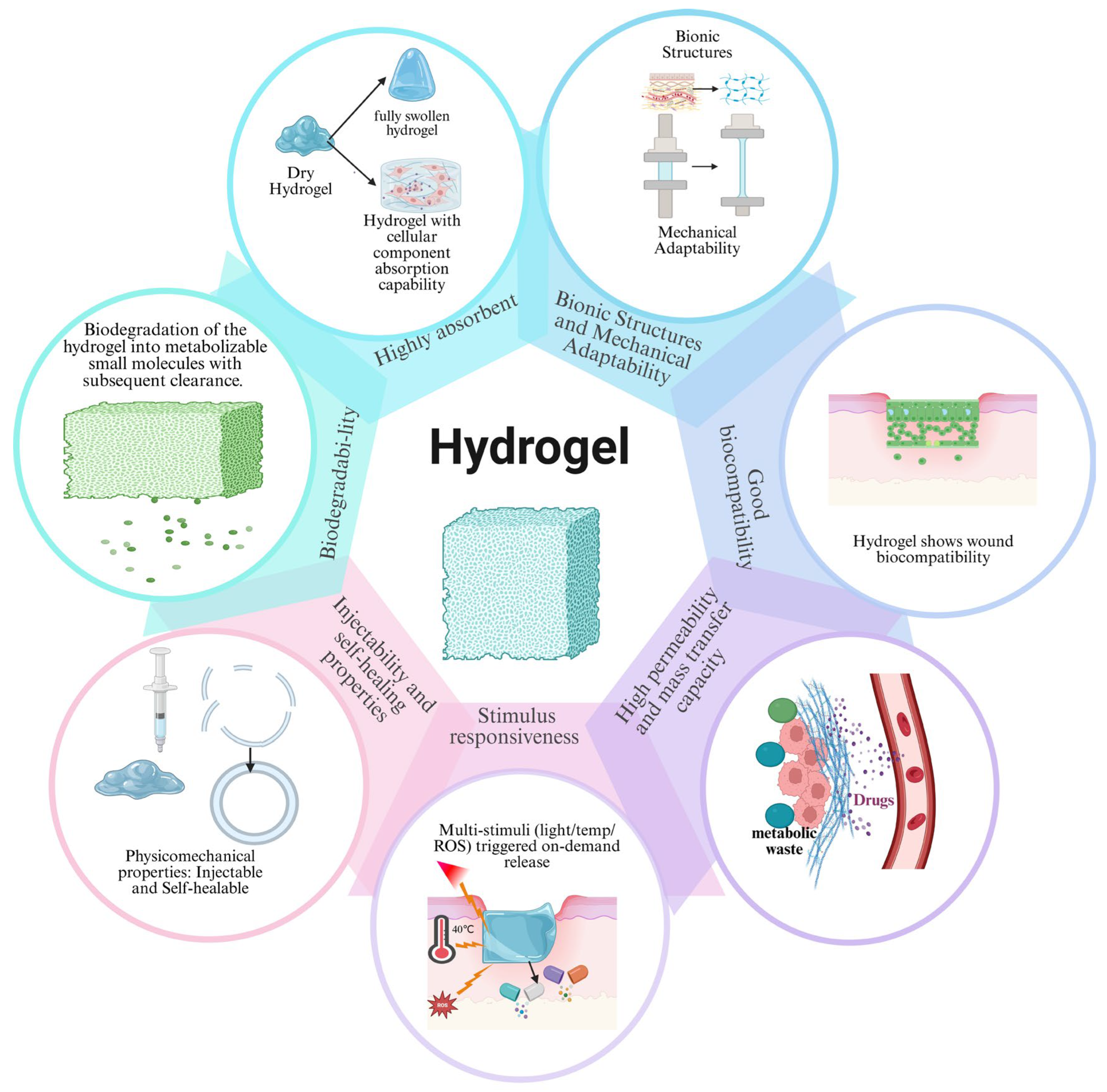
Owing to their highly hydrated three-dimensional networks, hydrogels provide exudate management, mechanical adaptability, biocompatibility, molecular transport, stimulus responsiveness, injectability, self-healing capability, and biodegradability. These integrated properties enable hydrogels to modulate the wound microenvironment and support tissue repair in diabetic wounds.
3. The Role of Multifunctional Hydrogels in Modulating the Microenvironment of Diabetic Wounds
Diabetic wounds are characterized by a complex pathological microenvironment involving persistent hyperglycemia, oxidative stress, dysregulation of the immune microenvironment, chronic inflammation, microbial infection, impaired angiogenesis, ECM remodeling disorders, and cellular senescence, as shown in Figure 2 [74,75,76,77].
Figure 2.
Schematic illustration of the pathological microenvironment in diabetic wounds.
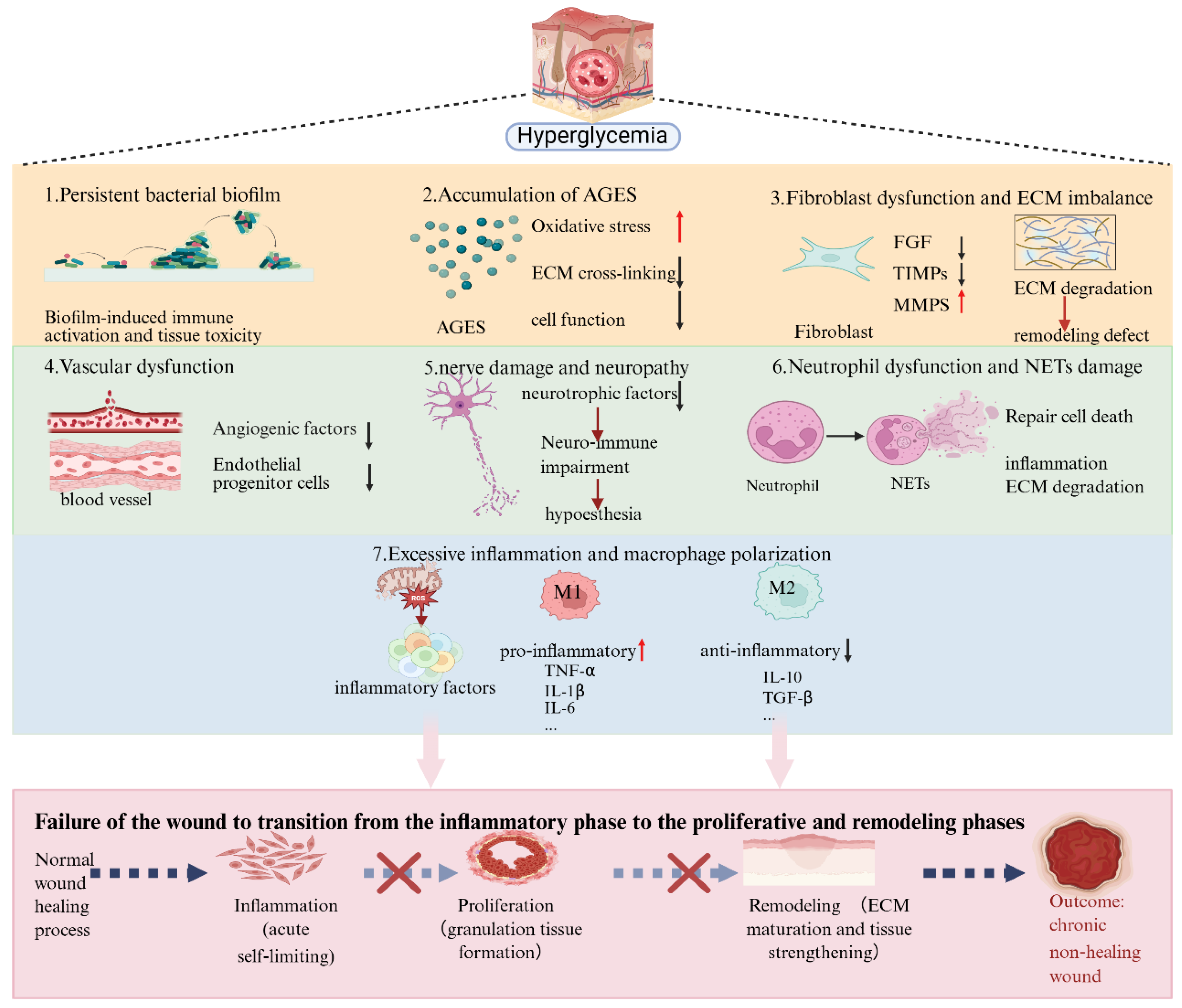
Persistent hyperglycemia triggers excessive ROS generation, chronic inflammation, abnormal pH conditions, and bacterial infection, resulting in macrophage polarization imbalance, impaired growth factor signaling, vascular dysfunction, peripheral neuropathy, and defective tissue repair. The synergistic effects of these pathological alterations ultimately lead to chronic, non-healing wounds.
Persistent hyperglycemia, a key pathological driver, induces immune cell dysfunction, including an imbalance in classically activated (M1) macrophage and alternatively activated (M2) macrophage polarization, and dysregulation of neutrophil extracellular trap formation (NETosis), thereby prolonging inflammation and suppressing tissue repair [78,79]. High glucose levels also promote excessive mitochondrial ROS production, exacerbating oxidative stress and further amplifying inflammatory signaling [80]. Additionally, hyperglycemia-induced sensory deficits delay injury detection, increasing the risk of infection [81,82], while elevated glucose levels provide nutrients for pathogenic microorganisms, facilitating biofilm formation and bacterial phenotypic shifts [82,83]. At the molecular level, sustained overexpression of pro-inflammatory cytokines, including Tumor Necrosis Factor-alpha (TNF-α), Interleukin-1 beta (IL-1β), and Interleukin-6 (IL-6), activates pathways such as Nuclear Factor kappa-light-chain-enhancer of activated B cells (NF-κB), establishing a positive feedback loop of inflammation while suppressing anti-inflammatory factors (e.g., IL-10) [84,85]. Growth factors (GF), essential mediators of tissue regeneration, exhibit severely impaired bioavailability and signaling activity under hyperglycemic conditions [86]. Local physicochemical factors, including pH [87] and humidity [88], further modulate these pathological processes, collectively destabilizing the wound microenvironment.
The interplay among these mechanisms and the resulting vicious cycle renders conventional hydrogels ineffective in reversing the pathological process. To overcome these limitations, recent advances in multifunctional hydrogel engineering have enabled the development of smart therapeutic platforms capable of simultaneously modulating multiple pathological processes through microenvironment-responsive and bioactive strategies [89]. Multifunctional hydrogels can be designed via structural optimization (e.g., dynamic networks, biomimetic architectures) and functional integration (e.g., drug delivery, incorporation of nanomaterials, and stimuli-responsive elements), allowing coordinated regulation of the diabetic wound microenvironment.
Building on the discussion above, the following sections will systematically delineate the specific mechanisms through which multifunctional hydrogels facilitate diabetic wound healing.
3.1. Metabolic Regulation: Targeting Hyperglycemia and Cellular Senescence
Hyperglycemia is a fundamental pathological driver of diabetic wound progression. Sustained elevations in glucose levels induce oxidative stress, advanced glycation end-product (AGE) accumulation, endothelial dysfunction, peripheral neuropathy, cellular senescence, and chronic inflammation, collectively impairing tissue regeneration and wound closure [90,91].
3.1.1. Classification of Hydrogel-Based Metabolic Regulation Strategies
Current hydrogel-based metabolic regulation strategies can be broadly categorized into three classes: (i) direct glucose consumption and microenvironment remodeling, (ii) localized delivery of hypoglycemic agents, and (iii) suppression of hyperglycemia-induced cellular senescence.
3.1.2. Representative Strategies and Examples
Microenvironment remodeling: The multifunctional COH-GB hydrogel developed by Ma [64] represents a typical microenvironment-remodeling strategy. By simultaneously reducing local glucose concentration, alleviating tissue hypoxia, scavenging ROS, and regulating pH, the hydrogel reconstructs the diabetic wound microenvironment. Moreover, glucose-responsive nitric oxide (NO) release enables simultaneous antibacterial and angiogenic activities, demonstrating the advantages of integrated metabolic regulation.
Localized drug delivery: Localized drug delivery represents another promising strategy. Zhou [54] incorporated insulin into an AP/SA hydrogel, achieving sustained local glucose reduction and prolonged therapeutic activity. Compared with conventional administration, hydrogel-mediated delivery provides more stable glucose control while simultaneously enhancing cell proliferation and angiogenesis.
Anti-senescence approach: In addition to regulating glucose levels directly, recent studies have increasingly focused on hyperglycemia-induced cellular senescence. The GelMA/PNS/Alg@IGF-1 hydrogel developed by Wang [92] delivers insulin-like growth factor-1 (IGF-1) to counteract cellular senescence while simultaneously reducing oxidative stress, thereby restoring cellular function and promoting vascular regeneration.
In short, these strategies demonstrate that effective metabolic regulation extends beyond glucose reduction alone and includes the prevention of senescence-associated dysfunction and restoration of cellular homeostasis.
In summary, metabolic regulation in diabetic wound healing extends beyond glucose control to include senescence modulation and cellular homeostasis restoration, yet its clinical translation is limited by poor physicochemical stability and insufficient spatiotemporal responsiveness to dynamic wound microenvironment signals [77,93,94]. Therefore, integrating metabolic modulation with antioxidant, anti-inflammatory, and pro-angiogenic functions within multifunctional hydrogel platforms is essential to address the multifactorial pathophysiology of diabetic wounds.
3.2. Regulation of the Immune Microenvironment
Persistent inflammation driven by an imbalance in macrophage polarization and excessive neutrophil activation is a major obstacle to diabetic wound healing. High glucose levels sustain M1-type macrophage activation while hindering M2-type polarization, which leads to prolonged inflammation, defective tissue repair, and eventual wound chronicity [95,96,97]. To counteract these immune disturbances, researchers have developed multifunctional hydrogels that can steer macrophage polarization, fine-tune inflammatory signaling pathways, limit NETosis, and restore cytokine balance.
3.2.1. Macrophage Repolarization
Hydrogels that encourage M2 polarization primarily work by restoring oxidative metabolism in macrophages, suppressing pro-inflammatory gene expression, and enhancing repair-related gene programs. For instance, the Ac2-26-loaded hydrogel reprograms macrophages via FPR2/PI3K/AKT signaling, which suppresses TLR-driven inflammation and restores oxidative phosphorylation to shift the diabetic wound niche from pro-inflammatory to pro-regenerative [98]. In another study, the SilMA-FGF21/CoS hydrogel not only promotes M2 polarization but also boosts angiogenesis through Janus kinase/signal transducer and activator of transcription (JAK/STAT)-mediated upregulation of vascular endothelial growth factor (VEGF), tackling both immune dysfunction and poor vascularization [99]. Natural polyphenol-based materials, such as protocatechuic aldehyde hydrogels, remodel the immune microenvironment by scavenging ROS, activating nuclear factor erythroid 2-related factor 2 (Nrf2), and suppressing NF-κB signaling—all without introducing external biological agents [100]. Transcriptomic analyses further show that the AP@HA-Si InjGel enhances the expression of reparative markers like arginase 1 (Arg1) and mannose receptor, C type 1 (Mrc1), confirming the molecular basis of macrophage reprogramming [101].
3.2.2. Modulation of Inflammatory Signaling Pathways
The coordinated regulation of multiple signaling pathways is central to immune modulation in diabetic wound healing. Impaired activation of the hypoxia-inducible factor-1 (HIF-1) pathway has been identified as a major cause of persistent wound non-healing, whereas activation of this pathway effectively promotes tissue repair [102,103,104]. Similarly, activation of the Nrf2 pathway [105] and inhibition of the NF-κB pathway [106] have been shown to markedly accelerate healing. These observations indicate that these signaling pathways represent promising therapeutic targets. The GelMA-FICZ hydrogel accelerates diabetic wound healing by activating AhR to restore impaired PINK1/Parkin-mediated mitophagy, thereby blocking mtDNA leakage and subsequent cGAS-STING/NLRP3 inflammasome activation, which reverses M1-dominant inflammation and promotes M2 polarization. [107]. The 4-hydroxychalcone (4HC) hydrogel developed by Li [108] promotes M2 macrophage polarization by inhibiting the TLR/IL-17/TNF signaling axis, thereby facilitating the wound transition from the inflammatory to the proliferative phase.
3.2.3. Cytokine Regulation and NETosis Control
Dysregulated cytokine expression and excessive NET formation further exacerbate chronic inflammation in diabetic wounds. Persistent elevation of TNF-α, IL-1β, and related cytokines directly impairs healing [109]. Hydrogel interventions such as quercetin nanoparticle-chitosan/fibronectin (QnChS) scaffolds reduce TNF-α and IL-1β levels while upregulating basic fibroblast growth factor (bFGF), achieving combined anti-inflammatory and regenerative effects [110]. Another study reported that the mPDA-PEI@GelMA micro-cage scavenges NETs via a non-contact electrostatic capture mechanism, confining cationic nanoparticles within GelMA microspheres to safely immobilize cfDNA from wound exudate, thereby alleviating NETs-driven inflammation and accelerating healing, as demonstrated in STZ-induced diabetic mice and human neutrophil models [111]. This NETs-scavenging capability represents a promising strategy for mitigating chronic inflammation and promoting wound repair.
Collectively, immunomodulatory hydrogels with promising translational relevance primarily exert their effects through macrophage M1–M2 polarization and reactive oxygen species (ROS) scavenging [112]. However, most of these strategies remain at the stage of mechanistic studies or preclinical animal models, with limited clinical validation (as shown in Table 3). Emerging approaches, including macrophage repolarization, NETosis regulation, multi-targeted hydrogel interventions, and signaling pathway modulation, underscore the pivotal role of immune microenvironment remodeling in accelerating diabetic wound healing, while highlighting the necessity of advancing these strategies toward clinical translation.
3.3. Scavenging ROS to Alleviate Oxidative Stress
Persistent oxidative stress induced by hyperglycemia represents a critical pathological mechanism underlying impaired wound healing in diabetes [80,113]. While moderate levels of reactive oxygen species (ROS) contribute to host defense, excessive accumulation induces cellular oxidative damage, DNA lesions, and pro-inflammatory factor release, thereby disrupting vascular endothelial function and impairing angiogenesis, which hinders wound healing [114]. At the molecular level, excessive ROS accumulation upregulates microRNA-200c (miR-200c) expression, mediating apoptosis and cellular senescence, whereas miR-200c inhibition enhances wound healing [115]. Therefore, modulation of ROS levels is a pivotal strategy for promoting diabetic wound healing.
Classification and Representative Examples of ROS-Modulating Hydrogels
In recent years, various ROS-modulating hydrogels have been developed and can be classified into three categories based on their mechanisms of action: (i) direct ROS scavengers, (ii) synergistic combination of direct ROS scavenging and source inhibition and (iii) systems that act synergistically with other reparative factors.
Direct ROS scavengers: The fullerene/sodium alginate composite hydrogel (6HPB@C60) developed by Wang [113] leverages the potent antioxidant properties of fullerene to markedly reduce ROS levels at the wound site and promote healing. Synergistic combination of direct ROS scavenging and source inhibition: The ORH hydrogel developed by Lee [116] exerts dual-mode ROS regulation by directly scavenging reactive oxygen species via catalase while simultaneously alleviating hypoxia through sustained oxygenation to curb ROS overproduction, thereby breaking the oxidative-inflammatory cycle and accelerating diabetic wound healing. Synergistic antioxidant-regenerative systems: The TA/PDGF co-delivery hydrogel developed by Kim [117] integrates tannic acid-mediated ROS elimination with platelet-derived growth factor (PDGF)-driven tissue regeneration, achieving a synergistic effect between “damage clearance” and “active repair.”
In summary, direct ROS scavenging provides rapid but consumptive neutralization, source-inhibiting systems enable on-demand intervention via ROS-triggered drug release, and synergistic platforms integrate multi-target functions to comprehensively reshape the inflammatory microenvironment—representing a logical evolution from passive supplementation to intelligent regulation and ultimately to integrated combination therapy.
3.4. Promoting Neurovascular Regeneration and Functional Recovery
3.4.1. Pathological Basis of Neurovascular Dysfunction
During the progression of diabetes, persistent hyperglycemia can induce peripheral neuropathy, manifesting as either reduced sensation or abnormal sensation [81]. Reduced sensation impairs the ability to detect injury, delaying timely intervention, whereas abnormal sensation, such as hyperalgesia, negatively affects treatment adherence. Together, these factors form a vicious cycle that hinders wound healing [118]. Concurrently, hyperglycemia-induced vascular lesions result in inadequate tissue perfusion, exacerbate ischemia and hypoxia, and consequently delay wound repair [119,120,121]. Therefore, promoting neurovascular regeneration and restoring functional integrity is critical for achieving complete wound healing.
3.4.2. Hydrogel-Based Strategies for Neurovascular Regeneration
To address these challenges, researchers have explored diverse design strategies for multifunctional hydrogels. One approach focuses on the delivery of bioactive factors; Xiong [122] developed a hydrogel that the hydrogel establishes a neurogenesis-angiogenesis crosstalk by activating Mg2+-triggered chemokine signaling (IL-8/CCL5) to recruit BMSCs, while G-sEVs deliver pro-neural cues that induce neurogenic differentiation and reprogram macrophages toward the M2 phenotype, collectively initiating a self-sustaining regenerative cycle that amplifies vascularization and tissue repair. In contrast, Kim [58] developed the AFGKLT hydrogel activates integrin-FAK mechanosignaling via aligned fibers to drive M2 macrophage polarization and Schwann cell maturation, while KLT peptide upregulates VEGF/PDGF through VEGFR, synergistically amplifying IL-10/VEGF/BDNF paracrine loops to establish a self-sustaining immune-angiogenic-neurogenic axis, ultimately accelerating diabetic wound closure with functional neovascularization and neurogenesis. The PABC hydrogel promotes diabetic wound healing through Cu2+-mediated activation of the HIF-1α/VEGF signaling axis in endothelial progenitor cells, thereby enhancing angiogenesis and collagen deposition. Remarkably, this pro-healing efficacy is achieved without the incorporation of any exogenous growth factors or bioactive molecules, underscoring its unique advantage over conventional growth factor-based therapies [123].
3.4.3. Emerging Targets: ECM Remodeling
Beyond vascular and neural repair, ECM remodeling has emerged as an increasingly important therapeutic target. Hyperglycemia-induced upregulation of matrix metalloproteinases (MMPs) and disruption of the MMP/tissue inhibitors of metalloproteinases (TIMP) balance accelerate ECM degradation, impairing tissue regeneration [124]. Therefore, hydrogels capable of restoring ECM homeostasis may significantly improve long-term healing outcomes. In diabetic wounds, MMP-9-responsive hydrogels—such as those incorporating metal-chelating dipeptides (e.g., L-carnosine) or anti-inflammatory agents (e.g., celecoxib)—effectively suppress MMP-9 activity, thereby mitigating excessive extracellular matrix degradation and accelerating wound healing [125].
Collectively, these strategies—ranging from bioactive factor delivery and biomimetic design to material intrinsic properties and ECM remodeling—provide a comprehensive framework for promoting neurovascular regeneration and tissue remodeling in diabetic wounds. Future designs may combine multiple mechanisms to achieve synergistic therapeutic benefits.
3.5. Antimicrobial and Anti-Biofilm Regulation
3.5.1. Pathological Basis and Challenges of Infection in Diabetic Wounds
Diabetic wounds are highly susceptible to infection due to multiple factors. Localized hyperglycemia disrupts the skin barrier and induces dysbiosis of the cutaneous microflora, thereby creating a microenvironment favorable for pathogen colonization [82,83]. Hyperglycemia not only supplies abundant nutrients for microbial proliferation but also promotes biofilm formation and bacterial phenotypic transformation, thereby complicating infection management. Among pathogens, Staphylococcus aureus is the most prevalent due to its adaptability to hyperglycemic environments and strong adhesion properties [126,127]. Biofilms protect microorganisms from antibiotics and host immune responses, further increasing treatment difficulty and promoting antimicrobial resistance. Conventional antibiotic therapy is often ineffective in the long term, as prolonged use readily induces resistant strains [82,128,129]. Conventional management primarily relies on systemic or topical antibiotics [130]; however, the chronic and refractory nature of diabetic wounds renders prolonged antibiotic use prone to inducing resistant strains. Currently, multidrug-resistant bacteria represent a major global public health challenge [131]. Therefore, the development of dressings that combine antimicrobial and pro-healing properties is a key strategy for addressing this challenge.
3.5.2. Representative Antimicrobial Hydrogel Systems
To address these challenges, multifunctional hydrogels have evolved from simple antimicrobial dressings toward more integrated antibacterial platforms. For instance, the PSE-AgNPs-PVA hydrogel [132] exerts broad-spectrum antibacterial activity through nano Ag+-mediated multi-target chemical damage, including bacterial membrane disruption, metabolic inhibition, and oxidative stress induction, effectively targeting E. coli, S. aureus, K. pneumoniae, and carbapenem-resistant A. baumannii (CRAB). In addition, it significantly enhances wound healing and tissue regeneration in both in vitro assays and a full-thickness skin defect mouse model. However, Ag+ exhibits potential cytotoxicity, and its long-term safety profile has not been fully evaluated, representing a major limitation for further clinical translation [133]. Another approach relies on physical membrane disruption without inducing drug resistance; In a MRSA-infected rat full-thickness wound model, the PVA/agarose double-network hydrogel incorporating hyperbranched polylysine (HBPL) and tannic acid (TA) achieves near-complete bacterial clearance through HBPL-mediated electrostatic membrane disruption, while also significantly accelerating wound healing and suppressing hypertrophic scar formation [134]. Natural compounds also offer promising alternatives. Baicalin-loaded hydrogels, for example, have demonstrated potent inhibitory effects against drug-resistant Pseudomonas aeruginosa (P. aeruginosa) [135]. The BPSFs@H hydrogel [8] employs black phosphorus nano-snowflakes (BPSFs) to generate ROS (1O2 and H2O2) via combined photothermal and photodynamic effects under near-infrared (NIR) irradiation, achieving 92.5% elimination of MRSA through oxidative damage. This target-independent physical–chemical synergistic strategy may help circumvent antibiotic resistance. However, its clinical translation is limited by the shallow tissue penetration of NIR light and the potential cytotoxicity associated with excessive ROS generation, which requires careful optimization in future designs. AMP-loaded hydrogels achieve antibacterial effects via charge-mediated selective membrane disruption, significantly reducing bacterial burden in STZ-induced diabetic mice and accelerating wound closure, while promoting re-epithelialization, angiogenesis, and collagen deposition; these effects are accompanied by reduced M1 macrophage polarization and ROS levels, but are limited by cytotoxicity at high concentrations, instability, and high production costs.
3.5.3. Anti-Biofilm and Antifungal Strategies
Beyond planktonic bacteria, biofilm-associated infections require specialized approaches. The HG1MB1 hydrogel [136] leverages photodynamic therapy with enhanced photosensitizer penetration and ROS-mediated degradation of extracellular polymers, providing an innovative strategy to eradicate persistent biofilms. Additionally, antimicrobial strategies have been extended to mycology; multifunctional hydrogels may also be effective against multidrug-resistant fungal infections [137].
Taken together, these antimicrobial hydrogel strategies—ranging from broad-spectrum chemical agents and physical membrane disruption to natural products, photodynamic therapy, and antifungal approaches—provide complementary mechanisms to combat infection while promoting wound healing, thereby addressing a crucial aspect of diabetic wound management.
3.6. Regulation of Other Aspects of the Wound
3.6.1. Regulation of Local pH
During diabetic wound healing, the wound microenvironment exhibits dynamic pH fluctuations: it is generally alkaline during the inflammatory or infectious phases and shifts toward a slightly acidic state during the reparative phase [138,139]. Such pH fluctuations directly affect the release kinetics of various growth factors, thereby differentially modulating the wound repair process [87]. Consequently, pH is recognized as a critical pathophysiological indicator and a regulatory target for diabetic wound healing [140]. In vitro and in vivo studies have demonstrated that maintaining a locally acidic environment promotes fibroblast proliferation and migration, epithelial regeneration, and collagen synthesis [141,142]. This effect is attributed to enhanced binding of growth factor receptors [87].
Leveraging these mechanisms, hydrogels have been engineered to actively regulate local pH. One category employs pH-responsive materials to enable visual color monitoring, thereby establishing an integrated “diagnosis and treatment” platform [143]. Another category consists of closed-loop systems with autonomous regulatory functions. For example, the OSA-GEL@GC hydrogel employs glucose oxidase (GOx) to convert endogenous glucose into gluconic acid, thereby achieving sustained local acidification [21].
3.6.2. Modulation of Growth Factor Activity
Growth factors play central roles in orchestrating wound healing, yet their bioavailability and signaling efficacy are severely compromised in the diabetic microenvironment due to excessive proteolytic degradation, oxidative stress, and glycation [124]. Multifunctional hydrogels address these limitations through several strategies. First, they serve as protective reservoirs, encapsulating growth factors (e.g., EGF, FGF, PDGF, VEGF) within their three-dimensional network to shield them from enzymatic degradation and enable sustained release [97]. Second, hydrogel matrices can be engineered to present growth factors in a spatially controlled manner, mimicking the natural extracellular matrix and promoting cell-specific responses [144]. Third, stimuli-responsive hydrogels achieve on-demand delivery of growth factors precisely when and where they are needed [99]. For instance, VEGF-loaded MMP-responsive hydrogels have been shown to enhance angiogenesis in diabetic wounds by releasing the growth factor in response to locally elevated MMP activity [124]. In summary, these hydrogel-based strategies restore growth factor function, thereby accelerating tissue repair and regeneration.
3.6.3. Prevention of Pathological Scarring
Inhibition of pathological scar formation constitutes another crucial aspect of functional wound healing. While normal repair involves physiological scarring, factors such as trauma, mechanical tension, or infection can disrupt this process [145]. Persistent inflammation drives excessive fibroblast activation via the TGF-β pathway, inducing aberrant collagen deposition and leading to hypertrophic scars or keloids [145,146,147].
To counteract these pathological mechanisms, hydrogels provide unique anti-scarring strategies, which can be broadly classified into two categories. The first strategy reduces wound tension through mechanical modulation, exemplified by HTA hydrogels that provide a tension-shielding barrier to protect the wound from mechanical stress [148]. The second strategy integrates multifunctional platforms, including TGF-β-targeting LA peptide hydrogels [149], PLGA/ZnO microneedle systems that combine antimicrobial, pro-healing, and anti-scarring properties [150], hydrogels loaded with asiaticoside or tannic acid [151,152], and smart responsive VP hydrogels [153]. Together, these strategies mitigate pathological scarring through distinct mechanisms, offering a multifaceted approach to achieve functional, aesthetically favorable wound healing.
In conclusion, regulating pH, modulating growth factors, and inhibiting scarring represent complementary strategies that jointly target the physicochemical microenvironment, molecular signaling, and long-term functional outcomes, each via distinct mechanisms.
In summary (as shown in Figure 3), multifunctional hydrogels are increasingly engineered to modulate the diabetic wound microenvironment, restoring its homeostasis and thereby accelerating wound healing. A large body of in vitro and in vivo studies has demonstrated their promising potential in diabetic wound treatment, and emerging clinical studies have provided preliminary validation of these findings (as Table 3). However, the translation of these research advances into clinical applications still faces multiple challenges, including large-scale manufacturing, long-term stability, standardized evaluation methods, safety assessment, and regulatory barriers associated with multi-component systems [154]. For example, stimuli-responsive hydrogels suffer from limited stability under physiological conditions, complex fabrication processes, and insufficient clinical evidence [155]. In addition, metal-ion–loaded systems (e.g., silver ions) still raise concerns regarding potential cytotoxicity and unclear long-term biosafety [156]. Moreover, most hydrogel systems generally face a trade-off between sensitivity and stability. Collectively, these factors continue to hinder the clinical application and widespread translation of multifunctional hydrogels.
Figure 3.
The role of multifunctional hydrogels in diabetic wounds. The green downward arrows indicate the next step in the process, the black upward arrows indicate an increase, the black downward arrows indicate a decrease, and the black horizontal arrows represent the outcomes of hydrogel treatment on the wound.
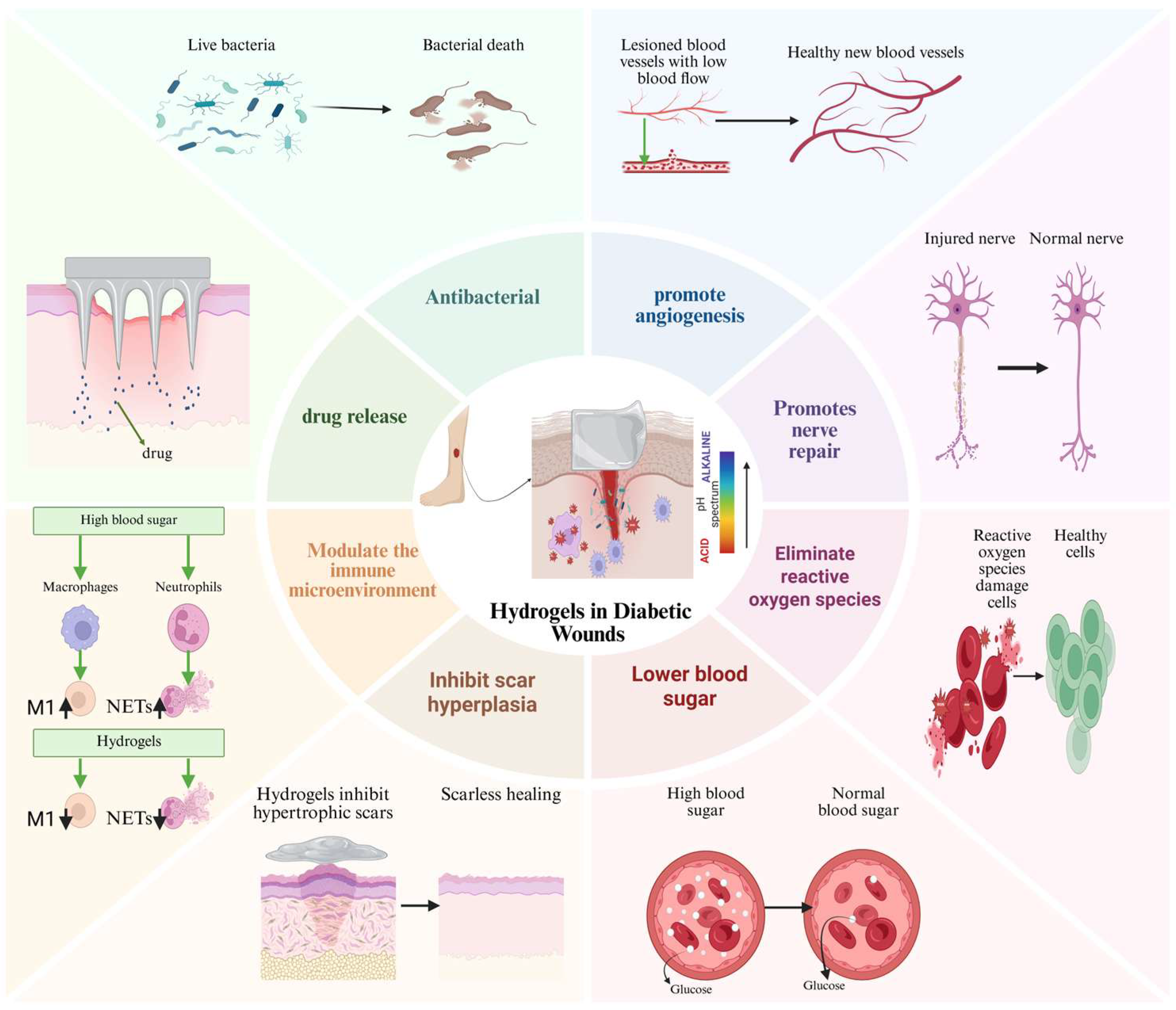
Multifunctional hydrogels not only possess the core functions of traditional hydro gels (water absorption and moisture retention) but also exhibit the following additional capabilities: (1) exerting antibacterial activity; (2) promoting angiogenesis; (3) promoting nerve repair; (4) scavenging reactive oxygen species; (5) lowering blood glucose; (6) inhib iting scar hyperplasia; (7) regulating the immune microenvironment; (8) enabling con trolled drug delivery. These functions directly address the complex pathophysiological mechanisms underlying diabetic wounds.
Table 3.
Representative Multifunctional Hydrogels for Diabetic Wound Healing: Composition, Mechanisms, and Functional Outcomes.
4. Outlook
In recent years, hydrogels have achieved notable advances in chronic wound management, with several hydrogel formulations entering clinical trials or receiving approval for applications in surgical sealing and tissue adhesion [161,162]. Nevertheless, their widespread clinical implementation still encounters three major challenges. First, the long-term biosafety of drug-loaded hydrogels necessitates comprehensive systematic evaluation, particularly for therapeutics with hepatotoxic, nephrotoxic, or neurotoxic potential (e.g., cyclosporine and metallic nanomaterials) [156,157,158,159,160,161,162,163]. Second, the preparation processes are complex and costly, and the absence of standardized protocols for efficacy assessment and quality control hinders scalable and reproducible manufacturing [29,44,164,165]. Third, intrinsic differences between animal models and humans limit the translatability of some effective strategies [166,167,168]; Furthermore, interdisciplinary barriers in mechanistic understanding and evaluation often lead to a disconnect between preclinical research and clinical requirements [20,169,170].
In response to these challenges, the future development of multifunctional hydrogels is likely to focus on three principal trends.
4.1. Intelligent Closed-Loop Systems and Multidisciplinary Collaboration
With the aid of programmable hydrogel technology [171] and AI-driven big data models [172], hydrogels are evolving from passive dressings into active, responsive therapeutic platforms. At the closed-loop therapeutic level, systems integrating “real-time monitoring, intelligent feedback, and precision treatment” can dynamically detect physiological signals and respond autonomously, thereby markedly enhancing both the precision and timeliness of intervention [172,173]. For instance, the GPP@ZnBG hydrogel responds to hyperglycemia and oxidative stress at the wound site by sequentially releasing zinc, calcium, and silicon ions in a pH-regulated manner, thereby achieving early-stage antibacterial effects and promoting late-stage angiogenesis. Preliminary clinical studies indicate that topical application for four weeks reduced wound area by 94.57%, with no reported adverse events. The tough, adhesive smart hydrogel developed by Shi [174] integrates multiple functionalities, including synergistic photothermal/nitric oxide antibacterial activity, angiogenesis promotion, and signal monitoring, achieving complete wound closure within 14 days. In parallel, the integration of wearable biosensors with smart hydrogels further advances wound management toward closed-loop systems by enabling real-time monitoring of electrical signals (e.g., impedance) and biochemical markers (e.g., pH, glucose, and inflammatory factors), which can trigger responsive interventions such as drug release, electrical stimulation, or photothermal therapy. Collectively, these systems allow coordinated regulation of infection, hypoxia, and inflammation to promote tissue regeneration [138,175,176]; however, they remain limited by insufficient sensing depth, trade-offs between sensitivity and long-term stability, and a lack of clinical validation.
At the research and development level, intensive collaboration among materials science, biology, clinical medicine, and engineering, combined with AI-assisted material design [168] and organoid model validation [177] holds promise for establishing a closed-loop research paradigm encompassing “computational design–high-throughput screening–experimental validation–clinical translation.” Zheng [178] systematically demonstrated how AI can integrate clinical and biological data to predict healing risks, optimize nanocarrier design, and dynamically tailor personalized treatment strategies. Furthermore, stimulus-responsive hydrogels based on cascade reactions have substantially enhanced the rational design and predictive validation of hydrogel systems by integrating AI-assisted modeling, biosensor feedback, and organ-on-a-chip technologies. Collectively, these studies validate the feasibility of closed-loop systems and multidisciplinary collaborative strategies, which are anticipated to substantially accelerate the translational process.
4.2. The Deep Integration of Traditional Chinese Medicine Theory and Hydrogel Technology
Beyond addressing the challenges mentioned above, the development of diversified therapeutic strategies represents an inevitable trend. The extensive clinical use of traditional Chinese medicine (TCM) dressings or medicated solutions in diabetic wound management [179,180] provides a solid clinical basis for such integration. Recently, considerable progress has been achieved in integrating bioactive components of TCM with modern smart hydrogel platforms. For example, quercetin, the principal bioactive component of Astragalus membranaceus, loaded onto chitosan nanoparticles within a GelMA hydrogel (QNPs@GelMA), exerts synergistic effects by downregulating the RAGE/NF-κB pathway, thereby providing anti-inflammatory and antioxidant benefits, promoting M2 macrophage polarization and angiogenesis, and accelerating diabetic wound healing [181]. Berberine-loaded glucose-responsive hydrogels (OHA/CMCS-BBR) enable high-glucose-triggered, on-demand drug release through dynamic boronate bonds. In a diabetic infected wound model, these hydrogels achieved a 99.7% wound closure rate within 14 days and completely eradicated bacterial pathogens [182]. A carrier-free hydrogel (Cu-SCU), formed by the self-assembly of scutellarin and copper ions through dynamic coordination bonds, can scavenge reactive oxygen species, induce M2 macrophage polarization, and activate the HIF-1α/VEGF pathway in a diabetic full-thickness wound model, thereby synergistically promoting epithelial regeneration, vascular maturation, and wound repair [183]. Overall, these studies integrate modern materials science with bioactive TCM components, providing an innovative paradigm for the development of multifunctional hydrogels with distinctive TCM characteristics, thereby enabling multi-targeted, synergistic therapy for diabetic wounds.
However, most current studies remain largely limited to single-component integration and have not yet fully realized synergistic design guided by the traditional Chinese medicine “emperor–minister–assistant–courier” (EMAC) compatibility theory [184]. In addition, dose–response relationships of bioactive TCM components remain poorly defined, while their extraction complexity, as well as the physicochemical properties, bioactivity, and potential toxicity of individual constituents, still require systematic characterization [185]. Furthermore, a key challenge lies in balancing the diversity of assembly strategies for incorporating active components within hydrogel matrices against the structural and functional stability of the resulting systems, which remains a critical bottleneck for clinical translation [186].
4.3. Standardized Quality Control Systems, Safety Assessment, and Intelligent Manufacturing
To overcome bottlenecks in safety assessment and large-scale production, a standardized quality control system encompassing the entire R&D process is essential. Specifically, drug safety prediction models leveraging organ-on-a-chip technology and computational toxicology should be developed to systematically evaluate pharmacokinetics and accumulation risks following transdermal drug delivery, as well as to establish standardized preclinical safety evaluation guidelines. Concurrently, the integration of automated and continuous production processes can reduce labor and time costs, thereby enabling high-quality, low-cost, large-scale manufacturing. In recent years, several hydrogel-based wound dressings have been translated into clinical use or clinical evaluation, demonstrating their potential for real-world wound management; for instance, INTRASITE* Gel Hydrogel Wound Dressing, DermiSphere hDRT, and the antimicrobial peptide hydrogel Amferia have all received FDA clearance or approval. From an industrial perspective, patent analysis indicates that more than 96,000 hydrogel-related patent documents had been published globally as of September 2024, with approximately 74.5% remaining in the application stage. This reflects a clear shift from single-function materials toward smart, integrated hydrogel platforms [187] (as Table 4). Current patent activity is mainly concentrated in three areas: stimuli-responsive systems, antimicrobial hydrogels, and bioactive delivery platforms [188]. These developments suggest that smart hydrogel dressings are at a critical juncture, transitioning rapidly from laboratory research to clinical application, thereby providing valuable practical guidance for industrial development.
Table 4.
Summary of representative patents and clinical trials of multifunctional hydrogels for diabetic wound healing.
In summary, the future development of multifunctional hydrogels will be driven by three key directions: “smart closed-loop systems and multidisciplinary collaboration,” “deep integration with traditional Chinese medicine,” and “standardized quality control, safety assessment, and intelligent manufacturing.” Among these, the most promising design principle is the construction of multifunctional, stimulus-responsive hydrogels capable of recognizing key pathological cues in diabetic wounds and coordinately regulating inflammation, angiogenesis, and tissue remodeling under clinically relevant thresholds, such as glucose-triggered responses at ~10 mM and pH-responsive activation within the physiological to mildly acidic range (pH 5.5–7.4) [189,190]. From a translational perspective, priority benchmarks should include long-term in vivo degradation of ≥12 months [191], ISO 10993-compliant biocompatibility evaluation, ≥70% complete wound closure within 12 weeks, and ≤20% recurrence within 6 months in randomized controlled trials [192]. However, successful clinical translation will still require standardized preclinical models, clearly defined regulatory pathways, rigorous long-term safety validation, and effective strategies to overcome practical barriers such as high production costs, limited manufacturing infrastructure, storage requirements, and insufficient clinical training resources, particularly in resource-limited healthcare settings [193,194,195].
Addressing these challenges will be essential for facilitating broader clinical adoption and commercialization of multifunctional hydrogels. Overall, this field is shifting toward adaptive, pathology-driven, and clinically translatable hydrogel systems that integrate therapeutic functionality with practical applicability, ultimately advancing precision wound management.
Author Contributions
Y.Z. conceived the review and drafted the manuscript. Y.H. participated in the conceptualization and revision of the figures. X.Z., L.L. (Li Li), D.C. and L.L. (Lin Li) contributed to literature analysis, critical revision of the manuscript, and intellectual input. All authors have read and agreed to the published version of the manuscript.
Funding
This work was supported by The National Natural Science Foundation of China (31960074, 31560087) and Guizhou Provincial Basic Research Program (Natural Science) (ZK [2023]496, MS (2025)359).
Data Availability Statement
No new data were created in this study. This article is a review of previously published literature, and all relevant data are available in the cited references.
Acknowledgments
The authors acknowledge the use of BioRender.com for the creation of Figure 1, Figure 2 and Figure 3 in this manuscript. The authors also acknowledge the use of DeepSeek (Version: DeepSeek-V4-Flash) and ChatGPT (Version: GPT-5.5) artificial intelligence tools for language polishing and partial translation during the preparation of this work.
Conflicts of Interest
The authors declare that the review was conducted in the absence of any commercial or financial relationships that could be construed as a potential conflict of interest.
References
- Genitsaridi, I.; Salpea, P.; Salim, A.; Sajjadi, S.F.; Tomic, D.; James, S.; Thirunavukkarasu, S.; Issaka, A.; Chen, L.; Basit, A.; et al. 11th edition of the IDF Diabetes Atlas: Global, regional, and national diabetes prevalence estimates for 2024 and projections for 2050. Lancet Diabetes Endocrinol. 2026, 14, 149–156. [Google Scholar] [CrossRef] [Scilit] [PubMed]
- He, J.; Chen, J.; Liu, T.; Qin, F.; Wei, W. Research Progress of Multifunctional Hydrogels in Promoting Wound Healing of Diabetes. Int. J. Nanomed. 2025, 20, 7549–7578. [Google Scholar] [CrossRef] [Scilit]
- Patel, S.; Srivastava, S.; Singh, M.R.; Singh, D. Mechanistic insight into diabetic wounds: Pathogenesis, molecular targets and treatment strategies to pace wound healing. Biomed. Pharmacother. 2019, 112, 108615. [Google Scholar] [CrossRef] [Scilit] [PubMed]
- Du, Y.; Wang, J.; Fan, W.; Huang, R.; Wang, H.; Liu, G. Preclinical study of diabetic foot ulcers: From pathogenesis to vivo/vitro models and clinical therapeutic transformation. Int. Wound J. 2023, 20, 4394–4409. [Google Scholar] [CrossRef] [Scilit] [PubMed]
- Eriksson, E.; Liu, P.Y.; Schultz, G.S.; Martins-Green, M.M.; Tanaka, R.; Weir, D.; Gould, L.J.; Armstrong, D.G.; Gibbons, G.W.; Wolcott, R.; et al. Chronic wounds: Treatment consensus. Wound Repair Regen. 2022, 30, 156–171. [Google Scholar] [CrossRef] [Scilit] [PubMed]
- Pereira, S.G.; Moura, J.; Carvalho, E.; Empadinhas, N. Microbiota of Chronic Diabetic Wounds: Ecology, Impact, and Potential for Innovative Treatment Strategies. Front. Microbiol. 2017, 8, 1791. [Google Scholar] [CrossRef] [Scilit] [PubMed]
- Hicks, C.W.; Canner, J.K.; Karagozlu, H.; Mathioudakis, N.; Sherman, R.L.; Black, J.H., 3rd; Abularrage, C.J. Contribution of 30-day readmissions to the increasing costs of care for the diabetic foot. J. Vasc. Surg. 2019, 70, 1263–1270. [Google Scholar] [CrossRef] [Scilit] [PubMed]
- Hicks, C.W.; Canner, J.K.; Mathioudakis, N.; Lippincott, C.; Sherman, R.L.; Abularrage, C.J. Incidence and Risk Factors Associated with Ulcer Recurrence Among Patients with Diabetic Foot Ulcers Treated in a Multidisciplinary Setting. J. Surg. Res. 2020, 246, 243–250. [Google Scholar] [CrossRef] [Scilit] [PubMed]
- Edmonds, M.; Manu, C.; Vas, P. The current burden of diabetic foot disease. J. Clin. Orthop. Trauma 2021, 17, 88–93. [Google Scholar] [CrossRef] [Scilit] [PubMed]
- Fu, T.; Stupnitskaia, P.; Matoori, S. Next-Generation Diagnostic Wound Dressings for Diabetic Wounds. ACS Meas. Sci. Au 2022, 2, 377–384. [Google Scholar] [CrossRef] [Scilit] [PubMed]
- Jodheea-Jutton, A.; Hindocha, S.; Bhaw-Luximon, A. Health economics of diabetic foot ulcer and recent trends to accelerate treatment. Foot 2022, 52, 101909. [Google Scholar] [CrossRef] [Scilit] [PubMed]
- Jiang, P.; Li, Q.; Luo, Y.; Luo, F.; Che, Q.; Lu, Z.; Yang, S.; Yang, Y.; Chen, X.; Cai, Y. Current status and progress in research on dressing management for diabetic foot ulcer. Front. Endocrinol. 2023, 14, 1221705. [Google Scholar] [CrossRef] [Scilit]
- Nguyen, H.M.; Ngoc Le, T.T.; Nguyen, A.T.; Thien Le, H.N.; Pham, T.T. Biomedical materials for wound dressing: Recent advances and applications. RSC Adv. 2023, 13, 5509–5528. [Google Scholar] [CrossRef] [Scilit] [PubMed]
- Qi, L.; Zhang, C.; Wang, B.; Yin, J.; Yan, S. Progress in Hydrogels for Skin Wound Repair. Macromol. Biosci. 2022, 22, e2100475. [Google Scholar] [CrossRef] [Scilit] [PubMed]
- Nešporová, K.; Pavlík, V.; Šafránková, B.; Vágnerová, H.; Odráška, P.; Žídek, O.; Císařová, N.; Skoroplyas, S.; Kubala, L.; Velebný, V. Effects of wound dressings containing silver on skin and immune cells. Sci. Rep. 2020, 10, 15216. [Google Scholar] [CrossRef] [Scilit] [PubMed]
- Zhang, Y.; Zhu, Y.; Ma, P.; Wu, H.; Xiao, D.; Zhang, Y.; Sui, X.; Zhang, L.; Dong, A. Functional carbohydrate-based hydrogels for diabetic wound therapy. Carbohydr. Polym. 2023, 312, 120823. [Google Scholar] [CrossRef] [Scilit] [PubMed]
- Alotaibi, B.S.; Khan, M.; Ibrahim, N.A.; Khan, A.K.; Liaqat, R.; Ijaz, M. Smart Nanofibers in Wound Healing: Exploring Novel Combinations and Applications. Curr. Drug Deliv. 2025, 23, 497–518. [Google Scholar]
- Khan, M.U.A.; Aslam, M.A.; Abdullah, M.F.B.; Arjan, W.S.A.; Stojanovic, G.M.; Hasan, A. Hydrogels: Classifications, fundamental properties, applications, and scopes in recent advances in tissue engineering and regenerative medicine—A comprehensive review. Arab. J. Chem. 2024, 17, 105968. [Google Scholar] [CrossRef] [Scilit]
- Hosseini, S.A.; Noruzi, S.; Kesharwani, P.; Sahebkar, A. Hydrogel-based dressing for wound healing: A systematic review of clinical trials. Int. J. Biol. Macromol. 2025, 308, 142322. [Google Scholar] [CrossRef] [Scilit] [PubMed]
- Zhang, J.; Yang, Y.; Bao, S.; Wang, Y.; Wen, Z.; Chen, S.; Yao, L.; Zhou, P.; Zhou, Y.; Chen, Y. Mapping the evolving landscape of conductive hydrogels in medicine: A bibliometric perspective. Regen. Ther. 2026, 31, 101038. [Google Scholar] [CrossRef] [Scilit] [PubMed]
- Cheng, Y.; Wang, Y.; Wang, Y.; Tan, P.C.; Yu, S.; Li, C.; Li, Z.Y.; Li, Q.F.; Zhou, S.B.; Wang, C.; et al. Microenvironment-feedback regulated hydrogels as living wound healing materials. Nat. Commun. 2025, 16, 6050. [Google Scholar] [CrossRef] [Scilit] [PubMed]
- Liu, B.; Chen, L.; Huang, C.; Zhang, H.; Zhou, H.; Chen, Y.; Liu, X.; Xiao, Z.; Liang, K.; Xie, X.; et al. A sprayable exosome-loaded hydrogel with controlled release and multifunctional synergistic effects for diabetic wound healing. Mater. Today Bio 2025, 34, 102159. [Google Scholar] [CrossRef] [Scilit] [PubMed]
- Riaz, S.; Waheed, H.; Ahmad, F.; Khan, M.I.; Shanableh, A. Natural and synthetic biomaterials, structural matrices-based wound dressings: Key properties, material correlation, and adaptability. Regenesis Repair Rehabil. 2025, 1, 47–65. [Google Scholar] [CrossRef] [Scilit]
- Soylu, Z.; Oktay, B.; Erarslan, A.; Ahlatcıoğlu Özerol, E. Multifunctional polymeric wound dressings. Polym. Bull. 2025, 82, 5325–5383. [Google Scholar] [CrossRef] [Scilit]
- Eudy, M.; Eudy, C.L.; Roy, S. Apligraf as an Alternative to Skin Grafting in the Pediatric Population. Cureus 2021, 13, e16226. [Google Scholar] [CrossRef] [Scilit] [PubMed]
- Holl, J.; Kowalewski, C.; Zimek, Z.; Fiedor, P.; Kaminski, A.; Oldak, T.; Moniuszko, M.; Eljaszewicz, A. Chronic Diabetic Wounds and Their Treatment with Skin Substitutes. Cells 2021, 10, 655. [Google Scholar] [CrossRef] [Scilit] [PubMed]
- Lyu, J.; Liu, X.; Yang, Q.; Zhang, Y.; Wang, X. Applications of Multifunctional Hydrogel in Tissue Engineering and Regenerative Medicine. MedComm 2026, 7, e70602. [Google Scholar]
- Wang, H.; Pei, B.; Yao, S.; Liu, Y.; Guo, X.; Tian, J. Intelligent Natural Hydrogels for Wound Therapy: Spatiotemporal Microenvironment Sensing, Synergistic Regulation, and Translational Pathways. Macromol. Rapid Commun. 2026, 47, e00766. [Google Scholar] [CrossRef] [Scilit] [PubMed]
- Varaprasad, K.; Raghavendra, G.M.; Jayaramudu, T.; Yallapu, M.M.; Sadiku, R. A mini review on hydrogels classification and recent developments in miscellaneous applications. Mater. Sci. Eng. C Mater. Biol. Appl. 2017, 79, 958–971. [Google Scholar] [CrossRef] [Scilit] [PubMed]
- Buwalda, S.J.; Boere, K.W.; Dijkstra, P.J.; Feijen, J.; Vermonden, T.; Hennink, W.E. Hydrogels in a historical perspective: From simple networks to smart materials. J. Control. Release 2014, 190, 254–273. [Google Scholar] [CrossRef] [Scilit] [PubMed]
- Kumar, A.; Pandey, S.; Kumar, K.; Krishnamoorthi, S.; Tungala, K. Hydrogels: Classification, cross-linking methods, characteristics, and current trends in biomedical applications. Polym. Bull. 2025, 83, 49. [Google Scholar] [CrossRef] [Scilit]
- Su, C.; Lin, D.; Huang, X.; Feng, J.; Jin, A.; Wang, F.; Lv, Q.; Lei, L.; Pan, W. Developing hydrogels for gene therapy and tissue engineering. J. Nanobiotechnol. 2024, 22, 182. [Google Scholar] [CrossRef] [Scilit]
- Di, J.; Li, J.; Sun, C.; Xu, L.; Li, X. Advances in Cellulose-Based Hydrogels for Drug Delivery: Preparation, Modification and Challenges. Gels 2025, 11, 938. [Google Scholar] [CrossRef] [Scilit] [PubMed]
- Rumon, M.; Rahman, M.S.; Akib, A.A.; Sohag, M.S.; Rakib, M.; Khan, M.; Yesmin, F.; Shakil, M.S.; Rahman Khan, M.M. Progress in hydrogel toughening: Addressing structural and crosslinking challenges for biomedical applications. Discov. Mater. 2025, 5, 5. [Google Scholar] [CrossRef] [Scilit]
- Hosseinzadeh, B.; Ahmadi, M. Degradable hydrogels: Design mechanisms and versatile applications. Mater. Today Sustain. 2023, 23, 100468. [Google Scholar] [CrossRef] [Scilit]
- Qi, Y.; Wang, F.; Liu, J.; Wang, C.; Liu, Y. Enzyme-mediated hydrogelation for biomedical applications: A review. Int. J. Biol. Macromol. 2025, 311, 143379. [Google Scholar] [CrossRef] [Scilit] [PubMed]
- Kuth, S.; Boccaccini, A.R. Enzymatic Insitu Crosslinking Can Improve Hydrogel Stability While Maintaining Matrix Stiffness. ChemistrySelect 2024, 9, e202401700. [Google Scholar] [CrossRef] [Scilit]
- Protsak, I.S.; Morozov, Y.M. Fundamentals and Advances in Stimuli-Responsive Hydrogels and Their Applications: A Review. Gels 2025, 11, 30. [Google Scholar] [CrossRef] [Scilit] [PubMed]
- Zhang, H.; Gao, Y. Recent Advances in Stimuli-Responsive Microgels and Their Biomedical Applications. Molecules 2025, 30, 4457. [Google Scholar] [CrossRef] [Scilit] [PubMed]
- Cheng, S.; Yang, J.; Song, J.; Cao, X.; Zhou, B.; Yang, L.; Li, C.; Wang, Y. A motion-responsive injectable lubricative hydrogel for efficient Achilles tendon adhesion prevention. Mater. Today Bio 2025, 30, 101458. [Google Scholar] [CrossRef] [Scilit] [PubMed]
- Du, B.; Ren, X.; Wang, X.; Wen, Y.; Yang, J.; Li, L.; Bai, P.; Lang, F.; Li, L.; Zhang, R. Four-in-one pH/glucose-responsive engineered hydrogel for diabetes wound healing. Nano Today 2025, 62, 102725. [Google Scholar] [CrossRef] [Scilit]
- Zhu, B.; Zou, K.; Huang, Y.; Huang, Q.; Zhang, H.; Yang, L.; Zhao, L.; Cao, H. Injectable and self-healing photothermal hydrogel with pH/photothermal dual-responsive mesoporous polydopamine for synergistic melanoma therapy. Int. J. Biol. Macromol. 2025, 335, 149336. [Google Scholar] [PubMed]
- Du, J.; Xian, C.; Liang, X.; Fan, S.; Wang, L.; Wu, J. Advanced Responsive Hydrogels for Diabetic Wound Healing: Design Principles, Controlled Drug Delivery, Therapeutic Strategies, and Application Prospects. MedComm—Biomater. Appl. 2025, 4, e70019. [Google Scholar] [CrossRef] [Scilit]
- Lian, H.; Liu, L.; Ke, L. Current Research Status of Biomedical Hydrogel and Challenges and Opportunities in Clinical Translation. Zhongguo Yi Liao Qi Xie Za Zhi 2025, 49, 520–526. [Google Scholar] [CrossRef] [Scilit] [PubMed]
- Khattak, S.; Ullah, I.; Yousaf, M.T.; Ullah, S.; Yousaf, H.; Li, Y.; Jin, H.; Shen, J.; Xu, H.T. Advancements in hydrogels: A comprehensive review of natural, synthetic, and hybrid innovations for wound healing. Int. J. Biol. Macromol. 2025, 327, 147270. [Google Scholar] [CrossRef] [Scilit] [PubMed]
- Zhu, T.; Ni, Y.; Biesold, G.M.; Cheng, Y.; Ge, M.; Li, H.; Huang, J.; Lin, Z.; Lai, Y. Recent advances in conductive hydrogels: Classifications, properties, and applications. Chem. Soc. Rev. 2023, 52, 473–509. [Google Scholar] [CrossRef] [Scilit] [PubMed]
- Guo, A.; Cao, Q.; Fang, H.; Tian, H. Recent advances and challenges of injectable hydrogels in drug delivery. J. Control. Release 2025, 385, 114021. [Google Scholar] [CrossRef] [Scilit] [PubMed]
- Ribeiro, M.; Simões, M.; Vitorino, C.; Mascarenhas-Melo, F. Hydrogels in Cutaneous Wound Healing: Insights into Characterization, Properties, Formulation and Therapeutic Potential. Gels 2024, 10, 188. [Google Scholar] [CrossRef] [Scilit] [PubMed]
- Khan, R.; Ahmad, U.; Yadav, I.K.; Khan, M.M.; Islam, A.; Akhtar, J.; Shubhra, Q.T.H. Engineering bioinspired pH-responsive hydrogels for smart wound repair. Nanoscale 2026. [Google Scholar] [CrossRef] [Scilit] [PubMed]
- Nezhad-Mokhtari, P.; Hasany, M.; Kohestanian, M.; Dolatshahi-Pirouz, A.; Milani, M.; Mehrali, M. Recent advancements in bioadhesive self-healing hydrogels for effective chronic wound care. Adv. Colloid Interface Sci. 2024, 334, 103306. [Google Scholar] [CrossRef] [Scilit] [PubMed]
- Zhang, T.; Zhong, X.C.; Feng, Z.X.; Lin, X.Y.; Chen, C.Y.; Wang, X.W.; Guo, K.; Wang, Y.; Chen, J.; Du, Y.Z.; et al. An active shrinkage and antioxidative hydrogel with biomimetic mechanics functions modulates inflammation and fibrosis to promote skin regeneration. Bioact. Mater. 2025, 45, 322–344. [Google Scholar] [CrossRef] [Scilit] [PubMed]
- Zhang, C.W.; Si, M.; Chen, C.; He, P.; Fei, Z.; Xu, N.; He, X. Hierarchical Engineering for Biopolymer-based Hydrogels with Tailored Property and Functionality. Adv. Mater. 2025, 37, e2414897. [Google Scholar] [CrossRef] [Scilit] [PubMed]
- Liang, Y.; He, J.; Guo, B. Functional Hydrogels as Wound Dressing to Enhance Wound Healing. ACS Nano 2021, 15, 12687–12722. [Google Scholar] [CrossRef] [Scilit] [PubMed]
- Zhou, Y.; Liang, X.; Shen, Z.; Zhang, R.; Zhang, G.; Yu, B.; Li, Y.; Xu, F.J. Glucose-responsive hydrogel with adaptive insulin release to modulate hyperglycemic microenvironment and promote wound healing. Biomaterials 2025, 326, 123641. [Google Scholar] [CrossRef] [Scilit] [PubMed]
- Wang, Y.; Jin, L.; Huang, C.; Bhutta, N.K.; Sun, L.; Li, J.; Han, B. Mechanically adaptive injectable hydrogels with dual-role oxidized cyclodextrin for pH-responsive drug delivery in chronic infected wound healing. Carbohydr. Polym. 2026, 372, 124530. [Google Scholar] [CrossRef] [Scilit] [PubMed]
- Wang, Q.; Zhang, Y.; Ma, Y.; Wang, M.; Pan, G. Nano-crosslinked dynamic hydrogels for biomedical applications. Mater. Today Bio 2023, 20, 100640. [Google Scholar] [CrossRef] [Scilit] [PubMed]
- Zhang, M.; Zhu, C. Dynamic Hydrogels against Infections: From Design to Applications. Gels 2024, 10, 331. [Google Scholar] [CrossRef] [Scilit] [PubMed]
- Kim, K.; Yang, J.; Li, C.; Yang, C.Y.; Hu, P.; Liu, Y.; Huang, Y.Y.; Sun, X.; Chi, M.; Huang, C.; et al. Anisotropic structure of nanofiber hydrogel accelerates diabetic wound healing via triadic synergy of immune-angiogenic-neurogenic microenvironments. Bioact. Mater. 2025, 47, 64–82. [Google Scholar] [CrossRef] [Scilit] [PubMed]
- Liu, F.; Li, G.; An, Z.; Wang, S.; Xu, S.; Liu, H. Dynamic Boronate Ester Based Hydrogel with Enhanced Mechanical Properties and Multi-Stimuli-Triggered Release for Tissue Repair and Antioxidant Therapy. Gels 2025, 11, 370. [Google Scholar] [CrossRef] [Scilit] [PubMed]
- Bi, S.; He, C.; Zhou, Y.; Liu, R.; Chen, C.; Zhao, X.; Zhang, L.; Cen, Y.; Gu, J.; Yan, B. Versatile conductive hydrogel orchestrating neuro-immune microenvironment for rapid diabetic wound healing through peripheral nerve regeneration. Biomaterials 2025, 314, 122841. [Google Scholar] [CrossRef] [Scilit] [PubMed]
- Shao, Y.; Zhou, X.; Zhou, S.; Long, J.; Jin, L.; Shi, X.; Zhou, L.; Zhang, Y.; Fan, D. Injectable DMM/GelMA hydrogel for diabetic wound healing via regulating mitochondrial metabolism and macrophage repolarization. Colloids Surf. B Biointerfaces 2025, 248, 114488. [Google Scholar] [CrossRef] [Scilit] [PubMed]
- Liu, Y.; Gilchrist, A.E.; Heilshorn, S.C. Engineered Protein Hydrogels as Biomimetic Cellular Scaffolds. Adv. Mater. 2024, 36, e2407794. [Google Scholar] [CrossRef] [Scilit] [PubMed]
- Cai, Y.; Gao, B.; Hong, J.; Zhu, X.; Cheang, L.H.; Yang, X.; Sun, C.; Zheng, S.; Li, Y.; Wang, H.; et al. Cell-derived extracellular matrix-loaded gelatin electrospun nanofibrous hydrogel for trauma repair: Fabrication and biomedical application. Int. J. Biol. Macromol. 2025, 322, 146724. [Google Scholar] [CrossRef] [Scilit] [PubMed]
- Ma, H.; Luo, Y.; Wang, Y.; Hao, Y.; Li, J.; Gao, X.; Xiong, Y.; He, L. Artificial multienzyme nanoflower composite hydrogel for efficiently promoting MRSA-infected diabetic wound healing via glucose-activated NO releasing and microenvironment regulation. Bioact. Mater. 2025, 49, 531–548. [Google Scholar] [CrossRef] [Scilit] [PubMed]
- Nie, L.; Wei, Q.; Li, J.; Deng, Y.; He, X.; Gao, X.; Ma, X.; Liu, S.; Sun, Y.; Jiang, G.; et al. Fabrication and desired properties of conductive hydrogel dressings for wound healing. RSC Adv. 2023, 13, 8502–8522. [Google Scholar] [CrossRef] [Scilit] [PubMed]
- Bisht, N.; Yeo, R.J.; Ramakrishna, S.; Sankaranarayanan, S.; Dhand, C.; Dwivedi, N. Shape Memorable and Self-Healable Smart Hydrogels and Emerging Directions. Adv. Healthc. Mater. 2026, 15, e03361. [Google Scholar] [CrossRef] [Scilit] [PubMed]
- Chocarro-Wrona, C.; Pleguezuelos-Beltrán, P.; López de Andrés, J.; Antich, C.; de Vicente, J.; Jiménez, G.; Arias-Santiago, S.; Gálvez-Martín, P.; López-Ruiz, E.; Marchal, J.A. A bioactive three-layered skin substitute based on ECM components effectively promotes skin wound healing and regeneration. Mater. Today Bio 2025, 31, 101592. [Google Scholar] [CrossRef] [Scilit] [PubMed]
- Agrawal, A.; Hussain, C.M. State-of-the-art in functionalized 3D/4D-printed magnetic hydrogels for environmental and biomedical applications. Adv. Colloid Interface Sci. 2026, 347, 103694. [Google Scholar] [CrossRef] [Scilit] [PubMed]
- Bai, Q.; Han, K.; Dong, K.; Zheng, C.; Zhang, Y.; Long, Q.; Lu, T. Potential Applications of Nanomaterials and Technology for Diabetic Wound Healing. Int. J. Nanomed. 2020, 15, 9717–9743. [Google Scholar] [CrossRef] [Scilit]
- Li, D.; Wu, N. Mechanism and application of exosomes in the wound healing process in diabetes mellitus. Diabetes Res. Clin. Pract. 2022, 187, 109882. [Google Scholar] [CrossRef] [Scilit] [PubMed]
- Hu, M.; Zhang, Q.; Qin, L. Innovative applications of multidimensional engineered hydrogels in wound healing. J. Adv. Res. 2026, 83, 347–378. [Google Scholar] [CrossRef] [Scilit] [PubMed]
- Feng, J.; Yao, Y.; Wang, Q.; Han, X.; Deng, X.; Cao, Y.; Chen, X.; Zhou, M.; Zhao, C. Exosomes: Potential key players towards novel therapeutic options in diabetic wounds. Biomed. Pharmacother. 2023, 166, 115297. [Google Scholar] [CrossRef] [Scilit] [PubMed]
- Hu, X.; Zhang, M.-X.; Xu, F.-W.; Fang, Q.-Q.; Cai, J.-Q.; Chen, Y.-Q.; Ma, J.-J.; Tan, W.-Q.; Wang, Y. Preparation Methods of Hydrogel Microspheres and Recent Advances in Their Application for Treating Diabetic Wounds. Int. J. Nanomed. 2025, 20, 10645–10666. [Google Scholar] [CrossRef] [Scilit]
- Chaves, R.G.R.; Costa, F.F.; Fuchs, L.A.; Rodrigues, L.S.; Moraes, R.A.N.; Junior, P.S.d.S.A.; Maciel, M.C.G.; Amaral, F.M.M.; Coutinho, D.F.; Reis, A.S. Mechanistic Insights into the Wound Healing Activity of Plant Species in Diabetic Ulcers. Curr. Issues Mol. Biol. 2025, 47, 972. [Google Scholar] [CrossRef] [Scilit] [PubMed]
- Han, C.; Singla, R.K.; Wang, C. Application of Biomaterials in Diabetic Wound Healing: The Recent Advances and Pathological Aspects. Pharmaceutics 2025, 17, 1295. [Google Scholar] [CrossRef] [Scilit] [PubMed]
- He, X.; Wei, Y.; Xu, K. Hydrogel-Based Treatment of Diabetic Wounds: From Smart Responsive to Smart Monitoring. Gels 2025, 11, 647. [Google Scholar] [CrossRef] [Scilit] [PubMed]
- Wu, J.; Yang, Q.; Cheng, H.; Zheng, H.; Chen, M. Metabolism, senescence, and natural products: New perspectives on wound healing in diabetes. Front. Nutr. 2025, 12, 1746827. [Google Scholar] [PubMed]
- Han, Y.; Ge, S.; Yin, H.; Han, D.; Wang, S. Advances in immunomodulatory microneedles for diabetic wound healing. J. Mater. Chem. B 2025, 13, 12349–12381. [Google Scholar] [CrossRef] [Scilit] [PubMed]
- Lin, L.; Liang, X.; Xu, Z.; Li, Y.; Guo, Z.; Liu, L.; Liu, H.; Cai, Q.; Chen, Y.; Yu, Z.; et al. Multifunctional hydrogel delivery of mesenchymal stem cell secretome suppresses neutrophil extracellular trap formation and promotes diabetic wound healing via PGE2/BMAL1 pathway. Biomaterials 2025, 327, 123764. [Google Scholar] [CrossRef] [Scilit] [PubMed]
- Callaghan, M.J.; Ceradini, D.J.; Gurtner, G.C. Hyperglycemia-induced reactive oxygen species and impaired endothelial progenitor cell function. Antioxid. Redox Signal. 2005, 7, 1476–1482. [Google Scholar] [CrossRef] [Scilit] [PubMed]
- Zhu, J.; Hu, Z.; Luo, Y.; Liu, Y.; Luo, W.; Du, X.; Luo, Z.; Hu, J.; Peng, S. Diabetic peripheral neuropathy: Pathogenetic mechanisms and treatment. Front. Endocrinol. 2023, 14, 1265372. [Google Scholar] [CrossRef] [Scilit]
- Li, W.; Sadeh, O.; Chakraborty, J.; Yang, E.; Basu, P.; Kumar, P. Multifaceted Antibiotic Resistance in Diabetic Foot Infections: A Systematic Review. Microorganisms 2025, 13, 2311. [Google Scholar] [CrossRef] [Scilit] [PubMed]
- Hsu, C.-Y.; Shu, J.-C.; Lin, M.-H.; Chong, K.-Y.; Chen, C.-C.; Wen, S.-M.; Hsieh, Y.-T.; Liao, W.-T. High Glucose Concentration Promotes Vancomycin-Enhanced Biofilm Formation of Vancomycin-Non-Susceptible Staphylococcus aureus in Diabetic Mice. PLoS ONE 2015, 10, e0134852. [Google Scholar] [CrossRef] [Scilit] [PubMed]
- David, J.A.; Rifkin, W.J.; Rabbani, P.S.; Ceradini, D.J. The Nrf2/Keap1/ARE Pathway and Oxidative Stress as a Therapeutic Target in Type II Diabetes Mellitus. J. Diabetes Res. 2017, 2017, 4826724. [Google Scholar] [CrossRef] [Scilit] [PubMed]
- Teh, H.X.; Phang, S.J.; Looi, M.L.; Kuppusamy, U.R.; Arumugam, B. Molecular pathways of NF-ĸB and NLRP3 inflammasome as potential targets in the treatment of inflammation in diabetic wounds: A review. Life Sci. 2023, 334, 122228. [Google Scholar] [CrossRef] [Scilit] [PubMed]
- Shang, J.; Jin, C.; Wang, F.; Zhang, Z.; Xie, L.; Zhou, Q.; Wang, Y.; Xu, X.; Zhang, S.; Li, J. Exploring the influence of growth factors in diabetic foot: A comprehensive bibliometric analysis. Medicine 2025, 104, e42716. [Google Scholar] [CrossRef] [Scilit] [PubMed]
- Pan, Q.; Fan, R.; Chen, R.; Yuan, J.; Chen, S.; Cheng, B. Weakly acidic microenvironment of the wound bed boosting the efficacy of acidic fibroblast growth factor to promote skin regeneration. Front. Bioeng. Biotechnol. 2023, 11, 1150819. [Google Scholar] [CrossRef] [Scilit] [PubMed]
- Liu, Z.; Zhang, X.; Cai, Z.; Ma, C.; Liu, M.; Zhao, P. Recent Advances in Wet Healing Research. Adv. Biotechnol. 2023, 13, 1650. [Google Scholar] [CrossRef] [Scilit]
- Fan, Z.; Li, J.; Zhong, C.; Liu, D.; Fan, H.; Jiang, L.; Wang, G. Comprehensive Review of Hydrogel-Mediated Strategies for Diabetic Wound Healing. Int. J. Mol. Sci. 2026, 27, 3915. [Google Scholar] [CrossRef] [Scilit] [PubMed]
- Shi, G.-J.; Shi, G.-R.; Zhou, J.-Y.; Zhang, W.-J.; Gao, C.-Y.; Jiang, Y.-P.; Zi, Z.-G.; Zhao, H.-H.; Yang, Y.; Yu, J.-Q. Involvement of growth factors in diabetes mellitus and its complications: A general review. Biomed. Pharmacother. 2018, 101, 510–527. [Google Scholar] [CrossRef] [Scilit] [PubMed]
- Fang, M.; Hu, J.; Jeon, Y.; Matsushita, K.; Selvin, E.; Hicks, C.W. Diabetic foot disease and the risk of major clinical outcomes. Diabetes Res. Clin. Pract. 2023, 202, 110778. [Google Scholar] [CrossRef] [Scilit] [PubMed]
- Yang, H.; Chen, Y.; Rong, Y.; Zhou, Y.; Li, S.; Li, X.; Wu, H.; Lv, D.; Cao, X.; Wang, P.; et al. Multifunctional hydrogel targeting senescence to accelerate diabetic wound healing through promoting angiogenesis. J. Nanobiotechnol. 2025, 23, 177. [Google Scholar] [CrossRef] [Scilit]
- Kita, A.; Yamamoto, S.; Saito, Y.; Chikenji, T.S. Cellular senescence and wound healing in aged and diabetic skin. Front. Physiol. 2024, 15, 1344116. [Google Scholar] [CrossRef] [Scilit] [PubMed]
- Wang, C.; Liu, Q.; Wang, N.; Fan, Y.; Cheng, M.; Zhang, X.; Liu, S.; Shao, X.; Pan, P.; Chen, J. A spatiotemporally programmed, matrix metalloproteinase-9-responsive hyaluronic acid/polyglutamate hydrogel for accelerating chronic diabetic wound healing. Carbohydr. Polym. 2026, 381, 125211. [Google Scholar] [CrossRef] [Scilit] [PubMed]
- Margaryan, S.; Poghosyan, D.; Ghonyan, S.; Hakobyan, L.; Martirosyan, A.; Manukyan, G. Long-term hyperglycaemia exerts contrasting effects on M1- and M2-like macrophages. Front. Immunol. 2025, 16, 1639650. [Google Scholar] [CrossRef] [Scilit] [PubMed]
- Wang, Z.; Zeng, X.; Feng, W.; Lu, Y.; Wei, P. Reframing chronic wound therapy: From growth factor delivery to regenerative immuno-engineering. Front. Immunol. 2025, 16, 1683591. [Google Scholar] [CrossRef] [Scilit] [PubMed]
- Zhang, Y.; Lin, S.; Xu, X.; Yao, Y.; Feng, S.; Jiang, S.; Wang, Y.; He, W.; Mo, R. Programmable hierarchical hydrogel dressing for sequential release of growth factor and DNase to accelerate diabetic wound healing. J. Control. Release 2025, 383, 113825. [Google Scholar] [CrossRef] [Scilit] [PubMed]
- Lu, Z.; Tan, K.; Xiang, S.; Zhang, Y.; Luo, F.; Liu, X.; Zhao, X.; Ouyang, L. Peptide loaded self-healing hydrogel promotes diabetic skin wound healing through macrophage orchestration and inflammation inhibition. Mater. Today Bio 2025, 32, 101690. [Google Scholar] [CrossRef] [Scilit] [PubMed]
- Chu, L.; Shen, J.M.; Xu, Z.; Huang, J.; Ning, L.; Feng, Z.; Jiang, Y.; Wu, P.; Gao, C.; Wang, W.; et al. Stimuli-responsive hydrogel with spatiotemporal co-delivery of FGF21 and H2S for synergistic diabetic wound repair. J. Control. Release 2025, 382, 113749. [Google Scholar] [CrossRef] [Scilit] [PubMed]
- Fu, Y.J.; Shi, Y.F.; Wang, L.Y.; Zhao, Y.F.; Wang, R.K.; Li, K.; Zhang, S.; Zha, X.; Wang, W.; Zhao, X.; et al. All-Natural Immunomodulatory Bioadhesive Hydrogel Promotes Angiogenesis and Diabetic Wound Healing by Regulating Macrophage Heterogeneity. Adv. Sci. 2023, 10, e2206771. [Google Scholar] [CrossRef] [Scilit]
- Wan, R.; Lin, Z.; Xu, M.; Luo, W.; Jia, H.; Hu, Z.; Fang, Z.; Sun, J.; Chen, Y.; Chen, S.; et al. An injectable hyaluronic acid-silanol hydrogel containing arginine and puerarin for immune modulation and enhanced diabetic wound healing. Bioact. Mater. 2025, 54, 850–870. [Google Scholar] [CrossRef] [Scilit] [PubMed]
- Li, G.; Ko, C.-N.; Li, D.; Yang, C.; Wang, W.; Yang, G.-J.; Di Primo, C.; Wong, V.K.W.; Xiang, Y.; Lin, L.; et al. A small molecule HIF-1α stabilizer that accelerates diabetic wound healing. Nat. Commun. 2021, 12, 3363. [Google Scholar] [CrossRef] [Scilit] [PubMed]
- Jeon, S.; Cho, S.; Yoo, S.; Lee, Y.; Goo, J.; Jeong, Y.J.; Nam, G.-H.; Shin, H.-T.; Park, J.-W.; Jeong, C.; et al. Controlled delivery of HIF-1α via extracellular vesicles with collagen-binding activity for enhanced wound healing. J. Control. Release 2025, 380, 330–347. [Google Scholar] [CrossRef] [Scilit] [PubMed]
- Zhu, D.; Wei, W.; Zhang, J.; Zhao, B.; Li, Q.; Jin, P. Mechanism of damage of HIF-1 signaling in chronic diabetic foot ulcers and its related therapeutic perspectives. Heliyon 2024, 10, e24656. [Google Scholar] [CrossRef] [Scilit] [PubMed]
- Schmidt, A.; Miebach, L.; Bagli, C.; Kantz, L.; Emmert, S.; von Woedtke, T.; Bekeschus, S. Medical gas plasma modifies Nrf2 signaling in diabetic wound healing. J. Adv. Res. 2026, 80, 179–195. [Google Scholar] [CrossRef] [Scilit] [PubMed]
- Yadav, J.P.; Verma, A.; Pathak, P.; Dwivedi, A.R.; Singh, A.K.; Kumar, P.; Khalilullah, H.; Jaremko, M.; Emwas, A.-H.; Patel, D.K. Phytoconstituents as modulators of NF-κB signalling: Investigating therapeutic potential for diabetic wound healing. Biomed. Pharmacother. 2024, 177, 117058. [Google Scholar] [CrossRef] [Scilit] [PubMed]
- Wang, Y.; Ni, T.; Zhang, Q.; Xu, Z.; Zhu, Z.; Xie, J.; Yi, M.; Tu, L.; Cheng, Z.; Gao, Y.; et al. AhR deficiency exacerbates inflammation in diabetic wounds via impaired mitophagy and cGAS-STING-NLRP3 activation: Therapeutic potential of hydrogels loaded with FICZ. Mater. Today Bio 2025, 34, 102119. [Google Scholar] [CrossRef] [Scilit] [PubMed]
- Li, H.-Y.; Li, M.-Y.; Cui, C.-X.; Yu, H.-M.; Sun, Y.; Yang, J.-J.; Li, Y.-R.; Li, Y.-Q.; Liu, T.-M. 4′-hydroxychalcone nanofiber hydrogel dressing promotes diabetic chronic wound healing by regulating macrophage polarization via the TLR/IL-17/TNF signaling pathway. Mater. Today Bio 2025, 35, 102474. [Google Scholar] [CrossRef] [Scilit] [PubMed]
- Qin, H.; Li, Z.; Li, S.; Huang, J.; Ren, J.; Wu, X. Advances in hydrogels for capturing and neutralizing inflammatory cytokines. J. Tissue Eng. 2025, 16, 20417314251342175. [Google Scholar] [CrossRef] [Scilit] [PubMed]
- Almasoudi, S.H. Bioengineered chitosan/silk scaffold encapsulated with quercetin nanoparticles accelerates wound healing in a diabetic rat skin defect model. Tissue Cell 2025, 98, 103119. [Google Scholar] [CrossRef] [Scilit] [PubMed]
- Xiao, Y.; Ding, T.; Fang, H.; Lin, J.; Chen, L.; Ma, D.; Zhang, T.; Cui, W.; Ma, J. Innovative Bio-based Hydrogel Microspheres Micro-Cage for Neutrophil Extracellular Traps Scavenging in Diabetic Wound Healing. Adv. Sci. 2024, 11, e2401195. [Google Scholar] [CrossRef] [Scilit]
- Du, L.; Lin, C.; Hu, H.; Zhao, Y.; Liao, J.; Al-Smadi, F.; Mi, B.; Hu, Y.; Liu, G. Recent advances and challenges in hydrogel-based delivery of immunomodulatory strategies for diabetic wound healing. Theranostics 2026, 16, 516–544. [Google Scholar] [CrossRef] [Scilit] [PubMed]
- Wang, Y.; Fang, H.; Zhou, G.; Zhang, G.; Wang, X.; Zhang, Z.; Zhao, Y.; Li, B. Hydrogels loaded with aqueous supramolecular fullerene complex for the promotion of diabetic chronic wound healing. Mater. Today Bio 2025, 35, 102250. [Google Scholar] [CrossRef] [Scilit] [PubMed]
- Venditti, P.; Di Meo, S. The Role of Reactive Oxygen Species in the Life Cycle of the Mitochondrion. Int. J. Mol. Sci. 2020, 21, 2173. [Google Scholar] [CrossRef] [Scilit] [PubMed]
- D’Agostino, M.; Sileno, S.; Lulli, D.; De Luca, N.; Scarponi, C.; Teson, M.; Torcinaro, A.; De Santa, F.; Cirielli, C.; Furgiuele, S.; et al. miR-200c inhibition and catalase accelerate diabetic wound healing. J. Biomed. Sci. 2025, 32, 21. [Google Scholar] [CrossRef] [Scilit] [PubMed]
- Lee, J.; Kim, J.; Kim, J.; Song, S.C. Synergistic Effect of Prolonged Oxygenation and Reactive Oxygen Species Scavenging on Diabetic Wound Healing Using an Injectable Thermoresponsive Hydrogel. Small 2025, 21, e2502399. [Google Scholar] [CrossRef] [Scilit] [PubMed]
- Kim, J.; Bong, K.W.; Cho, J.K.; Song, S.C. Thermo-responsive hydrogel via sustained Co-delivery of TA and PDGF to modulate the diabetic microenvironment and accelerate diabetic wound healing. J. Mater. Chem. B 2025, 13, 7090–7105. [Google Scholar] [CrossRef] [Scilit] [PubMed]
- Zaino, B.; Goel, R.; Devaragudi, S.; Prakash, A.; Vaghamashi, Y.; Sethi, Y.; Patel, N.; Kaka, N. Diabetic neuropathy: Pathogenesis and evolving principles of management. Dis. Mon. 2023, 69, 101582. [Google Scholar] [CrossRef] [Scilit] [PubMed]
- Okonkwo, U.A.; DiPietro, L.A. Diabetes and Wound Angiogenesis. Int. J. Mol. Sci. 2017, 18, 1419. [Google Scholar] [CrossRef] [Scilit] [PubMed]
- Luo, L.; An, Y.; Geng, K.; Wan, S.; Zhang, F.; Tan, X.; Jiang, Z.; Xu, Y. High glucose-induced endothelial STING activation inhibits diabetic wound healing through impairment of angiogenesis. Biochem. Biophys. Res. Commun. 2023, 668, 82–89. [Google Scholar] [CrossRef] [Scilit] [PubMed]
- Huang, K.; Mi, B.; Xiong, Y.; Fu, Z.; Zhou, W.; Liu, W.; Liu, G.; Dai, G. Angiogenesis during diabetic wound repair: From mechanism to therapy opportunity. Burn. Trauma 2025, 13, tkae052. [Google Scholar] [CrossRef] [Scilit]
- Xiong, Y.; Lin, Z.; Bu, P.; Yu, T.; Endo, Y.; Zhou, W.; Sun, Y.; Cao, F.; Dai, G.; Hu, Y.; et al. A Whole-Course-Repair System Based on Neurogenesis-Angiogenesis Crosstalk and Macrophage Reprogramming Promotes Diabetic Wound Healing. Adv. Mater. 2023, 35, e2212300. [Google Scholar] [CrossRef] [Scilit] [PubMed]
- Li, Y.; Xu, T.; Tu, Z.; Dai, W.; Xue, Y.; Tang, C.; Gao, W.; Mao, C.; Lei, B.; Lin, C. Bioactive antibacterial silica-based nanocomposites hydrogel scaffolds with high angiogenesis for promoting diabetic wound healing and skin repair. Theranostics 2020, 10, 4929–4943. [Google Scholar] [CrossRef] [Scilit] [PubMed]
- Mondal, N.S.; Islam, A.; Gautam, M.K.; Mondal, S.; Ahmed, M.; Jamir, S.I.; Khongwir, L.D.; Mahajon, B. Unraveling the Mechanisms of Diabetic Wounds: Insights into Pathogenesis and Advanced Treatment Strategies. Curr. Diabetes Rev. 2025, 22, E15733998368222. [Google Scholar]
- Sun, Y.; Sun, M.; Lin, X.; Sun, Y.; Mei, D.; Zhang, H.; Li, F.; Wang, S. MMP-responsive hydrogel: A new approach for tissue repair and regeneration. Mater. Des. 2026, 263, 115573. [Google Scholar] [CrossRef] [Scilit]
- Smith, K.; Collier, A.; Townsend, E.M.; O’Donnell, L.E.; Bal, A.M.; Butcher, J.; Mackay, W.G.; Ramage, G.; Williams, C. One step closer to understanding the role of bacteria in diabetic foot ulcers: Characterising the microbiome of ulcers. BMC Microbiol. 2016, 16, 54. [Google Scholar] [CrossRef] [Scilit] [PubMed]
- MacLeod, A.S. Bad “Staph” in the Wound Environment of Diabetic Foot Ulcers. Cell Host Microbe 2019, 25, 638–640. [Google Scholar] [CrossRef] [Scilit] [PubMed]
- Yan, X.; Song, J.F.; Zhang, L.; Li, X. Analysis of risk factors for multidrug-resistant organisms in diabetic foot infection. BMC Endocr. Disord. 2022, 22, 46. [Google Scholar] [CrossRef] [Scilit] [PubMed]
- Huihui, G.; Qiwei, S.; Siwei, M.; Zhenqiang, X.; Junjie, L.; Tao, N. Distribution of multidrug-resistant bacterial infections in diabetic foot ulcers and risk factors for drug resistance: A retrospective analysis. PeerJ 2023, 11, e16162. [Google Scholar] [CrossRef] [Scilit]
- Soldevila-Boixader, L.; Fernández, A.P.; Laguna, J.M.; Uçkay, I. Local Antibiotics in the Treatment of Diabetic Foot Infections: A Narrative Review. Antibiotics 2023, 12, 124. [Google Scholar] [CrossRef] [Scilit] [PubMed]
- Wp, S.E.; Norhidayah, M.; Ar, M. Factors associated with multidrug-resistant organism (MDRO) mortality: An analysis from the national surveillance of multidrug-resistant organism, 2018–2022. BMC Infect. Dis. 2025, 25, 60. [Google Scholar] [CrossRef] [Scilit] [PubMed]
- Shehzad, A.; Tariq, F.; Saidi, A.K.A.; Khan, K.A.; Khan, S.A.; Alshammari, F.H.; Ul-Islam, M. Green-synthesized silver nanoparticle-infused PVA hydrogels: A sustainable solution for skin repair. Results Chem. 2025, 17, 102584. [Google Scholar] [CrossRef] [Scilit]
- Albao, M.; Calsis, J.; Dancel, J.O.; De Juan-Corpuz, L.M.; Corpuz, R.D. Silver nanoparticle-infused hydrogels for biomedical applications: A comprehensive review. J. Chin. Chem. Soc. 2025, 72, 124–162. [Google Scholar]
- Liu, X.; Sun, Y.; Wang, J.; Kang, Y.; Wang, Z.; Cao, W.; Ye, J.; Gao, C. A tough, antibacterial and antioxidant hydrogel dressing accelerates wound healing and suppresses hypertrophic scar formation in infected wounds. Bioact. Mater. 2024, 34, 269–281. [Google Scholar] [CrossRef] [Scilit] [PubMed]
- Yuan, R.; Du, S.; Pan, S.; Lin, Z.; Zhang, N.; Zhang, C.; Zeng, Q.; Wei, Y.; Wu, Y.; Tao, L. Multifunctional hydrogel encapsulated with baicalin for full-layer regeneration of drug-resistant bacteria-infected wounds after radiotherapy. Bioact. Mater. 2025, 53, 20–31. [Google Scholar] [CrossRef] [Scilit] [PubMed]
- Melo, W.C.; Žalytė, E.; Abouhagger, A.; Lauciūtė, G.; Straksys, A.; Stirke, A. Photoactive Hydrogel-Based Therapy for Biofilm Disruption in Chronic Wound Infections. ACS Omega 2025, 10, 30525–30533. [Google Scholar] [CrossRef] [Scilit] [PubMed]
- Yao, S.; Han, Q.; Huang, X.; Lu, J.; Pi, W.; Wang, Z.; Zhao, Y.; Guo, F.; Tan, X.; Zhang, L.; et al. OH-optimized rational design of binary phytochemical hydrogels to combat multidrug-resistant fungal infections via CWI-MAPK pathway modulation. Biomaterials 2026, 325, 123598. [Google Scholar] [CrossRef] [Scilit] [PubMed]
- Zhang, Z.; Su, R.; Han, F.; Zheng, Z.; Liu, Y.; Zhou, X.; Li, Q.; Zhai, X.; Wu, J.; Pan, X.; et al. A soft intelligent dressing with pH and temperature sensors for early detection of wound infection. RSC Adv. 2022, 12, 3243–3252. [Google Scholar] [CrossRef] [Scilit] [PubMed]
- Wang, Y.; Miao, F.; Bai, J.; Wang, Z.; Qin, W. An observational study of the pH value during the healing process of diabetic foot ulcer. J. Tissue Viability 2024, 33, 208–214. [Google Scholar] [CrossRef] [Scilit] [PubMed]
- Tricou, L.P.; Al-Hawat, M.L.; Cherifi, K.; Manrique, G.; Freedman, B.R.; Matoori, S. Wound pH-Modulating Strategies for Diabetic Wound Healing. Adv. Wound Care 2024, 13, 446–462. [Google Scholar] [CrossRef] [Scilit]
- Sim, P.; Song, Y.; Yang, G.N.; Cowin, A.J.; Garg, S. In Vitro Wound Healing Properties of Novel Acidic Treatment Regimen in Enhancing Metabolic Activity and Migration of Skin Cells. Int. J. Mol. Sci. 2022, 23, 7188. [Google Scholar] [CrossRef] [Scilit] [PubMed]
- Sim, P.; Strudwick, X.L.; Song, Y.; Cowin, A.J.; Garg, S. Influence of Acidic pH on Wound Healing In Vivo: A Novel Perspective for Wound Treatment. Int. J. Mol. Sci. 2022, 23, 13655. [Google Scholar] [CrossRef] [Scilit] [PubMed]
- Sha, Z.; Li, J.; Song, Y.; Li, H.; Liu, H.; Fan, J.; Li, X.; Fei, X.; Zhu, M. Self-pumping, pH-responsive Janus fibrous dressing for enhanced immunomodulation and accelerated diabetic wound healing. Nano Today 2025, 65, 102847. [Google Scholar] [CrossRef] [Scilit]
- Lampel, A.; Werner, C. Expanding the matrix space—Modulating growth factor signals by cell instructive GAG-peptide hydrogels with customizable viscoelastic properties. ChemRxiv 2026. [Google Scholar] [CrossRef] [Scilit] [PubMed]
- Ogawa, R. Keloid and Hypertrophic Scars Are the Result of Chronic Inflammation in the Reticular Dermis. Int. J. Mol. Sci. 2017, 18, 606. [Google Scholar] [CrossRef] [Scilit] [PubMed]
- Kohlhauser, M.; Mayrhofer, M.; Kamolz, L.P.; Smolle, C. An Update on Molecular Mechanisms of Scarring—A Narrative Review. Int. J. Mol. Sci. 2024, 25, 11579. [Google Scholar] [CrossRef] [Scilit] [PubMed]
- Merlino, L.; Dominoni, M.; Pano, M.R.; Pasquali, M.F.; Senatori, R.; Zino, G.; Gardella, B. Recent Progress in Keloid Mechanism and Treatment: A Comprehensive Review. Biomedicines 2025, 13, 2276. [Google Scholar] [CrossRef] [Scilit] [PubMed]
- Fu, D.; Huang, J.; Wu, X.; Li, Y.; Zhang, Y.; Chen, L.; Liu, Z.; He, Y.; Zhou, Y.; Yang, L.; et al. Shape-fixing hydrogel promotes scarless healing of wounds under tension. Acta Biomater. 2024, 183, 173–190. [Google Scholar] [CrossRef] [Scilit] [PubMed]
- Li, Z.; Zhang, L.; Wang, Y.; Zhu, Y.; Shen, H.; Yuan, J.; Li, X.; Yu, Z.; Song, B. LA-peptide Hydrogel-Regulation of macrophage and fibroblast fates and their crosstalk via attenuating TGF-β to promote scarless wound healing. Bioact. Mater. 2025, 47, 417–431. [Google Scholar] [CrossRef] [Scilit] [PubMed]
- Zhang, Y.; Zhou, Z.; Liu, N.; Wang, J.; Guo, Q.; You, Y.; Mao, K.; Wang, Y.; Zhang, W.; Wu, T. Curcumin-loaded PLGA microparticles integrated with ZnO/GelMA hydrogel microneedles for infectious wound healing and reduction of hypertrophic scars. J. Nanobiotechnol. 2025, 23, 455. [Google Scholar] [CrossRef] [Scilit]
- Deng, J.; Li, J.; Yan, L.; Guo, W.; Ding, X.; Ding, P.; Liu, S.; Sun, Y.; Jiang, G.; Okoro, O.V.; et al. Accelerated, injectable, self-healing, scarless wound dressings using rGO reinforced dextran/chitosan hydrogels incorporated with PDA-loaded asiaticoside. Int. J. Biol. Macromol. 2024, 278, 134424. [Google Scholar] [CrossRef] [Scilit] [PubMed]
- Witkowska, K.; Paczkowska-Walendowska, M.; Miklaszewski, A.; Plech, T.; Michniak-Kohn, B.; Swora-Cwynar, E.; Cielecka-Piontek, J. Development of 3D-Printed chitosan-based hydrogels rich in Centella asiatica extract for enhanced wound healing applications. J. Drug Deliv. Sci. Technol. 2025, 111, 107143. [Google Scholar] [CrossRef] [Scilit]
- Zhang, C.; Yang, D.; Wang, T.-B.; Nie, X.; Chen, G.; Wang, L.-H.; You, Y.-Z.; Wang, Q. Biodegradable hydrogels with photodynamic antibacterial activity promote wound healing and mitigate scar formation. Biomater. Sci. 2022, 11, 288–297. [Google Scholar] [CrossRef] [Scilit] [PubMed]
- Xu, L.; Zhang, J.; Luo, J.; Cui, Y.; Chen, J.; Zeng, B.; Deng, Z.; Shao, L. Double-sided protector” Janus hydrogels for skin and mucosal wound repair: Applications, mechanisms, and prospects. J. Nanobiotechnol. 2025, 23, 387. [Google Scholar] [CrossRef] [Scilit]
- Lin, C.; Yang, M.J.; Zhang, F.H.; Liu, Y.J.; Leng, J.S. Stimuli-responsive smart materials for biomedical applications. Mater. Sci. Eng. R-Rep. 2026, 167, 101126. [Google Scholar] [CrossRef] [Scilit]
- Hadrup, N.; Sharma, A.K.; Loeschner, K. Toxicity of silver ions, metallic silver, and silver nanoparticle materials after in vivo dermal and mucosal surface exposure: A review. Regul. Toxicol. Pharmacol. 2018, 98, 257–267. [Google Scholar] [CrossRef] [Scilit] [PubMed]
- Qu, J.; Yang, X.; Sang, Z.; Zhang, Y.; Zhu, X.; Qu, Y.Y.; Chen, Z.; Li, C.; Li, L. Promotes MRSA infected wound healing by using photothermal responsive and ROS generating black phosphorus nano colloids enhanced hydrogel. Colloids Surf. B Biointerfaces 2026, 257, 115202. [Google Scholar] [CrossRef] [Scilit] [PubMed]
- Wang, F.; Wang, X.; Li, S.; Yang, Q.; Mu, H.; Li, J.; Yang, Y. Chitosan and gelatin based sprayable hydrogels incorporating photothermal and long-acting antibiotic sterilization for infected wound management with shape adaptability. Carbohydr. Polym. 2025, 350, 123046. [Google Scholar] [CrossRef] [Scilit] [PubMed]
- Della Pepa, G.; Lombardi, G.; Gianfrancesco, S.; Piccolo, R.; Chirico, G.; Pellegrino, M.; Santella, L.; Tecce, N.; Volpicelli, A.; Sollo, E.; et al. Triticum vulgare Extract and Polyhexanide (Fitostimoline® Hydrogel/Fitostimoline® Plus Gauze) versus Saline Gauze Dressing in Patients with Diabetic Foot Ulcers: Results of a Randomized Controlled Trial. J. Clin. Med. 2023, 12, 3596. [Google Scholar] [CrossRef] [Scilit] [PubMed]
- Zhao, L.; Chen, S.; Chen, S.; Sun, Y.; Xi, L.; Huang, S.; Zhang, Y.; Dong, B.; Liao, Y.; Li, J.; et al. Self-regulating hydrogel for diabetic wound healing: From animal models to a pilot clinical study. Sci. Adv. 2026, 12, eaed4981. [Google Scholar] [CrossRef] [Scilit] [PubMed]
- Wang, K.; Han, Z. Injectable hydrogels for ophthalmic applications. J. Control. Release 2017, 268, 212–224. [Google Scholar] [CrossRef] [Scilit] [PubMed]
- Clegg, J.R.; Adebowale, K.; Zhao, Z.; Mitragotri, S. Hydrogels in the clinic: An update. Bioeng. Transl. Med. 2024, 9, e10680. [Google Scholar] [CrossRef] [Scilit] [PubMed]
- Wan, M.; Xiao, J.; Liu, J.; Yang, D.; Wang, Y.; Liu, J.; Huang, L.; Liu, F.; Xiong, G.; Liao, X.; et al. Cyclosporine A induces hepatotoxicity in zebrafish larvae via upregulating oxidative stress. Comp. Biochem. Physiol. C Toxicol. Pharmacol. 2023, 266, 109560. [Google Scholar] [CrossRef] [Scilit] [PubMed]
- Huang, X.; Mo, Y.; Hu, C. 3D Printing Hydrogel, Perovskite Technology, and Preparation and Application of Hydrogel Perovskite Composite Materials. J. Polym. Sci. 2025, 63, 4003–4018. [Google Scholar] [CrossRef] [Scilit]
- Li, Y.R.; Lai, X.S.; Cheong, H.F.; Gui, D.K.; Zhao, Y.H.; Xu, Y.H. Advances in biomaterials and regenerative medicine for diabetic foot ulcer therapy. Ageing Res. Rev. 2025, 109, 102779. [Google Scholar] [CrossRef] [Scilit] [PubMed]
- Brito, S.; Baek, M.; Bin, B.H. Skin Structure, Physiology, and Pathology in Topical and Transdermal Drug Delivery. Pharmaceutics 2024, 16, 1403. [Google Scholar] [CrossRef] [Scilit] [PubMed]
- Di Francesco, D.; Marcello, E.; Casarella, S.; Copes, F.; Chevallier, P.; Carmagnola, I.; Mantovani, D.; Boccafoschi, F. Characterization of a decellularized pericardium extracellular matrix hydrogel for regenerative medicine: Insights on animal-to-animal variability. Front. Bioeng. Biotechnol. 2024, 12, 1452965. [Google Scholar] [CrossRef] [Scilit] [PubMed]
- Niu, Y.; Zhao, Z.; Yang, L.; Lv, D.; Sun, R.; Zhang, T.; Li, Y.; Bao, Q.; Zhang, M.; Wang, L.; et al. Towards Intelligent Wound Care: Hydrogel-Based Wearable Monitoring and Therapeutic Platforms. Polymers 2025, 17, 1881. [Google Scholar] [CrossRef] [Scilit] [PubMed]
- Hsu, C.R.; Tran, H.A.; Lau, K.; Li, Y.; Ho, M.H.; Zhang, Y.S.; Farrugia, B.L.; Wise, S.G.; Rnjak-Kovacina, J.; Serra, T.; et al. Clinically Unmet Needs of Conventional Diabetic Foot Ulcer Dressings. ACS Mater. Lett. 2025, 8, 2–15. [Google Scholar] [CrossRef] [Scilit]
- Yuan, Z.-Z.; Fan, Y.-Z.; Cheng, S.-J.; Wei, F.-J.; Gao, J.; Wang, C.-X.; Song, B.-S.; Tan, S.-L.; Gao, S.-L.; Kang, J.-J.; et al. A bibliometric analysis of hydrogel research in various fields: The trends and evolution of hydrogel application. J. Nanobiotechnol. 2025, 23, 70. [Google Scholar] [CrossRef] [Scilit]
- Xiang, G.; Yin, B.; Shiroud Heidari, B.; Youssef, G.; Gosecka, M.; Gosecki, M.; Torres, F.G.; Wong, S.H.D.; Dodda, J.M. Programmable Hydrogels: Frontiers in Dynamic Closed-Loop Systems, Biomimetic Synergy, and Clinical Translation. Adv. Sci. 2025, 12, e12037. [Google Scholar] [CrossRef] [Scilit]
- She, Y.; Liu, H.; Yuan, H.; Li, Y.; Liu, X.; Liu, R.; Wang, M.; Wang, T.; Wang, L.; Liu, M.; et al. Artificial Intelligence-Assisted Conductive Hydrogel Dressings for Refractory Wounds Monitoring. Nano-Micro Lett. 2025, 17, 319. [Google Scholar] [CrossRef] [Scilit]
- Huang, X.; Cheng, S.; Gong, F.; Yang, X.; Pei, Z.; Cui, X.; Hou, G.; Yang, N.; Han, Z.; Chen, Y.; et al. A closed-loop patch based on bioinspired infection sensor for wound management. Nano Today 2024, 57, 102400. [Google Scholar] [CrossRef] [Scilit]
- Shi, S.; Kang, Q.; Yang, C.; Liu, X.; Zhang, Y.; Sun, M.; Xiao, L.; Zhu, W.; Liu, Y. Fault-Tolerant Adhesive Intelligent Hydrogels Accelerate Diabetic Wound Healing through On-Demand Release of Photothermal/Nitric Oxide and Real-Time Monitoring. Biomacromolecules 2025, 26, 8606–8621. [Google Scholar] [CrossRef] [Scilit] [PubMed]
- Guan, K.; Ren, H.; Deng, S.; Li, Y.; Du, J.; Li, Z.; He, M.; Fu, X.; Huang, S.; Shi, Y.; et al. A peri-wound wearable system for dual-modal monitoring and management of healing dynamics. Biosens. Bioelectron. 2026, 296, 118344. [Google Scholar] [CrossRef] [Scilit] [PubMed]
- Li, B.; Li, M.; Wang, Y. Smart Hydrogels in Wearable Electronics for Wound Treatments. Small 2025, 21, e07368. [Google Scholar] [CrossRef] [Scilit] [PubMed]
- Zhang, Y.; Lu, Y.; Li, S.; Zheng, F.; Dong, Y.; Tang, H.; Wang, X.; Wang, J. Precision theranostics in cervical Cancer: Harnessing stimuli-responsive hydrogels for tumor microenvironment-targeted therapy and diagnosis. Mater. Today Bio 2025, 35, 102392. [Google Scholar] [CrossRef] [Scilit] [PubMed]
- Zheng, Y.; Lan, J.; Huang, Q.; Li, Q.; Liu, Y.; Li, B.; Wu, X.; Wang, Q.; Liao, Y.; Zhou, X.; et al. Novel AI-Driven Precision Strategies in Diabetic Wound Healing: Immunomodulation and Advances in Smart Composite Nanocarriers. Pharmaceutics 2026, 18, 252. [Google Scholar] [CrossRef] [Scilit] [PubMed]
- Chaudhary, R. The Role of Medicinal Plants in the Diabetic Wound Healing Process. Curr. Diabetes Rev. 2023, 19, e240222201410. [Google Scholar] [CrossRef] [Scilit] [PubMed]
- Xia, Y.; Wu, P.; Chen, Y.; Chen, Z. Current research progress on the use of traditional Chinese medicine in the treatment of diabetic foot ulcers. Front. Endocrinol. 2025, 16, 1637128. [Google Scholar] [CrossRef] [Scilit]
- Liu, M.; Zhou, H.; Wang, J.; Wang, Y.; Cai, C.; Han, C.; Dong, L.; Wang, X. Network pharmacology-guided self-assembling quercetin hydrogel from Astragalus membranaceous accelerates diabetic wound healing. Phytomedicine 2026, 155, 158137. [Google Scholar] [CrossRef] [Scilit] [PubMed]
- Li, X.; Huang, Y.; Du, Y.; Song, M.; Wang, L.; Li, H.; Zhao, Y.-Q.; Li, C.; Yan, H.; Shang, P.; et al. Glucose-responsive hyaluronic acid/carboxymethyl chitosan-based hydrogel with on-demand berberine release for diabetic wound healing. Int. J. Biol. Macromol. 2026, 339, 149772. [Google Scholar] [CrossRef] [Scilit] [PubMed]
- Xia, F.; Tu, Q.; Liu, Y.; Zhang, J.; Fan, M.; Zhong, L.; Fu, Y.; Guo, P.; Pang, H.; Meng, Y.; et al. Multifunctional Copper Ion-Mediated Carrier-Free Scutellarin Hydrogel for Diabetic Wound Healing. Adv. Healthc. Mater. 2026, 15, e03562. [Google Scholar] [CrossRef] [Scilit] [PubMed]
- Ren, Y.Y.; Yang, X.Y.; Lin, Y.T.; Hong, X.J.; Zhang, J.S. Hierarchical assembly of traditional chinese medicine-based hydrogels for wound healing. J. Nanobiotechnol. 2026, 24, 617. [Google Scholar] [CrossRef] [Scilit]
- Lin, S.; He, Z.; Xia, X.; Li, W.; Mao, Y.; Shi, H. Artificial intelligence-assisted design of Chinese herbal medicine based hydrogels. Colloids Surf. B Biointerfaces 2026, 265, 115745. [Google Scholar] [CrossRef] [Scilit] [PubMed]
- Zhang, R.; Tan, S.F.; Wang, Y.; Wu, J.; Zhang, C. Hydrogels incorporating active compounds from traditional Chinese medicine for diabetic wound healing: Mechanistic pathways and bioengineering progress. Front. Cell Dev. Biol. 2025, 13, 1666646. [Google Scholar] [CrossRef] [Scilit] [PubMed]
- Saadan, R.; Alaoui, C.H.; Quraishi, K.S. Recent Progress in Hydrogel-Based Bioinks for 3D Bioprinting: A Patent Landscape Analysis and Technology Updates. J. Res. Updat. Polym. Sci. 2024, 13, 130–146. [Google Scholar] [CrossRef] [Scilit]
- Fatimi, A.; Damiri, F.; El Arrach, N.; Hemdani, H.; Musuc, A.M.; Berrada, M. Hydrogel-Based Biomaterials: A Patent Landscape on Innovation Trends and Patterns. Gels 2025, 11, 216. [Google Scholar] [CrossRef] [Scilit] [PubMed]
- Alaee, R.; Mohammadi, T.; Mahinroosta, M. PH-responsive xanthan-glycerol hydrogel: An effective wound dressing for controlled amoxicillin delivery. Int. J. Biol. Macromol. 2025, 318, 145112. [Google Scholar] [CrossRef] [Scilit] [PubMed]
- Guo, Z.; Liu, H.; Shi, Z.; Lin, L.; Li, Y.; Wang, M.; Pan, G.; Lei, Y.; Xue, L. Responsive hydrogel-based microneedle dressing for diabetic wound healing. J. Mater. Chem. B 2022, 10, 3501–3511. [Google Scholar] [CrossRef] [Scilit] [PubMed]
- Kolouchova, K.; Humajova, J.; Matous, P.; Ritter, N.; Van Damme, L.; Hovorakova, M.; Pavlikova, Z.; Dalecka, L.; Steklikova, K.; Zeman, J.; et al. Non-destructive in vivo tracking of gelatin hydrogels for advancing tissue engineering. Commun. Mater. 2025, 6, 112. [Google Scholar] [CrossRef] [Scilit]
- Thanusha, A.V.; Koul, V. Biocompatibility evaluation for the developed hydrogel wound dressing—ISO-10993-11 standards—In vitro and in vivo study. Biomed. Phys. Eng. Express 2022, 8, 015010. [Google Scholar]
- Lo, W.Y.; Sin, C.H.; Wang, H.J. Topical Application of miR-200b-3p by Poloxamer 407-Based Hydrogel Accelerates Diabetic Wound Healing. Diabetes Metab. Syndr. Obes. 2025, 18, 3845–3864. [Google Scholar] [CrossRef] [Scilit] [PubMed]
- Tang, Y.Q.; Chen, H.; Fan, W.J.; Huang, R.Y.; You, Y.; Liu, G.B. Microenvironment Modulation-Based Nanomaterial-Loaded Hydrogel Dressings for Diabetic Foot Ulcers: Research Progress and Future Perspectives. Adv. Mater. Interfaces 2026, 13, e202500608. [Google Scholar] [CrossRef] [Scilit]
- Ranatunga, B.; Sekar, M.; Hashmi, A.R.; Zahra, F.; Ravi, R.N.; Prashantha Kumar, B.R.; Al Hamod, M.; Al Hamood, N.; Begum, M.Y.; Wong, L.S.; et al. Biopolymer Hydrogel-Based Nanocomposites Functionalized with Natural Products for Wound Dressings: Translational Advances in Drug Design, Development, and Therapeutic Wound Care. Drug Des. Dev. Ther. 2026, 20, 578261. [Google Scholar]

Disclaimer/Publisher’s Note: The statements, opinions and data contained in all publications are solely those of the individual author(s) and contributor(s) and not of MDPI and/or the editor(s). MDPI and/or the editor(s) disclaim responsibility for any injury to people or property resulting from any ideas, methods, instructions or products referred to in the content. |
© 2026 by the authors. Licensee MDPI, Basel, Switzerland. This article is an open access article distributed under the terms and conditions of the Creative Commons Attribution (CC BY) license.