Review

A Re-Evaluation of the Relationship between Morphology and Pathogenicity in Candida Species

by
David Kadosh
* and
Vasanthakrishna Mundodi
Department of Microbiology, Immunology and Molecular Genetics, University of Texas Health Science Center at San Antonio, 7703 Floyd Curl Dr., MC: 7758, San Antonio, TX 78229, USA
*
Author to whom correspondence should be addressed.
J. Fungi 2020, 6(1), 13; https://doi.org/10.3390/jof6010013
Submission received: 9 December 2019 / Revised: 7 January 2020 / Accepted: 8 January 2020 / Published: 13 January 2020
(This article belongs to the Special Issue Pathogenesis of Candidiasis)

Abstract

:
Many pathogenic Candida species possess the ability to undergo a reversible morphological transition from yeast to filamentous cells. In Candida albicans, the most frequently isolated human fungal pathogen, multiple lines of evidence strongly suggest that this transition is associated with virulence and pathogenicity. While it has generally been assumed that non-albicans Candida species (NACS) are less pathogenic than C. albicans, in part, because they do not filament as well, definitive evidence is lacking. Interestingly, however, a recent study suggests that filamentation of NACS is associated with reduced, rather than increased, pathogenicity. These findings, in turn, challenge conventional views and suggest that there are fundamental evolutionary differences in the morphology–pathogenicity relationship in C. albicans vs. NACS. The findings also raise many new and intriguing questions and open new avenues for future research, which are discussed.

1. Introduction

Candida species represent the fourth leading cause of hospital-acquired bloodstream infections in the U.S. [1,2]. In healthy individuals, these species are typically part of the normal human microbiota, residing on mucosal surfaces [3,4,5]. However, Candida species can also cause systemic infections and colonize internal organs in a variety of immunocompromised patients, as well as individuals who have received surgical implants [6,7,8]. Sepsis caused by Candida albicans has an associated mortality rate of about 40%, which is higher than that of any bacterial sepsis [2,9,10]. In the U.S. alone, approximately $1 billion per year is spent on the treatment of patients with hospital-acquired Candida infections [11].
Approximately 50% of all Candida infections can be attributed to C. albicans, the most frequently isolated human fungal pathogen, while the remainder are caused by the less pathogenic non-albicans Candida species (NACS) [12]. However, infections by NACS are on the rise and Candida glabrata, Candida tropicalis, Candida dubliniensis, Candida parapsilosis, Candida auris, and Candida krusei are considered as emerging opportunistic pathogens [13,14]. Of greater concern, several of these species show increased resistance to antifungal drugs [13,15] and have been added to the Centers for Disease Control (CDC) antibiotic resistance threat list (https://www.cdc.gov/drugresistance/biggest-threats.html#candida). Candida species are also known to possess a variety of properties that have typically been associated with pathogenesis, including the ability to adhere to host cells, form biofilms, secrete degradative enzymes, switch phenotypes, and undergo a morphological transition [4,5,16,17,18].

2. Morphogenesis in Candida Species

Candida species can typically grow in either the yeast or filamentous form. Yeasts are single oval-shaped budding cells, whereas filaments consist of elongated cells attached end-to-end [19,20]. There are two types of filamentous morphologies: pseudohyphae and hyphae. Pseudohyphae are elliptical in shape, have constrictions at cell junctions, and are typically highly branched. In contrast, hyphae possess parallel-sided cell walls, true septa (lacking constrictions), and generally show less branching [19]. In addition, in hyphae, the first nuclear division occurs in the initial germ tube, whereas in yeast and pseudohyphal cells, this event occurs across the mother cell–bud neck [21,22,23].
It is important to note that not all Candida species can grow in all three morphologies, and the proportion of cells that grow in a particular morphology can vary among Candida species. For example, C. glabrata grows almost exclusively in the yeast morphology, whereas C. albicans and C. tropicalis can be found in all three morphologies. Other species such as C. parapsilosis and Candida guilliermondii are only found in yeast and pseudohyphal morphologies but grow predominantly in the yeast form [16,20,24].
A fourth morphology, chlamydospores, can be found in several Candida species, including C. albicans and C. dubliniensis. Chlamydospores are thick-walled, large, round cells. They typically form at the ends of hyphal suspensor cells in response to nutrient starvation conditions and are rarely observed in infected tissues [25,26].

3. Relationship between Morphology and Pathogenicity in C. albicans

A large majority of previous studies have suggested that there is a strong correlation between the ability of C. albicans to undergo a reversible yeast–filament transition and virulence. Studies carried out on clinical isolates have shown a clear correlation between the invasion of reconstituted oral mucosal surfaces and an increased number of hyphal filaments [27]. In addition, early work showed that C. albicans strains locked in either the yeast or filamentous form were highly attenuated for virulence in a mouse model of systemic candidiasis [28,29,30]. A subsequent study provided more convincing evidence and showed that allowing a yeast-locked strain to transition to filaments at various time points during the course of a systemic infection was sufficient to promote virulence [31]. More specifically, NRG1, a strong transcriptional repressor of C. albicans filamentation, was placed under control of the E. coli tet operator in a strain constitutively expressing a tetR-transactivator fusion protein. In the absence of doxycycline (Dox), NRG1 was expressed at constitutively high levels, locking C. albicans in the yeast form and resulting in highly attenuated virulence. In the presence of Dox, NRG1 was shut off, allowing cells to transition to filaments in response to growth at 37 °C. Importantly, regulation of NRG1 expression could be controlled during a mouse systemic infection in vivo simply by the presence or absence of Dox in the drinking water. Using this system, Saville et al. found that the addition of Dox to the drinking water at various post-infection time points was sufficient to promote virulence, thus providing strong evidence that the C. albicans yeast–filament transition is required for pathogenesis [31]. A subsequent converse study demonstrated that expression of UME6, a key filament-specific transcription factor, under control of the E. coli tet operator, could drive strong hyphal growth as well as promote tissue invasion and pathogenicity in both a murine model of systemic candidiasis and 3-dimensional reconstituted model of the human oral epithelium [32,33].
One caveat to the experiments described above is that morphological effects were generated by deletion or overexpression of transcriptional regulators that control large sets of target genes. Therefore, it cannot be excluded that attenuated virulence is observed as a consequence of misexpression or overexpression of target genes that are involved in other virulence-related processes, rather than filamentation per se. However, an elegant study by the Wang laboratory has demonstrated that a C. albicans strain mutated for HGC1, which encodes a cyclin-related protein important for septin phosphorylation, is defective for filamentation and highly attenuated for virulence in a mouse model of disseminated candidiasis [34]. Importantly, the hgc1Δ/Δ mutant did not affect the expression of filament-specific genes, suggesting that the virulence defect was specifically due to reduced filamentation ability. In addition, a more recent large-scale functional genomic study has shown a general correlation between C. albicans mutants that are defective for filamentation and those that are attenuated for virulence [35]. However, a previous screen identified several mutants with defects in kidney infectivity that showed normal morphology, suggesting a more complex relationship between these processes [36].

4. Relationship between Morphology and Pathogenicity in Non-albicans Candida Species

Non-albicans Candida species are believed to be less pathogenic than C. albicans for a variety of reasons, including an increased sensitivity to host environmental stresses and reduced ability to form biofilms, adhere to host cells, and secrete a variety of degradative enzymes [16,37]. In addition, non-albicans Candida species do not filament as readily or robustly as C. albicans. For example, while C. albicans shows strong filament induction in response to a variety of host environmental cues, including serum and temperature, neutral/alkaline pH, nitrogen starvation, high CO2/O2 ratio, certain carbon sources and several human hormones (including progesterone, chorionic gonadotrophin, estradiol and leutinizing hormone), non-albicans Candida species such as C. tropicalis, C. parapsilosis and C. guilliermondii filament in response to very limited and defined growth conditions [4,37,38,39,40,41,42,43]. In addition, certain key transcriptional events associated with C. albicans filamentation appear to be only partially conserved in NACS [43]. For example, while UME6 is strongly induced during C. albicans filamentation, orthologs of UME6 are induced at lower levels, and in some cases with delayed timing, in C. tropicalis, C. parapsilosis and C. guilliermondii. Induction of UME6 orthologs in C. tropicalis and C. parapsilosis has been shown to drive strong filamentation and biofilm formation as well as expression of several (but not all) orthologs of C. albicans filament-specific genes in these species. NRG1 appears to have retained its function as a repressor of filamentation in C. tropicalis and C. parapsilosis but not C. guillermondii. Interestingly, however, unlike the situation in C. albicans, NRG1 transcript levels do not appear to be down-regulated in these species in response to filament-inducing conditions [43].
Based on the studies described above, as well as the strong correlation between filamentation and virulence in C. albicans, the reduced filamentation ability of NACS was expected to be an important contributor to the decreased pathogenicity of these species. Indeed, two initial in vitro studies indicated that C. tropicalis clinical isolates with increased filamentation ability showed enhanced invasion of a reconstituted human oral epithelial cell layer as well as increased epithelial cell damage [44,45]. In addition, a study using the mouse model of systemic candidiasis showed that strongly filamentous C. tropicalis clinical isolates demonstrated slight increases in virulence and tissue invasion, although this study was carried out with immunosuppressed mice [46]. However, previous studies have shown that the ability of C. parapsilosis isolates to form pseudohyphae does not correlate with the ability to invade in a reconstituted oral epithelium model and that a hyperfilamentous C. parapsilosis mutant is attenuated for pathogenicity in both Galleria and mouse models of candidiasis [47,48].
In order to more precisely define the relationship between the yeast–filament transition and pathogenicity in non-albicans Candida species, Banerjee et al. have recently carried out a study using C. tropicalis and C. parapsilosis tetO-UME6 strains genetically engineered to switch from yeast to filamentous form [49]. In vitro, these strains grow as yeast in the presence of Dox and as filaments in the absence of Dox as a consequence of constitutive high-level UME6 expression [43]. Mice placed on drinking water in the presence and absence of Dox and inoculated with C. tropicalis and C. parapsilosis tetO-UME6 strains showed equivalent fungal burdens at Day 1 post-infection [49]. Surprisingly, however, for both species, UME6 expression led to a dramatic decline in fungal burden and eventually complete organ clearance at later post-infection time points. Histological analysis confirmed that UME6 expression was driving filamentation during infection in vivo. In support of these findings, a C. tropicalis hyperfilamentous nrg1Δ/Δ mutant showed reduced organ fungal burdens and a filament-defective hgc1Δ/Δ mutant exhibited slightly increased fungal burdens [49]. Interestingly, comprehensive immune profiling did not reveal any significant alterations in the host immune response to C. tropicalis and C. parapsilosis UME6 expression. Instead, whole-genome transcriptional profiling experiments demonstrated that while, as expected, several genes involved in the physical process of generating filaments were induced, other genes involved in key virulence-related processes were significantly down-regulated [49]. These genes included those encoding secreted aspartyl proteases and a phospholipase, involved in host cell degradation, iron regulators, as well as genes involved in combatting host oxidative and nitrosative stress. These findings were consistent with a previous study suggesting that proteinases are not involved in the ability of C. tropicalis to promote tissue damage in a reconstituted model of the human oral epithelium [45]. Interestingly, unlike the situation in C. albicans where genes important for driving the physical process of filamentation are co-expressed with those involved in a variety of other virulence-related processes, in at least two non-albicans Candida species this does not appear to be the case. Instead, certain genes likely to be associated with pathogenesis processes appear to be down-regulated, rather than up-regulated, during filamentation which may, in part, explain the observed reduced pathogenicity (Figure 1).
Finally, recent work has shed new light on the relationship between filamentation and pathogenicity in the emerging human fungal pathogen C. auris. Until recently, this species was not believed to undergo filamentation. However, Kim et al. have demonstrated that C. auris can transition from yeast to filamentous cells in response to cell cycle arrest or depletion of the key molecular chaperone Hsp90, but not in response to conditions that normally drive C. albicans filamentation [51]. This study further highlights the differences between filamentation in C. albicans vs. NACS. Interestingly, another recent report demonstrated that C. auris can undergo a heritable phenotypic switch from yeast to both filamentation-competent yeast as well as filamentous cells upon passage through a mammalian host in vivo [52]. C. auris yeast and filamentous cells showed equivalent fungal burdens in an intraperitoneal infection. Equivalent fungal burdens were also observed in a systemic infection in kidneys, livers, and spleens, although higher burdens were observed for filamentous vs. yeast cells in brains and lungs. Filamentous cells also appeared to be more invasive in a skin infection model. One caveat to these experiments, however, is that they were carried out using high C. auris inoculum sizes, which could have affected the experimental outcome (see below). Interestingly, transcriptional profiling revealed that while orthologs of several C. albicans filament-specific genes were induced during C. auris filamentation, orthologs of two key positive transcriptional regulators of C. albicans filamentation were actually down-regulated. These findings suggest that if there is a positive correlation between filamentation and pathogenicity in C. auris, it appears to occur in a niche-specific manner.

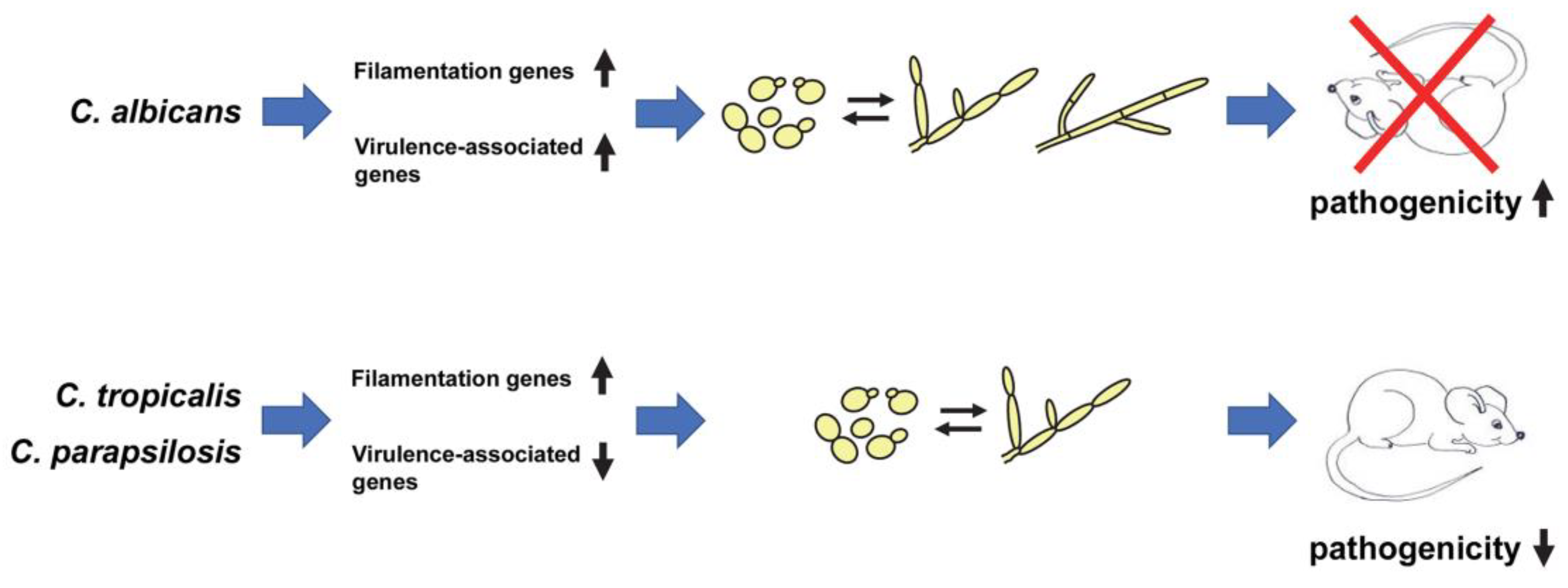
5. Evolutionary Differences in the Relationship between Morphology and Pathogenicity in C. albicans vs. Non-albicans Candida Species

The recent finding that enhanced filamentation is associated with reduced, rather than increased, pathogenicity of C. tropicalis and C. parapsilosis suggests that there are fundamental differences in the evolutionary relationship between morphology and pathogenicity when comparing C. albicans vs. NACS [49]. Importantly, these results challenge conventional views that NACS are less pathogenic than C. albicans because they do not filament as well and instead suggest that other virulence properties, besides filamentation, play a more prominent role in the pathogenesis of certain NACS. In this respect, certain NACS appear to be better adapted to survive in the host when growing in the yeast form. How did such important evolutionary differences in the relationship between morphology and pathogenicity arise among Candida species? Although a definitive answer to this question is lacking, transcriptional profiling experiments suggest that while genes important for filamentation per se are up-regulated in both C. albicans and NACS, several genes associated with other key virulence properties are significantly down-regulated in certain NACS [49]. In contrast, in C. albicans, genes important for filamentation are coordinately expressed with those involved in a wide variety of virulence-related processes, including adhesion and the secretion of degradative enzymes [53,54]. This coordinate expression, in turn, may provide C. albicans with a distinct evolutionary advantage during infection of the host, and could partially explain why approximately 50% of all Candida infections can be attributed to this species [12] (Figure 1). Coordinate expression of genes important for both filamentation and other virulence-related processes, however, may be more of an exception rather than the rule among Candida species. Another important question that arises is why have NACS retained the ability to filament if filamentation confers an evolutionary disadvantage during infection? One possible explanation is that filamentation may represent a vestigial nutrient scavenging response for these species. Alternatively, because C. tropicalis filaments are more readily apparent during mouse systemic infections at higher inoculum sizes [55], they may play a more prominent role in pathogenicity under these conditions. However, most naturally occurring infections in the clinic result from much lower inoculum sizes at which C. tropicalis is typically found in the yeast form [55]. In this case, a threshold level of C. tropicalis cells may need to be reached during infection, at which point filaments might promote pathogenesis rather than leading to organ clearance. While the role and purpose of filamentation in NACS remains unclear at this point, the recent findings of Banerjee et al. strongly suggest that not everything that has been learned about virulence properties of C. albicans can simply be applied to the NACS, but rather instead NACS need to be studied in their own right [49]. In addition, these findings raise many new and important questions for further study. Can reduced pathogenicity of NACS be attributed to lowered expression of particular genes or gene classes? Are filaments generated by NACS vs. C. albicans inherently less fit during infection in vivo and, if so, why? Do filamentous NACS strains have the potential to serve as live attenuated vaccines for protection against infections by multiple Candida species? Continued research in this area is likely to shed more light on the complex relationship between morphology and pathogenicity in Candida species, which may eventually provide information leading to the development of more effective broad-spectrum antifungal strategies.

Author Contributions

Conceptualization, D.K.; Writing-original draft preparation, D.K., V.M.; Writing-review and editing, D.K., V.M.; Funding acquisition, D.K. All authors have read and agreed to the published version of the manuscript.

Funding

This work was supported by grants R01AI127692, R21AI142560, R21AI130668, and R21AI129883 to D.K. from the NIH. The content is solely the responsibility of the authors and does not necessarily reflect the official views of the National Institutes of Health or the National Institute of Allergy and Infectious Diseases.

Acknowledgments

The authors thank Brian Wickes for critical reading of the manuscript and useful suggestions.

Conflicts of Interest

The authors declare no conflict of interest. The sponsors had no role in the design or writing of this article.

References

  1. Edmond, M.B.; Wallace, S.E.; McClish, D.K.; Pfaller, M.A.; Jones, R.N.; Wenzel, R.P. Nosocomial bloodstream infections in United States hospitals: A three-year analysis. Clin. Infect. Dis. 1999, 29, 239–244. [Google Scholar] [CrossRef] [PubMed] [Green Version]
  2. Wisplinghoff, H.; Bischoff, T.; Tallent, S.M.; Seifert, H.; Wenzel, R.P.; Edmond, M.B. Nosocomial bloodstream infections in US hospitals: Analysis of 24,179 cases from a prospective nationwide surveillance study. Clin. Infect. Dis. 2004, 39, 309–317. [Google Scholar] [CrossRef] [PubMed] [Green Version]
  3. Odds, F.C. Candida species and virulence. ASM News 1994, 60, 313–318. [Google Scholar]
  4. Odds, F.C. Candida and Candidosis, 2nd ed.; Baillière Tindall: London, UK, 1988; p. 468. [Google Scholar]
  5. Odds, F.C. Pathogenesis of Candida infections. J. Am. Acad. Dermatol. 1994, 31, S2–S5. [Google Scholar] [CrossRef]
  6. Dupont, P.F. Candida albicans, the opportunist. A cellular and molecular perspective. J. Am. Podiatr. Med. Assoc. 1995, 85, 104–115. [Google Scholar] [CrossRef]
  7. Weig, M.; Gross, U.; Muhlschlegel, F. Clinical aspects and pathogenesis of Candida infection. Trends Microbiol. 1998, 6, 468–470. [Google Scholar] [CrossRef]
  8. Perlroth, J.; Choi, B.; Spellberg, B. Nosocomial fungal infections: Epidemiology, diagnosis, and treatment. Med. Mycol. 2007, 45, 321–346. [Google Scholar] [CrossRef]
  9. Gudlaugsson, O.; Gillespie, S.; Lee, K.; Vande Berg, J.; Hu, J.; Messer, S.; Herwaldt, L.; Pfaller, M.; Diekema, D. Attributable mortality of nosocomial candidemia, revisited. Clin. Infect. Dis. 2003, 37, 1172–1177. [Google Scholar] [CrossRef]
  10. Picazo, J.J.; Gonzalez-Romo, F.; Candel, F.J. Candidemia in the critically ill patient. Int. J. Antimicrob. Agents 2008, 32 (Suppl. S2), S83–S85. [Google Scholar] [CrossRef]
  11. Miller, L.G.; Hajjeh, R.A.; Edwards, J.E., Jr. Estimating the cost of nosocomial candidemia in the United States. Clin. Infect. Dis. 2001, 32, 1110. [Google Scholar] [CrossRef] [Green Version]
  12. McCarty, T.P.; Pappas, P.G. Invasive Candidiasis. Infect. Dis. Clin. N. Am. 2016, 30, 103–124. [Google Scholar] [CrossRef] [PubMed]
  13. Caceres, D.H.; Forsberg, K.; Welsh, R.M.; Sexton, D.J.; Lockhart, S.R.; Jackson, B.R.; Chiller, T. Candida auris: A review of recommendations for detection and control in healthcare settings. J. Fungi 2019, 5, 111. [Google Scholar] [CrossRef] [PubMed] [Green Version]
  14. Pfaller, M.A.; Diekema, D.J. Epidemiology of invasive candidiasis: A persistent public health problem. Clin. Microbiol. Rev. 2007, 20, 133–163. [Google Scholar] [CrossRef] [Green Version]
  15. Patel, P.K.; Erlandsen, J.E.; Kirkpatrick, W.R.; Berg, D.K.; Westbrook, S.D.; Louden, C.; Cornell, J.E.; Thompson, G.R.; Vallor, A.C.; Wickes, B.L.; et al. The changing epidemiology of oropharyngeal candidiasis in patients with HIV/AIDS in the era of antiretroviral therapy. AIDS Res. Treat. 2012, 2012, 262471. [Google Scholar] [CrossRef] [PubMed] [Green Version]
  16. Moran, G.P.; Sullivan, D.J.; Coleman, D.C. Emergence of non-Candida albicans Candida species as pathogens. In Candida and Candidiasis; Calderone, R.A., Ed.; ASM Press: Washinton, DC, USA, 2002; pp. 37–54. [Google Scholar]
  17. Calderone, R.A.; Fonzi, W.A. Virulence factors of Candida albicans. Trends Microbiol. 2001, 9, 327–335. [Google Scholar] [CrossRef]
  18. Kumamoto, C.A.; Vinces, M.D. Contributions of hyphae and hypha-co-regulated genes to Candida albicans virulence. Cell. Microbiol. 2005, 7, 1546–1554. [Google Scholar] [CrossRef]
  19. Sudbery, P.; Gow, N.; Berman, J. The distinct morphogenic states of Candida albicans. Trends Microbiol. 2004, 12, 317–324. [Google Scholar] [CrossRef]
  20. Thompson, D.S.; Carlisle, P.L.; Kadosh, D. Coevolution of morphology and virulence in Candida species. Eukaryot. Cell 2011, 10, 1173–1182. [Google Scholar] [CrossRef] [Green Version]
  21. Gow, N.A.; Henderson, G.; Gooday, G.W. Cytological interrelationships between the cell cycle and duplication cycle of Candida albicans. Microbios 1986, 47, 97–105. [Google Scholar]
  22. Sudbery, P.E. The germ tubes of Candida albicans hyphae and pseudohyphae show different patterns of septin ring localization. Mol. Microbiol. 2001, 41, 19–31. [Google Scholar] [CrossRef]
  23. Warenda, A.J.; Konopka, J.B. Septin function in Candida albicans morphogenesis. Mol. Biol. Cell 2002, 13, 2732–2746. [Google Scholar] [CrossRef] [Green Version]
  24. Sutton, D.A.; Fothergill, A.W.; Rinaldi, M.G. Guide to Clinically Significant Fungi, 1st ed.; Williams and Wilkins: Baltimore, MD, USA, 1998. [Google Scholar]
  25. Staib, P.; Morschhauser, J. Chlamydospore formation in Candida albicans and Candida dubliniensis—An enigmatic developmental programme. Mycoses 2007, 50, 1–12. [Google Scholar] [CrossRef] [PubMed]
  26. Chabasse, D.; Bouchara, J.P.; de Gentile, L.; Chennebault, J.M. Candida albicans chlamydospores observed in vivo in a patient with AIDS. Ann. Biol. Clin. 1988, 46, 817–818. [Google Scholar]
  27. Bartie, K.L.; Williams, D.W.; Wilson, M.J.; Potts, A.J.; Lewis, M.A. Differential invasion of Candida albicans isolates in an in vitro model of oral candidosis. Oral Microbiol. Immunol. 2004, 19, 293–296. [Google Scholar] [CrossRef] [PubMed]
  28. Lo, H.J.; Kohler, J.R.; DiDomenico, B.; Loebenberg, D.; Cacciapuoti, A.; Fink, G.R. Nonfilamentous C. albicans mutants are avirulent. Cell 1997, 90, 939–949. [Google Scholar] [CrossRef] [Green Version]
  29. Braun, B.R.; Head, W.S.; Wang, M.X.; Johnson, A.D. Identification and characterization of TUP1-regulated genes in Candida albicans. Genetics 2000, 156, 31–44. [Google Scholar]
  30. Braun, B.R.; Johnson, A.D. Control of filament formation in Candida albicans by the transcriptional repressor TUP1. Science 1997, 277, 105–109. [Google Scholar] [CrossRef]
  31. Saville, S.P.; Lazzell, A.L.; Monteagudo, C.; Lopez-Ribot, J.L. Engineered control of cell morphology in vivo reveals distinct roles for yeast and filamentous forms of Candida albicans during infection. Eukaryot. Cell 2003, 2, 1053–1060. [Google Scholar] [CrossRef] [Green Version]
  32. Carlisle, P.L.; Banerjee, M.; Lazzell, A.; Monteagudo, C.; Lopez-Ribot, J.L.; Kadosh, D. Expression levels of a filament-specific transcriptional regulator are sufficient to determine Candida albicans morphology and virulence. Proc. Natl. Acad. Sci. USA 2009, 106, 599–604. [Google Scholar] [CrossRef] [Green Version]
  33. Banerjee, M.; Uppuluri, P.; Zhao, X.R.; Carlisle, P.L.; Vipulanandan, G.; Villar, C.C.; Lopez-Ribot, J.L.; Kadosh, D. Expression of UME6, a key regulator of Candida albicans hyphal development, enhances biofilm formation via Hgc1- and Sun41-dependent mechanisms. Eukaryot. Cell 2013, 12, 224–232. [Google Scholar] [CrossRef] [Green Version]
  34. Zheng, X.; Wang, Y.; Wang, Y. Hgc1, a novel hypha-specific G1 cyclin-related protein regulates Candida albicans hyphal morphogenesis. EMBO J. 2004, 23, 1845–1856. [Google Scholar] [CrossRef] [PubMed]
  35. O’Meara, T.R.; Veri, A.O.; Ketela, T.; Jiang, B.; Roemer, T.; Cowen, L.E. Global analysis of fungal morphology exposes mechanisms of host cell escape. Nat. Commun. 2015, 6, 6741. [Google Scholar] [CrossRef] [PubMed]
  36. Noble, S.M.; French, S.; Kohn, L.A.; Chen, V.; Johnson, A.D. Systematic screens of a Candida albicans homozygous deletion library decouple morphogenetic switching and pathogenicity. Nat. Genet. 2010, 42, 590–598. [Google Scholar] [CrossRef] [Green Version]
  37. Priest, S.J.; Lorenz, M.C. Characterization of virulence-related phenotypes in Candida species of the CUG clade. Eukaryot. Cell 2015, 14, 931–940. [Google Scholar] [CrossRef] [Green Version]
  38. Bramley, T.A.; Menzies, G.S.; Williams, R.J.; Kinsman, O.S.; Adams, D.J. Binding sites for LH in Candida albicans: Comparison with the mammalian corpus luteum LH receptor. J. Endocrinol. 1991, 130, 177–190. [Google Scholar] [CrossRef]
  39. Caticha, O.; Grover, S.; Winge, D.; Odell, W.D. Stimulation of Candida albicans transition by human chorionic gonadotrophin and a bacterial protein. Endocr. Res. 1992, 18, 133–143. [Google Scholar] [CrossRef]
  40. Kinsman, O.S.; Pitblado, K.; Coulson, C.J. Effect of mammalian steroid hormones and luteinizing hormone on the germination of Candida albicans and implications for vaginal candidosis. Mycoses 1988, 31, 617–626. [Google Scholar] [CrossRef]
  41. Merson-Davies, L.A.; Odds, F.C. A morphology index for characterization of cell shape in Candida albicans. J. Gen. Microbiol. 1989, 135, 3143–3152. [Google Scholar] [CrossRef] [Green Version]
  42. Brown, A.J. Expression of growth form-specific factors during morphogenesis in Candida albicans. In Candida and Candidiasis; Calderone, R.A., Ed.; ASM Press: Washington, DC, USA, 2002; pp. 87–93. [Google Scholar]
  43. Lackey, E.; Vipulanandan, G.; Childers, D.S.; Kadosh, D. Comparative evolution of morphological regulatory functions in Candida species. Eukaryot. Cell 2013, 12, 1356–1368. [Google Scholar] [CrossRef] [Green Version]
  44. Yu, S.; Li, W.; Liu, X.; Che, J.; Wu, Y.; Lu, J. Distinct expression levels of ALS, LIP, and SAP genes in Candida tropicalis with diverse virulent activities. Front. Microbiol. 2016, 7, 1175. [Google Scholar] [CrossRef]
  45. Silva, S.; Hooper, S.J.; Henriques, M.; Oliveira, R.; Azeredo, J.; Williams, D.W. The role of secreted aspartyl proteinases in Candida tropicalis invasion and damage of oral mucosa. Clin. Microbiol. Infect. 2011, 17, 264–272. [Google Scholar] [CrossRef] [Green Version]
  46. Jiang, C.; Li, Z.; Zhang, L.; Tian, Y.; Dong, D.; Peng, Y. Significance of hyphae formation in virulence of Candida tropicalis and transcriptomic analysis of hyphal cells. Microbiol. Res. 2016, 192, 65–72. [Google Scholar] [CrossRef]
  47. Silva, S.; Henriques, M.; Oliveira, R.; Azeredo, J.; Malic, S.; Hooper, S.J.; Williams, D.W. Characterization of Candida parapsilosis infection of an in vitro reconstituted human oral epithelium. Eur. J. Oral Sci. 2009, 117, 669–675. [Google Scholar] [CrossRef]
  48. Toth, R.; Cabral, V.; Thuer, E.; Bohner, F.; Nemeth, T.; Papp, C.; Nimrichter, L.; Molnar, G.; Vagvolgyi, C.; Gabaldon, T.; et al. Investigation of Candida parapsilosis virulence regulatory factors during host-pathogen interaction. Sci. Rep. 2018, 8, 1346. [Google Scholar] [CrossRef] [PubMed] [Green Version]
  49. Banerjee, M.; Lazzell, A.L.; Romo, J.A.; Lopez-Ribot, J.L.; Kadosh, D. Filamentation is associated with reduced pathogenicity of multiple non-albicans Candida species. mSphere 2019, 4. [Google Scholar] [CrossRef] [Green Version]
  50. Kadosh, D. Control of Candida albicans morphology and pathogenicity by post-transcriptional mechanisms. Cell. Mol. Life Sci. 2016, 73, 4265–4278. [Google Scholar] [CrossRef] [PubMed] [Green Version]
  51. Kim, S.H.; Iyer, K.R.; Pardeshi, L.; Munoz, J.F.; Robbins, N.; Cuomo, C.A.; Wong, K.H.; Cowen, L.E. Genetic analysis of Candida auris implicates Hsp90 in morphogenesis and azole tolerance and Cdr1 in azole resistance. mBio 2019, 10. [Google Scholar] [CrossRef] [PubMed] [Green Version]
  52. Yue, H.; Bing, J.; Zheng, Q.; Zhang, Y.; Hu, T.; Du, H.; Wang, H.; Huang, G. Filamentation in Candida auris, an emerging fungal pathogen of humans: Passage through the mammalian body induces a heritable phenotypic switch. Emerg. Microbes Infect. 2018, 7, 188. [Google Scholar] [CrossRef] [Green Version]
  53. Kadosh, D.; Johnson, A.D. Induction of the Candida albicans filamentous growth program by relief of transcriptional repression: A genome-wide analysis. Mol. Biol. Cell 2005, 16, 2903–2912. [Google Scholar] [CrossRef] [Green Version]
  54. Nantel, A.; Dignard, D.; Bachewich, C.; Harcus, D.; Marcil, A.; Bouin, A.P.; Sensen, C.W.; Hogues, H.; van het Hoog, M.; Gordon, P.; et al. Transcription profiling of Candida albicans cells undergoing the yeast-to-hyphal transition. Mol. Biol. Cell 2002, 13, 3452–3465. [Google Scholar] [CrossRef] [Green Version]
  55. Chen, Y.V.; Rosli, R.; Fong, S.H.; Sidik, S.M.; Pei, C.P. Histopathological characteristics of experimental Candida tropicalis induced acute systemic candidiasis in BALB/c mice. Int. J. Zool. Res. 2012, 8, 12–22. [Google Scholar]
Figure 1. Model for evolutionary differences in the relationship between morphology and pathogenicity among Candida species. In C. albicans, genes important for filamentation are coordinately expressed with those involved in other virulence-related processes, leading to a strong association of the yeast–filament transition with pathogenicity. In C. tropicalis and C. parapsilosis, genes involved in filamentation are also induced but there is an accompanying down-regulation of certain genes involved in virulence-related processes, resulting in an inverse correlation between filamentation and pathogenicity. For second column, upward and downward arrows indicate increased and decreased gene expression, respectively. For fourth column, upward and downward arrows indicate increased and decreased pathogenicity, respectively. Please note that not all virulence-related genes may be down-regulated during filamentation of NACS and that alternative mechanisms not associated with changes in gene expression may account for the reduced pathogenicity of C. tropicalis and C. parapsilosis upon filamentation. Although not depicted, C. tropicalis is capable of forming hyphal filaments (adapted from [20,50]).
Jof 06 00013 g001

