Article

Unveiling the Hidden Diversity of Termitomyces (Lyophyllaceae, Agaricales) in Northern Thailand: Identification of Five New Species and the First Report of Termitomyces acriumbonatus

by
Soumitra Paloi
1,2,
Jaturong Kumla
1,3,4,
Wiphawanee Phonrob
3,
Barsha Pratiher Paloi
1 and
Nakarin Suwannarach
1,3,4,*
1
Center of Excellence in Microbial Diversity and Sustainable Utilization, Chiang Mai University, Chiang Mai 50200, Thailand
2
Myco-Food Laboratory, Food Technology & Science Institute, TCG Centres of Research and Education in Science and Technology, Application Research & Development Center, 54/A/1, Block-DN, Sector-V, Salt Lake City, Kolkata 700091, India
3
Department of Biology, Faculty of Science, Chiang Mai University, Chiang Mai 50200, Thailand
4
Office of Research Administration, Chiang Mai University, Chiang Mai 50200, Thailand
*
Author to whom correspondence should be addressed.
J. Fungi 2025, 11(12), 830; https://doi.org/10.3390/jof11120830
Submission received: 3 October 2025 / Revised: 13 November 2025 / Accepted: 21 November 2025 / Published: 24 November 2025

Abstract

Members of the genus Termitomyces frequently grow in association with termites. During the monsoon season of 2022 and 2023, a total number of 13 Termitomyces samples were collected from the Chiang Mai University campus, Thailand. The objective of this study was to identify the collected samples. Based on morphological and molecular investigations, six distinct species were identified from the collected specimens. Five species (T. griseobulbus, T. griseobrunneus, T. planiperforatorius, T. pseudoheimii, and T. salmonicolor) are described herein as new to science, while one species (T. acriumbonatus) represents a new record for Thailand. The multi-gene phylogenetic analysis of the large subunit (nrLSU) of nuclear ribosomal DNA, the small subunit of mitochondrial DNA (mtSSU), and the nuclear internal transcribed spacer (nrITS) sequences confirmed that all six species belong to the genus Termitomyces. Full morphological descriptions, colour photographs, illustrations, and comparisons with phylogenetically and morphologically related species are provided.

1. Introduction

Fungi rank second among the major kingdoms in terms of known taxa, with approximately 155,000 species scientifically documented to date [1]; and approximately 2200 edible mushrooms have been reported throughout the world [2]. Among the wild edible mushrooms, most members of the genus Termitomyces R. Heim are well-known in different countries around the world, especially in Asia and Africa, due to their distinctive flavours. For example, in China, Termitomyces mushrooms are known as “Jizong”, meaning “chicken mushrooms”, and are commonly used as a food source [3]. Regarding their nutritional value, T. microcarpus contains a high amount of protein (30.69% of dry weight), fibre (11.60% of dry weight), and various minerals [4]. Medicinally, T. microcarpus has shown potential activity against different cancer cell lines [5]. Ethnomedicinally, T. clypeatus is used for the treatment of yellow fever in Nepal, pox in India, and constipation and gastritis in Ethiopia [6,7,8].
Termitomyces belongs to the family Lyophyllaceae Jülich, under the order Agaricales Underw. During the middle of the twentieth century, R. Heim established this genus, with T. striatus (Beeli) R. Heim as the type species [9]. According to Index Fungorum, there are 55 valid species reported to date [10]. Most of the taxa from Termitomyces are reported and described from different countries in Asia and Africa [11]. The highest diversity of Termitomyces is found in Africa [12]. Interestingly, Termitomyces grows in association with termites, mostly those in the genera Odontotermes, Macrotermes, and Microtermes under the only subfamily Macrotermitinae [13]. A diverse range of these termites is frequently found in tropical ecosystems [14]. Recently, many new Termitomyces species have been reported from different Asian countries based on morphological characteristics and single- or multi-gene phylogenetic analyses: T. floccosus S.M. Tang, Raspé & S.H. Li from Thailand [15]; T. fragilis L. Ye, Karun, J.C. Xu, K.D. Hyde & Mortimer, T. tigrinus S.M. Tang & Raspé, T. upsilocystidiatus S.M. Tang, Raspé & K.D. Hyde, and T. yunnanensis S.M. Tang & Raspé from China [3,15,16]; T. acriumbonatus Usman & Khalid, T. sheikhupurensis Izhar, Khalid & H. Bashir, T. islamabadensis S. Ashraf, Usman & Khalid, and T. pakistanensis Razaq from Pakistan [17,18,19,20]; T. gilvus C.S. Yee & J.S. Seelan from Malaysia [21]; T. srilankensis Ediriweera, Voto, Karun. & Kularathne from Sri Lanka [22]; T. assamicus L.R. Das, Narzary & A.K. Dutta from India [23]; and T. dhofarensis S. Hussain, Al-Yahya’ei, Al-Owaisi & Al-Sadi from Oman [24]. In Thailand, “Hed Khone” is the common name for Termitomyces. Different species are collected from local regions and sold in local or street markets, and 14 Termitomyces species have been reported from Thailand, including T. aurantiacus (R. Heim) R. Heim; T. bulborhizus T.Z. Wei, Y.J. Yao, Bo Wang and Pegler; T. clypeatus R. Heim; T. entolomoides R. Heim; T. eurrhizus (Berk.) R. Heim; T. flavus S.M. Tang and S.H. Li; T. floccosus; T. fuliginosus R. Heim; T. globulus R. Heim and Gooss.-Font.; T. heimii Natarajan; T. microcarpus (Berk. and Broome) R. Heim; T. perforans R. Heim; T. radicatus Natarajan; and T. striatus (Beeli) R. Heim [25,26,27,28,29].
Thailand is a biodiversity-rich country in Southeast Asia [30]. Geographically, the northern part is characterised by several mountain ranges and is home to several natural forests covered by a wide range of tree species [31]. Moreover, a total of 36 termite species belonging to 19 genera have been reported from northern Thailand, among which seven Odontotermes, four Macrotermes, and one Microtermes species are known to be associated with Termitomyces [32]. Therefore, given the diversity of termite hosts, a correspondingly high diversity of Termitomyces species can be expected within these habitats. However, knowledge of Termitomyces diversity in northern Thailand remains limited, highlighting the need for additional research. During a survey of macrofungi conducted on the Chiang Mai University campus, Chiang Mai Province, northern Thailand, in 2022 and 2023, 13 specimens of Termitomyces were collected. The objective of this study is to characterize and accurately identify these specimens. Based on their morphological characteristics and multi-gene phylogenetic analyses, the specimens were identified as five distinct new species, along with one previously known species.

2. Materials and Methods

2.1. Sample Collection and Morphological Study

Fresh basidiomata of Termitomyces were collected from the Chiang Mai University campus, Chiang Mai Province, northern Thailand, during the rainy seasons (May to August) of 2022 and 2023. The campus covers an area of approximately 229.5 acres and is situated at the foothills of Doi Suthep-Pui National Park. It is characterised by a diverse landscape comprising mixed deciduous and evergreen forests, landscaped gardens, and open grasslands. In the rainy season of 2022, the average daily temperatures ranged from 25 °C to 27 °C, while the relative humidity levels ranged from 79% to 86%. During the rainy season of 2023, the average daily temperature increased to 27 °C to 32 °C, with the relative humidity ranging from 75% to 85%. This weather information was sourced from the Northern Meteorological Center (https://cmmet.tmd.go.th; accessed on 15 April 2025). Macromorphological characteristics, ecological data, and chemical reactions (10% KOH) were noted in the field. Colour codes and terms of Kornerup and Wanscher [33] were used. Specimens were dried at 45 °C for 48 h, depending on the material. Microscopic characteristics were obtained from dried specimens using freehand sections by mounting them in 5% KOH, Congo red, and Melzer’s reagent. They were then viewed through an Olympus CH30 microscope. The size of the basidiospores was documented based on 30 measurements recorded from each specimen. The Q value denotes the length/width ratio of the basidiospores. Basidiospore and Q value statistics are presented as mean values (underlined). The terminology of the microscopic features followed that of Mossebo et al. [34]. The holotype and other examined specimens were deposited at the Chiang Mai University Herbarium of the Department of Biology (CMUB) and the Sustainable Development of Biological Resources Laboratory (SDBR), Faculty of Science, Chiang Mai University, Thailand.

2.2. DNA Extraction, PCR Amplification, and Sequencing

Genomic DNA was extracted from fresh specimens using a DNA Extraction Mini Kit (FAVORGEN, Pingtung City, Taiwan), according to the manufacturer’s protocol. The nrITS, nrLSU, and mtSSU regions were amplified by polymerase chain reaction (PCR) using the primer pairs ITS4/ITS5 [35], LROR/LR5 [36], and the Termitomyces-specific primers SSUFW105/SSUREV475 [13], respectively. These three domains were amplified in separate PCRs. The PCR programs for nrITS and nrLSU were established by following the methods employed by Paloi et al. [37]. The amplification program for mtSSU consisted of an initial denaturation step at 94 °C for 2 min, followed by 35 cycles of denaturation at 94 °C for 45 s, an annealing step at 59 °C for 45 s, an elongation step at 72 °C for 1 min, and an extension step at 72 °C for 2 min on a peqSTAR thermal cycler (PEQLAB Ltd., Fareham, UK). The PCR products were checked on 1% agarose gels stained with ethidium bromide under UV light. The PCR products were purified using a PCR clean-up Gel Extraction NucleoSpin® Gel and PCR Clean-up Kit (Macherey-Nagel, Düren, Germany), following the manufacturer’s protocol. The purified PCR products were directly sequenced using a genetic analyser at the 1st Base Company (Kembangan, Malaysia) with the PCR primers mentioned above.

2.3. Sequence Alignment and Phylogenetic Analyses

Sequences with a high degree of similarity to the newly generated sequences were retrieved from the GenBank database and recent publications [3,13,15,16,17,18,19,20,21,22,23,24,34,38,39,40,41,42], which are detailed in Table 1. Two Asterophora and Lyophyllum species, Asterophora lycoperdoides (Bull.) Ditmar, Asterophora parasitica (Bull. ex Pers.) Singer, Lyophyllum decastes (Fr.) Singer, and Lyophyllum shimeji (Kawam.) Hongo were used for rooting purposes. Multiple sequence alignment was performed using MUSCLE [43] and the default settings. The finalised alignment of the concatenated nrLSU, mtSSU, and nrITS sequences was deposited in Zenodo (https://zenodo.org; accessed on 15 October 2025) under the DOI 10.5281/zenodo.15528282. Phylogenetic analyses were carried out based on the combined dataset of the nrLSU, mtSSU, and nrITS sequences. A phylogenetic tree was constructed using the maximum likelihood (ML) and Bayesian inference (BI) methods. The ML analysis was carried out on RAxML-HPC2 version 8.2.10 [44] on the CIPRES web portal using the GTRCAT model and 1000 bootstrap replications. The BI analysis was performed using MrBayes version 3.2.6 [45]. The best substitution models for the BI analyses were estimated using Akaike Information Criterion (AIC) in jModeltest 2.1.10 [46]. The best substitution models were GTR + G for nrLSU, HKY + I + G for mtSSU, and HKY + G for nrITS. For the BI analysis, six simultaneous Markov chains were run for three million generations with random initial trees, and every 1000 generations were sampled. The burn-in was set to discard the first 2000 trees, and the remaining trees were used to construct the 50% majority-rule consensus phylogram with the calculated Bayesian posterior probabilities (PPs). Branches with bootstrap support (BS) and PP values greater than or equal to 70% and 0.75, respectively, were considered significantly supported [47,48]. Tree topologies were then visualised in FigTree version 1.4.0 [49].

3. Results and Discussion

3.1. Phylogenetic Analysis

The newly generated nrITS, nrLSU, and mtSSU sequences were deposited in the GenBank database with the accession numbers shown in Table 1. The combined sequence dataset (nrLSU + mtSSU + nrITS) consisted of a total of 42 species and three varieties of Termitomyces, and the aligned dataset comprised 1708 characters including gaps (nrLSU: 1–647; mtSSU: 648–1007; and nrITS: 1008–1708). The best-scoring RAxML tree was established with a final ML value of −9361.118837. The final average standard deviation value of the split frequencies at the end of the total MCMC generations was calculated to be 0.00984 through the BI analysis. The phylograms obtained from the ML and BI analyses exhibited similar topologies. Therefore, the phylogenetic tree obtained from the ML analysis was selected and is presented in this study (Figure 1).
The phylogenetic tree was divided into eight clades (Clade A to H). Clade A represents the closely related species of T. intermedius Har. Takah. & Taneyama. The species grouped in this clade are a mix of Asian and African representatives. Morphologically, all species produce medium-sized basidiomata. Our two specimens (CMUB40062 and SDBR-CMUSOU30), introduced as T. griseobulbus, formed a monophyletic group with strong support (BS = 99% and PP = 0.89) and are sister to T. intermedius, T. tigrinus, and T. radicatus. However, all these species are morphologically different from T. griseobulbus. Another four specimens (CMUB40063, SDBR-CMUNKN1211, SDBR-CMUNKN1248, and SDBR-CMUWP116), introduced as T. griseobrunneus, formed a monophyletic group. Termitomyces griseobrunneus formed a sister taxon to T. radicatus (voucher MRNo173), with sequences deposited based on the Thai collection. However, T. griseobrunneus can be easily distinguished from T. radicatus by having hymenial cystidia. According to IndexFungorum, the current name of T. striatus f. bibasidiatus Mossebo is T. striatus, but T. striatus f. subclypeatus Mossebo & Essouman is invalid according to Art. 40.7 of the Melbourne code. Another newly proposed species, T. assamicus from India, also clustered into this clade.
Clade B represents Termitomyces species that are mostly characterised by a smaller pileus size, and most of the species were described from Asia and a few are from Africa. Our specimen CMUB40061 was placed in T. acriumbonatus, described from Pakistan. Morphologically, it was similar to T. acriumbonatus. Other well-known Termitomyces species belonging to this clade include T. pakistanensis Razaq and T. sheikhupurensis from Pakistan; T. fragilis from China; T. microcarpus (Berk. & Broome) R. Heim; and T. entolomoides R. Heim from Africa. One sequence of T. schimperi (Pat.) R. Heim (voucher tgf18) clustered in this clade, possibly due to misidentified species.
Clade C represents the T. heimii complex with high support values (BS = 77% and PP = 1.00) in the phylogenetic tree (Figure 1). All species are medium-sized and were found in the Asian region. Here, two specimens of T. heimii (voucher Muid.sn and KM16528) were placed separately in this clade. It is important to recognise the sequence as the type sequence of T. heimii, and further detailed study is needed to resolve this complex. It was found that two specimens in this study (CMUB40069 and SDBR-CMUNKP2013), introduced as T. pseudoheimii, were monophyletic and formed a sister taxon to T. heimii (voucher Muid.sn). Additionally, T. islamabadensis, described from Pakistan, was placed in this clade. In Clade D, it included Asian and African Termitomyces species. Asian Termitomyces species in this clade were recently described, namely T. srilankensis and T. yunnanensis. Other African Termitomyces species belonging to this clade were T. eurrhizus; T. robustus (Beeli) R. Heim; and T. subumkowaan Mossebo. Clade E primarily represents Termitomyces species collected from Africa, but Asian specimens of T. le-testui and T. dhofarensis also belong to this clade.
All sequences of different Termitomyces species in Clade F are from the Asian continent. Two specimens (CMUB40064 and SDBR-CMUNKM1852), introduced as T. planiperforatorius, formed a monophyletic group with strong support (BS = 100% and PP = 0.97) and are sister to T. bulborhizus and unpublished Termitomyces sp. (vouchers YAAS 2021081126 and YAAS 2021081127) collected in Thailand. Moreover, two specimens, CMUB40006 and SDBR-CMUNK1760, identified as T. salmonicolor, were placed in this clade and formed a monophyletic group, clearly separating them from other species. Two other clades (Clades G and H) represented a few Asian and African Termitomyces species, respectively. Two species, T. floccosus and T. upsilocystidiatus, belonged to Clade G. Another species, T. medius R. Heim & Grassé (according to Index Fungorum, T. medius f. ochraceus Mossebo & Essouman is currently known as T. medius), belonged to Clade H.

3.2. Taxonomy

3.2.1. Termitomyces acriumbonatus Usman & Khalid, Phytotaxa 477: 221 (2020)

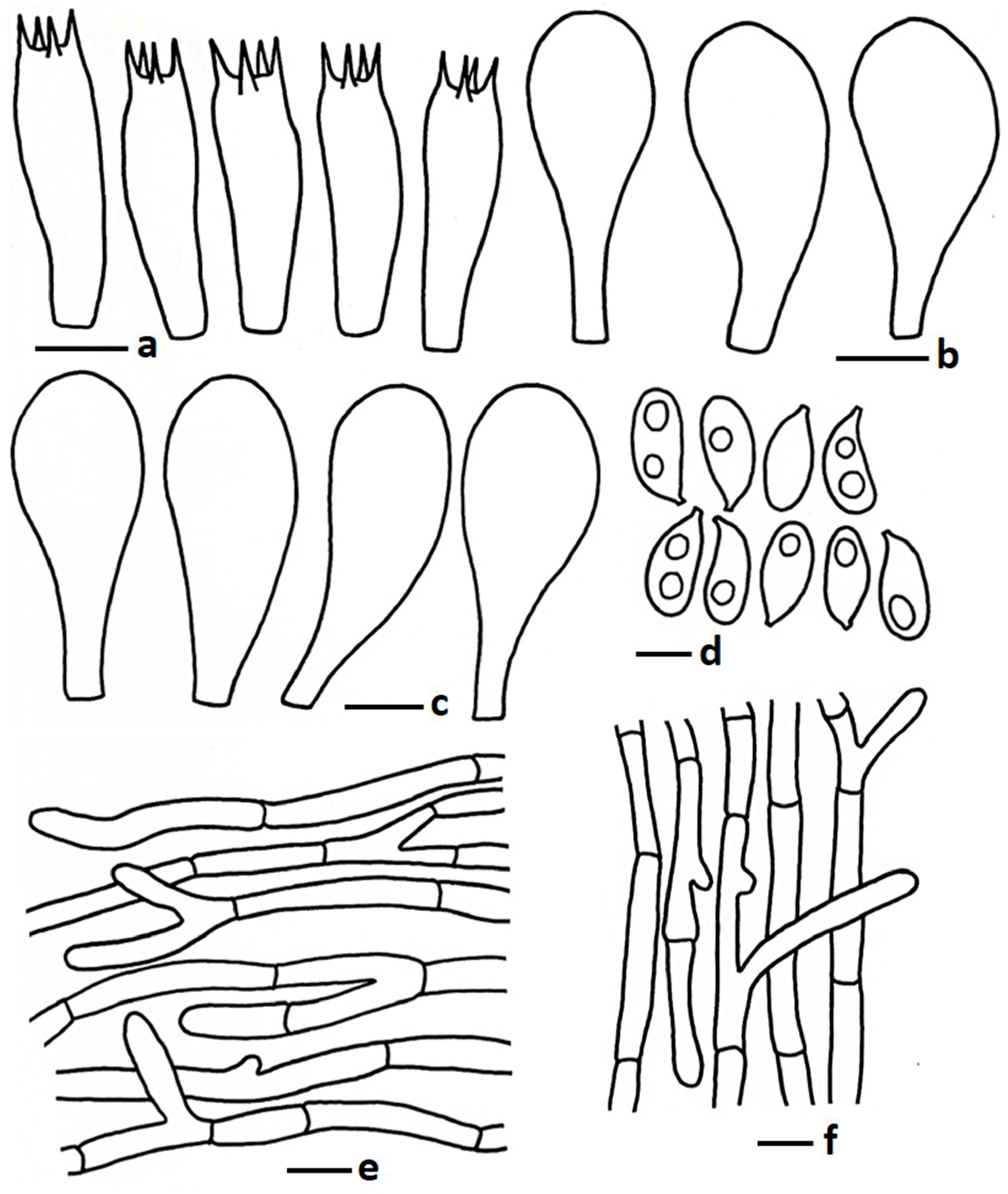
MycoBank: MB834937 (Figure 2a and Figure 3)
Description: Pileus 8–25 mm diam., paraboloid to hemispherical at early stage, becoming broadly convex to applanate to planoconcave; perforatorium prominent, sometimes pointed at early stage, abrupt at maturity; surface smooth at early stage, sometimes smooth or slowly becoming cracked at maturity; colour brownish grey (5D2, 6C2, 7D2) to greyish brown (5D3, 6D2) at centre, fading toward margin orange grey (5B2) to grey (6B1, 7C1) or reddish grey (9B2); margin split at maturity; context thin, white (1A1), no change after exposure to air. Lamellae free, 2–3 mm broad, regular, white (1A1) to off white or light grey; edge eroded, concolorous; lamellulae none. Stipe 17–55 × 2–3 mm, central, sometimes curved at centre, slightly bulbous at base, smooth, colour white (1A1) to grey (5B1); context white, fibrillose, no change after exposure to air. Pseudorhiza more than 20 mm long and 2–3 mm wide, white, extending deeply into and firmly attached to the termite nest. Odour mushroom-like.
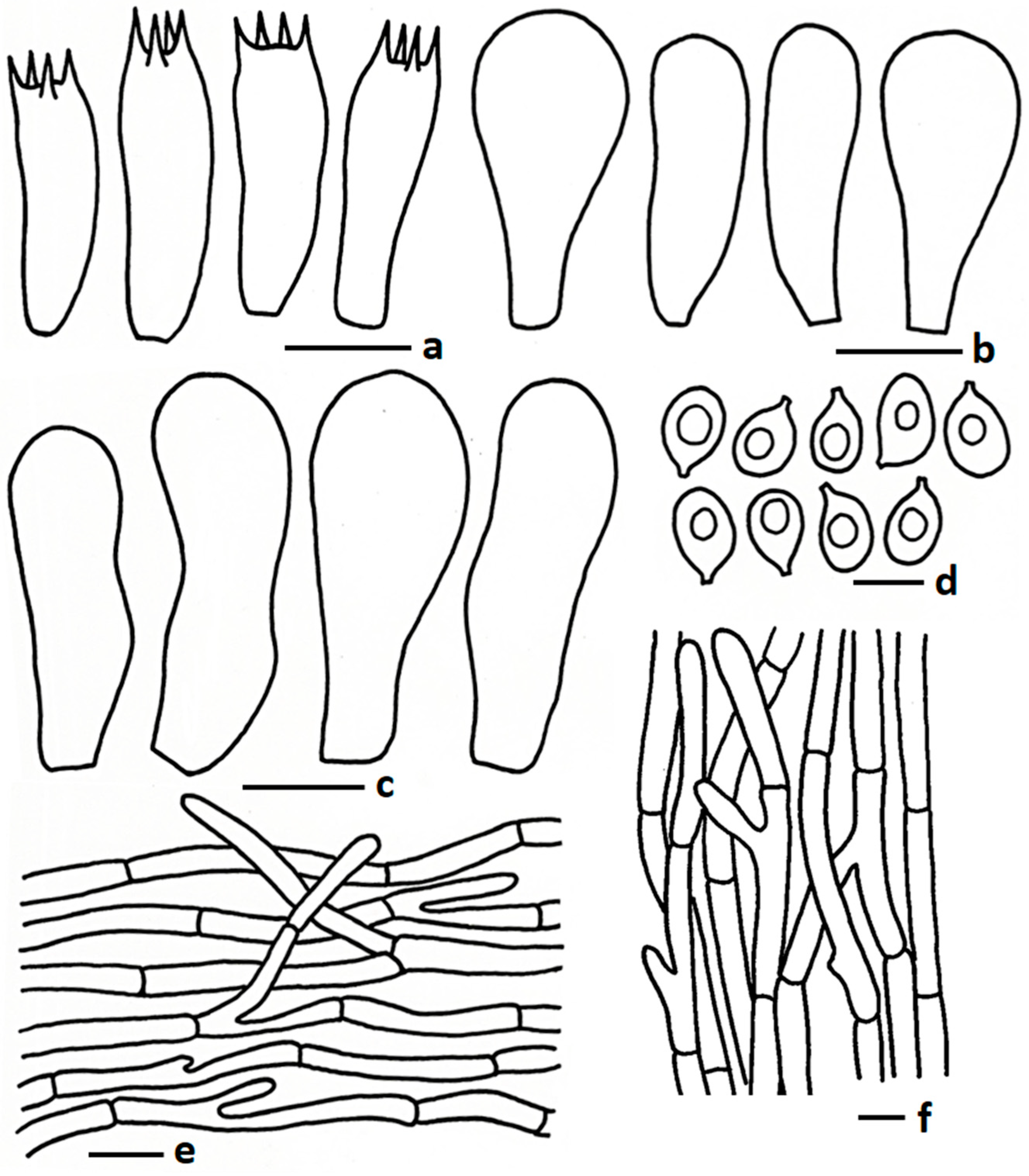
Basidiospores 5.30–6.67–7.86 × 4.02–4.55–5.34 µm, Q = 1.20–1.47–1.63, broadly ellipsoid to ellipsoid, thin-walled hyaline, sometimes 1–2-guttulate oil granules present when viewed with KOH; apiculus short. Basidia 16.09–25.50 × 5.50–8.10 µm, clavate to subclavate to sometimes cylindrical, thin walled, hyaline, oil granules present when viewed with KOH, 2–4-spored; sterigmata up to 4.00 µm long, cylindrical. Lamellae trama regular, composed of hyphae, and inflating cells, 4.23–11.49 µm wide, thin walled, hyaline, branched, septate. Cheilocystidia 18.32–28.00 × 5.81–13.20 µm, utriform to pyriform, thin walled, hyaline. Pleurocystidia 15.65–32.12 × 5.81–17.74 µm, same as cheilocystidia. Pileipellis two distinct layers; subpellis composed of inflating cells arranged in a chain, 25–110 × 12–25 µm, thin walled, hyaline, some inflated hyphae present, 7.00–12.5 µm broad, thin walled, branched; suprapellis composed of radially arranged hyphae, 2.97–6.50 µm broad, thin walled, hyaline, oil granules present with KOH, branched, septate. Stipitipellis composed of tightly arranged hyphae 2.50–8.00 µm broad, hyaline, thin walled, branched, oil granules present with KOH. Clamp connections absent in all tissues.
Habit and habitat: Solitary to gregarious, growing on humus-deposited grass field, above subterranean termite nest.
Edibility: Edible.
Material examined: THAILAND. Chiang Mai Province, Chiang Mai University, Angkeaw Reservoir, 18°48′22″ N 98°57′6″ E, elevation 340 m, 8 July 2022, W. Phonrob (CMUB40061). GenBank accession numbers PQ896627 (nrITS), PQ896626 (nrLSU), and PV020670 (mtSSU).
Notes: Termitomyces acriumbonatus was recently reported from Pakistan. Morphologically, this species is characterized a small pileus with a brownish pointed papilla, subglobose to ellipsoid basidiospores, and clamp connections in pileipellis and stipitipellis hyphae [17]. The newly collected Thai specimen is morphologically similar to T. acriumbonatus in most characteristics including basidiomata size, shape, colour, basidiospores, hymenial cystidia, and pileipellis cell arrangements, but this present collection from Thailand does not show any clamp connections in the pileipellis or stipitipellis. However, most of the morphological character similarities as well as phylogenetic placement with T. acriumbonatus suggest that the present Thai collection is conspecific with T. acriumbonatus. The morphologically similar species, T. microcarpus, can cause confusion in the field. However, T. microcarpus has a smaller pileus (up to 15 mm diam.) and T. acriumbonatus have brownish grey to greyish brown perforatorium [9].
Termitomyces pakistanensis, T. sheikhupurensis, and T. fragilis are closely related to T. acriumbonatus. Termitomyces pakistanensis has a smaller pileus (1.8–2 cm diam.), an umbonate cover with squamulose at the young stage, adnexed lamellae attachment, no pseudorhiza, and a pileus covered with larger terminal elements (35.5–76.5 × 10.5–28.5 μm) [20]. Termitomyces sheikhupurensis has a similar basidiomata size but white velar remnants present on the pileus margin, a stipe surface pale yellowish toward the pileus, a short pseudorhiza, and thin suprapellis hyphae (1–3 µm) [17]. Termitomyces fragilis, recently reported from China, has a fragile stipe, very long pseudorhiza (up to 72 mm), larger basidiospores (9.0–10.5 × 5.5–7.5 µm), and larger hymenial cystidia (40–80 × 12–31 µm) [3].

3.2.2. Termitomyces griseobulbus Paloi & N. Suwannar., sp. nov.

MycoBank: MB857473 (Figure 2b and Figure 4)
Holotype: THAILAND. Chiang Mai Province, Chiang Mai University, opposite to the volleyball court, 18°47′58.7″ N 98°57′25.2″ E, elevation 330 m, 16 August 2022, S. Paloi (CMUB40062). GenBank accession numbers PQ900075 (nrITS), PQ896628 (nrLSU), and PV020671 (mtSSU).
Diagnosis: Differs from closely related species by having a medium, brownish grey to grey pileus, free lamellae attachment, basidiospores 5.00–8.10 × 3.55–4.77 µm, basidia 16.00–27.50 × 5.90–9.44 µm, hymenial cystidia, and pileipellis composed of suberect to repent hyphae.
Etymology: “griseobulbus” represents the grey-coloured bulb present at the stipe base.
Description: Pileus 46–80 mm diam., obtusely conical, umbonate to hemispherical or sometimes campanulate at young stage, becoming convex, sub umbonate to applanate or plano concave with a central umbo at maturity; perforatorium round or sometimes blunt pointed; surface moist, velutinous to pubescent; colour brownish grey (5D2) to greyish brown (5D3) at very early stage, brownish grey (5D2, 7E2) to greyish brown (5D3) or grey (7D1) at centre becoming faded toward margin pale grey (1B2), medium grey (1E1), grey (2B1) to reddish grey (7B1) or sometimes similar to centre, no change after bruising; margin wavy at maturity, cracked, up to ½ of pileus, straight to inflexed; context up to 3 mm thick at middle, white to cream, no change after exposure to air. Lamellae free, up to 3 mm broad, densely crowded, regular, white (1A1) at young stage becoming, pale grey (1A2) to yellowish grey (2C2); edge serrate or eroded, concolorous; 2–3 lamellulae. Stipe 40–55 × 6–9 mm, central, sometimes slightly curved at centre, twisted at maturity, light grey (1C1) or medium grey (1E1) to grey (2C1), sometimes white (1A1) at early stage. turned light brownish with KOH; surface scabrous, moist, near ground forming a bulbous base, concolorous with stipe, sometimes darker than stipe; context solid, fibrillose, white, no change after exposure to air. Pseudorhiza 60–75 × 3–5 mm, white (1A1) or similar to the stipe, surface sometimes pubescent; context solid, white, extending deeply into and firmly attached to the termite nest. Odour mushroom-like.
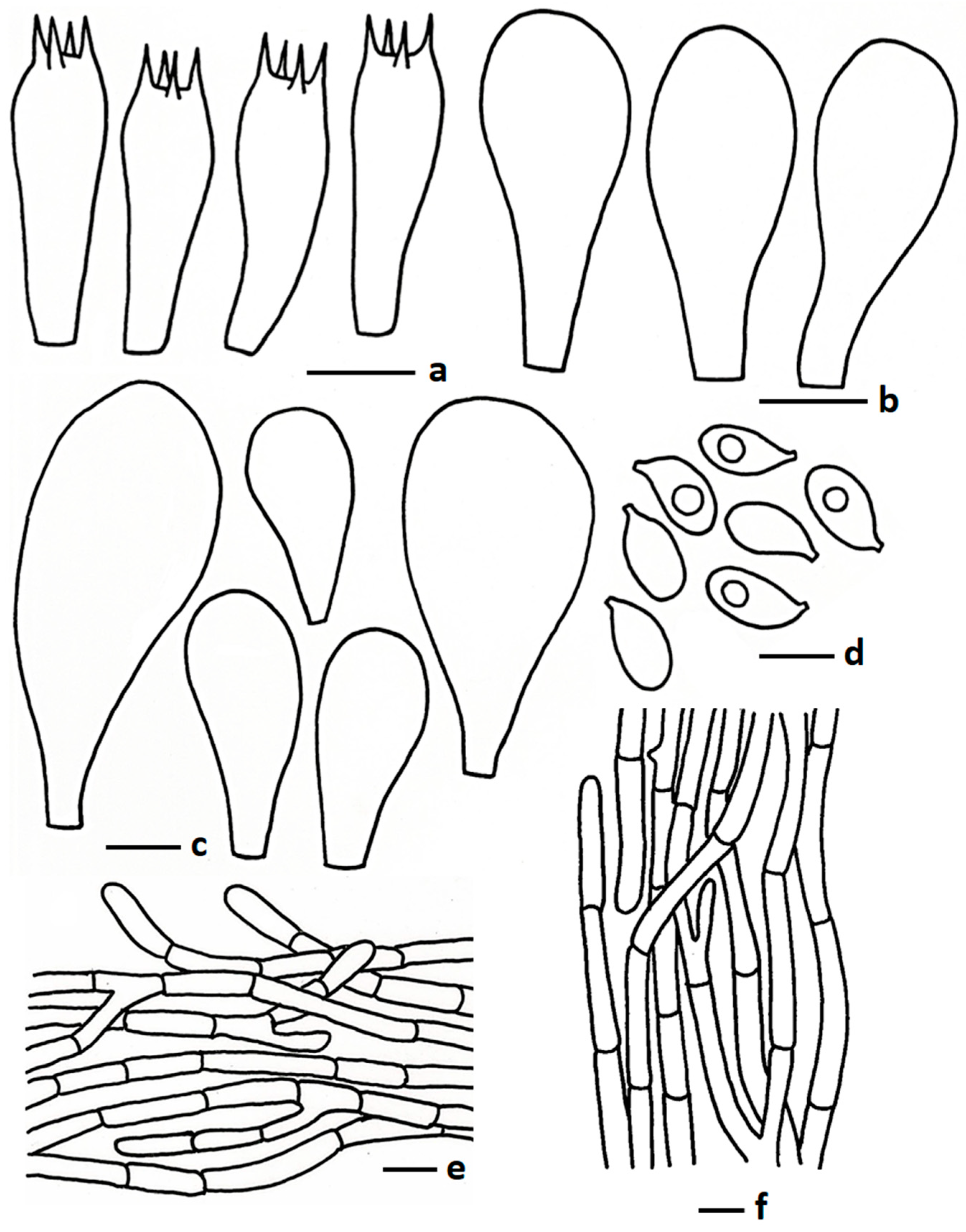
Basidiospores 5.00–6.76–8.10 × 3.55–3.99–4.77 µm, Q = 1.39–1.69–1.93, ellipsoid, thin walled, hyaline, oil granules present when viewed with KOH, sometimes 1–2-guttulate. Basidia 16.00–27.50 × 5.90–9.44 µm, clavate to subclavate, sometimes subcylindrical, thin walled, hyaline, 4-spored; sterigmata 1.09–2.5 µm long, cylindrical, sometimes obtuse apex. Lamellae trama regular, composed of hyphae, 6.50–14.00 µm broad, thin walled, hyaline, branched, septate. Cheilocystidia 23.50–39.50 × 8.75–14.5 µm, pyriform to clavate with round apex, frequently present in mature stage, thin walled, hyaline. Pleurocystidia two types, big pyriform, 51.50–79.50 × 17.75–32.00 µm, not frequent, thin walled, hyaline; small pyriform, with rounded apex, 30.50–49.00 × 13.50–26.50 µm, thin walled, hyaline, frequently present. Pileipellis two distinct layers; subpellis composed of hyphae, 5.50–12.00 µm wide, thin walled, hyaline, branched, hyaline; suprapellis composed of tightly arranged hyphae, 3.50–6.00 µm wide, sub erect to repent, sometimes few erect, thin walled, hyaline, oil granules present with KOH, branched, hyaline, hyphal end obtuse, sometimes round apex. Stipitipellis composed of hyphae, 3.50–12.00 µm wide, thin walled, hyaline, septate, branched. Clamp connections absent in all tissues.
Habit and habitat: Gregarious, rarely solitary, growing above subterranean termite nests.
Edibility: Edible.
Additional material examined: THAILAND. Chiang Mai province, Chiang Mai University, near CMU football ground, 18°47′54.7″ N 98°57′38.2″ E, elevation 323 m, 20 August 2022, S. Paloi (SDBR-CMUSOU30). GenBank accession numbers PQ900076 (nrITS), PQ896629 (nrLSU), and PV020672 (mtSSU).
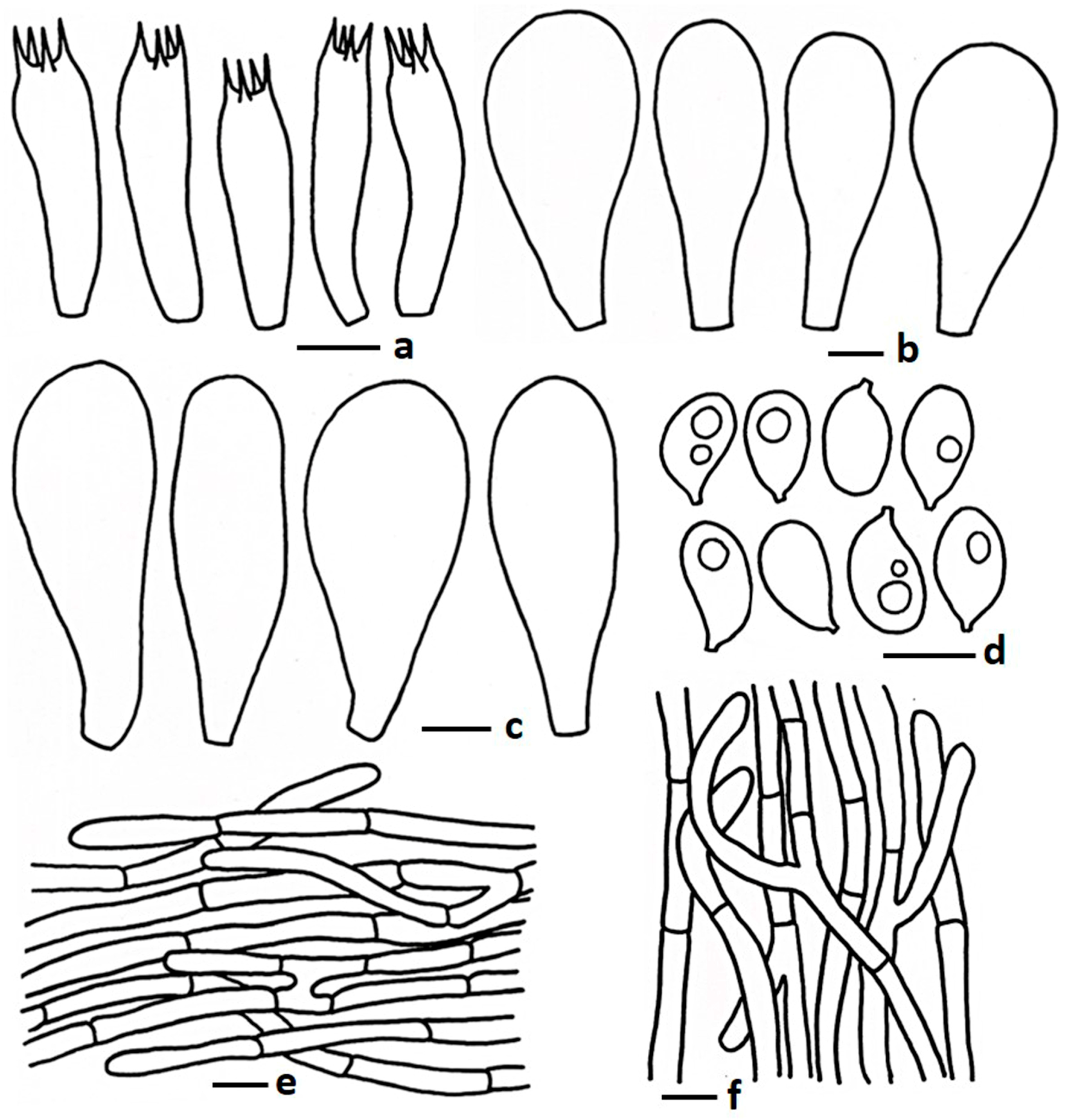
Notes: Morphologically and phylogenetically closely related species are T. intermedius, T. tigrinus, T. griseobrunneus, and T. radicatus. Termitomyces intermedius has a dark grey pileus, larger basidiospores (9.0–14.9 × 5.3–10.2 µm) and basidia (43–68 × 10–20 µm), and larger (40–169 × 19–34 µm) oblong, obovoid, or ellipsoid pleurocystidia [16]; T. tigrinus contains adnexed lamellae attachment and an ixocutis type of pileipellis and no pleurocystidia [16]; T. griseobrunneus differs by having a smaller pileus (25–45 mm in diam.) with a fibrillose surface, thin pileus context (1 mm), and smaller stipe (in this study). However, T. radicatus has a smaller (35 mm in diam.) orange white to orange grey pileus, short pseudorhiza (25 mm long), and no hymenial cystidia [50]. Termitomyces entolomoides is a somewhat morphologically similar species but differs by having a smaller pileus (30–40 mm in diam.) and a greyish pseudorhiza [51]. According to Wei et al. [52], T. entolomoides has larger cheilocystidia (28–60 × 13.0–31 µm) and rarely pleurocystidia.

3.2.3. Termitomyces griseobrunneus Paloi, W. Phonrob & N. Suwannar., sp. nov.

MycoBank: MB857474 (Figure 2c–e and Figure 5)
Holotype: THAILAND. Chiang Mai Province, Chiang Mai University campus, Plam Garden, 18°48′1″ N 98°57′19″ E, elevation 350 m, 18 July 2022, W. Phonrob (CMUB40063). GenBank accession numbers PQ899488 (nrITS), PQ896878 (nrLSU), and PV020673 (mtSSU).
Diagnosis: Brownish grey to dark brown pileus with pointed perforatorium, comparatively short stipe (22–35 × 4–9 mm) with bulbous base, basidiospores 5.50–8.25 × 3.50–5.40 µm, and two different types of cheilocystidia.
Etymology: “griseobrunneus” indicates a grey-brown coloured pileus.
Description: Pileus 25–45 mm diam., campanulate to papillate at young, becoming broadly convex to applanate or plano convex with pointed perforatorium at centre when mature; surface moist, shiny when dry, fibrillose-like, cracked, brownish grey (6E2, 8F2) to grey (8F1), greyish brown (8F3), dark brown (8F4) at centre, becoming faded toward margin grey (3C1) to greyish brown (6D2, 7C1, 11F1), brownish grey (9F2), greenish grey (30B2) or sometimes similar to the centre at early stage, no change after bruising; margin inflexed at young, becoming straight at maturity, wavy, cracked; context 1mm wide at middle, white to off-white, no change after exposure to air. Lamellae free, up to 6 mm broad, regular, white (1A1) at early stage, becoming brownish grey (5B2) at mature stage; edge serrate to eroded, concolorous; 2–3 lamellulae. Stipe 22–35 × 4–9 mm, central, sometimes curved at centre, tapering upward, base slightly bulbous to sometimes district bulbous, up to 16 mm wide; surface pubescent to minutely ribbed when mature, White (1A1) at early stage to grey (3D1, 7C1), sometimes violet grey (18D2) to greyish violet (19C3) at maturity, no change after bruising; context white to off-white, solid, fibrillose, no change after exposure to air. Pseudorhiza 25–55 × 2–5 mm, sometimes similar to the stipe or yellowish grey (3B2) to golden grey (4C2) or grey (4C1), solid, extending deeply into and firmly attached to the termite nest. Odour mushroom-like.
Basidiospores 5.50–7.10–8.25 × 3.50–4.40–5.40 µm, Q = 1.39–1.64–1.88, ellipsoid, thin walled, hyaline, oil granules present when viewed KOH, 1–2-guttulate, apiculus very short. Basidia 19.50–28.00 × 5.50–8.50 µm, clavate to subclavate or sometimes subcylindrical, thin walled, hyaline, oil granules present, 2–4-spored; sterigmata 1.50–3.50 µm long, cylindrical. Lamellae trama regular, composed of inflated hyphae, 5.00–13.00 µm wide, thin walled. Cheilocystidia 20.50–33.00 × 10.50–15.50 µm, pyriform, frequent; 24.50–37.00 × 8.50–13.50 µm, moniliform to short appendiculate or sometimes broadly clavate, thin walled, hyaline. Pleurocystidia 34.50–80.50 × 18.00–41.50 µm, pyriform, frequent; 31.50–43.00 × 11.50–16.00 µm, clavate with obtuse or less pointed apex, thin walled, hyaline. Pileipellis two layers; subpellis composed of inflated hyphae and/or chain of inflated cell, 5.00–11.50 µm wide, thin walled, septate; suprapellis up to 100 µm deep, composed of tightly arranged sub-erect or repent hyphae, 3.00–6.50 µm wide, thin walled, hyaline, branched, septate, oil granules present with KOH, hyphal end obtuse to sometimes round. Stipitipellis composed of tightly arranged hyphae, 3.00–8.50 µm wide, thin walled, hyaline, branched, septate, hyphal end obtuse. Clamp connections absent in all tissues.
Edibility: Edible.
Habit and habitat: Solitary to gregarious, growing on soil above subterranean termite nests.
Additional material examined: THAILAND. Chiang Mai Province, Chiang Mai University, Sala Dhamma, 18°48′15″ N; 98°57′10″ E, elevation 340 m, 19 July 2022, W. Phonrob (SDBR-CMUWP116), GenBank accession numbers PQ896901 (nrLSU), PV020674 (mtSSU); Chiang Mai University, Sala Dhamma, 18°48′15″ N; 98°57′10″ E, elevation 340 m, 25 July 2023 N. Suwannar. (SDBR-CMUNKN1211); GenBank accession numbers PQ899491 (nrLSU) and PV020675 (mtSSU); Chiang Mai University, Sala Dhamma, 18°48′15″ N; 98°57′10″ E, elevation 340 m, 10 August 2023, W. Phonrob (SDBR-CMUNKN1248). GenBank accession numbers PQ899488 (nrITS), PQ896878 (nrLSU), and PV020673 (mtSSU).
Notes: Morphologically and phylogenetically, T. griseobrunneus is very close to some Chinese and Thai species, such as T. intermedius, T. tigrinus, T. griseobulbus, and T. radicatus. Termitomyces intermedius is easily distinguished from these species by having a larger pileus (40–110 mm diam.), a rimose-squamulose surface on the pileus (dry condition), and larger basidiospores (9.0–14.9 × 5.3–10.2 μm) and basidia (43–68 × 10–20 μm), but the average Q value is similar in all these species, and they all have an ixocutis pileipellis [16]; T. tigrinus has a larger pileus (70–90 mm diam.), adnexed lamellae attachments, no pleurocystidia, and an ixocutis type of pileipellis [16]. Termitomyces griseobulbus differs from T. griseobrunneus by having a larger pileus size (46–80 mm in diam.), a velutinous to pubescent pileus surface, a comparatively thicker pileus context (3 mm), and a longer stipe (40–55 mm) with a prominent scabrous surface (in this manuscript). Termitomyces radicatus, originally described from India, differs based on characteristics like an orange white to orange grey pileus, yellow white to orange white stipe, and no hymenial cystidia [50]. The bulbous stipe base species T. bulborhizus differs by having a larger pileus (50–220 mm in diam.), a broadly round or blunt pointed perforatorium, and a longer pseudorhiza (800 mm) [53]; another species, T. gilvus, described from Malaysia, has a larger basidiomata, a thick pileus context (40 mm), no pleurocystidia and clamp connections [21].

3.2.4. Termitomyces planiperforatorius Paloi & N. Suwannar., sp. nov.

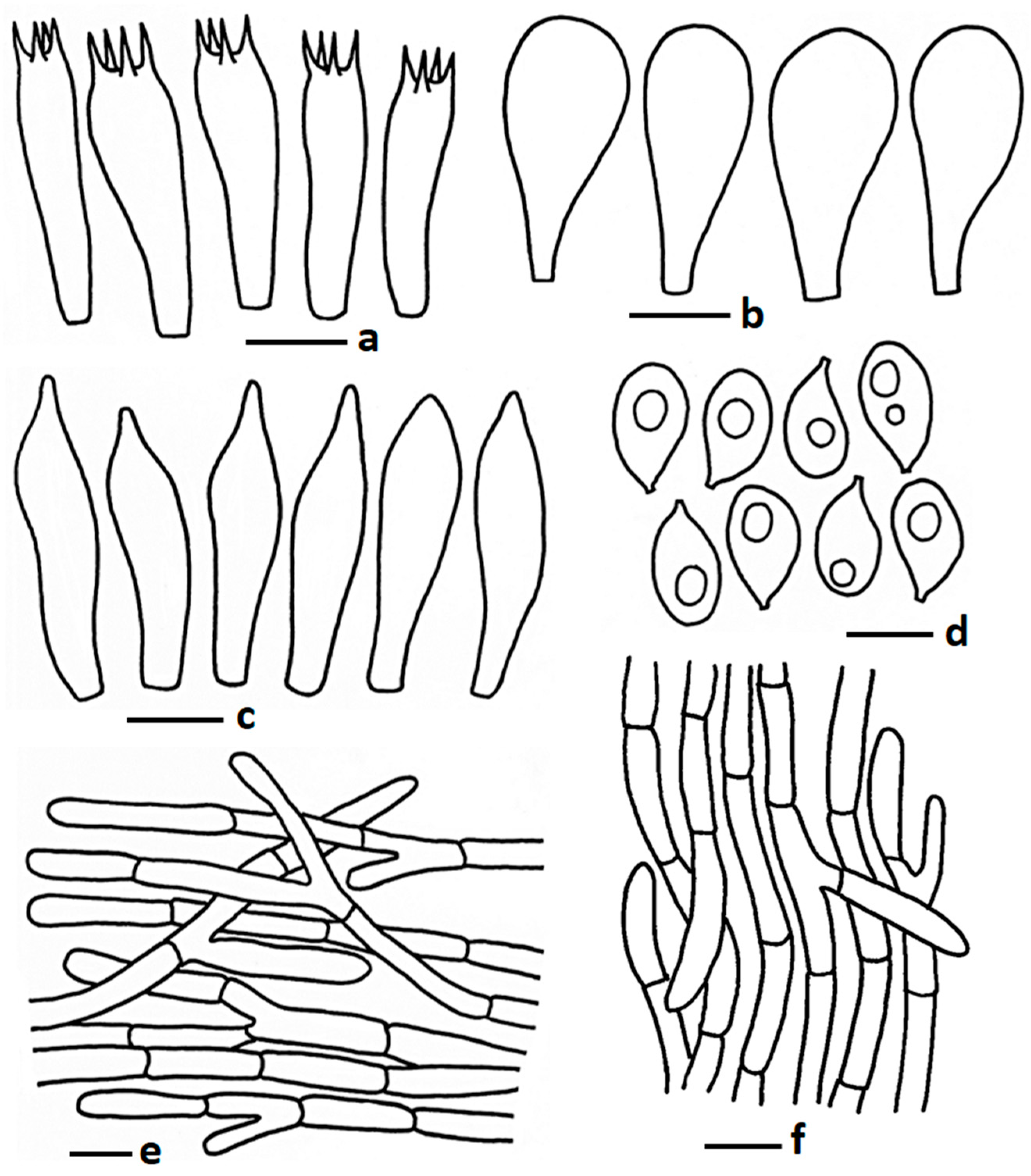
MycoBank: MB857475 (Figure 2f and Figure 6)
Holotype: THAILAND. Chiang Mai Province, Chiang Mai University campus, near Female Dormitory 3, 18°47′58.7″ N 98°57′11.4″ E, elevation 341 m, 4 August 2022, S. Paloi (CMUB40064). GenBank accession numbers PQ896983 (nrLSU) and PV020677 (mtSSU).
Diagnosis: Differs from closely related species by having a medium-sized pileus with a slightly round to flat but never pointed perforatorium, a cracked pileus surface when mature and the appearance of squamules on the surface, cheilocystidia 27.12–56.50 × 10.50–29.50 µm, and pyriform to utriform or clavate pleurocystidia with a round apex.
Etymology: “planiperforatorius” refers to the flat perforatorium present on the pileus surface.
Description: Pileus 22–97 mm diam., convex to broadly convex at early stage, becoming plano concave to concave at maturity; perforatorium slight round to flat, never pointed, moist to dry; surface greyish orange (5B4), brownish orange (5C3), greyish brown (6D3), to light brown (6D4), no change after bruising, smooth to slightly pubescent, cracked, look squamose-like, arranged as zonate, easily pilled; margin split, sometimes revolute to reflexed at maturity; context white (1A1) to cream, up to 2 mm thick at middle, no change after exposure to air. Lamellae free, regular, sometimes bifurcate, white (1A1) at early age to greyish white (1B1) at maturity; edge serrate to eroded, concolorous; 2 lamellulae. Stipe 55–72 × 7–19 mm, central, sometimes slightly curved at centre, more or less equal, sometimes slightly bulbous at base; surface scabrous, golden grey (4C2) to brownish grey (5D2) or greyish brown (5D3), no change after bruising; context solid, fibrillose, white to cream, no change after exposure to air. Pseudorhiza 35–50 × 3–5 mm, similar with stipe colour, sometimes off-white, extending deeply into and firmly attached to the termite nest. Odour mild.
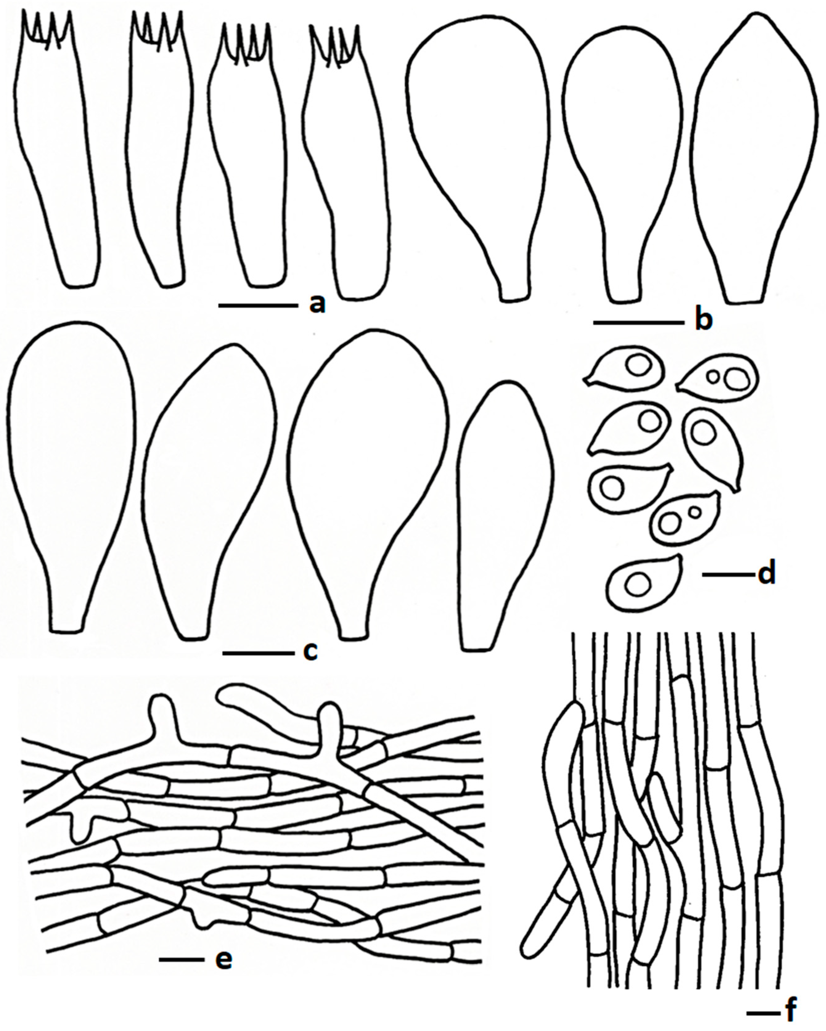
Basidiospores 5.00–6.40–7.60 × 3.40–3.95–4.50 µm, Q = 1.33–1.63–2.10, broadly ellipsoid to ellipsoid, thin walled, hyaline, oil granules present when viewed with KOH, 1–2-guttulate. Basidia 19.50–30.50 × 5.25–7.50 µm, subclavete to subcylindrical, sometimes clavate, thin walled, hyaline, 2–4-spored; sterigmata 1.00–3.00 µm long, pointed apex or sometimes obtuse apex. Lamellae trama regular, composed of inflated hyphae, 3.75–12.50 µm wide, thin walled. Cheilocystidia 27.12–56.50 × 10.50–29.50 µm, mostly pyriform to sometimes utriform or clavate, thin walled, hyaline. Pleurocystidia different types; pyriform, 47.00–69.50 × 17.50–26.00 µm, not very frequent; utriform 39.50–51.50 × 12.00–16.50 µm, frequent; clavate with round apex 41.50–54.50 × 16.00–23.50 µm, rare; thin walled, hyaline. Pileipellis two distinct layers; subpellis composed of radially arranged hyphae, thin walled; suprapellis up to 90 mm deep, composed of sub-erect to repent hyphae, 3.00–8.25 µm wide, thin walled, hyaline, branched, septate, hyphal end obtuse. Stipitipellis composed of tightly arranged hyphae, 4.0–10.00 µm wide, thin walled, hyaline, branched, septate, hyphal end obtuse or round. Clamp connections absent in all tissues.
Edibility: Edible.
Habit and habitat: Solitary to gregarious, growing on grass fields above subterranean termite nests.
Additional material examined: THAILAND. Chiang Mai province, Chiang Mai University, near Tat Chomphu Reservoir, 18°48′16.0″ N 98°57′08.2″ E, elevation 333 m, 11 August 2022, S. Paloi (SDBR-CMUNKM1852). GenBank accession numbers PQ896985 (nrLSU) and PV020678 (mtSSU).
Notes: Morphologically and phylogenetically, it is close to T. bulborhizus and T. gilvus. Termitomyces bulborhizus, originally described from China, differs from the present species by having a larger pileus (100–220 mm in diam.) with reddish brown to dark brown centre, fading from pale brown to brown toward the margin, prominent globose bulbous stipe base, larger cheilocystidia (19–60 × 12–34 µm) [52]; T. gilvus differs by having a dark brown, blunt pointed perforatorium on the pileus, a thicker pileus context (40 mm), a prominent globose bulbous base on the stipe, and clamp connections [20]. Some other look-alike species are T. umkowaan (Cooke & Massee) D.A. Reid and T. globulus R. Heim & Gooss.-Font. Termitomyces umkowaan, described from South Africa and widely distributed in Asia and Africa, which differs by having a larger pileus (80–220 mm diam.), a bulbous stipe base, comparatively larger basidiospores (6.1–13 × 4.3–6 µm), and narrow suprapellis hyphae (3.1–4.7 µm wide) [54]. Another species, T. globulus, differs by having a larger (150–200 mm in diam.) subglobose to bell-shaped pileus and a larger but not prominent perforatorium [50].

3.2.5. Termitomyces pseudoheimii Paloi, N. Suwannar. & J. Kumla sp. nov.

MycoBank: MB857476 (Figure 2g and Figure 7)
Holotype: THAILAND. Chiang Mai Province, Chiang Mai University, 18°47′49″ N 98°57′30″ E, elevation 332 m, 20 August 2022, N. Suwannarach (CMUB40069). GenBank accession numbers PQ897224 (nrITS), PQ897223 (nrLSU), and PV020679 (mtSSU).
Diagnosis: Differs from closely related species by having a smooth to velutinous pileus surface, ellipsoid to elongate basidiospores (7.53–10.89 × 3.67–6.13 µm, Q = 1.60–3.6), and comparatively wider suprapellis hyphae 4.22–8.2 µm wide.
Etymology: “pseudoheimii”—“pseudo” means false, referring to the morphological characteristics that are easily mistaken for T. heimii.
Description: Pileus 29–99 mm diam., hemispherical to convex at early stage, becoming broadly convex to subumbonate or applanate with apical papilla; perforatorium not pointed, surface smooth to velutinous, moist, brownish grey (5C2), greyish brown (5D4) to light brown (6D4) or brown (6E4) at centre (papilla), white (1A1) to sometimes orange white (5A2) at early stage, becoming white (1A1) at maturity, sometimes brownish tent present; margin cracked up to half of the pileus; context white (1A1), unchanged when exposure to air. Lamellae free, up to 5 mm broad, regular, white (1A1), becoming slightly yellowish white (4A2) to orange white (5A2) at maturity, sometimes light brownish after handling; edge smooth to wavy or serrate at maturity, concolorous; 2–3 lamellulae in a series. Stipe 45–65 × 7–12 mm, central, sometimes curved, smooth, white (1A1), to very light brown toward base at maturity; context white, fibrillose, no change after exposure to air; annulus present, light brown (6D4) to brown (5E4–F4). Pseudorhiza up to 170 mm long and 8–10 mm wide, white (1A1) to off white or brownish grey (5C2) to brownish orange (5C3), extending deeply into and firmly attached to the termite nest. Odour mild.
Basidiospores 7.53–9.33–10.89 × 3.67–4.67–6.13 µm, Q = 1.60–2.02–2.33, ellipsoid to elongate, thin walled, hyaline, sometimes 1–3-guttules, apiculus short, inamyloid. Basidia 15.75–26.5 × 6.91–8.75 µm, clavate to sub–clavate, thin walled, hyaline, oil granules present with KOH, mostly 2–4-spored, rarely 1-spored; sterigmata 1.0–4.5 µm long, cylindrical. Lamellae trama regular, composed of hyphae, 6.0–12.5 µm broad, thin walled, hyaline, branched, septate. Cheilocystidia 18.5–35.0 × 8.0–12.5 µm, pyriform to sometimes broadly clavate, thin walled, hyaline. Pleurocystidia 32.5–41.75 × 11.5–15.25 µm, pyriform, thin walled, hyaline. Pileipellis two distinct layers; subpellis composed of radially arranged chain of ellipsoids cells, 6.01–11.0 µm broad, thin walled, hyaline, branched; suprapellis up to 250 µm deep, composed of sub-erect to repent hyphae, 4.22–8.2 µm broad, thin walled, hyaline, branched, septate, hyphal end obtuse, gelatinised. Stipitipellis interwoven hyphae, 3.32–8.0 µm broad, thin walled, hyaline, branched, septate, oil granules present with KOH. Clanp connections absent in all tissues.
Habit and habitat: Gregarious, growing on grass fields above subterranean termite nests.
Edibility: Edible.
Other material examined: THAILAND. Chiang Mai Province, Chiang Mai University, 18°47′49″ N 98°57′30″ E, elevation 332 m, 13 September 2022, N. Suwannarach (SDBR-CMUNKP2013). GenBank accession numbers PQ897225 (nrITS), PQ897226 (nrLSU), and PV020680 (mtSSU).
Notes: Morphologically, T. pseudoheimii is close to T. heimii and T. islamabadensis. Termitomyces heimii is a well-known edible mushroom in several Asian countries and differs from the present species due to velar squamules covering the pileus and stipe surfaces, smaller basidiospores, and pileipellis repent epicutis formed by narrow hyphae (2.0–4.5 µm) [52,55]. Previously, Jannual et al. [27] described T. heimii from Thailand as having a comparatively very small pileus (up to 35 mm diam.) with a smaller stipe length (up to 40 mm long), and smaller-diameter pileipellis hyphae (2.0–4.5 µm). Another T. heimii from Kanchanaburi Province, Thailand, has smaller basidiospores (5–7 × 3.5–4.5 µm) [26], but T. pseudoheimii differs from these described species due to its larger pileus (up to 99 mm in diam.) and basidiospore size (7.53–10.89 × 3.67–6.13 µm). Termitomyces islamabadensis differs from the present species due to a creamy white or whitish grey pileus margin, 0 to 2 lamellulae, obovoid to lacrymoid basidiospores (Q value = 1.45–1.8), and comparatively narrow pileipellis hyphae (3.5–4.6 µm broad) [18]. Based on basidiomata size and colour, T. mammiformis R. Heim and T. upsilocystidiatus can cause confusion with T. pseudoheimii in the field. Termitomyces mammiformis has unique characteristics: mammiform, scrobiculate perforatorium on the pileus, and pale grey to pale brown velar squamules present on the pileus surface when mature [52,56]. Additionally, T. upsilocystidiatus, recently described from China, differs from T. pseudoheimii by having smaller basidiospores (4.8–6.8 × 3.5–4.2 μm) and Y-shaped cheilocystidia [15].

3.2.6. Termitomyces salmonicolor Paloi, W. Phonrob & N. Suwannar., sp. nov.

MycoBank: MB857477 (Figure 2h and Figure 8)
Holotype: THAILAND. Chiang Mai Province, Chiang Mai University, 18°47′45.5″ N 98°57′21.8″ E, elevation 332 m, 4 August 2022, S. Paloi and W. Phonrob (CMUB40070). GenBank accession numbers PQ897227 (nrLSU) and PV020681 (mtSSU).
Diagnosis: Differs from closely related species by having a smaller pileus with a smooth to velutinous surface, a slight to absent bulbous base, smaller basidiospores (4.67–6.68 × 3.10–4.26 µm), cheilocystidia 16.25–29.50 × 8.50–10.50 µm, and without clamp connections.
Etymology: “salmonicolor” refers to the light salmon colour of the pileus.
Description: Pileus 30–62 mm diam., convex, umbonate to papillate at young stage, becoming broadly convex to applanate with a pointed perforatorium at centre when mature; surface smooth to velutinous, moist, light brown (6D4), brown (6E5), dark brown (7F5) at centre, becoming faded toward margin, orange grey (6B2), brownish grey (6C2), reddish grey (7B2), greyish red (7B3), brownish orange (7C3) sometimes reddish white (8A2) or dull red (8C3); no change after bruising; margin regular, wavy, cracked, sometimes inflexed; context 1 mm thick at middle, white (1A1) to cream, no change after exposure to air. Lamellae free, regular, sometimes frocked near margin, white (1A1) at early stage, becoming greyish white (1A2), yellowish grey (4B2) at maturity; edge eroded, concolorous, 1–2 lamellulae. Stipe 34–65 × 5–12 mm, central, sometimes slightly curved a centre, base slightly bulbous or sometimes more or less equal, surface smooth to longitudinally striate, white (1A1) and greyish white (1A2) to yellowish grey (4B2) or grey (5B1); no change after bruising; context white to off-white, fibrillose, no change after exposure to air. Pseudorhiza 30–45 × 2–4 mm, similar colour as stipe or sometimes orange grey (5B2) to greyish orange (5B3); context solid, extending deeply into and firmly attached to the termite nest. Odour mild.
Basidiospores 4.67–5.75–6.68 × 3.10–3.68–4.26 µm, Q = 1.33–1.57–1.84, broadly ellipsoid to ellipsoid, thin walled, hyaline, oil granules present when viewed with KOH, 1–2-guttulate, short apiculus. Basidia 17.00–31.50 × 5.50–8.00 µm, clavate to sub clavate or sometimes subcylindrical, thin walled, hyaline, oil granules present with KOH, 2–4-spored; sterigmata 1.00–2.75 µm long, cylindrical. Lamellae trama regular, composed of inflated hyphae, 5.00–15.50 µm wide, thin walled, hyaline. Cheilocystidia 16.25–29.50 × 8.50–10.50 µm, pyriform, sometimes broadly clavate with round apex, thin walled, hyaline. Pleurocystidia 23.50–32.50 × 5.75–8.00 µm, short appendiculate to moniliform or pointed apex, thin walled, hyaline. Pileipellis two layers; subpellis composed of a chain of inflated cells, 9.60–24.15 µm wide, thin walled, hyaline, septate; suprapellis up to 80 µm deep, composed of erect to sub-erect or repent hyphae, 4.67–8.50 µm wide, thin walled, hyaline, septate, branched, hyphal end obtuse to sometimes round. Stipitipellis composed of tightly arranged hyphae, 3.11–6.00 µm wide, thin walled, hyaline, septate, branched, oil granules present with KOH, hyphal end obtuse. Clamp connections absent in all tissues.
Habit and habitat: Solitary to gregarious, growing on soil above subterranean termite nests.
Edibility: Edible.
Additional material examined: THAILAND. Chiang Mai University, near Tat Chomphu Reservoir, 18°48′14.8″ N 98°56′51.6″ E, elevation 342.3 m, 19 August 2022, W. Phonrob (SDBR-CMUNKN1261). GenBank accession numbers PQ897229 (nrLSU) and PV020682 (mtSSU).
Notes: Morphologically similar species, namely T. clypeatus R. Heim, T. striatus, T. aurantiacus (R. Heim) R. Heim, and T. tylerianus Otieno, can be confused with T. salmonicolor. Termitomyces clypeatus differs by having a larger pileus (25–140 mm in diam.) with a sharp spiny perforatorium (5–12 mm high), a long cylindrical stipe, longer pseudorhiza (up to 220 mm long), larger basidiospores (6.0–9.0 × 4.0–6.0 µm), and narrow suprapellis hyphae (2.0–5.0 µm) [52]. Termitomyces striatus has a white to pale grey or greyish brown pileus, larger basidiospores (5.5–8.0 × 3.5–5.5 µm), and narrow pileipellis hyphae [52]. Termitomyces aurantiacus has a larger pileus size and very long pseudorhiza (up to 340 mm), lacks pleurocystidia, and has narrow pileipellis hyphae (1.5–4.5 µm) [52]. Termitomyces tylerianus, originally described from Kenya, differs from the present species due to characteristics such as a small pileus (10–20 mm in diam.) with a sharp pointed perforatorium, no cheilocystidia and pleurocystidia, and narrow pileipellis hyphae (2.0–5.0 µm) [52]. Phylogenetically, T. salmonicolor is close to T. bulborhizus, T. gilvus, and T. planiperforatorius. Termitomyces bulborhizus has a larger basidiomata size, prominent bulbous base, comparatively larger basidiospores (6–9 × 4–6 µm), cheilocystidia (19–60 × 12–34 µm), and pleurocystidia (19–78 × 10–32 µm) [53]. Termitomyces gilvus has a larger pileus (80–130 mm in diam.), thick pileus context (40 mm), prominent bulbous base, comparatively larger basidiospores (6–8.5 × 3.7–5.4 µm), and clamp connections [21]. Termitomyces planiperforatorius, also collected from the same region and also lacking a bulbous base, differs due to characteristics such as a larger basidiomata with a prominent squamulose pileus surface and larger hymenial cystidia (this study).

4. Conclusions

Termitomyces species are globally recognised as a tropical delicacy, with almost all species being edible and nutritious. In this study, five new Termitomyces species (T. griseobulbus, T. griseobrunneus, T. planiperforatorius, T. pseudoheimii, and T. salmonicolor) and one species (T. acriumbonatus) previously identified outside of Thailand were discovered in northern Thailand and described based on a combination of morphological characteristics and multi-gene phylogenetic analyses. These newly discovered macrofungi can be used for further research in the food, pharmaceutical, and other industries. Furthermore, this finding has increased the number of Termitomyces species found in Thailand. The results of this study are an important step in stimulating further research on wild edible mushrooms in Thailand and may help researchers to better understand the distribution of Termitomyces in Asia and around the world. Future research on Termitomyces in Thailand should include the identification of the associated termite hosts and should consider the potential impacts of climate change, as both factors may influence species diversity, distribution, and habitat dynamics.

Author Contributions

Conceptualisation, S.P. and N.S.; methodology, S.P., W.P., J.K. and N.S. software, S.P., J.K. and N.S.; validation, S.P. and N.S.; formal analysis, S.P., W.P., J.K. and N.S.; investigation, N.S.; resources, N.S.; data curation, S.P., J.K. and N.S.; writing—original draft preparation, S.P. and B.P.P.; writing—review and editing, S.P., W.P., J.K., B.P.P. and N.S.; visualisation, S.P. and N.S.; supervision, N.S.; project administration, N.S.; funding acquisition, N.S. All authors have read and agreed to the published version of the manuscript.

Funding

This research was financially supported by Chiang Mai University, Thailand and Fundamental Fund (Grant number: FF207587/2568), and the National Research Council of Thailand (Grant number: N42A650198).

Institutional Review Board Statement

Not applicable.

Informed Consent Statement

Not applicable.

Data Availability Statement

The DNA sequence data obtained from this study were deposited in GenBank (https://www.ncbi.nlm.nih.gov/nucleotide, accessed on 18 April 2025) under the following accession numbers: nrLSU (PQ896626, PQ896628, PQ896629, PQ896878, PQ896983, PQ896985, PQ896901, PQ897223, PQ897226, PQ897227, PQ897229, PQ899490, and PQ899491), nrITS (PQ896627, PQ897224, PQ897225, PQ899488, PQ899489, PQ900075, and PQ900076), and mtSSU (PV202670 to PV202682). Specimens of the new taxa identified in this study have been deposited in MycoBank (https://www.mycobank.org/, accessed on 20 April 2025) under numbers MB857473 to MB857477. The alignment of the concatenated nrLSU, mtSSU, and nrITS sequences was deposited in Zenodo under the DOI number 10.5281/zenodo.15528282.

Acknowledgments

We express our thanks to Chiang Mai University’s Post-Doctoral Fellowship and the National Research Council of Thailand (NRCT) for permitting Soumitra Paloi to continue carrying out research in Thailand.

Conflicts of Interest

The authors declare no conflicts of interest.

References

  1. Antonelli, A.; Fry, C.; Smith, R.J.; Eden, J.; Govaerts, R.H.A.; Kersey, P.J.; Lughadha, E.M.N.; Onstein, R.E.; Simmonds, M.S.J.; Zizka, R.; et al. State of the World’s Plants and Fungi; Royal Botanic Gardens, Kew: Richmond, UK, 2023; pp. 1–96.
  2. Gopal, J.; Sivanesan, I.; Muthu, M.; Oh, J.-W. Scrutinizing the nutritional aspects of Asian mushrooms, its commercialization and scope for value-added products. Nutrients 2022, 14, 3700. [Google Scholar] [CrossRef]
  3. Ye, L.; Karunarathna, S.C.; Li, H.; Xu, J.; Hyde, K.D.; Mortimer, P.E. A survey of Termitomyces (Lyophyllaceae, Agaricales), including a new species, from a subtropical forest in Xishuangbanna, China. Mycobiology 2019, 47, 391–400. [Google Scholar] [CrossRef] [PubMed]
  4. Teke, N.A.; Kinge, T.R.; Bechem, E.; Nji, T.M.; Ndam, L.M.; Mih, A.M. Ethnomycological study in the Kilum-Ijim mountain forest, Northwest Region, Cameroon. J. Ethnobiol. Ethnomed. 2018, 14, 25. [Google Scholar] [CrossRef] [PubMed]
  5. Njue, A.W.; Omolo, J.O.; Cheplogoi, P.K.; Langat, M.K.; Mulholland, D.A. Cytotoxic ergostane derivatives from the edible mushroom Termitomyces microcarpus (Lyophyllaceae). Biochem. Syst. Ecol. 2018, 76, 12–14. [Google Scholar] [CrossRef]
  6. Manna, S.; Ray, D.; Roy, A. Tribal relation to spatio-temporal variation of wild mushrooms in eastern lateritic part of India. Ethnobot. Res. Appl. 2014, 12, 15–24. [Google Scholar]
  7. Aryal, H.P.; Budhathoki, U. Ethnomycology of Termitomyces R. Heim in Nepal. J. Yeast Fungal Res. 2016, 7, 28–38. [Google Scholar]
  8. Sitotaw, R.; Lulekal, E.; Abate, E. Ethnomycological study of edible and medicinal mushrooms in Menge District, Asossa Zone, Benshangul Gumuz Region, Ethiopia. J. Ethnobiol. Ethnomed. 2020, 16, 11. [Google Scholar] [CrossRef]
  9. Heim, R. Nouvelles études descriptives sur les agarics termitophiles d’Afrique tropicale. Arch. Mus. Natl. d’Hist. Nat. 6ème Sér. 1942, 18, 107–166. [Google Scholar]
  10. Index Fungorum. Available online: https://www.indexfungorum.org/names/names.asp (accessed on 1 April 2025).
  11. Paloi, S.; Kumla, J.; Paloi, B.P.; Srinuanpan, S.; Hoijang, S.; Karunarathna, S.C.; Acharya, K.; Suwannarach, N.; Lumyong, S. Termite mushrooms (Termitomyces), a potential source of nutrients and bioactive compounds exhibiting human health benefits: A review. J. Fungi 2023, 9, 112. [Google Scholar] [CrossRef]
  12. Osiemo, Z.B.; Marten, A.; Kaib, M.; Gitonga, L.M.; Boga, H.I.; Brandl, R. Open relationships in the castles of clay: High diversity and low host specificity of Termitomyces fungi associated with fungus-growing termites in Africa. Insectes Sociaux 2010, 57, 351–363. [Google Scholar] [CrossRef]
  13. Aanen, D.K.; Eggleton, P.; Rouland-Lefèvre, C.; Guldberg-Frøslev, T.; Rosendahl, S.; Boomsma, J.J. The evolution of fungus growing termites and their mutualistic fungal symbionts. Proc. Natl. Acad. Sci. USA 2002, 99, 14887–14892. [Google Scholar] [CrossRef] [PubMed]
  14. Koné, N.A.; Yéo, K.; Konaté, S.; Linsenmair, K.E. Socio-economical aspects of the exploitation of Termitomyces fruit bodies in central and southern Côted’Ivoire: Raising awareness for their sustainable use. J. Appl. Biosci. 2013, 70, 5580–5590. [Google Scholar] [CrossRef]
  15. Tang, S.M.; He, M.Q.; Raspé, O.; Luo, X.; Zhang, X.L.; Li, Y.; Su, K.M.; Li, S.H.; Thongklang, N.; Hyde, K.D. Two new species of Termitomyces (Agaricales, Lyophyllaceae) from China and Thailand. Phytotaxa 2020, 439, 231–242. [Google Scholar] [CrossRef]
  16. Tang, S.M.; Vadthanarat, S.; He, J.; Raghoonundon, B.; Yu, F.M.; Karunarathna, S.C.; Li, S.H.; Raspé, O. Morphological and molecular analyses reveal two new species of Termitomyces (Agaricales, Lyophyllaceae) and morphological variability of T. intermedius. MycoKeys 2023, 95, 61–82. [Google Scholar] [CrossRef]
  17. Izhar, A.; Khalid, A.N.; Bashir, H. Termitomyces sheikhupurensis sp. nov. (Lyophyllaceae, Agaricales) from Pakistan, evidence from morphology and DNA sequences data. Turk. J. Bot. 2022, 44, 694–704. [Google Scholar] [CrossRef]
  18. Usman, M.; Khalid, A.N. Termitomyces acriumbonatus sp. nov. (Lyophyllaceae, Agaricales) from Pakistan. Phytotaxa 2020, 477, 217–228. [Google Scholar] [CrossRef]
  19. Ashraf, S.; Usman, M.; Khalid, A.N. Termitomyces islamabadensis sp. nov. (Lyophyllaceae, Agaricales) from the Foothills of the Pakistani Himalayas. Biol. Bull. Russ. Acad. Sci. 2022, 49, S66–S76. [Google Scholar] [CrossRef]
  20. Razaq, A.; Ishaq, A.; Ilyas, S.; Sadia, S. Termitomyces pakistanensis, a new mushroom species from Pakistan based on scanning electron microscopy and ITS-rDNA barcoding. Microsc. Res. Tech. 2023, 86, 115–121. [Google Scholar] [CrossRef]
  21. Seelan, J.S.S.; Yee, C.S.; Fui, F.S.; Dawood, M.; Tan, Y.S.; Kim, M.J.; Park, M.S.; Lim, Y.W. New species of Termitomyces (Lyophyllaceae, Basidiomycota) from Sabah (Northern Borneo), Malaysia. Mycobiology 2020, 48, 95–103. [Google Scholar] [CrossRef]
  22. Ediriweera, A.N.; Voto, P.; Karunarathna, S.C.; Dilshan, B.C. Termitomyces srilankensis sp. nov. (Lyophyllaceae, Agaricales), a new species from Sri Lanka. MycolObs 2023, 6, 47–53. [Google Scholar]
  23. Das, L.R.; Chattopadhyay, P.; Dutta, A.K.; Narzary, D.; Rana, T.S. Termitomyces assamicus (Lyophyllaceae)—A new species of Termitomyces from India. Phytotaxa 2023, 599, 126–136. [Google Scholar] [CrossRef]
  24. Hussain, S.; Al-Kharousi, M.; Al-Owaisi, A.A.; Al-Maqbali, D.; Al-Muharabi, M.A.; Al-Shabibi, Z.; Al-Balushi, A.H.; Al Saady, N.; Velazhahan, R.; Rashan, L.; et al. The genus Termitomyces: Outline, phylogeny, and divergence times estimation with description of a new edible species from Arabian Peninsula. Sydowia 2024, 76, 187–200. [Google Scholar]
  25. Sangvichien, E.; Taylor-Hawksworth, P.A. Termitomyces mushrooms: A tropical delicacy. Mycologist 2001, 15, 31–33. [Google Scholar] [CrossRef]
  26. Sawhasan, P.; Worapong, J.; Vinijsanun, T. Morphological and molecular studies of selected Termitomyces species collected from 8 districts of Kanchanaburi Province, Thailand. Thai J. Agric. Sci. 2011, 44, 183–196. [Google Scholar]
  27. Jannual, N.; Nipitwattanaphon, M.; Hasin, S.; Kaewgrajang, T. Morphological and molecular characterization of Termitomyces (Lyophyllaceae, Agaricales) in Thailand. Biodiversitas 2020, 22, 2481–2491. [Google Scholar] [CrossRef]
  28. Srikram, A.; Supapvanich, S. Proximate compositions and bioactive compounds of edible wild and cultivated mushrooms from Northeast Thailand. Agric. Nat. Resour. 2016, 50, 432–436. [Google Scholar] [CrossRef]
  29. Tang, S.-M.; Li, E.-X.; Luo, H.-M.; Ao, C.-C.; Lv, T.; Li, S.-H. Termitomyces flavus sp. nov. (Lyophyllaceae, Agaricales), a new species from northern Thailand. Chiang Mai J. Sci. 2024, 51, e2024060. [Google Scholar] [CrossRef]
  30. Hughes, A.C. Mapping priorities for conservation in Southeast Asia. Biol. Conserv. 2017, 209, 395–405. [Google Scholar] [CrossRef]
  31. Liengsiri, C.; Luangjame, J.; Ponoy, B.; Sukhotanang, A.; Visuthitepkul, S.; Thienhirun, S.; Vacharangkura, T.; Luangviriyasaeng, V.; Semsuntud, N.; Wichiennopparat, W.; et al. Forestry in Thailand; Royal Forest Department, Ministry of Natural Resources and Environment: Bangkok, Thailand, 2009; pp. 1–48.
  32. Lertlumnaphakul, W.; Ngoen-Klan, R.; Vongkaluang, C.; Chareonviriyaphap, T. A review of termite species and their distribution in Thailand. Insects 2022, 13, 186. [Google Scholar] [CrossRef]
  33. Kornerup, A.; Wanscher, J.H. Taschenlexikon der Farben, 3rd ed.; Musterschmidt: Northeim-Sudheim, Germany, 1981. [Google Scholar]
  34. Mossebo, D.C.; Essouman, E.; Machouart, M.; Gueidan, C. Phylogenetic relationships, taxonomic revision and new taxa of Termitomyces (Lyophyllaceae, Basidiomycota) inferred from combined nLSU and mtSSU rDNA sequences. Phytotaxa 2017, 321, 71–102. [Google Scholar] [CrossRef]
  35. White, T.J.; Bruns, T.D.; Lee, S.B.; Taylor, J.W. Amplification and Direct Sequencing of Fungal Ribosomal RNA Genes for Phylogenetics. In PCR Protocols: A Guide to Methods and Applications; Innis, M.A., Gelfand, D.H., Sninsky, J.J., White, T.J., Eds.; Academic Press: New York, NY, USA, 1990; pp. 315–322. [Google Scholar] [CrossRef]
  36. Vilgalys, R.; Hester, M. Rapid genetic identification and mapping of enzymatically amplified ribosomal DNA from several Cryptococcus species. J. Bacteriol. 1990, 172, 4238–4246. [Google Scholar] [CrossRef]
  37. Paloi, S.; Suwannarach, N.; Kumla, J.; Phonrob, W.; Karunarathna, S.C.; Lumyong, S. An update on species diversity, distribution and sequence data of Tulostoma in Asia with the addition of Tulostoma exasperatum, a new record for Thailand. Chiang Mai J. Sci. 2023, 50, e2023018. [Google Scholar] [CrossRef]
  38. Moncalvo, J.M.; Lutzoni, F.M.; Rehner, S.A.; Johnson, J.; Vilgalys, R. Phylogenetic relationships of agaric fungi based on nuclear large subunit ribosomal DNA sequences. Syst. Biol. 2000, 49, 278–305. [Google Scholar] [CrossRef] [PubMed]
  39. Hofstetter, V.; Clémençon, H.; Vilgalys, R.; Moncalvo, J.M. Phylogenetic analyses of the Lyophylleae (Agaricales, Basidiomycota) based on nuclear and mitochondrial rDNA sequences. Mycol. Res. 2002, 106, 1043–1059. [Google Scholar] [CrossRef]
  40. Frøslev, T.G.; Aanen, D.K.; Læssøe, T.; Rosendahl, S. Phylogenetic relationships of Termitomyces and related taxa. Mycol. Res. 2003, 107, 1277–1286. [Google Scholar] [CrossRef] [PubMed]
  41. Willis, E. The Taxonomic Identity of Termitomyces aurantiacus Using the Internal Transcribed Spacer Regions 1 & 2 (ITS1 & ITS2) of the Ribosomal DNA (rDNA). Ph.D. Thesis, University of Putra, Serdang, Malaysia, 2007. [Google Scholar]
  42. Hofstetter, V.; Redhead, S.A.; Kauff, F.; Moncalvo, J.M.; Matheny, P.B.; Vilgalys, R. Taxonomic revision and examination of ecological transitions of the Lyophyllaceae (Basidiomycota, Agaricales) based on a multigene phylogeny. Cryptogam. Mycol. 2014, 35, 399–425. [Google Scholar] [CrossRef]
  43. Edgar, R.C. MUSCLE: Multiple sequence alignment with high accuracy and high throughput. Nucleic Acids Res. 2004, 32, 1792–1797. [Google Scholar] [CrossRef]
  44. Stamatakis, A. RAxML version 8: A tool for phylogenetic analysis and post-analysis of large phylogenies. Bioinformatics 2014, 30, 1312–1313. [Google Scholar] [CrossRef]
  45. Ronquist, F.; Teslenko, M.; van der Mark, P.; Ayres, D.L.; Darling, A.; Höhna, S.; Larget, B.; Liu, L.; Suchard, M.A.; Huelsenbeck, J.P. MrBayes 3.2: Efficient Bayesian phylogenetic inference and model choice across a large model space. Syst. Biol. 2012, 61, 539–542. [Google Scholar] [CrossRef]
  46. Darriba, D.; Taboada, G.L.; Doallo, R.; Posada, D. jModelTest 2: More models, new heuristics and parallel computing. Nat. Methods 2012, 9, e772. [Google Scholar] [CrossRef]
  47. Hillis, D.M.; Bull, J.J. An empirical test of bootstrapping as a method for assessing confidence in phylogenetic analysis. Syst. Biol. 1993, 42, 182–192. [Google Scholar] [CrossRef]
  48. Alfaro, M.E.; Zoller, S.; Lutzoni, F. Bayes or bootstrap? A simulation study comparing the performance of bayesian markov chain monte carlo sampling and bootstrapping in assessing phylogenetic confidence. Mol. Biol. Evol. 2003, 20, 255–266. [Google Scholar] [CrossRef]
  49. Rambaut, A. FigTree, v1.3.1. 2009. Available online: http://tree.bio.ed.ac.uk/software/figtree (accessed on 10 February 2025).
  50. Natarajan, K. A new species of Termitomyces from India. Curr. Sci. 1977, 46, 679–680. [Google Scholar]
  51. Heim, R. Les Termitomyces du Congo Belge recueillis par Madame M. Goossens-Fontana. Bull. Jard. Bot. État. Brux. 1951, 21, 205–222. [Google Scholar] [CrossRef]
  52. Wei, T.Z.; Tang, B.H.; Yao, Y.J. Revision of Termitomyces in China. Mycotaxon 2009, 108, 257–285. [Google Scholar] [CrossRef]
  53. Wei, T.Z.; Yao, Y.J.; Wang, B.; Pegler, D.N. Termitomyces bulborhizus sp. nov. from China, with a key to allied species. Mycol. Res. 2004, 108, 1458–1462. [Google Scholar] [CrossRef]
  54. Westhuizen, G.C.A.; Eicker, A. Species of Termitomyces occurring in South Africa. Mycol. Res. 1990, 94, 923–937. [Google Scholar] [CrossRef]
  55. Natarajan, K. South Indian Agaricales V: Termitomyces heimii. Mycologia 1979, 71, 853–855. [Google Scholar] [CrossRef]
  56. Wei, T.Z.; Tang, B.H.; Yao, Y.J.; Pegler, D.N. A revision of Sinotermitomyces, a synonym of Termitomyces (Agaricales). Fungal Divers. 2006, 21, 225–237. [Google Scholar]
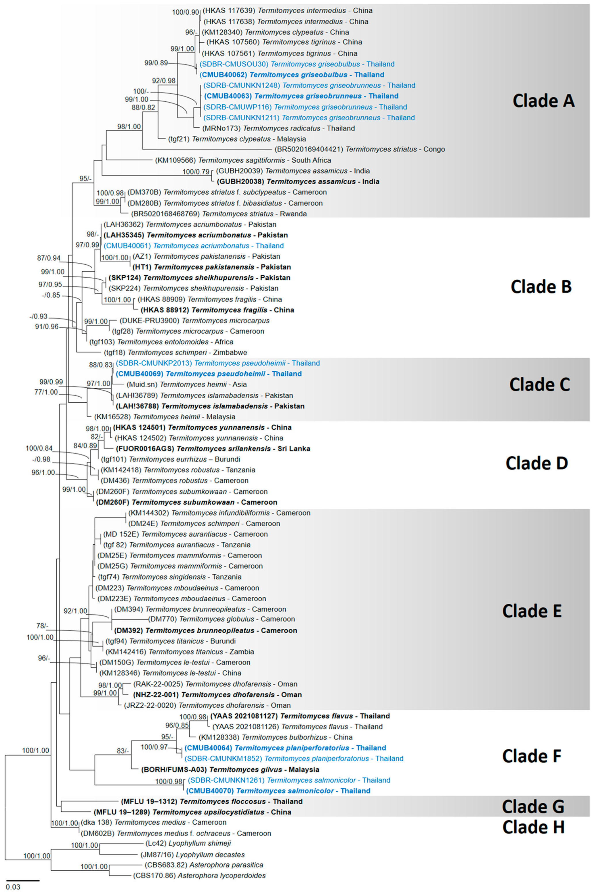
Figure 1. Phylogenetic tree derived from maximum likelihood analysis of 82 taxa of combined nrLSU + mtSSU + nrITS data. Asterophora lycoperdoides, Asterophora parasitica, Lyophyllum decastes, and Lyophyllum shimeji were used for rooting purposes. The numbers above branches represent maximum likelihood bootstrap percentages (right) and Bayesian posterior probabilities (left). Bootstrap values ≥ 70% and Bayesian posterior probabilities ≥ 0.75 are shown above the branches. The scale bar represents the expected number of nucleotide substitutions per site. Type species are highlighted in bold font. Newly reported species in this study are highlighted (blue font).
Jof 11 00830 g001
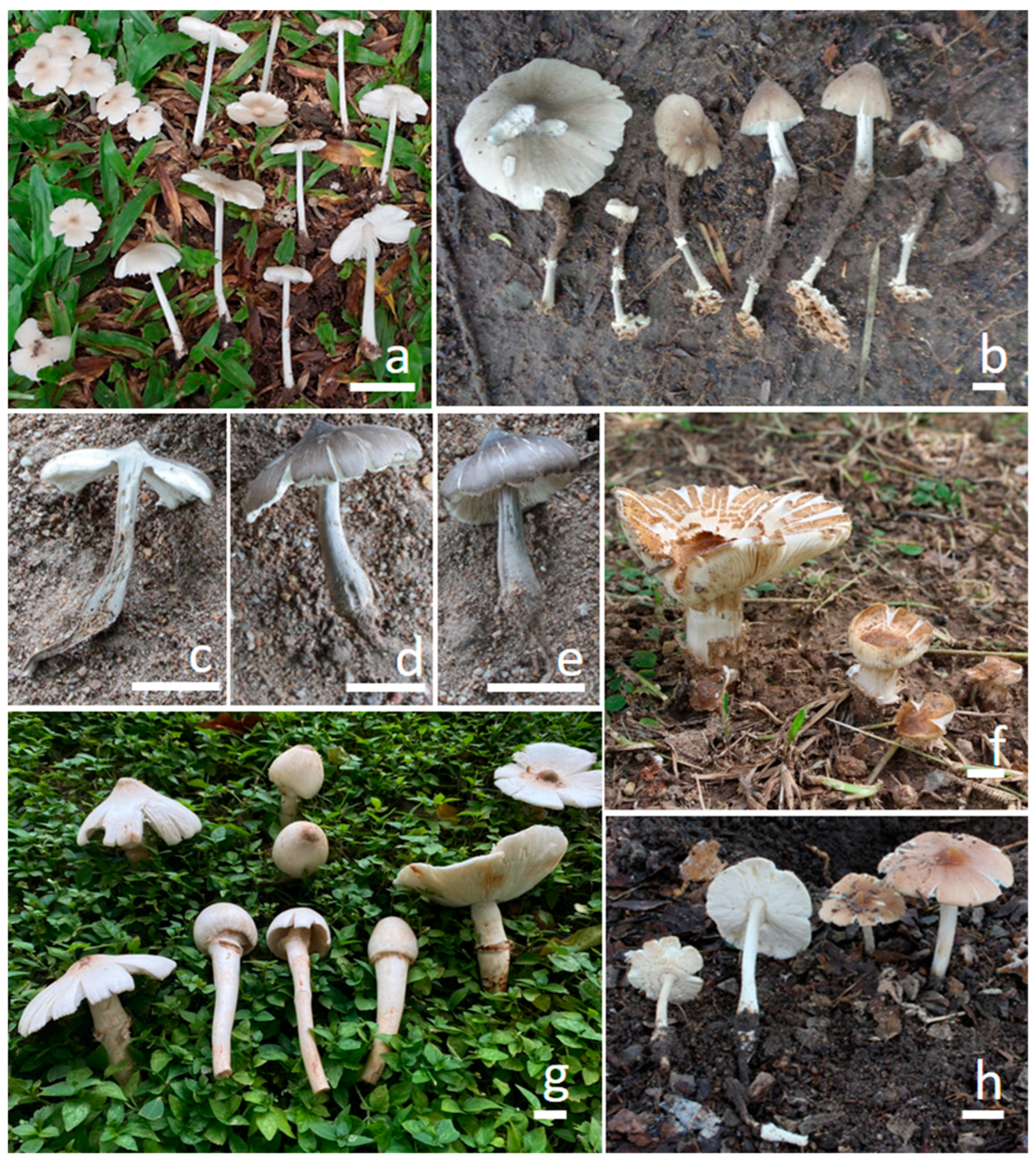
Figure 2. Field photograph of basidiomata. (a) Termitomyces acriumbonatus (CMUB40061, new record). (b) T. griseobulbus (CMUB40062, holotype). (ce) T. griseobrunneus (CMUB40063, holotype). (f) T. planiperforatorius (CMUB40064, holotype). (g) T. pseudoheimii (CMUB40069, holotype). (h) T. salmonicolor (CMUB40070, holotype). Scale bars = 20 mm.
Jof 11 00830 g002
Figure 3. Microscopic features of Termitomyces acriumbonatus (CMUB40061, new record): (a) basidia; (b) cheilocystidia; (c) pleurocystidia; (d) basidiospores; (e) pileipellis; (f) stipitipellis. Scale bars: (ac,e,f) = 10 μm and (d) = 5 μm.
Jof 11 00830 g003
Figure 4. Microscopic features of Termitomyces griseobulbus (CMUB40062, holotype): (a) basidia; (b) cheilocystidia; (c) pleurocystidia; (d) basidiospores; (e) pileipellis; (f) stipitipellis. Scale bars: (ac,e,f) = 10 μm and (d) = 5 μm.
Jof 11 00830 g004
Figure 5. Microscopic features of Termitomyces griseobrunneus (CMUB40063, holotype): (a) basidia; (b) cheilocystidia; (c) pleurocystidia; (d) basidiospores; (e) pileipellis; (f) stipitipellis. Scale bars: (ac,e,f) = 10 μm and (d) = 5 μm.
Jof 11 00830 g005
Figure 6. Microscopic features of Termitomyces planiperforatorius (CMUB40064, holotype): (a) basidia; (b) cheilocystidia; (c) pleurocystidia; (d) basidiospores; (e) pileipellis; (f) stipitipellis. Scale bars: (ac,e,f) = 10 μm and (d) = 5 μm.
Jof 11 00830 g006
Figure 7. Microscopic features of Termitomyces pseudoheimii (CMUB40069, holotype): (a) basidia; (b) cheilocystidia; (c) pleurocystidia; (d) basidiospores; (e) pileipellis; (f) stipitipellis. Scale bars: (ac,e,f) = 10 μm and (d) = 5 μm.
Jof 11 00830 g007
Figure 8. Microscopic features of Termitomyces salmonicolor (CMUB40070, holotype): (a) basidia; (b) cheilocystidia; (c) pleurocystidia; (d) basidiospores; (e) pileipellis; (f) stipitipellis. Scale bars: (ac,e,f) = 10 μm and (d) = 5 μm.
Jof 11 00830 g008
Table 1. Taxa, voucher numbers, countries, GenBank accession numbers, references, and type sequences used in the phylogenetic analyses conducted in this study.
TaxaVoucher NumberCountryGenBank Accession NumberReference
nrITSnrLSUmtSSU
Termitomyces acriumbonatus TLAH35345PakistanMT179688MT179689NA[17]
T. acriumbonatusLAH36362PakistanMT179687MT179690NA[17]
T. acriumbonatusCMUB40061ThailandPQ896627PQ896626PV020670This study
T. assamicus TGUBH20038IndiaOQ346313NANA[23]
T. assamicusGUBH20039IndiaOQ976999NANA[23]
T. aurantiacusDM 152ECameroonNAKY809234KY809186[41]
T. aurantiacustgf 82TanzaniaNAAY127804AY127852[13]
T. brunneopileatus TDM392CameroonNAKY809273KY809225[34]
T. brunneopileatusDM394CameroonNAKY809244KY809197[34]
T. bulborhizusKM128338ChinaNAKY809261KY809213[34]
T. clypeatusKM128340ChinaNAKY809262KY809214[34]
T. clypeatustgf21MalaysiaNAAY127802AY127850[13]
T. dhofarensisJRZ2-22-0020OmanOR297695OR338598NA[24]
T. dhofarensisRAK-22-0025OmanOR297696OR338597NA[24]
T. dhofarensis TNHZ-22-001OmanOR297694NANA[24]
T. entolomoidestgf103AfricaNAAY232693AY232680[40]
T. eurrhizustgf101BurundiNAAY232694NA[40]
T. floccosus TMFLU 19–1312Thailand MN633305MN701029[15]
T. fragilis THKAS 88912ChinaKY214475NANA[3]
T. fragilisHKAS 88909ChinaKY214476NANA[3]
T. gilvus TBORH/FUMS-A03MalaysiaNAMK472701MK478904[21]
T. globulusDM770CameroonNAKY809252KY809204[39]
T. griseobulbus TCMUB40062ThailandPQ900075PQ896628PV020671This study
T. griseobulbusSDBR–CMUSOU30ThailandPQ900076PQ896629PV020672This study
T. griseobrunneus TCMUB40063ThailandPQ899488PQ896878PV020673This study
T. griseobrunneusSDBR–CMUNKN1248ThailandPQ899489PQ899490PV020676This study
T. griseobrunneusSDBR–CMUWP116ThailandNAPQ896901PV020674This study
T. griseobrunneusSDBR–CMUNKN1211ThailandNAPQ899491PV020675This study
T. heimiiKM16528MalaysiaNAKY809253KY809205[34]
T. heimiiMuid.snAsiaNAAF042586AF357091[38]
T. infundibiliformisKM144302CameroonNAKY809245NA[34]
T. intermediusHKAS 117638ChinaON557369ON556484ON557367[16]
T. intermediusHKAS 117639ChinaON557370ON556485ON557368[16]
T. islamabadensis TLAH!36788PakistanMW520178OM100949NA[18]
T. islamabadensisLAH!36789PakistanMW520179OM100950NA[18]
T. le-testuiDM150GCameroonNAKY809231KY809184[34]
T. le-testuiKM128346ChinaNAKY809263KY809215[34]
T. mammiformisDM25ECameroonNAKY809229KY809182[34]
T. mammiformisDM25GCameroonNAKY809230KY809183[34]
T. mboudaeinusDM223CameroonNAKY809274KY809226[34]
T. mboudaeinusDM223ECameroonNAKY809237KY809189[34]
T. mediusdka 138CameroonNAAY127796AY127844[13]
T. medius f. ochraceusDM602BCameroonNAKY809246KY809198[34]
T. microcarpustgf28CameroonNAAY127799AY127847[13]
T. microcarpusDUKE-PRU3900NAAF357023AF042587AF357092[39]
T. pakistanensisHT1PakistanOP688120NANA[20]
T. pakistanensisAZ1PakistanOP688121NANA[20]
T. planiperforatorius TCMUB40064ThailandNAPQ896983PV020677This study
T. planiperforatoriusSDBR–CMUNKM1852ThailandNAPQ896985PV020678This study
T. pseudoheimii TCMUB40069ThailandPQ897224PQ897223PV020679This study
T. pseudoheimiiSDBR–CMUNKP2013ThailandPQ897225PQ897226PV020680This study
T. radicatusMRNo173ThailandLC068787NANAUnP
T. robustusKM142418TanzaniaNAKY809265KY809217[34]
T. robustusDM436CameroonNAKY809271KY809223[34]
T. sagittiformisKM109566South AfricaNAKY809260KY809212[34]
T. salmonicolor TCMUB40070ThailandNAPQ897227PV020681This study
T. salmonicolorSDBR–CMUNKN1261ThailandNAPQ897229PV020682This study
T. schimperiDM24ECameroonNAKY809228KY809181[34]
T. schimperitgf18ZimbabweNAAY232712AY232686[40]
T. sheikhupurensis TSKP124PakistanMT192217MT192228NA[19]
T. sheikhupurensisSKP224PakistanMT192218NANA[19]
T. singidensistgf74TanzaniaNAAY232713AY232687[40]
T. srilankensis TFUOR0016AGSSri LankaON685313NANA[22]
T. striatusBR5020169404421CongoOP179299OP168080OP179293[16]
T. striatusBR5020168468769RwandaOP179297OP168081OP179294[16]
T. striatus f. bibasidiatusDM280BCameroonNAKY809241KY809193[34]
T. striatus f. subclypeatusDM370BCameroonNAKY809268KY809220[34]
T. subumkowaanDM260FCameroonNAKY809239KY809190[34]
T. subumkowaan TDM260BCameroonNAKY809275KY809227[34]
T. tigrinusHKAS 107560ChinaMT683156MT679729MT683152[16]
T. tigrinus THKAS 107561ChinaMT683157MT679730MT683153[16]
T. titanicustgf94BurundiNAAY127801AY127849[13]
T. titanicusKM142416ZambiaNAKY809264KY809216[34]
T. upsilocystidiatus TMFLU 19–1289ChinaNAMN636637MN636642[15]
T. yunnanensis THKAS 124501ChinaOP179295OP168083OP179290[16]
T. yunnanensisHKAS 124502ChinaOP179296OP168084OP179291[16]
Termitomyces flavus TYAAS 2021081127ThailandPP264695PP264703PP264701[29]
Termitomyces flavusYAAS 2021081126ThailandPP264696PP264704PP264702[29]
Asterophora lycoperdoidesCBS170.86NAAF357037AF223190AF357109[42]
A. parasiticaCBS683.82NAAF357038AF223191AF357110[42]
Lyophyllum shimejiLc42NAAF357060AF357078AF357137[42]
L. decastesJM87/16NAAF357059AF042583AF357136[42]
“NA” = indicates that no data are available in the GenBank database or could not be obtained. “UnP” = Unpublished; “T” = Holotype specimen.
Disclaimer/Publisher’s Note: The statements, opinions and data contained in all publications are solely those of the individual author(s) and contributor(s) and not of MDPI and/or the editor(s). MDPI and/or the editor(s) disclaim responsibility for any injury to people or property resulting from any ideas, methods, instructions or products referred to in the content.