Article

Co-Culture of Monascus purpureus and Aspergillus niger Isolated from Wuyi Hongqu to Enhance Monascus Pigments Production While Inhibiting Citrinin Production

1
College of Food Science and Technology, Huazhong Agricultural University, Wuhan 430070, China
2
College of Food Science and Technology, Wuhan Business University, Wuhan 430056, China
*
Authors to whom correspondence should be addressed.
J. Fungi 2025, 11(12), 829; https://doi.org/10.3390/jof11120829
Submission received: 16 October 2025 / Revised: 13 November 2025 / Accepted: 14 November 2025 / Published: 24 November 2025
(This article belongs to the Special Issue Monascus spp. and Their Relative Products)

Abstract

Wuyi Hongqu (WYH), also called black-skin-red-koji, which has been utilizing as a fermentation starter for more than one thousand years in China, is a symbiotic combination of Monascus spp. and Aspergillus niger formed through long-term application and domestication. In this study, the strains of Monascus purpureus and A. niger isolated from WYH samples were used to investigate their mutual influence, especially the effects on three main secondary metabolites from M. purpureus, Monascus pigments (MPs), monacolin K (MK), and citrinin (CIT), using a double-sided Petri dish (DSPD). The results showed that co-cultivation of M. purpureus and A. niger strains was favorable to increase the MPs production while inhibiting the CIT production by M. purpureus, especially when M. purpureus strains (M1-1 or M9) were co-cultivated with certain A. niger strains (An1-2 or An9), respectively, and both Monascus strains hardly produced detectable CIT. The expression levels of CIT-related genes in M. purpureus M1-1 or M9 were greatly restricted when co-cultivated with A. niger An1-2 or An9 confirmed by RT-qPCR. This study provides important insights into the selection of WYH production strains and the effects of fungal interactions.

1. Introduction

Wuyi Hongqu (WYH), also called as black-skin-red-koji, is a traditional fermentation agent for the production of Chinese rice wine (Huangjiu), one of the oldest and most representative fermented alcoholic beverages in East Asia [1,2,3]. WYH has been produced and used for more than one thousand years in China [3]. WYH is a symbiotic product mainly containing Monascus species and A. niger, through long-term domestication and application, which makes it a good model for studying microbial symbiosis [4,5]. Although there has been some research about the microbial community in WYH [6,7], the microbial interaction and the resulting secondary metabolite changes remain unknown.
As one of the main fungi of WYH, Monascus spp. is traditionally used for making Hongqu, also called as red yeast rice or red mold rice, which has been applied as food colorants and Chinese herbal medicines for nearly two thousands of years [8,9,10], since Monascus spp. can produce an abundance of beneficial secondary metabolites, such as Monascus pigments (MPs) [10,11,12], and monacolin K (MK), also known as lovastatin, a lipid-lowering active compound [13,14]. However, some Monascus strains may also produce a harmful secondary metabolite, mycotoxin citrinin (CIT), affecting the food safety of Hongqu [15]. In recent years, therefore, lots of research has been conducted to increase the productions of MPs [16,17] and MK [18,19], and inhibit the CIT production [17,20].
Microbial co-culture involves two or more microorganisms growing in the same environment, which may stimulate or hinder the relative microbial growth and metabolism by triggering some regulatory mechanisms [21,22,23,24]. For example, co-culturing M. purpureus and Aspergillus oryzae can lead to the production of two novel substances by M. purpureus, monaspins A and B [25]. Co-culture of Aspergillus flavus with Bacillus subtilis can significantly increase the production of the anticancer drug paclitaxel by A. flavus [26]. Co-cultivation of Rhodotorula mucilaginosa and M. purpureus can significantly improve the yields of carotenoids of R. mucilaginosa and MPs of M. purpureus [27]. In our previous study, when M. ruber M7 and Aspergillus niger CBS 513.88 strains were jointly cultivated by double-sided Petri dish (DSPD), MPs production by M. ruber M7 was enhanced and two novel natural products were discovered [28].
In the current study, to investigate the interrelationship of M. purpureus and A. niger isolated from WYH, samples of traditional craftsmanship-made WYH were collected, and M. purpureus and A. niger were isolated and identified. Then, using DSPD, morphological observations were conducted, and MPs, MK, and CIT produced by M. purpureus were analyzed when M. purpureus and A. niger were co-cultivated. The results revealed that co-cultivation affected the growth and secondary metabolism of M. purpureus, promoting the production of MPs while suppressing CIT biosynthesis.

2. Materials and Methods

2.1. Samples

Nine Wuyi Hongqu (WYH) samples were collected from multiple locations in Fujian and Zhejiang Provinces in China, where are the WYH main production areas (Figure 1). All samples were produced with the traditional craft method. All isolated strains were named according to the numbers of the 9 collected WYH samples. The Monascus purpureus strains were designated as M1-1 to M9, and the Aspergillus niger strains were designated as An1-1 to An9 (Figure 1).

2.2. Media

Potato dextrose agar (PDA): Potato (200 g/L), sugar (20 g/L), and agar power (2 g/L) [28]. PDA was used for the isolation and purification of A. niger, the preservation and morphological observation of isolated strains from WYH.
A total of 6% ethanol PDA: After PDA was sterilized and cooled to about 50 °C, 6% ethanol was added into PDA. It was used for the isolation and purification of M. purpureus [29].
Czapek yeast extract agar (CYA): NaNO3 (3 g/L), K2HPO4 (1 g/L), KCl (0.5 g/L), MgSO4·7H2O (0.5 g/L), FeSO4 (0.01 g/L), sucrose (30 g/L), yeast extracts power (1 g/L), and agar power (2 g/L).
Malt extract agar (MEA): Malt extract power (30 g/L), peptone (1 g/L), glucose (20 g/L), and agar powder (15 g/L).
A total of 25% glycerol nitrate agar (G25N): CYA with 25% glycerol. CYA, MEA, and G25N were used for morphological observation of isolated strains from WYH [28].
Rice powders broth (RB): rice power (50 g/L). RB was used for the preparation of M. purpureus inoculants.
Rice powders agar (RA): RB with 20g/L agar power [28], which was used for morphological observation of M. purpureus and A. niger.
All media were sterilized at 121 °C for 20 min.

2.3. Strain Isolation

Monascus spp. isolation: based on the previous method [29] and appropriately modified, 40 μL of sterile 10% lactic acid solution was added to the center of 6% ethanol PDA plate. Then, 0.5 g of WYH powder was put into the lactic acid solution. Once the lactic acid solution had been absorbed, the PDA plates were inverted and incubated at 28 °C for 3–5 d, and then single colonies were picked out and purified 3–4 times.
Aspergillus spp. isolation: 0.5 g of WYH powder was put into sterile water and diluted 10−7 to 10−8 times. Then, 200 μL of diluted solution was spread on the PDA plate and incubated at 28 °C for 1–2 d, and then single colonies were picked out and purified 3–4 times.

2.4. Colonial and Microscopic Morphologies of Strains Isolated from WYH

Preparation of strains spore suspension: The fungal strains isolated from WYH samples were cultured on PDA slants at 28 °C for 7 d, then sterile water was added into the slants and spores were scraped using an inoculating loop. After the mycelia were filtered by sterile filter paper, the spore concentration was adjusted to 1 × 10⁶ spores/mL [30].
Colonial morphological observation: 10 μL of the spore suspension was inoculated at the plate centers of PDA, MEA, CYA, G25N, and RA media, respectively. The colonial morphologies including growth rates, aerial mycelia status, and pigment production of Monascus spp. and Aspergillus spp. were observed and recorded at 28 °C on 3, 5, 7, and 9 d, respectively.
Microscopic morphological observation: 200 μL spore suspensions (106 spores/mL) of Monascus spp. or Aspergillus spp. were spread on the plates of PDA, MEA, CYA, G25N, and RA media, respectively. Then, the sterilized glass slides were inserted into the media at a 45-degree angle, respectively. After incubating at 28 °C for 7 d, the slides were taken out and put under the optical microscope (BH-2, Olympus, Tokyo, Japan) to observe the microscopic morphologies such as mycelia, conidia, and cleistothecia.

2.5. DNA Extraction, Sequencing, and Microbial Classification

A total of 200 μL spore suspensions (106 spores/mL) of Monascus spp. or Aspergillus spp. were severally spread on the PDA plates coated with sterile cellophane, and incubated for 5 d at 28 °C. Then, DNA from 20 mg mycelia was extracted using the CTAB method [29], followed by PCR amplification of the ITS, LSU, BenA, CaM, and RPB2 genes with the corresponding primer pairs (Table S2) [31,32]. After that, the PCR products were verified by 1% agarose gel electrophoresis and sequenced by Wuhan Tianyi Huayu Gene Technology Co., Ltd. (Wuhan, China). Finally, these gene sequences were assembled in the order ITS-LSU-BenA-CaM-RPB2 with PhyloSuite v1.2.3 [33], and multi-gene phylogenetic trees were constructed using the maximum likelihood method in IQ-TREE v2.2.0 to identify and classify the fungal strains isolated from the WYH samples.

2.6. Preparation for Double-Sided Petri Dish

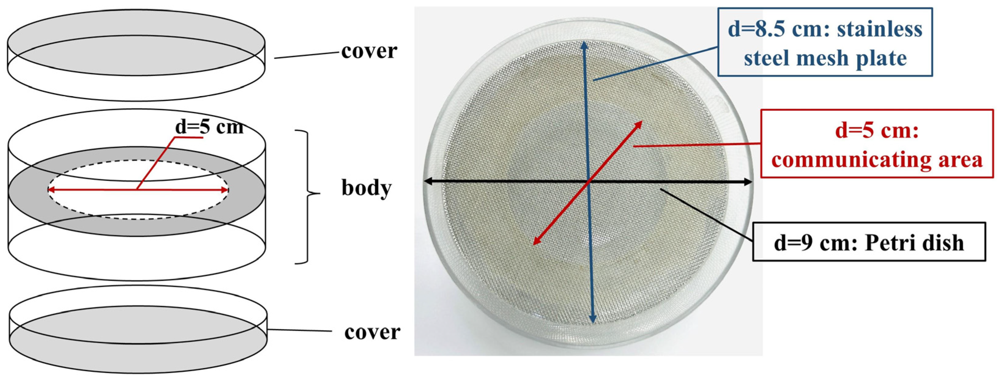
Based on the previous report [28], a double-sided Petri dish (DSPD) was prepared, which consists of a body (double-sided dish bottom) and two covers (Figure 2). The preparation process of DSPD is as follows: two resin Petri dish bottoms (d = 9 cm) were stuck together by glue, and a circular hole (d = 5 cm) was drilled in the center as communicating area. Then, a stainless steel mesh plate (40 mesh, d = 8.5 cm) was put into one side of the DSPD body to support the media. When DSPD was used, a sheet of sterile cellophane (d = 9 cm) was put onto the stainless steel mesh, and the media were poured into it. After the media solidified, the DSPD was reversed, and the same media were poured into another side of the DSPD body.

2.7. Co-Culture Methods for Fungi

Preparation of fungal inoculants: 5 mL M. purpureus spore suspension (106 spores/mL) was inoculated in RB media and incubated at 28 °C for 7 d at 120 rpm to prepare the inoculants of M. purpureus. The A. niger spore suspension was adjusted to 1 × 10⁵ spores/mL.
Morphological observations: 10 μL M. purpureus inoculant and A. niger spore suspension were inoculated on the center of RA media on each side of DSPD, respectively, and cultivated for 7 d at 28 °C. At the same time, 10 μL of M. purpureus inoculant and A. niger spore suspension was severally inoculated on the center of RA plate of normal Petri dish as the monoculture control, respectively. The colonial morphologies of M. purpureus and A. niger of monoculture and co-culture were observed and recorded on 3, 5, and 7 d, respectively. For microscopic morphological observations, 200 μL M. purpureus inoculant and A. niger spore suspension was spread on RA plates of each side of DSPD and normal Petri dish, respectively. Then, the microscopic morphology was observed after 7 days of cultivation.

2.8. Detection of Secondary Metabolites

The sterile cellophane was placed on an RA plate of DSPD and the normal Petri dish, then, 200 μL of M. purpureus inoculant and A. niger spore suspension was spread on the cellophane, respectively. After incubation at 28 °C for 7 d, the mycelia and media were collected and freeze-dried (FD8-5, Golden-SIM, Miami, FL, USA), respectively. Then, the dried samples were extracted with 80% methanol solvent at 40 °C for 30 min under ultrasonication (KQ2200DE, Kunshan Ultrasonic Instrument Co., Ltd., Kunshan, China), and centrifuged (Neofuge 18R, Heal Force, Shanghai, China) at 12,000 rpm for 10 min, and the supernatants were filtered through 0.22 µm membrane (Jinteng, Tianjin, China). Thereafter, MPs, MK, and CIT were detected, respectively, according to the previously reported methods [34,35]. MPs and MK were detected using high-performance liquid chromatography (HPLC, LC-20A, Shimadzu, Kyoto, Japan) with an ODS-3 (4.6 mm × 250 mm, 5 µm) column and an injection volume of 10 μL. CIT was analyzed using ultra-performance liquid chromatography (UPLC, ACPUITY Y UPLC I-class, Waters, Milford, MA, USA) with C18 column (Waters, 2.1 × 100 mm, 1.7 μm) and an injection volume of 2 μL. Acetonitrile and 0.1% formic acid were utilized as the mobile phase with gradient elution for MPs and CIT detection [28,36]. Acetonitrile and 0.5% phosphoric acid (60:40) were applied for MK detection [37]. The contents of MPs, MK and CIT, were expressed as their peak areas (AU·min or EU·min).

2.9. RT-qPCR

The expression levels of MPs and CIT biosynthetic genes in M. purpureus under both monoculture and co-culture were quantified by RT-qPCR analyses. Total RNA was extracted from mycelia cultured at 28 °C on RA for 5 d using the TransZol Up Plus RNA Kit (TransGen Biotech, Beijing, China). RNA was converted to cDNA using the HiScript II 1st Strand cDNA Synthesis Kit (Vazyme, Nanjing, China), with the beta-actin gene as an internal control (Table S5). RT-qPCR detection was performed using the AceQ qPCR SYBR Green Master Mix (Vazyme, Nanjing, China) [36]. The relative expression levels of genes relative to MPs and CIT biosynthesis were calculated following the previously described formula [38].

3. Results

3.1. Isolation and Classification of Fungal Strains from WYH Samples

Nine Wuyi Hongqu (WYH) samples (Figure 1) made by with the traditional handmade craft were collected from Fujian and Zhejiang Provinces in China, which are the main production areas of WYH [3]. Except Sample 8, the grains surface features of WYH samples exhibited uniform black–red (Sample 1), gray–black (Samples 2, 4, and 6), and deep black colors (Samples 3, 5, and 7), and the cross-section properties of WYH grains were black coat, red wall, and white heart (Figure 1).
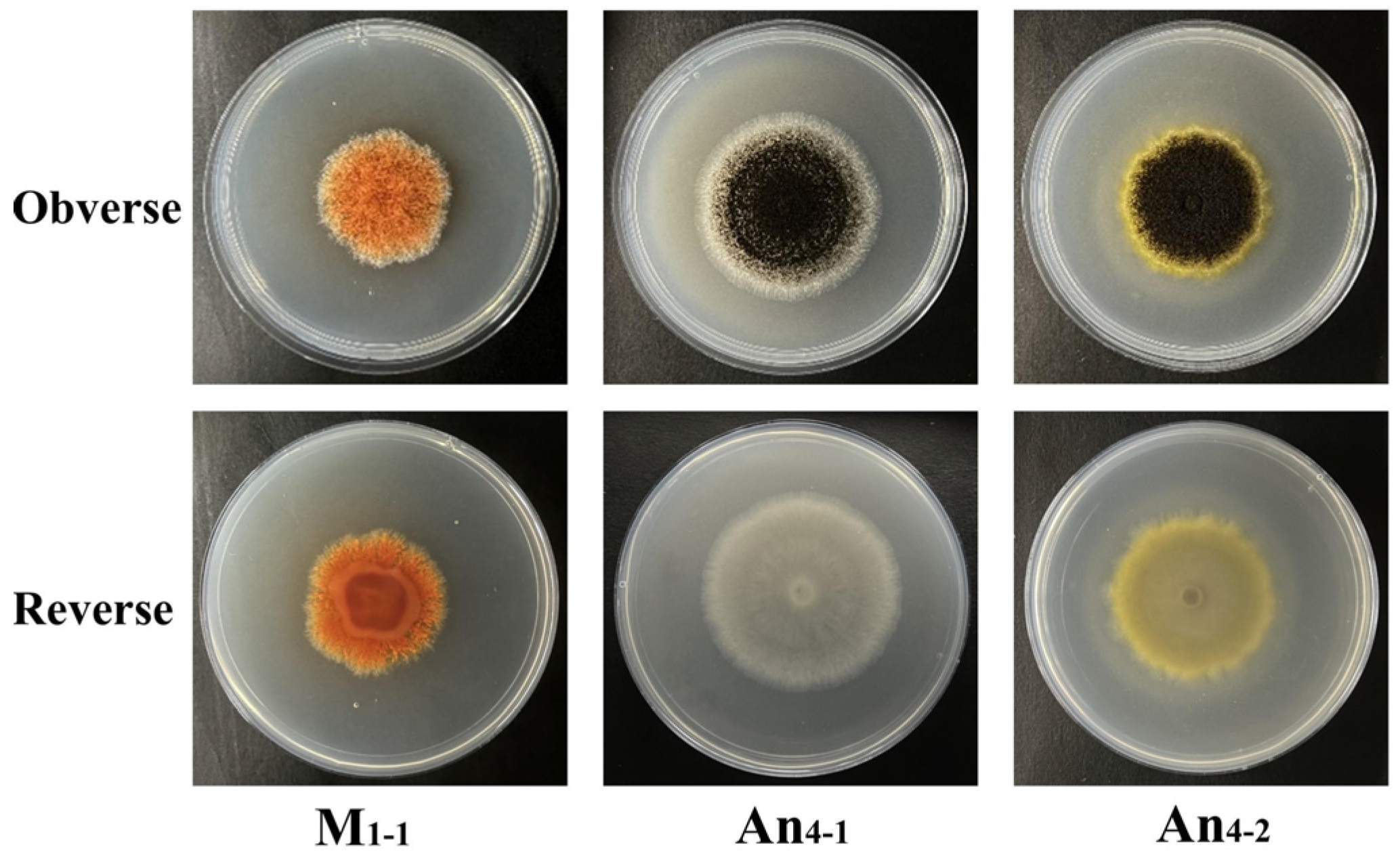
A total of 32 fungal strains were isolated from the WYH samples. Among them, 18 strains exhibited the characteristic morphologies of Monascus spp., while 14 strains exhibited morphologies consistent with Aspergillus niger. The colonial and microscopic morphologies of the fungal strains were observed and compared on PDA, CYA, MEA, G25N, and RA media, respectively, and the colonial and microscopic photos of these strains are shown in Figure S1, and their characters are described in Table S1. The colonial morphologies of the putative Monascus strains in above-mentioned different media were same as those reported in previous research [28,39], and putative Aspergillus strains were simply divided into pigment-producing Aspergillus strains and non-pigment-producing Aspergillus strains based on the colors of the margin and reverse sides of colonies (Figure S1). The morphologies of three typical strains of Monascus and A. niger are shown in Figure 3. Microscopic examination revealed slender, septate vegetative mycelia for putative Monascus strains, with abundant granules exhibiting orange, yellow and red colors, and cleistothecia and conidia formation was significantly influenced by media. For example, no cleistothecium was observed on CYA or G25N media (Figure S2). For putative Aspergillus strains, their conidiophores were mainly spherical and black, but some of them were orange–yellow and reddish–brown (Figure S2, Table S1).
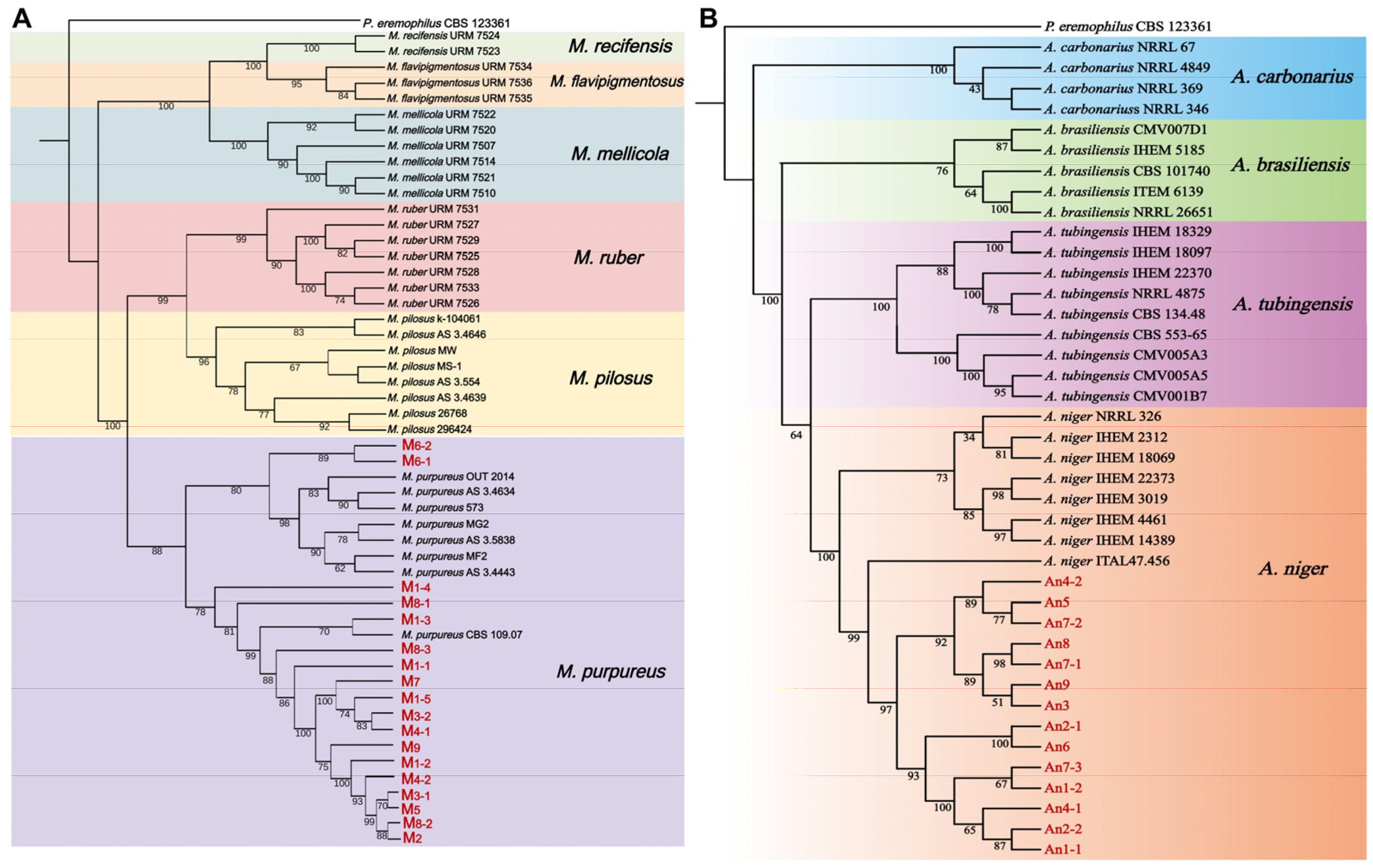
In order to classify and identify the isolated fungal strains, the ITS, LSU, BenA, CaM, and RPB2 genes from the isolated fungal strains from WYH samples were amplified and sequenced (Table S3), and multi-gene joint phylogenetic trees (Figure 4) were constructed to classify these strains through combining ITS-LSU-BenA-CaM-RPB2 gene sequences based on the DNA sequences from reference strains (Table S4) and the tested fungal strains. Due to the lack of LSU sequences from reference strains of Aspergillus spp. [31], the isolated putative Aspergillus strains were identified by ITS-BenA-CaM-RPB2 gene sequences. The results showed that all putative Monascus and Aspergillus strains were identified as M. purpureus and A. niger, respectively.

3.2. Morphological Properties and Biomass in Co-Culture of M. purpureus and A. niger

Based on the morphological characteristics of M. purpureus and A. niger (Table S1, Figure S1), six strains of M. purpureus and seven strains of A. niger from different WYH samples were selected. To study the effects of fungal interactions on growth and metabolism of M. purpureus, 14 groups of fungal co-culture experiments were conducted on RA media using DSPD. The combinations of M. purpureus and A. niger strains are shown in Table 1.
M. purpureus and A. niger were co-cultured on a double-sided Petri dish (DSPD), and their colonial and microscopic morphologies are shown in Figures S4 and S5, and their morphological characters are described in Table 2, and their biomasses are revealed in Figure 5. Compared with monoculture, the colonies of M. purpureus were smaller and orange in the co-culture (red in monoculture) and the numbers of conidia and cleistothecia were changed, while the colonies of A. niger after co-culture were larger than monoculture and there was no significant difference in micro-morphologies. Specially, when M. purpureus was co-cultured with pigment-producing A. niger, they possessed larger colonies than those with non-pigment-producing A. niger (Figure S5). After co-cultivation, the biomasses of most M. purpureus decreased slightly, but not significantly, while the biomasses of A. niger increased significantly (Figure 5). Among them, when M. purpureus was co-cultured with pigment-producing A. niger (An4-1 and An7-3), the biomasses of M. purpureus increased, especially in M. purpureus strains (M4-1 and M8-3) with weaker pigment-production capacities.

3.3. Co-Cultivation Effects on M. purpureus Secondary Metabolites

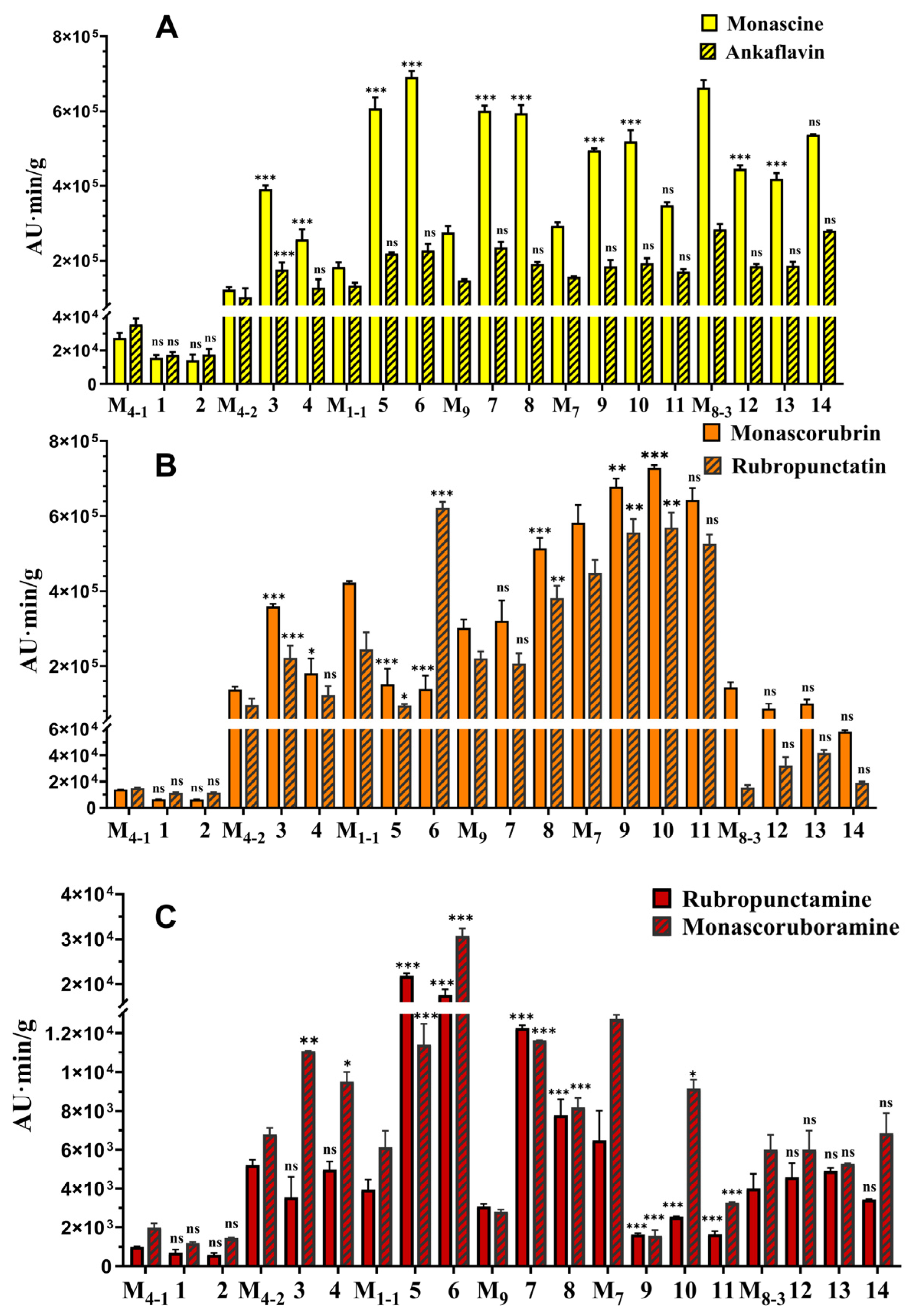
To further investigate the effects of A. niger on the secondary metabolites (SMs) of M. purpureus, its main SMs including Monascus pigments (MPs), monacolin K (MK), and citrinin (CIT) [28] were analyzed. The results (Figure 6) showed that after co-cultivation with A. niger for 7 d, all M. purpureus strains in monoculture and co-culture did not produce MK (the results not revealed), and the main six classic MPs including yellow pigments (YPs, Monascin and Ankaflavin), orange pigments (OPs, Rubropunctatin and Monascorubrin) and red pigments (RPs, Rubropunctamine and Monascorubramine) [12,40] were increased to varying degrees, except M4-1 and M8-3 (Figure 6). CIT detection revealed that after co-cultivation of M. purpureus and A. niger, both intracellular and extracellular CIT contents were significantly reduced, decreasing to 0–28.57% and 0–45.71%, compared to monoculture, respectively (Figure 7). Among them, when M. purpureus M1-1 and M9 were co-cultured with A. niger An1-2 and An9, respectively, they produced undetectable citrinin. Additionally, when co-cultured with non-pigment-producing A. niger, the pigment yields of M. purpureus significantly increased and the contents of CIT significantly decreased, but when co-cultured with pigment-producing A. niger, these changes were inhibited to a certain extent.

3.4. RT-qPCR for Analyzing Expression Levels of Genes Relative MPs and CIT Production

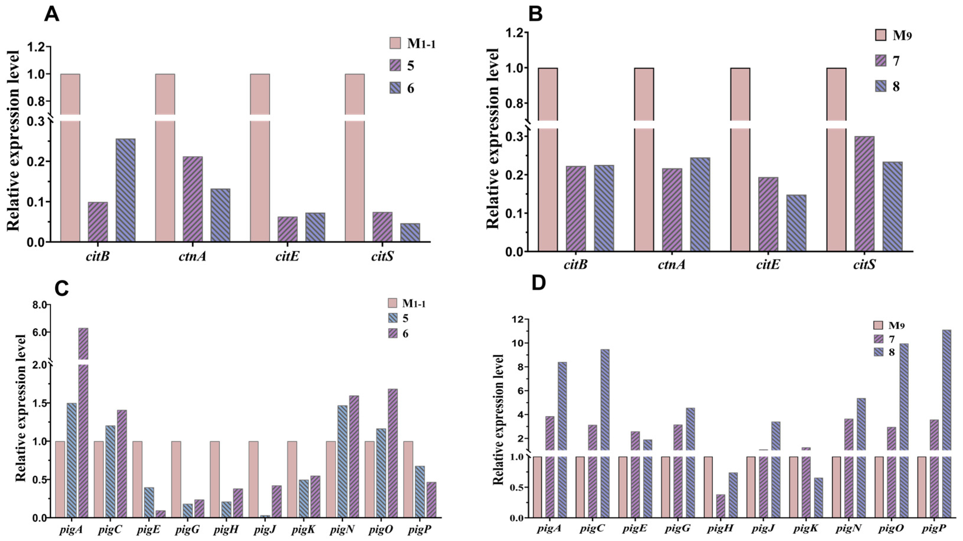
After co-culturing M. purpureus M1-1 and M9 with A. niger, respectively, they produced undetectable intracellular and extracellular CIT and significantly higher yields of MPs (Figure 6 and Figure 7). Therefore, the expression levels of key genes [12,40,41] in the CIT and MPs biosynthetic gene clusters of M. purpureus M1-1 and M9 in mono- and co-culture were analyzed and compared. As determined by RT-qPCR, the expression levels of key genes relative to CIT in strains M1-1 and M9 were significantly reduced after co-culture (Figure 8A,B). Meanwhile, the expression levels of MPs after co-culture of M1-1 and M9 increased and decreased to varying degrees (Figure 8C,D). The gene expression levels of M1-1 and M9 after co-culture were consistent with the previous results of CIT and MPs contents (Figure 6 and Figure 7).

4. Discussion

WYH is a fungal co-cultivation fermentation product mainly including Monascus spp. and Aspergillus niger, and is used in the production of yellow wine and Monascus vinegar in China [3,28]. Up to now, the co-cultivation of Monascus spp. and A. niger from WYH has had little research. A double-sided Petri dish (DSPD) provides an interface for communication between M. purpureus and A. niger, facilitating the separation of mycelia for metabolite analyses, and can also simulate the exchange state between these two fungi during WYH fermentation [28]. When co-cultured using DSPD, A. niger exhibited a significant effect on the growths (Figures S4 and S5) and metabolite productions (Figure 6 and Figure 7) of M. purpureus. However, co-cultivation using DSPD represents an indirect approach in which metabolites were exchanged through the media, whereas certain interactions may only occur during direct physical contact [42]. Therefore, various direct co-culture strategies, including the simulation of WYH fermentation on rice grains, are being investigated to further elucidate the interaction mechanisms between M. purpureus and A. niger.
As an important secondary metabolite of Monascus spp., Monacolin K (MK) was not detected in any of the examined M. purpureus strains examined. Previous genomic analyses have suggested that Monascus strains are unlikely to simultaneously produce MPs, MK, and CIT, as the complete biosynthetic gene clusters responsible for these metabolites rarely coexist within the same genome [43]. Furthermore, MK biosynthesis appeared to be species-dependent: M. ruber and M. pilosus possessed conserved and complete MK biosynthetic gene clusters, whereas M. purpureus usually lacked a complete MK biosynthetic gene cluster [44,45,46,47]. These findings are consistent with the results of the present study.
Previously, co-cultivation of M. ruber M7 with A. niger CBS 513.88 led to the discovery of two novel compounds [28]. In the present study, two similar compounds were also detected in the products of M. purpureus M1-1 co-cultured with A. niger, which are currently being isolated and characterized. These findings suggest that co-cultivation of M. purpureus with A. niger not only influences fungal growths and metabolic activities, but may also activate silent gene clusters to yield additional secondary metabolites. In the future, expanding fungal co-culture approaches may provide opportunities for the discovery of further novel secondary metabolites (SMs) [48]. It is noteworthy that analytical techniques such as high-performance liquid chromatography (HPLC) and mass spectrometry (MS) are commonly employed for metabolite detection in co-culture systems [23]. However, the concentrations of novel SMs produced under co-cultivation conditions are often relatively low, which may limit their detection by conventional instrumental methods. Therefore, enhancing SMs extraction efficiency and applying more sensitive and precise analytical platforms are recommended for subsequent investigations.
In this study, the growths and pigment-producing capacities of M. purpureus were found to exert a substantial influence on the outcomes of co-cultivation. For instance, strains M4-1 and M8-3, which exhibited relatively weak pigment production, showed a decline in pigment yields after co-cultivation. By contrast, strains with strong growth potentials, such as M1-1 and M9, displayed increased pigment production and decreased CIT (Figure 6 and Figure 7). These results highlight the critical importance of strain selection in WYH production. Specifically, the use of M. purpureus strains with strong pigment-producing abilities, in combination with non-pigmenting A. niger strains, is recommended to ensure high-quality and safe WYH products. In addition, MK, a lipid-lowering secondary metabolite produced by Monascus spp., represents another valuable target for strain selection. Thus, co-cultivation of Monascus strains with high MK biosynthetic potential and A. niger could provide a promising strategy for developing functional WYH in the future. Nevertheless, our previous experiments revealed that co-cultivation of the high MK-producing Monascus strain MS-1 with A. niger CBS 513.88 or CBS 113.46 failed to yield detectable levels of MK [28]. These observations suggest that further research is necessary to determine whether high MK-producing Monascus strains are suitable for WYH fermentation.
In addition, owing to the substantial variability among strains and the lack of a clear classification system for A. niger SMs [49], the impact of co-cultivation on A. niger SMs was not addressed in this study. Nevertheless, differences were observed in the effects of pigment-producing and non-pigment-producing A. niger strains on the SMs of M. purpureus (Figure 6). In particular, pigment-producing strains of A. niger (An4-2 and An7-3) appeared to suppress the increase in MPs and attenuate the decrease in CIT. Therefore, the principal SMs produced by pigment-producing and non-pigmenting A. niger strains are currently being isolated and identified to provide deeper insights into the mechanisms through which signaling molecules mediate interspecies interactions.

5. Conclusions

This study revealed the interaction between Monascus purpureus and Aspergillus niger through co-cultures of M. purpureus and A. niger isolated from WYH, and the effects of A. niger on the growths and metabolites of M. purpureus. Morphological results showed that M. purpureus and A. niger have significantly changed after co-cultivation. Metabolite analysis demonstrated that A. niger could promote the production of MPs and inhibits the production of CIT. The results of this study provide useful guidance for the selection of strains for production of WYH and serves as a good model for further research to enhance MPs production while reducing CIT levels.

Supplementary Materials

The following supporting information can be downloaded at: https://www.mdpi.com/article/10.3390/jof11120829/s1, Figure S1: Colonial morphologies of strains isolated from WYH on different media; Figure S2: Microscopic morphologies of Monascus spp. and Aspergillus spp. isolated from WYH; Figure S3: Agarose electrophoresis of PCR amplification products; Figure S4: Colonial morphologies of M. purpureus and A. niger co-cultured using DSPD on RA media at 28 °C; Figure S5: Microscopic morphologies of M. purpureus and A. niger co-cultured using DSPD on RA media at 28 °C; Figure S6: HPLC chromatography diagram of MPs; Figure S7: HPLC chromatography diagram of Monascus red pigments; Figure S8: UPLC chromatography diagram of intracellular CIT; Figure S9: UPLC chromatography diagram of extracellular CIT; Figure S10: Total RNA of 6 samples for RNA in 1% agarose gel electrophoresis; Table S1: The colonial and microscopic morphologies of putative Monascus and Aspergillus strains on different media; Table S2: Primers for gene amplification and fragment length; Table S3: GenBank accession No. of M. purpureus and A. niger strains; Table S4: Strains and sequences used in molecular study; Table S5: Primers for RT-qPCR of Monascus MPs and CIT biosynthetic genes.

Author Contributions

Conceptualization: Q.Y.; methodology: Q.Y. and X.Y.; resources: F.C.; writing—original draft preparation: Q.Y.; writing—review and editing: Q.Y., X.Y. and F.C.; visualization: Q.Y.; supervision: F.C. and X.Y.; project administration: F.C.; funding acquisition: F.C. All authors have read and agreed to the published version of the manuscript.

Funding

This research was supported by the Technology Innovation Plan Project of Hubei Province. (No. 2024BCB024: to F.C.) and the Doctoral Research Fund of Wuhan Business University (Grant No. 2024KB011).

Institutional Review Board Statement

Not applicable.

Informed Consent Statement

Not applicable.

Data Availability Statement

The original contributions presented in this study are included in the article/Supplementary Materials. Further inquiries can be directed to the corresponding authors.

Conflicts of Interest

The authors declare no conflicts of interest.

References

  1. Zhao, W.; Qian, M.; Dong, H.; Liu, X.; Bai, W.; Liu, G.; Lv, X. Effect of Hong Qu on the flavor and quality of Hakka yellow rice wine (Huangjiu) produced in Southern China. LWT 2022, 160, 113264. [Google Scholar] [CrossRef]
  2. Wang, J.; Yuan, C.; Gao, X.; Kang, Y.; Huang, M.; Wu, J.; Liu, Y.; Zhang, J.; Li, H.; Zhang, Y. Characterization of key aroma compounds in Huangjiu from northern China by sensory-directed flavor analysis. Food Res. INT 2020, 134, 109238. [Google Scholar] [CrossRef]
  3. Mao, Q.; Tu, X.; Jia, X.; Zhou, L. The fermentationtechnology of black-skin-red-koji and the brewing of indica rice wine. Jiangsu Condiment Subsid. Food 2011, 28, 28–31, 40. (In Chinese) [Google Scholar]
  4. Huang, Y.Y.; Liang, Z.C.; Lin, X.Z.; He, Z.G.; Ren, X.Y.; Li, W.X.; Molnár, I. Fungal community diversity and fermentation characteristics in regional varieties of traditional fermentation starters for Hong Qu glutinous rice wine. Food Res. Int. 2021, 141, 110146. [Google Scholar] [CrossRef]
  5. Lv, X.C.; Huang, R.L.; Chen, F.; Zhang, W.; Rao, P.F.; Ni, L. Bacterial community dynamics during the traditional brewing of Wuyi Hong Qu glutinous rice wine as determined by culture-independent methods. Food Control 2013, 34, 300–306. [Google Scholar] [CrossRef]
  6. Park, K.H.; Liu, Z.; Park, C.S.; Ni, L. Microbiota associated with the starter cultures and brewing process of traditional Hong Qu glutinous rice wine. Food Sci. Biotechnol. 2016, 25, 649–658. [Google Scholar] [CrossRef] [PubMed]
  7. Lv, X.C.; Huang, Z.Q.; Zhang, W.; Rao, P.F.; Ni, L. Identification and characterization of filamentous fungi isolated from fermentation starters for Hong Qu glutinous rice wine brewing. J. Gen. Appl. Microbiol. 2012, 58, 33–42. [Google Scholar] [CrossRef]
  8. Arruda, G.L.; Reis, W.S.M.; Raymundo, M.T.F.R.; Shibukawa, V.P.; Cruz-Santos, M.M.; Silos, N.O.; Prado, C.A.; Marcelino, P.R.F.; Silva, S.S.; Santos, J.C. Biotechnological potential of Monascus: Biological aspects, metabolites of interest, and opportunities for new products. Microbiol. Res. 2025, 297, 128177. [Google Scholar] [CrossRef] [PubMed]
  9. Farawahida, A.H.; Palmer, J.; Flint, S. Monascus spp. and citrinin: Identification, selection of Monascus spp. isolates, occurrence, detection and reduction of citrinin during the fermentation of red fermented rice. Int. J. Food Microbiol. 2022, 379, 109829. [Google Scholar] [CrossRef]
  10. Feng, Y.; Shao, Y.; Chen, F. Monascus pigments. Appl. Microbiol. Biot. 2012, 96, 1421–1440. [Google Scholar] [CrossRef]
  11. Husakova, M.; Patakova, P. Purified Monascus pigments: Biological activities and mechanisms of action. J. Nat. Prod. 2025, 88, 607–615. [Google Scholar] [CrossRef]
  12. Chen, W.; Feng, Y.; Molnár, I.; Chen, F. Nature and nurture: Confluence of pathway determinism with metabolic and chemical serendipity diversifies Monascus azaphilone pigments. Nat. Prod. Rep. 2019, 36, 561–572. [Google Scholar] [CrossRef]
  13. Dai, W.; Shao, Y.; Chen, F. Production of Monacolin K in Monascus pilosus: Comparison between industrial strains and analysis of its gene klusters. Microorganisms 2021, 9, 747. [Google Scholar] [CrossRef] [PubMed]
  14. Zhang, Y.; Chen, Z.; Wen, Q.; Xiong, Z.; Cao, X.; Zheng, Z.; Zhang, Y.; Huang, Z. An overview on the biosynthesis and metabolic regulation of monacolin K/lovastatin. Food Func. 2020, 11, 5738–5748. [Google Scholar] [CrossRef] [PubMed]
  15. Gui, Y.; Tang, G.; Man, H.; Wang, J.; Han, J.; Zhao, J. Transportation of citrinin is regulated by the CtnC gene in the medicinal fungus Monascus purpureus. J. Zhejiang Univ. Sci. B 2023, 24, 543–548. [Google Scholar] [CrossRef]
  16. Shi, J.; Zhao, W.; Lu, J.; Wang, W.; Yu, X.; Feng, Y. Insight into Monascus pigments production promoted by glycerol based on physiological and transcriptome analyses. Process Biochem. 2021, 102, 141–149. [Google Scholar] [CrossRef]
  17. Zhen, Z.; Xiong, X.; Liu, Y.; Zhang, J.; Wang, S.; Li, L.; Gao, M. NaCl inhibits citrinin and stimulates Monascus pigments and Monacolin K production. Toxins 2019, 11, 118. [Google Scholar] [CrossRef]
  18. Yang, X.; Xiang, L.; Zhang, C.; Cao, Y.; Wang, C. Promotion of monacolin K production in Monascus extractive fermentation: The variation in fungal morphology and in the expression levels of biosynthetic gene clusters. J. Sci. Food Agric. 2021, 101, 5652–5659. [Google Scholar] [CrossRef] [PubMed]
  19. Zhang, C.; Liang, J.; Yang, L.; Chai, S.; Zhang, C.; Sun, B.; Wang, C. Glutamic acid promotes monacolin K production and monacolin K biosynthetic gene cluster expression in Monascus. AMB Express 2017, 7, 22. [Google Scholar] [CrossRef]
  20. Zhang, H.; Ahima, J.; Yang, Q.; Zhao, L.; Zhang, X.; Zheng, X. A review on citrinin: Its occurrence, risk implications, analytical techniques, biosynthesis, physiochemical properties and control. Food Res. Int. 2021, 141, 110075. [Google Scholar] [CrossRef]
  21. Li, Y.Z.; Zhang, W.Q.; Hu, P.F.; Yang, Q.; Molnár, I.; Xu, P.; Zhang, B. Harnessing microbial co-culture to increase the production of known secondary metabolites. Nat. Prod. Rep. 2025, 42, 623–637. [Google Scholar] [CrossRef]
  22. Xu, S.; Li, M.; Hu, Z.; Shao, Y.; Ying, J.; Zhang, H. The potential use of fungal co-culture strategy for discovery of new secondary metabolites. Microorganisms 2023, 11, 464. [Google Scholar] [CrossRef]
  23. Knowles, S.L.; Raja, H.A.; Roberts, C.D.; Oberlies, N.H. Fungal–fungal co-culture: A primer for generating chemical diversity. Nat. Prod. Rep. 2022, 39, 1557–1573. [Google Scholar] [CrossRef]
  24. Tomm, H.A.; Ucciferri, L.; Ross, A.C. Advances in microbial culturing conditions to activate silent biosynthetic gene clusters for novel metabolite production. J. Ind. Microbiol. Biot. 2019, 46, 1381–1400. [Google Scholar] [CrossRef] [PubMed]
  25. Meng, Z.; Tan, Y.; Duan, Y.L.; Li, M. Monaspin B a novel cyclohexyl-furan from cocultivation of Monascus purpureus and Aspergillus oryzae, exhibits potent antileukemic activity. J. Agric. Food Chem. 2024, 72, 1114–1123. [Google Scholar] [CrossRef] [PubMed]
  26. El-sayed, A.S.A.; Shindia, A.A.; Abouzeid, A.; Koura, A.; Hassanein, S.E.; Ahmed, R.M. Triggering the biosynthetic machinery of Taxol by Aspergillus flavipes via cocultivation with Bacillus subtilis: Proteomic analyses emphasize the chromatin remodeling upon fungal-bacterial interaction. Environ. Sci. Pollut. Res. 2021, 28, 39866–39881. [Google Scholar] [CrossRef] [PubMed]
  27. Ju, M.; Zhang, J.; Mai, T.; Li, L.; Gu, T.; Liu, Y.; Gao, M. Co-culture of Rhodotorula mucilaginosa and Monascus purpureus increased the yield of carotenoids and Monascus pigments. LWT 2023, 183, 114949. [Google Scholar] [CrossRef]
  28. Yuan, X.; Chen, F. Cocultivation study of Monascus spp. and Aspergillus niger inspired from black-skin-red-koji by a double-sided petri dish. Front. Microbiol. 2021, 12, 670684. [Google Scholar] [CrossRef]
  29. Zheng, C. Isolation of High-Yield Esterase and Non-Citrinin Monascus Strain and Its Esterase Production Condition Optimation. Master’s Thesis, Huazhong Agricultural University, Wuhan, China, 2017. (In Chinese). [Google Scholar]
  30. Feng, Y.; Shao, Y.; Zhou, Y.; Chen, F. Monacolin K production by citrinin-free Monascus pilosus MS-1 and fermentation process monitoring. Eng. Life Sci. 2014, 14, 538–545. [Google Scholar] [CrossRef]
  31. Bian, C.; Kusuya, Y.; Sklenář, F.; D’hooge, E.; Yaguchi, T.; Ban, S.; Visagie, C.M.; Houbraken, J.; Takahashi, H.; Hubka, V. Reducing the number of accepted species in Aspergillus series. Nigri. Stud. Mycol. 2022, 102, 95–132. [Google Scholar] [CrossRef]
  32. Barbosa, R.N.; Leong, S.L.; Vinnere-Pettersson, O.; Chen, A.J.; Souza-Motta, C.M.; Frisvad, J.C.; Samson, R.A.; Oliveira, N.T.; Houbraken, J. Phylogenetic analysis of Monascus and new species from honey, pollen and nests of stingless bees. Stud. Mycol. 2017, 86, 29–51. [Google Scholar] [CrossRef]
  33. Zhang, D.; Gao, F.; Jakovlić, I.; Zhou, H.; Zhang, J.; Li, W.; Wang, G. PhyloSuite: An integrated and scalable desktop platform for streamlined molecular sequence data management and evolutionary phylogenetics studies. Mol. Ecol. Resour. 2020, 20, 348–355. [Google Scholar] [CrossRef]
  34. Wang, X.; Wei, J.; Tang, F.; Chen, F. Effects of blue light on pigment and citrinin production in Monascus ruber M7 via MrcreD, encoding an arrestin-like protein. Int. J. Biol. Macromol. 2025, 288, 138604. [Google Scholar] [CrossRef] [PubMed]
  35. Li, L.; Chen, S.; Gao, M.; Ding, B.; Zhang, J.; Zhou, Y.; Liu, Y.; Yang, H.; Wu, Q.; Chen, F. Acidic conditions induce the accumulation of orange Monascus pigments during liquid-state fermentation of Monascus ruber M7. Appl. Microbiol. Biot. 2019, 103, 8393–8402. [Google Scholar] [CrossRef] [PubMed]
  36. Zheng, Y.; Huang, Y.; Mao, Z.; Shao, Y. Histone deacetylase MrRpd3 plays a major regulational role in the mycotoxin production of Monascus ruber. Food Control 2022, 132, 108457. [Google Scholar] [CrossRef]
  37. Yao, T.; Wang, X.; Chen, F. The role of enoyl reductase in the Monacolin K biosynthesis pathway in Monascus spp. J. Fungi 2025, 11, 199. [Google Scholar] [CrossRef] [PubMed]
  38. Livak, K.J.; Schmittgen, T.D. Analysis of relative gene expression data using real-time quantitative PCR and the 2−ΔΔCT method. Methods 2001, 25, 402–408. [Google Scholar] [CrossRef]
  39. Tang, F.; Zhao, Y.; Mao, Z.; Huang, Y.; Hu, Y.; Liu, B.; Huang, Y.; Shao, Y. Consistency of gene expression and protein acetylation modification with physiological and biochemical changes responding to the deficiency of histone deacetylase 1 homolog in Monascus ruber. Int. J. Biol. Macromol. 2025, 308, 142485. [Google Scholar] [CrossRef]
  40. Chen, W.; Chen, R.; Liu, Q.; He, Y.; He, K.; Ding, X.; Kang, L.; Guo, X.; Xie, N.; Zhou, Y.; et al. Orange, red, yellow: Biosynthesis of azaphilone pigments in Monascus fungi. Chem. Sci. 2017, 8, 4917–4925. [Google Scholar] [CrossRef]
  41. He, Y.; Cox, R.J. The molecular steps of citrinin biosynthesis in fungi. Chem. Sci. 2016, 7, 2119–2127. [Google Scholar] [CrossRef]
  42. Okada, B.K.; Seyedsayamdost, M.R. Antibiotic dialogues: Induction of silent biosynthetic gene clusters by exogenous small molecules. FEMS Microbiol. Rev. 2017, 41, 19–33. [Google Scholar] [CrossRef] [PubMed]
  43. Liu, A.; Chen, A.J.; Liu, B.; Wei, Q.; Bai, J.; Hu, Y.C. Investigation of citrinin and monacolin K gene clusters variation among pigment producer Monascus species. Fungal Genet. Biol. 2022, 160, 103687. [Google Scholar] [CrossRef] [PubMed]
  44. Lin, T.S.; Chiu, S.H.; Chen, C.C.; Lin, C.H. Investigation of monacolin K, yellow pigments, and citrinin production capabilities of Monascus purpureus and Monascus ruber (Monascus pilosus). J. Food Drug Anal. 2023, 31, 85–94. [Google Scholar] [CrossRef] [PubMed]
  45. Higa, Y.; Kim, Y.S.; Amin, A.U.; Huang, M.; Ono, N.; Kanaya, S. Divergence of metabolites in three phylogenetically close Monascus species (M. pilosus, M. ruber, and M. purpureus) based on secondary metabolite biosynthetic gene clusters. BMC Genom. 2020, 21, 679. [Google Scholar] [CrossRef]
  46. Kwon, H.J.; Balakrishnan, B.; Kim, Y.K. Some Monascus purpureus genomes lack the Monacolin K biosynthesis locus. J. Appl. Biol. Chem. 2016, 59, 45–47. [Google Scholar] [CrossRef]
  47. Chen, Y.P.; Yuan, G.F.; Hsieh, S.Y.; Lin, Y.S.; Wang, W.Y.; Liaw, L.L.; Tseng, C.P. Identification of the mokH gene encoding transcription factor for the upregulation of monacolin K biosynthesis in Monascus pilosus. J. Agric. Food Chem. 2010, 58, 287–293. [Google Scholar] [CrossRef]
  48. Alanzi, A.; Elhawary, E.A.; Ashour, M.L.; Moussa, A.Y. Aspergillus co-cultures: A recent insight into their secondary metabolites and microbial interactions. Arch. Pharm. Res. 2023, 46, 273–298. [Google Scholar] [CrossRef]
  49. Yu, R.; Liu, J.; Wang, Y.; Wang, H.; Zhang, H. Aspergillus niger as a secondary metabolite factory. Front. Chem. 2021, 9, 701022. [Google Scholar] [CrossRef]
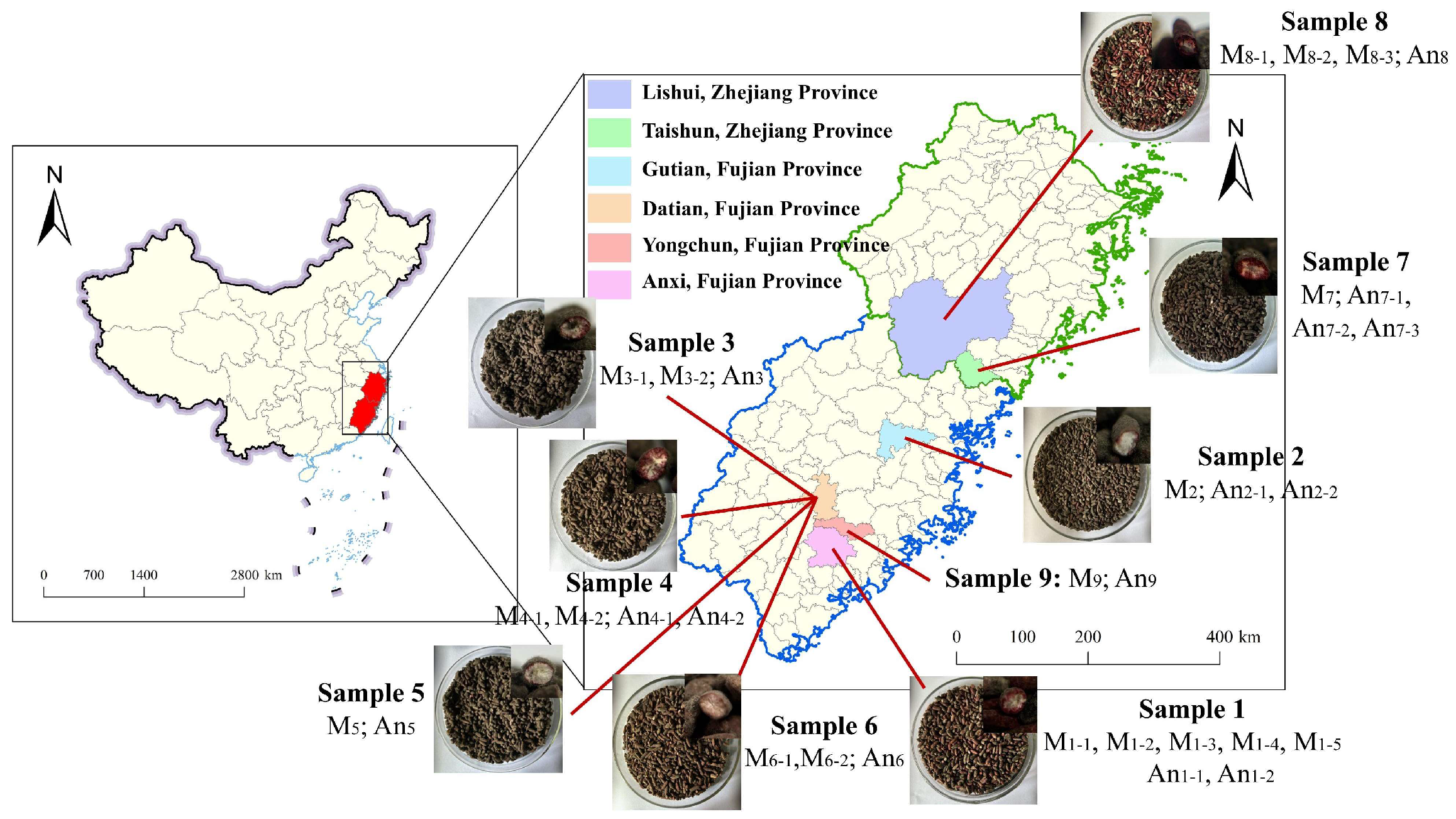
Figure 1. WYH samples were collected from Fujian and Zhejiang provinces of China *. *: The upper right corner of the WYH images is the cross-section of WYH grains; the M9 and An9 strains were isolated by our previous study and preserved in our laboratory; M1-1~M9: M. purpureus strains; An1-1~An9: A. niger strains.
Jof 11 00829 g001
Figure 2. The schematic diagram of the structural composition of a double-sided Petri dish.
Jof 11 00829 g002
Figure 3. Colonial morphologies of typical putative Monascus and Aspergillus strains isolated from WYH. M1-1 was typical putative Monascus strain, An4-1 was the non-pigment-producing Aspergillus strain, and A4-2 was the pigment-producing Aspergillus strain. The putative Monascus and Aspergillus strains in the above colonies were cultured at 28 °C for 9 d and 5 d, respectively.
Jof 11 00829 g003
Figure 4. Phylogenetic trees based on multiple genes of the putative Monascus and Aspergillus strains. (A): Phylogenetic tree of Monascus strains based on ITS-LSU-BenA-CaM-RPB2; (B): phylogenetic tree of Aspergillus strains based on ITS-BenA-CaM-RPB2.
Jof 11 00829 g004
Figure 5. Biomasses of M. purpureus and A. niger after 7 d of co-cultivation on RA media. (A): Biomass of M. purpureus; (B): biomass of A. niger. The data are represented as the mean ± standard error (n = 3). * p < 0.05, ** p < 0.01, *** p < 0.005, ns: not significant compared to monoculture.
Jof 11 00829 g005
Figure 6. MPs produced by M. purpureus co-cultured with A. niger on RA media for 7 d. (A): M. purpureus YPs; (B): M. purpureus OPs; (C): M. purpureus RPs. The data were represented as the mean ± standard error (n = 3). * p < 0.05, ** p < 0.01, *** p < 0.005, ns: not significant compared to monoculture. AU: Absorbance Unit, AU·min: peak area.
Jof 11 00829 g006
Figure 7. CIT produced by M. purpureus co-cultured with A. niger on RA media for 7 d. (A): Intracellular CIT; (B): extracellular CIT. EU: Electrical Unit, EU·min: peak area.
Jof 11 00829 g007
Figure 8. Expression levels of CIT and MPs biosynthetic-related genes of M. purpureus M1-1 and M9, under monoculture and co-culture with A. niger An1-2 and An9. (A,B): CIT gene expression levels; (C,D): MPs gene expression levels. Except for the genes in the figure, other genes were poorly expressed or unexpressed and are not shown in the results. Here, 5: M1-1 and An1-2, 6: M1-1 and An9, 7: M9 and An1-2, and 8: M9 and An9.
Jof 11 00829 g008
Table 1. The co-culture experimental combination for M. purpureus and A. niger.
No. of Co-Culture Strain NamesStrain SourcesPigment-Production AbilitiesGrowth Abilities
1M4-1 & An4-1Sample 4M4-1(+)M4-1(+)
2M4-1 & An4-2M4-2(++)M4-2(++)
3M4-2 & An4-1An4-1(−)An4-1(+++)
4M4-1 & An4-2An4-2(+)An4-2(++)
5M1-1 & An1-2Sample 1 and Sample 9M1-1(++)M1-1(++)
6M1-1 & An9M9(++)M9(++)
7M9 & An1-2An1-2(−)An1-2(+++)
8M9 & An9An9(−)An9(+++)
9M7 & An7-1Sample 7 and Sample 8M7(++)
M8-3(+)
An7-1(−)
An7-3(+)
An8(−)
M7(++)
M8-3(+)
An7-1(+++)
An7-3(++)
An8(+++)
10M7 & An7-3
11M7 & An8
12M8-3 & An7-1
13M8-3 & An7-3
14M8-3 & An8
+: indicates pigment production, and the number of ‘+’ symbols represents the relative strength of pigment-producing and growth abilities; −: indicates no pigment production.
Table 2. The colonial and microscopic morphologies of M. purpureus and A. niger on co-culture using DSPD on RA media.
Strain TypesCulture TypesColonial SizesColonies ColorsAerial HyphaePigment Granules ShapesCleistothecia NumberConidia Number
M. purpureusMonoculture++red+Combs. 1, 2, 10, 11: block-like to needle-like; Comb. 5: needle -like to block -likeCombs. 2, 12, 13: increased.Combs. 7, 8: increased; Com3,4: decreased.
Co-culture+orange or yellow++
A. nigerMonoculture++black////
Co-culture+++black////
+: the number indicates colonies size or aerial hyphae relative change of monoculture and co-culture; /: no relevant morphological characteristics or no significant change.
Disclaimer/Publisher’s Note: The statements, opinions and data contained in all publications are solely those of the individual author(s) and contributor(s) and not of MDPI and/or the editor(s). MDPI and/or the editor(s) disclaim responsibility for any injury to people or property resulting from any ideas, methods, instructions or products referred to in the content.