Article

Unveiling the Unique Mitogenome Structure of Phylloporus: Implications for Phylogeny and Evolution in Boletaceae

Key Laboratory of Natural Microbial Medicine Research of Jiangxi Province, College of Life Science, Jiangxi Science and Technology Normal University, Nanchang 330013, China
*
Authors to whom correspondence should be addressed.
J. Fungi 2025, 11(12), 831; https://doi.org/10.3390/jof11120831
Submission received: 15 October 2025 / Revised: 18 November 2025 / Accepted: 20 November 2025 / Published: 25 November 2025

Abstract

The genus Phylloporus (Boletaceae, Boletales) is a group of ectomycorrhizal fungi, distinguished from other members of Boletaceae by its unique lamellate hymenophore. Although some molecular data exist for this genus, its mitogenomic characteristics remain poorly understood. In our study, we sequenced, assembled, and annotated the complete mitogenomes of eight species representing seven major subclades of Boletaceae collected in Jiangxi Province, China, with a focus on four Phylloporus species. We found that Phylloporus mitogenomes are circular, ranging in size from 35,117 bp to 38,908 bp, and contain 14–15 core protein-coding genes (PCGs), 24–28 tRNA genes, and 2 rRNA genes. Our comparative analysis revealed that Phylloporus species share many features, such as gene content, gene length, tRNA repertoire, and gene order, while Boletaceae as a whole shows a lot of diversity. Codon usage patterns are quite similar across the family. The Ka/Ks ratios of most 15 core PCGs were less than 1, suggesting these genes have been preserved through purifying selection over time. By using Bayesian inference (BI) and maximum likelihood (ML) methods and combining 28 other mitotic genomes in the NCBI database, our phylogenetic analysis produced highly consistent and well-supported trees (BPP ≥ 0.98, BS ≥ 71). It is noted that this family is divided into seven subfamilies, which is consistent with previous taxonomic studies. Altogether, our findings shed light on the unique features of Phylloporus and its connections to other members of Boletaceae. These findings not only provide valuable insights into the taxonomy, phylogeny, genetic diversity, and resource conservation of Boletaceae but also serve as a valuable genomic resource for future research.

1. Introduction

The genus Phylloporus was established by Quélet in 1888 [1], with Ph. pelletieri (Lév.) Quél. as type species. This genus is distinguished from other members of the family Boletaceae by its lamellate hymenophore. Phylloporus species are common edible and medicinal fungi that form ectomycorrhizal associations with both coniferous and broad-leaved trees [2]. This symbiotic relationship plays a crucial role in maintaining the stability of global forest ecosystems and the carbon cycle [3,4].
Phylloporus is widely distributed in tropical, subtropical, and temperate regions of the Northern Hemisphere. Jiangxi Province, with a subtropical humid climate in southern China [5], is suitable for the growth and reproduction of macrofungi. A total of 84 species belonging to 27 genera of Boletaceae have been reported here, including six lamellate boletes [6,7]. In recent years, we collected four Phylloporus species from Jiangxi Province.
The mitochondrial genome is becoming an important tool for studying species origin, evolution, and classification due to its high copy number, low mutation rate, and rapid evolutionary rate [8]. For example, the structural characteristics, gene sequences, and genetic information from protein-coding genes of mitochondrial genomes can serve as a potential alternative tool for fungal taxonomy and phylogenetic analysis [9,10]. Therefore, determining the mitogenome sequence and analyzing its basic features is of pivotal importance [11,12]. Indeed, although there are about forty thousand species of basidiomycetes, fewer than 120 fungal mitogenomes have been deposited in public databases such as GenBank, the Joint Genome Institute, and the European Nucleotide Archive [13]. Consequently, the mitochondrial characteristics of Phylloporus species remain unclear.
In this study, five specimens of Phylloporus collected from Jiangxi Province, China, were subjected to second-generation sequencing, assembly, and annotation. By analyzing the structural features of their mitogenomes, protein-coding genes, codon usage bias, and other characteristics, we aim to reveal their genetic traits and evolutionary adaptations. Furthermore, five specimens from Boletaceae (except for Phylloporus) were also newly sequenced. Combining those with 26 Boletaceae species available in GenBank and using Paxillaceae as an outgroup, a phylogenetic framework based on 28 mitogenomes was constructed to confirm the phylogenetic position of the genus Phylloporus.

2. Materials and Methods

2.1. Sample Collection and DNA Extraction

Nine specimens belonging to eight species of Boletaceae were collected from Jiangxi Province, China. They were first identified by their macroscopic characteristics, such as the pileus, stipe, and hymenophore, and their microscopic characteristics, such as the size of the spores. Additionally, molecular analyses further confirm their identity. The total DNA was extracted from basidiocarp tissues using the cetyltrimethylammonium bromide (CTAB) method [13]. The specimens are stored at the Cryptogamic Herbarium, Kunming Institute of Botany, Chinese Academy of Sciences (HKAS). The species names are as follows: Phylloporus brunneiceps, Phylloporus grossus, Phylloporus rubrosquamosus, Phylloporus luxiensis, Austroboletus fusisporus, Chalciporus radiatus, Chiua viridula, Zangia olivaceobrunnea; their corresponding specimen collection numbers are HKAS1059–HKAS1064 and HKAS105268–HKAS105270. They were successfully sequenced. Sequencing was performed by Sangon Biotech Co., Ltd. (Shanghai, China) to generate high-quality sequence reads.

2.2. Mitogenomes Assembly and Annotation

GetOrganelle v.1.6.2 software [14] was used for genome assembly with the fungal database (-F fungus_mt) to identify, filter, and assemble target-related reads. Annotation was conducted using the MITOS web server with the genetic code table 4 [15]. Annotated protein-coding genes (PCGs) were further optimized using the NCBI Open Reading Frame (ORF) Finder (https://www.ncbi.nlm.nih.gov/orffinder/, accessed on 5 December 2024). tRNA genes were identified using tRNAscan-SE v1.3.1 [16]. Intron types (if found) were verified with RNAweasel v5.2.1 [17]. Gene maps were generated using OGDRAW v1.3.1 [18]. tRNA genes were analyzed using the software tRNAscan-SE v.2.0.9 [19], and the secondary structures of tRNA genes were drawn with the software VARNA v.3-93 [20].

2.3. Sequence Analysis

The GC content of mitogenomes was calculated using Geneious v9.0.2. Synteny analysis was performed with the built-in Mauve plugin [21]. Start and stop codons of PCGs were identified using PhyloSuite v1.2.3 [22]. Base composition of the nine Boletaceae mitogenomes was analyzed with DNASTAR Lasergene v7.1 (http://www.dnastar.com/, accessed on 20 December 2024). Strand asymmetry was calculated as follows: AT skew = (A − T)/(A + T) and GC skew = (G − C)/(G + C) [23]. BLASTN searches were conducted to detect interspersed repeats or large intragenomic duplications (https://blast.ncbi.nlm.nih.gov/Blast.cgi/, accessed on 20 December 2024). Tandem repeats were identified using Tandem Repeats Finder [24]. Furthermore, TBtools-II and DnaSP v6 [25] were used to estimate the nonsynonymous (Ka) and synonymous (Ks) substitution rates of 15 core PCGs (atp6, atp8, atp9, cob, cox1, cox2, cox3, nad1, nad2, nad3, nad4, nad4L, nad5, nad6, and rps3) across the nine mitogenomes.

2.4. Phylogenetic Analysis

Mitochondrial genomes of nine specimens from Boletaceae, including five Phylloporus specimens, were newly sequenced for this study. Additionally, 19 publicly available mitogenomes were retrieved from NCBI based on previous studies [26,27,28,29,30], comprising 17 samples from Boletaceae and two from Paxillaceae (order Boletales) designated as the outgroup (Table 1). Phylogenetic analysis was performed using PhyloSuite v1.2.3 [22,31]. Fifteen core protein-coding genes (PCGs) were extracted and individually aligned with MAFFT as implemented in PhyloSuite [32]. The resulting alignments were trimmed, respectively, by the Gblock tool and then integrated into a single matrix using the sequence concatenation tool in PhyloSuite. The final concatenated alignment had a total length of 14,131 bp, containing 2016 parsimony-informative sites. Through the PartitionFinder2 software, by using the greedy search algorithm and the associated branch length and based on the Bayesian Information Criterion (BIC) and the modified Akaike Information Criterion (AICc), the GTR + I + G model was determined as the single partition and the best nucleotide replacement model. Phylogenetic trees were reconstructed using both Bayesian Inference (BI) and Maximum Likelihood (ML) methods. The BI analysis was conducted with two independent runs of four Markov chain Monte Carlo chains each for 1,000,000 generations, sampling every 1000 generations. The average standard deviation of split frequencies reached 0.005 (<0.01), indicating satisfactory convergence. Posterior probabilities (PP) were calculated for each node after discarding 25% of the samples as burn-in. The ML tree was inferred with IQ-TREE [33] within PhyloSuite, employing ultrafast bootstrap approximation with 10,000 replicates under an edge-linked partition model [34,35] to assess branch support. Final trees were visualized in FigTree v1.4.2 and aesthetically adjusted using Adobe Illustrator CS5.

2.5. Data Availability

The complete mitogenomes of the nine Boletaceae specimens were deposited in the GenBank database under the accession numbers of PV424033-PV424039, PQ821895, and PQ846490.

3. Results

3.1. Characterization of Mitogenomes in Phylloporus

The complete mitogenomes of Phylloporus species were all circular DNA molecules (Figure 1A–F), with sizes ranging from 35,117 bp to 38,908 bp and an average size of 36,182 bp. These mitogenomes exhibited low GC content, ranging from 21.69% to 24.49%, with an average of 23.91%. All Phylloporus species showed relatively high GC content, each exceeding 24.24%. The AT skew values were positive across the genus, ranging from −0.03 to 0.06 and averaging 0.027. Except Austroboletus fusisporus, which showed a GC skew of 0.01, all other genomes displayed negative GC skew, ranging from −0.04 to −0.02(Table 2).
The mitogenomes contained 15 protein-coding genes (PCGs), including one ribosomal protein S3 (rps3), three ATP synthase subunits (atp6, atp8, and atp9), three cytochrome c oxidase subunits (cox1, cox2, and cox3), seven NADH dehydrogenase subunits (nad1, nad2, nad3, nad4, nad4L, nad5, and nad6), one cytochrome b (cob), and two ribosomal RNAs. The number of tRNA genes among the nine mitogenomes ranged from 20 to 26, with an average of 23. These tRNAs encode all 20 standard amino acids.

3.2. Analysis of Repetitive Sequences

Repetitive sequences can be classified into three main categories: simple sequence repeats (SSRs), tandem repeats, and dispersed repeats. The evolution of genomes is characterized by a substantial abundance of repetitive sequences, which play roles highly dependent on specific structural features and modes of multiplication [40]. It has been hypothesized that variation in these repetitive sequences significantly influences mitochondrial (mt) genome rearrangement and evolution. Analysis of SSR composition and distribution frequency identified mononucleotide (83.14%), dinucleotide (13.48%), and trinucleotide (3.37%) repeats as the dominant types, with A/T, AT/TA, and TCT as the most common motifs, respectively (Table S1).
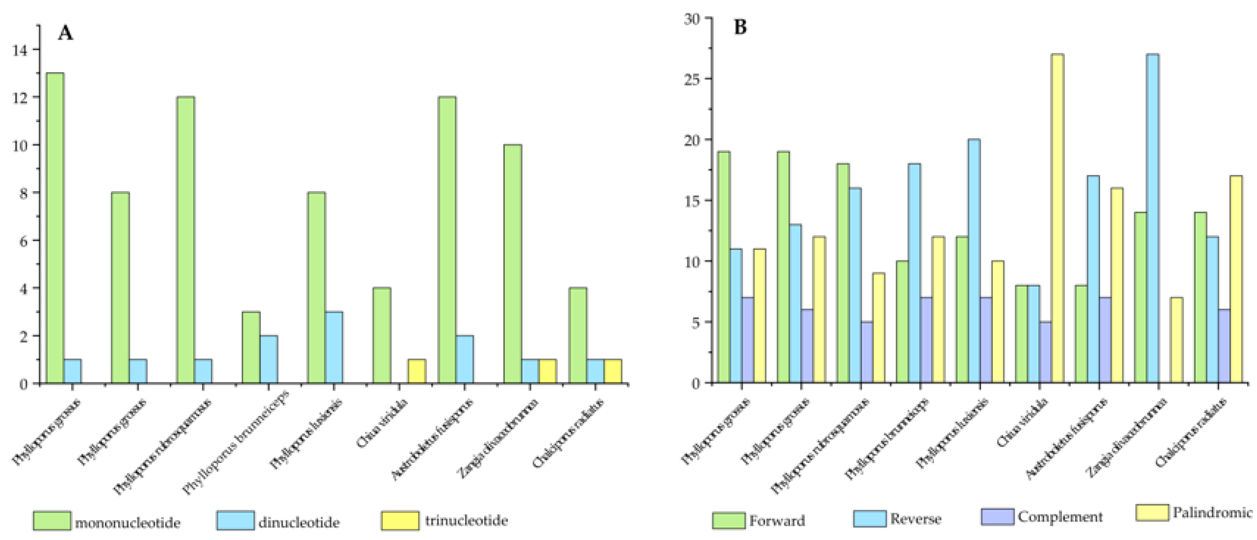
Simple sequence repeats (SSRs) are widely used as molecular markers in kinship comparison, genetic diversity analysis, variety identification, and breeding research [41]. Analysis of SSRs in the mitogenomes of eight Boletaceae species detected 5 to 14 SSR loci, with lengths ranging from 10 to 72 bp. Mononucleotide, dinucleotide, and trinucleotide repeats were identified as the dominant types: among these, mononucleotide repeats were predominant, accounting for 83.14%, while dinucleotide and trinucleotide repeats accounted for 13.48% and 3.37%, respectively. Their most common motifs were A/T, AT/TA, and TCT, respectively. Notably, no trinucleotide repeats were detected in any of the Phylloporus species (Figure 2A).
A total of 86 tandem repeat sequences with a match rate ≥ 95% were identified across the eight species. Among these, Phylloporus grossus contained the fewest tandem repeats, with only three, contrasting sharply with Chalciporus radiatus, which contained twenty-seven (Table S2).
Dispersed repeat analysis revealed that all Phylloporus species, along with most other taxa examined, contained forward (F), reverse (R), complementary (C), and palindromic (P) repeats. Among the species studied, the number of forward repeats was generally similar to that of reverse repeats. In the mitogenomes of Phylloporus species, the number of forward repeats was relatively high, ranging from 10 (Phylloporus brunneiceps) to 19 (Phylloporus grossus). Complementary repeats were the least common type among all species, ranging from 0 to 7 (Table S3). Zangia olivaceobrunnea was the only species that lacked complementary repeats (Figure 2B).

3.3. Genome Rearrangement Analysis

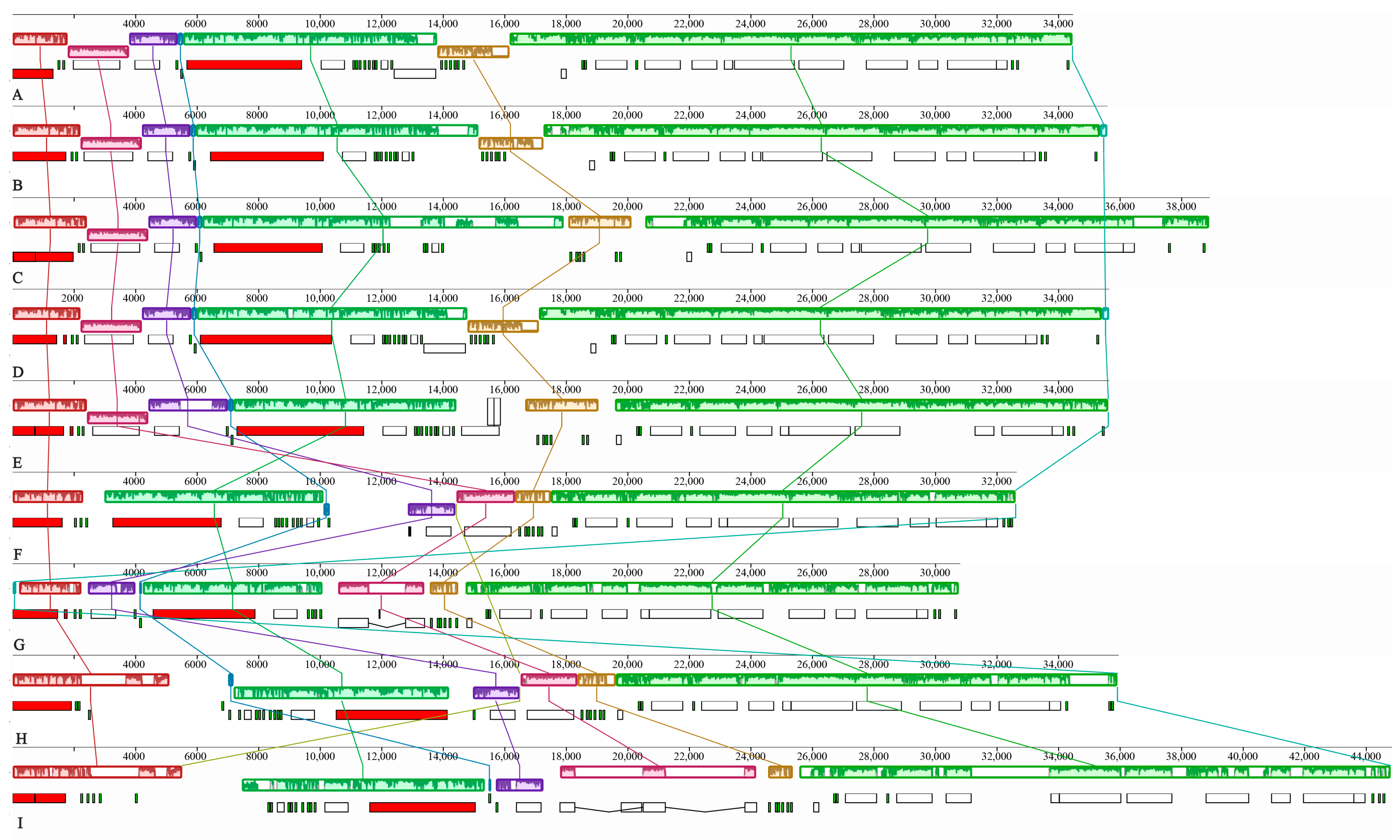
Collinear analysis of the mitogenomes of four Phylloporus species and four other Boletaceae species revealed the presence of seven homologous blocks, designated A through G, across the nine mitogenomes (Figure 3). The order of these seven homologous regions was completely consistent within the genus Phylloporus. In contrast, the arrangement of blocks B, C, and D showed noticeable differences among species from different genera within the Boletaceae family.
The sizes of these seven homologous blocks were not uniform. Block G was the largest of all four Phylloporus species, while blocks D and G were relatively conserved. The lengths of the remaining blocks exhibited some degree of variation. Compared to the Phylloporus species, the mitogenome of Chalciporus radiatus is larger, and its homologous blocks are relatively longer, indicating a positive correlation between the size of the homologous blocks and the overall genome size.
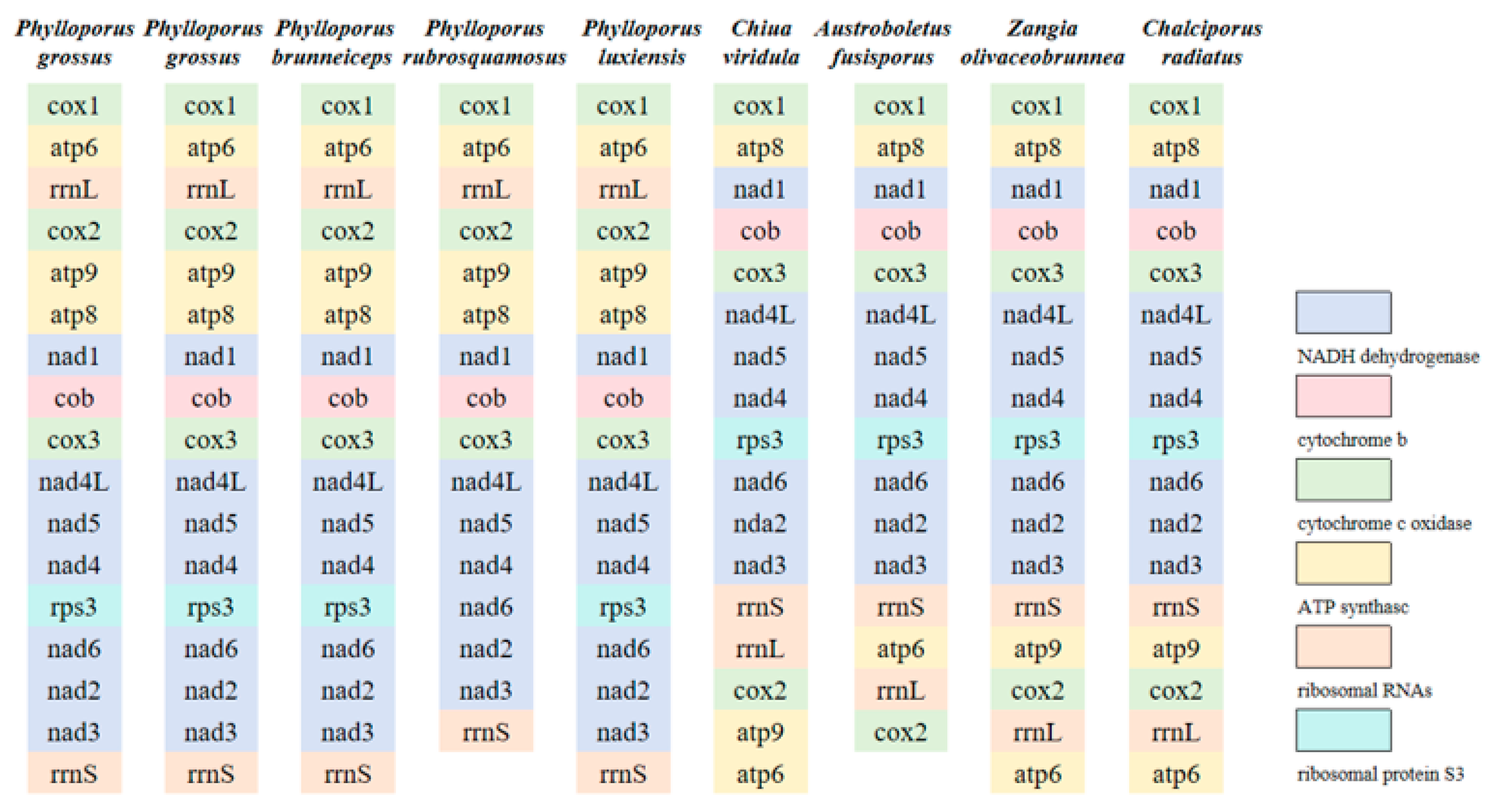
The gene order is largely consistent among the five Phylloporus mitogenomes. However, mitogenome rearrangements were observed among species within the Boletaceae mitogenomes (Figure 4). These rearrangements may be attributed to gene duplication and insertion events.
The gene order between atp8 and rrnS was relatively conserved in the mitogenomes of Boletaceae. Rearrangements were identified in five protein-coding genes (cox1, cox2, atp6, atp9) and two rRNA genes (rrnS, rrnL). Compared to other Boletaceae mitogenomes, two specific gene inversions were observed in the Phylloporus mitogenomes: the first involved an inversion between the cox1 and atp6 genes, and the second was an inversion centered around the cox2 gene, involving atp9 and rrnL.

3.4. Protein-Coding Genes of the Nine Boletaceae Mitogenomes

In the mitogenomes of the nine Boletaceae species, we found the dominance of ATG as the Start Codon in core protein-coding genes, and TAA is the most common stop codon. Whereas the cox1 gene of Phylloporus grossus and Phylloporus rubrosquamosus employs TTG as the start codon, and their atp8 gene utilizes TAG as the stop codon.
The cox2 gene of Chiua viridula uses TTG as the start codon, while its nad1 and atp8 genes use TAG as the stop codon. The cox1 gene of Austroboletus fusisporus uses TTG as the start codon, and its nad3 gene uses TAG as the stop codon. The atp8 gene of Zangia olivaceobrunnea uses TAG as the stop codon (Table S4).
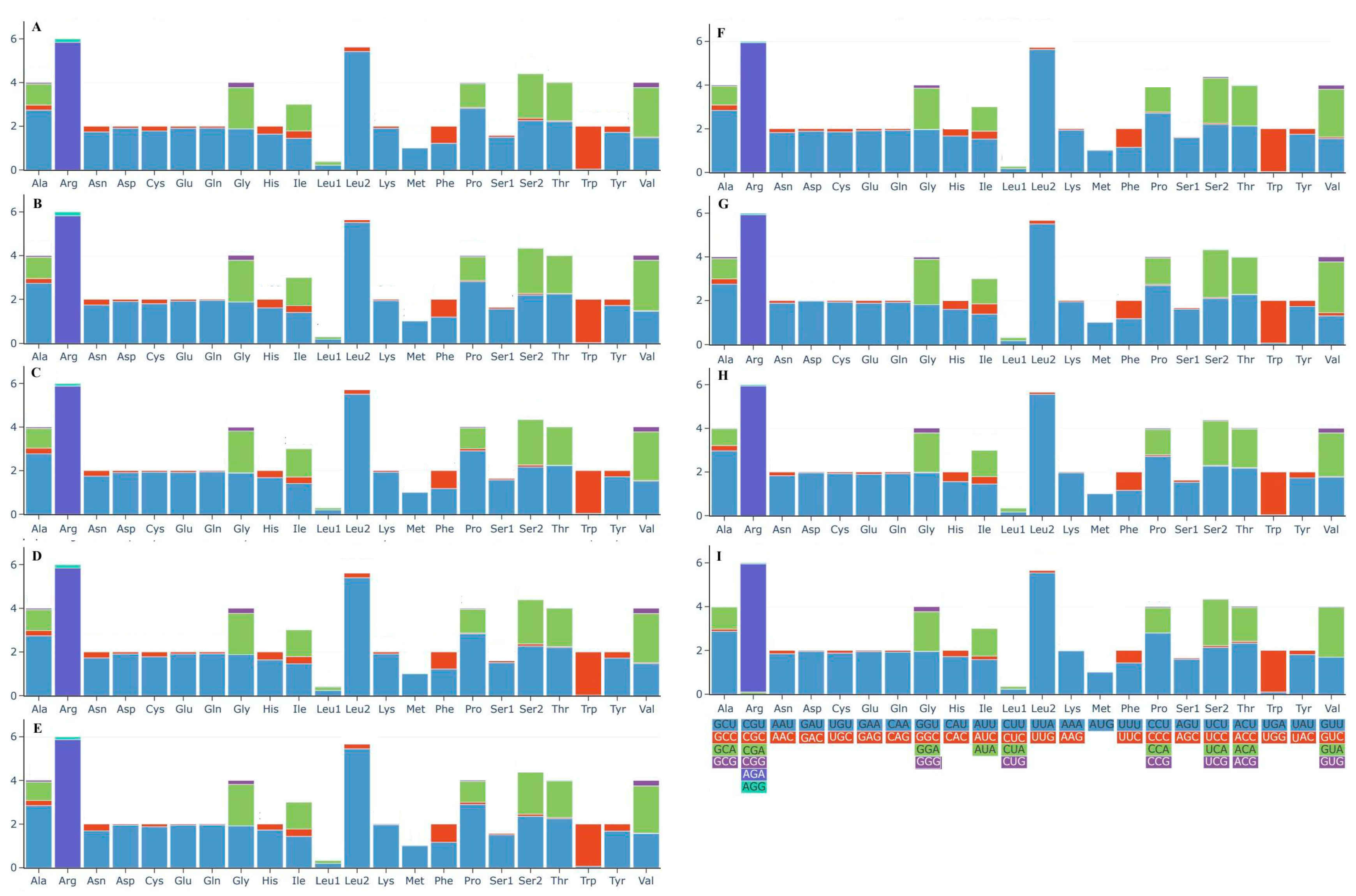
Analysis of codon usage and relative synonymous codon usage (RSCU) in the nine mitogenomes showed that they are highly similar in codon usage patterns, but there are significant differences in the RSCU of each amino acid (Figure 5). Among them, AGA (for glutamine; Arg) and UUA (for cysteine; Leu2) are the most frequently used codons, and these two amino acids are also the most abundant. In contrast, Met (methionine) is only encoded by AUG, with a low usage frequency of only 2.15%. Additionally, among the 62 analyzed codons, 27 have an RSCU value greater than 1.0.

3.5. Analysis of the tRNA Secondary Structure in Phylloporus
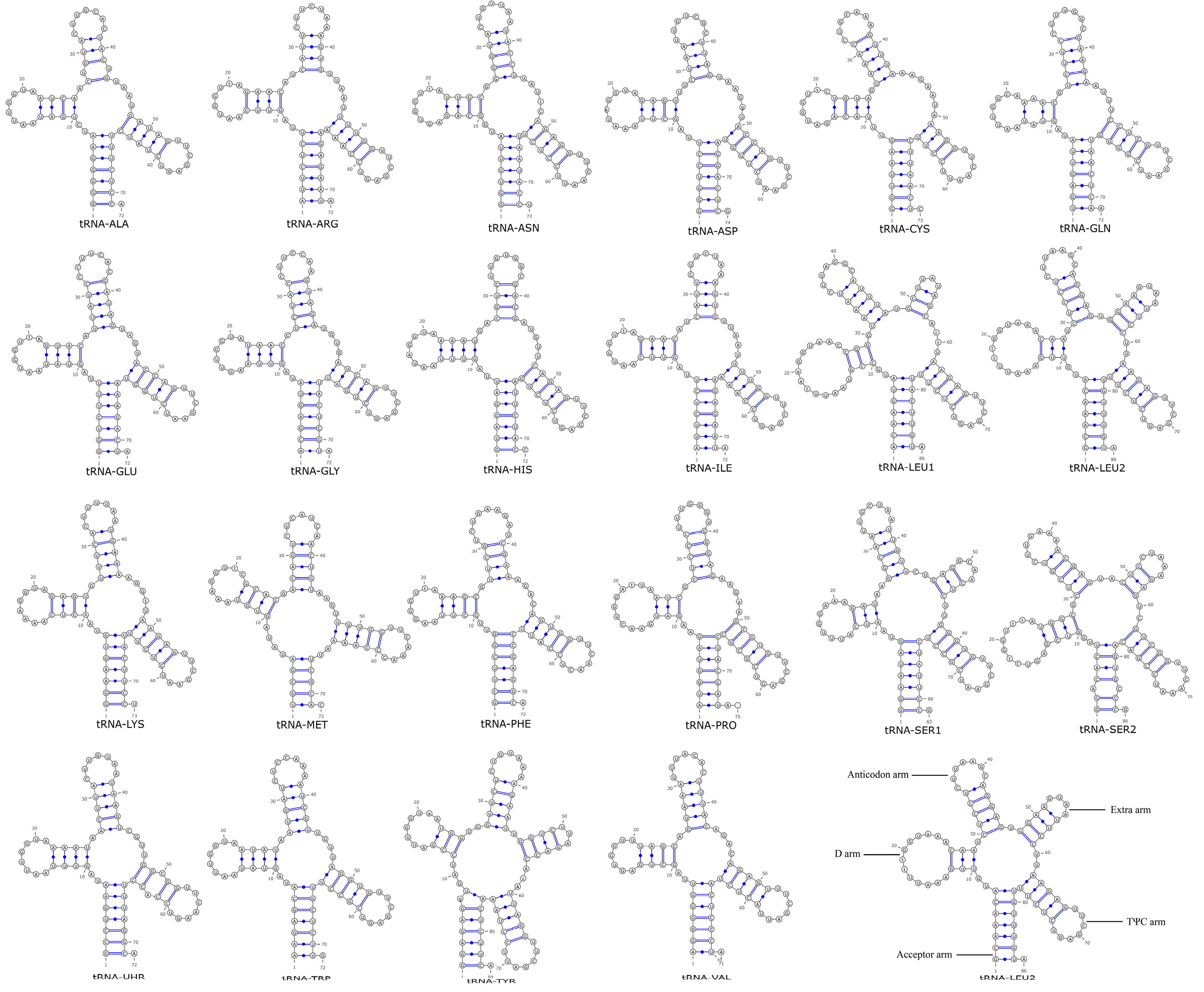
In the mitochondrial genome of Phylloporus, there are 24 tRNA genes, which are capable of transporting all 20 amino acids. Among them, the secondary structures of tRNA-Leu, tRNA-Ser, and tRNA-Tyr possess one extra loop, while the rest exhibit the typical cloverleaf structure (Figure 6). Among the four Phylloporus species, the mitochondrial genome tRNA genes contain 2 to 4 base mismatches, with Phylloporus brunneiceps and Phylloporus rubrosquamosus having the fewest base mismatches. Compared to other species in the Boletaceae family, the tRNA-Leu of Phylloporus has an additional base G in the D-loop. Austroboletus fusisporus shows the highest number of base mismatches compared to Phylloporus, with 6 mismatches. In all Boletaceae species studied, the secondary structures of tRNA-Leu, tRNA-Ser, and tRNA-Tyr feature one extra loop (Figures S1–S8).
Figure 5. Codon usage in the mitogenomes of the nine Boletaceae specimens. Frequency of codon usage is plotted on the y-axis. Different colors respectively represent the corresponding codons. (A) Phylloporus grossus HKAS105262; (B) Phylloporus grossus HKAS105260; (C) Phylloporus brunneiceps HKAS105261; (D) Phylloporus rubrosquamosus HKAS105259; (E) Phylloporus luxiensis HKAS105263; (F) Chiua viridula HKAS105270; (G) Austroboletus fusisporus HKAS105269; (H) Zangia olivaceobrunnea HKAS105268; (I) Chalciporus radiatus HKAS105264.
Jof 11 00831 g005

3.6. Evolutionary Rates of Core Genes in Boletaceae

To further investigate the evolutionary dynamics of Boletaceae, selection pressure analysis was conducted on protein-coding genes (PCGs) at the amino acid level. Due to the skewed distribution of the data, the median was used to represent the overall trend (Figure 7).
Among the 15 PCGs analyzed, the rps3 gene exhibited the highest nonsynonymous substitution rate (Ka), while the atp9 gene showed the lowest Ka value. Overall, all 15 genes had Ka values below 0.35. In terms of synonymous substitution rates (Ks), cox1 displayed the highest value, whereas atp8 had the lowest. All Ks values remained under 0.7.
The majority of core PCGs across Boletaceae species exhibited Ka/Ks ratios less than 1, indicating the prevalence of purifying selection at the amino acid level. Notably, nad4L and cox1 displayed significantly low Ka/Ks values, reflecting stronger purifying selection pressures on these genes.

3.7. Phylogenetic Relationships

Based on specimens of eight species from Jiangxi Province and mitogenome data of 19 related species (17 Boletaceae, 2 Paxillaceae) retrieved from NCBI [28,29,36,37,38,39,42], 15 core mitochondrial protein-coding genes (PCGs) were extracted (Table 1). Individual gene matrices were constructed and concatenated for phylogenetic analysis. With Paxillaceae designated as the outgroup, maximum likelihood (ML) and Bayesian inference (BI) methods were used to reconstruct phylogenetic trees.
Figure 6. Structure prediction of mitochondrial genome tRNA of Phylloporus luxiensis.
Jof 11 00831 g006
Figure 7. Evolutionary rates of genes in Boletaceae species. Ka represents the nonsynonymous substitution rate per nonsynonymous site, and Ks denotes the synonymous substitution rate per synonymous site.
Jof 11 00831 g007
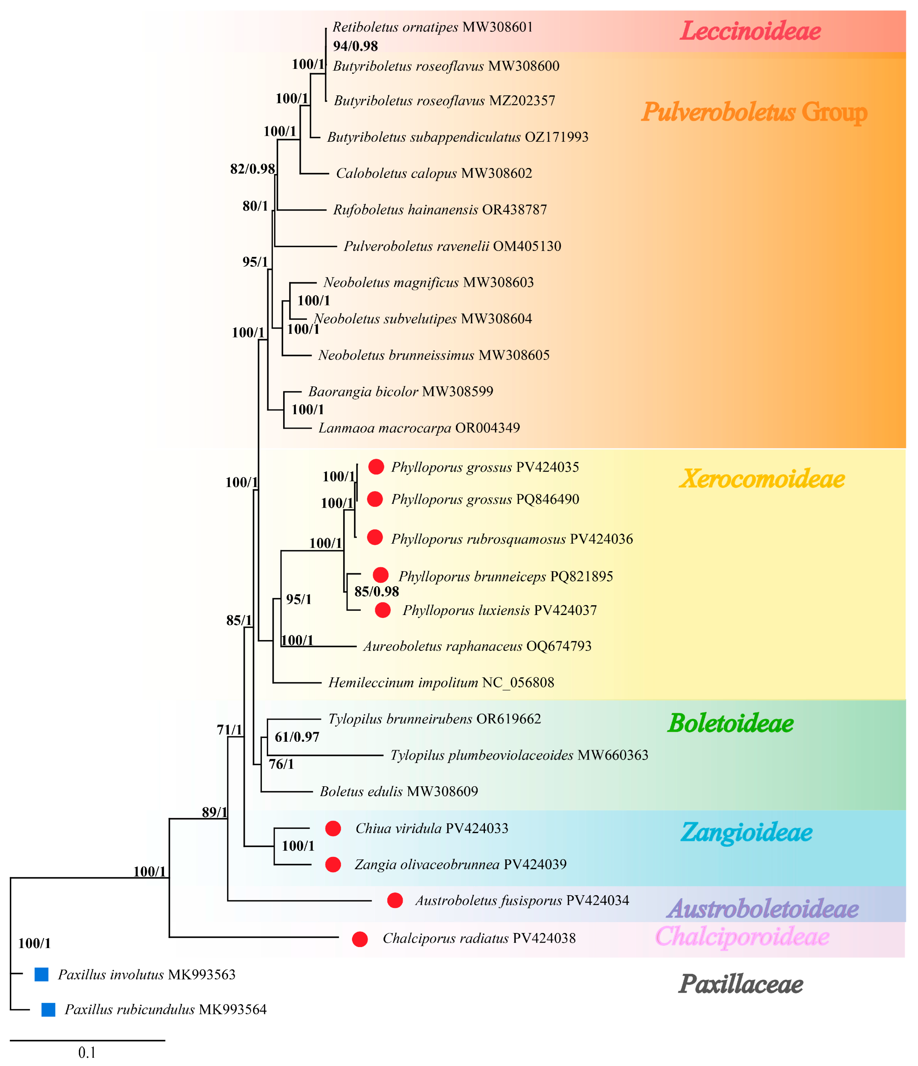
The resulting ML and BI trees exhibited identical topology. For clarity, only the ML tree is presented (Figure 8), with BI posterior probabilities indicated at corresponding nodes.
The phylogeny supported seven distinct major clades within Boletaceae, each confirming the monophyly of the corresponding subfamily. This structure aligns with the recently proposed classification system of seven subfamilies: Austroboletoideae, Boletoideae, Chalciporoideae, Leccinoideae, Xerocomoideae, Zangioideae, and the Pulveroboletus Group [43,44]. The genus Phylloporus was clustered within the Xerocomoideae subfamily and showed the closest affinity to the Boletoideae subfamily. There are subclades within the genus; one clade includes Phylloporus grossus and Phylloporus rubrosquamosus, the other clade includes Phylloporus brunneiceps and Phylloporus luxiensis. The phylogenetic relationships revealed in this study are consistent with prior studies based on 28S, TEF1, RPB1, and RPB2 molecular markers [44].

4. Discussion

Accurate taxonomic identification is a prerequisite for the efficient utilization and safe development of fungal resources within the Boletaceae family. Phylloporus species are readily identified at the genus level and belong to the subfamily Boletoideae, yet species delimitation based on morphology remains difficult due to ambiguous boundaries. Recent advances in molecular phylogenetics have led to the reevaluation of several taxa within the genus and the description of new species, greatly improving our understanding of its diversity in China [45,46,47]. As of 2024, 70 toxic fungal species have been documented in China within the order Boletales [48]. Interspecific morphological differences in Boletaceae are often subtle and overlapping, making species classification and identification based solely on morphological characteristics challenging [49].
Although nuclear genomes and molecular markers are widely used in fungal identification [50], mitogenomes have emerged as a powerful tool for phylogenetic studies due to their relatively small size, ease of acquisition, and substantially greater information content compared to single molecular markers [8]. The evolutionary dynamics of mitochondrial genomes provide rich insights into the history and diversity of life on Earth [51]. Understanding these dynamics offers critical context for interpreting phylogenetic relationships, elucidating species divergence, and unravelling the molecular mechanisms underpinning adaptation and speciation [52]. At present, the available mitogenome data for fungi remain very limited [28]. There are some thirty sequenced mitochondrial genomes in the family Boletaceae, while the mitochondrial genomes of Phylloporus have not been reported.
This study presents the first complete mitogenomes of eight species from seven subfamilies of Boletaceae in Jiangxi Province. The genomes were circular, ranging from 30,801 bp to 44,823 bp, consistent with the documented size range for this family [28]. Comparative analysis reveals that the genus Phylloporus possesses a distinctive mitogenomic architecture, characterized by a combination of a relatively conserved gene order, a moderate genome size, and notable variations in intergenic spacer length and repetitive element content.
DNA base composition is a fundamental genomic trait, with variations influencing species distribution, ecological adaptation, and life-history strategies [53]. We comparatively analyzed the genomic characteristics of 24 species in the Boletaceae family and 2 species in the Paxillus genus (Table 3). Among the species examined, ranging from 30,801 bp to 44,823 bp, and GC content ranges from 21.03% to 24.49%. Species within Phylloporus exhibit notably higher GC content compared to other groups in the Boletaceae family. Elevated GC content may increase mutation rates [54], which could be associated with the distinctive lamellate hymenophore structure in Phylloporus. However, this potential relationship is a tentative hypothesis that requires further research to verify. Crucially, the factors leading to differences in gene size are the accumulation of intron and repeat-driven changes.
When compared to other genera within the Boletaceae, this profile of Phylloporus stands in clear contrast. For instance, in this study, the mitogenome of Chalciporus radiatus is the largest, and it also contains the most tandem repeat sequences, indicating that repetitive elements play a certain role in genomic expansion across the family. Conversely, Austroboletus fusisporus maintains the smallest genome despite harboring a group I intron, underscoring that intronic regions are not the predominant driver of size in these lineages and further highlighting the unique Phylloporus model where repetitive elements, not introns, modulate size. However, due to the presence of introns, the mitochondrial genome size of species in the Boletaceae is much smaller than that of the Suillus genus [55]. This comparative analysis confirmed that the accumulation of repetitive sequences and the number of introns is common drivers of mitochondrial genome size variation in Boletaceae species.
Repetitive sequences play a crucial role in genomic recombination and restructuring [56], and their accumulation can lead to genomic expansion or contraction over evolutionary time [57,58]. The consistency in SSR types across Boletaceae mitogenomes suggests a high degree of sequence conservation within the family. Boletaceae species have fewer SSR types than plants, and hexameric SSRs are present in Hydrangea chinensis [59]. The absence of trinucleotide repeats in Phylloporus may be attributed to mitochondrial genomic characteristics and shorter sequence lengths. Recombination events have been shown to play a crucial role in genome evolution and adaptation [60]. The presence of a large number of SSRs further highlights the dynamic nature of the mitochondrial genome. It is known that SSRs can lead to genomic instability and variability, which may in turn affect gene expression and mitochondrial function [61].
Although mitochondrial genes are generally conserved due to their essential roles in cellular metabolism, gene order in fungal mitochondria can vary considerably. The accumulation of repetitive DNA elements in intergenic regions is a key driver of mitochondrial gene rearrangements. Frequent rearrangements have been reported in the genus Boletus [28], suggesting divergent evolutionary trajectories among lineages. Gene order analysis here reveals significant intergeneric variation in mitogenomes within Boletaceae, whereas Phylloporus exhibits high intragenomic conservation, possibly due to intramolecular recombination in the ancestral Basidiomycete mitogenome [62]. This contrast between intrageneric stability and intergeneric plasticity supports the utility of mitochondrial gene rearrangements as taxonomic markers.
Codon usage bias is critical for protein function and translational accuracy and can provide insights into evolutionary and environmental adaptation mechanisms [63]. In this study, codon preference patterns are highly conserved across Boletaceae mitogenomes, though minor differences exist in the start and stop codons of core PCGs. Among the 62 codons analyzed, 27 have RSCU values > 1.0, a pattern potentially shaped by long-term environmental stress.
The Ka/Ks ratios for all 15 core PCGs are below 1, indicating dominance of purifying selection across the family. However, based on qualitative observations, the atp9 gene shows signs of positive selection in Phylloporus luxiensis, Phylloporus rubrosquamosus, and Phylloporus grossus, possibly reflecting ecological adaptation. Significantly lower Ka/Ks values for nad4L and cox1 suggest intensified purifying selection, which may reduce genetic diversity and distort site frequency spectra—effects that could mimic population expansion or positive selection [64]. These differential selection pressures provide new insights into the adaptive evolution of functional genes during the diversification of Phylloporus.
Phylogenetic tree (Figure 7) confirmed seven major subfamilial clades within Boletaceae [44]. Our phylogenetic analysis groups Phylloporus brunneiceps with P. luxiensis, and P. rubrosquamosus with P. grossus. This is consistent with the nuclear gene-based phylogeny by Zeng et al. [1], where P. brunneiceps and P. luxiensis formed part of Clade I, while P. rubrosquamosus fell within Clade VI. The phylogenetic tree we reconstructed based on ITS sequences also confirms this (Figure S9).
Nonetheless, compared to topologies inferred from nuclear gene fragments, mitochondrial-based trees show slight differences in branching patterns and subfamilial placements, possibly due to the autonomous replication, transcription, and translation of mitogenomes, their lack of recombination, and maternal inheritance [8]. Alternatively, limited taxonomic sampling may affect clustering accuracy. These findings demonstrate that concatenated mitochondrial gene sets can accurately recover deep evolutionary relationships, highlighting their value as a robust tool for phylogenetic inference in Basidiomycetes, and providing a reliable basis for taxonomic revision, species identification, and conservation planning in Boletaceae.
The evolution of the mitochondrial genome of Phylloporus, an ectomycorrhizal fungus, may be the result of a balance between functional conservation and ecological adaptability. The purification and selection of core energy metabolism genes ensure their basic symbiotic functions, while the adaptive mutations of local genes and the plasticity of genomic structure promote the adaptation of fungi to hosts and the environment. Ultimately, through the optimization of the symbiotic system between fungi and hosts, the stability of the forest ecosystem is maintained.

5. Conclusions

Phylloporus is an important ectomycorrhizal and edible fungus, yet its molecular data are scarce. Mitochondrial genomes are valuable for phylogeny and species differentiation due to their high copy number and ease of acquisition. While many Boletales mitogenomes have been published, none are available for the genus Phylloporus. In this study, we presented the first complete mitochondrial genomes of four Phylloporus species from Jiangxi Province, China, along with four additional Boletaceae species, providing the first mitogenomic insights into the genus Phylloporus. Our results revealed that the mitogenomes of Phylloporus are exhibiting a relatively conserved gene order and content yet are showing notable variations in repetitive sequences and intergenic regions. Notably, Phylloporus mitogenomes lacked trinucleotide SSRs and contained few introns, suggesting that repetitive elements rather than intronic expansions are the primary drivers of genome size variation within this genus.
Comparative genomic analyses highlighted both shared and distinct evolutionary features between Phylloporus and other Boletaceae genera. While codon usage bias was largely conserved across the family, gene order and repetitive element profiles served as informative taxonomic markers. Phylogenetic reconstruction based on 15 core PCGs strongly supported the monophyly of Phylloporus and its placement within the subfamily Xerocomoideae, consistent with previous nuclear-based studies. Furthermore, the Ka/Ks ratios indicated that most PCGs are under purifying selection, possibly reflecting ecological adaptation. These characteristics represent not only molecular signatures of evolutionary divergence but also reflect long-term adaptive strategies to subtropical forest ecosystems, offering new perspectives on the evolution of ectomycorrhizal fungi.
This study not only enriches the mitochondrial genomic resources for Boletaceae but also demonstrates the utility of mitogenomes in resolving phylogenetic relationships and understanding genomic evolution in ectomycorrhizal fungi. The distinct mitogenomic architecture of Phylloporus provides a valuable model for studying fungal adaptation and diversification. Future efforts should focus on expanding mitogenome sequencing to include more taxonomically diverse and morphologically cryptic taxa within Basidiomycota to further elucidate the evolutionary history and functional genomics of these ecologically and economically significant fungi.

Supplementary Materials

The following supporting information can be downloaded at: https://www.mdpi.com/article/10.3390/jof11120831/s1. Table S1: Short tandem repeats. Table S2: tandem repeats. Table S3: dispersed repeats. Table S4: Codon usage. Figure S1: Austroboletus fusisporus. Figure S2: Chalciporus radiatus. Figure S3: Chiua viridula. Figure S4: Phylloporus rubrosquamosus. Figure S5: Phylloprous grossus. Figure S6: Phylloprus grossus 1301. Figure S7: Phylloprus brunneiceps. Figure S8: Zangia olivaceobrunnea. Figure S9: Phylogenetic analysis of Phylloporus.

Author Contributions

Conceptualization, K.Z. and S.Y.; methodology, J.-Y.H.; validation, Z.Z. and M.-W.M.; formal analysis, J.-Y.H.; investigation, J.-Y.H.; resources, M.-W.M.; data curation, Z.Z.; writing—original draft preparation, J.-Y.H.; writing—review and editing, K.Z.; visualization, J.-Y.H.; supervision, S.Y.; project administration, K.Z.; funding acquisition, K.Z. All authors have read and agreed to the published version of the manuscript.

Funding

This work was supported by the National Natural Science Foundation of China [32360153] and the Inbreeding and Genetic Differentiation Among Geographic Populations of Lactarius hatsudake in Southwest China [XJ202511318140].

Data Availability Statement

The original contributions presented in this study are included in the article/Supplementary Materials. Further inquiries can be directed to the corresponding authors.

Conflicts of Interest

The authors declare no conflicts of interest. The funders had no role in the design of the study; in the collection, analyses, or interpretation of data; in the writing of the manuscript; or in the decision to publish the results.

References

  1. Zeng, N.-K.; Tang, L.-P.; Li, Y.-C.; Tolgor, B.; Zhu, X.-T.; Zhao, Q.; Yang, Z.L. The genus Phylloporus (Boletaceae, Boletales) from China: Morphological and multilocus DNA sequence analyses. Fungal Divers. 2012, 58, 73–101. [Google Scholar] [CrossRef]
  2. Ramos, A.; Leticia, M.; Bandala, V.M. Morphological and molecular characterization of ectomycorrhizas of Phylloporus (Boletales) and Quercus sapotifolia from tropical oak forest of eastern Mexico. Symbiosis 2023, 91, 45–54. [Google Scholar] [CrossRef]
  3. Courty, P.-E.; Buée, M.; Diedhiou, A.G.; Frey-Klett, P.; Le Tacon, F.; Rineau, F.; Turpault, M.-P.; Uroz, S.; Garbaye, J.J.S.B. The role of ectomycorrhizal communities in forest ecosystem processes: New perspectives and emerging concepts. Soil Biol. Biochem. 2010, 42, 679–698. [Google Scholar] [CrossRef]
  4. Wu, G.; Miyauchi, S.; Morin, E.; Kuo, A.; Drula, E.; Varga, T.; Kohler, A.; Feng, B.; Cao, Y.; Lipzen, A.; et al. Evolutionary innovations through gain and loss of genes in the ectomycorrhizal Boletales. New Phytol. 2022, 233, 1383–1400. [Google Scholar] [CrossRef] [PubMed]
  5. Li, H.; Zhong, X.; Ma, Z.; Tang, G.; Ding, L.; Sui, X.; Xu, J.; He, Y. Climate changes and their teleconnections with ENSO over the last 55 years, 1961–2015, in floods-dominated basin, Jiangxi province, China. Earth Space Sci. 2020, 7, e2019EA001047. [Google Scholar] [CrossRef]
  6. Zhang, J.; Song, H.; Hu, D. A Checklist of macrofungi in Jiangxi province. Biol. Hazard Sci. 2016, 39, 1–13. [Google Scholar] [CrossRef]
  7. Cao, K.Z.R. Jiangxi Jiuling Mountain Macrofungi Atlas; Jiangxi People’s Publishing House: Nanchang, China, 2022; ISBN 9787210143239. [Google Scholar]
  8. Zeb, U.; Aziz, T.; Azizullah, A.; Zan, X.Y.; Khan, A.A.; Bacha, S.A.S.; Cui, F.J. Complete mitochondrial genomes of edible mushrooms: Features, evolution, and phylogeny. Physiol. Plant. 2024, 176, e14363. [Google Scholar] [CrossRef] [PubMed]
  9. Sun, T.; Li, T.-h.; Huang, O.; Zhu, K.-f.; Chen, S.-q.; Wang, Y.-a.; Wang, Y.; Wang, Y.-b.; Yu, H. Phylogenetic analysis of the mitochondrial genome of the Samsoniella hepiali holotype strain. Mycosystema 2022, 41, 1572–1584. [Google Scholar] [CrossRef]
  10. Ma, J.; Wang, Z.; Yang, Z.; Sun, T.; Yang, S.; Yu, H. Morphology, phylogeny, and mitogenomics reveal a new entomopathogenic fungus, Blackwellomyces changningensis (Hypocreales, Clavicipitaceae), from southwestern China. Mycologia 2024, 117, 166–182. [Google Scholar] [CrossRef]
  11. Yu, H.; Zhang, M.; Sun, Y.; Li, Q.; Liu, J.; Song, C.; Shang, X.; Tan, Q.; Zhang, L.; Yu, H. Whole-genome sequence of a high-temperature edible mushroom Pleurotus giganteus (zhudugu). Front. Microbiol. 2022, 13, 941889. [Google Scholar] [CrossRef]
  12. Liu, J.; Li, T.; Song, L.; Kong, J.; Niu, Q.; Wang, Y.; Wu, C.; Deng, B.; Wang, H.; Gai, Y. Comparative transcriptome of isonuclear alloplasmic strain revealed the important role of mitochondrial genome in regulating Flammulina filiformis. Agronomy 2023, 13, 998. [Google Scholar] [CrossRef]
  13. Li, Q.; Bao, Z.; Tang, K.; Feng, H.; Tu, W.; Li, L.; Han, Y.; Cao, M.; Zhao, C. First two mitochondrial genomes for the order Filobasidiales reveal novel gene rearrangements and intron dynamics of Tremellomycetes. IMA Fungus 2022, 13, 7. [Google Scholar] [CrossRef]
  14. Jin, J.J.; Yu, W.B.; Yang, J.B.; Song, Y.; dePamphilis, C.W.; Yi, T.S.; Li, D.Z. GetOrganelle: A fast and versatile toolkit for accurate de novo assembly of organelle genomes. Genome Biol. 2020, 21, 241. [Google Scholar] [CrossRef] [PubMed]
  15. Donath, A.; Juhling, F.; Al-Arab, M.; Bernhart, S.H.; Reinhardt, F.; Stadler, P.F.; Middendorf, M.; Bernt, M. Improved annotation of protein-coding genes boundaries in metazoan mitochondrial genomes. Nucleic Acids Res. 2019, 47, 10543–10552. [Google Scholar] [CrossRef]
  16. Lowe, T.M.; Chan, P.P. tRNAscan-SE On-line: Integrating search and context for analysis of transfer RNA genes. Nucleic Acids Res. 2016, 44, W54–W57. [Google Scholar] [CrossRef] [PubMed]
  17. Lang, B.F.; Laforest, M.J.; Burger, G. Mitochondrial introns: A critical view. Trends Genet. 2007, 23, 119–125. [Google Scholar] [CrossRef] [PubMed]
  18. Greiner, S.; Lehwark, P.; Bock, R. OrganellarGenomeDRAW (OGDRAW) version 1.3.1: Expanded toolkit for the graphical visualization of organellar genomes. Nucleic Acids Res. 2019, 47, W59–W64. [Google Scholar] [CrossRef]
  19. Chan, P.P.; Lin, B.Y.; Mak, A.J.; Lowe, T.M. tRNAscan-SE 2.0: Improved detection and functional classification of transfer RNA genes. Nucleic Acids Res. 2021, 49, 9077–9096. [Google Scholar] [CrossRef]
  20. Darty, K.; Denise, A.; Ponty, Y. VARNA: Interactive drawing and editing of the RNA secondary structure. Bioinformatics 2009, 25, 1974–1975. [Google Scholar] [CrossRef]
  21. Darling, A.C.; Mau, B.; Blattner, F.R.; Perna, N.T. Mauve: Multiple alignment of conserved genomic sequence with rearrangements. Genome Res. 2004, 14, 1394–1403. [Google Scholar] [CrossRef]
  22. Zhang, D.; Gao, F.; Jakovlic, I.; Zou, H.; Zhang, J.; Li, W.X.; Wang, G.T. PhyloSuite: An integrated and scalable desktop platform for streamlined molecular sequence data management and evolutionary phylogenetics studies. Mol. Ecol. Resour. 2020, 20, 348–355. [Google Scholar] [CrossRef]
  23. Perna, N.T.; Kocher, T.D. Patterns of nucleotide composition at fourfold degenerate sites of animal mitochondrial genomes. J. Mol. Evol. 1995, 41, 353–358. [Google Scholar] [CrossRef] [PubMed]
  24. Benson, G. Tandem repeats finder: A program to analyze DNA sequences. Nucleic Acids Res. 1999, 27, 573–580. [Google Scholar] [CrossRef]
  25. Rozas, J.; Ferrer-Mata, A.; Sanchez-DelBarrio, J.C.; Guirao-Rico, S.; Librado, P.; Ramos-Onsins, S.E.; Sanchez-Gracia, A. DnaSP 6: DNA sequence polymorphism analysis of large data sets. Mol. Biol. Evol. 2017, 34, 3299–3302. [Google Scholar] [CrossRef]
  26. Miyauchi, S.; Kiss, E.; Kuo, A.; Drula, E.; Kohler, A.; Sanchez-Garcia, M.; Morin, E.; Andreopoulos, B.; Barry, K.W.; Bonito, G.; et al. Large-scale genome sequencing of mycorrhizal fungi provides insights into the early evolution of symbiotic traits. Nat. Commun. 2020, 11, 5125. [Google Scholar] [CrossRef] [PubMed]
  27. Li, Q.; Ren, Y.; Xiang, D.; Shi, X.; Zhao, J.; Peng, L.; Zhao, G. Comparative mitogenome analysis of two ectomycorrhizal fungi (Paxillus) reveals gene rearrangement, intron dynamics, and phylogeny of basidiomycetes. IMA Fungus 2020, 11, 12. [Google Scholar] [CrossRef] [PubMed]
  28. Li, Q.; Wu, P.; Li, L.; Feng, H.; Tu, W.; Bao, Z.; Xiong, C.; Gui, M.; Huang, W. The first eleven mitochondrial genomes from the ectomycorrhizal fungal genus (Boletus) reveal intron loss and gene rearrangement. Int. J. Biol. Macromol. 2021, 172, 560–572. [Google Scholar] [CrossRef]
  29. Shi, W.; Song, W.; Peng, Y.; Wang, S.; Yang, G.; Shi, C. The complete mitochondrial genome sequence and annot-ation of Tylopilus plumbeoviolaceoides (Boletaceae, Boletoideae). Mitochondrial DNA Part B Resour. 2022, 7, 999–1000. [Google Scholar] [CrossRef]
  30. Zheng, Y.t.; Chen, L.-l.; Zhao, K. Complete mitochondrial genome sequence of Lanmaoa macrocarpa (Boletales, Basidiomycota). Mitochondrial DNA Part B Resour. 2023, 8, 1067–1070. [Google Scholar] [CrossRef]
  31. Nguyen, L.T.; Schmidt, H.A.; Haeseler, A.; Minh, B.Q. IQ-TREE: A fast and effective stochastic algorithm for estimating maximum-likelihood phylogenies. Mol. Biol. Evol. 2015, 32, 268–274. [Google Scholar] [CrossRef]
  32. Katoh, K.; Standley, D.M. MAFFT multiple sequence alignment software version 7: Improvements in performance and usability. Mol. Biol. Evol. 2013, 30, 772–780. [Google Scholar] [CrossRef]
  33. Minh, B.Q.; Schmidt, H.A.; Chernomor, O.; Schrempf, D.; Woodhams, M.D.; von Haeseler, A.; Lanfear, R. IQ-TREE 2: New models and efficient methods for phylogenetic inference in the genomic era. Mol. Biol. Evol. 2020, 37, 1530–1534. [Google Scholar] [CrossRef]
  34. Minh, B.Q.; Nguyen, M.A.; von Haeseler, A. Ultrafast approximation for phylogenetic bootstrap. Mol. Biol. Evol. 2013, 30, 1188–1195. [Google Scholar] [CrossRef] [PubMed]
  35. Hoang, D.T.; Chernomor, O.; von Haeseler, A.; Minh, B.Q.; Vinh, L.S. UFBoot2: Improving the ultrafast bootstrap approximation. Mol. Biol. Evol. 2018, 35, 518–522. [Google Scholar] [CrossRef]
  36. Mu, X.H.; Liang, X.X.; Zheng, Y.T.; Zhao, K. Complete mitochondrial genome sequence of Aureoboletus raphanaceus (Boletales, Basidiomycota). Mitochondrial DNA Part B Resour. 2024, 9, 20–23. [Google Scholar] [CrossRef]
  37. Cho, S.E.; Kwag, Y.N.; Han, S.K.; Lee, D.H.; Kim, C.S. Complete mitochondrial genome sequence of Pulveroboletus ravenelii (Boletales, Basidiomycota). Mitochondrial DNA Part B Resour. 2022, 7, 1581–1582. [Google Scholar] [CrossRef]
  38. Zeng, Y.P.; Huang, J.Y.; Tu, L.; Zhao, K. Complete mitochondrial genome sequence of Butyriboletus hainanensis (Boletales, Basidiomycota). Mitochondrial DNA Part B Resour. 2024, 9, 46–49. [Google Scholar] [CrossRef]
  39. Huang, J.Y.; Tu, L.; Lv, Y.; Zhao, K. Complete mitochondrial genome sequence and phylogenetic analysis of Tylo-pilus brunneirubens (Boletales, Basidiomycota). Mitochondrial DNA Part B Resour. 2024, 9, 616–620. [Google Scholar] [CrossRef] [PubMed]
  40. Torresen, O.K.; Star, B.; Mier, P.; Andrade-Navarro, M.A.; Bateman, A.; Jarnot, P.; Gruca, A.; Grynberg, M.; Kajava, A.V.; Promponas, V.J.; et al. Tandem repeats lead to sequence assembly errors and impose multi-level challenges for genome and protein databases. Nucleic Acids Res. 2019, 47, 10994–11006. [Google Scholar] [CrossRef]
  41. Dang, Z.; Huang, L.; Jia, Y.; Lockhart, P.J.; Fong, Y.; Tian, Y. Identification of genic SSRs provide a perspective for studying environmental adaptation in the endemic shrub Tetraena mongolica. Genes 2020, 11, 322. [Google Scholar] [CrossRef] [PubMed]
  42. Maharachchikumbura, S.S.; Larignon, P.; Hyde, K.D.; Al-Sadi, A.M.; Liu, Z. Characterization of Neopestalotiopsis, Pestalotiopsis and Truncatella species associated with grapevine trunk diseases in France. Phytopathol. Mediterr. 2016, 55, 380–390. [Google Scholar] [CrossRef]
  43. Wu, G.; Feng, B.; Xu, J.; Zhu, X.-T.; Li, Y.-C.; Zeng, N.-K.; Hosen, M.I.; Yang, Z.L. Molecular phylogenetic analyses redefine seven major clades and reveal 22 new generic clades in the fungal family Boletaceae. Fungal Divers. 2014, 69, 93–115. [Google Scholar] [CrossRef]
  44. Wu, G.; Li, Y.; Zhu, X.; Zhao, K.; Han, L.; Cui, Y.; Li, F.; Xu, J.; Yang, Z. One hundred noteworthy boletes from China. Fungal Divers. 2016, 81, 25–188. [Google Scholar] [CrossRef]
  45. Chuankid, B.; Vadthanarat, S.; Hyde, K.D.; Thongklang, N.; Zhao, R.; Lumyong, S.; Raspé, O. Three new Phylloporus species from tropical China and Thailand. Mycol. Prog. 2019, 18, 603–614. [Google Scholar] [CrossRef]
  46. Zhang, Y.; Xue, R.; Su, M.; Wu, L.; Zeng, N. Phylloporus rubiginosus, a noteworthy lamellar bolete from tropical Asia. Guizhou Sci. 2019, 37, 1–5. [Google Scholar] [CrossRef]
  47. Wu, L.-L.; Liang, Z.-Q.; Su, M.-S.; Fan, Y.-G.; Zhang, P.; Jiang, S.; Chen, Y.-L.; Hao, Y.-J.; Zeng, N.-K. Updated taxonomy of Chinese Phylloporus (Boletaceae, Boletales): Six new taxa and four redescribed species. Mycol. Prog. 2021, 20, 1243–1273. [Google Scholar] [CrossRef]
  48. Kai, Z.N.; Ying, D.Q. Poisonous fungi of the order Boletomycetes in China. Mycol. Res. 2024, 22, 322–332. [Google Scholar] [CrossRef]
  49. Dentinger, B.T.; Ammirati, J.F.; Both, E.E.; Desjardin, D.E.; Halling, R.E.; Henkel, T.W.; Moreau, P.A.; Nagasawa, E.; Soytong, K.; Taylor, A.F.; et al. Molecular phylogenetics of porcini mushrooms (Boletus section Boletus). Mol. Phylogenet. Evol. 2010, 57, 1276–1292. [Google Scholar] [CrossRef]
  50. Li, H.; Wu, S.; Ma, X.; Chen, W.; Zhang, J.; Duan, S.; Gao, Y.; Kui, L.; Huang, W.; Wu, P.; et al. The Genome Sequences of 90 Mushrooms. Sci. Rep. 2018, 8, 9982. [Google Scholar] [CrossRef]
  51. Malik, M.; Malik, F.; Fatma, T.; Qasim Hayat, M.; Jamal, A.; Gul, A.; Faraz Bhatti, M. The complete mitochondrial genome of Penicillium expansum: Insights into the fungal evolution and phylogeny. Gene 2024, 910, 148315. [Google Scholar] [CrossRef]
  52. Li, Y.; Yang, T.; Qiao, J.; Liang, J.; Li, Z.; Sa, W.; Shang, Q. Whole-genome sequencing and evolutionary analysis of the wild edible mushroom, morchella eohespera. Front. Microbiol. 2023, 14, 1309703. [Google Scholar] [CrossRef]
  53. Yu, Y.; Li, H.T.; Wu, Y.H.; Li, D.Z. Correlation analysis reveals an important role of GC content in accumulation of deletion mutations in the coding region of angiosperm plastomes. J. Mol. Evol. 2021, 89, 73–80. [Google Scholar] [CrossRef]
  54. Yu, Y.; Yang, J.B.; Ma, W.Z.; Pressel, S.; Liu, H.M.; Wu, Y.H.; Schneider, H. Chloroplast phylogenomics of liverworts: A reappraisal of the backbone phylogeny of liverworts with emphasis on Ptilidiales. Cladistics 2020, 36, 184–193. [Google Scholar] [CrossRef]
  55. Tao, J.; Wang, X.; Long, Y.; Gao, Z.; Zhang, G.; Guo, Z.; Wang, G.; Xu, G.; Wang, Y.; Liu, H. Determining gene order patterns in the Suillus and Boletales through comparative analysis of their mitogenomes. Int. J. Mol. Sci. 2024, 25, 9597. [Google Scholar] [CrossRef] [PubMed]
  56. Lin, P.; Yan, Z.F.; Kook, M.; Li, C.T.; Yi, T.H. Genetic and chemical diversity of edible mushroom Pleurotus species. Biomed. Res. Int. 2022, 2022, 6068185. [Google Scholar] [CrossRef]
  57. Boiko, S.M. The trends in the spread of simple sequence repeats in the genomes of Schizophyllum commune. Mycologia 2023, 115, 288–298. [Google Scholar] [CrossRef]
  58. Gong, F.; Geng, Y.; Zhang, P.; Zhang, F.; Fan, X.; Liu, Y. Genetic diversity and structure of a core collection of Huangqi (Astragalus ssp.) developed using genomic simple sequence repeat markers. Genet. Resour. Crop Evol. 2022, 70, 571–585. [Google Scholar] [CrossRef]
  59. Ye, K.; Qin, J.; Yonghong, H. Decoding the complete mitochondrial genome of Hydrangea chinensis maxim.: Insights into genomic recombination, gene transfer, and RNA editing dynamics. BMC Plant Biol. 2025, 25, 1078. [Google Scholar] [CrossRef] [PubMed]
  60. Martin, W. Gene transfer from organelles to the nucleus: Frequent and in big chunks. Proc. Natl. Acad. Sci. USA 2003, 100, 8612–8614. [Google Scholar] [CrossRef]
  61. Smith, D.R.; Keeling, P.J. Mitochondrial and plastid genome architecture: Reoccurring themes, but significant differences at the extremes. Proc. Natl. Acad. Sci. USA 2015, 112, 10177–10184. [Google Scholar] [CrossRef]
  62. Beaudet, D.; Nadimi, M.; Iffis, B.; Hijri, M. Rapid mitochondrial genome evolution through invasion of mobile elements in two closely related species of arbuscular mycorrhizal fungi. PLoS ONE 2013, 8, e60768. [Google Scholar] [CrossRef] [PubMed]
  63. Almutairi, M.M.; Almotairy, H.M. Analysis of heat shock proteins based on amino acids for the tomato genome. Genes 2022, 13, 2014. [Google Scholar] [CrossRef] [PubMed]
  64. Cvijovic, I.; Good, B.H.; Desai, M.M. The effect of strong purifying selection on genetic diversity. Genetics 2018, 209, 1235–1278. [Google Scholar] [CrossRef] [PubMed]
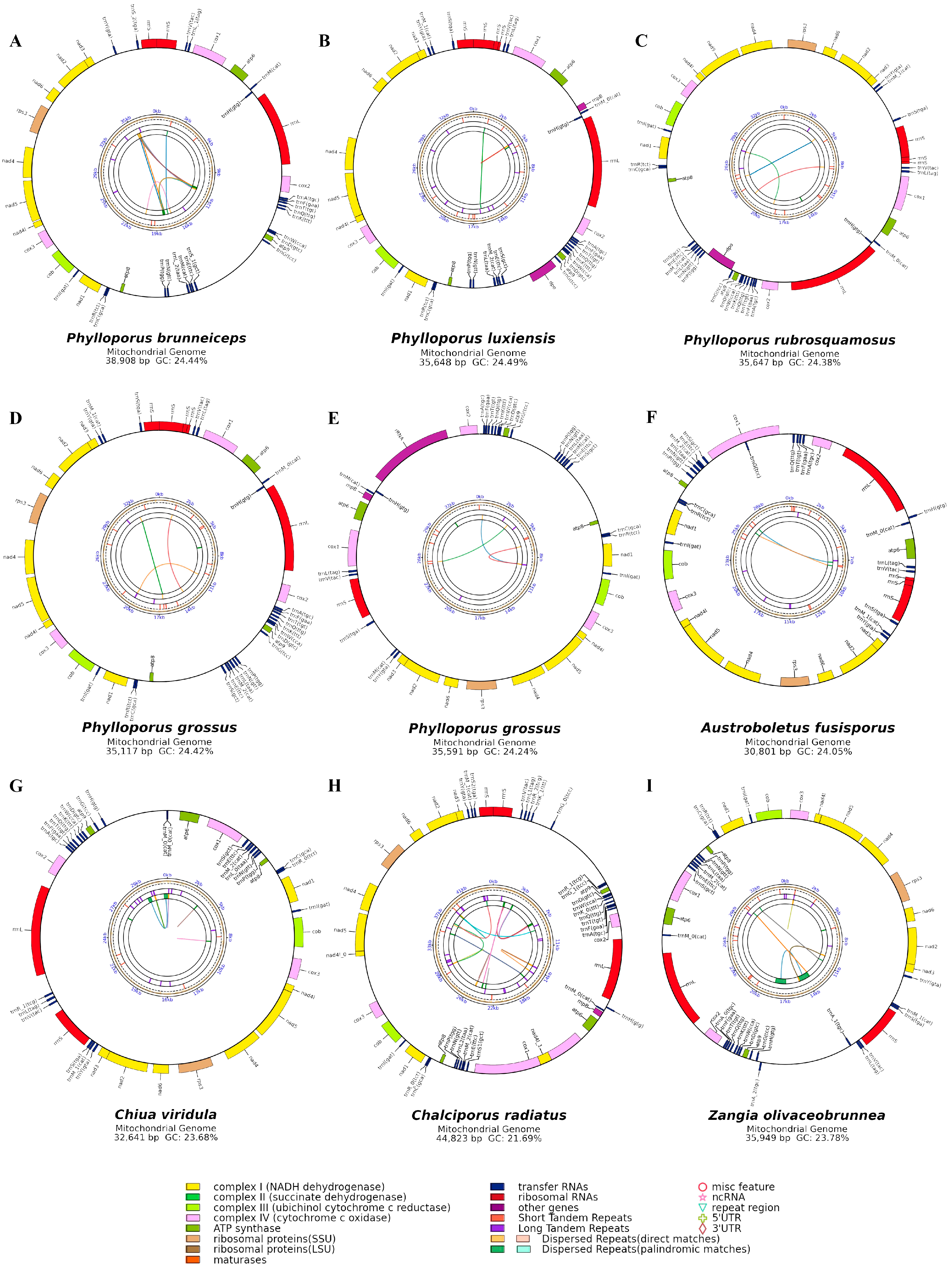
Figure 1. Circular maps of the nine mitogenomes from Boletaceae. (A) Phylloporus brunneiceps * HKAS105261; (B) Phylloporus luxiensis HKAS105263; (C) Phylloporus rubrosquamosus HKAS105259; (D) Phylloporus grossus HKAS105262; (E) Phylloporus grossus HKAS105260; (F) Austroboletus fusisporus HKAS105269; (G) Chiua viridula HKAS105270; (H) Chalciporus radiatus HKAS105264; (I) Zangia olivaceobrunnea HKAS105268). Each gene is represented by a corresponding colored block. Colored blocks outside the ring indicate genes located on the direct strand, while those inside the ring represent genes on the reverse strand. Colored lines for intraspecies collinearity analysis.
Jof 11 00831 g001
Figure 2. (A) Short tandem repeats and (B) dispersed repeats in the mitogenomes of the nine Boletaceae specimens.
Jof 11 00831 g002
Figure 3. Collinear analysis of mitogenomes in Boletaceae. The colored rectangles beneath the homologous blocks represent the positions and lengths of different types of genes. The same-origin sections are connected by lines of corresponding colors. (A) Phylloporus grossus HKAS105262; (B) Phylloporus grossus HKAS105260; (C) Phylloporus brunneiceps HKAS105261; (D) Phylloporus rubrosquamosus HKAS105259; (E) Phylloporus luxiensis HKAS105263; (F) Chiua viridula HKAS105270; (G) Austroboletus fusisporus HKAS105269; (H) Zangia olivaceobrunnea HKAS105268; (I) Chalciporus radiatus HKAS105264.
Jof 11 00831 g003
Figure 4. Gene order analysis in nine specimens of Boletaceae collected from Jiangxi Province. Each colored block represents a distinct type of gene. A total of 14–15 PCGs and 2 rRNA genes were used in this analysis.
Jof 11 00831 g004
Figure 8. Phylogenetic tree constructed based on Bayesian inference (BI) and maximum likelihood (ML) analyses of 15 core protein-coding genes (atp6, atp8, atp9, cob, cox1, cox2, cox3, nad1, nad2, nad3, nad4, nad4L, nad5, nad6, and rps3). RAxML likelihood bootstrap support values (98%) and Bayesian posterior probabilities (PP > 0.71) are indicated above or below the branches as BS/PP. The red circles mark the newly sequenced species, and the blue rectangles indicate the outgroups.
Jof 11 00831 g008
Table 1. Information of samples for phylogenetic analysis.
FamilySpeciesGenbank NumberSize (bp)References
BoletaceaeAureoboletus raphanaceusNC07966242,157[36]
Baorangia bicolorMW30859938,082[28]
Boletus edulisMW30860934,763[28]
Butyriboletus subappendiculatusOZ17199335,023Unpublished
Butyriboletus roseoflavusMW30860036,622[28]
Butyriboletus roseoflavusMZ20235736,551Unpublished
Caloboletus calopusMW30860232,883[28]
Hemileccinum impolitumNC05680839,362Unpublished
Lanmaoa macrocarpaNC08088538,139[30]
Neoboletus brunneissimusMW30860542,147[28]
Neoboletus magnificusMW30860339,449[28]
Neoboletus obscureumbrinusMW30860739,929[28]
Pulveroboletus raveneliiNC06166643,528[37]
Rufoboletus hainanensisOR43878736,592[38]
Retiboletus ornatipesMW30860136,785[28]
Tylopilus brunneirubensNC08429132,389[39]
Tylopilus plumbeoviolaceoidesNC05683537,242[29]
PaxillaceaePaxillus involutusNC04520339,109[27]
Paxillus rubicundulusNC04520441,061[27]
Table 2. The nine newly sequenced mitochondrial genomes of Boletaceae in Jiangxi Province.
Species NameGenbank NumberLength(bp)GCRate(%)AT-SkewGC-SkewtRNAsIntronsNumber of Repeats
Austroboletus fusisporusPV42403430,80124.05−0.030.012015
Chalciporus radiatusPV42403844,82321.69−0.02−0.0224027
Chiua viridulaPV42403332,64123.680.06−0.0225015
Phylloporus brunneicepsPQ82189538,90824.440.04−0.042806
Phylloporus grossusPQ84649035,59124.240.06−0.032403
Phylloporus grossusPV42403535,11724.420.05−0.032403
Phylloporus luxiensisPV42403735,64824.490.06−0.0324116
Phylloporus rubrosquamosusPV42403635,64724.380.05−0.032414
Zangia olivaceobrunneaPV42403935,94923.78−0.02−0.032607
Table 3. Comparative genomic characteristics of 24 species o (22 Boletaceae, 2 Paxillaceae).
Species NameGenbank NumberLength (bp)GC Rate (%)AT-SkewGC-SkewNumber of Repeats
Austroboletus fusisporusPV42403430,80124.05−0.030.015
Chalciporus radiatusPV42403844,82321.69−0.02−0.0227
Chiua viridulaPV42403332,64123.680.06−0.0215
Phylloporus brunneicepsPQ82189538,90824.440.04−0.046
Phylloporus grossusPQ84649035,59124.240.06−0.033
Phylloporus grossusPV42403535,11724.420.05−0.033
Phylloporus luxiensisPV42403735,64824.490.06−0.0316
Phylloporus rubrosquamosusPV42403635,64724.380.05−0.034
Zangia olivaceobrunneaPV42403935,94923.78−0.02−0.037
Aureoboletus raphanaceusNC07966242,15722.730.030.038
Baorangia bicolorMW30859938,08223.610.020.0112
Boletus edulisMW30860934,76323.830.02−0.0612
Butyriboletus subappendiculatusOZ17199335,02322.820.030.0213
Butyriboletus roseoflavusMW30860036,62223.130.030.0214
Butyriboletus roseoflavusMZ20235736,55123.130.030.0212
Caloboletus calopusMW30860232,88323.210.030.0217
Hemileccinum impolitumNC05680839,36223.980.020.015
Lanmaoa macrocarpaNC08088538,13923.50.030.0314
Neoboletus brunneissimusMW30860542,14722.960.030.0214
Neoboletus magnificusMW30860339,44922.930.030.0211
Neoboletus obscureumbrinusMW30860739,92923.10.030.049
Pulveroboletus raveneliiNC06166643,52823.35−0.020.0512
Rufoboletus hainanensisOR43878736,59223.95−0.04−0.025
Retiboletus ornatipesMW30860136,78523.190.030.0113
Tylopilus brunneirubensNC08429132,38923.8−0.020.059
Tylopilus plumbeoviolaceoidesNC05683537,24223.020.020.019
Paxillus involutusNC04520339,10921.75−0.090.0653
Paxillus rubicundulusNC04520441,06121.03−0.090.0547
Disclaimer/Publisher’s Note: The statements, opinions and data contained in all publications are solely those of the individual author(s) and contributor(s) and not of MDPI and/or the editor(s). MDPI and/or the editor(s) disclaim responsibility for any injury to people or property resulting from any ideas, methods, instructions or products referred to in the content.