1. Introduction
Mycoplasma bovis (
M. bovis) is a significant pathogen responsible for bovine respiratory disease complex (BRDC) and systemic infections in cattle, including pneumonia, mastitis, arthritis, otitis, keratoconjunctivitis, and reproductive disorders [
1,
2]. Neonatal and weaned calves, as well as cattle subjected to environmental stressors (e.g., transportation, regrouping, or seasonal changes), are particularly susceptible. The absence of a cell wall renders
M. bovis intrinsically resistant to β-lactam antibiotics (e.g., penicillin) and prone to developing resistance to other antimicrobials [
3,
4]. Compounded by the lack of effective vaccines and targeted therapeutics,
M. bovis has emerged as a critical threat to global cattle production, with severe economic consequences for both dairy and beef industries [
3,
5,
6].
Systematic analyses reveal that
M. bovis infections lead to substantial economic losses, including a 15–30% decline in milk production, elevated somatic cell counts (>500,000 cells/mL) [
7,
8], and increased mortality and culling rates (5–20%) [
9,
10,
11]. Chronic infections and synergistic interactions with respiratory pathogens (e.g.,
Mannheimia haemolytica) further exacerbate disease severity and complicate treatment [
12]. These challenges highlight the urgent need for reliable diagnostic and control strategies, particularly given the pathogen’s rapid transmission within herds and persistence once established [
13].
Current diagnostic methods for
M. bovis include pathogen isolation, molecular techniques (e.g., PCR), and serological assays [
14,
15]. However, chronic infections and prolonged antibiotic use often hinder bacterial culture, while serological tests, especially enzyme-linked immunosorbent assays (ELISAs), offer high sensitivity and specificity for detecting antibodies in infected cattle [
16,
17]. Among these, the competitive ELISA (cELISA) exhibits prominent advantages: it quantifies target antibodies via competitive binding against specific epitopes, which effectively reduces cross-reactivity induced by contaminating proteins present in coated antigens [
9,
18]. Accordingly, cELISA may improve specificity when based on well-characterized antigen–antibody competition systems [
19].
In our previous work, using a proteomics approach, we identified the
M. bovis membrane protein MbovP579 and demonstrated that it is a conserved, sensitive, and specific antigen among
M. bovis strains, indicating its potential as a diagnostic target [
10]. Subsequently, a monoclonal antibody was developed [
20]. Here, we developed a cELISA using recombinant MbovP579 as the coating antigen and a monoclonal antibody (1A2) as the competitive probe. This assay aims to provide a cost-effective, high-throughput tool for accurate
M. bovis serodiagnosis, supporting improved disease management in clinical and field settings.
2. Materials and Methods
2.1. Animal Ethics
Ten-week-old BALB/c mice were obtained from the Experimental Animals of Huazhong Agricultural University and used to develop the mAbs. Naturally immunized cattle and M. bovis-negative cattle from a farm in Hubei Province, China, were sampled with the consent of the farm owners. The animal experiments in this study were conducted in strict accordance with the Guide for the Care and Use of Laboratory Animals, Hubei Province, China. The protocols were approved by the Ethics Committee of Huazhong Agricultural University (Agreement No. HZAUMO-2023-0038; Agreement No. HZAUCA-2022-0017).
2.2. Serum Sample Collection
The negative serum samples for
M. bovis in each experiment were verified using a commercial
M. bovis antibody test kit (Biovet
®, Saint-Hyacinthe, QC, Canada) and PCR [
11,
21], corresponding to cattle that showed no clinical symptoms [
22,
23]. Information regarding the positive serum samples for experimental groups is presented in
Table 1.
Antisera against the M. bovis HB150 strain, Mycoplasma bovirhinis, Mycoplasma agalactiae, Mycoplasma arginini, Pasteurella multocida, and Mannheimia haemolytica were prepared via vaccination. For M. bovis, calves were immunized with an attenuated M. bovis HB150 strain (antigen concentration of 108 CFU/mL). Negative calves were intranasally inoculated with 2 × 10⁸ CFU/2.0 mL. Blood samples were obtained from the caudal (tail) or jugular vein at 0, 7, 14, 21, 28, and 35 days post-vaccination, and positivity was determined using the Biovet® kit.
All the serum samples were subsequently stored at −20 °C in the National Key Laboratory of Agricultural Microbiology, Huazhong Agricultural University.
For the other pathogens, aluminum hydroxide gel-inactivated vaccines were prepared using Mycoplasma bovirhinis, Mycoplasma agalactiae, Mycoplasma arginini, Pasteurella multocida, and Mannheimia haemolytica via a 60 °C water bath treatment for 30 min (antigen concentration of 109 CFU/mL). Negative calves were subcutaneously inoculated with 2 × 10⁹ CFU/2.0 mL and boosted with the same dose 30 days later. Blood samples were collected one week post-boost, and positive sera were confirmed via an in-house indirect ELISA.
For the in-house iELISA, ultrasound-fragmented bacterial proteins (quantified by an Enhanced Bicinchoninic Acid Protein Assay kit) were coated at 1.0 μg/mL overnight at 4 °C, blocked with 200 μL 1% fish gelatin (Sigma, Burlington, MA, USA) in PBS for 1 h at 37 °C, and incubated with serum samples. Following standard washing with PBST, the plates were incubated with TMB (Seracare, Shanghai, China) for 10 min in the dark, stopped with 2 M H2SO4, and read at 450 nm using a microplate reader (BMG LABTECH, Offenburg, BW, Germany). Samples were classified as positive if their OD450 value was ≥2.1 times that of the negative control.
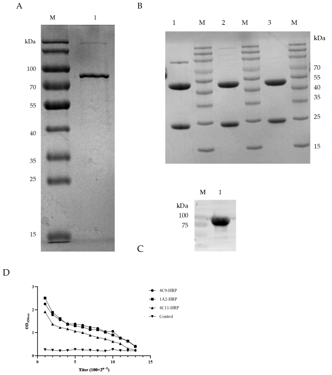
2.3. Expression and Purification of MbovP579 Protein
The recombinant plasmid encoding the MbovP579 protein was previously constructed in our lab. Briefly, the
Mbov579 DNA fragment was inserted into the pET30a(+) plasmid and transformed into
E. coli BL21 (DE3) for expression [
10]. MbovP579 was purified by a Ni Sepharose
TM 6 Fast Flow resin gravity column (GE Healthcare, IL, USA). Purity was evaluated by 10% sodium dodecyl sulfate–polyacrylamide gel electrophoresis (SDS-PAGE), and the concentration was determined by the Enhanced BCA Protein Assay kit (Beyotime, Shanghai, China).
2.4. Production of Monoclonal Antibodies Against the MbovP579 Protein
Three hybridoma cell lines, 1A2, 4C9, and 4C11, were developed with the procedure as follows. Briefly, BALB/c mice were immunized with purified MbovP579 antigen emulsified in Freund’s adjuvant. Spleen cells were fused with SP2/0 myeloma cells using PEG-mediated fusion, and positive clones were selected using in-house iELISA. Furthermore, 10-week-old SPF BALB/c mice were injected with 500 µL of Freund’s incomplete adjuvant (IFA, Sigma, USA), followed 5–7 days later by intraperitoneal injection of hybridoma cells (5 × 10⁵ to 1 × 10⁶) to generate ascites. Ascites fluid was collected and purified using rProtein G Beads 4FF columns (Smart Life Sciences, Nanjing, China), and the purified monoclonal antibodies were quantified using the BCA assay.
2.5. HRP-Conjugated mAbs and Blocking Assay Evaluation
Monoclonal antibody labeling was performed using a modified periodate oxidation method. Briefly, 5 mg of HRP (Sigma, USA) was dissolved in 1 mL of double-distilled water, mixed with 500 µL of 0.1 M sodium periodate solution, and incubated at 4 °C for 30 min. Then, 0.5 mL of 0.16 M ethylene glycol was added, reacting at room temperature in the dark for 30 min. Purified mAb (5 mg) was added, and the solution was dialyzed against 0.05 M carbonate–bicarbonate buffer (pH 9.5) at 4 °C for 15 h. Subsequently, 0.2 mL of 4 mg/mL NaBH4 (Sinopharm, Beijing, China) was added and incubated at 4 °C for 2 h. The conjugate was precipitated with saturated ammonium sulfate, centrifuged, resuspended in phosphate buffer (PB), and dialyzed. The HRP-conjugated mAbs were verified via iELISA and stored at −80 °C.
To validate blocking efficacy, a cELISA was performed. Plates were coated with MbovP579 protein (100 ng/well, 100 µL/well) at 4 °C for 12 h, blocked with 5% skim milk (200 µL/well) at 37 °C for 60 min, and washed. Negative and positive sera (100 µL/well) were incubated at 37 °C for 60 min. After washing, HRP-labeled mAbs (1A2, 4C9, 4C11) at dilutions of 1:1000, 1:2000, and 1:5000 were added and OD
450 was measured. The percentage inhibition (PI) was calculated as follows [
24,
25]:
2.6. Establishment of MbovP579-cELISA
The optimal coating concentration of MbovP579 (0.5–4.0 μg/mL) and dilution of HRP-conjugated 1A2 mAb (0.28–2.8 μg/mL) were determined by checkerboard titration. The ratio of OD450 values of the negative control serum (N) to the positive control serum (P), expressed as N/P, was calculated. The conditions yielding the highest N/P ratio were considered optimal. Optimal serum dilution (1:2–1:10) and reaction times (45, 60, or 90 min) were subsequently determined using the same maximization principle.
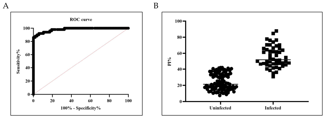
2.7. Determination of Cut-Off Value, Diagnostic Sensitivity, and Specificity
Fifty
M. bovis-positive (
Table 1) and 104
M. bovis-negative (
Table A3) serum samples were tested to determine the cut-off value. The diagnostic cut-off was established by receiver operating characteristic (ROC) curve analysis. The optimal threshold was selected to maximize Youden’s index (J = sensitivity + specificity − 1). Then, diagnostic sensitivity and specificity were calculated based on a 2 × 2 contingency table of true disease status vs. test results. Diagnostic sensitivity was defined as the ability of the assay to correctly identify
M. bovis-positive samples, whereas diagnostic specificity was defined as the ability of the assay to correctly identify
M. bovis-negative samples [
26].
2.8. Evaluation of cELISA
Sensitivity was evaluated using two approaches. First, nine clinically healthy calves confirmed to be negative for M. bovis were inoculated intranasally with the M. bovis HB150 strain at a dose of 2 × 10⁸ CFU in 2.0 mL. Blood samples collected at 0, 7, 14, 21, 28, and 35 days post-inoculation were tested in parallel using the cELISA and the Biovet® kit. Second, strongly positive serum samples were subjected to serial dilutions ranging from 1- to 64-fold to determine the analytical limit of detection, and the results were compared with those obtained using the commercial Biovet® kit.
Analytical specificity was assessed by testing antisera against M. bovis HB150, M. bovirhinis, M. agalactiae, M. arginini, P. multocida and M. haemolytica.
Repeatability was assessed by testing 10 positive and 10 negative samples in triplicate across three plates from the same batch (intra-assay) and different batches (inter-assay). The coefficient of variation (CV) was calculated using the formula CV = SD/X × 100%, where SD represents the standard deviation, and X represents the mean PI value [
27].
2.9. Metabolic Inhibition Test (MIT) of M. bovis
M. bovis cultured to the logarithmic phase was diluted to 104 CFU/mL using PPLO liquid medium for use. For the assay, eight wells were designated on a cell culture plate, including 3 test wells, 3 negative serum control wells, and 2 blank control wells.
Aliquots of the diluted bacterial suspension (100 μL per well) were dispensed into both test wells and negative serum control wells. Subsequently, 100 μL of M. bovis antiserum was pipetted into the test wells, while 100 μL of antibody negative serum was added to the negative serum control wells. All mixtures were gently mixed to ensure homogeneity. Finally, 200 μL of PPLO medium alone was added to the blank control wells.
The plate was incubated at 37 °C for 48 h prior to result observation. The anticipated outcomes are as follows: no noticeable color change should be observed in the medium of test wells and blank control wells, whereas the medium in negative serum control wells should turn distinctly yellow [
28].
2.10. Application for the cELISA
The established cELISA was used to test 1069 clinical bovine serum samples from four different farms. The results were compared with the commercial Biovet
® kit to calculate the sensitivity and agreement. Discrepant specimens were further characterized by the metabolic inhibition test (MIT) to facilitate subsequent specificity evaluation [
28].
2.11. Statistical Analysis
Statistical analysis was performed using Graphpad Prism V.8.0.2 (San Diego, CA, USA). Parameters were calculated using the online Epitools platform (
http://www.ausvet.com.au/ URL (accessed on 5 July 2025)).
4. Discussion
The diagnosis of
M. bovis infection relies on diverse methodologies, each with distinct advantages. PCR-based techniques are widely used for pathogen detection because of their high sensitivity of 83–100% and specificity of 93–99% [
29,
30,
31], while serological assays provide complementary value for early outbreak detection and herd surveillance. ELISA demonstrates robust performance, with reported sensitivities of 90–97% and specificities of 94–98% in field validations [
10,
32,
33,
34].
In previous work, we identified MbovP579 as a highly immunogenic membrane protein harboring multiple T-cell and B-cell epitopes [
10]. Importantly, mAb 1A2, raised against MbovP579, exhibited no cross-reactivity with other
Mycoplasma species, including
M. agalactiae [
10], underscoring its exceptional species specificity. Furthermore, the high genetic conservation of MbovP579 across
M. bovis strains (95–100% sequence identity) supports its utility as a universal diagnostic target.
Building upon these findings, we developed a cELISA using mAb 1A2 and recombinant MbovP579. By conjugating mAb 1A2 directly with HRP, we eliminated the need for secondary antibodies, streamlining the procedure. The assay performed comparably to a commercial kit, achieving a 100% seropositivity rate by day 21 post-vaccination and maintaining detection at a 1:32 dilution.
In the clinical validation using 1069 bovine serum samples, the cELISA showed high overall agreement with the commercial kit (90.46%; kappa = 0.81). Discrepant samples were retested using the MIT assay, whose results confirmed that the commercially available Biovet® kit yielded more false-positive results, further corroborating the superior specificity of the newly developed cELISA.
Compared with conventional indirect ELISAs, the MbovP579-based cELISA offers enhanced specificity without compromising sensitivity, aligning with emerging trends in veterinary serology that favor recombinant protein-based assays for their reproducibility and reduced batch-to-batch variability. This cELISA is currently being validated using clinical specimens; pending favorable validation results, the reagent kit will proceed to regulatory submission for commercialization—thereby improving diagnostic accuracy and supporting disease prevention and control efforts.



 ,
,