Canine Cognitive Dysfunction and Alzheimer’s Disease: Pathophysiological Relationships and the Impact of Glymphatic System Impairment on Neurodegeneration
 , , , , , , and
Simple Summary
Abstract
1. Introduction
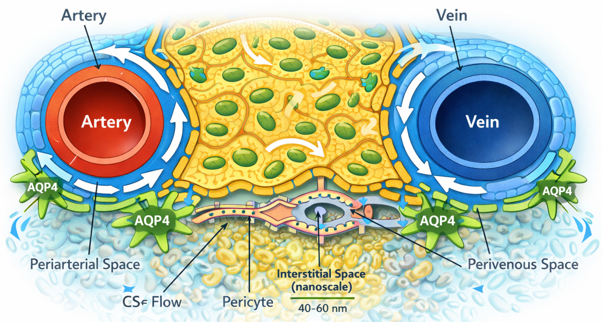
2. Anatomy of Glymphatic System
2.1. Cerebrospinal Fluid and Periarterial Central Nervous System
2.2. Astrocytes, AQP4 Polarization, and the Perivascular Glial Sheath
2.3. Parenchymal Interstitial Pathways: From Periarterial Spaces to Perivenous Routes
2.4. Ultrastructural Anatomy of Glymphatic System
3. Cerebrospinal Fluid, the Glymphatic System and the Aging Dog
3.1. Aquaporin-4 Channels
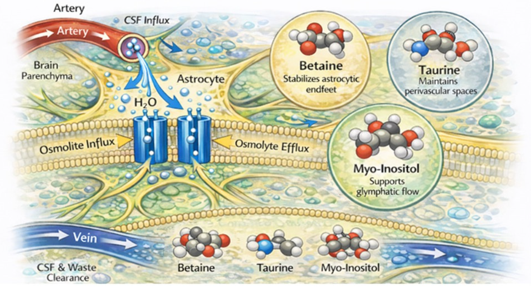
3.2. Role of Osmolytes in Alzheimer’s Disease and Canine Cognitive Dysfunction
3.2.1. Betaine
3.2.2. Taurine
3.2.3. Myo-Inositol
- Osmoregulation in the brain: MI acts as a compatible osmolyte in neural tissue, aiding neurons and astrocytes in adapting to osmotic stress and contributing to the regulation of cell volume [81].
- Signal transduction, neurotransmission, and neurobehavioral effects: inositol derivatives participate in pathways that modulate serotonin, dopamine, noradrenaline, and acetylcholine neurotransmission, processes central to mood regulation, cognition, and stress responses [163].
- Neuronal connectivity and synaptic maturation: in non-canine models, including mice, rats, and human neurons, MI has been shown to promote synaptic connectivity by enhancing excitatory synapse density and postsynaptic structure, suggesting a role in synaptic development and neuronal network formation [164].

| Osmolyte | Main Cellular Localization | Primary Transporters | Osmoregulation Function | Additional Neurobiological Roles | Alterations in Brain Pathology | Key References |
|---|---|---|---|---|---|---|
| Taurine | Astrocytes > neurons | TauT (SLC6A6) | Major neuro-osmolyte regulating intracellular osmotic balance; mediates regulatory volume decrease under hypo-osmotic stress and prevents cellular dehydration under hyperosmotic conditions; stabilizes astrocytic volume and perivascular space integrity and may support of glymphatic clearance | Neuromodulation (modulate GABAergic signaling); Ca2+ homeostasis, membrane stabilization, antioxidant and cytoprotective effects; trophic factor during brain development; anti-inflammatory modulation of microglial activation; | Altered taurine homeostasis is associated with cerebral edema, ischemia, and neurodegeneration; increased vulnerability to excitotoxicity insufficient taurine-mediated volume regulation may contribute to impaired glymphatic clearance in AD; canine data are limited but suggest relevance for age-related cognitive decline | [81,105,128,129,135,136,156,183] |
| Betaine | Astrocytes and neurons (region-dependent) | BGT-1/GAT2 (SLC6A12) | Compatible organic osmolyte involved in cell volume regulation, particularly under hyperosmotic stress; prevents astrocyte swelling and supports perivascular space integrity | Methyl donor for remethylation of homocysteine; chemical chaperone for protein conformation; modulation of oxidative stress, neuroprotection, regulation of microglial polarization; potential support of astrocytic endfeet integrity and AQP4-related water homeostasis and of glymphatic function modulation of GABAergic neurotransmission | Evidence suggests neuroprotective effects in AD models, including inhibition of amyloid-β aggregation, suppression of inflammasome signaling, promotion of anti-inflammatory microglial phenotypes, and modulation of GABA metabolism; reduced transport or dysregulation may impair glymphatic clearance and exacerbate neuronal stress; data in dogs are scarce, but metabolomic and comparative physiology studies support a putative protective role in cognitive dysfunction | [63,82,84,87,95,97,98,99,100,107] |
| Myo-inositol | Predominantly astrocytes | SMIT1 (SLC5A3); SMIT2 (SLC5A11) HMIT1 (SLC2A13) | Slow but sustained osmoadaptation; accumulation during chronic osmotic stress | Precursor of phosphoinositides, intracellular signaling, synaptic connectivity, neurotransmission, and osmoregulation A of astrocyte markers in neuroimaging | Elevated levels in Alzheimer’s disease reflect gliosis and osmotic stress, afailure to resolve inflammation, a loss of normal astrocytic support functions, and imbalance of the oxidant/antioxidant system. Altered MI functions may influence glymphatic dysfunction | [81,156,159,167,168] |
4. Pathogenesis
4.1. Genetics
4.2. Amyloid Deposition and Amyloidogenic Processing
4.3. APOE4, Alzheimer’s Disease, and Canine Cognitive Dysfunction: Molecular Parallels and Translational Implications
4.4. Presenilin
4.5. Tau Biology and Isoforms
4.5.1. Tau Isoforms in Pathology
4.5.2. Tau Misfolding, Aggregation, and Thermodynamic Perspective
Hyperphosphorylation and Post-Translational Modification
Thermodynamic Hypothesis of Tau Aggregation
Prion-like Propagation of Tau in AD and CCD
4.5.3. Interplay Between Tau Pathology, Parenchymal Amyloid Plaques, and Cerebral Amyloid Angiopathy
4.6. Cerebral Amyloid Angiopathy (CAA)
4.7. Neuronal and Synaptic Loss
4.8. Neurotransmitter System Dysfunction
4.9. Mitochondrial Dysfunction and Oxidative Stress
4.10. Neuroinflammation and Glial Activation
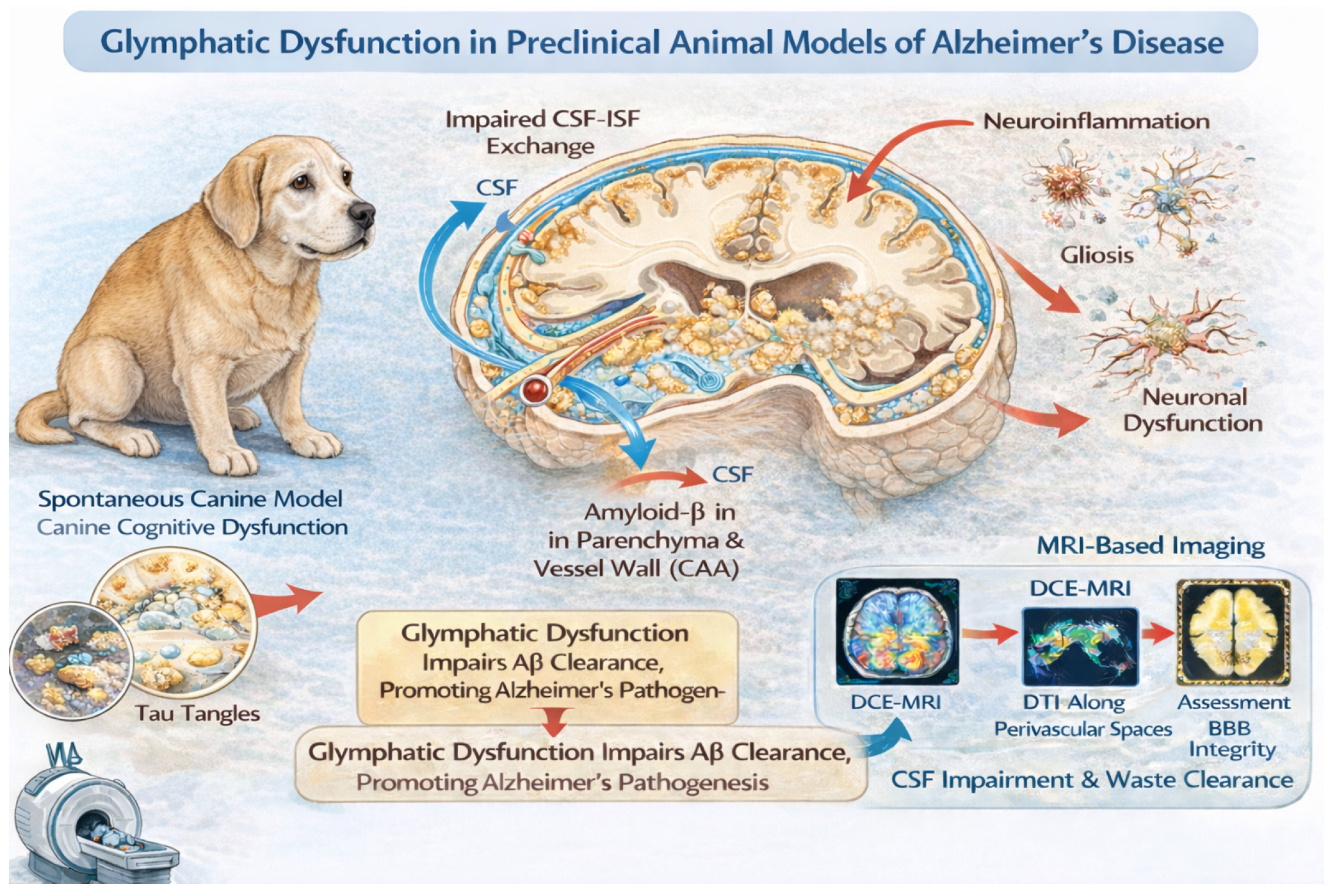
4.11. Glymphatic System
5. Neuropathology of Alzheimer’s Disease
5.1. Gross Pathology
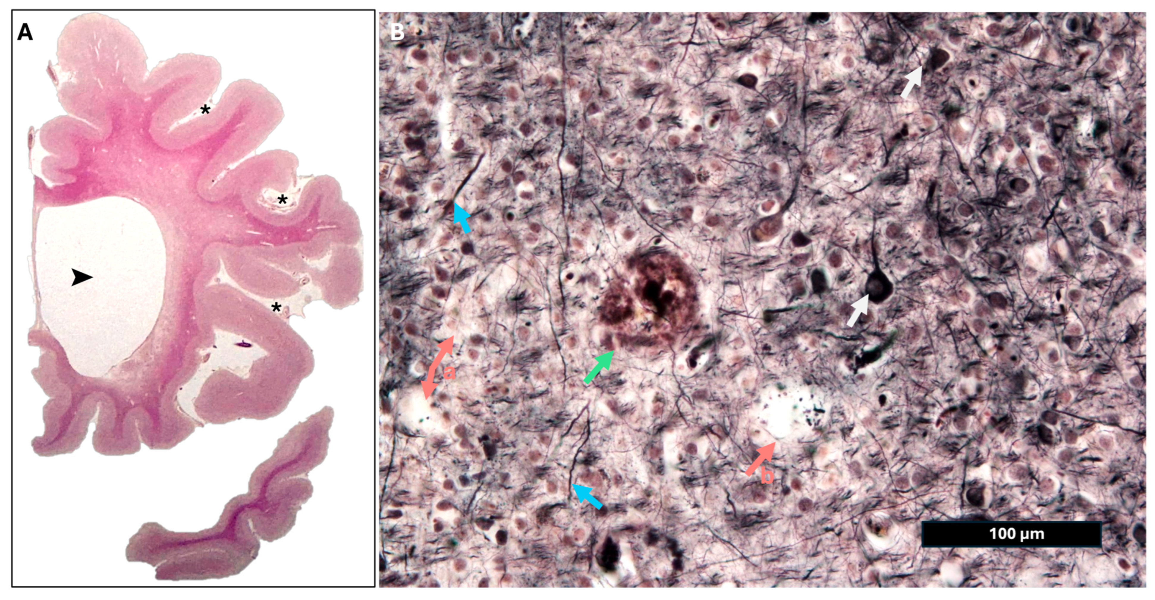
5.2. Histopathology
- (a)
- Plaques are composed predominantly of Aβ peptides (Aβ40/Aβ42) derived from the amyloid precursor protein (APP) via β- and γ-secretase cleavage.
- (b)
- (c)
- Diffuse plaques, often Aβ42-rich, appear earlier in disease and may lack neuritic elements and dense fibrils.
- (d)
- Plaque deposition is initially prominent in the neocortex, then involves the hippocampus and limbic structures and finally subcortical regions as pathology advances [259].
5.3. Neurofibrillary Tangles and Tau Pathology
5.4. Neuronal and Synaptic Pathology
5.5. Glial and Vascular Changes
5.6. Immunohistochemistry in Alzheimer’s Disease
5.7. National Institute on Aging–Alzheimer’s Association (NIA-AA) Guidelines and ABC Scoring System
6. Neuropathology of Canine Cognitive Dysfunction
6.1. Gross Pathology
6.2. Histopathology
6.2.1. Amyloid-β Pathology
6.2.2. Tau and Other Proteinopathies
6.2.3. Neuronal and Synaptic Changes
6.2.4. Glial and Vascular Changes
6.2.5. Immunohistochemistry in Canine Cognitive Dysfunction
7. Pathological Diversity and Similarity Between Alzheimer’s Disease and Canine Cognitive Dysfunction
8. Translational Impact Related to Alzheimer’s Disease and Canine Cognitive Dysfunction Through Transmission Electron Microscopy
8.1. Alzheimer’s Disease, Canine Cognitive Dysfunction and Transmission Electron Microscopy
- (a)
- To validate candidate biomarkers across species;
- (b)
- To evaluate disease-modifying interventions at synaptic and fibrillar scales;
- (c)
- To define the temporal relationship between Aβ accumulation, synaptic alterations, and cognitive impairment.
8.1.1. Amyloid Fibrils and Plaques
8.1.2. Neurofibrillary Tangles
8.1.3. Synapses and Organelles
8.1.4. Myelin and Axons
9. Neuroclinical Aspects
10. Magnetic Resonance Imaging
11. Conclusions
- (a)
- A naturally occurring, environmentally relevant platform to study how Aβ, vascular pathology, and impaired glymphatic/perivascular clearance interact over time.
- (b)
- The opportunity to link behavior, advanced imaging, fluid biomarkers, and ultrastructure in ways that are difficult to achieve in humans and not fully recapitulated in rodent models.
- (c)
- A clinically meaningful setting in which to test multimodal interventions aimed at reducing Aβ burden, modulating tau phosphorylation and aggregation, restoring neurovascular and glymphatic function, and supporting synaptic and mitochondrial health [280].
Author Contributions
Funding
Institutional Review Board Statement
Informed Consent Statement
Data Availability Statement
Conflicts of Interest
References
- Fast, R.; Schütt, T.; Toft, N.; Møller, A.; Berendt, M. An Observational Study with Long-Term Follow-up of Canine Cognitive Dysfunction: Clinical Characteristics, Survival, and Risk Factors. J. Vet. Intern. Med. 2013, 27, 822–829. [Google Scholar] [CrossRef] [PubMed]
- Vite, C.H.; Head, E. Aging in the Canine and Feline Brain. Vet. Clin. N. Am. Small Anim. Pract. 2014, 44, 1113–1129. [Google Scholar] [CrossRef]
- Blanchard, T.; Eppe, J.; Mugnier, A.; Delfour, F.; Meynadier, A. Enhancing Cognitive Functions in Aged Dogs and Cats: A Systematic Review of Enriched Diets and Nutraceuticals. Geroscience 2025, 47, 2925–2947. [Google Scholar] [CrossRef] [PubMed]
- Prpar Mihevc, S.; Majdič, G. Canine Cognitive Dysfunction and Alzheimer’s Disease—Two Facets of the Same Disease? Front. Neurosci. 2019, 13, 604. [Google Scholar] [CrossRef]
- Cummings, B.J.; Head, E.; Afagh, A.J.; Milgram, N.W.; Cotman, C.W. Beta-Amyloid Accumulation Correlates with Cognitive Dysfunction in the Aged Canine. Neurobiol. Learn. Mem. 1996, 66, 11–23. [Google Scholar] [CrossRef]
- Colle, M.A.; Hauw, J.J.; Crespeau, F.; Uchihara, T.; Akiyama, H.; Checler, F.; Pageat, P.; Duykaerts, C. Vascular and Parenchymal Abeta Deposition in the Aging Dog: Correlation with Behavior. Neurobiol. Aging 2000, 21, 695–704. [Google Scholar] [CrossRef]
- Uchida, K.; Nakayama, H.; Goto, N. Pathological Studies on Cerebral Amyloid Angiopathy, Senile Plaques and Amyloid Deposition in Visceral Organs in Aged Dogs. J. Vet. Med. Sci. 1991, 53, 1037–1042. [Google Scholar] [CrossRef]
- Head, E.; Liu, J.; Hagen, T.M.; Muggenburg, B.A.; Milgram, N.W.; Ames, B.N.; Cotman, C.W. Oxidative Damage Increases with Age in a Canine Model of Human Brain Aging. J. Neurochem. 2002, 82, 375–381. [Google Scholar] [CrossRef]
- Serrano-Pozo, A.; Frosch, M.P.; Masliah, E.; Hyman, B.T. Neuropathological Alterations in Alzheimer Disease. Cold Spring Harb. Perspect. Med. 2011, 1, a006189. [Google Scholar] [CrossRef]
- Head, E.; Rofina, J.; Zicker, S. Oxidative Stress, Aging, and Central Nervous System Disease in the Canine Model of Human Brain Aging. Vet. Clin. N. Am. Small Anim. Pract. 2008, 38, 167–178, vi. [Google Scholar] [CrossRef]
- Head, E.; McCleary, R.; Hahn, F.F.; Milgram, N.W.; Cotman, C.W. Region-Specific Age at Onset of Beta-Amyloid in Dogs. Neurobiol. Aging 2000, 21, 89–96. [Google Scholar] [CrossRef]
- Jessen, N.A.; Munk, A.S.F.; Lundgaard, I.; Nedergaard, M. The Glymphatic System: A Beginner’s Guide. Neurochem. Res. 2015, 40, 2583–2599. [Google Scholar] [CrossRef] [PubMed]
- Iliff, J.J.; Wang, M.; Liao, Y.; Plogg, B.A.; Peng, W.; Gundersen, G.A.; Benveniste, H.; Vates, G.E.; Deane, R.; Goldman, S.A.; et al. A Paravascular Pathway Facilitates CSF Flow through the Brain Parenchyma and the Clearance of Interstitial Solutes, Including Amyloid β. Sci. Transl. Med. 2012, 4, 147ra111. [Google Scholar] [CrossRef] [PubMed]
- Iliff, J.J.; Wang, M.; Zeppenfeld, D.M.; Venkataraman, A.; Plog, B.A.; Liao, Y.; Deane, R.; Nedergaard, M. Cerebral Arterial Pulsation Drives Paravascular CSF-Interstitial Fluid Exchange in the Murine Brain. J. Neurosci. 2013, 33, 18190–18199. [Google Scholar] [CrossRef] [PubMed]
- Mestre, H.; Hablitz, L.M.; Xavier, A.L.; Feng, W.; Zou, W.; Pu, T.; Monai, H.; Murlidharan, G.; Rivera, R.M.C.; Simon, M.J.; et al. Aquaporin-4-Dependent Glymphatic Solute Transport in the Rodent Brain. eLife 2018, 7, e40070. [Google Scholar] [CrossRef]
- de Lahunta, A.; Glass, E.; Kent, M. Veterinary Neuroanatomy and Clinical Neurology, 5th ed.; Elsevier Mosby: Maryland Heights, MO, USA, 2021. [Google Scholar]
- Taoka, T.; Masutani, Y.; Kawai, H.; Nakane, T.; Matsuoka, K.; Yasuno, F.; Kishimoto, T.; Naganawa, S. Evaluation of Glymphatic System Activity with the Diffusion MR Technique: Diffusion Tensor Image Analysis along the Perivascular Space (DTI-ALPS) in Alzheimer’s Disease Cases. Jpn. J. Radiol. 2017, 35, 172–178. [Google Scholar] [CrossRef]
- Nielsen, S.; Nagelhus, E.A.; Amiry-Moghaddam, M.; Bourque, C.; Agre, P.; Ottersen, O.P. Specialized Membrane Domains for Water Transport in Glial Cells: High-Resolution Immunogold Cytochemistry of Aquaporin-4 in Rat Brain. J. Neurosci. 1997, 17, 171–180. [Google Scholar] [CrossRef]
- Nagelhus, E.A.; Ottersen, O.P. Physiological Roles of Aquaporin-4 in Brain. Physiol. Rev. 2013, 93, 1543–1562. [Google Scholar] [CrossRef]
- Papadopoulos, M.C.; Verkman, A.S. Aquaporin Water Channels in the Nervous System. Nat. Rev. Neurosci. 2013, 14, 265–277. [Google Scholar] [CrossRef]
- Iliff, J.J.; Chen, M.J.; Plog, B.A.; Zeppenfeld, D.M.; Soltero, M.; Yang, L.; Singh, I.; Deane, R.; Nedergaard, M. Impairment of Glymphatic Pathway Function Promotes Tau Pathology after Traumatic Brain Injury. J. Neurosci. 2014, 34, 16180–16193. [Google Scholar] [CrossRef]
- Álvarez, P.; Blasco, E.; Pumarola, M.; Wessmann, A. Aquaporin-4 Protein Expression in Normal Canine Brains. BMC Vet. Res. 2021, 17, 29. [Google Scholar] [CrossRef]
- Kress, B.T.; Iliff, J.J.; Xia, M.; Wang, M.; Wei, H.S.; Zeppenfeld, D.; Xie, L.; Kang, H.; Xu, Q.; Liew, J.A.; et al. Impairment of Paravascular Clearance Pathways in the Aging Brain. Ann. Neurol. 2014, 76, 845–861. [Google Scholar] [CrossRef]
- Klostranec, J.M.; Vucevic, D.; Bhatia, K.D.; Kortman, H.G.J.; Krings, T.; Murphy, K.P.; terBrugge, K.G.; Mikulis, D.J. Current Concepts in Intracranial Interstitial Fluid Transport and the Glymphatic System: Part I-Anatomy and Physiology. Radiology 2021, 301, 502–514. [Google Scholar] [CrossRef]
- Morris, A.W.J.; Sharp, M.M.; Albargothy, N.J.; Fernandes, R.; Hawkes, C.A.; Verma, A.; Weller, R.O.; Carare, R.O. Vascular Basement Membranes as Pathways for the Passage of Fluid into and out of the Brain. Acta Neuropathol. 2016, 131, 725–736. [Google Scholar] [CrossRef] [PubMed]
- Hasan-Olive, M.M.; Enger, R.; Hansson, H.-A.; Nagelhus, E.A.; Eide, P.K. Loss of Perivascular Aquaporin-4 in Idiopathic Normal Pressure Hydrocephalus. Glia 2019, 67, 91–100. [Google Scholar] [CrossRef]
- Rasschaert, M.; Schroeder, J.A.; Wu, T.-D.; Marco, S.; Emerit, A.; Siegmund, H.; Fischer, C.; Fretellier, N.; Idée, J.-M.; Corot, C.; et al. Multimodal Imaging Study of Gadolinium Presence in Rat Cerebellum: Differences Between Gd Chelates, Presence in the Virchow-Robin Space, Association with Lipofuscin, and Hypotheses About Distribution Pathway. Investig. Radiol. 2018, 53, 518–528. [Google Scholar] [CrossRef]
- Harvey, N.D. How Old Is My Dog? Identification of Rational Age Groupings in Pet Dogs Based Upon Normative Age-Linked Processes. Front. Vet. Sci. 2021, 8, 643085. [Google Scholar] [CrossRef]
- López-Otín, C.; Blasco, M.A.; Partridge, L.; Serrano, M.; Kroemer, G. The Hallmarks of Aging. Cell 2013, 153, 1194–1217. [Google Scholar] [CrossRef]
- López-Otín, C.; Blasco, M.A.; Partridge, L.; Serrano, M.; Kroemer, G. Hallmarks of Aging: An Expanding Universe. Cell 2023, 186, 243–278. [Google Scholar] [CrossRef] [PubMed]
- Guelfi, G.; Capaccia, C.; Tedeschi, M.; Bufalari, A.; Leonardi, L.; Cenci-Goga, B.; Maranesi, M. Dog Aging: A Comprehensive Review of Molecular, Cellular, and Physiological Processes. Cells 2024, 13, 2101. [Google Scholar] [CrossRef] [PubMed]
- Damkier, H.H.; Brown, P.D.; Praetorius, J. Cerebrospinal Fluid Secretion by the Choroid Plexus. Physiol. Rev. 2013, 93, 1847–1892. [Google Scholar] [CrossRef]
- Nielsen, S.; Smith, B.L.; Christensen, E.I.; Agre, P. Distribution of the Aquaporin CHIP in Secretory and Resorptive Epithelia and Capillary Endothelia. Proc. Natl. Acad. Sci. USA 1993, 90, 7275–7279. [Google Scholar] [CrossRef]
- Damkier, H.H.; Praetorius, J. Genetic Ablation of Slc4a10 Alters the Expression Pattern of Transporters Involved in Solute Movement in the Mouse Choroid Plexus. Am. J. Physiol. Cell Physiol. 2012, 302, C1452–C1459. [Google Scholar] [CrossRef] [PubMed]
- Oshio, K.; Watanabe, H.; Song, Y.; Verkman, A.S.; Manley, G.T. Reduced Cerebrospinal Fluid Production and Intracranial Pressure in Mice Lacking Choroid Plexus Water Channel Aquaporin-1. FASEB J. 2005, 19, 76–78. [Google Scholar] [CrossRef]
- Bulat, M.; Lupret, V.; Orehković, D.; Klarica, M. Transventricular and Transpial Absorption of Cerebrospinal Fluid into Cerebral Microvessels. Coll. Antropol. 2008, 32, 43–50. [Google Scholar]
- Orešković, D.; Klarica, M. A New Look at Cerebrospinal Fluid Movement. Fluids Barriers CNS 2014, 11, 16. [Google Scholar] [CrossRef] [PubMed]
- Yamada, S.; Miyazaki, M.; Yamashita, Y.; Ouyang, C.; Yui, M.; Nakahashi, M.; Shimizu, S.; Aoki, I.; Morohoshi, Y.; McComb, J.G. Influence of Respiration on Cerebrospinal Fluid Movement Using Magnetic Resonance Spin Labeling. Fluids Barriers CNS 2013, 10, 36. [Google Scholar] [CrossRef]
- Xie, L.; Kang, H.; Xu, Q.; Chen, M.J.; Liao, Y.; Thiyagarajan, M.; O’Donnell, J.; Christensen, D.J.; Nicholson, C.; Iliff, J.J.; et al. Sleep Drives Metabolite Clearance from the Adult Brain. Science 2013, 342, 373–377. [Google Scholar] [CrossRef] [PubMed]
- Lee, D.-H.; Torchetti, M.K.; Winker, K.; Ip, H.S.; Song, C.-S.; Swayne, D.E. Intercontinental Spread of Asian-Origin H5N8 to North America through Beringia by Migratory Birds. J. Virol. 2015, 89, 6521–6524. [Google Scholar] [CrossRef]
- Rasmussen, M.K.; Mestre, H.; Nedergaard, M. The Glymphatic Pathway in Neurological Disorders. Lancet Neurol. 2018, 17, 1016–1024. [Google Scholar] [CrossRef]
- Nedergaard, M.; Goldman, S.A. Glymphatic Failure as a Final Common Pathway to Dementia. Science 2020, 370, 50–56. [Google Scholar] [CrossRef]
- Buccellato, F.R.; D’Anca, M.; Serpente, M.; Arighi, A.; Galimberti, D. The Role of Glymphatic System in Alzheimer’s and Parkinson’s Disease Pathogenesis. Biomedicines 2022, 10, 2261. [Google Scholar] [CrossRef]
- Huang, S.-Y.; Zhang, Y.-R.; Guo, Y.; Du, J.; Ren, P.; Wu, B.-S.; Feng, J.-F.; Cheng, W.; Yu, J.-T. Alzheimer’s Disease Neuroimaging Initiative. Glymphatic System Dysfunction Predicts Amyloid Deposition, Neurodegeneration, and Clinical Progression in Alzheimer’s Disease. Alzheimers Dement. 2024, 20, 3251–3269. [Google Scholar] [CrossRef]
- Silva, I.; Silva, J.; Ferreira, R.; Trigo, D. Glymphatic System, AQP4, and Their Implications in Alzheimer’s Disease. Neurol. Res. Pract. 2021, 3, 5. [Google Scholar] [CrossRef]
- Ni, R. Magnetic Resonance Imaging in Animal Models of Alzheimer’s Disease Amyloidosis. Int. J. Mol. Sci. 2021, 22, 12768. [Google Scholar] [CrossRef]
- Keil, S.A.; Jansson, D.; Braun, M.; Iliff, J.J. Glymphatic Dysfunction in Alzheimer’s Disease: A Critical Appraisal. Science 2025, 389, eadv8269. [Google Scholar] [CrossRef] [PubMed]
- Simon, M.; Wang, M.X.; Ismail, O.; Braun, M.; Schindler, A.G.; Reemmer, J.; Wang, Z.; Haveliwala, M.A.; O’Boyle, R.P.; Han, W.Y.; et al. Loss of Perivascular Aquaporin-4 Localization Impairs Glymphatic Exchange and Promotes Amyloid β Plaque Formation in Mice. Alzheimers Res. Ther. 2022, 14, 59. [Google Scholar] [CrossRef] [PubMed]
- MacAulay, N. Molecular Mechanisms of Brain Water Transport. Nat. Rev. Neurosci. 2021, 22, 326–344. [Google Scholar] [CrossRef]
- Rossi, A.; Ratelade, J.; Papadopoulos, M.C.; Bennett, J.L.; Verkman, A.S. Neuromyelitis Optica IgG Does Not Alter Aquaporin-4 Water Permeability, Plasma Membrane M1/M23 Isoform Content, or Supramolecular Assembly. Glia 2012, 60, 2027–2039. [Google Scholar] [CrossRef]
- Saini, H.; Fernandez, G.; Kerr, D.; Levy, M. Differential Expression of Aquaporin-4 Isoforms Localizes with Neuromyelitis Optica Disease Activity. J. Neuroimmunol. 2010, 221, 68–72. [Google Scholar] [CrossRef] [PubMed]
- Lien, C.F.; Hazai, D.; Yeung, D.; Tan, J.; Füchtbauer, E.-M.; Jancsik, V.; Górecki, D.C. Expression of Alpha-Dystrobrevin in Blood-Tissue Barriers: Sub-Cellular Localisation and Molecular Characterisation in Normal and Dystrophic Mice. Cell Tissue Res. 2007, 327, 67–82. [Google Scholar] [CrossRef] [PubMed]
- Neely, J.D.; Amiry-Moghaddam, M.; Ottersen, O.P.; Froehner, S.C.; Agre, P.; Adams, M.E. Syntrophin-Dependent Expression and Localization of Aquaporin-4 Water Channel Protein. Proc. Natl. Acad. Sci. USA 2001, 98, 14108–14113. [Google Scholar] [CrossRef] [PubMed]
- Salman, M.M.; Kitchen, P.; Halsey, A.; Wang, M.X.; Törnroth-Horsefield, S.; Conner, A.C.; Badaut, J.; Iliff, J.J.; Bill, R.M. Emerging Roles for Dynamic Aquaporin-4 Subcellular Relocalization in CNS Water Homeostasis. Brain 2022, 145, 64–75. [Google Scholar] [CrossRef]
- Nycz, B.; Mandera, M. The Features of the Glymphatic System. Auton. Neurosci. 2021, 232, 102774. [Google Scholar] [CrossRef]
- Reith, W.; Haußmann, A. Importance of Virchow-Robin spaces. Radiologe 2018, 58, 142–147. [Google Scholar] [CrossRef]
- Verkman, A.S.; Tradtrantip, L.; Smith, A.J.; Yao, X. Aquaporin Water Channels and Hydrocephalus. Pediatr. Neurosurg. 2017, 52, 409–416. [Google Scholar] [CrossRef]
- Kitchen, P.; Salman, M.M.; Halsey, A.M.; Clarke-Bland, C.; MacDonald, J.A.; Ishida, H.; Vogel, H.J.; Almutiri, S.; Logan, A.; Kreida, S.; et al. Targeting Aquaporin-4 Subcellular Localization to Treat Central Nervous System Edema. Cell 2020, 181, 784–799.e19. [Google Scholar] [CrossRef]
- Taya, K.; Marmarou, C.R.; Okuno, K.; Prieto, R.; Marmarou, A. Effect of Secondary Insults upon Aquaporin-4 Water Channels Following Experimental Cortical Contusion in Rats. J. Neurotrauma 2010, 27, 229–239. [Google Scholar] [CrossRef]
- Hu, H.; Yao, H.; Zhang, W.; Zhang, L.; Ding, W.; Zhang, S.; Chen, Z.; Wei, E. Increased Expression of Aquaporin-4 in Human Traumatic Brain Injury and Brain Tumors. J. Zhejiang Univ. Sci. B 2005, 6, 33–37. [Google Scholar] [CrossRef] [PubMed]
- Braun, M.; Sevao, M.; Keil, S.A.; Gino, E.; Wang, M.X.; Lee, J.; Haveliwala, M.A.; Klein, E.; Agarwal, S.; Pedersen, T.; et al. Macroscopic Changes in Aquaporin-4 Underlie Blast Traumatic Brain Injury-Related Impairment in Glymphatic Function. Brain 2024, 147, 2214–2229. [Google Scholar] [CrossRef]
- Schmidt, M.J.; Rummel, C.; Hauer, J.; Kolecka, M.; Ondreka, N.; McClure, V.; Roth, J. Increased CSF Aquaporin-4, and Interleukin-6 Levels in Dogs with Idiopathic Communicating Internal Hydrocephalus and a Decrease after Ventriculo-Peritoneal Shunting. Fluids Barriers CNS 2016, 13, 12. [Google Scholar] [CrossRef][Green Version]
- Burg, M.B.; Ferraris, J.D.; Dmitrieva, N.I. Cellular Response to Hyperosmotic Stresses. Physiol. Rev. 2007, 87, 1441–1474. [Google Scholar] [CrossRef]
- Kushwah, N.; Jain, V.; Yadav, D. Osmolytes: A Possible Therapeutic Molecule for Ameliorating the Neurodegeneration Caused by Protein Misfolding and Aggregation. Biomolecules 2020, 10, 132. [Google Scholar] [CrossRef]
- Mueed, Z.; Mehta, D.; Rai, P.K.; Kamal, M.A.; Poddar, N.K. Cross-Interplay between Osmolytes and mTOR in Alzheimer’s Disease Pathogenesis. Curr. Pharm. Des. 2020, 26, 4699–4711. [Google Scholar] [CrossRef]
- Khan, S.; Siraj, S.; Shahid, M.; Haque, M.M.; Islam, A. Osmolytes: Wonder Molecules to Combat Protein Misfolding against Stress Conditions. Int. J. Biol. Macromol. 2023, 234, 123662. [Google Scholar] [CrossRef]
- Woo, J.; Kim, J.E.; Im, J.J.; Lee, J.; Jeong, H.S.; Park, S.; Jung, S.-Y.; An, H.; Yoon, S.; Lim, S.M.; et al. Astrocytic Water Channel Aquaporin-4 Modulates Brain Plasticity in Both Mice and Humans: A Potential Gliogenetic Mechanism Underlying Language-Associated Learning. Mol. Psychiatry 2018, 23, 1021–1030. [Google Scholar] [CrossRef]
- Nilius, B.; Eggermont, J.; Voets, T.; Buyse, G.; Manolopoulos, V.; Droogmans, G. Properties of Volume-Regulated Anion Channels in Mammalian Cells. Prog. Biophys. Mol. Biol. 1997, 68, 69–119. [Google Scholar] [CrossRef]
- Parkerson, K.A.; Sontheimer, H. Biophysical and Pharmacological Characterization of Hypotonically Activated Chloride Currents in Cortical Astrocytes. Glia 2004, 46, 419–436. [Google Scholar] [CrossRef] [PubMed]
- Han, Y.-E.; Kwon, J.; Won, J.; An, H.; Jang, M.W.; Woo, J.; Lee, J.S.; Park, M.G.; Yoon, B.-E.; Lee, S.E.; et al. Tweety-Homolog (Ttyh) Family Encodes the Pore-Forming Subunits of the Swelling-Dependent Volume-Regulated Anion Channel (VRACswell) in the Brain. Exp. Neurobiol. 2019, 28, 183–215. [Google Scholar] [CrossRef] [PubMed]
- Chen, S.; Wang, H.; Zhang, L.; Xi, Y.; Lu, Y.; Yu, K.; Zhu, Y.; Regina, I.; Bi, Y.; Tong, F. Glymphatic System: A Self-Purification Circulation in Brain. Front. Cell Neurosci. 2025, 19, 1528995. [Google Scholar] [CrossRef] [PubMed]
- Mestre, H.; Mori, Y.; Nedergaard, M. The Brain’s Glymphatic System: Current Controversies. Trends Neurosci. 2020, 43, 458–466. [Google Scholar] [CrossRef]
- Pasantes-Morales, H.; Cardin, V.; Tuz, K. Signaling Events during Swelling and Regulatory Volume Decrease. Neurochem. Res. 2000, 25, 1301–1314. [Google Scholar] [CrossRef] [PubMed]
- Pasantes-Morales, H.; Franco, R.; Torres-Marquez, M.E.; Hernández-Fonseca, K.; Ortega, A. Amino Acid Osmolytes in Regulatory Volume Decrease and Isovolumetric Regulation in Brain Cells: Contribution and Mechanisms. Cell Physiol. Biochem. 2000, 10, 361–370. [Google Scholar] [CrossRef] [PubMed]
- Pasantes-Morales, H.; Lezama, R.A.; Ramos-Mandujano, G.; Tuz, K.L. Mechanisms of Cell Volume Regulation in Hypo-Osmolality. Am. J. Med. 2006, 119, S4–S11. [Google Scholar] [CrossRef]
- Pasantes-Morales, H.; Franco, R.; Ordaz, B.; Ochoa, L.D. Mechanisms Counteracting Swelling in Brain Cells during Hyponatremia. Arch. Med. Res. 2002, 33, 237–244. [Google Scholar] [CrossRef]
- Zhou, Z.; Zhan, J.; Cai, Q.; Xu, F.; Chai, R.; Lam, K.; Luan, Z.; Zhou, G.; Tsang, S.; Kipp, M.; et al. The Water Transport System in Astrocytes-Aquaporins. Cells 2022, 11, 2564. [Google Scholar] [CrossRef] [PubMed]
- Melton, J.E.; Patlak, C.S.; Pettigrew, K.D.; Cserr, H.F. Volume Regulatory Loss of Na, Cl, and K from Rat Brain during Acute Hyponatremia. Am. J. Physiol. 1987, 252, F661–F669. [Google Scholar] [CrossRef]
- Verbalis, J.G.; Gullans, S.R. Hyponatremia Causes Large Sustained Reductions in Brain Content of Multiple Organic Osmolytes in Rats. Brain Res. 1991, 567, 274–282. [Google Scholar] [CrossRef] [PubMed]
- Sterns, R.H.; Baer, J.; Ebersol, S.; Thomas, D.; Lohr, J.W.; Kamm, D.E. Organic Osmolytes in Acute Hyponatremia. Am. J. Physiol. 1993, 264, F833–F836. [Google Scholar] [CrossRef]
- Pasantes-Morales, H.; Cruz-Rangel, S. Brain Volume Regulation: Osmolytes and Aquaporin Perspectives. Neuroscience 2010, 168, 871–884. [Google Scholar] [CrossRef]
- Lang, F.; Busch, G.L.; Ritter, M.; Völkl, H.; Waldegger, S.; Gulbins, E.; Häussinger, D. Functional Significance of Cell Volume Regulatory Mechanisms. Physiol. Rev. 1998, 78, 247–306. [Google Scholar] [CrossRef]
- Kopecká, J.; Krijt, J.; Raková, K.; Kožich, V. Restoring Assembly and Activity of Cystathionine β-Synthase Mutants by Ligands and Chemical Chaperones. J. Inherit. Metab. Dis. 2011, 34, 39–48. [Google Scholar] [CrossRef]
- Kempson, S.A.; Zhou, Y.; Danbolt, N.C. The Betaine/GABA Transporter and Betaine: Roles in Brain, Kidney, and Liver. Front. Physiol. 2014, 5, 159. [Google Scholar] [CrossRef]
- De Angelis, E.; Borghetti, P.; Passeri, B.; Cavalli, V.; Ferrari, L.; Andrani, M.; Martelli, P.; Saleri, R. Hyperosmotic Stress Induces the Expression of Organic Osmolyte Transporters in Porcine Intestinal Cells and Betaine Exerts a Protective Effect on the Barrier Function. Biomedicines 2024, 12, 2391. [Google Scholar] [CrossRef]
- de Angelis, E.; Petronini, P.G.; Borghetti, P.; Borghetti, A.F.; Wheeler, K.P. Induction of Betaine-γ-Aminobutyric Acid Transport Activity in Porcine Chondrocytes Exposed to Hypertonicity. J. Physiol. 1999, 518, 187–194. [Google Scholar] [CrossRef]
- Tanaka, K.; Masu, M.; Nakanishi, S. Structure and Functional Expression of the Cloned Rat Neurotensin Receptor. Neuron 1990, 4, 847–854. [Google Scholar] [CrossRef] [PubMed]
- Gadea, A.; López-Colomé, A.M. Glial Transporters for Glutamate, Glycine, and GABA: II. GABA Transporters. J. Neurosci. Res. 2001, 63, 461–468. [Google Scholar] [CrossRef] [PubMed]
- Eulenburg, V.; Gomeza, J. Neurotransmitter Transporters Expressed in Glial Cells as Regulators of Synapse Function. Brain Res. Rev. 2010, 63, 103–112. [Google Scholar] [CrossRef] [PubMed]
- Lopez-Corcuera, B.; Liu, Q.R.; Mandiyan, S.; Nelson, H.; Nelson, N. Expression of a Mouse Brain cDNA Encoding Novel Gamma-Aminobutyric Acid Transporter. J. Biol. Chem. 1992, 267, 17491–17493. [Google Scholar] [CrossRef] [PubMed]
- Borden, L.A.; Smith, K.E.; Gustafson, E.L.; Branchek, T.A.; Weinshank, R.L. Cloning and Expression of a Betaine/GABA Transporter from Human Brain. J. Neurochem. 1995, 64, 977–984. [Google Scholar] [CrossRef]
- Burnham, C.E.; Buerk, B.; Schmidt, C.; Bucuvalas, J.C. A Liver-Specific Isoform of the Betaine/GABA Transporter in the Rat: cDNA Sequence and Organ Distribution. Biochim. Biophys. Acta 1996, 1284, 4–8. [Google Scholar] [CrossRef]
- Rasola, A.; Galietta, L.J.; Barone, V.; Romeo, G.; Bagnasco, S. Molecular Cloning and Functional Characterization of a GABA/Betaine Transporter from Human Kidney. FEBS Lett. 1995, 373, 229–233. [Google Scholar] [CrossRef] [PubMed]
- Nishimura, T.; Yagi, R.; Usuda, M.; Oda, K.; Yamazaki, M.; Suda, S.; Takahashi, Y.; Okazaki, F.; Sai, Y.; Higuchi, K.; et al. System A Amino Acid Transporter SNAT2 Shows Subtype-Specific Affinity for Betaine and Hyperosmotic Inducibility in Placental Trophoblasts. Biochim. Biophys. Acta 2014, 1838, 1306–1312. [Google Scholar] [CrossRef] [PubMed]
- Zhu, X.-M.; Ong, W.-Y. A Light and Electron Microscopic Study of Betaine/GABA Transporter Distribution in the Monkey Cerebral Neocortex and Hippocampus. J. Neurocytol. 2004, 33, 233–240. [Google Scholar] [CrossRef]
- Rowley, N.M.; Smith, M.D.; Lamb, J.G.; Schousboe, A.; White, H.S. Hippocampal Betaine/GABA Transporter mRNA Expression Is Not Regulated by Inflammation or Dehydration Post-Status Epilepticus. J. Neurochem. 2011, 117, 82–90. [Google Scholar] [CrossRef]
- Knight, L.S.; Piibe, Q.; Lambie, I.; Perkins, C.; Yancey, P.H. Betaine in the Brain: Characterization of Betaine Uptake, Its Influence on Other Osmolytes and Its Potential Role in Neuroprotection from Osmotic Stress. Neurochem. Res. 2017, 42, 3490–3503. [Google Scholar] [CrossRef]
- Ibi, D.; Tsuchihashi, A.; Nomura, T.; Hiramatsu, M. Involvement of GAT2/BGT-1 in the Preventive Effects of Betaine on Cognitive Impairment and Brain Oxidative Stress in Amyloid β Peptide-Injected Mice. Eur. J. Pharmacol. 2019, 842, 57–63. [Google Scholar] [CrossRef]
- Zhang, Y.; Ke, Z.; Luo, J.; Chen, Q.; Jiang, X.; Xiong, J.; Deng, L. Betaine: A Promising Natural Product for Neurological and Psychiatric Diseases. Curr. Neuropharmacol. 2025. [Google Scholar] [CrossRef]
- Santoro, A.; Buonocore, M.; D’Ursi, A.M. Effect of Osmolytes on the Conformational Stability of Aβ(25-35): A Circular Dichroism Analysis. Biochim. Biophys. Acta Biomembr. 2025, 1867, 184420. [Google Scholar] [CrossRef]
- Zhao, H.; Qu, J.; Li, Q.; Cui, M.; Wang, J.; Zhang, K.; Liu, X.; Feng, H.; Chen, Y. Taurine Supplementation Reduces Neuroinflammation and Protects against White Matter Injury after Intracerebral Hemorrhage in Rats. Amino Acids 2018, 50, 439–451. [Google Scholar] [CrossRef] [PubMed]
- Amiraslani, B.; Sabouni, F.; Abbasi, S.; Nazem, H.; Sabet, M. Recognition of Betaine as an Inhibitor of Lipopolysaccharide-Induced Nitric Oxide Production in Activated Microglial Cells. Iran. Biomed. J. 2012, 16, 84–89. [Google Scholar] [CrossRef]
- Zhang, Y.; Jia, J. Betaine Mitigates Amyloid-β-Associated Neuroinflammation by Suppressing the NLRP3 and NF-κB Signaling Pathways in Microglial Cells. J. Alzheimers Dis. 2023, 94, S9–S19. [Google Scholar] [CrossRef]
- Shi, H.; Wang, X.-L.; Quan, H.-F.; Yan, L.; Pei, X.-Y.; Wang, R.; Peng, X.-D. Effects of Betaine on LPS-Stimulated Activation of Microglial M1/M2 Phenotypes by Suppressing TLR4/NF-κB Pathways in N9 Cells. Molecules 2019, 24, 367. [Google Scholar] [CrossRef]
- Kim, H.Y.; Kim, H.V.; Yoon, J.H.; Kang, B.R.; Cho, S.M.; Lee, S.; Kim, J.Y.; Kim, J.W.; Cho, Y.; Woo, J.; et al. Taurine in Drinking Water Recovers Learning and Memory in the Adult APP/PS1 Mouse Model of Alzheimer’s Disease. Sci. Rep. 2014, 4, 7467. [Google Scholar] [CrossRef]
- Pukale, D.D.; Farrag, M.; Gudneppanavar, R.; Baumann, H.J.; Konopka, M.; Shriver, L.P.; Leipzig, N.D. Osmoregulatory Role of Betaine and Betaine/γ-Aminobutyric Acid Transporter 1 in Post-Traumatic Syringomyelia. ACS Chem. Neurosci. 2021, 12, 3567–3578. [Google Scholar] [CrossRef]
- Bhatt, M.; Di Iacovo, A.; Romanazzi, T.; Roseti, C.; Bossi, E. Betaine-The Dark Knight of the Brain. Basic. Clin. Pharmacol. Toxicol. 2023, 133, 485–495. [Google Scholar] [CrossRef] [PubMed]
- Buonaiuto, G.; Federiconi, A.; Vecchiato, C.G.; Benini, E.; Mordenti, A.L. Betaine Dietary Supplementation: Healthy Aspects in Human and Animal Nutrition. Antioxidants 2025, 14, 771. [Google Scholar] [CrossRef] [PubMed]
- Jewell, D.E.; Tavener, S.K.; Creech, R.; Panickar, K.S. Betaine and L-Carnitine Synergistically Influence the Metabolome and Immune Response in Dogs. Animals 2024, 14, 357. [Google Scholar] [CrossRef] [PubMed]
- Jewell, D.E.; Jackson, M.I. Dietary Betaine and Fatty Acids Change Circulating Single-Carbon Metabolites and Fatty Acids in the Dog. Animals 2022, 12, 768. [Google Scholar] [CrossRef]
- Abu Ahmad, N.; Raizman, M.; Weizmann, N.; Wasek, B.; Arning, E.; Bottiglieri, T.; Tirosh, O.; Troen, A.M. Betaine Attenuates Pathology by Stimulating Lipid Oxidation in Liver and Regulating Phospholipid Metabolism in Brain of Methionine-Choline-Deficient Rats. FASEB J. 2019, 33, 9334–9349. [Google Scholar] [CrossRef]
- Ephraim, E.; Jewell, D.E. Effect of Added Dietary Betaine and Soluble Fiber on Metabolites and Fecal Microbiome in Dogs with Early Renal Disease. Metabolites 2020, 10, 370. [Google Scholar] [CrossRef]
- Ko, J.W.; Lee, Y.; Jang, Y.; Kwon, Y.H. Protective Effects of Taurine and Betaine against Neurotoxicity via Inhibition of Endoplasmic Reticulum Stress and Inflammation Signaling in the Brain of Mice Fed a Western Diet. J. Funct. Foods 2024, 113, 106022. [Google Scholar] [CrossRef]
- Yang, Z.-J.; Huang, S.-Y.; Zhong, K.-Y.; Huang, W.-G.; Huang, Z.-H.; He, T.-T.; Yang, M.-T.; Wusiman, M.; Zhou, D.-D.; Chen, S.; et al. Betaine Alleviates Cognitive Impairment Induced by Homocysteine through Attenuating NLRP3-Mediated Microglial Pyroptosis in an m6A-YTHDF2-Dependent Manner. Redox Biol. 2024, 69, 103026. [Google Scholar] [CrossRef]
- Pauline Rovers-Paap, Central Technical Manager, Orffa Additives, The Netherlands. The Unrivalled Benefit of Betaine in Pet Food. Available online: https://files.orffa.com/production/publications/ORF230605-Publication-Pet-Food-Supplement.pdf?dm=1762251666 (accessed on 5 February 2026).
- Freeman, L.M.; Stern, J.A.; Fries, R.; Adin, D.B.; Rush, J.E. Diet-Associated Dilated Cardiomyopathy in Dogs: What Do We Know? J. Am. Vet. Med. Assoc. 2018, 253, 1390–1394. [Google Scholar] [CrossRef]
- Furlanello, T.; Masti, R.; Bertolini, F.M.; Ongaro, V.; Zoia, A.; Sanchez Del Pulgar, J. Development and Validation of a Robust and Straightforward LC-MS Method for Measuring Taurine in Whole Blood and Plasma of Dogs and Reference Intervals Calculation. Animals 2024, 15, 3. [Google Scholar] [CrossRef]
- Pion, P.D.; Kittleson, M.D.; Rogers, Q.R.; Morris, J.G. Myocardial Failure in Cats Associated with Low Plasma Taurine: A Reversible Cardiomyopathy. Science 1987, 237, 764–768. [Google Scholar] [CrossRef]
- Wu, J.-Y.; Prentice, H. Role of Taurine in the Central Nervous System. J. Biomed. Sci. 2010, 17, S1. [Google Scholar] [CrossRef]
- Schaffer, S.; Kim, H.W. Effects and Mechanisms of Taurine as a Therapeutic Agent. Biomol Ther 2018, 26, 225–241. [Google Scholar] [CrossRef] [PubMed]
- Hernández-Benítez, R.; Pasantes-Morales, H.; Saldaña, I.T.; Ramos-Mandujano, G. Taurine Stimulates Proliferation of Mice Embryonic Cultured Neural Progenitor Cells. J. Neurosci. Res. 2010, 88, 1673–1681. [Google Scholar] [CrossRef] [PubMed]
- Hernández-Benítez, R.; Ramos-Mandujano, G.; Pasantes-Morales, H. Taurine Stimulates Proliferation and Promotes Neurogenesis of Mouse Adult Cultured Neural Stem/Progenitor Cells. Stem Cell Res. 2012, 9, 24–34. [Google Scholar] [CrossRef] [PubMed]
- Pasantes-Morales, H.; Ramos-Mandujano, G.; Hernández-Benítez, R. Taurine Enhances Proliferation and Promotes Neuronal Specification of Murine and Human Neural Stem/Progenitor Cells. Adv. Exp. Med. Biol. 2015, 803, 457–472. [Google Scholar] [CrossRef]
- Miyamoto, T.-A.; Ueno, T.; Iguro, Y.; Yotsumoto, G.; Fukumoto, Y.; Nakamura, K.; Sakata, R. Taurine-Mediated Cardioprotection Is Greater When Administered upon Reperfusion than Prior to Ischemia. Adv. Exp. Med. Biol. 2009, 643, 27–36. [Google Scholar] [CrossRef]
- Terrill, J.R.; Pinniger, G.J.; Graves, J.A.; Grounds, M.D.; Arthur, P.G. Increasing Taurine Intake and Taurine Synthesis Improves Skeletal Muscle Function in the Mdx Mouse Model for Duchenne Muscular Dystrophy. J. Physiol. 2016, 594, 3095–3110. [Google Scholar] [CrossRef]
- Ahmadian, M.; Roshan, V.D.; Aslani, E.; Stannard, S.R. Taurine Supplementation Has Anti-Atherogenic and Anti-Inflammatory Effects before and after Incremental Exercise in Heart Failure. Ther. Adv. Cardiovasc. Dis. 2017, 11, 185–194. [Google Scholar] [CrossRef]
- Heidari, R.; Jamshidzadeh, A.; Niknahad, H.; Mardani, E.; Ommati, M.M.; Azarpira, N.; Khodaei, F.; Zarei, A.; Ayarzadeh, M.; Mousavi, S.; et al. Effect of Taurine on Chronic and Acute Liver Injury: Focus on Blood and Brain Ammonia. Toxicol. Rep. 2016, 3, 870–879. [Google Scholar] [CrossRef] [PubMed]
- Tognoni, C.M.; Biswas, R.G.; Suar, Z.M.; Carreras, I.; Dedeoglu, A.; Jenkins, B.G. Protection of Multiple Aspects of Alzheimer’s Disease Pathology Using Dietary Supplementation with Taurine. Res. Sq. 2025, 16, 2919. [Google Scholar] [CrossRef]
- Huf, F.; Gutierres, J.M.; da Silva, G.N.; Zago, A.M.; Koenig, L.F.C.; Fernandes, M.C. Neuroprotection Elicited by Taurine in Sporadic Alzheimer-like Disease: Benefits on Memory and Control of Neuroinflammation in the Hippocampus of Rats. Mol. Cell Biochem. 2024, 479, 2663–2678. [Google Scholar] [CrossRef] [PubMed]
- Jang, H.; Lee, S.; Choi, S.L.; Kim, H.Y.; Baek, S.; Kim, Y. Taurine Directly Binds to Oligomeric Amyloid-β and Recovers Cognitive Deficits in Alzheimer Model Mice. Adv. Exp. Med. Biol. 2017, 975, 233–241. [Google Scholar] [CrossRef]
- Oh, S.J.; Lee, H.-J.; Jeong, Y.J.; Nam, K.R.; Kang, K.J.; Han, S.J.; Lee, K.C.; Lee, Y.J.; Choi, J.Y. Evaluation of the Neuroprotective Effect of Taurine in Alzheimer’s Disease Using Functional Molecular Imaging. Sci. Rep. 2020, 10, 15551. [Google Scholar] [CrossRef] [PubMed]
- Xu, S.; He, M.; Zhong, M.; Li, L.; Lu, Y.; Zhang, Y.; Zhang, L.; Yu, Z.; Zhou, Z. The Neuroprotective Effects of Taurine against Nickel by Reducing Oxidative Stress and Maintaining Mitochondrial Function in Cortical Neurons. Neurosci. Lett. 2015, 590, 52–57. [Google Scholar] [CrossRef]
- Zhang, B.; Yang, X.; Gao, X. Taurine Protects against Bilirubin-Induced Neurotoxicity in Vitro. Brain Res. 2010, 1320, 159–167. [Google Scholar] [CrossRef]
- Das, J.; Ghosh, J.; Manna, P.; Sinha, M.; Sil, P.C. Taurine Protects Rat Testes against NaAsO(2)-Induced Oxidative Stress and Apoptosis via Mitochondrial Dependent and Independent Pathways. Toxicol. Lett. 2009, 187, 201–210. [Google Scholar] [CrossRef]
- Baliou, S.; Adamaki, M.; Ioannou, P.; Pappa, A.; Panayiotidis, M.I.; Spandidos, D.A.; Christodoulou, I.; Kyriakopoulos, A.M.; Zoumpourlis, V. Protective Role of Taurine against Oxidative Stress (Review). Mol. Med. Rep. 2021, 24, 605. [Google Scholar] [CrossRef] [PubMed]
- Wu, H.; Jin, Y.; Wei, J.; Jin, H.; Sha, D.; Wu, J.-Y. Mode of Action of Taurine as a Neuroprotector. Brain Res. 2005, 1038, 123–131. [Google Scholar] [CrossRef] [PubMed]
- Ripps, H.; Shen, W. Review: Taurine: A “Very Essential” Amino Acid. Mol. Vis. 2012, 18, 2673–2686. [Google Scholar] [PubMed]
- Menzie, J.; Prentice, H.; Wu, J.-Y. Neuroprotective Mechanisms of Taurine against Ischemic Stroke. Brain Sci. 2013, 3, 877–907. [Google Scholar] [CrossRef]
- Kumari, N.; Prentice, H.; Wu, J.-Y. Taurine and Its Neuroprotective Role. Adv. Exp. Med. Biol. 2013, 775, 19–27. [Google Scholar] [CrossRef] [PubMed]
- Liu, C.; He, P.; Guo, Y.; Tian, Q.; Wang, J.; Wang, G.; Zhang, Z.; Li, M. Taurine Attenuates Neuronal Ferroptosis by Regulating GABAB/AKT/GSK3β/β-Catenin Pathway after Subarachnoid Hemorrhage. Free Radic. Biol. Med. 2022, 193, 795–807. [Google Scholar] [CrossRef]
- Li, K.; Wang, D.; Zhou, X.; Shao, J.; Li, Y.; Liu, X.; Zhang, C.; Zuo, E.; Shi, X.; Piao, F.; et al. Taurine Protects Against Arsenic-Induced Apoptosis Via PI3K/Akt Pathway in Primary Cortical Neurons. Adv. Exp. Med. Biol. 2019, 1155, 747–754. [Google Scholar] [CrossRef]
- Ahmed, S.; Ma, N.; Kawanokuchi, J.; Matsuoka, K.; Oikawa, S.; Kobayashi, H.; Hiraku, Y.; Murata, M. Taurine Reduces Microglia Activation in the Brain of Aged Senescence-Accelerated Mice by Increasing the Level of TREM2. Sci. Rep. 2024, 14, 7427. [Google Scholar] [CrossRef]
- Su, Y.; Fan, W.; Ma, Z.; Wen, X.; Wang, W.; Wu, Q.; Huang, H. Taurine Improves Functional and Histological Outcomes and Reduces Inflammation in Traumatic Brain Injury. Neuroscience 2014, 266, 56–65. [Google Scholar] [CrossRef]
- Reeta, K.H.; Singh, D.; Gupta, Y.K. Chronic Treatment with Taurine after Intracerebroventricular Streptozotocin Injection Improves Cognitive Dysfunction in Rats by Modulating Oxidative Stress, Cholinergic Functions and Neuroinflammation. Neurochem. Int. 2017, 108, 146–156. [Google Scholar] [CrossRef]
- Aytan, N.; Choi, J.-K.; Carreras, I.; Brinkmann, V.; Kowall, N.W.; Jenkins, B.G.; Dedeoglu, A. Fingolimod Modulates Multiple Neuroinflammatory Markers in a Mouse Model of Alzheimer’s Disease. Sci. Rep. 2016, 6, 24939. [Google Scholar] [CrossRef] [PubMed]
- Liu, Q.R.; López-Corcuera, B.; Mandiyan, S.; Nelson, H.; Nelson, N. Molecular Characterization of Four Pharmacologically Distinct Gamma-Aminobutyric Acid Transporters in Mouse Brain [Corrected]. J. Biol. Chem. 1993, 268, 2106–2112. [Google Scholar] [CrossRef] [PubMed]
- Ramamoorthy, S.; Leibach, F.H.; Mahesh, V.B.; Han, H.; Yang-Feng, T.; Blakely, R.D.; Ganapathy, V. Functional Characterization and Chromosomal Localization of a Cloned Taurine Transporter from Human Placenta. Biochem. J. 1994, 300, 893–900. [Google Scholar] [CrossRef] [PubMed]
- Kang, Y.-S.; Ohtsuki, S.; Takanaga, H.; Tomi, M.; Hosoya, K.-I.; Terasaki, T. Regulation of Taurine Transport at the Blood-Brain Barrier by Tumor Necrosis Factor-Alpha, Taurine and Hypertonicity. J. Neurochem. 2002, 83, 1188–1195. [Google Scholar] [CrossRef]
- Han, X.; Patters, A.B.; Jones, D.P.; Zelikovic, I.; Chesney, R.W. The Taurine Transporter: Mechanisms of Regulation. Acta Physiol. 2006, 187, 61–73. [Google Scholar] [CrossRef]
- Ramírez-Guerrero, S.; Guardo-Maya, S.; Medina-Rincón, G.J.; Orrego-González, E.E.; Cabezas-Pérez, R.; González-Reyes, R.E. Taurine and Astrocytes: A Homeostatic and Neuroprotective Relationship. Front. Mol. Neurosci. 2022, 15, 937789. [Google Scholar] [CrossRef]
- Vitvitsky, V.; Garg, S.K.; Banerjee, R. Taurine Biosynthesis by Neurons and Astrocytes. J. Biol. Chem. 2011, 286, 32002–32010. [Google Scholar] [CrossRef]
- Hernández-Benítez, R.; Sedeño-Cortés, A.; Ramos-Mandujano, G.; Pasantes-Morales, H. Regulatory Volume Decrease in Neural Precursor Cells: Taurine Efflux and Gene Microarray Analysis. Cell Physiol. Biochem. 2014, 34, 2038–2048. [Google Scholar] [CrossRef]
- Olson, J.E.; Martinho, E. Regulation of Taurine Transport in Rat Hippocampal Neurons by Hypo-Osmotic Swelling. J. Neurochem. 2006, 96, 1375–1389. [Google Scholar] [CrossRef]
- Hackett, M.J.; Paterson, P.G.; Pickering, I.J.; George, G.N. Imaging Taurine in the Central Nervous System Using Chemically Specific X-Ray Fluorescence Imaging at the Sulfur K-Edge. Anal. Chem. 2016, 88, 10916–10924. [Google Scholar] [CrossRef]
- Pasantes Morales, H.; Schousboe, A. Volume Regulation in Astrocytes: A Role for Taurine as an Osmoeffector. J. Neurosci. Res. 1988, 20, 503–509. [Google Scholar] [CrossRef]
- Wu, G. Roles of Nutrients in the Brain Development, Cognitive Function, and Mood of Dogs and Cats. Adv. Exp. Med. Biol. 2024, 1446, 177–202. [Google Scholar] [CrossRef] [PubMed]
- Milgram, N.W.; Zicker, S.C.; Head, E.; Muggenburg, B.A.; Murphey, H.; Ikeda-Douglas, C.J.; Cotman, C.W. Dietary Enrichment Counteracts Age-Associated Cognitive Dysfunction in Canines. Neurobiol. Aging 2002, 23, 737–745. [Google Scholar] [CrossRef] [PubMed]
- Margolis, R.U.; Press, R.; Altszuler, N.; Stewart, M.A. Inositol Production by the Brain in Normal and Alloxan-Diabetic Dogs. Brain Res. 1971, 28, 535–539. [Google Scholar] [CrossRef]
- Lepore, E.; Lauretta, R.; Bianchini, M.; Mormando, M.; Di Lorenzo, C.; Unfer, V. Inositols Depletion and Resistance: Principal Mechanisms and Therapeutic Strategies. Int. J. Mol. Sci. 2021, 22, 6796. [Google Scholar] [CrossRef] [PubMed]
- Uldry, M.; Ibberson, M.; Horisberger, J.D.; Chatton, J.Y.; Riederer, B.M.; Thorens, B. Identification of a Mammalian H(+)-Myo-Inositol Symporter Expressed Predominantly in the Brain. EMBO J. 2001, 20, 4467–4477. [Google Scholar] [CrossRef]
- Fu, H.; Li, B.; Hertz, L.; Peng, L. Contributions in Astrocytes of SMIT1/2 and HMIT to Myo-Inositol Uptake at Different Concentrations and pH. Neurochem. Int. 2012, 61, 187–194. [Google Scholar] [CrossRef]
- Dai, G.; Yu, H.; Kruse, M.; Traynor-Kaplan, A.; Hille, B. Osmoregulatory Inositol Transporter SMIT1 Modulates Electrical Activity by Adjusting PI(4,5)P2 Levels. Proc. Natl. Acad. Sci. USA 2016, 113, E3290–E3299. [Google Scholar] [CrossRef]
- Shaldubina, A.; Buccafusca, R.; Johanson, R.A.; Agam, G.; Belmaker, R.H.; Berry, G.T.; Bersudsky, Y. Behavioural Phenotyping of Sodium-Myo-Inositol Cotransporter Heterozygous Knockout Mice with Reduced Brain Inositol. Genes Brain Behav. 2007, 6, 253–259. [Google Scholar] [CrossRef]
- Paquette, A.F.; Carbone, B.E.; Vogel, S.; Israel, E.; Maria, S.D.; Patil, N.P.; Sah, S.; Chowdhury, D.; Kondratiuk, I.; Labhart, B.; et al. The Human Milk Component Myo-Inositol Promotes Neuronal Connectivity. Proc. Natl. Acad. Sci. USA 2023, 120, e2221413120. [Google Scholar] [CrossRef] [PubMed]
- Bizzarri, M.; Fuso, A.; Dinicola, S.; Cucina, A.; Bevilacqua, A. Pharmacodynamics and Pharmacokinetics of Inositol(s) in Health and Disease. Expert. Opin. Drug Metab. Toxicol. 2016, 12, 1181–1196. [Google Scholar] [CrossRef] [PubMed]
- Cantley, L.C. The Phosphoinositide 3-Kinase Pathway. Science 2002, 296, 1655–1657. [Google Scholar] [CrossRef]
- Balla, T. Phosphoinositides: Tiny Lipids with Giant Impact on Cell Regulation. Physiol. Rev. 2013, 93, 1019–1137. [Google Scholar] [CrossRef]
- Berridge, M.J. The Inositol Trisphosphate/Calcium Signaling Pathway in Health and Disease. Physiol. Rev. 2016, 96, 1261–1296. [Google Scholar] [CrossRef] [PubMed]
- Voevodskaya, O.; Simmons, A.; Nordenskjöld, R.; Kullberg, J.; Ahlström, H.; Lind, L.; Wahlund, L.-O.; Larsson, E.-M.; Westman, E. Alzheimer’s Disease Neuroimaging Initiative. The Effects of Intracranial Volume Adjustment Approaches on Multiple Regional MRI Volumes in Healthy Aging and Alzheimer’s Disease. Front. Aging Neurosci. 2014, 6, 264. [Google Scholar] [CrossRef]
- Miller, B.L.; Moats, R.A.; Shonk, T.; Ernst, T.; Woolley, S.; Ross, B.D. Alzheimer Disease: Depiction of Increased Cerebral Myo-Inositol with Proton MR Spectroscopy. Radiology 1993, 187, 433–437. [Google Scholar] [CrossRef]
- Haris, M.; Cai, K.; Singh, A.; Hariharan, H.; Reddy, R. In Vivo Mapping of Brain Myo-Inositol. Neuroimage 2011, 54, 2079–2085. [Google Scholar] [CrossRef]
- Huang, W.; Alexander, G.E.; Daly, E.M.; Shetty, H.U.; Krasuski, J.S.; Rapoport, S.I.; Schapiro, M.B. High Brain Myo-Inositol Levels in the Predementia Phase of Alzheimer’s Disease in Adults with Down’s Syndrome: A 1H MRS Study. Am. J. Psychiatry 1999, 156, 1879–1886. [Google Scholar] [CrossRef]
- Borghys, H.; Schwab, A.; Keppler, B. Middle-Aged Dogs with Low and High Aβ CSF Concentrations Show Differences in Energy and Stress Related Metabolic Profiles in CSF. Heliyon 2024, 10, e39104. [Google Scholar] [CrossRef]
- Ali, F.; Manzoor, U.; Bhattacharya, R.; Bansal, A.K.; Chandrashekharaiah, K.S.; Singh, L.R.; Saraswati, S.M.; Uversky, V.; Dar, T.A. Brain Metabolite, Myo-Inositol, Inhibits Catalase Activity: A Mechanism of the Distortion of the Antioxidant Defense System in Alzheimer’s Disease. ACS Omega 2022, 7, 12690–12700. [Google Scholar] [CrossRef] [PubMed]
- Wang, X.; Zheng, W. Ca2+ Homeostasis Dysregulation in Alzheimer’s Disease: A Focus on Plasma Membrane and Cell Organelles. FASEB J. 2019, 33, 6697–6712. [Google Scholar] [CrossRef]
- Fenili, D.; Brown, M.; Rappaport, R.; McLaurin, J. Properties of Scyllo-Inositol as a Therapeutic Treatment of AD-like Pathology. J. Mol. Med. 2007, 85, 603–611. [Google Scholar] [CrossRef] [PubMed]
- Arar, S.; Haque, M.A.; Bhatt, N.; Zhao, Y.; Kayed, R. Effect of Natural Osmolytes on Recombinant Tau Monomer: Propensity of Oligomerization and Aggregation. ACS Chem. Neurosci. 2024, 15, 1366–1377. [Google Scholar] [CrossRef] [PubMed]
- Videen, J.S.; Michaelis, T.; Pinto, P.; Ross, B.D. Human Cerebral Osmolytes during Chronic Hyponatremia. A Proton Magnetic Resonance Spectroscopy Study. J. Clin. Investig. 1995, 95, 788–793. [Google Scholar] [CrossRef]
- Cotman, C.W.; Head, E.; Muggenburg, B.A.; Zicker, S.; Milgram, N.W. Brain Aging in the Canine: A Diet Enriched in Antioxidants Reduces Cognitive Dysfunction. Neurobiol. Aging 2002, 23, 809–818. [Google Scholar] [CrossRef]
- Pan, Y.; Landsberg, G.; Mougeot, I.; Kelly, S.; Xu, H.; Bhatnagar, S.; Gardner, C.L.; Milgram, N.W. Efficacy of a Therapeutic Diet on Dogs with Signs of Cognitive Dysfunction Syndrome (CDS): A Prospective Double Blinded Placebo Controlled Clinical Study. Front. Nutr. 2018, 5, 127. [Google Scholar] [CrossRef]
- Pero, M.E.; Cortese, L.; Mastellone, V.; Tudisco, R.; Musco, N.; Scandurra, A.; D’Aniello, B.; Vassalotti, G.; Bartolini, F.; Lombardi, P. Effects of a Nutritional Supplement on Cognitive Function in Aged Dogs and on Synaptic Function of Primary Cultured Neurons. Animals 2019, 9, 393. [Google Scholar] [CrossRef]
- Haake, J.; Meyerhoff, N.; Meller, S.; Twele, F.; Charalambous, M.; Wilke, V.; Volk, H. Investigating Owner Use of Dietary Supplements in Dogs with Canine Cognitive Dysfunction. Animals 2023, 13, 3056. [Google Scholar] [CrossRef]
- Milgram, N.W.; Head, E.; Zicker, S.C.; Ikeda-Douglas, C.J.; Murphey, H.; Muggenburg, B.; Siwak, C.; Tapp, D.; Cotman, C.W. Learning Ability in Aged Beagle Dogs Is Preserved by Behavioral Enrichment and Dietary Fortification: A Two-Year Longitudinal Study. Neurobiol. Aging 2005, 26, 77–90. [Google Scholar] [CrossRef]
- Hein, Z.M.; Vishnumukkala, T.; Karikalan, B.; Alkatiri, A.; Hussan, F.; Jagadeesan, S.; Kamaruzzaman, M.A.; Che Ramli, M.D.; Che Mohd Nassir, C.M.N.; Gopalakrishna, P.K. Autophagy and Alzheimer’s Disease: Mechanisms and Impact Beyond the Brain. Cells 2025, 14, 911. [Google Scholar] [CrossRef]
- Schütt, T.; Toft, N.; Berendt, M. Cognitive Function, Progression of Age-Related Behavioral Changes, Biomarkers, and Survival in Dogs More Than 8 Years Old. J. Vet. Intern. Med. 2015, 29, 1569–1577. [Google Scholar] [CrossRef]
- Youssef, S.A.; Capucchio, M.T.; Rofina, J.E.; Chambers, J.K.; Uchida, K.; Nakayama, H.; Head, E. Pathology of the Aging Brain in Domestic and Laboratory Animals, and Animal Models of Human Neurodegenerative Diseases. Vet. Pathol. 2016, 53, 327–348. [Google Scholar] [CrossRef]
- Scheltens, P.; De Strooper, B.; Kivipelto, M.; Holstege, H.; Chételat, G.; Teunissen, C.E.; Cummings, J.; van der Flier, W.M. Alzheimer’s Disease. Lancet 2021, 397, 1577–1590. [Google Scholar] [CrossRef] [PubMed]
- Bekris, L.M.; Yu, C.-E.; Bird, T.D.; Tsuang, D.W. Genetics of Alzheimer Disease. J. Geriatr. Psychiatry Neurol. 2010, 23, 213–227. [Google Scholar] [CrossRef] [PubMed]
- Jansen, I.E.; Savage, J.E.; Watanabe, K.; Bryois, J.; Williams, D.M.; Steinberg, S.; Sealock, J.; Karlsson, I.K.; Hägg, S.; Athanasiu, L.; et al. Genome-Wide Meta-Analysis Identifies New Loci and Functional Pathways Influencing Alzheimer’s Disease Risk. Nat. Genet. 2019, 51, 404–413. [Google Scholar] [CrossRef]
- Kunkle, B.W.; Grenier-Boley, B.; Sims, R.; Bis, J.C.; Damotte, V.; Naj, A.C.; Boland, A.; Vronskaya, M.; van der Lee, S.J.; Amlie-Wolf, A.; et al. Genetic Meta-Analysis of Diagnosed Alzheimer’s Disease Identifies New Risk Loci and Implicates Aβ, Tau, Immunity and Lipid Processing. Nat. Genet. 2019, 51, 414–430. [Google Scholar] [CrossRef] [PubMed]
- Raichlen, D.A.; Alexander, G.E. Exercise, APOE Genotype, and the Evolution of the Human Lifespan. Trends Neurosci. 2014, 37, 247–255. [Google Scholar] [CrossRef]
- Yamazaki, Y.; Zhao, N.; Caulfield, T.R.; Liu, C.-C.; Bu, G. Apolipoprotein E and Alzheimer Disease: Pathobiology and Targeting Strategies. Nat. Rev. Neurol. 2019, 15, 501–518. [Google Scholar] [CrossRef]
- Parhizkar, S.; Holtzman, D.M. APOE Mediated Neuroinflammation and Neurodegeneration in Alzheimer’s Disease. Semin. Immunol. 2022, 59, 101594. [Google Scholar] [CrossRef]
- Awano, T.; Johnson, G.S.; Wade, C.M.; Katz, M.L.; Johnson, G.C.; Taylor, J.F.; Perloski, M.; Biagi, T.; Baranowska, I.; Long, S.; et al. Genome-Wide Association Analysis Reveals a SOD1 Mutation in Canine Degenerative Myelopathy That Resembles Amyotrophic Lateral Sclerosis. Proc. Natl. Acad. Sci. USA 2009, 106, 2794–2799. [Google Scholar] [CrossRef] [PubMed]
- Awano, T.; Katz, M.L.; O’Brien, D.P.; Sohar, I.; Lobel, P.; Coates, J.R.; Khan, S.; Johnson, G.C.; Giger, U.; Johnson, G.S. A Frame Shift Mutation in Canine TPP1 (the Ortholog of Human CLN2) in a Juvenile Dachshund with Neuronal Ceroid Lipofuscinosis. Mol. Genet. Metab. 2006, 89, 254–260. [Google Scholar] [CrossRef]
- Katz, M.L.; Farias, F.H.; Sanders, D.N.; Zeng, R.; Khan, S.; Johnson, G.S.; O’Brien, D.P. A Missense Mutation in Canine CLN6 in an Australian Shepherd with Neuronal Ceroid Lipofuscinosis. J. Biomed. Biotechnol. 2011, 2011, 198042. [Google Scholar] [CrossRef] [PubMed]
- Zeng, R.; Coates, J.R.; Johnson, G.C.; Hansen, L.; Awano, T.; Kolicheski, A.; Ivansson, E.; Perloski, M.; Lindblad-Toh, K.; O’Brien, D.P.; et al. Breed Distribution of SOD1 Alleles Previously Associated with Canine Degenerative Myelopathy. J. Vet. Intern. Med. 2014, 28, 515–521. [Google Scholar] [CrossRef]
- Melville, S.A.; Wilson, C.L.; Chiang, C.S.; Studdert, V.P.; Lingaas, F.; Wilton, A.N. A Mutation in Canine CLN5 Causes Neuronal Ceroid Lipofuscinosis in Border Collie Dogs. Genomics 2005, 86, 287–294. [Google Scholar] [CrossRef]
- Yamato, O.; Ochiai, K.; Masuoka, Y.; Hayashida, E.; Tajima, M.; Omae, S.; Iijima, M.; Umemura, T.; Maede, Y. GM1 Gangliosidosis in Shiba Dogs. Vet. Rec. 2000, 146, 493–496. [Google Scholar] [CrossRef]
- Haskins, M.E.; Desnick, R.J.; DiFerrante, N.; Jezyk, P.F.; Patterson, D.F. Beta-Glucuronidase Deficiency in a Dog: A Model of Human Mucopolysaccharidosis VII. Pediatr. Res. 1984, 18, 980–984. [Google Scholar] [CrossRef]
- Kolicheski, A.; Johnson, G.S.; Villani, N.A.; O’Brien, D.P.; Mhlanga-Mutangadura, T.; Wenger, D.A.; Mikoloski, K.; Eagleson, J.S.; Taylor, J.F.; Schnabel, R.D.; et al. GM2 Gangliosidosis in Shiba Inu Dogs with an In-Frame Deletion in HEXB. J. Vet. Intern. Med. 2017, 31, 1520–1526. [Google Scholar] [CrossRef]
- Raj, K.; Berman-Booty, L.; Foureman, P.; Giger, U. ARSB Gene Variants Causing Mucopolysaccharidosis VI in Miniature Pinscher and Miniature Schnauzer Dogs. Anim. Genet. 2020, 51, 982–986. [Google Scholar] [CrossRef] [PubMed]
- Wrzosek, M.; Giza, E.; Płonek, M.; Podgórski, P.; Vandevelde, M. Alexander Disease in a Dog: Case Presentation of Electrodiagnostic, Magnetic Resonance Imaging and Histopathologic Findings with Review of Literature. BMC Vet. Res. 2015, 11, 115. [Google Scholar] [CrossRef]
- Selkoe, D.J.; Hardy, J. The Amyloid Hypothesis of Alzheimer’s Disease at 25 Years. EMBO Mol. Med. 2016, 8, 595–608. [Google Scholar] [CrossRef] [PubMed]
- Rofina, J.E.; van Ederen, A.M.; Toussaint, M.J.M.; Secrève, M.; van der Spek, A.; van der Meer, I.; Van Eerdenburg, F.J.C.M.; Gruys, E. Cognitive Disturbances in Old Dogs Suffering from the Canine Counterpart of Alzheimer’s Disease. Brain Res. 2006, 1069, 216–226. [Google Scholar] [CrossRef] [PubMed]
- Landsberg, G.M.; Nichol, J.; Araujo, J.A. Cognitive Dysfunction Syndrome: A Disease of Canine and Feline Brain Aging. Vet. Clin. N. Am. Small Anim. Pract. 2012, 42, 749–768, vii. [Google Scholar] [CrossRef]
- Walsh, D.M.; Selkoe, D.J. A Beta Oligomers—A Decade of Discovery. J. Neurochem. 2007, 101, 1172–1184. [Google Scholar] [CrossRef]
- Shankar, G.M.; Li, S.; Mehta, T.H.; Garcia-Munoz, A.; Shepardson, N.E.; Smith, I.; Brett, F.M.; Farrell, M.A.; Rowan, M.J.; Lemere, C.A.; et al. Amyloid-Beta Protein Dimers Isolated Directly from Alzheimer’s Brains Impair Synaptic Plasticity and Memory. Nat. Med. 2008, 14, 837–842. [Google Scholar] [CrossRef] [PubMed]
- Shen, J.; Kelleher, R.J. The Presenilin Hypothesis of Alzheimer’s Disease: Evidence for a Loss-of-Function Pathogenic Mechanism. Proc. Natl. Acad. Sci. USA 2007, 104, 403–409. [Google Scholar] [CrossRef] [PubMed]
- Terry, R.D.; Masliah, E.; Salmon, D.P.; Butters, N.; DeTeresa, R.; Hill, R.; Hansen, L.A.; Katzman, R. Physical Basis of Cognitive Alterations in Alzheimer’s Disease: Synapse Loss Is the Major Correlate of Cognitive Impairment. Ann. Neurol. 1991, 30, 572–580. [Google Scholar] [CrossRef]
- Corder, E.H.; Saunders, A.M.; Strittmatter, W.J.; Schmechel, D.E.; Gaskell, P.C.; Small, G.W.; Roses, A.D.; Haines, J.L.; Pericak-Vance, M.A. Gene Dose of Apolipoprotein E Type 4 Allele and the Risk of Alzheimer’s Disease in Late Onset Families. Science 1993, 261, 921–923. [Google Scholar] [CrossRef]
- Farrer, L.A.; Cupples, L.A.; Haines, J.L.; Hyman, B.; Kukull, W.A.; Mayeux, R.; Myers, R.H.; Pericak-Vance, M.A.; Risch, N.; van Duijn, C.M. Effects of Age, Sex, and Ethnicity on the Association between Apolipoprotein E Genotype and Alzheimer Disease. A Meta-Analysis. APOE and Alzheimer Disease Meta Analysis Consortium. JAMA 1997, 278, 1349–1356. [Google Scholar] [CrossRef]
- Liu, C.-C.; Liu, C.-C.; Kanekiyo, T.; Xu, H.; Bu, G. Apolipoprotein E and Alzheimer Disease: Risk, Mechanisms and Therapy. Nat. Rev. Neurol. 2013, 9, 106–118. [Google Scholar] [CrossRef]
- Bu, G. Apolipoprotein E and Its Receptors in Alzheimer’s Disease: Pathways, Pathogenesis and Therapy. Nat. Rev. Neurosci. 2009, 10, 333–344. [Google Scholar] [CrossRef]
- Verghese, P.B.; Castellano, J.M.; Holtzman, D.M. Apolipoprotein E in Alzheimer’s Disease and Other Neurological Disorders. Lancet Neurol. 2011, 10, 241–252. [Google Scholar] [CrossRef]
- Haller, S.; Montandon, M.-L.; Rodriguez, C.; Ackermann, M.; Herrmann, F.R.; Giannakopoulos, P. APOE*E4 Is Associated with Gray Matter Loss in the Posterior Cingulate Cortex in Healthy Elderly Controls Subsequently Developing Subtle Cognitive Decline. AJNR Am. J. Neuroradiol. 2017, 38, 1335–1342. [Google Scholar] [CrossRef] [PubMed]
- Jack, C.R.; Shiung, M.M.; Gunter, J.L.; O’Brien, P.C.; Weigand, S.D.; Knopman, D.S.; Boeve, B.F.; Ivnik, R.J.; Smith, G.E.; Cha, R.H.; et al. Comparison of Different MRI Brain Atrophy Rate Measures with Clinical Disease Progression in AD. Neurology 2004, 62, 591–600. [Google Scholar] [CrossRef] [PubMed]
- Castro-Fuentes, R.; Socas-Pérez, R. Unveiling the Significance of Dog Domestication in Cognitive Dysfunction: Are Wolves Protected? Open Vet. J. 2025, 15, 6126. [Google Scholar] [CrossRef]
- Dewey, C.W.; Davies, E.S.; Xie, H.; Wakshlag, J.J. Canine Cognitive Dysfunction: Pathophysiology, Diagnosis, and Treatment. Vet. Clin. N. Am. Small Anim. Pract. 2019, 49, 477–499. [Google Scholar] [CrossRef] [PubMed]
- Head, E. A Canine Model of Human Aging and Alzheimer’s Disease. Biochim. Biophys. Acta 2013, 1832, 1384–1389. [Google Scholar] [CrossRef]
- Haapasalo, A.; Kovacs, D.M. The Many Substrates of Presenilin/γ-Secretase. J. Alzheimers Dis. 2011, 25, 3–28. [Google Scholar] [CrossRef]
- Saura, C.A.; Choi, S.-Y.; Beglopoulos, V.; Malkani, S.; Zhang, D.; Shankaranarayana Rao, B.S.; Chattarji, S.; Kelleher, R.J.; Kandel, E.R.; Duff, K.; et al. Loss of Presenilin Function Causes Impairments of Memory and Synaptic Plasticity Followed by Age-Dependent Neurodegeneration. Neuron 2004, 42, 23–36. [Google Scholar] [CrossRef]
- Lee, J.-H.; Yu, W.H.; Kumar, A.; Lee, S.; Mohan, P.S.; Peterhoff, C.M.; Wolfe, D.M.; Martinez-Vicente, M.; Massey, A.C.; Sovak, G.; et al. Lysosomal Proteolysis and Autophagy Require Presenilin 1 and Are Disrupted by Alzheimer-Related PS1 Mutations. Cell 2010, 141, 1146–1158. [Google Scholar] [CrossRef]
- Sun, L.; Zhou, R.; Yang, G.; Shi, Y. Analysis of 138 Pathogenic Mutations in Presenilin-1 on the in Vitro Production of Aβ42 and Aβ40 Peptides by γ-Secretase. Proc. Natl. Acad. Sci. USA 2017, 114, E476–E485. [Google Scholar] [CrossRef]
- Tanzi, R.E. The Genetics of Alzheimer Disease. Cold Spring Harb. Perspect. Med. 2012, 2, a006296. [Google Scholar] [CrossRef]
- Head, E.; Callahan, H.; Muggenburg, B.A.; Cotman, C.W.; Milgram, N.W. Visual-Discrimination Learning Ability and Beta-Amyloid Accumulation in the Dog. Neurobiol. Aging 1998, 19, 415–425. [Google Scholar] [CrossRef]
- Weingarten, M.D.; Lockwood, A.H.; Hwo, S.Y.; Kirschner, M.W. A Protein Factor Essential for Microtubule Assembly. Proc. Natl. Acad. Sci. USA 1975, 72, 1858–1862. [Google Scholar] [CrossRef] [PubMed]
- Cleveland, D.W.; Hwo, S.Y.; Kirschner, M.W. Physical and Chemical Properties of Purified Tau Factor and the Role of Tau in Microtubule Assembly. J. Mol. Biol. 1977, 116, 227–247. [Google Scholar] [CrossRef]
- Andreadis, A.; Brown, W.M.; Kosik, K.S. Structure and Novel Exons of the Human Tau Gene. Biochemistry 1992, 31, 10626–10633. [Google Scholar] [CrossRef] [PubMed]
- Wang, Y.; Mandelkow, E. Tau in Physiology and Pathology. Nat. Rev. Neurosci. 2016, 17, 5–21. [Google Scholar] [CrossRef]
- Morris, M.; Maeda, S.; Vossel, K.; Mucke, L. The Many Faces of Tau. Neuron 2011, 70, 410–426. [Google Scholar] [CrossRef] [PubMed]
- Goedert, M.; Spillantini, M.G.; Jakes, R.; Rutherford, D.; Crowther, R.A. Multiple Isoforms of Human Microtubule-Associated Protein Tau: Sequences and Localization in Neurofibrillary Tangles of Alzheimer’s Disease. Neuron 1989, 3, 519–526. [Google Scholar] [CrossRef]
- Spillantini, M.G.; Goedert, M. Tau Pathology and Neurodegeneration. Lancet Neurol. 2013, 12, 609–622. [Google Scholar] [CrossRef]
- El Mammeri, N.; Dregni, A.J.; Duan, P.; Wang, H.K.; Hong, M. Microtubule-Binding Core of the Tau Protein. Sci. Adv. 2022, 8, eabo4459. [Google Scholar] [CrossRef]
- Shi, Y.; Zhang, W.; Yang, Y.; Murzin, A.G.; Falcon, B.; Kotecha, A.; van Beers, M.; Tarutani, A.; Kametani, F.; Garringer, H.J.; et al. Structure-Based Classification of Tauopathies. Nature 2021, 598, 359–363. [Google Scholar] [CrossRef]
- Smolek, T.; Madari, A.; Farbakova, J.; Kandrac, O.; Jadhav, S.; Cente, M.; Brezovakova, V.; Novak, M.; Zilka, N. Tau Hyperphosphorylation in Synaptosomes and Neuroinflammation Are Associated with Canine Cognitive Impairment. J. Comp. Neurol. 2016, 524, 874–895. [Google Scholar] [CrossRef] [PubMed]
- Buée, L.; Bussière, T.; Buée-Scherrer, V.; Delacourte, A.; Hof, P.R. Tau Protein Isoforms, Phosphorylation and Role in Neurodegenerative Disorders. Brain Res. Brain Res. Rev. 2000, 33, 95–130. [Google Scholar] [CrossRef] [PubMed]
- Petersen, K.K.; Milà-Alomà, M.; Li, Y.; Du, L.; Xiong, C.; Tosun, D.; Saef, B.; Saad, Z.S.; Du-Cuny, L.; Coomaraswamy, J.; et al. Predicting Onset of Symptomatic Alzheimer’s Disease with Plasma p-Tau217 Clocks. Nat. Med. 2026. [Google Scholar] [CrossRef] [PubMed]
- Samelson, A.J.; Ariqat, N.; McKetney, J.; Rohanitazangi, G.; Parra Bravo, C.; Bose, R.S.; Travaglini, K.J.; Lam, V.L.; Goodness, D.; Ta, T.; et al. CRISPR Screens in iPSC-Derived Neurons Reveal Principles of Tau Proteostasis. Cell 2026, 189, 1517–1534.e19. [Google Scholar] [CrossRef]
- Avila, J.; Lucas, J.J.; Perez, M.; Hernandez, F. Role of Tau Protein in Both Physiological and Pathological Conditions. Physiol. Rev. 2004, 84, 361–384. [Google Scholar] [CrossRef]
- Hernández, F.; Avila, J. Tauopathies. Cell. Mol. Life Sci. 2007, 64, 2219–2233. [Google Scholar] [CrossRef]
- Zempel, H.; Mandelkow, E. Lost after Translation: Missorting of Tau Protein and Consequences for Alzheimer Disease. Trends Neurosci. 2014, 37, 721–732. [Google Scholar] [CrossRef]
- von Bergen, M.; Friedhoff, P.; Biernat, J.; Heberle, J.; Mandelkow, E.M.; Mandelkow, E. Assembly of Tau Protein into Alzheimer Paired Helical Filaments Depends on a Local Sequence Motif ((306)VQIVYK(311)) Forming Beta Structure. Proc. Natl. Acad. Sci. USA 2000, 97, 5129–5134. [Google Scholar] [CrossRef]
- Sawaya, M.R.; Sambashivan, S.; Nelson, R.; Ivanova, M.I.; Sievers, S.A.; Apostol, M.I.; Thompson, M.J.; Balbirnie, M.; Wiltzius, J.J.W.; McFarlane, H.T.; et al. Atomic Structures of Amyloid Cross-Beta Spines Reveal Varied Steric Zippers. Nature 2007, 447, 453–457. [Google Scholar] [CrossRef]
- Fitzpatrick, A.W.P.; Falcon, B.; He, S.; Murzin, A.G.; Murshudov, G.; Garringer, H.J.; Crowther, R.A.; Ghetti, B.; Goedert, M.; Scheres, S.H.W. Cryo-EM Structures of Tau Filaments from Alzheimer’s Disease. Nature 2017, 547, 185–190. [Google Scholar] [CrossRef]
- Braak, H.; Braak, E. Neuropathological Stageing of Alzheimer-Related Changes. Acta Neuropathol. 1991, 82, 239–259. [Google Scholar] [CrossRef]
- Tartaglia, G.G.; Pechmann, S.; Dobson, C.M.; Vendruscolo, M. Life on the Edge: A Link between Gene Expression Levels and Aggregation Rates of Human Proteins. Trends Biochem. Sci. 2007, 32, 204–206. [Google Scholar] [CrossRef]
- Baldwin, A.J.; Knowles, T.P.J.; Tartaglia, G.G.; Fitzpatrick, A.W.; Devlin, G.L.; Shammas, S.L.; Waudby, C.A.; Mossuto, M.F.; Meehan, S.; Gras, S.L.; et al. Metastability of Native Proteins and the Phenomenon of Amyloid Formation. J. Am. Chem. Soc. 2011, 133, 14160–14163. [Google Scholar] [CrossRef] [PubMed]
- Vecchi, G.; Sormanni, P.; Mannini, B.; Vandelli, A.; Tartaglia, G.G.; Dobson, C.M.; Hartl, F.U.; Vendruscolo, M. Proteome-Wide Observation of the Phenomenon of Life on the Edge of Solubility. Proc. Natl. Acad. Sci. USA 2020, 117, 1015–1020. [Google Scholar] [CrossRef]
- Knowles, T.P.J.; Vendruscolo, M.; Dobson, C.M. The Amyloid State and Its Association with Protein Misfolding Diseases. Nat. Rev. Mol. Cell Biol. 2014, 15, 384–396. [Google Scholar] [CrossRef]
- Goedert, M.; Eisenberg, D.S.; Crowther, R.A. Propagation of Tau Aggregates and Neurodegeneration. Annu. Rev. Neurosci. 2017, 40, 189–210. [Google Scholar] [CrossRef] [PubMed]
- Sanders, D.W.; Kaufman, S.K.; DeVos, S.L.; Sharma, A.M.; Mirbaha, H.; Li, A.; Barker, S.J.; Foley, A.C.; Thorpe, J.R.; Serpell, L.C.; et al. Distinct Tau Prion Strains Propagate in Cells and Mice and Define Different Tauopathies. Neuron 2014, 82, 1271–1288. [Google Scholar] [CrossRef] [PubMed]
- Zlokovic, B.V. Neurovascular Pathways to Neurodegeneration in Alzheimer’s Disease and Other Disorders. Nat. Rev. Neurosci. 2011, 12, 723–738. [Google Scholar] [CrossRef]
- Asai, H.; Ikezu, S.; Tsunoda, S.; Medalla, M.; Luebke, J.; Haydar, T.; Wolozin, B.; Butovsky, O.; Kügler, S.; Ikezu, T. Depletion of Microglia and Inhibition of Exosome Synthesis Halt Tau Propagation. Nat. Neurosci. 2015, 18, 1584–1593. [Google Scholar] [CrossRef]
- Giannakopoulos, P.; Herrmann, F.R.; Bussière, T.; Bouras, C.; Kövari, E.; Perl, D.P.; Morrison, J.H.; Gold, G.; Hof, P.R. Tangle and Neuron Numbers, but Not Amyloid Load, Predict Cognitive Status in Alzheimer’s Disease. Neurology 2003, 60, 1495–1500. [Google Scholar] [CrossRef] [PubMed]
- Jack, C.R.; Bennett, D.A.; Blennow, K.; Carrillo, M.C.; Dunn, B.; Haeberlein, S.B.; Holtzman, D.M.; Jagust, W.; Jessen, F.; Karlawish, J.; et al. NIA-AA Research Framework: Toward a Biological Definition of Alzheimer’s Disease. Alzheimers Dement. 2018, 14, 535–562. [Google Scholar] [CrossRef] [PubMed]
- Busche, M.A.; Hyman, B.T. Synergy between Amyloid-β and Tau in Alzheimer’s Disease. Nat. Neurosci. 2020, 23, 1183–1193. [Google Scholar] [CrossRef]
- Roberson, E.D.; Scearce-Levie, K.; Palop, J.J.; Yan, F.; Cheng, I.H.; Wu, T.; Gerstein, H.; Yu, G.-Q.; Mucke, L. Reducing Endogenous Tau Ameliorates Amyloid Beta-Induced Deficits in an Alzheimer’s Disease Mouse Model. Science 2007, 316, 750–754. [Google Scholar] [CrossRef] [PubMed]
- Thal, D.R.; Rüb, U.; Orantes, M.; Braak, H. Phases of A Beta-Deposition in the Human Brain and Its Relevance for the Development of AD. Neurology 2002, 58, 1791–1800. [Google Scholar] [CrossRef] [PubMed]
- Ossenkoppele, R.; Schonhaut, D.R.; Schöll, M.; Lockhart, S.N.; Ayakta, N.; Baker, S.L.; O’Neil, J.P.; Janabi, M.; Lazaris, A.; Cantwell, A.; et al. Tau PET Patterns Mirror Clinical and Neuroanatomical Variability in Alzheimer’s Disease. Brain 2016, 139, 1551–1567. [Google Scholar] [CrossRef]
- Greenberg, S.M.; Charidimou, A. Diagnosis of Cerebral Amyloid Angiopathy: Evolution of the Boston Criteria. Stroke 2018, 49, 491–497. [Google Scholar] [CrossRef]
- Tarasoff-Conway, J.M.; Carare, R.O.; Osorio, R.S.; Glodzik, L.; Butler, T.; Fieremans, E.; Axel, L.; Rusinek, H.; Nicholson, C.; Zlokovic, B.V.; et al. Clearance Systems in the Brain-Implications for Alzheimer Disease. Nat. Rev. Neurol. 2015, 11, 457–470. [Google Scholar] [CrossRef]
- Smith, E.E.; Greenberg, S.M. Beta-Amyloid, Blood Vessels, and Brain Function. Stroke 2009, 40, 2601–2606. [Google Scholar] [CrossRef]
- Carrano, A.; Hoozemans, J.J.M.; van der Vies, S.M.; Rozemuller, A.J.M.; van Horssen, J.; de Vries, H.E. Amyloid Beta Induces Oxidative Stress-Mediated Blood-Brain Barrier Changes in Capillary Amyloid Angiopathy. Antioxid. Redox Signal. 2011, 15, 1167–1178. [Google Scholar] [CrossRef]
- Attems, J.; Jellinger, K.A. Only Cerebral Capillary Amyloid Angiopathy Correlates with Alzheimer Pathology--a Pilot Study. Acta Neuropathol. 2004, 107, 83–90. [Google Scholar] [CrossRef]
- van Duinen, S.G.; Castaño, E.M.; Prelli, F.; Bots, G.T.; Luyendijk, W.; Frangione, B. Hereditary Cerebral Hemorrhage with Amyloidosis in Patients of Dutch Origin Is Related to Alzheimer Disease. Proc. Natl. Acad. Sci. USA 1987, 84, 5991–5994. [Google Scholar] [CrossRef] [PubMed]
- Gómez-Isla, T.; Hollister, R.; West, H.; Mui, S.; Growdon, J.H.; Petersen, R.C.; Parisi, J.E.; Hyman, B.T. Neuronal Loss Correlates with but Exceeds Neurofibrillary Tangles in Alzheimer’s Disease. Ann. Neurol. 1997, 41, 17–24. [Google Scholar] [CrossRef]
- DeKosky, S.T.; Scheff, S.W. Synapse Loss in Frontal Cortex Biopsies in Alzheimer’s Disease: Correlation with Cognitive Severity. Ann. Neurol. 1990, 27, 457–464. [Google Scholar] [CrossRef]
- Bartus, R.T.; Dean, R.L.; Beer, B.; Lippa, A.S. The Cholinergic Hypothesis of Geriatric Memory Dysfunction. Science 1982, 217, 408–414. [Google Scholar] [CrossRef]
- Francis, P.T.; Palmer, A.M.; Snape, M.; Wilcock, G.K. The Cholinergic Hypothesis of Alzheimer’s Disease: A Review of Progress. J. Neurol. Neurosurg. Psychiatry 1999, 66, 137–147. [Google Scholar] [CrossRef]
- Perry, E.K.; Tomlinson, B.E.; Blessed, G.; Bergmann, K.; Gibson, P.H.; Perry, R.H. Correlation of Cholinergic Abnormalities with Senile Plaques and Mental Test Scores in Senile Dementia. Br. Med. J. 1978, 2, 1457–1459. [Google Scholar] [CrossRef] [PubMed]
- Insua, D.; Corredoira, A.; González-Martínez, A.; Suárez, M.-L.; Santamarina, G.; Sarasa, M.; Pesini, P. Expression of P75(NTR), a Marker for Basal Forebrain Cholinergic Neurons, in Young and Aged Dogs with or without Cognitive Dysfunction Syndrome. J. Alzheimers Dis. 2012, 28, 291–296. [Google Scholar] [CrossRef] [PubMed]
- Marien, M.R.; Colpaert, F.C.; Rosenquist, A.C. Noradrenergic Mechanisms in Neurodegenerative Diseases: A Theory. Brain Res. Rev. 2004, 45, 38–78. [Google Scholar] [CrossRef] [PubMed]
- Reddy, P.H.; Beal, M.F. Amyloid Beta, Mitochondrial Dysfunction and Synaptic Damage: Implications for Cognitive Decline in Aging and Alzheimer’s Disease. Trends Mol. Med. 2008, 14, 45–53. [Google Scholar] [CrossRef]
- Swerdlow, R.H. Mitochondria and Mitochondrial Cascades in Alzheimer’s Disease. J. Alzheimers Dis. 2018, 62, 1403–1416. [Google Scholar] [CrossRef]
- Smith, M.A.; Perry, G.; Richey, P.L.; Sayre, L.M.; Anderson, V.E.; Beal, M.F.; Kowall, N. Oxidative Damage in Alzheimer’s. Nature 1996, 382, 120–121. [Google Scholar] [CrossRef]
- Butterfield, D.A.; Halliwell, B. Oxidative Stress, Dysfunctional Glucose Metabolism and Alzheimer Disease. Nat. Rev. Neurosci. 2019, 20, 148–160. [Google Scholar] [CrossRef]
- Heneka, M.T.; Carson, M.J.; El Khoury, J.; Landreth, G.E.; Brosseron, F.; Feinstein, D.L.; Jacobs, A.H.; Wyss-Coray, T.; Vitorica, J.; Ransohoff, R.M.; et al. Neuroinflammation in Alzheimer’s Disease. Lancet Neurol. 2015, 14, 388–405. [Google Scholar] [CrossRef]
- Akiyama, H.; Barger, S.; Barnum, S.; Bradt, B.; Bauer, J.; Cole, G.M.; Cooper, N.R.; Eikelenboom, P.; Emmerling, M.; Fiebich, B.L.; et al. Inflammation and Alzheimer’s Disease. Neurobiol. Aging 2000, 21, 383–421. [Google Scholar] [CrossRef]
- Dias, D.; Socodato, R. Beyond Amyloid and Tau: The Critical Role of Microglia in Alzheimer’s Disease Therapeutics. Biomedicines 2025, 13, 279. [Google Scholar] [CrossRef]
- Aspelund, A.; Antila, S.; Proulx, S.T.; Karlsen, T.V.; Karaman, S.; Detmar, M.; Wiig, H.; Alitalo, K. A Dural Lymphatic Vascular System That Drains Brain Interstitial Fluid and Macromolecules. J. Exp. Med. 2015, 212, 991–999. [Google Scholar] [CrossRef] [PubMed]
- Mansour, G.K.; Bolgova, O.; Hajjar, A.W.; Mavrych, V. Neurovascular Dysfunction and Glymphatic Impairment: An Unexplored Therapeutic Frontier in Neurodegeneration. Int. J. Mol. Sci. 2025, 26, 11843. [Google Scholar] [CrossRef] [PubMed]
- Da Mesquita, S.; Louveau, A.; Vaccari, A.; Smirnov, I.; Cornelison, R.C.; Kingsmore, K.M.; Contarino, C.; Onengut-Gumuscu, S.; Farber, E.; Raper, D.; et al. Functional Aspects of Meningeal Lymphatics in Ageing and Alzheimer’s Disease. Nature 2018, 560, 185–191. [Google Scholar] [CrossRef] [PubMed]
- Zeppenfeld, D.M.; Simon, M.; Haswell, J.D.; D’Abreo, D.; Murchison, C.; Quinn, J.F.; Grafe, M.R.; Woltjer, R.L.; Kaye, J.; Iliff, J.J. Association of Perivascular Localization of Aquaporin-4 with Cognition and Alzheimer Disease in Aging Brains. JAMA Neurol. 2017, 74, 91–99. [Google Scholar] [CrossRef]
- Valenza, M.; Facchinetti, R.; Steardo, L.; Scuderi, C. Altered Waste Disposal System in Aging and Alzheimer’s Disease: Focus on Astrocytic Aquaporin-4. Front. Pharmacol. 2019, 10, 1656. [Google Scholar] [CrossRef]
- Ishida, K.; Yamada, K.; Nishiyama, R.; Hashimoto, T.; Nishida, I.; Abe, Y.; Yasui, M.; Iwatsubo, T. Glymphatic System Clears Extracellular Tau and Protects from Tau Aggregation and Neurodegeneration. J. Exp. Med. 2022, 219, e20211275. [Google Scholar] [CrossRef]
- Harrison, I.F.; Ismail, O.; Machhada, A.; Colgan, N.; Ohene, Y.; Nahavandi, P.; Ahmed, Z.; Fisher, A.; Meftah, S.; Murray, T.K.; et al. Impaired Glymphatic Function and Clearance of Tau in an Alzheimer’s Disease Model. Brain 2020, 143, 2576–2593. [Google Scholar] [CrossRef]
- Zou, W.; Pu, T.; Feng, W.; Lu, M.; Zheng, Y.; Du, R.; Xiao, M.; Hu, G. Blocking Meningeal Lymphatic Drainage Aggravates Parkinson’s Disease-like Pathology in Mice Overexpressing Mutated α-Synuclein. Transl. Neurodegener. 2019, 8, 7. [Google Scholar] [CrossRef]
- Rainey-Smith, S.R.; Mazzucchelli, G.N.; Villemagne, V.L.; Brown, B.M.; Porter, T.; Weinborn, M.; Bucks, R.S.; Milicic, L.; Sohrabi, H.R.; Taddei, K.; et al. Genetic Variation in Aquaporin-4 Moderates the Relationship between Sleep and Brain Aβ-Amyloid Burden. Transl. Psychiatry 2018, 8, 47. [Google Scholar] [CrossRef] [PubMed]
- Burfeind, K.G.; Murchison, C.F.; Westaway, S.K.; Simon, M.J.; Erten-Lyons, D.; Kaye, J.A.; Quinn, J.F.; Iliff, J.J. The Effects of Noncoding Aquaporin-4 Single-Nucleotide Polymorphisms on Cognition and Functional Progression of Alzheimer’s Disease. Alzheimers Dement. 2017, 3, 348–359. [Google Scholar] [CrossRef] [PubMed]
- Chandra, A.; Farrell, C.; Wilson, H.; Dervenoulas, G.; De Natale, E.R.; Politis, M. Alzheimer’s Disease Neuroimaging Initiative Aquaporin-4 Polymorphisms Predict Amyloid Burden and Clinical Outcome in the Alzheimer’s Disease Spectrum. Neurobiol. Aging 2021, 97, 1–9. [Google Scholar] [CrossRef]
- Fang, Y.; Dai, S.; Jin, C.; Si, X.; Gu, L.; Song, Z.; Gao, T.; Chen, Y.; Yan, Y.; Yin, X.; et al. Aquaporin-4 Polymorphisms Are Associated with Cognitive Performance in Parkinson’s Disease. Front. Aging Neurosci. 2021, 13, 740491. [Google Scholar] [CrossRef]
- Ringstad, G.; Vatnehol, S.A.S.; Eide, P.K. Glymphatic MRI in Idiopathic Normal Pressure Hydrocephalus. Brain 2017, 140, 2691–2705. [Google Scholar] [CrossRef]
- Lee, H.-J.; Lee, D.A.; Shin, K.J.; Park, K.M. Glymphatic System Dysfunction in Obstructive Sleep Apnea Evidenced by DTI-ALPS. Sleep Med. 2022, 89, 176–181. [Google Scholar] [CrossRef]
- Ozsahin, I.; Zhou, L.; Wang, X.; Garetti, J.; Jamison, K.; Xi, K.; Tanzi, E.; Jaywant, A.; Patchell, A.; Maloney, T.; et al. Diffusion Tensor Imaging Along Perivascular Spaces (DTI-ALPS) to Assess Effects of Age, Sex, and Head Size on Interstitial Fluid Dynamics in Healthy Subjects. J. Alzheimers Dis. Rep. 2024, 8, 355–361. [Google Scholar] [CrossRef]
- Zhang, W.; Zhou, Y.; Wang, J.; Gong, X.; Chen, Z.; Zhang, X.; Cai, J.; Chen, S.; Fang, L.; Sun, J.; et al. Glymphatic Clearance Function in Patients with Cerebral Small Vessel Disease. Neuroimage 2021, 238, 118257. [Google Scholar] [CrossRef]
- Gold, B.T.; Shao, X.; Sudduth, T.L.; Jicha, G.A.; Wilcock, D.M.; Seago, E.R.; Wang, D.J.J. Water Exchange Rate across the Blood-Brain Barrier Is Associated with CSF Amyloid-β 42 in Healthy Older Adults. Alzheimers Dement. 2021, 17, 2020–2029. [Google Scholar] [CrossRef]
- Sudlow, C.; Gallacher, J.; Allen, N.; Beral, V.; Burton, P.; Danesh, J.; Downey, P.; Elliott, P.; Green, J.; Landray, M.; et al. UK Biobank: An Open Access Resource for Identifying the Causes of a Wide Range of Complex Diseases of Middle and Old Age. PLoS Med. 2015, 12, e1001779. [Google Scholar] [CrossRef]
- Jack, C.R.; Knopman, D.S.; Jagust, W.J.; Petersen, R.C.; Weiner, M.W.; Aisen, P.S.; Shaw, L.M.; Vemuri, P.; Wiste, H.J.; Weigand, S.D.; et al. Tracking Pathophysiological Processes in Alzheimer’s Disease: An Updated Hypothetical Model of Dynamic Biomarkers. Lancet Neurol. 2013, 12, 207–216. [Google Scholar] [CrossRef] [PubMed]
- Kopeć, K.; Koziorowski, D.; Szlufik, S. The Therapeutic Potential of Glymphatic System Activity to Reduce the Pathogenic Accumulation of Cytotoxic Proteins in Alzheimer’s Disease. Int. J. Mol. Sci. 2025, 26, 7552. [Google Scholar] [CrossRef]
- Lilius, T.O.; Blomqvist, K.; Hauglund, N.L.; Liu, G.; Stæger, F.F.; Bærentzen, S.; Du, T.; Ahlström, F.; Backman, J.T.; Kalso, E.A.; et al. Dexmedetomidine Enhances Glymphatic Brain Delivery of Intrathecally Administered Drugs. J. Control. Release 2019, 304, 29–38. [Google Scholar] [CrossRef] [PubMed]
- Aryal, M.; Azadian, M.M.; Hart, A.R.; Macedo, N.; Zhou, Q.; Rosenthal, E.L.; Airan, R.D. Noninvasive Ultrasonic Induction of Cerebrospinal Fluid Flow Enhances Intrathecal Drug Delivery. J. Control. Release 2022, 349, 434–442. [Google Scholar] [CrossRef] [PubMed]
- Lilius, T.O.; Mortensen, K.N.; Deville, C.; Lohela, T.J.; Stæger, F.F.; Sigurdsson, B.; Fiordaliso, E.M.; Rosenholm, M.; Kamphuis, C.; Beekman, F.J.; et al. Glymphatic-Assisted Perivascular Brain Delivery of Intrathecal Small Gold Nanoparticles. J. Control. Release 2023, 355, 135–148. [Google Scholar] [CrossRef]
- Ohara, M.; Hattori, T. The Glymphatic System in Cerebrospinal Fluid Dynamics: Clinical Implications, Its Evaluation, and Application to Therapeutics. Neurodegener. Dis. 2025, 25, 189–201. [Google Scholar] [CrossRef]
- Hyman, B.T.; Phelps, C.H.; Beach, T.G.; Bigio, E.H.; Cairns, N.J.; Carrillo, M.C.; Dickson, D.W.; Duyckaerts, C.; Frosch, M.P.; Masliah, E.; et al. National Institute on Aging–Alzheimer’s Association Guidelines for the Neuropathologic Assessment of Alzheimer’s Disease. Alzheimers Dement. 2012, 8, 1–13. [Google Scholar] [CrossRef]
- Whitehouse, P.J.; Price, D.L.; Struble, R.G.; Clark, A.W.; Coyle, J.T.; Delon, M.R. Alzheimer’s Disease and Senile Dementia: Loss of Neurons in the Basal Forebrain. Science 1982, 215, 1237–1239. [Google Scholar] [CrossRef] [PubMed]
- Vinters, H.V. Cerebral Amyloid Angiopathy. A Critical Review. Stroke 1987, 18, 311–324. [Google Scholar] [CrossRef]
- Arvanitakis, Z.; Capuano, A.W.; Leurgans, S.E.; Bennett, D.A.; Schneider, J.A. Relation of Cerebral Vessel Disease to Alzheimer’s Disease Dementia and Cognitive Function in Elderly People: A Cross-Sectional Study. Lancet Neurol. 2016, 15, 934–943. [Google Scholar] [CrossRef]
- Iqbal, K.; Liu, F.; Gong, C.-X. Tau and Neurodegenerative Disease: The Story so Far. Nat. Rev. Neurol. 2016, 12, 15–27. [Google Scholar] [CrossRef]
- Arriagada, P.V.; Growdon, J.H.; Hedley-Whyte, E.T.; Hyman, B.T. Neurofibrillary Tangles but Not Senile Plaques Parallel Duration and Severity of Alzheimer’s Disease. Neurology 1992, 42, 631–639. [Google Scholar] [CrossRef]
- Attems, J.; Jellinger, K.; Thal, D.R.; Van Nostrand, W. Review: Sporadic Cerebral Amyloid Angiopathy. Neuropathol. Appl. Neurobiol. 2011, 37, 75–93. [Google Scholar] [CrossRef] [PubMed]
- Montine, T.J.; Phelps, C.H.; Beach, T.G.; Bigio, E.H.; Cairns, N.J.; Dickson, D.W.; Duyckaerts, C.; Frosch, M.P.; Masliah, E.; Mirra, S.S.; et al. National Institute on Aging-Alzheimer’s Association Guidelines for the Neuropathologic Assessment of Alzheimer’s Disease: A Practical Approach. Acta Neuropathol. 2012, 123, 1–11. [Google Scholar] [CrossRef] [PubMed]
- Tapp, P.D.; Siwak, C.T.; Gao, F.Q.; Chiou, J.-Y.; Black, S.E.; Head, E.; Muggenburg, B.A.; Cotman, C.W.; Milgram, N.W.; Su, M.-Y. Frontal Lobe Volume, Function, and Beta-Amyloid Pathology in a Canine Model of Aging. J. Neurosci. 2004, 24, 8205–8213. [Google Scholar] [CrossRef]
- Waller, R.; Baxter, L.; Fillingham, D.J.; Coelho, S.; Pozo, J.M.; Mozumder, M.; Frangi, A.F.; Ince, P.G.; Simpson, J.E.; Highley, J.R. Iba-1-/CD68+ Microglia Are a Prominent Feature of Age-Associated Deep Subcortical White Matter Lesions. PLoS ONE 2019, 14, e0210888. [Google Scholar] [CrossRef] [PubMed]
- DeTure, M.A.; Dickson, D.W. The Neuropathological Diagnosis of Alzheimer’s Disease. Mol. Neurodegener. 2019, 14, 32. [Google Scholar] [CrossRef]
- MacQuiddy, B.; Moreno, J.A.; Kusick, B.; McGrath, S. Assessment of Risk Factors in Dogs with Presumptive Advanced Canine Cognitive Dysfunction. Front. Vet. Sci. 2022, 9, 958488. [Google Scholar] [CrossRef]
- Pan, Y.; Kennedy, A.D.; Jönsson, T.J.; Milgram, N.W. Cognitive Enhancement in Old Dogs from Dietary Supplementation with a Nutrient Blend Containing Arginine, Antioxidants, B Vitamins and Fish Oil. Br. J. Nutr. 2018, 119, 349–358. [Google Scholar] [CrossRef]
- Ozawa, M.; Chambers, J.K.; Uchida, K.; Nakayama, H. The Relation between Canine Cognitive Dysfunction and Age-Related Brain Lesions. J. Vet. Med. Sci. 2016, 78, 997–1006. [Google Scholar] [CrossRef]
- Ozawa, M.; Inoue, M.; Uchida, K.; Chambers, J.K.; Takeuch, Y.; Nakayama, H. Physical Signs of Canine Cognitive Dysfunction. J. Vet. Med. Sci. 2019, 81, 1829–1834. [Google Scholar] [CrossRef]
- Katina, S.; Farbakova, J.; Madari, A.; Novak, M.; Zilka, N. Risk Factors for Canine Cognitive Dysfunction Syndrome in Slovakia. Acta Vet. Scand. 2016, 58, 17. [Google Scholar] [CrossRef]
- Wrightson, R.; Albertini, M.; Pirrone, F.; McPeake, K.; Piotti, P. The Relationship between Signs of Medical Conditions and Cognitive Decline in Senior Dogs. Animals 2023, 13, 2203. [Google Scholar] [CrossRef] [PubMed]
- Vitturini, C.; Cerquetella, M.; Spaterna, A.; Bazzano, M.; Marchegiani, A. Diagnosis of Canine Cognitive Dysfunction Syndrome: A Narrative Review. Vet. Sci. 2025, 12, 781. [Google Scholar] [CrossRef]
- González-Martínez, Á.; Rosado, B.; Pesini, P.; Suárez, M.-L.; Santamarina, G.; García-Belenguer, S.; Villegas, A.; Monleón, I.; Sarasa, M. Plasma β-Amyloid Peptides in Canine Aging and Cognitive Dysfunction as a Model of Alzheimer’s Disease. Exp. Gerontol. 2011, 46, 590–596. [Google Scholar] [CrossRef] [PubMed]
- Pan, Y. Nutrients, Cognitive Function, and Brain Aging: What We Have Learned from Dogs. Med. Sci. 2021, 9, 72. [Google Scholar] [CrossRef] [PubMed]
- Dewey, C.W.; Rishniw, M.; Johnson, P.J.; Platt, S.; Robinson, K.; Sackman, J.; O’Donnell, M. Canine Cognitive Dysfunction Patients Have Reduced Total Hippocampal Volume Compared with Aging Control Dogs: A Comparative Magnetic Resonance Imaging Study. Open Vet. J. 2021, 10, 438–442. [Google Scholar] [CrossRef] [PubMed]
- Yoon, J.-W.; Nam, C.-S.; Lee, K.-S.; Dan, T.-J.; Jeon, H.-J.; Kang, M.-A.; Park, H.-M. Evaluation of Blood-Based Diagnostic Biomarkers for Canine Cognitive Dysfunction Syndrome. Animals 2025, 15, 1974. [Google Scholar] [CrossRef]
- Kang, M.-H.; Jeong, W.-P.; Nam, C.-S.; Yoon, J.-W.; Choi, D.-M.; Lee, G.-S.; Kim, Y.-J.; Dan, T.-J.; Park, H.-M. Case Report: Ischemic Brain Infarction and Cognitive Dysfunction Syndrome in an Aged Dog. Front. Vet. Sci. 2025, 12, 1563798. [Google Scholar] [CrossRef]
- Hasegawa, D.; Yayoshi, N.; Fujita, Y.; Fujita, M.; Orima, H. Measurement of Interthalamic Adhesion Thickness as a Criteria for Brain Atrophy in Dogs with and without Cognitive Dysfunction (Dementia). Vet. Radiol. Ultrasound 2005, 46, 452–457. [Google Scholar] [CrossRef]
- Noh, D.; Choi, S.; Choi, H.; Lee, Y.; Lee, K. Evaluation of Interthalamic Adhesion Size as an Indicator of Brain Atrophy in Dogs with and without Cognitive Dysfunction. Vet. Radiol. Ultrasound 2017, 58, 581–587. [Google Scholar] [CrossRef]
- van der Flier, W.M.; Skoog, I.; Schneider, J.A.; Pantoni, L.; Mok, V.; Chen, C.L.H.; Scheltens, P. Vascular Cognitive Impairment. Nat. Rev. Dis. Primers 2018, 4, 18003. [Google Scholar] [CrossRef] [PubMed]
- Chapagain, D.; Range, F.; Huber, L.; Virányi, Z. Cognitive Aging in Dogs. Gerontology 2018, 64, 165–171. [Google Scholar] [CrossRef]
- Dewey, C.W.; da Costa, R.C. Practical Guide to Canine and Feline Neurology, 3rd ed.; Wiley-Blackwell: Hoboken, NJ, USA, 2015. [Google Scholar]
- Hodshon, A.W.; Hecht, S.; Thomas, W.B. Use of the T2*-Weighted Gradient Recalled Echo Sequence for Magnetic Resonance Imaging of the Canine and Feline Brain. Vet. Radiol. Ultrasound 2014, 55, 599–606. [Google Scholar] [CrossRef]
- Kerwin, S.C.; Levine, J.M.; Budke, C.M.; Griffin, J.F.; Boudreau, C.E. Putative Cerebral Microbleeds in Dogs Undergoing Magnetic Resonance Imaging of the Head: A Retrospective Study of Demographics, Clinical Associations, and Relationship to Case Outcome. J. Vet. Intern. Med. 2017, 31, 1140–1148. [Google Scholar] [CrossRef] [PubMed]
- Li, Q.; Yang, Y.; Reis, C.; Tao, T.; Li, W.; Li, X.; Zhang, J.H. Cerebral Small Vessel Disease. Cell Transplant. 2018, 27, 1711–1722. [Google Scholar] [CrossRef] [PubMed]


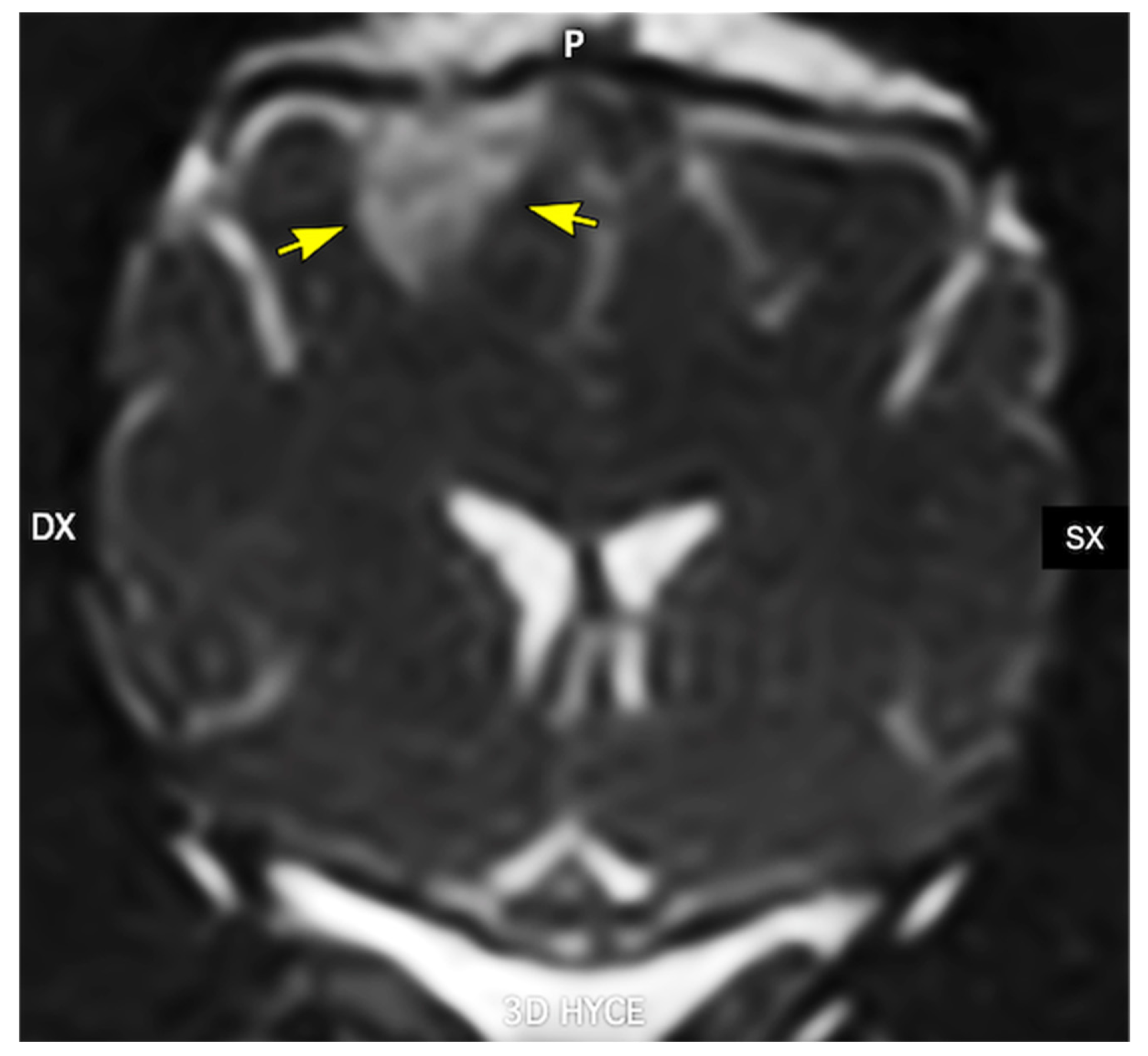

| Item/Disease | Exon 10 (R2) Status | Tau Repeat Type in Fibrils | Main Isoforms/Pool | Predominant Isoform Composition | Notes/Key References |
|---|---|---|---|---|---|
| Structural rule | Exon 10 excluded | 3R | 0N3R, 1N3R, 2N3R | — | Exon 10− → loss of R2 → 3-repeat tau (3R) [229,235] |
| Structural rule | Exon 10 included | 4R | 0N4R, 1N4R, 2N4R | — | Exon 10+ → inclusion of R2 → 4-repeat tau (4R) [229,235] |
| Adult human brain tau | Both exon 10− and exon 10+ | 3R + 4R | Six major isoforms (352–441 aa) | Mixed 3R + 4R pool | MAPT alternative splicing generates 0N/1N/2N × 3R/4R [229]. |
| Alzheimer’s disease (AD) | Mixed exon 10−/exon 10+ | 3R + 4R | Mixed 3R and 4R isoforms in filaments | Mixed 3R + 4R | Classic mixed 3R/4R tauopathy [235]. |
| Primary age-related tauopathy (PART) | Mixed exon 10−/exon 10+ | 3R + 4R | Mixed 3R and 4R | Mixed 3R + 4R | AD-like but with more restricted distribution [235]. |
| Chronic traumatic encephalopathy (CTE) | Mixed exon 10−/exon 10+ | 3R + 4R | Mixed 3R and 4R | Mixed 3R + 4R | Trauma-associated mixed 3R/4R fold [235]. |
| Pick’s disease (PiD) | Mainly exon 10− | Predominantly 3R | 3R-only filaments | 3R | Prototypical 3R-dominant tauopathy [235]. |
| Corticobasal degeneration (CBD) | Mainly exon 10+ | Predominantly 4R | 4R-only filaments | 4R | Prototypical 4R tauopathy [235]. |
| Argyrophilic grain disease (AGD) | Mainly exon 10+ | Predominantly 4R | 4R-only filaments | 4R | 4R-dominant tauopathy [235]. |
| Progressive supranuclear palsy (PSP) | Mainly exon 10+ | Predominantly 4R | 4R-only filaments | 4R | 4R tauopathy with characteristic PSP fold [235]. |
| Globular glial tauopathy (GGT) | Mainly exon 10+ | Predominantly 4R | 4R-only filaments | 4R | 4R-dominant glial tauopathy [235] |
| Canine cognitive dysfunction (CCD) | Not structurally resolved (AD-like) | Likely mixed 3R + 4R pool *** | Canine tau orthologues of 3R/4R isoforms | AD-like, presumed 3R + 4R | Hyperphosphorylated/aggregated tau with AD-like pathology; 3R/4R fibril composition not yet defined [185,236] |
| Step | Element | Key Details | References |
|---|---|---|---|
| 1 | Initial tau misfolding | Soluble tau → misfolded/aggregated tau assemblies; early sites: transentorhinal cortex, hippocampus | [246] |
| 2 | Release of misfolded tau from affected neurons | Active release: exocytosis, ectosomes (microvesicles), exosomes; passive release: leakage from degenerating neurons | [251] |
| 3 | Extracellular tau seeds in brain interstitium | Misfolded tau persists in extracellular space; in chronic vascular insufficiency, BBB compromise, low-grade neuroinflammation → enhanced extracellular persistence and interstitial transport | [251,253,254] |
| 4 | Uptake of tau seeds by neighboring cells | Mechanisms: endocytosis, macropinocytosis, receptor-mediated uptake | [251] |
| 5 | Intracellular seeding and templated misfolding | Internalized seeds induce misfolding of endogenous soluble tau; amplification of misfolded/aggregated tau within neuron | [251,252] |
| 6 | Spread along anatomically connected networks | Seed-containing neurons release new tau assemblies; spread via synaptic and network connections; mirrors Braak staging: transentorhinal/hippocampal regions → association cortices → primary sensory–motor areas | [246] |
| 7 | Regional tau pathology and clinical progression | Progressive accumulation of tau pathology in connected regions; neurodegeneration and cognitive decline; in chronic vascular insufficiency: vascular, BBB, and neuroinflammatory factors further facilitate propagation | [246,253,254,255] |
| Aspect | Alzheimer’s Disease (AD) | Canine Cognitive Dysfunction (CCD) | Key References |
|---|---|---|---|
| Gross lesions—distribution and atrophy pattern | Marked diffuse cortical atrophy, most pronounced in medial temporal lobes (hippocampus, entorhinal cortex) and association neocortex; relative early sparing of primary motor/sensory cortex; hippocampus and amygdala shrunken; enlarged ventricles (ex vacuo hydrocephalus); atrophy of basal forebrain (nucleus basalis of Meynert). | Generalized cortical atrophy, usually less severe than in advanced AD; sulcal widening and ventricular enlargement; atrophy particularly in frontal and temporal (± parietal) cortex; hippocampal volume loss correlates with cognitive decline; cerebellum relatively spared. | [6,9,10,185,246,305,306,313] |
| Gross lesions—vascular/hemorrhagic changes | Cerebral amyloid angiopathy (CAA) frequent; may cause lobar microbleeds or macrohemorrhages, cortical superficial siderosis; more prevalent/severe in some genetic backgrounds (e.g., APOE ε4). | CAA common in aged dogs with or without CCD; Aβ in leptomeningeal and cortical vessel walls; associated with microbleeds and vasculopathy; overt large hemorrhages less systematically described than in human AD. | [6,7,307,308,311] |
| Histopathology—Aβ plaques | Abundant Aβ plaques: diffuse and neuritic (cored) plaques. Neuritic plaques have dense fibrillar Aβ core with dystrophic neurites, microglia, astrocytes. Early deposition in neocortex → limbic → subcortical areas (Thal phases 1–5). Aβ40 and Aβ42 species present; Aβ42 enriched in parenchymal plaques. | Aβ deposition is the most consistent lesion: mainly in association cortices (frontal, temporal, parietal) and less in hippocampus. Diffuse plaques common in aged dogs; cored/neuritic plaques more frequent or abundant in clinically affected CCD cases. Aβ42 predominates in plaques; Aβ40 more in vascular deposits. | [5,9,11,220,259,305] |
| Histopathology—Tau/NFTs | Defining lesion: abundant neurofibrillary tangles (NFTs) composed of hyperphosphorylated tau (paired helical filaments) in neuronal soma and dendrites; associated neuropil threads and tau-positive dystrophic neurites. Stereotyped progression (Braak stages I–VI) from transentorhinal/entorhinal → hippocampus/limbic → widespread neocortex. NFT/tau burden correlates strongly with cognitive decline. | Classic NFTs largely absent or very sparse. Phospho-tau-positive neurons/threads occasionally reported but without widespread, layered Braak-like progression. No standardized staging system analogous to Braak in dogs. CCD is therefore considered primarily an Aβ-centric pathology with limited tauopathy. | [185,246,309,310] |
| Histopathology—neuronal & synaptic changes | Marked neuronal loss in hippocampal CA1, entorhinal cortex, association cortices, and basal forebrain cholinergic nuclei; prominent synapse loss (e.g., in frontal and temporal cortex) tightly correlates with severity of dementia. | Neuronal loss/shrinkage in frontal and temporal cortex and hippocampus, but generally less dramatic and more variable; synaptic loss (e.g., decreased synaptophysin) correlates with cognitive deficits; white matter degeneration/demyelination, especially frontal, can contribute to dysfunction. | [9,10,185,205,210,268,306] |
| Histopathology—glial and inflammatory changes | Robust astrogliosis and microgliosis surrounding plaques and tangles; chronic innate immune activation in affected regions; microglia often clustered around plaques, astrocytes hypertrophic and GFAP-positive; modest white matter degeneration. | Astrogliosis and microgliosis often associated with cortical Aβ plaques and CAA; gliosis more variable but present in regions with heavy Aβ burden; white matter gliosis and degeneration described in aged dogs. Extent of neuroinflammation generally less well characterized than in human AD. | [5,9,205,220,278] |
| Histopathology—vascular pathology | CAA common: Aβ (especially Aβ40) in leptomeningeal and cortical vessel walls, sometimes with smooth muscle cell loss and fibrosis; associated microbleeds, microinfarcts, superficial siderosis; often coexists with small-vessel disease. | CAA frequent in aged dogs: Aβ in cortical and leptomeningeal arterioles/arteries; associated wall thickening, smooth muscle degeneration, and occasional microbleeds; microinfarcts and small-vessel disease less systematically described but present in some series. | [6,7,307,308,311] |
| Immunohistochemistry—Aβ | Anti-Aβ antibodies (e.g., 4G8, 6E10, Aβ40, Aβ42) used to detect diffuse and cored plaques and CAA; support Thal amyloid phase scoring; enable quantification of plaque burden and vascular deposition. | Human Aβ antibodies cross-react with canine Aβ; 4G8, 6E10, Aβ42, Aβ40 commonly used to grade plaque and CAA burden; IHC confirms predominance of Aβ42 in plaques and Aβ40 in vessels; used to correlate lesion load with cognitive scores. | [5,11,220,259,305,312] |
| Immunohistochemistry—tau | Phospho-tau antibodies (AT8, PHF-1, CP13, etc.) robustly label NFTs, neuropil threads, and dystrophic neurites; applied to assign Braak NFT stages (NIA-AA “B” score). | Phospho-tau antibodies (e.g., AT8) usually show minimal to focal immunoreactivity; classic NFT morphology and extensive distribution are rare; no established canine tau staging scheme. | [185,246,305,309,312] |
| Immunohistochemistry—neuronal & synaptic markers | NeuN, MAP2 for neuronal cell bodies and dendrites; synaptophysin, PSD-95 for synapses; reduced immunoreactivity in hippocampus and association cortex correlates with cognitive impairment. | NeuN/MAP2 used to document neuronal loss/atrophy; synaptophysin and other synaptic markers diminished in frontal cortex and hippocampus in CCD, correlating with behavioral measures of cognitive dysfunction. | [9,10,185,210,268] |
| Immunohistochemistry—glial markers | GFAP stains reactive astrocytes; Iba1, CD68, and other microglial markers highlight activated microglia surrounding plaques and tangles; support assessment of neuroinflammation. | GFAP and Iba1/CD68 demonstrate plaque-associated astrocytosis and microgliosis; patterns broadly similar to AD but often less extensive and less systematically staged in the literature. | [5,9,220,278] |
| Immunohistochemistry—integrated staging frameworks | NIA–AA “ABC” scheme: A = Thal Aβ phase; B = Braak NFT stage; C = CERAD neuritic plaque score; IHC for Aβ and phospho-tau central to classification. | No universally accepted standardized staging comparable to NIA–AA. Most studies use semi-quantitative or regional scoring of Aβ plaques and CAA, with limited tau assessment and without a formal composite stage. | [220,305,312] |
| Dimension | Similarities (AD-CCD) | Differences (AD vs. CCD) | References |
|---|---|---|---|
| Global brain atrophy | Both show age-associated cortical and hippocampal atrophy, with ventricular enlargement and sulcal widening. Hippocampal volume loss correlates with cognitive decline in both species. | AD typically exhibits more severe and widespread atrophy in advanced stages, especially in medial temporal structures and association neocortex; CCD atrophy tends to be milder and more variable, often accentuated in frontal lobes. | [9,10,185,246,305,313] |
| Aβ plaque pathology | Both develop cortical Aβ plaques (diffuse and cored/neuritic), particularly in association cortices; Aβ42 is prominent in parenchymal plaques in both species. Plaque burden generally correlates with aspects of cognitive impairment. | AD usually shows a more stereotyped spatiotemporal evolution of Aβ deposition (Thal phases) and frequently high neuritic plaque density; CCD may have abundant diffuse plaques in cognitively normal aged dogs, with less clear threshold between “normal aging” and disease; overall neuritic change tends to be less pronounced than in advanced AD. | [5,9,11,220,259] |
| Tau pathology | Both species express tau and can show phospho-tau immunoreactivity, and isolated tau-positive neurons/threads may appear in aged dog brains. | AD is defined by abundant hyperphosphorylated tau NFTs, neuropil threads, and tau-positive dystrophic neurites, with well-defined Braak staging that strongly correlates with cognitive decline; CCD typically lacks classic NFTs and does not show a Braak-like laminar progression; tau pathology is sparse, inconsistent, and not central to diagnosis. | [185,246,309,310] |
| Neuronal and synaptic degeneration | Both exhibit neuronal loss and synaptic depletion in cortex and hippocampus; synapse loss correlates with cognitive dysfunction in each disease. Both show neuron shrinkage and dendritic abnormalities in affected regions. | Neuronal and synaptic loss are generally more extensive and stereotyped in AD, involving hippocampus, entorhinal cortex, multimodal association cortices, and cholinergic basal forebrain nuclei. In CCD, neuronal and synaptic loss can be significant but is more variable, and it is harder to distinguish from changes of “normal” canine aging; cholinergic system involvement is less well characterized. | [9,185,205,210,268,306] |
| Glial response and neuroinflammation | Both show plaque-associated astrocytosis and microgliosis; GFAP-positive astrocytes and Iba1/CD68-positive microglia cluster around Aβ deposits; chronic low-grade neuroinflammation is a shared feature. | In AD, microglial and astrocytic activation has been deeply characterized and linked to genetic risk (e.g., TREM2, APOE) and disease progression; in CCD, glial responses are documented but less comprehensively studied, and their temporal dynamics and causal contribution to cognitive decline remain less clear. | [5,9,205,220,278] |
| Vascular Aβ and CAA | Both develop CAA with Aβ deposition in leptomeningeal and cortical vessel walls; Aβ40 frequently enriched in vascular deposits; CAA can be associated with microbleeds and microinfarcts in both species. | In AD, the clinical and pathological spectrum of CAA (microbleeds, macrohemorrhages, superficial siderosis, ischemic lesions) is well defined and integrated into mixed-dementia concepts; in CCD, CAA is common but the full clinical impact and relationship to cognitive signs are less clearly delineated and less systematically quantified. | [6,7,307,308,311] |
| Clinicopathologic correlation & staging frameworks | In both, increasing cortical/hippocampal pathology (Aβ, neuronal/synaptic loss) tracks with worsening cognition; aged but cognitively normal individuals may still have some Aβ burden. | AD has formal NIA–AA neuropathologic criteria (ABC score) integrating Aβ phase, NFT stage, and neuritic plaque density; cognition correlates particularly with Braak stage and synaptic loss. CCD lacks standardized staging; most studies apply semi-quantitative scoring for Aβ and gliosis, with limited tau assessment, making cross-study comparisons and clinicopathologic correlations less robust. | [210,220,305,312] |
| Overall disease “type” (proteinopathy profile) | Combined Aβ- and tau-driven proteinopathy with strong tau contribution to neurodegenerative cascade; often accompanied by vascular and other co-pathologies in aged humans. | Largely Aβ-dominant encephalopathy with prominent plaques and CAA but minimal classic tauopathy; therefore, considered a partial analog of AD, modeling especially the amyloid and vascular aspects rather than the full tau-driven neurodegenerative spectrum. | [9,220,246,259] |
| Pathological Feature (TEM) | Ultrastructural Observations | Translational Impact | References |
|---|---|---|---|
| Amyloid fibrils and plaques | Fibrillar Aβ deposits with defined diameter, periodicity, and packing in human cerebrovascular and parenchymal amyloid; analogous Aβ fibrils within plaques and vessel walls in aged dogs | Confirms that dogs develop human-like Aβ fibrils and cerebral amyloid angiopathy, supporting aged dogs as a spontaneous large-animal model for amyloidogenesis and anti-amyloid therapies | [6,210,220,245] |
| Neurofibrillary tangles and tau filaments | Paired helical filaments and straight tau filaments with conserved ultrastructural motifs in human AD; age-related tau alterations documented in canine brain | Structural conservation of tau assemblies informs cross-species mechanisms of tauopathy, enabling preclinical evaluation of tau-targeting agents in dogs that model early or limited tau pathology | [220,245,315] |
| Synaptic degeneration | Reduced synaptic density, dystrophic synaptic profiles, altered vesicle pools, and associated mitochondrial abnormalities in AD cortex and hippocampus; comparable synaptic degeneration in CCD | Establishes synapse loss and ultrastructural synaptic pathology as common correlates of cognitive decline in humans and dogs, supporting synaptic metrics as translational outcome measures for disease-modifying interventions | [5,6,210,220] |
| Mitochondrial and autophagic changes | Abnormal mitochondrial morphology (swelling, cristae disruption) and accumulation of autophagic vacuoles in neurons and neurites in AD; similar mitochondrial alterations described in aged/CCD canine brain | Supports conserved pathways of bioenergetic failure and impaired proteostasis across species, justifying the use of dogs for testing mitochondrial and autophagy-modulating therapies | [210,220] |
| Myelin and axonal pathology | Axonal swellings, spheroids, myelin splitting, and degeneration in aging human white matter; TEM evidence of analogous age-related axon/myelin pathology in dogs | Provides a structural basis for shared white matter dysfunction and cerebrovascular contributions to cognitive impairment, enabling translational studies of vascular and myelin-protective strategies | [6,10,220] |
| Correlation of Aβ burden with cognition | Quantitative TEM and histopathology show that increasing amyloid load and neuritic pathology correlate with cognitive deficits in both species | Validates aged dogs as a model of early–intermediate AD stages, in which Aβ load and neuritic pathology can be linked to behavioral measures, improving prediction of clinical efficacy from preclinical trials | [1,5,185,219] |
Disclaimer/Publisher’s Note: The statements, opinions and data contained in all publications are solely those of the individual author(s) and contributor(s) and not of MDPI and/or the editor(s). MDPI and/or the editor(s) disclaim responsibility for any injury to people or property resulting from any ideas, methods, instructions or products referred to in the content. |
© 2026 by the authors. Licensee MDPI, Basel, Switzerland. This article is an open access article distributed under the terms and conditions of the Creative Commons Attribution (CC BY) license.
Share and Cite
Dondi, M.; Bianchi, E.; Borghetti, P.; Di Lecce, R.; Gnudi, G.; Guarnieri, C.; Buffagni, V.; Ravanetti, F.; Saleri, R.; Corradi, A. Canine Cognitive Dysfunction and Alzheimer’s Disease: Pathophysiological Relationships and the Impact of Glymphatic System Impairment on Neurodegeneration. Vet. Sci. 2026, 13, 298. https://doi.org/10.3390/vetsci13030298
Dondi M, Bianchi E, Borghetti P, Di Lecce R, Gnudi G, Guarnieri C, Buffagni V, Ravanetti F, Saleri R, Corradi A. Canine Cognitive Dysfunction and Alzheimer’s Disease: Pathophysiological Relationships and the Impact of Glymphatic System Impairment on Neurodegeneration. Veterinary Sciences. 2026; 13(3):298. https://doi.org/10.3390/vetsci13030298
Chicago/Turabian StyleDondi, Maurizio, Ezio Bianchi, Paolo Borghetti, Rosanna Di Lecce, Giacomo Gnudi, Chiara Guarnieri, Valentina Buffagni, Francesca Ravanetti, Roberta Saleri, and Attilio Corradi. 2026. "Canine Cognitive Dysfunction and Alzheimer’s Disease: Pathophysiological Relationships and the Impact of Glymphatic System Impairment on Neurodegeneration" Veterinary Sciences 13, no. 3: 298. https://doi.org/10.3390/vetsci13030298
APA StyleDondi, M., Bianchi, E., Borghetti, P., Di Lecce, R., Gnudi, G., Guarnieri, C., Buffagni, V., Ravanetti, F., Saleri, R., & Corradi, A. (2026). Canine Cognitive Dysfunction and Alzheimer’s Disease: Pathophysiological Relationships and the Impact of Glymphatic System Impairment on Neurodegeneration. Veterinary Sciences, 13(3), 298. https://doi.org/10.3390/vetsci13030298