Article

Analysis of Potential Genes, Acute Phase Proteins and Hormonal Profiles Associated with Methicillin-Resistant Staphylococcus aureus (MRSA) Isolation from Pneumonic Sheep

1
Department of Biology, College of Science, Princess Nourah bint Abdulrahman University, P.O. Box 84428, Riyadh 11671, Saudi Arabia
2
Department of Animal Health and Poultry, Animal and Poultry Production Division, Desert Research Center (DRC), Cairo 11753, Egypt
3
Department of Animal Medicine (Internal Medicine), Faculty of Veterinary Medicine, Arish University, Arish 45511, Egypt
4
Department of Development of Animal Wealth, Faculty of Veterinary Medicine, Mansoura University, Mansoura 35516, Egypt
5
Department of Chemistry, Faculty of Science, Majmaah University, Al Majmaah 11952, Saudi Arabia
*
Author to whom correspondence should be addressed.
Vet. Sci. 2025, 12(6), 584; https://doi.org/10.3390/vetsci12060584
Submission received: 19 May 2025 / Revised: 9 June 2025 / Accepted: 10 June 2025 / Published: 13 June 2025
(This article belongs to the Section Veterinary Internal Medicine)

Simple Summary

Staphylococcus aureus is a significant bacterial pathogen responsible for a wide range of infections in both humans and animals. In this study, 100 pneumonic sheep and 100 clinically healthy sheep were examined to assess biochemical parameters and gene expression profiles. The findings revealed notable differences in gene expression, nucleotide sequences, and biochemical markers between healthy and pneumonic animals. Additionally, MRSA isolates displayed complete resistance to amoxicillin, cloxacillin, and erythromycin, along with high resistance to penicillin and tetracycline. However, all MRSA isolates remained fully susceptible to vancomycin. The observed alterations in molecular and biochemical marker profiles may contribute to the pathogenesis and clinical manifestations of pneumonia in sheep.

Abstract

Staphylococcus aureus is a significant bacterial pathogen responsible for a wide range of diseases in both humans and animals. This study aimed to investigate nucleotide sequence variations, gene expression patterns, and serum biomarkers, including acute phase proteins (APPs), hormonal fluctuations, and iron profile parameters in sheep affected by pneumonia. Additionally, the study focused on the isolation and characterization of S. aureus from pneumonic sheep, with particular emphasis on the prevalence of methicillin-resistant S. aureus (MRSA) strains. Blood samples were collected from both healthy and pneumonic sheep for gene expression and biochemical analyses, while nasal swabs from pneumonic sheep were used for bacterial isolation and identification. Out of 100 nasal swabs analyzed, 44% tested positive for Staphylococcus spp., and 61.4% of these were confirmed as S. aureus by PCR. The mecA gene, a key marker of methicillin resistance, was identified in 17 isolates (38.6% of the S. aureus-positive samples). MRSA isolates showed complete resistance to amoxicillin, cloxacillin, and erythromycin, and high resistance to penicillin, amoxicillin, and tetracycline; however, all MRSA strains remained fully susceptible to vancomycin. Gene expression analysis revealed that TLR2, CLEC4E, PTX3, CXCL8, and IL15RA were significantly upregulated (p < 0.05) in pneumonic ewes, while SOCS3 expression was markedly downregulated. Sequence analysis of immune-related genes revealed notable nucleotide differences between healthy and affected animals. Furthermore, the pneumonic group exhibited significantly elevated levels of APPs, cortisol, and growth hormone, along with reduced levels of insulin, T3, and T4. These findings underscore the zoonotic risk posed by MRSA and emphasize the need for robust surveillance and antibiotic stewardship to control its spread. The study also highlights the importance of molecular diagnostics in accurately identifying MRSA and elucidating resistance mechanisms, thereby facilitating targeted treatment and informed management strategies.

1. Introduction

Pneumonia is a major challenge in the global sheep industry, affecting approximately 30–40% of flocks and contributing to 40–70% of total sheep losses. In addition to mortality, pneumonia adversely impacts growth rates, leads to carcass condemnation, and substantially increases treatment and vaccination costs [1,2]. Collectively, these factors impose considerable financial burdens on sheep production systems.
Respiratory infections in sheep have a complex etiology involving interactions between the host immune response, environmental stressors, and a range of pathogenic organisms, including bacteria, mycoplasmas, viruses, and fungi. Major predisposing factors include transportation stress, overcrowding, abrupt weather changes, poor ventilation, and malnutrition. In Egypt, pneumonia is most prevalent during transitional seasons, particularly from autumn to early winter and from spring to early summer—when sheep experience sudden fluctuations between warm and cold temperatures. Additionally, elevated dust levels during spring and autumn can facilitate the deep penetration of pathogens into the respiratory tract [3,4,5,6,7].
Staphylococcus aureus is among the bacterial pathogens associated with pneumonia. This species is particularly significant due to its involvement in a wide range of diseases affecting both humans and animals. As reported in [8,9,10], S. aureus is a facultative anaerobic, Gram-positive, non-motile, and non-spore-forming coccus known for its remarkable adaptability and ability to persist in diverse environments. Approximately 25–40% of human and animal populations are asymptomatically colonized by S. aureus, primarily in the nasal passages and on the skin. In addition to causing cutaneous and soft tissue infections [11], S. aureus is a major contributor to foodborne illnesses, pneumonia, bacteremia, meningitis, sepsis, pericarditis, and other serious systemic infections [12,13].
The increasing antimicrobial resistance of Staphylococcus aureus infections is a significant public health concern. The emergence of methicillin-resistant S. aureus (MRSA) is attributed to bacterial strains that have developed resistance to methicillin and other β-lactam antibiotics. As reported in [14], these strains commonly exhibit resistance to a range of β-lactam antibiotics, including amoxicillin, penicillin, oxacillin, and methicillin. According to [15], monitoring the antibiotic resistance patterns of S. aureus isolates is crucial for minimizing treatment failures and implementing effective control measures.
Methicillin-resistant Staphylococcus aureus (MRSA) has emerged as a major pathogen in both human and veterinary medicine, with infections documented in hospital environments as well as within the community [16,17,18]. Since the first report of an MRSA strain in 1961 [19], its global prevalence has increased significantly, contributing to a rise in multidrug-resistant infections [20]. This rapid escalation in antibiotic resistance has posed serious challenges to conventional treatment strategies. MRSA, now considered one of the most common antimicrobial-resistant organisms worldwide, spreads efficiently in healthcare settings and exhibits high resistance to β-lactam antibiotics. This resistance is primarily mediated by the production of a modified penicillin-binding protein (PBP2a), which inhibits the action of β-lactam agents [21].
Initial treatment options for MRSA infections included daptomycin, linezolid, and vancomycin. However, MRSA has also developed resistance to these agents through various genetic adaptations, including alterations in SCCmec elements and single nucleotide polymorphisms (SNPs) within open reading frames (ORFs) [22]. The mecA gene, located on the staphylococcal cassette chromosome mec (SCCmec), encodes the penicillin-binding protein PBP2a and has undergone significant evolutionary modifications. These changes have played a critical role in the persistence and global dissemination of MRSA.
Compared to methicillin-sensitive Staphylococcus aureus (MSSA), MRSA infections are associated with poorer clinical outcomes [23,24]. The Centers for Disease Control and Prevention (CDC) and the World Health Organization (WHO) have identified MRSA as a significant global health threat [25]. The genetic diversity and antibiotic resistance patterns of S. aureus vary across geographical regions and host populations [26]. A major contributor to the emergence of MRSA in both humans and animals is the widespread and uncontrolled use of antibiotics [27]. Zoonotic transmission of MRSA has been extensively documented, with evidence supporting bidirectional transmission between humans and animals [28]. According to [29], asymptomatic colonization in the nasal and rectal regions further supports the role of animals as reservoirs for MRSA, facilitating its spread to humans and other animals.
The polymerase chain reaction (PCR) is considered one of the most rapid and effective techniques for detecting microbial infections in clinical samples. As noted in [30], PCR is particularly advantageous for identifying pathogens that are difficult to culture or require prolonged incubation periods. This method enables faster diagnosis and treatment, reduces the need for antimicrobial use, minimizes costs, and lowers the risk of adverse effects [31]. Although PCR-based assays enhance the molecular detection of MRSA, conventional bacterial culture remains the gold standard for definitive diagnosis [32].
Acute-phase proteins (APPs), primarily synthesized by the liver, are present in the blood of infected animals and play a crucial role in maintaining homeostasis, limiting tissue damage, and inhibiting microbial growth independently of antibodies [33]. During infection, inflammation, or other internal and external challenges, the concentration of APPs in plasma either increases (positive APPs) or decreases (negative APPs). These changes, which exhibit notable species-specific variation, provide valuable diagnostic and prognostic information in the context of infection and inflammation [34].
Advanced molecular genetic approaches have the potential to enhance animal health and support disease control efforts [35]. Several genetic markers, predominantly single nucleotide polymorphisms (SNPs), have been successfully identified to determine livestock susceptibility to various diseases [36,37,38]. This indicates that genetic variation among animals contributes to differences in disease vulnerability [39]. Marker-assisted selection (MAS) is a genomic strategy that uses these genetic markers as selection criteria, shifting the focus from observable disease symptoms to specific alleles at the DNA level. MAS improves selection accuracy and enables the identification of genetically resistant animals without the need for exposure to disease challenges, thereby offering a promising tool for breeding disease-resistant livestock [40].
Currently, limited information is available regarding the relationship between methicillin-resistant Staphylococcus aureus (MRSA) isolated from pneumonic sheep and changes in acute-phase proteins (APPs), hormonal levels, and gene expression. The development of effective preventive and therapeutic strategies is further hindered by the lack of reliable diagnostic tools and associated biomarkers for these conditions. This study aims to address these gaps by examining the gene expression patterns and serum profiles of APPs, hormonal markers, and iron-related parameters in Barki sheep infected with MRSA. Furthermore, the research will focus on the molecular identification of key antibiotic resistance genes, assess the prevalence of MRSA in nasal swabs, and characterize the antibiotic resistance profiles of the isolated strains. By elucidating the underlying molecular mechanisms, this approach seeks to provide essential insights to improve the management of ovine health and enhance disease monitoring strategies.

2. Material and Methods

2.1. Animals and Study Design

Two hundred Barki ewes weighing between 30 and 45 kg (mean ± SD: 37.5 ± 4.7) and between the ages of 3.5 and 5.5 (mean ± SD: 4.4 ± 0.6) were included in the study. The study was conducted between November 2023 and January 2024, corresponding to the late autumn and early winter seasons in the Matrouh Governorate, Egypt. During this period, the region experiences frequent fluctuations in temperature, increased humidity, and high atmospheric dust levels, particularly during early mornings and evenings. These environmental factors are well-documented contributors to respiratory stress and the onset of pneumonia in sheep. Recording these climatic conditions provides important context for interpreting the disease prevalence and biomarker fluctuations observed in the study. Following a comprehensive clinical examination in accordance with the technique described by [41], the ewes were split into two groups based on evaluations of their rumen, heart, and lung health, as well as other vital signs. The clinically healthy sheep in Group 1 (n = 100) were placed in the healthy control group (HCG), which had normal body temperature, pulse, respiration rate, clean eyes, no lacrimal or nasal discharge, normal posture, and appetite. One hundred sheep in the pneumonic group (PG) had clinical symptoms of respiratory disease, such as fever, belly breathing, mucopurulent nasal discharge, general weakness, appetite loss, and abnormal lung sounds (crackles and wheezes) identified through auscultation. The ewes were kept in semi-open, shaded quarters and fed 750 g of concentrated feed mixture (CFM) and 750 g of alfalfa hay per head that was given twice a day along with unlimited access to water. They also had access to green herbage, grass, berseem, darawa, and other natural grazing when it was available. The CFM composition consisted of 240 kg of wheat bran, 230 kg of soybean meal, 530 kg of maize, 5 kg of sodium chloride, 10 kg of calcium carbonate, 1 kg of premix, 0.5 kg of Netro-Nill, and 0.5 kg of Fylax.

2.2. Sampling

2.2.1. Blood Sampling

Ten milliliters of fresh blood were drawn from each ewe in each group at 10 o’clock in the morning using a jugular vein puncture, and the blood was then split into three portions. The first and second parts were supplemented with EDTA and heparin calcium 5000 I.U., respectively, to disrupt the coagulation cascade. The EDTA blood was used for real-time PCR, and the second and third parts were centrifuged to extract serum and plasma, respectively, at 3000 r.p.m. for 20 min at 37 °C. They were then aliquoted and stored at −20 °C. The acute phase proteins (APPs), hormones, and iron profile parameters were then examined.

2.2.2. Nasal Swabs

One hundred nose swabs were gathered in total. An assistant identified each animal and secured it while keeping it bound. A sterile cotton-tipped swab was placed within the nostril and rotated against the nasal cavity wall after the nasal exterior had been cleaned with 70% alcohol. Next, the swab was put into a sterile, labeled test tube with 10 mL of nutritional broth at 4 °C. Before being transported to the Microbiology Department laboratory at the Development of Matrouh Resources, Desert Research Center, Matrouh, the samples were kept in an icebox and kept under strict sanitary conditions for bacteriological examination.

2.3. Bacteriological Examination

2.3.1. Isolation and Identification of Staphylococcus aureus

Each sample was inoculated into Baird-Parker agar plates supplemented with egg yolk tellurite using a 1 mL aliquot. Following a 24 to 48 h incubation period at 37 °C, suspicious colonies that were black, glossy, with a slight white border, and encircled by a clear zone that extended into the opaque medium began to expand.
The suspicious colonies were separated and streaked onto mannitol salt agar (MSA) medium, where they were incubated for 24 to 48 h at 37 °C to confirm the presence of Staphylococcus aureus. S. aureus-positive colonies were defined as those that showed yellow on MSA. Following the procedure outlined by [42], purified isolates were subjected to biochemical testing and Gram staining.

2.3.2. Antimicrobial Susceptibility Testing

The disk diffusion approach was used to test for antibiotic susceptibility in accordance with the Clinical and Laboratory Standards Institute’s recommendations (CLSI, 2012). The following antibiotics were examined as follows: β-lactam antibiotics: methicillin (ME; 5 μg), ampicillin (A; 10 μg), cloxacillin (OB; 5 μg), oxacillin (OX; 1 μg), amoxicillin (AML; 25 μg), amoxicillin/clavulanic acid (AMC; 20 μg), penicillin (P; 10 IU), aminoglycosides, and gentamycin (G; 10 μg); and fluoroquinolones: ciprofloxacin (CIP; 5 μg). The approach followed the guidelines provided by Refs [43,44].

2.3.3. Antimicrobial Susceptibility Testing (AST) for MRSA Strains

On Mueller–Hinton agar (Merck, Darmstadt, Germany), the traditional disk diffusion method was used to assess the susceptibility of positive Staphylococcus aureus isolates, including identified methicillin-resistant S. aureus (MRSA) strains, to fourteen antimicrobial drugs. Following the 0.5 McFarland standard (1.5 × 10⁸ CFU/mL), the test was conducted, and the Clinical and Laboratory Standards Institute (CLSI) criteria were followed for analysis [45]. The following fourteen antibacterial agents were tested as follows: β-lactam antibiotics: penicillin (P; 10 IU), amoxicillin (AML; 25 μg), amoxicillin/clavulanic acid (AMC; 20 μg), ceftriaxone (CTR), and cloxacillin (OB; 5 μg); aminoglycosides: gentamycin (G; 10 μg); fluoroquinolones: ciprofloxacin (CIP; 5 μg), levofloxacin (LEV), and ofloxacin (OFL); and other classes: vancomycin (VAN), cetirimoxazole (SMX), erythromycin (ERY), tetracycline (TET), and chloramphenicol (CHL). To establish susceptibility, the inhibition zones were measured in millimeters.

2.4. Molecular Confirmation of MRSA Strains and Detection of Virulence and Resistance Genes

In accordance with the manufacturer’s instructions, polymerase chain reaction (PCR) was used to molecularly confirm the presence of Staphylococcus aureus and methicillin-resistant S. aureus (MRSA). The positive control was Staphylococcus aureus subsp. aureus (ATCC® 8096).
The QIAamp DNA Mini Kit (Qiagen GmbH, Hilden, Germany) was used to extract DNA from samples, with minor adjustments made to the manufacturer’s instructions. To summarize, 200 μL of the sample or tissue homogenate suspension was treated for 10 min at 56 °C with 10 μL of proteinase K and 200 μL of lysis buffer. Following incubation, the lysate was mixed with 200 μL of 100% ethanol. In accordance with the manufacturer’s recommendations, the sample was subsequently cleaned and centrifuged. A total of 100 μL of elution buffer was used to elute the nucleic acids.
Primers used in the study were supplied by Metabion (Planegg, Germany) and are listed in Table 1. The presence of the nuc gene was detected using specific primers, as described by [12]. Detection of the mecA gene, responsible for methicillin resistance, was carried out using primers reported by [46].
The following ingredients were used in the 25 μL reaction mixture used for PCR amplification: 1 μL of each primer (20 pmol concentration), 5.5 μL of nuclease-free water, 5 μL of DNA template, and 12.5 μL of EmeraldAmp Max PCR Master Mix (Takara, Kusatsu, Japan) were used. A 2720 thermal cycler from Applied Biosystems (Foster City, CA, USA) was used to carry out the procedures.
PCR products were electrophoresed using a voltage gradient of 5 V/cm on a 1.5% agarose gel (Applichem GmbH, Darmstadt, Germany) in 1× TBE buffer at room temperature. A total of 20 μL of uniplex PCR products and 40 μL of duplex PCR products were placed into each gel slot for the gel analysis. The fragment size was determined using the GeneRuler 100 bp Ladder (Fermentas, Darmstadt, Germany) and the GelPilot 100 bp Plus Ladder (Qiagen, Hilden, Germany). A gel documentation system (Alpha Innotech, San Leandro, CA, USA; Biometra, Gottingen, Germany) was used to display the gel, and computer software was used to evaluate the data.

2.5. Genetic Polymorphisms Between Healthy and Pneumonic Ewes

2.5.1. Total RNA Extraction, Reverse Transcription and Quantitative Real Time PCR

In accordance with the manufacturer’s recommendations, total RNA was isolated from ewe blood using Trizol reagent (RNeasy Mini Kit, catalog no. 74104). The NanoDrop® ND-1000 Spectrophotometer was used to quantify and qualify the amount of isolated RNA. Each sample’s cDNA was created in accordance with the manufacturing process (Thermo Fisher, Waltham, WA, USA, catalog no, EP0441). Using quantitative RT-PCR with SYBR Green PCR Master Mix (2× SensiFastTM SYBR, Bioline, CAT no: Bio-98002), the gene expression patterns for coding segments of genes encoding immunity (TLR2, CLEC4E, PTX3, SOCS3, CXCL8, and IL15RA) were evaluated. Via real-time PCR with the SYBR Green PCR Master Mix (Quantitect SYBR green PCR kit, catalog no. 204141), the relative mRNA level was measured.
Primers were created based on the Ovis aries sequence that was published in PubMed, as indicated in Table 2. As a constitutive control for normalization, the housekeeping gene GAPDH was employed. The reaction mixture, which had a total volume of 25 µL, contained 3 µL of total RNA, 4 µL of 5× Trans Amp buffer, 0.25 µL of reverse transcriptase, 0.5 µL of each primer, 12.5 µL of 2× Quantitect SYBR green PCR master mix, and 8.25 µL of RNase-free water. After putting the finished reaction mixture in a thermal cycler, the following procedure was run: reverse transcription for 30 min at 50 °C, primary denaturation for 10 min at 94 °C, 40 cycles of 94 °C for 15 s, annealing temperatures for 1 min as indicated in Table 2, and 72 °C for 30 s. A melting curve analysis was conducted at the conclusion of the amplification step to verify the PCR product’s specificity. Using the 2−ΔΔCt technique, the relative expression of each gene per sample was compared to that of the GAPDH gene [47].

2.5.2. DNA Sequencing and Polymorphism Detection

Removal of primer dimers, nonspecific bands, and other contaminants was performed prior to DNA sequencing. As described by [48], a PCR purification kit was used in accordance with the manufacturer’s instructions to purify real-time PCR products with the desired size (target bands) (Jena Bioscience # pp-201×s/ Jena, Germany). The Nanodrop (UV–Vis spectrophotometer Q5000/USA) was used to quantify the PCR product in order to produce high-quality results and guarantee sufficient concentrations and purity of the real-time PCR products [49]. PCR results containing the target band were sent for forward and reverse DNA sequencing using an ABI 3730XL DNA sequencer (Applied Biosystem, USA) in order to identify SNPs in genes examined in control and pneumonia-affected ewes. This was performed using an enzyme-chain terminator technique created by [50]. Chromas 1.45 and blast 2.0 tools were used to analyze the DNA sequencing data [51]. PCR results of the genes under investigation and reference sequences found in GenBank were compared, and differences were categorized as single-nucleotide polymorphisms (SNPs). Based on DNA sequencing data alignment, the MEGA6 software tool was used to compare the amino acid sequences of the genes under investigation among enrolled ewes [52].

2.6. Biochemical Analysis

IBL International Corp (Blainville, QC, Canada)® ELISA kits were used to measure serum haptoglobin (Hp), plasma fibrinogen (Fb), and serum amyloid A (SAA). Turbidimetric analysis of serum transferrin (Tf) and caeruloplasmin (Cp) utilizing Elabscience USA® (College Park, MD, USA) kits. Chemiluminescence immunoassay (CLIA) with Abnova® kits (Taipei, Taiwan) for serum ferritin. Spectrophotometric measurement of serum iron (SI) and total iron-binding capacity (TIBC) using commercial kits from Biodiagnostic Company® (Giza, Egypt). ELISA kits from MyBioSecure Company® (Cheyenne, WY, USA) are used for hormonal assays (cortisol, insulin, TSH, T3, T4, and growth hormone (GH). Calculations were carried out as follows: Transferrin Saturation Percentage (Tf sat. %): Tf sat. % = SI/TIBC × 100, Unsaturated Iron-Binding Capacity (UIBC), and UIBC = TIBC − SI

2.7. Statistical Analysis

With SPSS software (version 23), an independent-sample T-test was used to do statistical comparisons between the investigated groups. The data observed in each group were tested for normality using the Kolmogorov–Smirnov test, and all parameters were not statistically significant, which indicates that the data are parametric (normally distributed), so the results were expressed as mean ± SD. Relationships between the genetic markers and serum profile of APP variables were evaluated using Pearson’s correlation. The difference in the frequencies of each gene SNP between pneumonic and healthy sheep was statistically assessed using the chi-square test to match the distribution of the recognized SNPs among the two groups using SPSS version 23, USA. A Linear Discriminant Analysis (LDA) was conducted to determine whether gene-level SNP averages could differentiate between pneumonic and healthy sheep. The 6 gene average scores served as predictor variables, and the health status (pneumonic vs. healthy) was the grouping variable. Statistical consequence was established at p < 0.05. A significance level of p < 0.05 was established. The sensitivity, specificity, cut-off values, and likelihood ratios (LR) for acute-phase proteins (APPs), hormonal and iron profiles were computed using GraphPad Prism software (version 8).
Predictive metrics and accuracy rates were determined using the following formulas:
PPV = true positive ÷ total positive × 100.
NPV = true negative ÷ total negative × 100.
Accuracy rate = (true positive + True negative) ÷ total population × 100.
Percentage of increase = (the mean value of the marker concentration in diseased group − the mean value of its concentration in CG) ÷ the mean value of its concentration in CG × 100.

3. Results

3.1. Clinical Findings

This study observed that respiratory distress symptoms such as rhinitis, congested mucous membranes, and a wet, harsh cough are common in affected sheep (Figure S1). The most frequent clinical signs included serous or mucoid nasal discharge, elevated respiratory and pulse rates, and increased rectal temperature exceeding 41 °C. Additional signs such as ataxia, ruminal atony, depression, and abnormal milk production were also recorded. Early-stage auscultation revealed pleuritic friction rubs, which progressively became muffled as the disease advanced.

3.2. Staphylococcus Isolates

The presence of Staphylococcus aureus and methicillin-resistant S. aureus (MRSA) was evaluated in 100 nasal swab samples. Initial identification of suspected Staphylococcus species was based on colony morphology on Baird-Parker agar, where colonies appeared black with a surrounding clear halo. Further confirmation was performed using mannitol salt agar (MSA), on which S. aureus colonies were characterized by a yellow coloration. Based on these culture characteristics, 44 out of the 100 samples (44%) were presumptively identified as Staphylococcus spp.

3.3. Molecular Identification

PCR amplification of the nuc gene yielded a 395 bp amplicon, which was used for the molecular confirmation of S. aureus (Figure 1A). Further identification of MRSA isolates was achieved by detecting the mecA gene, characterized by a 310 bp amplicon (Figure 1B). Among the 44 suspected Staphylococcus isolates, 27 (61.4%) were confirmed as S. aureus based on nuc gene amplification. Of these, 17 isolates (38.6%) tested positive for the mecA gene and were classified as MRSA.

3.4. Antimicrobial Susceptibility of Staphylococcus aureus Isolated from Nasal Swabs of Sheep

The antibiotic susceptibility of S. aureus isolates obtained from nasal swabs was evaluated. As shown in Table 3, a high level of resistance was observed against cotrimoxazole and penicillin. Notably, all tested isolates exhibited multidrug resistance (MDR). The MRSA susceptibility profile revealed that 100% of isolates were resistant to amoxicillin, amoxicillin/clavulanate, cloxacillin, penicillin, erythromycin, chloramphenicol, tetracycline, and cotrimoxazole. Conversely, susceptibility was highest to levofloxacin (96%), followed by ofloxacin (70%) and gentamicin (15%). A detailed summary of these results is presented in Table 4.

3.5. Patterns for Transcript Levels of Immune Indicators

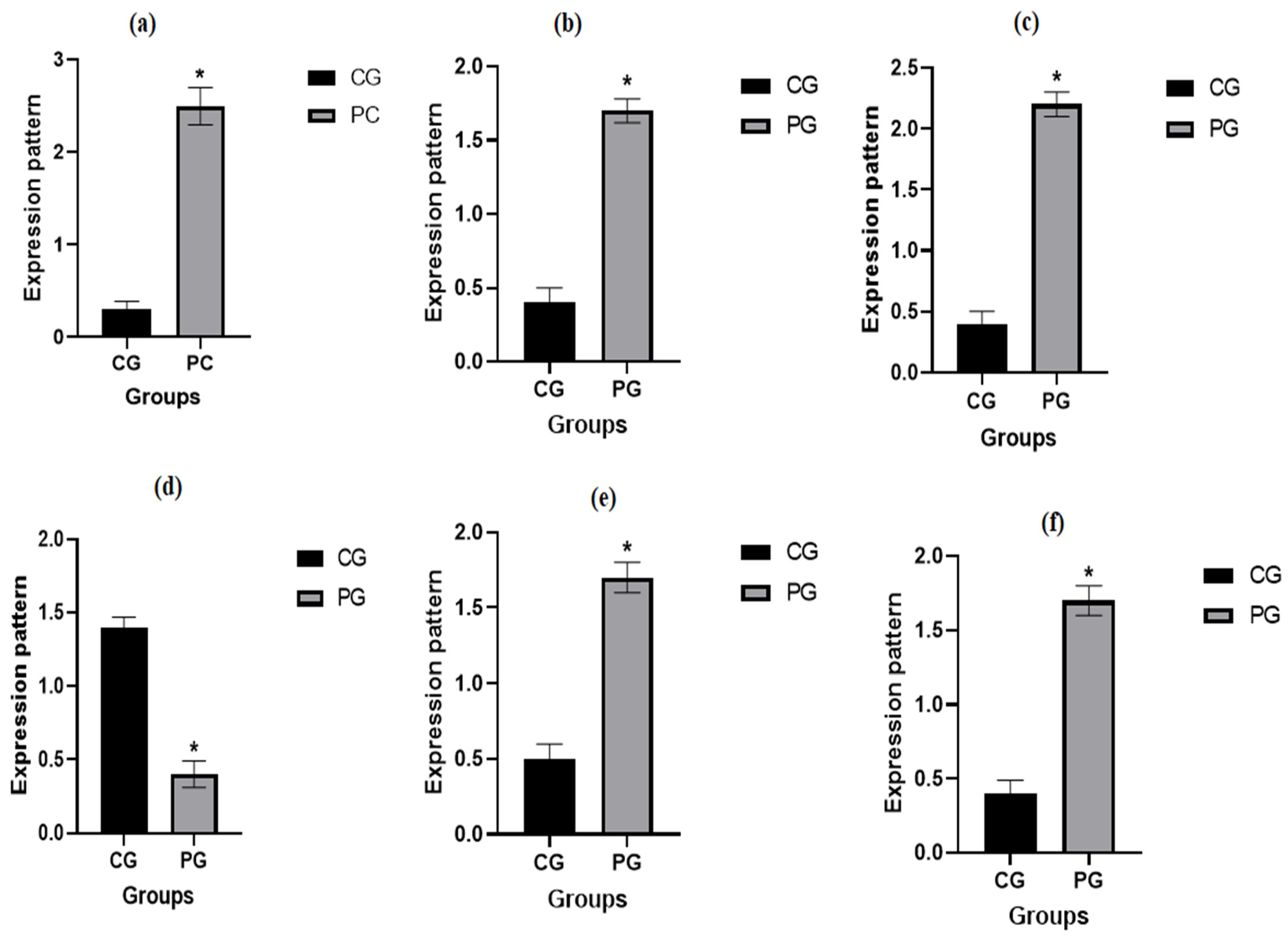
Figure 2 illustrates the transcript patterns of the evaluated immunological markers. The genes TLR2, CLEC4E, PTX3, CXCL8, and IL15RA exhibited significantly higher expression levels in pneumonia-affected ewes compared to healthy controls. In contrast, SOCS3 expression was significantly reduced in the affected group. Among pneumonic ewes, TLR2 showed the highest mRNA expression level (2.52 ± 0.22), while SOCS3 had the lowest (0.43 ± 0.16). Conversely, in healthy ewes, SOCS3 demonstrated the highest mRNA level (1.40 ± 0.13), and TLR2 had the lowest (0.37 ± 0.16).

3.6. Genetic Polymorphisms of Immune Genes

Using PCR-DNA sequence verdicts, the TLR2 (354 bp), CLEC4E (443 bp), PTX3 (363 bp), CXCL8 (300 bp), SOCS3 (480 bp) and IL15RA (378 bp) genes were found to have different SNPs in the amplified DNA bases linked to pneumonia. Based on the DNA sequence variations between immunological indicators examined in the ewes under study and the reference gene sequences retrieved from GenBank (Figures S1–S6), all of the identified SNPs were approved.
The coding DNA sequences of the affected ewes differed from those of the healthy ewes due to the exonic region abnormalities that were seen in Table 5 in all of the immunological markers tested. Using DNA sequencing of immune genes, seven SNPs were found; four of them were non-synonymous and three were synonymous. The four non-synonymous SNPs were associated with healthy ewes. The incidence of pneumonia, on the other hand, was linked to the identified three synonymous SNPs.
The TLR2 (354 bp), CLEC4E (443 bp), and IL15RA (378 bp) genes each contain one recurrent SNP for each marker, according to DNA sequencing, which causes non-synonymous mutations L57E, C15Y, and S25N, respectively. The 300 bp CXCL8 and 480 bp SOCS3 genes both contained two synonymous SNPs. The amino acids 146T and 52L were linked to the two SNPs, G438C and C159T, respectively. Two recurrent SNPs were found when the PTX3 (363 bp) gene was sequenced. In 30A, synonymous mutation G90C was implicated. In contrast, the C283T SNP caused the non-synonymous mutation L95F.
A significant difference was detected in the frequencies of all examined gene SNPs among pneumonic and healthy sheep (p < 0.005). Chi-square analysis was carried out for comparison of the distribution of all identified SNPs in all genes between pneumonic and healthy sheep. The total chi-square value showed significant variation among the identified SNPs in all genes between resistant and affected animals (p < 0.05) (Table 5).
Table 6 showed the discriminant analysis for the arrangement of kinds of genes and healthy status. The organizational consequences showed that the model properly classified 100% of the cases overall, either healthy sheep or pneumonic sheep. These results indicate that the SNP markers included in the model possess a good level of discriminatory power and may be useful as potential genetic indicators for pneumonia susceptibility in ewes.

3.7. Biochemical Profile

APPs (blood concentrations of acute-phase proteins (APPs) fibrinogen (Fb), haptoglobin (Hp), serum amyloid A (SAA), and ceruloplasmin (Cp) along with cortisol, growth hormone (GH), and thyroid-stimulating hormone (TSH) were significantly (p < 0.05) elevated in the pneumonia group (PG) compared to the control group (CG). In contrast, serum levels of insulin, triiodothyronine (T3), and thyroxine (T4) were significantly (p < 0.05) lower in PG. The iron profile of PG showed a significant (p < 0.05) increase in ferritin, total iron-binding capacity (TIBC), and unsaturated iron-binding capacity (UIBC), alongside a significant (p < 0.05) decrease in serum iron (SI), transferrin (Tf), and transferrin saturation percentage (Tf sat. %) compared to CG (Table 7).
Regarding the diagnostic and prognostic performance of APPs, hormones, and iron profile parameters, most markers demonstrated high sensitivity, specificity, positive predictive value (PPV), negative predictive value (NPV), and accuracy rates, all exceeding 70% at an area under the curve (AUC) > 0.7. An exception was TSH, which showed lower sensitivity and NPV (<70%). Likelihood ratios (LRs) varied among markers: SI, TIBC, and Tf sat. % exhibited low LRs (<5), T3 and GH had high LRs (~20), and the remaining markers showed moderate LRs (5–10). TSH and UIBC did not have calculable LRs due to their specificity values (LR = sensitivity/(1 − specificity)). Notably, among all predicted markers, Hp displayed the most pronounced percentage increase (Table 8).

3.8. Correlation Between Gene Expression Pattern of Immunological Markers and Serum Profile of Acute Phase Proteins in Healthy and Pneumonic Ewes

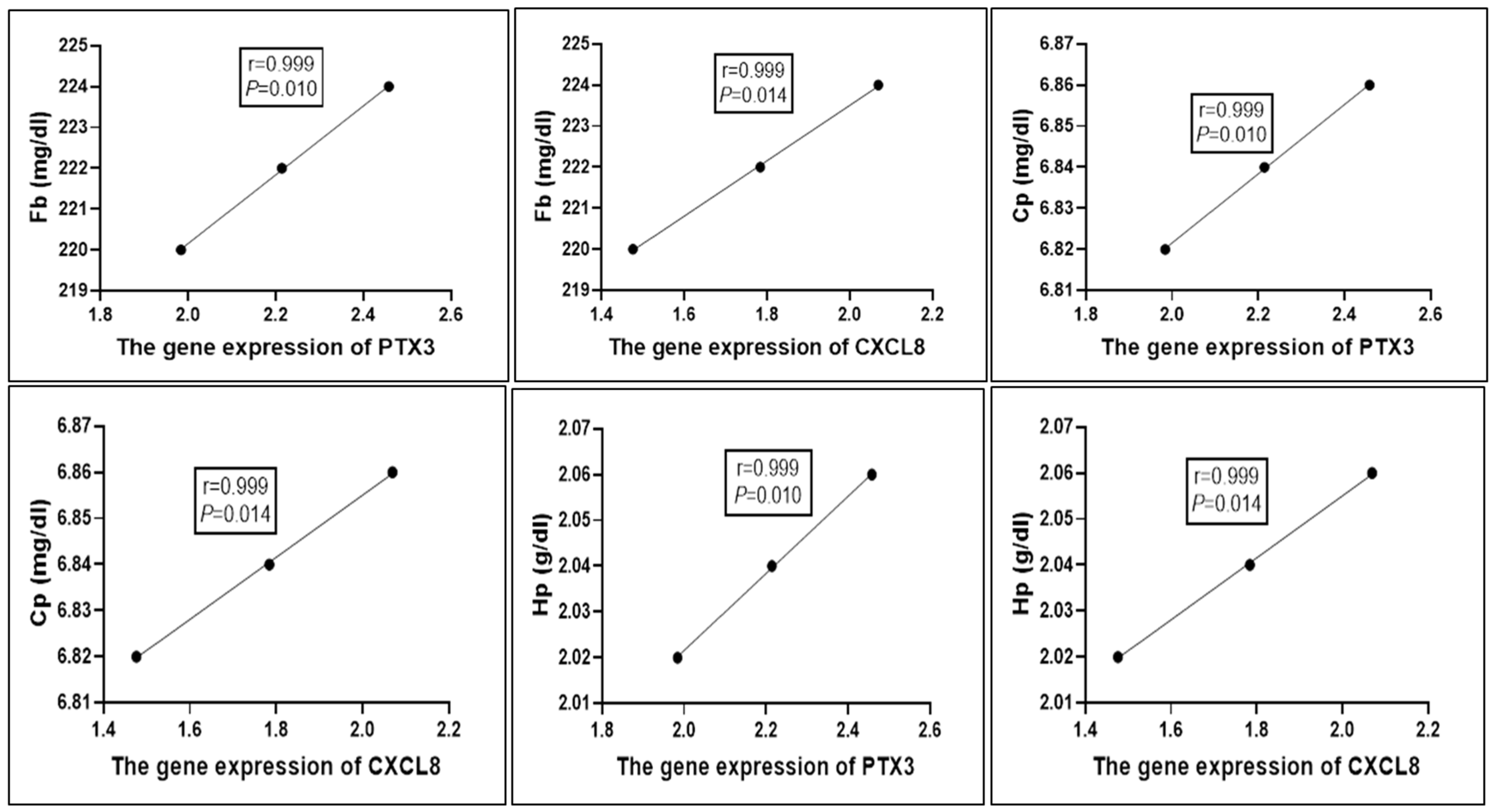
The correlation between the serum profile of acute phase proteins in the CG and PG and the gene expression patterns of immunological markers is shown in Figure 3. The mRNA levels of PTX3 and CXCL8 were positively correlated with the blood levels of Fb, Cp, and Hp (r = 0.999, p = 0.01).

4. Discussion

According to [53], pneumonia is a major cause of economic loss due to its high morbidity and mortality rates, frequent occurrence across sheep of all ages and breeds, and the associated impacts on growth rates and veterinary costs. Environmental stressors, such as extreme summer heat and harsh winter cold, increase the susceptibility of desert-dwelling animals to microbial infections, particularly respiratory diseases [54,55]. This study aims to investigate gene expression patterns and serum profiles of acute-phase proteins (APPs), hormonal markers, and iron profile parameters in Barki sheep infected with methicillin-resistant Staphylococcus aureus (MRSA). Additionally, it will determine the prevalence of MRSA in nasal swabs, analyze the antibiotic resistance profiles of isolated MRSA strains, and perform molecular identification of key antibiotic resistance genes. This comprehensive approach seeks to provide critical insights into the molecular mechanisms underlying these infections to support more effective management strategies for ovine health.

4.1. Clinical Examination

The pneumonic sheep in this study exhibited clinical signs including cyanotic mucous membranes, coughing, dyspnea, bilateral nasal discharge, elevated body temperature, and increased heart and respiratory rates compared to healthy controls. These findings align with observations reported in calves [56,57,58] and sheep [2,59,60]. The observed anorexia, depression, and lethargy may result from muscular weakness caused by intracellular potassium leakage, hyperkalemia, and hypoglycemia [61]. Additionally, infection and inflammation, along with the release of pyrogenic substances such as prostaglandins and stimulation of the hypothalamic thermoregulatory center, contribute to pain responses and hyperthermia [62].

4.2. Prevalence of Staphylococcus aureus in Pneumonic Sheep

Staphylococcus aureus is one of the most frequently isolated bacterial pathogens in pneumonic sheep. In this study, S. aureus was detected in 44% (44/100) of nasal swab samples. This prevalence is consistent with previous reports from Egypt, such as those by [63] (43%), [64] (42%), and [65] (40%). However, higher isolation rates have also been documented, including 50% [66], 55% [67], and 66% [68]. Conversely, lower recovery rates have been reported by [69] (15.5%), [70] (26%), and [71] (28.9%). The lowest recorded isolation rates are 1.23% in Southern Italy (Caruso et al., 2015) and 2.3% in Iran [72]. Management approaches, antibiotic use, isolation techniques, and environmental conditions can all have an impact on isolation frequencies [73].

4.3. Emergence of MRSA in Sheep

Due to its zoonotic potential, methicillin-resistant Staphylococcus aureus (MRSA) represents a significant public health concern, particularly among individuals in close contact with livestock [74]. The transmission of MRSA through the food chain further exacerbates this risk [75]. Timely and accurate diagnosis is essential for effective antibiotic therapy and infection control strategies [76]. Molecular techniques, including conventional PCR and real-time PCR, have substantially enhanced the detection of MRSA [77]. In the present study, the mecA gene—an indicator of methicillin resistance—was detected in 17 out of 44 (38.6%) S. aureus isolates, identifying them as MRSA. This prevalence is consistent with findings from Egypt [37] (36.6%), Nigeria [78,79] (34.7% and 43%, respectively), and Ethiopia [80] (32.4%). However, lower MRSA prevalence rates have been reported in Iran [81] (9.1%), Egypt [82] (25.3%), and European nations like France (6%), Ireland (5%), and the UK (2%) [83]. However, research in Egypt revealed rates as high as 85.7% [84] and even 100% [85], while [86] in Pakistan observed an extremely high MRSA incidence of 83%.
Several factors may account for the elevated MRSA frequency. Chief among them are inadequate infection control measures, including limited training opportunities, understaffing, heavy workloads, and resource constraints, which impede the implementation of standard precautions. In addition, widespread self-medication and the inappropriate use of antibiotics contribute significantly to resistance development. The increased detection of MRSA in Egyptian sheep may also be attributed to the expanded use of molecular diagnostic tools and the unregulated, extensive use of antibiotics in livestock production systems [87,88].

4.4. Antimicrobial Resistance of S. aureus and MRSA

Antimicrobial susceptibility testing showed that 21 out of 27 (77.7%) S. aureus isolates were susceptible to gentamicin, which is consistent with research by [89,90]. The 74% ofloxacin susceptibility was comparable to those from [91,92] in Nepal. Nonetheless, MRSA isolates showed high rates of resistance: 100% susceptibility to vancomycin, 96% susceptibility to levofloxacin, 70% susceptibility to ofloxacin, and 100% resistance to amoxicillin, amoxicillin/clavulanate, cloxacillin, penicillin, erythromycin, chloramphenicol, tetracycline, and cotrimoxazole. According to [93], the mecA gene, which encodes penicillin-binding protein 2a (PBP2a), is largely responsible for MRSA’s resistance mechanism. This gene allows the bacteria to withstand β-lactam drugs. As a result of its high prevalence in Europe, North Africa, the Middle East, and East Asia, MRSA has now gained international attention [27]. Antibiotic resistance in S. aureus has been greatly exacerbated by the careless use of antibiotics without a prescription in underdeveloped nations [89]. According to investigations conducted in Owerri, Nigeria [94], cloxacillin was previously very successful against S. aureus, with 78% susceptibility and 85.4% susceptibility, respectively. But according to recent research, such as [95], there has been a notable drop in susceptibility to 38.5%, most likely as a result of the increase in methicillin resistance. Likewise, this study’s findings on tetracycline, chloramphenicol, penicillin, and cotrimoxazole resistance are consistent with those from Eritrea [96] and Nigeria [89,95].

4.5. Nucleotide Sequence Variants of Investigated Immune Genes Linked to Pneumonia

We investigated the alterations in the immunological states of ewes with pneumonia in comparison to healthy ewes by assessing the mRNA levels of immune genes. The expression of the genes TLR2, CLEC4E, PTX3, CXCL8, and IL15RA was much higher in the afflicted ewes than in the healthy ones. However, SOCS3 levels were decreased. Defensin, SP110, SPP1, BP1, A2M, ADORA3, CARD15, IRF3, and SCART1 are among the immune genes whose levels differ in goats with and without pneumonia [97]. Gene expression profiling has connected viral pneumonia in sheep to complement genes and Toll-like receptors (TLRs) [98].
This study used a PCR-DNA sequencing technique to characterize the immunological (TLR2, CLEC4E, PTX3, CXCL8, SOCS3 and IL15RA) genes in ewes with pneumonia and healthy ewes. The results demonstrate that there are differences in the SNPs involving the two categories. It is crucial to stress that, in contrast to the similar datasets obtained from GenBank, the polymorphisms discovered and made available in this context offer extra information for the evaluated indicators. This link is initially shown by the Ovis aris gene sequences used in our study, which were published in PubMed.
Susceptibility to pneumonia in ruminants has been investigated using the candidate gene approach, revealing notable differences in the nucleotide sequences of immune-related genes between pneumonic and healthy lambs [99]. In pneumonia-affected sheep, the expression levels of genes such as IL-1α, IL1B, IL6, TNF-α, LFA-1, CR2, IL17, IL13, DEFB123, SCART1, ICAM1, NOS, and HMOX1 were significantly upregulated compared to resistant ewes. Conversely, genes involved in anti-inflammatory and antioxidant responses (IL10, SOD1, CAT, GPX1, and NQO1) were downregulated. For example, susceptibility or resistance to Mycoplasma ovipneumoniae in sheep has been associated with polymorphisms in the MHC-DRB1 exon 2 region [100]. The influence of TMEM154 gene variations on sheep susceptibility to ovine progressive pneumonia virus following natural exposure was examined by [101]. Additionally, single nucleotide polymorphisms (SNPs) linked to pneumonia susceptibility and resistance were identified in several immune-related genes—SLC11A1, CD14, CCL2, TLR1, TLR7, TLR8, TLR9, defensin, SP110, SPP1, BP1, A2M, ADORA3, CARD15, IRF3, and SCART1—in Baladi goats affected by pneumonia, compared to a healthy control group, using PCR-DNA sequencing [97].
We found that the TLR2 (354 bp), CLEC4E (443 bp), and IL15RA (378 bp) genes each had one recurrent SNP for each marker, which leads to non-synonymous mutations L57E, C15Y, and S25N, respectively, based on DNA sequencing. Goats, cattle, and buffalo preserve the modified base in the TLR2 gene (GenBank accession numbers XM_005217446.5, JN608796.1, and JQ342089.1, respectively). A conserved G43A SNP for the CLEC4E gene is found in bighorn sheep (Ovis canadensis) (GenBank accession number XM_069585824.1). Additionally, scimitar-horned oryx (Oryx dammah; GenBank accession code XM_040248264.1) databases contain the A74G SNP for the IL15RA gene.
There were two synonymous SNPs in the 300 bp CXCL8 and 480 bp SOCS3 genes. The amino acids 146T and 52L, respectively, were linked to the two SNPs, G438C and C159T. It was found that the altered nucleotide in SOCS3 was conserved when compared to the genomes of cattle, buffalo, and bighorn sheep (GenBank accession numbers NM_174466.2, XM_025279609.3, and ON390895.1, respectively). Similarly, databases for cattle, buffalo, and bighorn sheep were found to include the C159T SNP in CXCL8, which has the GenBank accession numbers NM_173925.2, FJ595833.1, and EF524263.1, respectively.
Sequencing of the PTX3 (363 bp) gene revealed two recurrent SNPs. G90C was identified as a synonymous mutation in 30A. Conversely, the non-synonymous mutation L95F was brought on by the C283T SNP. The cattle has a conserved G90C SNP (GenBank accession number NM_001076259.2), and the goat has a conserved C283T mutated base (GenBank accession number XM_018048475.1).
Immune gene DNA sequencing revealed seven SNPs, three of which were synonymous and four of which were non-synonymous. A correlation was found between the four non-synonymous SNPs and healthy ewes. In contrast, three synonymous SNPs were shown to be associated with the incidence of pneumonia. Mutation is the central cause of selection and adaptability [102]. Exonic area mutations were presented in all examined markers under investigation in this case, leading to different coding DNA sequences in pneumonic as opposed to healthy ewes. Four non-synonymous and three synonymous SNPs were identified through DNA sequencing of investigated genes. Protein sequences are altered by non-synonymous mutations, and natural selection usually targets animals with these changes [102]. Genetic discrepancy caused by non-synonymous SNPs adjusts the fixed amino acid at the mutant site, potentially leading to structural and practical deviations in the mutated protein [103]. It was long thought that there was either no selection on synonymous mutations or very little selection [102]. The examined genes must be precisely characterized at the molecular level in order to comprehend the physiological alterations between pneumonic and healthy ewes in terms of resistance and susceptibility. According to our research, polymorphisms based on the translated DNA sequence of ewes are more valued than intronic regions.
According to the afoementioned findings, the C169T (L→F) substitution in TLR2 and G43A (C→Y) in CLEC4E were predicted to be “probably damaging” due to alterations in conserved hydrophobic domains critical for receptor-ligand binding. The A74G (S→N) variant in IL15RA lies within a domain involved in cytokine receptor activity and was also predicted to have a moderate impact on protein function [102,103].
In the face of microbes, the first line of defense depends on innate immune systems, especially Toll-like receptors and antimicrobial peptides [104]. Pathogen-associated molecular forms are seen in the ten Toll-like receptor (TLR) family members that are typically encoded in the mammalian genome [105]. TLR2 is able to identify bacterial lipids like lipoteichoic acid (LTA) [54].
Different cell types express the glycoprotein pentraxin 3 (PTX3) in response to primary inflammatory stimuli, including those mediated by TNFα, IL-1β, and TLR agonists [106,107]. According to [108], the PTX3 gene is primarily responsible for regulating innate resistance to pathogens and inflammatory responses. It also functions as an antibacterial agent that may help protect the mammary gland against chronic and subclinical infections. According to several studies, the goats’ udder’s first line of defense against S. aureus infection is PTX3, which is markedly up-regulated [108,109]. In the examined Holstein and Montbéliarde dairy cows, single nucleotide polymorphisms (SNPs) and gene expression profile changes related to mastitis resistance/susceptibility were found in the PTX3 gene [110].
As an immune response gene linked to genetic resistance and susceptibility to a wide range of diseases, CLEC4E is expressed on the surface of macrophages [111] and is crucial in the immune system’s recognition of bacterial glycolipids [112]. In goats vulnerable to nematode infection, Ref. [113] demonstrated that CLEC4E was a down-regulated gene implicated in the immune response system. When examining the in vivo transcriptional response of mammary epithelial cells during the early phases of infection with Staphylococcus aureus, CLEC4E was elevated in other goat investigations [112].
Numerous cell types, including macrophages, epithelial cells, and endothelial cells, produce the chemokine interleukin-8 (IL8), also known as chemokine (C-X-C motif) ligand 8, or CXCL8 [114]. This chemokine acts on CXCR1 and CXCR2 receptors and is one of the main mediators of the inflammatory response [115].
A cytokine receptor with a high affinity for interleukin 15 (IL15) is encoded by the IL15RA gene. The IL-15 signaling pathways and their main biological effects on various immune cell types and innate immunity systems are examples of similar pathways. SNPs in IL15RA were linked to macro-pathogen richness in humans [116]. Goats’ resistance to gastrointestinal illnesses and mastitis was found to be influenced by the IL15RA gene [117].
A protein that inhibits cytokine signaling, SOCS3 belongs to the suppressors of cytokine signaling (SOCS) family [118]. The role of mammary epithelial cells in the immune response during the initial phases of a bacterial infection with S. aureus in goats was investigated by [112].
The entire collection of genes in the genome that are consistently and effectively expressed under a range of physiological and pathological circumstances is referred to as the “transcriptome” [119]. It has proven useful in identifying new therapeutic or diagnostic targets and is widely employed in inflammatory illnesses to assess the immune system and detect pathogenic, diagnostic, and prognostic markers [120].
This alteration in immunological marker expression patterns in ewes with pneumonia may be due to the fact that wounded tissues are more susceptible to free radical reactions than healthy tissues [121]. By binding to cell surface receptors, pneumonia also produces a range of cellular immunological components that control and regulate the immune system and inflammatory response [122]. The intricate interactions among these immunological components have been demonstrated by numerous studies. In the case of acute-phase reactive proteins, complement proteins, and other immune globulins, for instance, the interaction of these proteins results in a complex network structure [123]. Therefore, our hypothesis is that infectious pneumonia may have affected most of the sheep in our study. Furthermore, our real-time PCR data clearly shows that sheep with pneumonia had a marked inflammatory response.

4.6. Biochemical Markers

APP levels in the PG were considerably higher than those in the CG, which is indicative of the biochemical changes in the pneumonic sheep in this investigation. This outcome was consistent with earlier research showing elevated APP concentrations in pneumonia cases in a variety of animal species [4,124,125]. In the innate immune response, APPs are essential because they serve as the body’s initial line of defense against infection, damage, and cancer. When disease strikes, APPs, which are produced by hepatocytes in response to pro-inflammatory cytokines, quickly increase in number and stay in the bloodstream for a long time. While each protein has distinct qualities, APPs work in concert to identify infections and limit microbial proliferation. Through the binding of free hemoglobin, Hp reduces oxidative damage and denies bacteria the iron they need to proliferate. This has both bacteriostatic and antioxidant benefits. Because it remains in the bloodstream longer than SAA, Hp is also a good predictor of tissue damage and disease consequences. SAA, on the other hand, reacts quickly to infections by binding bacteria and making it easier for neutrophils and macrophages to absorb them [1,4,124,125]. Because of its anti-inflammatory and antioxidant qualities, Cp helps to regulate the immune system. As an extracellular peroxide scavenger, it transforms poisonous ferrous iron into its harmless ferric form, neutralizes dangerous reactive oxygen species, and decreases neutrophil adherence to the endothelium. According to [4,124,125], Fb is essential for regulating hemostasis, coordinating the inflammatory response, and promoting tissue healing. When taken as a whole, these APPs strengthen the body’s defenses against infections, inflammation, and the initiation of healing processes.
Pneumonia causes major hormonal changes in addition to being an inflammatory illness. The complex interaction between the immunological and endocrine systems is highlighted by this study, which shows that PG patients had significant hypercortisolemia, hypoinsulinemia, increased growth hormone (GH), and decreased T3 and T4 levels. The endocrine system tries to maintain immune function by reversing the significant metabolic changes brought on by the immunological response during infection [126]. The endocrine system tries to maintain immune function by reversing the significant metabolic changes brought on by the immunological response during infection [126]. The hypothalamic–pituitary–adrenal (HPA) axis is directly stimulated by pro-inflammatory cytokines, which results in the release of adrenocorticotropic hormone (ACTH) and adrenal activation, which raises cortisol levels. The increase in cortisol is necessary for preventing inflammation-induced hypoglycemia and providing a sufficient amount of energy for vital physiological functions as well as immunological defense systems. Increased pituitary GH production, decreased cellular glucose absorption, and impaired insulin synthesis are just some of the metabolic consequences of hypercortisolemia [127,128]. It also supports energy homeostasis by suppressing thyroid function and encouraging gluconeogenesis and glycogenolysis. A powerful immunomodulator, cortisol inhibits the generation of pro-inflammatory cytokines and shifts immunological activity from innate to humoral immunity, in addition to its metabolic function [126].
Thyroid hormone (T3 and T4) reduction, increased growth hormone, and hypoinsulinemia in PG are all consistent with these adaptive endocrine responses. In order to restore thyroid hormone levels, a corresponding increase in TSH most likely serves as a compensatory mechanism. Because of this complex hormonal interaction, the endocrine system plays a vital role in preserving immunological and metabolic homeostasis during pneumonia [126].
PG’s iron profile showed hyperferritinemia, hypoferremia, and hypotransferrinemia. These alterations were influenced by the immunological response in multiple ways. To prevent the invasive bacteria from growing and multiplying, the activated pro-inflammatory cytokines obstruct their access to iron. Therefore, they decrease intestinal iron absorption, decrease transferrin activity, promote hepcidin release (which competes with transferrin for iron), and promote iron storage as ferritin [3,129]. Because ferritin is a positive acute phase reactant and transferrin is a negative acute phase reactant, the acute phase response, which typically accompanies respiratory and lung diseases, also contributes to these changes [130,131]. Furthermore, the observed hyperferritinemia may be a result of oxidative stress associated with pneumonia, as the accumulated free radicals attack and break the membrane of the red blood cells, resulting in the release of a significant amount of free iron. Consequently, the body increased ferritin production to eliminate these detrimental consequences of free iron [129]. This hypoferremia is rationally explained by the increased TIBC and UIBC and decreased Tf sat. %.
APPs, hormones, and iron profile measures showed high sensitivity, specificity, PPV, NPV, and accuracy rate (except for TSH), with an AUC > 0.7, in terms of their diagnostic and prognostic value. These results are somewhat consistent with earlier studies that used APPs for reliable indicators of pneumonia in sheep and other animals. The study’s potential use of iron profile and hormonal parameters as useful disease indicators, however, is a significant new finding [4,124,125]. The study strongly suggests UIBC because of its exceptional diagnostic performance, T3 and GH because of their high LRs, and Hp because of its distinct percentage increase among the tested markers. These results highlight the potential benefits of combining iron profile measures, hormone markers, and APPs for improved pneumonia diagnosis and prognosis.
The current work has several restrictions that should be addressed in upcoming research. First, the study was conducted on a small number of sheep; therefore, studies involving larger sample sizes are needed. Second, the study focused on a single breed, so applying this research to different calf breeds would provide more accurate health assessments. Third, only a limited set of immune genes were examined, indicating the need for broader gene profiling in subsequent studies. Fourth, pharmacokinetic/pharmacodynamic (PK/PD) studies, therapeutic efficacy trials, and field validation across multiple regions must be integrated for a more thorough and representative treatment approach. As a result, studies that expand the geographical scope and include clinical treatment trials are necessary to verify the effectiveness, safety, and economic viability of the recommended therapeutic agents. This would ensure that the selected treatments are not only microbiologically effective but also clinically practical and sustainable under diverse field conditions.

5. Conclusions

This study highlights the significant prevalence of methicillin-resistant Staphylococcus aureus (MRSA) among pneumonic sheep in Egypt and underscores its multidrug-resistant profile, particularly its complete resistance to several β-lactam antibiotics. Gene expression analysis revealed significant upregulation of immune-related markers (TLR2, CLEC4E, PTX3, CXCL8, IL15RA) and notable SNP variations associated with pneumonia susceptibility. Biochemical profiling showed marked alterations in acute-phase proteins, hormones, and iron metabolism, supporting their potential diagnostic and prognostic value. These findings suggest that integrating molecular diagnostics, SNP-based genetic markers, and serum biomarker profiling can enhance early detection, targeted therapy, and selective breeding strategies to mitigate pneumonia in sheep flocks.

Supplementary Materials

The following supporting information can be downloaded at https://www.mdpi.com/article/10.3390/vetsci12060584/s1, Figure S1: Sheep showing signs of pneumonia. Figure S2: Representative alignment of the TLR2 gene (354 bp) between healthy (H) and pneumonia-affected ewes (P) compared to reference GenBank accession number (NM_001048231.1). Figure S3: Representative alignment of the CLEC4E gene (443 bp) between healthy (H) and pneumonia-affected ewes (P) compared to reference GenBank accession number (XM_042247586.1). Figure S4: Representative alignment of the PTX3 gene (363 bp) between healthy (H) and pneumonia-affected ewes (P) compared to reference GenBank accession number (XM_004003220.5). Figure S5: Representative alignment of the SOCS3 gene (480 bp) between healthy (H) and pneumonia-affected ewes (P) compared to reference GenBank accession number (XM_027974200.3). Figure S6: Representative alignment of the CXCL8 gene (300 bp) between healthy (H) and pneumonia-affected ewes (P) compared to reference GenBank accession number (NM_001009401.2). Figure S7: Representative alignment of the IL15RA gene (378 bp) between healthy (H) and pneumonia-affected ewes (P) compared to reference GenBank accession number (XM_042230436.2).

Author Contributions

E.A.N., A.E.-S., and A.D. designed the experiment, collected the samples, interpreted the results, conducted blood biochemical analysis, and drafted the main manuscript. H.M.A., M.T.R., A.H., A.E., A.A., K.M.A., and M.A.B. contributed to the paper’s authoring and planning the article. All authors have read and agreed to the published version of the manuscript.

Funding

This research was funded by Princess Nourah bint Abdulrahman University Researchers Supporting Project number (PNURSP2025R454), Princess Nourah bint Abdulrahman University, Riyadh, Saudi Arabia.

Institutional Review Board Statement

The Ethics Committee of Desert Research Centre (DRC), Egypt, approved the methods used to collect samples and care for animals in this experiment (code DRC-05-3-2025).

Informed Consent Statement

All farmers gave their informed consent to participate in the project.

Data Availability Statement

Upon reasonable request, the appropriate author will share supporting data for the study’s conclusions.

Acknowledgments

The authors acknowledge Princess Nourah bint Abdulrahman University Researchers Supporting Project number (PNURSP2025R454), Princess Nourah bint Abdulrahman University, Riyadh, Saudi Arabia. The authors extend the appreciation to the deanship of postgraduate studies and scientific research at Majmaah University for funding this research work through the project number (R-2025-1842).

Conflicts of Interest

The authors declare no conflicts of interest.

References

  1. El-Deeb, W.M.; Elmoslemany, A.M. The diagnostic accuracy of acute phase proteins and proinflammatory cytokines in sheep with pneumonic pasteurellosis. PeerJ 2016, 4, e2161. [Google Scholar] [CrossRef]
  2. Arbaga, A.; Hassan, H.; Anis, A.; Othman, N.; Kamr, A. Clinicopathological and Electrophoretic Pattern of Serum Protein Alterations in Acute Pneumonic Sheep. Pak. Vet. J. 2023, 43, 303–308. [Google Scholar]
  3. Fernández, S.; Galapero, J.; Rey, J.; Pérez, C.; Ramos, A. Cytokines study in lungs infected with Pasteurellaceae and Mycoplasma spp. from fattening lambs. J. Med. Microbiol. Immunol. Res. 2018, 2, 1–7. [Google Scholar]
  4. Ramadan, M.A.; Abdel-Raoof, Y.M.; Zeineldin, M.M.; El Attar, H.E.M.; Abdelghany, A.H.; Ghanem, M.M. Assessment of pulmonary function test, acute phase proteins, cytokines and electrocardiographic changes in naturally occurring bovine respiratory disease of feedlot cattle calves. Benha Vet. Med. J. 2019, 37, 1–5. [Google Scholar] [CrossRef]
  5. Rizk, M.A.; El-Sayed, S.A.E.-S.; Salman, D.; Marghani, B.H.; Gadalla, H.E.; Sayed-Ahmed, M.Z. Immunomodulatory effect of vitamin C on proinflammatory cytokines production in Ossimi Lambs (Ovis aries) with Pneumonic Pasteurellosis. Animals 2021, 11, 3374. [Google Scholar] [CrossRef]
  6. Aly, M.M.; El-Sanousi, A.A.; Mahmoud, A.A. Environmental factors influencing respiratory infections in small ruminants in arid zones. Vet. World 2024, 17, 567–574. [Google Scholar]
  7. El-Sayed, A.; Ahmed, M.; Hassan, M.A. Etiological agents and risk factors of ovine respiratory diseases in Egypt: A comprehensive review. J. Vet. Med. Res. 2023, 30, 201–213. [Google Scholar]
  8. Cucarella, C.; Tormo, M.A.; Ubeda, C.; Trotonda, M.P.; Monzón, M.; Peris, C.; Amorena, B.; Lasa, Í.; Penadés, J.R. Role of biofilm-associated protein bap in the pathogenesis of bovine Staphylococcus aureus. Infect. Immun. 2004, 72, 2177–2185. [Google Scholar] [CrossRef]
  9. Tong, S.Y.; Davis, J.S.; Eichenberger, E.; Holland, T.L.; Fowler, V.G., Jr. Staphylococcus aureus infections: Epidemiology, pathophysiology, clinical manifestations, and management. Clin. Microbiol. Rev. 2015, 28, 603–661. [Google Scholar] [CrossRef]
  10. Becker, K.; Heilmann, C.; Peters, G. Coagulase-negative staphylococci. Clin. Microbiol. Rev. 2014, 27, 870–926. [Google Scholar] [CrossRef]
  11. Zaher, H.A.; El Baz, S.; Alothaim, A.S.; Alsalamah, S.A.; Alghonaim, M.I.; Alawam, A.S.; Eraqi, M.M. Molecular basis of methicillin and vancomycin resistance in Staphylococcus aureus from cattle, sheep carcasses and slaughterhouse workers. Antibiotics 2023, 12, 205. [Google Scholar] [CrossRef]
  12. Gao, J.; Stewart, G.C. Regulatory elements of the Staphylococcus aureus protein A (Spa) promoter. J. Bacteriol. 2004, 186, 3738–3748. [Google Scholar] [CrossRef]
  13. Turner, N.A.; Sharma-Kuinkel, B.K.; Maskarinec, S.A.; Eichenberger, E.M.; Shah, P.P.; Carugati, M.; Holland, T.L.; Fowler, V.G., Jr. Methicillin-resistant Staphylococcus aureus: An overview of basic and clinical research. Nat. Rev. Microbiol. 2019, 17, 203–218. [Google Scholar] [CrossRef]
  14. Wichelhaus, T.; Schulze, J.; Hunfeld, K.; Schäfer, V.; Brade, V. Clonal heterogeneity, distribution, and pathogenicity of methicillin-resistant Staphylococcus aureus. Eur. J. Clin. Microbiol. Infect. Dis. 1997, 16, 893–897. [Google Scholar] [CrossRef]
  15. Acar, J.; Rostel, B. Antimicrobial resistance: An overview. Rev. Sci. Tech.-Off. Int. Des Epizoot. 2001, 20, 797–810. [Google Scholar] [CrossRef]
  16. Moran, G.J.; Krishnadasan, A.; Gorwitz, R.J.; Fosheim, G.E.; McDougal, L.K.; Carey, R.B.; Talan, D.A. Methicillin-resistant S. aureus infections among patients in the emergency department. N. Engl. J. Med. 2006, 355, 666–674. [Google Scholar] [CrossRef]
  17. Conturba, B.; Lo Feudo, C.M.; Stucchi, L.; Stancari, G.; Olivieri, L.; Ferrucci, F. Recurrent equine capillary haemangioma treated with adjunctive laser photocoagulation therapy: A case report. Vet. Dermatol. 2021, 32, 290. [Google Scholar] [CrossRef]
  18. Lee, A.S.; De Lencastre, H.; Garau, J.; Kluytmans, J.; Malhotra-Kumar, S.; Peschel, A.; Harbarth, S. Methicillin-resistant Staphylococcus aureus. Nat. Rev. Dis. Primers 2018, 4, 18033. [Google Scholar] [CrossRef]
  19. Boucher, H.W.; Talbot, G.H.; Bradley, J.S.; Edwards, J.E.; Gilbert, D.; Rice, L.B.; Scheld, M.; Spellberg, B.; Bartlett, J. Bad bugs, no drugs: No ESKAPE! An update from the Infectious Diseases Society of America. Clin. Infect. Dis. 2009, 48, 1334. [Google Scholar] [CrossRef]
  20. Li, Y.; Cao, Y.; Wang, Y.; Yang, Y.; Liu, Y. Genomic adaptations contributing to linezolid and daptomycin resistance in clinical MRSA isolates. Front. Microbiol. 2022, 13, 853270. [Google Scholar]
  21. Shore, A.C.; Deasy, E.C.; Slickers, P.; Brennan, G.; O’Connell, B.; Monecke, S.; Ehricht, R.; Coleman, D.C. Detection of staphylococcal cassette chromosome mec type XI carrying highly divergent mecA, mecI, mecR1, blaZ, and ccr genes in human clinical isolates of clonal complex 130 methicillin-resistant Staphylococcus aureus. Antimicrob. Agents Chemother. 2011, 55, 3765–3773. [Google Scholar] [CrossRef]
  22. Alghamdi, B.A.; Al-Johani, I.; Al-Shamrani, J.M.; Alshamrani, H.M.; Al-Otaibi, B.G.; Almazmomi, K.; Yusof, N.Y. Antimicrobial resistance in methicillin-resistant Staphylococcus aureus. Saudi J. Biol. Sci. 2023, 30, 103604. [Google Scholar] [CrossRef]
  23. David, M.Z.; Daum, R.S. Community-associated methicillin-resistant Staphylococcus aureus: Epidemiology and clinical consequences of an emerging epidemic. Clin. Microbiol. Rev. 2010, 23, 616–687. [Google Scholar] [CrossRef]
  24. Klein, E.Y.; Van Boeckel, T.P.; Martinez, E.M.; Pant, S.; Gandra, S.; Levin, S.A.; Goossens, H.; Laxminarayan, R. Global increase and geographic convergence in antibiotic consumption between 2000 and 2015. Proc. Natl. Acad. Sci. USA 2018, 115, E3463–E3470. [Google Scholar] [CrossRef]
  25. Talaat, M.; Zayed, B.; Tolba, S.; Abdou, E.; Gomaa, M.; Itani, D.; Hutin, Y.; Hajjeh, R. Increasing antimicrobial resistance in world health organization eastern mediterranean region, 2017–2019. Emerg. Infect. Dis. 2022, 28, 717. [Google Scholar] [CrossRef]
  26. Jauro, S.; Hamman, M.M.; Malgwi, K.D.; Musa, J.A.; Ngoshe, Y.B.; Gulani, I.A.; Kwoji, I.D.; Iliya, I.; Abubakar, M.B.; Fasina, F.O. Antimicrobial resistance pattern of methicillin-resistant Staphylococcus aureus isolated from sheep and humans in Veterinary Hospital Maiduguri, Nigeria. Vet. World 2022, 15, 1141. [Google Scholar] [CrossRef]
  27. Lakhundi, S.; Zhang, K. Methicillin-resistant Staphylococcus aureus: Molecular characterization, evolution, and epidemiology. Clin. Microbiol. Rev. 2018, 31, e00020-18. [Google Scholar] [CrossRef]
  28. Saleha, A.; Zunita, Z. Methicillin resistant Staphylococcus aureus (MRSA): An emerging veterinary and zoonotic pathogen of public health concern and some studies in Malaysia. J. Anim. Vet. Adv. 2010, 9, 1094–1098. [Google Scholar] [CrossRef]
  29. Kralik, P.; Ricchi, M. A basic guide to real time PCR in microbial diagnostics: Definitions, parameters, and everything. Front. Microbiol. 2017, 8, 108. [Google Scholar] [CrossRef]
  30. Yamamoto, Y. PCR in diagnosis of infection: Detection of bacteria in cerebrospinal fluids. Clin. Vaccine Immunol. 2002, 9, 508–514. [Google Scholar] [CrossRef]
  31. Kerremans, J.; Verboom, P.; Stijnen, T.; Hakkaart-van Roijen, L.; Goessens, W.; Verbrugh, H.; Vos, M. Rapid identification and antimicrobial susceptibility testing reduce antibiotic use and accelerate pathogen-directed antibiotic use. J. Antimicrob. Chemother. 2008, 61, 428–435. [Google Scholar] [CrossRef]
  32. Mostafa Abdalhamed, A.; Zeedan, G.S.G.; Ahmed Arafa, A.; Shafeek Ibrahim, E.; Sedky, D.; Abdel nabey Hafez, A. Detection of Methicillin-Resistant Staphylococcus aureus in Clinical and Subclinical Mastitis in Ruminants and Studying the Effect of Novel Green Synthetized Nanoparticles as One of the Alternative Treatments. Vet. Med. Int. 2022, 2022, 6309984. [Google Scholar] [CrossRef]
  33. Ceciliani, F.; Ceron, J.; Eckersall, P.; Sauerwein, H. Acute phase proteins in ruminants. J. Proteom. 2012, 75, 4207–4231. [Google Scholar] [CrossRef]
  34. Miglio, A.; Moscati, L.; Scoccia, E.; Maresca, C.; Antognoni, M.T.; Felici, A. Reference values for serum amyloid A, haptoglobin, lysozyme, zinc and iron in healthy lactating Lacaune sheep. Acta Vet. Scand. 2018, 60, 46. [Google Scholar] [CrossRef]
  35. Canelli, E.; Catella, A.; Borghetti, P.; Ferrari, L.; Ogno, G.; De Angelis, E.; Corradi, A.; Passeri, B.; Bertani, V.; Sandri, G. Phenotypic characterization of a highly pathogenic Italian porcine reproductive and respiratory syndrome virus (PRRSV) type 1 subtype 1 isolate in experimentally infected pigs. Vet. Microbiol. 2017, 210, 124–133. [Google Scholar] [CrossRef]
  36. Pierce, C.F.; Brown, V.R.; Olsen, S.C.; Boggiatto, P.; Pedersen, K.; Miller, R.S.; Speidel, S.E.; Smyser, T.J. Loci associated with antibody response in feral swine (Sus scrofa) infected with Brucella suis. Front. Vet. Sci. 2020, 7, 554674. [Google Scholar] [CrossRef]
  37. Wang, X.; Zhang, X.; Lu, T.; Niu, L.; Wang, L.; Zhan, S.; Guo, J.; Cao, J.; Li, L.; Zhang, H. Nucleotide variants in TheTLR5 gene and promoter methylation with A susceptibility to brucellosis in Chinese goats. Folia Biol. (Kraków) 2022, 70, 55–66. [Google Scholar] [CrossRef]
  38. Sallam, A.M.; Abou-Souliman, I.; Reyer, H.; Wimmers, K.; Rabee, A.E. New insights into the genetic predisposition of brucellosis and its effect on the gut and vaginal microbiota in goats. Sci. Rep. 2023, 13, 20086. [Google Scholar] [CrossRef]
  39. Adams, L.; Schutta, C.J. Natural resistance against brucellosis: A review. Open Vet. Sci. J. 2010, 4, 61–71. [Google Scholar] [CrossRef]
  40. El-Shafaey, E.-S.; Ateya, A.; Ramadan, H.; Saleh, R.; Elseady, Y.; Abo El Fadl, E.; El-Khodery, S. Single nucleotide polymorphisms in IL8 and TLR4 genes as candidates for digital dermatitis resistance/susceptibility in Holstein cattle. Anim. Biotechnol. 2017, 28, 131–137. [Google Scholar] [CrossRef]
  41. Constable, P.D.; Hinchcliff, K.W.; Done, S.H.; Grünberg, W. Veterinary Medicine: A Textbook of the Diseases of Cattle, Horses, Sheep, Pigs and Goats; Elsevier Health Sciences: Amsterdam, The Netherlands, 2016. [Google Scholar]
  42. Uchida-Fujii, E.; Niwa, H.; Kinoshita, Y.; Nukada, T. Matrix-assisted laser desorption ionization–time of flight mass spectrometry (MALDI-TOF MS) for identification of bacterial isolates from horses. J. Equine Vet. Sci. 2020, 95, 103276. [Google Scholar] [CrossRef]
  43. Rocha, L.A.; Aleixo, A.; Allen, G.; Almeda, F.; Baldwin, C.C.; Barclay, M.V.; Bates, J.M.; Bauer, A.; Benzoni, F.; Berns, C. Specimen collection: An essential tool. Science 2014, 344, 814–815. [Google Scholar] [CrossRef] [PubMed]
  44. Clinical and Laboratory Standards Institute. Performance Standards for Antimicrobial Susceptibility Testing; Clinical and Laboratory Standards Institute: Wayne, PA, USA, 2020. [Google Scholar]
  45. Weinstein, M.P.; Lewis, J.S. The clinical and laboratory standards institute subcommittee on antimicrobial susceptibility testing: Background, organization, functions, and processes. J. Clin. Microbiol. 2020, 58, e01864-19. [Google Scholar] [CrossRef] [PubMed]
  46. McClure, J.-A.; Conly, J.M.; Lau, V.; Elsayed, S.; Louie, T.; Hutchins, W.; Zhang, K. Novel multiplex PCR assay for detection of the staphylococcal virulence marker Panton-Valentine leukocidin genes and simultaneous discrimination of methicillin-susceptible from-resistant staphylococci. J. Clin. Microbiol. 2006, 44, 1141–1144. [Google Scholar] [CrossRef]
  47. Pfaffl, M.W. A new mathematical model for relative quantification in real-time RT–PCR. Nucleic Acids Res. 2001, 29, e45. [Google Scholar] [CrossRef]
  48. Boom, R.; Sol, C.; Salimans, M.; Jansen, C.; Wertheim-van Dillen, P.; Van der Noordaa, J. Rapid and simple method for purification of nucleic acids. J. Clin. Microbiol. 1990, 28, 495–503. [Google Scholar] [CrossRef] [PubMed]
  49. Boesenberg-Smith, K.A.; Pessarakli, M.M.; Wolk, D.M. Assessment of DNA yield and purity: An overlooked detail of PCR troubleshooting. Clin. Microbiol. Newsl. 2012, 34, 1, 3–6. [Google Scholar] [CrossRef]
  50. Sanger, F.; Nicklen, S.; Coulson, A.R. DNA sequencing with chain-terminating inhibitors. Proc. Natl. Acad. Sci. USA 1977, 74, 5463–5467. [Google Scholar] [CrossRef]
  51. Altschul, S.F.; Gish, W.; Miller, W.; Myers, E.W.; Lipman, D.J. Basic local alignment search tool. J. Mol. Biol. 1990, 215, 403–410. [Google Scholar] [CrossRef]
  52. Tamura, K.; Stecher, G.; Peterson, D.; Filipski, A.; Kumar, S. MEGA6: Molecular evolutionary genetics analysis version 6.0. Mol. Biol. Evol. 2013, 30, 2725–2729. [Google Scholar] [CrossRef]
  53. Abdalla, O.; Haidy, G.; Yousseff, F.; Rehab, A. Clinicopathological alterations in blood and sera of sheep due to respiratory affections. J. Clin. Pathol. Forecast 2019, 2, 1006. [Google Scholar]
  54. Mandal, R.; Gupta, V.; Joshi, V.; Kumar, S.; Mondal, D. Study of Clinico Hematobiochemical Changes and Therapeutic Management of Naturally Infected Cases of Respiratory Disease in Non-Descript Goats of Bareilly Region. Int. J. Livest. Res. 2017, 7, 211–218. [Google Scholar] [CrossRef]
  55. Teng, T.; Yang, H.; Xu, T.; Sun, G.; Song, X.; Bai, G.; Shi, B. Activation of inflammatory networks in the lungs caused by chronic cold stress is moderately attenuated by glucose supplementation. Int. J. Mol. Sci. 2022, 23, 10697. [Google Scholar] [CrossRef]
  56. Bozukluhan, K.; Merhan, O.; Kiziltepe, S.; Egritag, H.E.; Akyuz, E.; Gokce, H.I. Determination of haptoglobin, some biochemical and oxidative stress parameters in calves with pneumonia. Fresenius Env. Bull 2021, 30, 9492–9496. [Google Scholar]
  57. Su, M.; She, Y.; Deng, M.; Guo, Y.; Li, Y.; Liu, G.; Sun, B.; Liu, D. Effect of capsaicin addition on antioxidant capacity, immune performance and upper respiratory microbiota in nursing calves. Microorganisms 2023, 11, 1903. [Google Scholar] [CrossRef]
  58. Saher, A.S.; Raza, A.; Qiu, F.; Mehmood, K.; Hussain, R.; Qayyum, A.; Idris, M.; Almutairi, M.H.; Li, K. Detection of haptoglobin and serum amyloid A as biomarkers in naturally infected Mycoplasma bovis calves. Acta Trop. 2024, 254, 107215. [Google Scholar] [CrossRef]
  59. AL-Dujaily, A.H.; Abeed, S.A.; Sahib, A.M. Hematological, Biochemical, Pathological, Serology and Molecular Detection of Mycoplasma Ovipneumoniae from Awassi Sheep in Al-Najaf Province, Iraq. Ann. For. Res 2023, 66, 1410–1422. [Google Scholar]
  60. Madhi, K.S.; Jasim, A.S.; Nasear, H.A.; Ibraheim, H.K.; Gharban, H.A. Phylogenetic analysis of Klebsiella pneumoniae isolates of respiratory tract infections in humans and sheep. Open Vet. J. 2024, 14, 2325. [Google Scholar] [CrossRef]
  61. Radostits, O.M.; Gay, C.C.; Hinchcliff, K.W.; Constable, P.D. Veterinary Medicine E-Book; Elsevier Health Sciences: Amsterdam, The Netherlands, 2006. [Google Scholar]
  62. Risalde, M.; Molina, V.; Sánchez-Cordón, P.; Pedrera, M.; Panadero, R.; Romero-Palomo, F.; Gómez-Villamandos, J. Response of proinflammatory and anti-inflammatory cytokines in calves with subclinical bovine viral diarrhea challenged with bovine herpesvirus-1. Vet. Immunol. Immunopathol. 2011, 144, 135–143. [Google Scholar] [CrossRef] [PubMed]
  63. Hassan, A.; Oraby, N.; Mohamed, A.; Mahmoud, H. The possibility of using zinc oxide nanoparticles in controlling some fungal and bacterial strains isolated from buffaloes. Egypt. J. Appl. Sci. 2014, 29, 58–83. [Google Scholar]
  64. Mohamed, D.; El-Gheit, A.A.; Eid, S.; Khadra, S.A.; Elsadik, G.; Mohamed, A.; Hamed, T. Prevalence of Staphylococcus aureus and Pasteurella multocida incriminated in calf pneumonia and their treatment. Egypt. J. Anim. Health 2023, 3, 21–39. [Google Scholar]
  65. Hassan, A.; Howayda, M.; Hanan, K.M. Antimicrobial Potential of Ozone on Fungal and Bacterial Contamination of Animal Feed That Caused Diseases in Some Buffalo Farms. In Proceedings of the 1st International Conference, Animal Health Research Institute, ARC, Giza, Egypt, 9–13 November 2017; pp. 9–13. [Google Scholar]
  66. Chu, C.; Yu, C.; Lee, Y.; Su, Y. Genetically divergent methicillin-resistant Staphylococcus aureus and sec-dependent mastitis of dairy goats in Taiwan. BMC Vet. Res. 2012, 8, 39. [Google Scholar] [CrossRef]
  67. Murdoch, D.R.; Anderson, T.P.; Beynon, K.A.; Chua, A.; Fleming, A.M.; Laing, R.T.; Town, G.I.; Mills, G.D.; Chambers, S.T.; Jennings, L.C. Evaluation of a PCR assay for detection of Streptococcus pneumoniae in respiratory and nonrespiratory samples from adults with community-acquired pneumonia. J. Clin. Microbiol. 2003, 41, 63–66. [Google Scholar] [CrossRef]
  68. Sallam, M.G.A.E.; Khalifa, N.; Barakat, A.; Salem, L. Occurrence of Staphylococcus aureus in dairy farms and humans with reference to antimicrobial profile in Qalyobia Governorate. Benha Vet. Med. J. 2023, 45, 40–45. [Google Scholar] [CrossRef]
  69. Franco, M.F.; Gaeta, N.C.; Alemán, M.A.; Mellville, P.A.; Timenetsky, J.; Balaro, M.F.; Gregory, L. Bacteria isolated from the lower respiratory tract of sheep and their relationship to clinical signs of sheep respiratory disease. Pesqui. Vet. Bras. 2019, 39, 796–801. [Google Scholar] [CrossRef]
  70. Ali, A.; Abu-Zaid, K. Study on Klebsiella pneumoniae causing respiratory infection in small ruminants. Anim. Health Res. J 2019, 7, 57–67. [Google Scholar]
  71. El-Mashad, A.-B.I.; Moustafa, S.A.; Amin, A.; Samy, E.M. Pathological Studies on lung affections in sheep and goat at Kalubia Governorate. Benha Vet. Med. J. 2020, 38, 17–23. [Google Scholar] [CrossRef]
  72. IB, M.-S.; Okon, K.; Adamu, N.; Askira, U.; Isyaka, T.; Adamu, S.; Mohammed, A. Methicillin-resistant Staphylococcus aureus (MRSA) colonization rate among ruminant animals slaughtered for human consumption and contact persons in Maiduguri, Nigeria. Afr. J. Microbiol. Res. 2014, 8, 2643–2649. [Google Scholar]
  73. Graveland, H.; Duim, B.; Van Duijkeren, E.; Heederik, D.; Wagenaar, J.A. Livestock-associated methicillin-resistant Staphylococcus aureus in animals and humans. Int. J. Med. Microbiol. 2011, 301, 630–634. [Google Scholar] [CrossRef]
  74. Wirusanti, N.I.; Baldridge, M.T.; Harris, V.C. Microbiota regulation of viral infections through interferon signaling. Trends Microbiol. 2022, 30, 778–792. [Google Scholar] [CrossRef]
  75. Kluytmans, J. Methicillin-resistant Staphylococcus aureus in food products: Cause for concern or case for complacency? Clin. Microbiol. Infect. 2010, 16, 11–15. [Google Scholar] [CrossRef] [PubMed]
  76. Calfee, D.P.; Salgado, C.D.; Milstone, A.M.; Harris, A.D.; Kuhar, D.T.; Moody, J.; Aureden, K.; Huang, S.S.; Maragakis, L.L.; Yokoe, D.S. Strategies to prevent methicillin-resistant Staphylococcus aureus transmission and infection in acute care hospitals: 2014 update. Infect. Control Hosp. Epidemiol. 2014, 35, S108–S132. [Google Scholar] [CrossRef] [PubMed]
  77. Al-Talib, H.; Yean, C.Y.; Al-Khateeb, A.; Hasan, H.; Ravichandran, M. Rapid detection of methicillin-resistant Staphylococcus aureus by a newly developed dry reagent-based polymerase chain reaction assay. J. Microbiol. Immunol. Infect. 2014, 47, 484–490. [Google Scholar] [CrossRef] [PubMed]
  78. Taiwo, S.; Bamidele, M.; Omonigbehin, E.; Akinsinde, K.; Smith, S.; Onile, B.; Olowe, A. Molecular epidemiology of methicillan-resistant Staphylococcus aureus in Ilorin, Nigeria. West Afr. J. Med. 2005, 24, 100–106. [Google Scholar] [CrossRef]
  79. Ikeh, E. Methicilin-resistant Staphylococcus aureus (MRSA) at Jos University Teaching Hospital. Afr. J. Clin. Exp. Microbiol. 2003, 4, 52–55. [Google Scholar] [CrossRef]
  80. Tesfaye, K.; Gizaw, Z.; Haile, A.F. Prevalence of mastitis and phenotypic characterization of methicillin-resistant Staphylococcus aureus in lactating dairy cows of selected dairy farms in and around adama town, Central Ethiopia. Environ. Health Insights 2021, 15, 11786302211021297. [Google Scholar] [CrossRef]
  81. Rahimi, H.; Saei, H.D.; Ahmadi, M. Nasal carriage of Staphylococcus aureus: Frequency and antibiotic resistance in healthy ruminants. Jundishapur J. Microbiol. 2015, 8, e22413. [Google Scholar] [CrossRef]
  82. Ramadan, H.A.; El-Baz, A.M.; Goda, R.M.; El-Sokkary, M.M.; El-Morsi, R.M. Molecular characterization of enterotoxin genes in methicillin-resistant S. aureus isolated from food poisoning outbreaks in Egypt. J. Health Popul. Nutr. 2023, 42, 86. [Google Scholar] [CrossRef]
  83. Denton, M.; O’Connell, B.; Bernard, P.; Jarlier, V.; Williams, Z.; Henriksen, A.S. The EPISA study: Antimicrobial susceptibility of Staphylococcus aureus causing primary or secondary skin and soft tissue infections in the community in France, the UK and Ireland. J. Antimicrob. Chemother. 2008, 61, 586–588. [Google Scholar] [CrossRef]
  84. Aa, A.; Ibrahim, E.; Fouad, E.; Gaber, E. Antibiotic resistance of staphylococci concerning strains included in food industry in Egypt. Int. J. Pharm. Clin. Res. 2016, 8, 1583–1589. [Google Scholar]
  85. Naeim, D.E.; Eldesoukey, I.E.; Moawad, A.A.; Ahmed, A.M. Molecular detection of methicillin-resistant Staphylococcus aureus isolated from different foodstuffs in Egypt. Vet. Res. Forum 2023, 14, 243–248. [Google Scholar]
  86. Mehta, A.; Rodrigues, C.; Sheth, K.; Jani, S.; Hakiniyan, A.; Fazalbhoy, N. Control of methicillin resistant Staphylococcus aureus in a tertiary care Centre-A five year study. Indian J. Med. Microbiol. 1998, 16, 31–34. [Google Scholar]
  87. Pyatov, V.; Vrtková, I.; Knoll, A. Detection of selected antibiotic resistance genes using multiplex PCR assay in mastitis pathogens in the Czech Republic. Acta Vet. Brno 2017, 86, 167–174. [Google Scholar] [CrossRef]
  88. Kaliyeva, S.S.; Lavrinenko, A.V.; Tishkambayev, Y.; Zhussupova, G.; Issabekova, A.; Begesheva, D.; Simokhina, N. Microbial landscape and antibiotic susceptibility dynamics of skin and soft tissue infections in Kazakhstan 2018–2020. Antibiotics 2022, 11, 659. [Google Scholar] [CrossRef] [PubMed]
  89. Onwubiko, N.E.; Sadiq, N.M. Antibiotic sensitivity pattern of Staphylococcus aureus from clinical isolates in a tertiary health institution in Kano, Northwestern Nigeria. Pan Afr. Med. J. 2011, 8, 4. [Google Scholar] [CrossRef]
  90. Ndip, R.; Ebah, L.; Onile, B. Antibiogram of Staphylococcus aureus from clinical Syndromes in Ilorin, Nigeria. J. Med. Lab. Sci. 1997, 6, 24–26. [Google Scholar]
  91. Ikeagwu, I.; Amadi, E.; Iroha, I. Antibiotic sensitivity pattern of Staphylococcus aureus in Abakaliki. Pak. J. Med. Sci. 2008, 24, 231–235. [Google Scholar]
  92. Ashish, K.; Gurung, R.; Kinney, M.V.; Sunny, A.K.; Moinuddin, M.; Basnet, O.; Paudel, P.; Bhattarai, P.; Subedi, K.; Shrestha, M.P. Effect of the COVID-19 pandemic response on intrapartum care, stillbirth, and neonatal mortality outcomes in Nepal: A prospective observational study. Lancet Glob. Health 2020, 8, e1273–e1281. [Google Scholar]
  93. Schulte, R.H.; Munson, E. Staphylococcus aureus resistance patterns in Wisconsin 2018 surveillance of Wisconsin organisms for trends in antimicrobial resistance and epidemiology (SWOTARE) Program Report. Clin. Med. Res. 2019, 17, 72–81. [Google Scholar] [CrossRef]
  94. Uwaezuoke, J.; Aririatu, L. A survey of antibiotic resistant Staphylococcus aureus strains from clinical sources in Owerri. J. Appl. Sci. Environ. Mgt. 2004, 8, 67–69. [Google Scholar] [CrossRef]
  95. Obiazi, H.; Nmorsi, O.; Ekundayo, A.; Ukwandu, N. Prevalence and antibiotic susceptibility pattern of Staphylococcus aureus from clinical isolates grown at 37 and 44oC from Irrua, Nigeria. Afr. J. Microbiol. Res. 2007, 1, 57–60. [Google Scholar]
  96. Naik, D.; Teclu, A. A study on antimicrobial susceptibility pattern in clinical isolates of Staphylococcus aureus in Eritrea. Pan Afr. Med. J. 2009, 3, 1. [Google Scholar] [CrossRef] [PubMed]
  97. Ateya, A.; Al-Sharif, M.; Abdo, M.; Fericean, L.; Essa, B. Individual genomic loci and mRNA levels of immune biomarkers associated with pneumonia susceptibility in baladi goats. Vet. Sci. 2023, 10, 185. [Google Scholar] [CrossRef]
  98. Abendaño, N.; Esparza-Baquer, A.; Bernales, I.; Reina, R.; de Andrés, D.; Jugo, B.M. Gene expression profiling reveals new pathways and genes associated with Visna/Maedi viral disease. Animals 2021, 11, 1785. [Google Scholar] [CrossRef]
  99. Sayed, A.E.; Hafez, A.; Ateya, A.; Darwish, A.; Tahoun, A. Single nucleotide polymorphisms, gene expression and evaluation of immunological, antioxidant, and pathological parameters associated with bacterial pneumonia in Barki sheep. Ir. Vet. J. 2025, 78, 11. [Google Scholar] [CrossRef]
  100. Wang, K.; Liu, X.; Li, Q.; Wan, K.; Gao, R.; Han, G.; Li, C.; Xu, M.; Jia, B.; Shen, X. MHC-DRB1 exon 2 polymorphism and its association with mycoplasma ovipneumonia resistance or susceptibility genotypes in sheep. J. Genet. 2020, 99, 22. [Google Scholar] [CrossRef]
  101. Leymaster, K.; Chitko-McKown, C.; Clawson, M.; Harhay, G.; Heaton, M. Effects of TMEM154 haplotypes 1 and 3 on susceptibility to ovine progressive pneumonia virus following natural exposure in sheep. J. Anim. Sci. 2013, 91, 5114–5121. [Google Scholar] [CrossRef] [PubMed]
  102. Barton, N.H. Mutation and the evolution of recombination. Philos. Trans. R. Soc. B Biol. Sci. 2010, 365, 1281–1294. [Google Scholar] [CrossRef]
  103. Dakal, T.C.; Kala, D.; Dhiman, G.; Yadav, V.; Krokhotin, A.; Dokholyan, N.V. Predicting the functional consequences of non-synonymous single nucleotide polymorphisms in IL8 gene. Sci. Rep. 2017, 7, 6525. [Google Scholar] [CrossRef]
  104. Botos, I.; Segal, D.M.; Davies, D.R. The structural biology of Toll-like receptors. Structure 2011, 19, 447–459. [Google Scholar] [CrossRef]
  105. Fujita, M.; Into, T.; Yasuda, M.; Okusawa, T.; Hamahira, S.; Kuroki, Y.; Eto, A.; Nisizawa, T.; Morita, M.; Shibata, K.-I. Involvement of leucine residues at positions 107, 112, and 115 in a leucine-rich repeat motif of human Toll-like receptor 2 in the recognition of diacylated lipoproteins and lipopeptides and Staphylococcus aureus peptidoglycans. J. Immunol. 2003, 171, 3675–3683. [Google Scholar] [CrossRef] [PubMed]
  106. Han, B.; Mura, M.; Andrade, C.F.; Okutani, D.; Lodyga, M.; dos Santos, C.C.; Keshavjee, S.; Matthay, M.; Liu, M. TNFα-induced long pentraxin PTX3 expression in human lung epithelial cells via JNK. J. Immunol. 2005, 175, 8303–8311. [Google Scholar] [CrossRef] [PubMed]
  107. Salvatori, G.; Campo, S. Current understanding of PTX3 protective activity on Aspergillus fumigatus infection. Med. Mycol. 2012, 50, 225–233. [Google Scholar] [CrossRef]
  108. Cremonesi, P.; Capoferri, R.; Pisoni, G.; Del Corvo, M.; Strozzi, F.; Rupp, R.; Caillat, H.; Modesto, P.; Moroni, P.; Williams, J.L. Response of the goat mammary gland to infection with Staphylococcus aureus revealed by gene expression profiling in milk somatic and white blood cells. BMC Genom. 2012, 13, 540. [Google Scholar] [CrossRef]
  109. Filipe, J.; Bronzo, V.; Curone, G.; Pollera, C.; Dall’Ara, P.; Luzzago, C.; Turin, L.; Vigo, D.; Casula, A.; Roccabianca, P. PTX3 is up-regulated in epithelial mammary cells during S. aureus intramammary infection in goat. Health Anim. Sci. Food Saf. 2015, 2, 8–16. [Google Scholar] [CrossRef]
  110. Essa, B.; Al-Sharif, M.; Abdo, M.; Fericean, L.; Ateya, A. New insights on nucleotide sequence variants and mRNA levels of candidate genes assessing resistance/susceptibility to mastitis in Holstein and montbéliarde dairy cows. Vet. Sci. 2023, 10, 35. [Google Scholar] [CrossRef]
  111. Arce, I.; Martínez-Muñoz, L.; Roda-Navarro, P.; Fernández-Ruiz, E. The human C-type lectin CLECSF8 is a novel monocyte/macrophage endocytic receptor. Eur. J. Immunol. 2004, 34, 210–220. [Google Scholar] [CrossRef]
  112. Brenaut, P.; Lefèvre, L.; Rau, A.; Laloë, D.; Pisoni, G.; Moroni, P.; Bevilacqua, C.; Martin, P. Contribution of mammary epithelial cells to the immune response during early stages of a bacterial infection to Staphylococcus aureus. Vet. Res. 2014, 45, 16. [Google Scholar] [CrossRef] [PubMed]
  113. Bhuiyan, A.A.; Li, J.; Wu, Z.; Ni, P.; Adetula, A.A.; Wang, H.; Zhang, C.; Tang, X.; Bhuyan, A.A.; Zhao, S. Exploring the genetic resistance to gastrointestinal nematodes infection in goat using RNA-sequencing. Int. J. Mol. Sci. 2017, 18, 751. [Google Scholar] [CrossRef]
  114. Hedges, J.C.; Singer, C.A.; Gerthoffer, W.T. Mitogen-activated protein kinases regulate cytokine gene expression in human airway myocytes. Am. J. Respir. Cell Mol. Biol. 2000, 23, 86–94. [Google Scholar] [CrossRef]
  115. Russo, R.C.; Garcia, C.C.; Teixeira, M.M.; Amaral, F.A. The CXCL8/IL-8 chemokine family and its receptors in inflammatory diseases. Expert Rev. Clin. Immunol. 2014, 10, 593–619. [Google Scholar] [CrossRef] [PubMed]
  116. Fumagalli, M.; Pozzoli, U.; Cagliani, R.; Comi, G.P.; Riva, S.; Clerici, M.; Bresolin, N.; Sironi, M. Parasites represent a major selective force for interleukin genes and shape the genetic predisposition to autoimmune conditions. J. Exp. Med. 2009, 206, 1395–1408. [Google Scholar] [CrossRef] [PubMed]
  117. Ilie, D.E.; Kusza, S.; Sauer, M.; Gavojdian, D. Genetic characterization of indigenous goat breeds in Romania and Hungary with a special focus on genetic resistance to mastitis and gastrointestinal parasitism based on 40 SNPs. PLoS ONE 2018, 13, e0197051. [Google Scholar] [CrossRef] [PubMed]
  118. Krebs, D.L.; Hilton, D.J. SOCS proteins: Negative regulators of cytokine signaling. Stem Cells 2001, 19, 378–387. [Google Scholar] [CrossRef]
  119. Kukurba, K.R.; Montgomery, S.B. RNA sequencing and analysis. Cold Spring Harb. Protoc. 2015, 2015, pdb-top084970. [Google Scholar] [CrossRef]
  120. Burel, J.G.; Babor, M.; Pomaznoy, M.; Lindestam Arlehamn, C.S.; Khan, N.; Sette, A.; Peters, B. Host rranscriptomics as a tool to identify diagnostic and mechanistic immune signatures of tuberculosis. Front. Immunol 2019, 19, 221. [Google Scholar]
  121. Aslan, O.; Goksu, A.G.; Apaydin, Ν. The evaluation of oxidative stress in lambs with Pestivirus infection. J. Hell. Vet. Med. Soc. 2017, 68, 299–306. [Google Scholar] [CrossRef]
  122. Blattman, A.; Kinghorn, B.; Gray, G.; Hulme, D.; JBeh, K.; Woolaston, R. A search for associations between major histocompatibility complex restriction fragment length polymorphism bands and resistance to Haemonchus contortus infection in sheep. Anim. Genet. 1993, 24, 277–282. [Google Scholar] [CrossRef]
  123. Zhang, W.; Li, J.; McManus, D.P. Concepts in immunology and diagnosis of hydatid disease. Clin. Microbiol. Rev. 2003, 16, 18–36. [Google Scholar] [CrossRef]
  124. El-Deeb, W.; Fayez, M.; Elsohaby, I.; Salem, M.; Alhaider, A.; Kandeel, M. Investigation of acute-phase proteins and cytokines response in goats with contagious caprine pleuropneumonia with special reference to their diagnostic accuracy. PeerJ 2020, 8, e10394. [Google Scholar] [CrossRef]
  125. Kaplan, Y.; Tekerli, M. Environmental Factors Affecting Economically Important Traits of Anatolian Buffalo in Yozgat. Kocatepe Vet. J. 2024, 17, 95–103. [Google Scholar] [CrossRef]
  126. Cruz-Topete, D.; Myers, P.; Foley, J.; Willis, M.; Cidlowski, J. Stress Hormones are Critical in Maintaining Cardiac Gene Expression and Function in Mice. FASEB J. 2015, 29, 1037.1. [Google Scholar] [CrossRef]
  127. Pérez-Aguilar, M.C.; Rondón-Mercado, R. Neuroimmunoendocrine system in health and disease. EC Microbiol 2018, 14, 2–6. [Google Scholar]
  128. Mehdi, S.F.; Qureshi, M.H.; Pervaiz, S.; Kumari, K.; Saji, E.; Shah, M.; Abdullah, A.; Zahoor, K.; Qadeer, H.A.; Katari, D.K. Endocrine and metabolic alterations in response to systemic inflammation and sepsis: A review article. Mol. Med. 2025, 31, 16. [Google Scholar] [CrossRef]
  129. Allam, T.S.; Saleh, N.S.; Abo-Elnaga, T.R.; Darwish, A.A. Cytokine Response and Immunological Studies in Camels (Camelus Dromedarius) with Respiratory Diseases at Matrouh Province. Alex. J. Vet. Sci. 2017, 53, 116–124. [Google Scholar] [CrossRef]
  130. Iliev, P.; Georgieva, T. Acute phase proteins in sheep and goats-function, reference ranges and assessment methods: An overview. Bulg. J. Vet. Med. 2018, 21, 1–16. [Google Scholar] [CrossRef]
  131. Reczyńska, D.; Zalewska, M.; Czopowicz, M.; Kaba, J.; Zwierzchowski, L.; Bagnicka, E. Acute phase protein levels as an auxiliary tool in diagnosing viral diseases in ruminants—A review. Viruses 2018, 10, 502. [Google Scholar] [CrossRef]
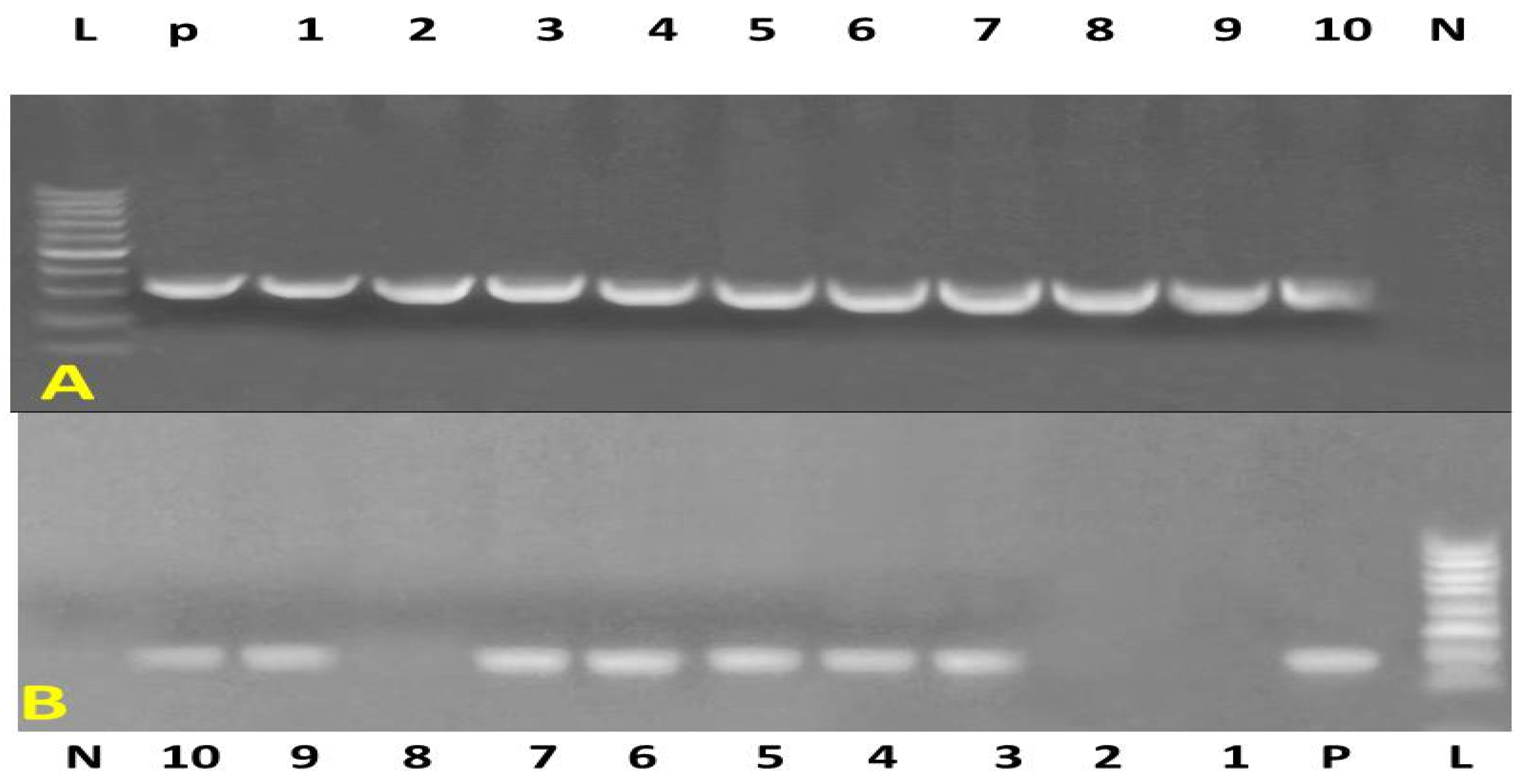
Figure 1. Agarose gel electrophoresis of PCR products. (A) Amplification of the nuc gene (395 bp). Lane 1: DNA ladder (100–1000 bp); Lane N: negative control; Lane P: positive control; Lanes 1–10: positive samples, except lanes 1, 7, and 10, which are negative. (B) Amplification of the mecA gene (310 bp). Lane 1: DNA ladder (100–1000 bp); Lane N: negative control; Lane P: positive control; Lanes 1–10: all positive samples.
Vetsci 12 00584 g001
Figure 2. Normal and pneumonia-affected ewes have different immune genes: (a) = TLR2; (b) = CLEC4E; (c) = PTX3; (d) = SOCS3; (e) = CXCL8; and (f) = IL15RA transcript levels. When p is less than 0.05, significance is indicated by the symbol *. CG: control group; PG: pneumonic group.
Vetsci 12 00584 g002
Figure 3. The correlation between the genetic markers and serum profile of acute phase proteins in CG and PG (Pearson’s Correlation test), considered significant when p < 0.05.
Vetsci 12 00584 g003
Table 1. Primers sequences, target genes, amplicon sizes and cycling conditions for MRSA and some virulence genes.
Target GenePrimers SequencesAmplified Segment (bp)Primary
Denaturation
Amplification (35 Cycles)Final ExtensionReference
Secondary DenaturationAnnealingExtension
Staph nucATATGTATGGCAATCGTTTCAAT39594 °C
5 min.
94 °C
30 sec.
55 °C
40 sec
72 °C
40 sec.
72 °C
10 min.
[12]
GTAAATGCACTTGCTTCAGGAC
Staph mecAGTA GAA ATG ACT GAA CGT CCG ATA A31094 °C
5 min.
94 °C
30 sec.
50 °C
30 sec
72 °C
30 sec
72 °C
10 min
[46]
CCA ATT CCA CAT TGT TTC GGT CTA A
Table 2. Forward-reverse oligonucleotide-based real-time PCR primers for the immunological genes being studied.
Investigated MarkerPrimerProduct Size (bp)Annealing Temperature (°C)GenBank IsolateOrigin
TLR2F5′-GCCTGGCTCCAGGCCAAGAGGA-3
R5′-TCCTCTTGGCCTGGAGCCAGGC-3′
35458NM_001048231.1
CLEC4EF5′-ATTCATCCACATCACCAGCATCA-3
R5′-GTGACCCTCGACCACCTGGTC-3′
44355XM_042247586.1Present Research
PTX3F5′-CTGGAGGAGCTGCGGCGGACGC-3′
R5′-GGTGCTGCACAGATGGGTCCATG-3′
36358XM_004003220.5
SOCS3F5′-ACCTTCCTCATCCGCGACAGCTC-3′
R5′-ATCGTACTGGTCCAGGAACTC-3′
48058XM_027974200.3
CXCL8F5′-ATGACTTCCAAGCTGGCTGTTG-3′
R5′-ATCTTGCTTCTCAGCTCTCTTC-3′
30055NM_001009401.2
IL15RAF5′-CCGGCCACGCCGGGCATCACCTG-3′
R5′-CACCAGGCACACTGCAAAGAC-3′
37858XM_042230436.2
GAPDHF5′-GTGAAGGTCGGAGTGAACGG-3′
R5′-TTGACTGTGCCGTGGAACTT-3′
17358HM043737.1
Table 3. Antibiotic sensitivity and resistance pattern of Staphylococcus aureus from isolates.
Type of AntibioticNo. Sensitive/27No. Resistant/27% Sensitive% Resistant
Gentamicin21677.722.2
Amoxycillin81929.670.4
Amoxycillin/clavullanate17106337.0
Ceftriaxone2077426
Cloxacillin10173763
Ciprofloxacin21677.722.2
Levofloxacin2701000
Penicillin1263.796.3
Erythromycin141351,848
Chloramphenicol161159.240.7
Ofloxacin2077426
Tetracycline81929.670.3
Cotrimoxazole42314.885.2
vancomycin1701000
Table 4. Antibiotic sensitivity pattern of methicillin resistant isolates.
Type of AntibioticNo. Sensitive/17No. Resistant/17% Sensitive% Resistant
Gentamicin21511.788.3
Amoxycillin0170100
Amoxycillin/clavullanate0170100
Ceftriaxone71041.258.8
Cloxacillin0170100
Ciprofloxacin985347
Levofloxacin161945.8
Penicillin0170100
Erythromycin0170100
Chloramphenicol0170100
Ofloxacin12570.629.4
Tetracycline0170100
Cotrimoxazole0170100
vancomycin1701000
Table 5. Pneumonic and healthy ewes’ immunological marker distributions with a single base differential and possible genetic change.
GeneSNPsHealthy
n = 100
Pneumonia
n = 100
Total
n = 200
Chi Square
X2
p Value Kind of Inherited ChangeAmino Acid Order and Sort
TLR2C169T73/100-/10073/200115<0.005Non-synonymous57 L to F
CLEC4EG43A59/100-/10059/20083.69<0.005Non-synonymous15 C to Y
PTX3G90C-/10084/10084/200144.8<0.005Synonymous30 A
C2383T39/100-/10039/20048.45<0.005Non-synonymous95 L to F
SOCS3G438C-/10048/10048/20063.13<0.005Synonymous146 T
CXCL8C154T-/10074/10074/200117.5<0.005Synonymous52 L
IL15RAA74G43/100-/10043/200 54.8<0.005Non-synonymous25 S to N
Table 6. Discriminant analysis for classification of type of genes and healthy status of examined sheep.
Predicted Group MembershipTotal
HealthyDiseases
CountHealthy1000100
Diseased0100100
%Healthy100.00.0100.0
Diseased0.0100.0100.0
Table 7. Biochemical parameters of control and pneumonic groups.
ParametersControl GroupPneumonic Group
Fb (mg/dL)122.01 ± 8.49225.01 ± 3.47 *
Cp (mg/dL)2.30 ± 1.156.24 ± 0.02 *
Hp (g/dL)0.15 ± 0.022.59 ± 0.49 *
SAA (mg/L)2.32 ± 0.156.96 ± 0.16 *
Cortisol (μg/dL)1.79 ± 0.166.44 ± 0.05 *
Insulin (μIU/mL)8.41 ± 0.157.08 ± 0.17 *
T3(ng/mL)1.74 ± 0.151.02 ± 0.02 *
T4 (μg/mL)0.85 ± 0.080.65 ± 0.02 *
TSH (μIU/mL)0.010 ± 0.0020.022 ± 0.012 *
GH (ng/dL)12.39 ± 1.4716.80 ± 0.05 *
SI (μg/dL)106.89 ± 2.4689.13 ± 1.62 *
TIBC (μg/dL)327.39 ± 2.16341.42 ± 3.11 *
UIBC (μg/dL)220.50 ± 2.24252.29 ± 3.72 *
Transferrin(mg/dL)124.65 ± 2.7486.47 ± 0.22 *
Tf sat. %32.65 ± 0.6626.11 ± 0.56 *
Ferritin (ng/mL)13.60 ± 1.0519.35 ± 0.53 *
Significant differences between the two groups were indicated by (*) when p < 0.05.
Table 8. Roc curve analysis of APPs, hormones, and iron profile parameters in PG compared to CG.
ParametersCut-Off PointSensitivitySpecificityLRPPVNPVAR% of Increase or Decrease
Fb (mg/dL)132.50100%85%6.6790.91%100%94%84.41%
Cp (mg/dL)3.60100%80%588.24%100%92%171.30%
Hp (g/dL)0.185100%90%1093.75%100%96%1626.67%
SAA (mg/L)2.45100%80%588.24%100%92%200%
Cortisol (μg/dL)1.99100%85%6.6790.91%100%94%259.77%
Insulin (μIU/mL)8. 26100%80%588.24%100%92%−15.81%
T3(ng/mL)1.57100%95%2096.77%100%98%−41.37%
T4 (μg/mL)0.78100%80%588.24%100%92%−23.52%
TSH (μIU/mL)0.01560%100%-100%62.50%76%120%
GH (ng/dL)14.39100%95%2096.77%100%98%35.59%
SI (μg/dL)106100%75%485.71%100%90%−16.61%
TIBC (μg/dL)328100%75%485.71%100%90%4.28%
UIBC (μg/dL)232.9100%100%-100%100%100%14.41%
Transferrin(mg/dL)121.50100%85%6.6790.91%100%94%−30.62%
Tf sat. %32.51100%75%485.71%100%90%−20.03%
Ferritin (ng/mL)14.50100%80%588.24%100%92%42.28%
LR = 0.5–5: low; LR = 5–10: moderate; LR > 10: high.
Disclaimer/Publisher’s Note: The statements, opinions and data contained in all publications are solely those of the individual author(s) and contributor(s) and not of MDPI and/or the editor(s). MDPI and/or the editor(s) disclaim responsibility for any injury to people or property resulting from any ideas, methods, instructions or products referred to in the content.