




Intracranial Subdural Fluid Accumulation Associated with a Choroid Plexus Carcinoma in a Dog
Simple Summary
Abstract
1. Introduction
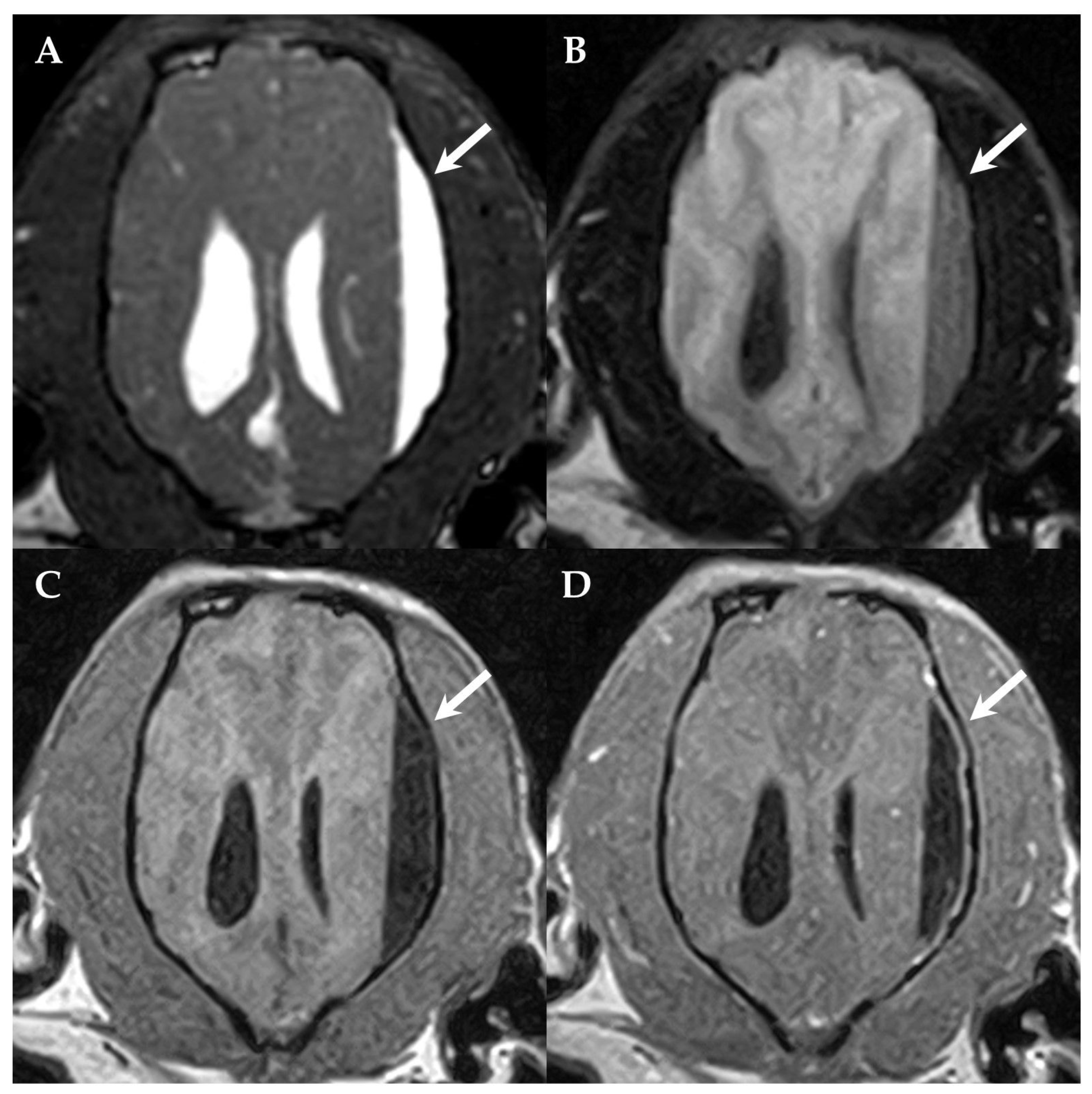
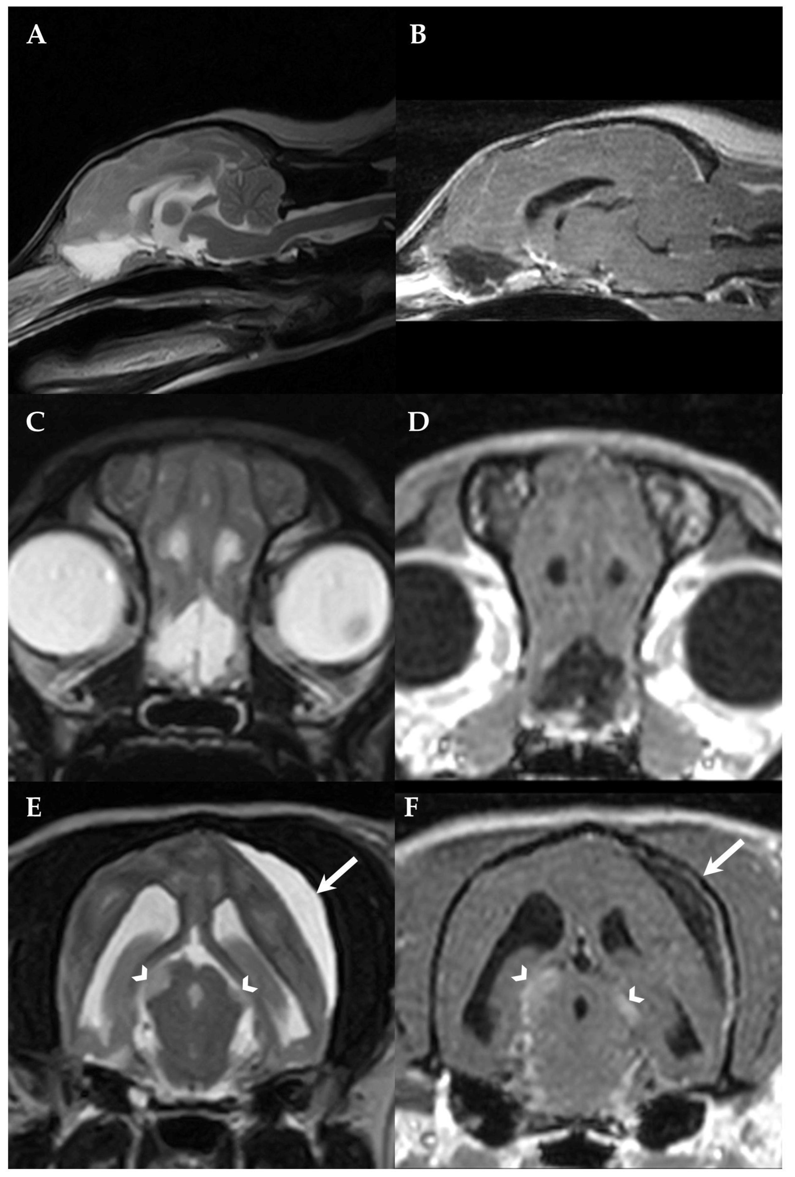
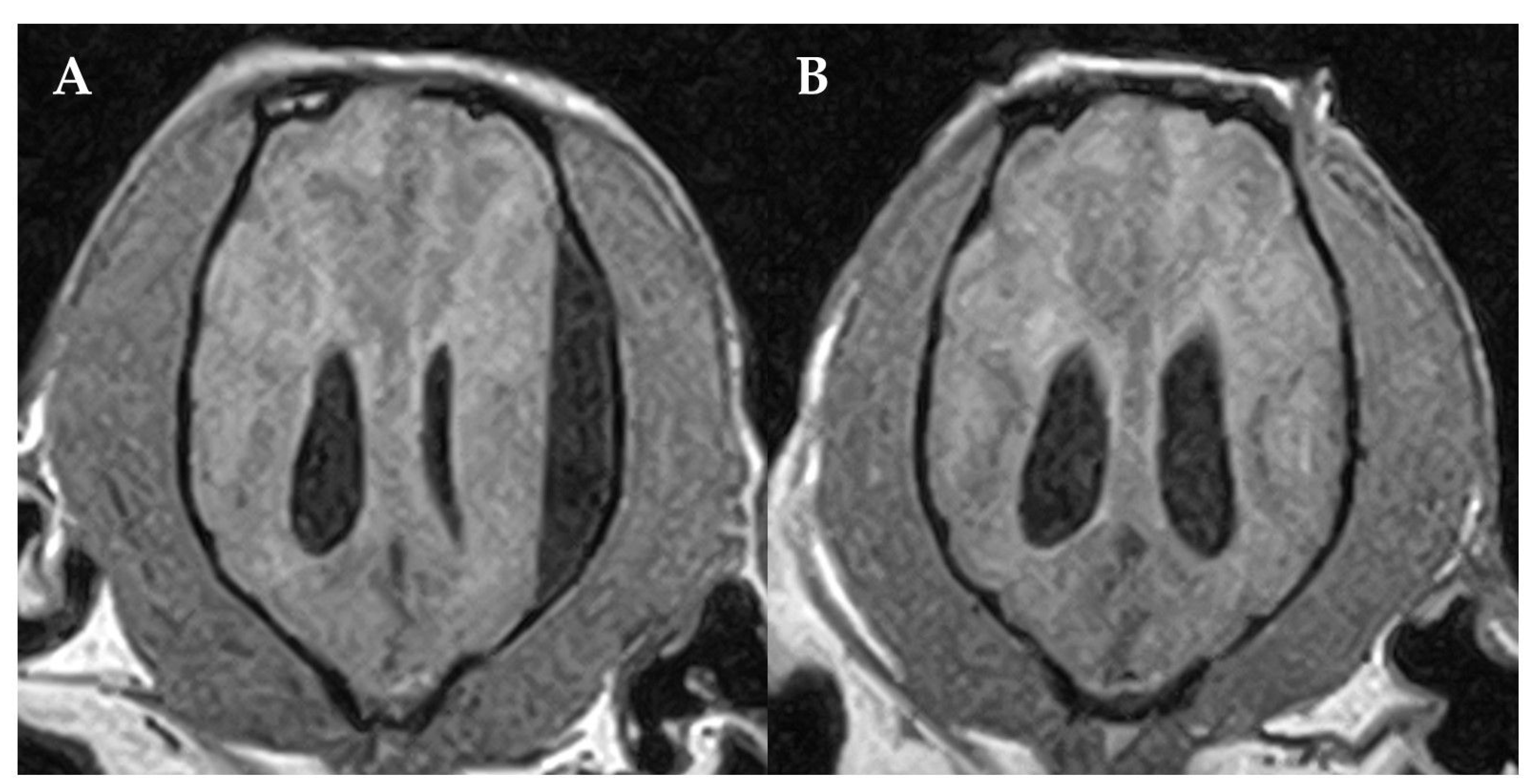
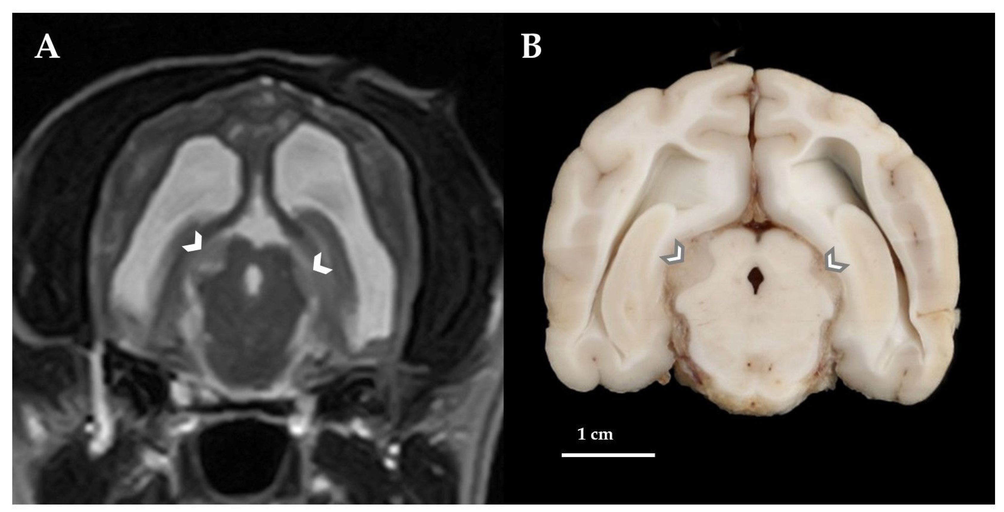
2. Case Presentation
3. Discussion
4. Conclusions
Author Contributions
Funding
Institutional Review Board Statement
Informed Consent Statement
Data Availability Statement
Acknowledgments
Conflicts of Interest
References
- Snyder, J.M.; Shofer, F.S.; Van Winkle, T.J.; Massicotte, C. Canine Intracranial Primary Neoplasia: 173 Cases (1986–2003). J. Vet. Intern. Med. 2006, 20, 669–675. [Google Scholar] [CrossRef]
- Higgins, R.J.; Bollen, A.W.; Dickinson, P.J.; Sisó-Llonch, S. Tumors of the Nervous System. In Tumors in Domestic Animals; John Wiley & Sons, Inc.: Hoboken, NJ, USA, 2016; pp. 834–891. ISBN 9780813806761. [Google Scholar]
- Westworth, D.R.; Dickinson, P.J.; Vernau, W.; Johnson, E.G.; Bollen, A.W.; Kass, P.H.; Sturges, B.K.; Vernau, K.M.; LeCouteur, R.A.; Higgins, R.J. Choroid Plexus Tumors in 56 Dogs (1985–2007). J. Vet. Intern. Med. 2008, 22, 1157–1165. [Google Scholar] [CrossRef] [Scilit] [PubMed]
- Itoh, T.; Uchida, K.; Nishi, A.; Shii, H.; Nagayoshi, T.; Sakamoto, H. Choroid Plexus Papilloma in a Dog Surviving for 15 Months after Diagnosis with Symptomatic Therapy. J. Vet. Med. Sci. 2016, 78, 167–169. [Google Scholar] [CrossRef] [Scilit] [PubMed]
- Lin, H.; Leng, X.; Qin, C.; Du, Y.; Wang, W.; Qiu, S. Choroid Plexus Tumours on MRI: Similarities and Distinctions in Different Grades. Cancer Imaging 2019, 19, 17. [Google Scholar] [CrossRef] [Scilit] [PubMed]
- Wisner, E.R.; Dickinson, P.J.; Higgins, R.J. Magnetic Resonance Imaging Features of Canine Intracranial Neoplasia. Vet. Radiol. Ultrasound 2011, 52, S52–S61. [Google Scholar] [CrossRef] [Scilit] [PubMed]
- Rickert, C.H.; Paulus, W. Tumors of the Choroid Plexus. Microsc. Res. Tech. 2001, 52, 104–111. [Google Scholar] [CrossRef] [PubMed]
- Mallick, S.; Benson, R.; Melgandi, W.; Rath, G.K. Effect of Surgery, Adjuvant Therapy, and Other Prognostic Factors on Choroid Plexus Carcinoma: A Systematic Review and Individual Patient Data Analysis. Int. J. Radiat. Oncol. 2017, 99, 1199–1206. [Google Scholar] [CrossRef] [Scilit]
- Louis, D.N.; Perry, A.; Reifenberger, G.; von Deimling, A.; Figarella-Branger, D.; Cavenee, W.K.; Ohgaki, H.; Wiestler, O.D.; Kleihues, P.; Ellison, D.W. The 2016 World Health Organization Classification of Tumors of the Central Nervous System: A Summary. Acta Neuropathol. 2016, 131, 803–820. [Google Scholar] [CrossRef] [Scilit] [PubMed]
- Wrede, B.; Liu, P.; Wolff, J.E.A. Chemotherapy Improves the Survival of Patients with Choroid Plexus Carcinoma: A Meta-Analysis of Individual Cases with Choroid Plexus Tumors. J. Neurooncol. 2007, 85, 345–351. [Google Scholar] [CrossRef] [Scilit]
- Meyers, S.P.; Khademian, Z.P.; Chuang, S.H.; Pollack, I.F.; Korones, D.N.; Zimmerman, R.A. Choroid Plexus Carcinomas in Children: MRI Features and Patient Outcomes. Neuroradiology 2004, 46, 770–780. [Google Scholar] [CrossRef] [Scilit]
- Bentley, R.T. Magnetic Resonance Imaging Diagnosis of Brain Tumors in Dogs. Vet. J. 2015, 205, 204–216. [Google Scholar] [CrossRef] [Scilit] [PubMed]
- Oura, T.J.; Early, P.J.; Jennings, S.H.; Lewis, M.J.; Tobias, J.R.; Thrall, D.E. Canine Choroid Plexus Tumor with Intracranial Dissemination Presenting as Multiple Cystic Lesions. Case Rep. Vet. Med. 2013, 2013, 1–4. [Google Scholar] [CrossRef] [Scilit]
- Hughes, J.R.; Taylor-Brown, F.E.; Greville-Heygate, O.; Constantino-Casas, F.; Williams, D.L.; Genain, M.A. Multimodality Characteristics of Multifocal Choroid Plexus Carcinoma with Bilateral Calvarial Defects in a Dog. Vet. Radiol. Ultrasound 2021, 62, E20–E25. [Google Scholar] [CrossRef] [Scilit] [PubMed]
- Pelegrini, L.F.; Silva, N.F.; Campos, O.P.S.; Nery, C.C.; Silva, F.M.; Lemos, R.S.; Yamauchi, K.C.I.; Amude, A.M. Medical Therapy Using Omeprazole in 12 Hydrocephalic Dogs: Clinical, Diagnostic, and Therapeutic Findings. Pesqui. Veterinária Bras. 2019, 39, 823–829. [Google Scholar] [CrossRef] [Scilit]
- Choi, E.J.; Sloma, E.A.; Miller, A.D. Kir7.1 Immunoreactivity in Canine Choroid Plexus Tumors. J. Vet. Diagn. Investig. 2016, 28, 464–468. [Google Scholar] [CrossRef] [Scilit]
- Hasselblatt, M.; Böhm, C.; Tatenhorst, L.; Dinh, V.; Newrzella, D.; Keyvani, K.; Jeibmann, A.; Buerger, H.; Rickert, C.H.; Paulus, W. Identification of Novel Diagnostic Markers for Choroid Plexus Tumors. Am. J. Surg. Pathol. 2006, 30, 66–74. [Google Scholar] [CrossRef] [Scilit]
- Blutke, A.; Knebel, J.; Brühschwein, A.; Breuer, W.; Hermanns, W. Hemangiopericytoma in a Cat: A Case Report. Vet. Med. (Praha). 2012, 57, 263–269. [Google Scholar] [CrossRef] [Scilit]
- Nakamura, N.; Suzuki, Y.; Sakuta, H.; Ookata, K.; Kawahara, K.; Hirose, S. Inwardly Rectifying K+ Channel Kir7.1 Is Highly Expressed in Thyroid Follicular Cells, Intestinal Epithelial Cells and Choroid Plexus Epithelial Cells: Implication for a Functional Coupling with Na+,K+-ATPase. Biochem. J. 1999, 342, 329. [Google Scholar] [CrossRef]
- Forward, A.K.; Plessas, I.N.; Guilherme, S.; De Decker, S. Retrospective Evaluation of the Clinical Presentation, Magnetic Resonance Imaging Findings, and Outcome of Dogs Diagnosed with Intracranial Empyema (2008–2015): 9 Cases. J. Vet. Emerg. Crit. Care 2019, 29, 431–438. [Google Scholar] [CrossRef] [Scilit]
- Yanai, H.; Tapia-Nieto, R.; Cherubini, G.B.; Caine, A. Results of Magnetic Resonance Imaging Performed within 48 Hours after Head Trauma in Dogs and Association with Outcome: 18 Cases (2007–2012). J. Am. Vet. Med. Assoc. 2015, 246, 1222–1229. [Google Scholar] [CrossRef] [Scilit]
- Nykamp, S.; Scrivani, P.; Delahunta, A.; Yu-Speight, A.; Rus, R. Chronic Subdural Hematomas and Hydrocephalus in a Dog. Vet. Radiol. Ultrasound 2001, 42, 511–514. [Google Scholar] [CrossRef] [Scilit] [PubMed]
- Asakawa, M.G.; MacKillop, E.; Olby, N.J.; Robertson, I.D.; Cullen, J.M. Imaging Diagnosis—Ceroid Lipofuscinosis with a Chronic Subdural Hematoma. Vet. Radiol. Ultrasound 2010, 51, 155–158. [Google Scholar] [CrossRef] [Scilit] [PubMed]
- Kitagawa, M.; Sakai, T.; Kanayama, K. Subdural Accumulation of Fluid in a Dog after the Insertion of a Ventriculoperitoneal Shunt. Vet. Rec. 2005, 156, 206–208. [Google Scholar] [CrossRef] [Scilit] [PubMed]
- Motta, L. Suspected Chronic Subdural Haemorrhage as a Sequela of Subdural Empyema in an Irish Setter. J. Small Anim. Pract. 2021, 62, 715. [Google Scholar] [CrossRef] [Scilit] [PubMed]
- Cramer, X.J.A.; Rassner, X.U.A.; Hedlund, X.G.L. Limitations of T2 *– Gradient Recalled-Echo and Susceptibility- Weighted Imaging in Characterizing Chronic Subdural Hemorrhage in Infant Survivors of Abusive Head Trauma. Am. J. Neuroradiol. 2016, 37, 1752–1756. [Google Scholar] [CrossRef] [Scilit] [PubMed]
- Vinchon, M.; Joriot, S.; Jissendi-Tchofo, P.; Dhellemmes, P. Postmeningitis Subdural Fluid Collection in Infants: Changing Pattern and Indications for Surgery. J. Neurosurg. 2006, 104, 383–387. [Google Scholar] [CrossRef] [Scilit]
- Pastorello, A.; Constantino-Casas, F.; Archer, J. Choroid Plexus Carcinoma Cells in the Cerebrospinal Fluid of a Staffordshire Bull Terrier. Vet. Clin. Pathol. 2010, 39, 505–510. [Google Scholar] [CrossRef] [Scilit]
- Webb, A.A. Potential Sources of Neck and Back Pain in Clinical Conditions of Dogs and Cats: A Review. Vet. J. 2003, 165, 193–213. [Google Scholar] [CrossRef] [Scilit]
- Patnaik, A.K.; Erlandson, R.A.; Lieberman, P.H.; Fenner, W.R.; Prata, R.G. Choroid Plexus Carcinoma with Meningeal Carcinomatosis in a Dog. Vet. Pathol. 1980, 17, 381–385. [Google Scholar] [CrossRef] [Scilit]
- Lipsitz, D.; Levitski, R.E.; Chauvet, A.E. Magnetic Resonance Imaging of a Choroid Plexus Carcinoma and Meningeal Carcinomatosis in a Dog. Vet. Radiol. Ultrasound 1999, 40, 246–250. [Google Scholar] [CrossRef] [Scilit]
- Miller, A.D.; Miller, C.R.; Rossmeisl, J.H. Canine Primary Intracranial Cancer: A Clinicopathologic and Comparative Review of Glioma, Meningioma, and Choroid Plexus Tumors. Front. Oncol. 2019, 9, 1151. [Google Scholar] [CrossRef] [Scilit] [PubMed]
- Sun, M.Z.; Ivan, M.E.; Clark, A.J.; Oh, M.C.; Delance, A.R.; Oh, T.; Safaee, M.; Kaur, G.; Bloch, O.; Molinaro, A.; et al. Gross Total Resection Improves Overall Survival in Children with Choroid Plexus Carcinoma. J. Neurooncol. 2014, 116, 179–185. [Google Scholar] [CrossRef] [Scilit] [PubMed]
- Antonakakis, M.G.; Carletti, B.E.; Anselmi, C.; McGrath, S.; Minguez, J.J. Use of a Telovelar Approach for Complete Resection of a Choroid Plexus Tumor in a Dog. Vet. Surg. 2022, 51, 1273–1279. [Google Scholar] [CrossRef] [Scilit] [PubMed]
- Lehner, L.; Czeibert, K.; Benczik, J.; Jakab, C.; Nagy, G. Transcallosal Removal of a Choroid Plexus Tumor From the Lateral Ventricle in a Dog. Case Report. Front. Vet. Sci. 2020, 7, 536. [Google Scholar] [CrossRef] [Scilit]
- Hosmann, A.; Hinker, F.; Dorfer, C.; Slavc, I.; Haberler, C.; Dieckmann, K.; Knosp, E.; Czech, T. Management of Choroid Plexus Tumors—An Institutional Experience. Acta Neurochir. (Wien) 2019, 161, 745–754. [Google Scholar] [CrossRef] [Scilit]
- Mazloom, A.; Wolff, J.E.; Paulino, A.C. The Impact of Radiotherapy Fields in the Treatment of Patients With Choroid Plexus Carcinoma. Int. J. Radiat. Oncol. 2010, 78, 79–84. [Google Scholar] [CrossRef] [Scilit]
- Schwarz, P.; Meier, V.; Soukup, A.; Drees, R.; Besserer, J.; Beckmann, K.; Roos, M.; Rohrer Bley, C. Comparative Evaluation of a Novel, Moderately Hypofractionated Radiation Protocol in 56 Dogs with Symptomatic Intracranial Neoplasia. J. Vet. Intern. Med. 2018, 32, 2013–2020. [Google Scholar] [CrossRef] [Scilit]
- Spugnini, E.P.; Thrall, D.E.; Price, G.S.; Sharp, N.J.; Munana, K.; Page, R.L. Primary Irradiation of Canine Iintracranial Masses. Vet. Radiol. 2000, 41, 377–380. [Google Scholar] [CrossRef] [Scilit]
- Mariani, C.L.; Schubert, T.A.; House, R.A.; Wong, M.A.; Hopkins, A.L.; Barnes Heller, H.L.; Milner, R.J.; Lester, N.V.; Lurie, D.M.; Rajon, D.A.; et al. Frameless Stereotactic Radiosurgery for the Treatment of Primary Intracranial Tumours in Dogs. Vet. Comp. Oncol. 2015, 13, 409–423. [Google Scholar] [CrossRef] [Scilit]
- Cornelis, I.; Van Ham, L.; Gielen, I.; De Decker, S.; Bhatti, S.F.M. Clinical Presentation, Diagnostic Findings, Prognostic Factors, Treatment and Outcome in Dogs with Meningoencephalomyelitis of Unknown Origin: A Review. Vet. J. 2019, 244, 37–44. [Google Scholar] [CrossRef] [Scilit]
- Scott-Moncrieff, J.C.R.; Chan, T.C.K.; Samuels, M.L.; Cook, J.R.; Coppoc, G.L.; DeNicola, D.B.; Richardson, R.C. Plasma and Cerebrospinal Fluid Pharmacokinetics of Cytosine Arabinoside in Dogs. Cancer Chemother. Pharmacol. 1991, 29, 13–18. [Google Scholar] [CrossRef] [Scilit] [PubMed]
- Marconato, L.; Bonfanti, U.; Stefanello, D.; Lorenzo, M.R.; Romanelli, G.; Comazzi, S.; Zini, E. Cytosine Arabinoside in Addition to VCAA-Based Protocols for the Treatment of Canine Lymphoma with Bone Marrow Involvement: Does It Make the Difference? Vet. Comp. Oncol. 2008, 6, 80–89. [Google Scholar] [CrossRef] [Scilit]
- Muzumdar, D.; Biyani, N.; Deopujari, C. Subdural Empyema in Children. Child’s Nerv. Syst. 2018, 34, 1881–1887. [Google Scholar] [CrossRef] [Scilit]
- Witten, A.J.; Mendenhall, S.K.; DeWitt, L.S.; Vortmeyer, A.; Cohen-Gadol, A. Cerebellopontine Angle Primary Choroid Plexus Carcinoma Present in an Adult: Case Report and Literature Review. Cureus 2021, 13, e13268. [Google Scholar] [CrossRef] [Scilit] [PubMed]
- Orlandi, R.; Vasilache, C.G.; Mateo, I. Palliative Ventriculoperitoneal Shunting in Dogs with Obstructive Hydrocephalus Caused by Tumors Affecting the Third Ventricle. J. Vet. Intern. Med. 2020, 34, 1556–1562. [Google Scholar] [CrossRef] [Scilit] [PubMed]

| Pretreatment | Antibody | Dilution | Source | |
|---|---|---|---|---|
| Kir7.1 | Boiling (pH 6) | First antibody: Polyclonal rabbit anti human potassium channel Kir7.1 | 1:2000 | kindly provided by S. Hirose [19] |
| Secondary antibody/detection: EnVision FLEX + Rabbit (LINKER) (visualization system for automated IHC-staining systems) | ready-to-use | part N° K800921 Agilent Technologies Inc., Santa Clara, CA, USA | ||
| Cytokeratin | Proteinase K (* Code S3020) | First antibody: Monoclonal mouse anti-Human cytokeratin | 1:50 | * clones AE1/AE3, code No. M3515, |
| Secondary antibody: Peroxidase conjugated rabbit anti-mouse immunoglobulins | 1:100 | * code No. P0161 |
Disclaimer/Publisher’s Note: The statements, opinions and data contained in all publications are solely those of the individual author(s) and contributor(s) and not of MDPI and/or the editor(s). MDPI and/or the editor(s) disclaim responsibility for any injury to people or property resulting from any ideas, methods, instructions or products referred to in the content. |
© 2022 by the authors. Licensee MDPI, Basel, Switzerland. This article is an open access article distributed under the terms and conditions of the Creative Commons Attribution (CC BY) license (https://creativecommons.org/licenses/by/4.0/).
Share and Cite
Schneider, N.; Blutke, A.; Matiasek, K.; Parzefall, B. Intracranial Subdural Fluid Accumulation Associated with a Choroid Plexus Carcinoma in a Dog. Vet. Sci. 2023, 10, 24. https://doi.org/10.3390/vetsci10010024
Schneider N, Blutke A, Matiasek K, Parzefall B. Intracranial Subdural Fluid Accumulation Associated with a Choroid Plexus Carcinoma in a Dog. Veterinary Sciences. 2023; 10(1):24. https://doi.org/10.3390/vetsci10010024
Chicago/Turabian StyleSchneider, Nina, Andreas Blutke, Kaspar Matiasek, and Birgit Parzefall. 2023. "Intracranial Subdural Fluid Accumulation Associated with a Choroid Plexus Carcinoma in a Dog" Veterinary Sciences 10, no. 1: 24. https://doi.org/10.3390/vetsci10010024
APA StyleSchneider, N., Blutke, A., Matiasek, K., & Parzefall, B. (2023). Intracranial Subdural Fluid Accumulation Associated with a Choroid Plexus Carcinoma in a Dog. Veterinary Sciences, 10(1), 24. https://doi.org/10.3390/vetsci10010024

