
Numerical Simulations of Red-Blood Cells in Fluid Flow: A Discrete Multiphysics Study


Abstract
:1. Introduction
2. Methodology
2.1. General Equations
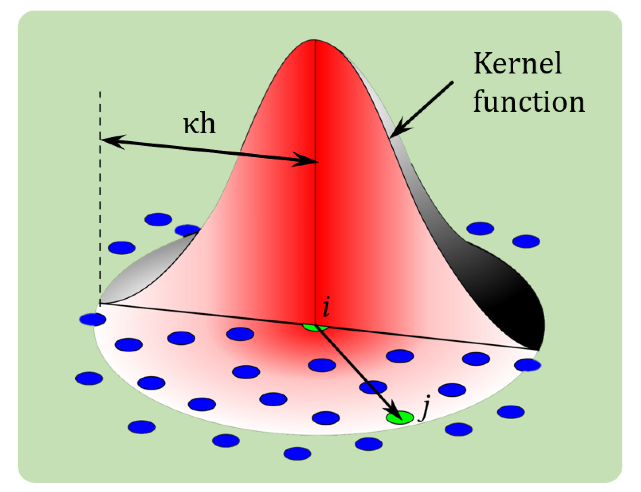
2.2. The SPH Method
2.3. The MSM
2.4. Coupling of SPH and MSM
2.5. Numerical Algorithm
3. Problem Set-Up
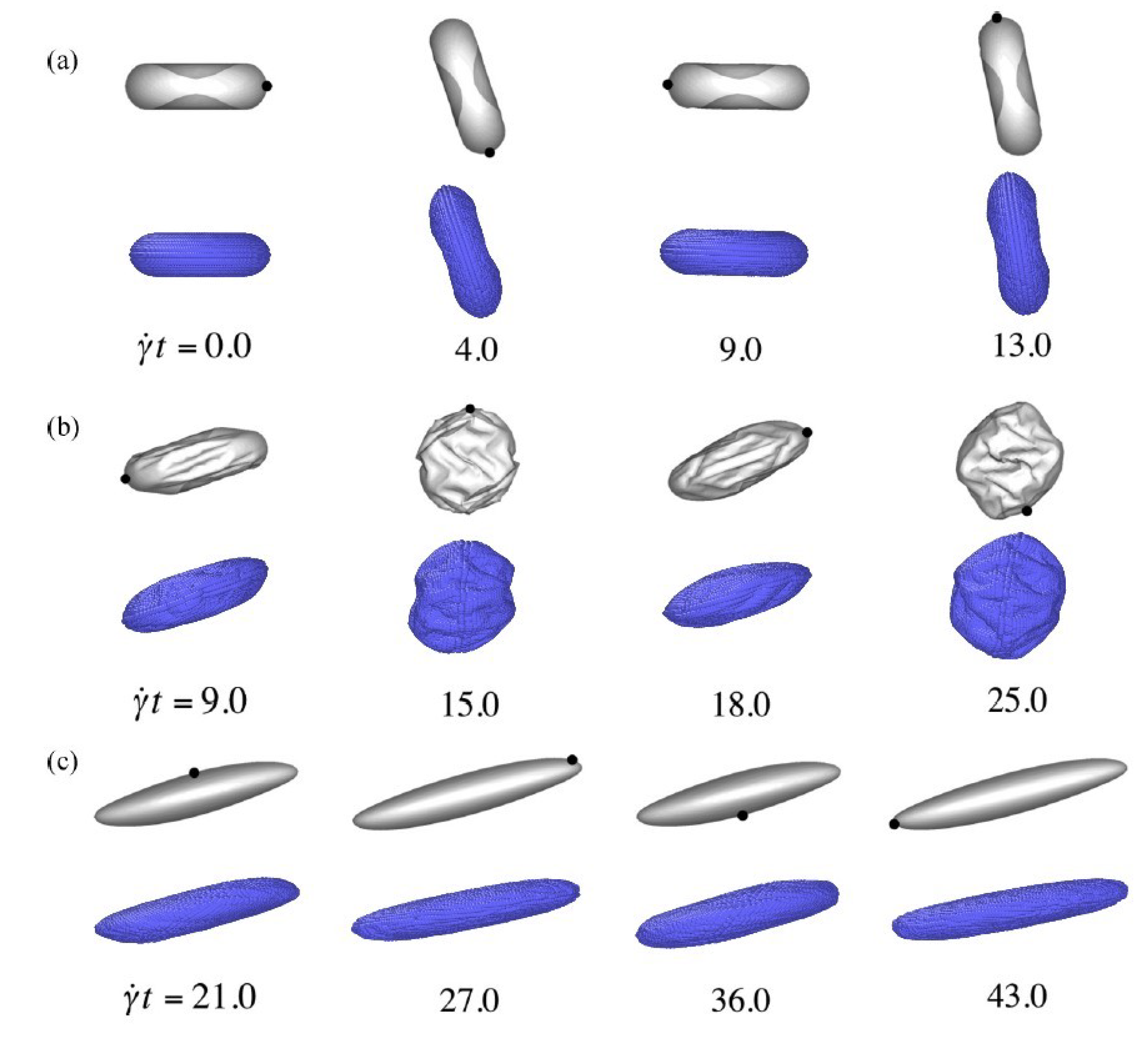
4. Results
5. Conclusions
Author Contributions
Funding
Institutional Review Board Statement
Informed Consent Statement
Data Availability Statement
Conflicts of Interest
Abbreviations
References
- Mohandas, N.; Evans, E. Mechanical properties of the red cell membrane in relation to molecular structure and genetic defects. Annu. Rev. Biophys. Biomol. Struct. 1994, 23, 787–818. [Google Scholar] [CrossRef] [PubMed]
- Mulquiney, P.J.; Bubb, W.A.; Kuchel, P.W. Model of 2, 3-bisphosphoglycerate metabolism in the human erythrocyte based on detailed enzyme kinetic Equations (1): In vivo kinetic characterization of 2, 3-bisphosphoglycerate synthase/phosphatase using 13C and 31P NMR. Biochem. J. 1999, 342, 567–580. [Google Scholar] [CrossRef]
- Mulquiney, P.J.; Kuchel, P.W. Model of 2, 3-bisphosphoglycerate metabolism in the human erythrocyte based on detailed enzyme kinetic equations: Equations and parameter refinement. Biochem. J. 1999, 342, 581–596. [Google Scholar] [CrossRef] [PubMed]
- Mulquiney, P.J.; Kuchel, P.W. Model of 2, 3-bisphosphoglycerate metabolism in the human erythrocyte based on detailed enzyme kinetic equations: Computer simulation and metabolic control analysis. Biochem. J. 1999, 342, 597–604. [Google Scholar] [CrossRef]
- Wan, J.; Ristenpart, W.D.; Stone, H.A. Dynamics of shear-induced ATP release from red blood cells. Proc. Natl. Acad. Sci. USA 2008, 105, 16432–16437. [Google Scholar] [CrossRef] [PubMed] [Green Version]
- Shishmarev, D.; Kuchel, P.W.; Pagès, G.; Wright, A.J.; Hesketh, R.L.; Kreis, F.; Brindle, K.M. Glyoxalase activity in human erythrocytes and mouse lymphoma, liver and brain probed with hyperpolarized 13 C-methylglyoxal. Commun. Biol. 2018, 1, 1–8. [Google Scholar] [CrossRef] [PubMed]
- Kuchel, P.W.; Shishmarev, D. Accelerating metabolism and transmembrane cation flux by distorting red blood cells. Sci. Adv. 2017, 3, eaao1016. [Google Scholar] [CrossRef] [PubMed] [Green Version]
- Dupin, M.; Halliday, I.; Care, C.; Munn, L. Lattice Boltzmann modelling of blood cell dynamics. Int. J. Comput. Fluid Dyn. 2008, 22, 481–492. [Google Scholar] [CrossRef]
- Shi, X.; Lin, G.; Zou, J.; Fedosov, D.A. A lattice Boltzmann fictitious domain method for modeling red blood cell deformation and multiple-cell hydrodynamic interactions in flow. Int. J. Numer. Methods Fluids 2013, 72, 895–911. [Google Scholar] [CrossRef]
- Zhang, J. Lattice Boltzmann method for microfluidics: Models and applications. Microfluid. Nanofluid. 2011, 10, 1–28. [Google Scholar] [CrossRef]
- Chen, M.; Boyle, F.J. Investigation of membrane mechanics using spring networks: Application to red-blood-cell modelling. Mater. Sci. Eng. C 2014, 43, 506–516. [Google Scholar] [CrossRef] [PubMed] [Green Version]
- Eggleton, C.D.; Popel, A.S. Large deformation of red blood cell ghosts in a simple shear flow. Phys. Fluids 1998, 10, 1834–1845. [Google Scholar] [CrossRef] [PubMed]
- Omori, T.; Ishikawa, T.; Barthès-Biesel, D.; Salsac, A.V.; Imai, Y.; Yamaguchi, T. Tension of red blood cell membrane in simple shear flow. Phys. Rev. E 2012, 86, 056321. [Google Scholar] [CrossRef] [Green Version]
- Hosseini, S.M.; Feng, J.J. A particle-based model for the transport of erythrocytes in capillaries. Chem. Eng. Sci. 2009, 64, 4488–4497. [Google Scholar] [CrossRef]
- Freund, J.B. Numerical simulation of flowing blood cells. Annu. Rev. Fluid Mech. 2014, 46, 67–95. [Google Scholar] [CrossRef] [Green Version]
- Wu, T.; Feng, J.J. Simulation of malaria-infected red blood cells in microfluidic channels: Passage and blockage. Biomicrofluidics 2013, 7, 044115. [Google Scholar] [CrossRef] [Green Version]
- Boryczko, K.; Dzwinel, W.; Yuen, D.A. Dynamical clustering of red blood cells in capillary vessels. J. Mol. Model. 2003, 9, 16–33. [Google Scholar] [CrossRef] [PubMed]
- Tsubota, K.; Wada, S.; Yamaguchi, T. Particle method for computer simulation of red blood cell motion in blood flow. Comput. Methods Programs Biomed. 2006, 83, 139–146. [Google Scholar] [CrossRef]
- Hochmuth, R.; Mohandas, N. Uniaxial loading of the red-cell membrane. J. Biomech. 1972, 5, 501–509. [Google Scholar] [CrossRef]
- Hochmuth, R.; Wiles, H.; Evans, E.; McCown, J. Extensional flow of erythrocyte membrane from cell body to elastic tether. II. Experiment. Biophys. J. 1982, 39, 83–89. [Google Scholar] [CrossRef] [Green Version]
- Pozrikidis, C. Effect of membrane bending stiffness on the deformation of capsules in simple shear flow. J. Fluid Mech. 2001, 440, 269–291. [Google Scholar] [CrossRef]
- Zhang, J.; Johnson, P.C.; Popel, A.S. An immersed boundary lattice Boltzmann approach to simulate deformable liquid capsules and its application to microscopic blood flows. Phys. Biol. 2007, 4, 285. [Google Scholar] [CrossRef] [PubMed]
- Bagchi, P. Mesoscale simulation of blood flow in small vessels. Biophys. J. 2007, 92, 1858–1877. [Google Scholar] [CrossRef] [PubMed] [Green Version]
- Barthes-Biesel, D.; Diaz, A.; Dhenin, E. Effect of constitutive laws for two-dimensional membranes on flow-induced capsule deformation. J. Fluid Mech. 2002, 460, 211–222. [Google Scholar] [CrossRef]
- Evans, E.A.; Skalak, R. Mechanics and Thermodynamics of Biomembranes; CRC Press: Boca Raton, FL, USA, 1980. [Google Scholar]
- Sui, Y.; Chew, Y.; Roy, P.; Cheng, Y.; Low, H. Dynamic motion of red blood cells in simple shear flow. Phys. Fluids 2008, 20, 112106. [Google Scholar] [CrossRef]
- Lac, E.; Barthes-Biesel, D.; Pelekasis, N.; Tsamopoulos, J. Spherical capsules in three-dimensional unbounded Stokes flows: Effect of the membrane constitutive law and onset of buckling. J. Fluid Mech. 2004, 516, 303–334. [Google Scholar] [CrossRef] [Green Version]
- Rahmat, A.; Barigou, M.; Alexiadis, A. Deformation and rupture of compound cells under shear: A discrete multiphysics study. Phys. Fluids 2019, 31, 051903. [Google Scholar] [CrossRef]
- Secomb, T.W.; Styp-Rekowska, B.; Pries, A.R. Two-dimensional simulation of red blood cell deformation and lateral migration in microvessels. Ann. Biomed. Eng. 2007, 35, 755–765. [Google Scholar] [CrossRef]
- Fedosov, D.A.; Caswell, B.; Karniadakis, G.E. A multiscale red blood cell model with accurate mechanics, rheology, and dynamics. Biophys. J. 2010, 98, 2215–2225. [Google Scholar] [CrossRef] [Green Version]
- Fedosov, D.A.; Peltomäki, M.; Gompper, G. Deformation and dynamics of red blood cells in flow through cylindrical microchannels. Soft Matter 2014, 10, 4258–4267. [Google Scholar] [CrossRef] [PubMed] [Green Version]
- Ziherl, P.; Svetina, S. Nonaxisymmetric phospholipid vesicles: Rackets, boomerangs, and starfish. EPL Europhys. Lett. 2005, 70, 690. [Google Scholar] [CrossRef]
- Svetina, S.; Ziherl, P. Morphology of small aggregates of red blood cells. Bioelectrochemistry 2008, 73, 84–91. [Google Scholar] [CrossRef] [PubMed]
- MacMeccan, R.M.; Clausen, J.; Neitzel, G.; Aidun, C. Simulating deformable particle suspensions using a coupled lattice-Boltzmann and finite-element method. J. Fluid Mech. 2009, 618, 13. [Google Scholar] [CrossRef]
- Janoschek, F.; Toschi, F.; Harting, J. Simplified particulate model for coarse-grained hemodynamics simulations. Phys. Rev. E 2010, 82, 056710. [Google Scholar] [CrossRef] [PubMed] [Green Version]
- Melchionna, S. A Model for Red Blood Cells in Simulations of Large-scale Blood Flows. Macromol. Theory Simul. 2011, 20, 548–561. [Google Scholar] [CrossRef] [Green Version]
- Alexiadis, A. A smoothed particle hydrodynamics and coarse-grained molecular dynamics hybrid technique for modelling elastic particles and breakable capsules under various flow conditions. Int. J. Numer. Methods Eng. 2014, 100, 713–719. [Google Scholar] [CrossRef]
- Alexiadis, A. The discrete multi-hybrid system for the simulation of solid-liquid flows. PLoS ONE 2015, 10, e0124678. [Google Scholar] [CrossRef] [PubMed] [Green Version]
- Alexiadis, A. A new framework for modelling the dynamics and the breakage of capsules, vesicles and cells in fluid flow. In Proceedings of the Iutam Symposium on Dynamics of Capsules, Vesicles and Cells in Flow, Rio de Janeiro, Brazil, 8–12 September 2015; Barthes Biesel, D., Blyth, M.G., Salsac, A.V., Eds.; Elsevier: Amsterdam, The Netherlands, 2015; pp. 80–88. [Google Scholar]
- Ariane, M.; Vigolo, D.; Brill, A.; Nash, F.; Barigou, M.; Alexiadis, A. Using Discrete Multi-Physics for studying the dynamics of emboli in flexible venous valves. Comput. Fluids 2018, 166, 57–63. [Google Scholar] [CrossRef]
- Mohammed, A.M.; Ariane, M.; Alexiadis, A. Using Discrete Multiphysics Modelling to Assess the Effect of Calcification on Hemodynamic and Mechanical Deformation of Aortic Valve. ChemEngineering 2020, 4, 48. [Google Scholar] [CrossRef]
- Schütt, M.; Stamatopoulos, K.; Simmons, M.; Batchelor, H.; Alexiadis, A. Modelling and simulation of the hydrodynamics and mixing profiles in the human proximal colon using Discrete Multiphysics. Comput. Biol. Med. 2020, 121, 103819. [Google Scholar] [CrossRef]
- Gingold, R.A.; Monaghan, J.J. Smoothed Particle Hydrodynamics: Theory and application to non-spherical stars. Mon. Not. R. Astron. Soc. 1977, 181, 375–389. [Google Scholar] [CrossRef]
- Lucy, L.B. A numerical approach to the testing of the fission hypothesis. Astron. J. 1977, 82, 1013–1024. [Google Scholar] [CrossRef]
- Monaghan, J.J. Simulating free surface flows with SPH. J. Comput. Phys. 1994, 110, 399–406. [Google Scholar] [CrossRef]
- Monaghan, J.J.; Kocharyan, A. SPH simulation of multi-phase flow. Comput. Phys. Commun. 1995, 87, 225–235. [Google Scholar] [CrossRef]
- Monaghan, J.; Kos, A. Solitary waves on a Cretan beach. J. Waterw. Port. Coast. Ocean Eng. 1999, 125, 145–155. [Google Scholar] [CrossRef]
- Ozbulut, M.; Tofighi, N.; Goren, O.; Yildiz, M. Investigation of Wave Characteristics in Oscillatory Motion of Partially Filled Rectangular Tanks. J. Fluids Eng. 2018, 140, 041204. [Google Scholar] [CrossRef]
- Rahmat, A.; Tofighi, N.; Yildiz, M. Numerical simulation of the electrohydrodynamic effects on bubble rising using the SPH method. Int. J. Heat Fluid Flow 2016, 62, 313–323. [Google Scholar] [CrossRef]
- Rahmat, A.; Tofighi, N.; Shadloo, M.; Yildiz, M. Numerical simulation of wall bounded and electrically excited Rayleigh-Taylor Instability using incompressible Smoothed Particle Hydrodynamics. Colloids Surf. A Physicochem. Eng. Asp. 2014, 460, 60–70. [Google Scholar] [CrossRef]
- Shadloo, M.; Rahmat, A.; Yildiz, M. A Smoothed Particle Hydrodynamics study on the electrohydrodynamic deformation of a droplet suspended in a neutrally buoyant Newtonian fluid. Comput. Mech. 2013, 52, 693–707. [Google Scholar] [CrossRef]
- Rahmat, A.; Barigou, M.; Alexiadis, A. Numerical simulation of dissolution of solid particles in fluid flow using the SPH method. Int. J. Numer. Methods Heat Fluid Flow 2019. [Google Scholar] [CrossRef]
- Rahmat, A.; Nasiri, H.; Goodarzi, M.; Heidaryan, E. Numerical investigation of anguilliform locomotion by the SPH method. Int. J. Numer. Methods Heat Fluid Flow 2019. [Google Scholar] [CrossRef]
- Monaghan, J.J.; Lattanzio, J.C. A refined particle method for astrophysical problems. Astron. Astrophys. 1985, 149, 135–143. [Google Scholar]
- Morris, J.P.; Fox, P.J.; Zhu, Y. Modeling low Reynolds number incompressible flows using SPH. J. Comput. Phys. 1997, 136, 214–226. [Google Scholar] [CrossRef]
- Tofighi, N.; Ozbulut, M.; Rahmat, A.; Feng, J.; Yildiz, M. An incompressible Smoothed Particle Hydrodynamics method for the motion of rigid bodies in fluids. J. Comput. Phys. 2015, 297, 207–220. [Google Scholar] [CrossRef]
- Morris, J.P. Simulating surface tension with Smoothed Particle Hydrodynamics. Int. J. Numer. Methods Fluids 2000, 33, 333–353. [Google Scholar] [CrossRef]
- Fatehi, R.; Rahmat, A.; Tofighi, N.; Yildiz, M.; Shadloo, M. Density-Based Smoothed Particle Hydrodynamics Methods for Incompressible Flows. Comput. Fluids 2019, 185, 22–33. [Google Scholar] [CrossRef]
- Hopp-Hirschler, M.; Shadloo, M.S.; Nieken, U. A Smoothed Particle Hydrodynamics approach for thermo-capillary flows. Comput. Fluids 2018, 176, 1–19. [Google Scholar] [CrossRef]
- Kilimnik, A.; Mao, W.; Alexeev, A. Inertial migration of deformable capsules in channel flow. Phys. Fluids 2011, 23, 123302. [Google Scholar] [CrossRef]
- Lloyd, B.; Székely, G.; Harders, M. Identification of spring parameters for deformable object simulation. IEEE Trans. Vis. Comput. Graph. 2007, 13. [Google Scholar] [CrossRef]
- Esmon, C.T. Basic mechanisms and pathogenesis of venous thrombosis. Blood Rev. 2009, 23, 225–229. [Google Scholar] [CrossRef] [Green Version]
- Rahmat, A.; Meng, J.; Emerson, D.; Wu, C.Y.; Barigou, M.; Alexiadis, A. A practical approach for extracting mechanical properties of microcapsules using a hybrid numerical model. Microfluid. Nanofluid. 2021, 25, 1–17. [Google Scholar] [CrossRef]
- Rahmat, A.; Weston, D.; Madden, D.; Usher, S.; Barigou, M.; Alexiadis, A. Modeling the agglomeration of settling particles in a dewatering process. Phys. Fluids 2020, 32, 123314. [Google Scholar] [CrossRef]
- Kuchel, P.W.; Fackerell, E.D. Parametric-equation representation of biconcave erythrocytes. Bull. Math. Biol. 1999, 61, 209–220. [Google Scholar] [CrossRef] [PubMed]


Publisher’s Note: MDPI stays neutral with regard to jurisdictional claims in published maps and institutional affiliations. |
© 2021 by the authors. Licensee MDPI, Basel, Switzerland. This article is an open access article distributed under the terms and conditions of the Creative Commons Attribution (CC BY) license (https://creativecommons.org/licenses/by/4.0/).
Share and Cite
Rahmat, A.; Kuchel, P.; Barigou, M.; Alexiadis, A. Numerical Simulations of Red-Blood Cells in Fluid Flow: A Discrete Multiphysics Study. ChemEngineering 2021, 5, 33. https://doi.org/10.3390/chemengineering5030033
Rahmat A, Kuchel P, Barigou M, Alexiadis A. Numerical Simulations of Red-Blood Cells in Fluid Flow: A Discrete Multiphysics Study. ChemEngineering. 2021; 5(3):33. https://doi.org/10.3390/chemengineering5030033
Chicago/Turabian StyleRahmat, Amin, Philip Kuchel, Mostafa Barigou, and Alessio Alexiadis. 2021. "Numerical Simulations of Red-Blood Cells in Fluid Flow: A Discrete Multiphysics Study" ChemEngineering 5, no. 3: 33. https://doi.org/10.3390/chemengineering5030033
APA StyleRahmat, A., Kuchel, P., Barigou, M., & Alexiadis, A. (2021). Numerical Simulations of Red-Blood Cells in Fluid Flow: A Discrete Multiphysics Study. ChemEngineering, 5(3), 33. https://doi.org/10.3390/chemengineering5030033