Article

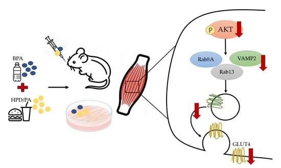
Co-Exposure to Bisphenol A and a High-Fat Diet Induces Insulin Resistance via Suppression of Insulin Signaling Molecule Expression and GLUT4 Translocation

1
Institute of Public Health, Guangzhou Medical University & Guangzhou Center for Disease Control and Prevention, Guangzhou 510440, China
2
School of Basic Medicine and Public Health, Jinan University, Guangzhou 510632, China
3
Department of Science Research, Guangzhou Center for Disease Control and Prevention, Guangzhou 510440, China
*
Author to whom correspondence should be addressed.
Toxics 2026, 14(2), 146; https://doi.org/10.3390/toxics14020146
Submission received: 23 December 2025 / Revised: 22 January 2026 / Accepted: 29 January 2026 / Published: 1 February 2026
(This article belongs to the Special Issue Health Risks and Toxicity of Emerging Contaminants)

Abstract

While the adverse health effects of bisphenol A (BPA) or high-fat diet (HFD) exposure alone have been relatively well documented, the mechanisms underlying their combined impact on insulin resistance and type 2 diabetes remain poorly understood. In this study, we observed the effects of 90 days of treatment with BPA and an HFD on insulin resistance in mouse gastrocnemius muscle, as well as the expression of signaling molecules and proteins potentially associated with glucose transporter type 4 (GLUT4) translocation. Additionally, C2C12 myotubes were co-treated with BPA and palmitic acid (PA) to observe the effects on insulin signaling molecules, GLUT4 translocation, and insulin resistance. Specifically, in vitro cellular experiments further demonstrated that BPA and PA inhibited GLUT4 translocation from the nucleus to the cell membrane. Taken together, co-exposure to BPA and an HFD (or PA) treatment significantly altered the expression of insulin signaling molecules in both gastrocnemius muscle and C2C12 cells, suggesting a potential link to their impacts on insulin resistance and GLUT4 translocation.

Graphical Abstract

1. Introduction

Diabetes mellitus is a common chronic metabolic disease. Model predictions have suggested that by 2045, the number of people with diabetes worldwide may reach 629 million [1]. Type 2 diabetes mellitus (T2DM) is the most prevalent form of this disease.
The etiology of T2DM is highly complex and potentially involves genetics, behavior, environment, nutritional factors, and their intricate interplay [2]. Insulin resistance (IR) is also recognized as part of the etiology of T2DM [3]. With the acceleration of urbanization, corresponding shifts in dietary patterns have emerged, characterized by a widespread increase in the consumption of ultra-processed foods, which are typically rich in refined carbohydrates, saturated fats, and added sugars. This modern lifestyle, exemplified by high-fat diets (HFD), now poses a significant health threat, and the incidence of related diseases has been increasing annually. An HFD induces T2DM by disrupting insulin-mediated metabolic functions [4,5,6]. Additionally, studies have indicated that endocrine disrupting chemicals (EDCs) can induce IR and T2DM by altering glucose homeostasis and insulin secretion [7,8]. Bisphenol A (BPA), a typical EDC, is widely used in electronic devices, plastic bottles, metal food can linings, and thermal paper, as well as in the synthesis of polycarbonate, epoxy resins, and other polymeric materials [9]. Due to its widespread application and resistance to degradation [10], humans are universally and continually exposed to BPA [11,12] primarily through oral ingestion, with absorption and subsequent metabolism occurring in the intestine and liver [9]. As a lipophilic compound, BPA has the ability to accumulate in different human and animal tissues, compromising their physiological functions and exerting deleterious effects on health [13]. Epidemiological studies have demonstrated a correlation between BPA exposure and elevated IR risk [14].
IR, a key pathological process and primary pathological mechanism in the development of T2DM, is characterized by elevated fasting blood glucose, decreased insulin sensitivity, and impaired glucose tolerance [15,16]. Because skeletal muscle accounts for approximately 80% of postprandial glucose uptake [15,17,18], it is a critical organ for regulating blood glucose levels; moreover, skeletal muscle IR is often considered a key factor affecting the onset of T2DM. The insulin signaling pathway and the translocation of glucose transporter type 4 (GLUT4) play essential roles in glucose uptake and metabolism [19,20,21]. Rab proteins have been identified to participate in the mobilization of insulin-stimulated GLUT4 storage vesicles in adipocytes and skeletal muscle cells, and Rab8A and Rab13 predominate in the skeletal muscle [22]. While high concentrations of BPA are known to affect Rab protein function [23], the effects of environmentally relevant doses of BPA on Rab proteins remain unclear. Furthermore, soluble N-ethylmaleimide-sensitive factor attachment protein receptor (SNARE) complex proteins play crucial roles in GLUT4 transport and plasma membrane fusion [24,25], and synaptosome-associated protein 23 (SNAP23), Syntaxin-4, and vesicle-associated membrane protein 2 (VAMP2) are highly expressed in skeletal muscle tissue and control GLUT4 transport in muscle cells [26,27]. Dysfunction and changes in SNARE protein levels can cause T2DM [28].
In recent years, an HFD has become a major lifestyle aspect associated with increased diabetes incidence [29]. People rich in fat may confer simultaneous exposure to BPA and HFD. Therefore, studies are necessary to explore the toxic effects of the combined exposure to BPA and HFD on insulin signaling pathway-associated molecules. Herein, we investigated the effects of BPA and HFD co-exposure on glucose metabolism using the gastrocnemius muscle of mice. Additionally, we employed C2C12 cells to further explore the impact of BPA and palmitic acid (PA) co-exposure on GLUT4 translocation and its underlying mechanisms.

2. Materials and Methods

2.1. Ethics

All methods in this experiment, including animal euthanasia, were strictly carried out in accordance with the guidelines and regulations of the Animal Care and Use Committee of the Guangzhou Center for Disease Control and Prevention. Animal procedures followed the experimental ethics standards approved by this committee (ethics approval number: No. 2021-012, approval date: 23 August 2020). All methods and protocols in this study comply with the requirements of the international ARRIVE 2.0 guidelines.

2.2. Animal Experimental Design

6–8 week-old SPF-grade C57BL/6J mice were purchased from the Guangdong Provincial Medical Laboratory Animal Center. After 1 week of acclimatization feeding, male and female mice were randomly divided into six groups, respectively. Three BPA doses (5, 50, and 500 μg/kg/d) were selected with reference to the U.S. and European tolerable daily intake (TDI) (50 and 4 μg/kg/d, respectively). The two factorial design (based on the presence or absence of BPA exposure and HFD exposure) comprised four groups: a control group (non-high-fat diet, no BPA, C0), high-fat diet group (high-fat diet, no BPA, C1), 50 μg/kg/day BPA group (non-high-fat diet, with BPA, C2), and 50 μg/kg/day BPA + high-fat diet group (with high-fat diet and BPA, TM). Additionally, to investigate the dose–response relationship of BPA under high-fat conditions, we added a 5 μg/kg/day BPA + high-fat diet group (TL) and a 500 μg/kg/day BPA + high-fat diet group (TH). Regular chow was obtained from the Guangdong Provincial Medical Laboratory Animal Center, and the HFD (with 45% energy from fat) was purchased from Nantong Troph Animal Feed Co., Ltd. (Nantong, China) The HFD was formulated with lard and soybean oil as the primary lipid sources, containing 23.66% crude protein, 23.6% crude fat, and 40.86% carbohydrate on a dry matter basis. BPA was dissolved in corn oil and administered daily by gavage at 9:00 AM for 90 days. Animals in control group received an equivalent volume of corn oil via gavage. Mouse body weight was measured at fixed times each week, and trends were recorded and analyzed. During the experiment, mice were given free access to water, the light/dark cycle was 12 h/12 h (changing at 7:00 AM and 7:00 PM), and the environment was maintained at a temperature of (22 ± 2) °C and a relative humidity of 40–70%. After 90 days, mice were anesthetized via intraperitoneal injection of 1% sodium pentobarbital, and the gastrocnemius muscle was isolated for various measurements.

2.3. Fasting Blood Glucose Measurement

At day 90 of the experiment, after a 12 h fast, mouse tail vein blood was collected and tested with a Roche glucometer.

2.4. Fasting Plasma Insulin Measurement

After 90 days of feeding, the mice were euthanized, and blood was drawn from the inferior vena cava and centrifuged at 4 °C at 3000 rpm for 10 min. The supernatant was used for plasma insulin measurement with an insulin ELISA kit (JONLNBIO, Shanghai, China, JL11459-96T).

2.5. Mouse HOMA-IR Measurement

IR was assessed using the homeostasis model assessment of insulin resistance (HOMA-IR), calculated from fasting insulin (FI) and fasting glucose (FG) levels as follows [30]:
H O M A I R = F I ( m I U / L ) × F G ( m m o l / L ) 22.5

2.6. Glucose Tolerance Test

Glucose tolerance tests were performed at day 60 (on mice fasted for 12 h before the experiment). The tail tip of the mouse was cut with scissors, and blood was squeezed onto a Roche glucometer (ACCU-CHEK Guide Me, Shanghai, China) for fasting blood glucose measurement, i.e., the 0 min glucose value. Subsequently, glucose was injected intraperitoneally (1 g/kg body weight), and the counting of timepoints started after the injection. Blood glucose values were measured and recorded at 15, 30, 60, 90, and 120 min post-injection.

2.7. Immunohistochemistry for Protein Expression

Immunohistochemistry was performed on tissues to observe GLUT4 localization and expression levels. Paraffin sections were deparaffinized and hydrated, then incubated with antibodies to GLUT4 (abcam, Cambridge, UK, ab33780, 1:1000) at 4 °C overnight and subsequently with goat anti-rabbit secondary antibodies at room temperature for 1 h. Staining was performed with a DAB staining kit (Beyotime, Shanghai, China, P0203), and sections were observed and analyzed after being mounted with neutral gum.

2.8. Cell Experiment Design

C2C12 myoblasts were cultured in high-glucose DMEM (Gibco, Shanghai, China, C11995500BT) containing 10% fetal bovine serum (Gibco, Carlsbad, CA, USA, 5669701), 100 U/mL penicillin, and 100 μg/mL streptomycin (Gibco, Waltham, MA, USA, 15140122) at 37 °C in an incubator with 5% CO2. Upon reaching 80–90% confluence, the growth medium was replaced with differentiation medium consisting of DMEM containing 2% horse serum (Solarbio, Beijing, China, S9050), 100 U/mL penicillin, and 100 μg/mL streptomycin. Based on preliminary screening of cell viability and glucose uptake (2-NBDG assay), the differentiated myotubes were treated with various agents in DMEM containing 3% FBS for 48 h. The treatment groups were as follows: control group (containing 2% BSA and 1% DMSO), PA group (200 μM PA + 1% DMSO), BPA group (102 nM BPA + 2% BSA), and PA + BPA group (200 μM PA + 102 nM BPA).

2.9. Cell Survival Assay

The cell viability of C2C12 cells were detected by CCK-8 assay. C2C12 cells were cultured in a 96-well plate and treated with BPA and PA at varying concentrations and for different durations. Then, every well was added to 10 μL CCK-8 reagent (Beyotime, Shanghai, China, C0038) for 1 h. The absorbance was measured at 450 nm with a Microplate Reader (Bio Tek, Winooski, VT, USA). All the experiments were carried out in triplicate.

2.10. 2-NBDG Glucose Uptake Assays

After cell treatment, 2-NBDG (Cayman chemical, Ann Arbor, MI, USA, 11046) was added to Krebs buffer containing 2% FA-free bovine serum albumin, and the cells were incubated with 100 nM insulin for 2 h. After incubation, cells were lysed with lysis buffer for 10 min. The lysates were then centrifuged at 16,000× g for 15 min at 4 °C to collect the supernatant. Fluorescence intensity was measured with a fluorescence plate reader at 475 nm/550 nm (excitation wavelength/emission wavelength). BCA quantification and protein correction were performed for each well to determine the glucose uptake of control and experimental group myotubes.

2.11. Cellular Immunofluorescence Staining for GLUT4

After cell treatment, cells were fixed with 4% paraformaldehyde at room temperature for 1 h, permeabilized with 0.2% Triton X-100, and then blocked with 3% FBS at 37 °C for 30 min. Anti-GLUT4 antibody (Abcam, Cambridge, UK, ab33780, 1:1000) was added and incubated overnight, and cells were subsequently incubated with fluorescent goat anti-rabbit IgG at 37 °C for 1 h. Nuclei were stained with DAPI for 10 min, and cells were observed and photographed under a fluorescence microscope.

2.12. Western Blotting for AKT-GLUT4 Signaling Molecules

Proteins from gastrocnemius tissue or C2C12 cells were extracted with tissue lysis buffer or cell lysis buffer, respectively. Proteins were incubated with monoclonal antibodies to AKT (CST, Beverly, MA, USA, 4685, 1:1000), p-AKTSer473 (CST, Beverly, MA, USA, 4060, 1:1000), p-AKTThr308 (CST, Beverly, MA, USA, 13038, 1:1000), Rab8A (Abcam, Cambridge, UK, ab188674, 1:1000), Rab13 (Merck, Darmstadt, Germany, SAB4200057, 1:1000), Syntaxin4 (Abcam, Cambridge, UK, ab184545, 1:1000), VAMP2 (Abcam, Cambridge, UK, ab181869, 1:5000), GLUT4 (Abcam, Cambridge, UK, ab33780, 1:1000), GSK3β (CST, Beverly, MA, USA, 12456, 1:1000), pGSK3βSer9 (CST, Beverly, MA, USA, 9322, 1:1000), and GAPDH (Abcam, Cambridge, UK, ab8245, 1:1000),and subsequently incubated with horseradish peroxidase-labeled goat anti-rabbit secondary antibodies. Bands were developed, and band density was analyzed with ImageJ 1.46r(USA National Institutes of Health). The relative expression of proteins was calculated as the ratio of the band density of the protein to that of GAPDH.

2.13. Cell Membrane Isolation

According to the manufacturer’s instructions, membrane proteins were isolated with a Mem-PER Plus Membrane Protein Extraction Kit (Thermo Scientific, Waltham, MA, USA). Expression of GLUT4 (Abcam, Cambridge, UK, ab33780, 1:1000) on the plasma membrane was determined by Western blotting with the membrane marker Na+-K+-ATPase (Abcam, Cambridge, UK, ab254025, 1:1000) serving as a control.

2.14. Statistical Analysis

All quantitative data are expressed as mean ± standard deviation (SD). Statistical analyses were primarily performed using SPSS25 (IBM, New York, USA). Repeated measures ANOVA was used for mouse body weight, and a factorial design was used to assess the interaction effects of BPA and HFD in the in vivo animal experiments for the C0, C1, C2, and TM groups, as well as the four groups in the in vitro cell experiments. One-way ANOVA was used for multiple group comparisons, and Dunnett tests were used for pairwise comparisons of multiple sample means. A p-value < 0.05 was considered statistically significant. Image analysis was performed with ImageJ 1.46r (USA National Institutes of Health), and data analysis was conducted in GraphPad Prism 7 (GraphPad Software, San Diego, CA, USA) software for graphing.

3. Results

3.1. BPA in Combination with an HFD Exacerbates IR

Repeated measures ANOVA indicated a significant interaction effect between various treatments and changes in the body weight of male and female mice over time (p < 0.05). The effects of various treatments on mouse body weight were statistically significant (p < 0.05). Mice in the HFD treatment groups (C1, TL, TM, and TH groups) had significantly higher body weights than those in the normal diet groups (C0 and C2; Figure 1a,b).
Following glucose administration, blood glucose levels peaked at 30 min in all groups. At 120 min post-injection, levels in male mice from the C1, TL, TM, and TH groups remained elevated above baseline (time 0) (Figure 1c,d). Compared with the control group, these four groups also showed a significant increase in the area under the curve (AUC) (Figure 1e). These results demonstrate that co-exposure to BPA and a high-fat diet induces glucose intolerance in male mice. In contrast, the combined exposure did not significantly affect glucose tolerance in female mice.
Calculation of IR and insulin sensitivity indices based on fasting blood glucose and fasting serum insulin values revealed that co-exposure to BPA and HFD induced an upward trend in glucose and insulin levels in both male and female mice. Notably, the high-dose co-exposure group (TH group) exhibited more pronounced elevation in insulin levels in both male and female mice (Figure 1f–h). All male mice experimental groups showed significantly higher HOMA-IR values compared to controls (p < 0.05), with particularly marked increases in the co-exposure groups accompanied by reduced insulin sensitivity. In female mice, all co-exposure groups to BPA and HFD (TL, TM, and TH groups) demonstrated significantly elevated HOMA-IR values relative to controls (p < 0.05) (Figure 1h). These findings collectively indicate that combined BPA and HFD exposure exacerbates insulin resistance in mice.

3.2. BPA and HFD Co-Exposure Affects Insulin Signaling Molecule Expression in Gastrocnemius Tissue

Analysis of the expression of key insulin signal transduction molecules in mice gastrocnemius tissue revealed that co-exposure to BPA and HFD (TL, TM, and TH groups) in both male and female mice resulted in a downward trend in pAKTSer473 expression compared to the control group, with a statistically significant reduction observed in the high-dose co-exposure group (TH group) (p < 0.05) (Figure 2a–c). The expression of VAMP2, a key molecule regulating the translocation and fusion of GLUT4 with the plasma membrane, was significantly reduced in both male and female mice following co-exposure to BPA and HFD groups (TL, TM, and TH groups) compared to the control group (p < 0.05). In contrast, no significant trend was observed for Rab8A (Figure 2d–f). Compared with the control group, the high-dose co-exposure group to BPA and HFD (TH group) in both female and male mice showed a significant increase in Syntaxin4 expression (Figure 2d, g; p < 0.05).
Furthermore, the expression of GLUT4 in male mice of the high-dose co-exposure group (TH group) was significantly lower than that in the control group (Figure 2d,h; p < 0.05). Immunohistochemical results also indicated significantly lower expression of GLUT4 in the gastrocnemius tissue in the co-exposure groups to BPA and HFD groups (TL, TM, and TH groups) than the corresponding control groups (Figure 2i,j).
Inhibition of the AKT signaling pathway reduces GSK3β phosphorylation, leading to increased GSK3β activity that suppresses glycogen synthase (GS). This inhibits glycogen synthesis and ultimately elevates blood glucose levels, constituting a key mechanism in the development of insulin resistance [31]. In this study, co-exposure to BPA and HFD affected the expression of the AKT signaling pathway.
The protein expression of pGSK3βSer9 in the gastrocnemius tissue of mice in the co-exposure to BPA and HFD (TM, and TH groups) was significantly lower than that in the control group, in both male and female mice (p < 0.05). Particularly in female mice, the expression of pGSK3βSer9 protein in the TM and TH groups was not only lower than that in the C0 group (control group) but also significantly lower than that in the C1 group (HFD treatment group) (p < 0.05) (Figure 2k,m). These findings were further supporting the hypothesis that BPA and HFD exacerbate IR.

3.3. BPA in Combination with Palmitic Acid (PA) Significantly Decreases Glucose Uptake Levels in C2C12 Cells

To assess the effects of BPA and PA exposure on cell viability, we treated C2C12 cells with various concentrations of BPA (1–105 nM) and PA (100–700 μM) for 48 h, and evaluated cell activity with CCK8 assays. Compared with that in the control group, cell activity was significantly lower with BPA treatment at concentrations of 102, 103, 104, and 105 nM (p < 0.05; Figure 3a). PA at 400 μM decreased the cell activity to 61% (Figure 3b). Therefore, BPA concentrations of 102 nM, 103 nM, 104 nM, and PA concentrations of 200 μM, 300 μM, and 450 μM were selected for glucose uptake experiments to determine the optimal combined dosage. Fluorescent Enzyme Labeler analysis (Figure 3c,d) indicated that glucose uptake was significantly lower after treatment with 104 nM BPA and 300 μM, 450 μM PA for 48 h than observed in the insulin-stimulated control group (p < 0.05). Considering multiple factors such as cell activity and glucose uptake, we selected a combination of 300 μM PA, and 102 nM or 103 nM BPA for 48 h. At that timepoint, glucose uptake in all groups was significantly lower than observed in the control group (p < 0.05). Our findings indicated that 300 μM PA and 102 nM or 103 nM BPA induced IR in C2C12 cells (Figure 3e). Moreover, after 48 h of exposure to 200 μM PA and BPA, the glucose uptake was also significantly lower than observed in the control group (p < 0.05); these findings suggested that BPA decreased the concentration of PA necessary to induce IR (Figure 3f). When the exposure time was decreased to 24 h, we observed no significant change in cell glucose uptake (p > 0.05; Figure 3g). These findings suggested that BPA decreased the concentration of PA required for exposure, and their combined action induced IR in C2C12 myocytes. On the basis of the above results, we selected dosages and exposure times of 200 μM PA and 102 nM BPA for 48 h in C2C12 myotubes. Compared with that in the insulin-stimulated control group, the glucose uptake of the co-exposed group was significantly lower (p < 0.05; Figure 3h). According to the CCK8 cell viability assay results, the cell activity of the groups exposed to 200 μM PA alone and 102 nM BPA alone exceeded 80%, and after 48 h of co-exposure to 200 μM PA and 102 nM BPA, the cell activity decreased to 73% and showed statistically significant differences among groups (p < 0.05; Figure 3i).

3.4. Co-Exposure to BPA and PA Inhibits Insulin Signaling and GLUT4 Translocation in C2C12 Cells

The PI3K/AKT signaling pathway is an important regulatory route for glucose uptake [19,32]. To investigate the potential molecular mechanisms through which BPA and PA affect insulin sensitivity, we examined the expression of AKT and p-AKT, as well as their downstream signaling molecules and proteins associated with GLUT4 translocation (Figure 4a–c). Compared with that in the control group, the expression of p-AKT (Ser473 and Thr308) was significantly lower in the group co-exposed to 200 μM PA and 102 nM BPA (p < 0.01), as was the expression of AKTThr308 in the group exposed to 102 nM BPA alone (p < 0.05).
The total GLUT4 expression was significantly lower in all experimental groups than the control group (p < 0.05; Figure 4d). After separation of cell membranes, we determined that the expression of GLUT4 on the cell membrane in the group exposed to 102 nM BPA alone (p < 0.05) and in the group co-exposed to 200 μM PA and 102 nM BPA (p < 0.05) was significantly lower than that in the control group (Figure 4e). These findings suggested that co-exposure to BPA and PA significantly inhibited GLUT4 expression on the cell membrane (i.e., decreased translocation of GLUT4 from the cytoplasm to the plasma membrane). Immunofluorescence staining of C2C12 myotubes indicated significantly lower GLUT4 expression in the experimental groups than the control group (p < 0.05). Moreover, the GLUT4 expression in the group exposed to 200 μM PA alone (p < 0.05) and in the group co-exposed to 200 μM PA and 102 nM BPA (p < 0.05) was significantly lower than that in the group exposed to 102 nM BPA alone (Figure 4f,g).
AKT downstream molecules such as AS160 activate Rab proteins (e.g., Rab8A/Rab13), which along with VAMP2 and Syntaxin4 drive GLUT4 vesicle fusion with the plasma membrane and subsequent glucose release. In this study, the protein expression of Rab8A and Rab13 in the group co-exposed to 200 μM PA and 102 nM BPA was significantly lower than that in the control group (p < 0.05). The expression of Syntaxin4 protein in the group exposed to 102 nM BPA alone (p < 0.05) and in the group co-exposed to 200 μM PA and 102 nM BPA (p < 0.05) was significantly higher than observed in the control group. However, the expression level of VAMP2 did not exhibit significant changes (Figure 4h–k).

4. Discussion

With changes in lifestyle and dietary habits, excessive energy intake coupled with insufficient physical activity leads to obesity and associated IR, thus posing a substantial threat to human health. Compounding this issue is the widespread presence of EDCs such as BPA. As a high-production-volume chemical monomer and plasticizer, BPA has an annual output exceeding 100 million pounds [9] and has been detected in various human biological matrices, including serum, urine, amniotic fluid, and placental tissue [33]. Long-term exposure to both BPA and an HFD is associated with the occurrence of IR or T2DM, yet the combined effects and mechanisms of both factors remain unclear. By simulating the widespread scenario of high-fat dietary behavior combined with continuous environmental-dose BPA co-exposure, this study aims to elucidate the underlying mechanisms of insulin resistance in the gastrocnemius muscle, focusing on the expression regulation and membrane translocation dynamics of GLUT4, thereby providing critical experimental evidence for the intervention of insulin resistance and related metabolic diseases.
To clarify the relationships and mechanisms underlying the combined effects of an HFD and BPA on animal IR, we observed the effects of co-exposure to an HFD and low doses of BPA on tissue or cellular GLUT4 expression and IR. For the animal experiments, BPA doses were selected based on established safety thresholds and human exposure relevance. The U.S. tolerable daily intake for BPA is 50 μg/kg/day, which is 1000-fold lower than the lowest observed adverse effect level [34]. Additionally, a dose of 50 μg/kg/day in mice is approximately equivalent to 4 μg/kg/day in humans, a value significantly lower than the U.S. EPA reference dose [35] and aligned with the European proposed TDI of 4 μg/kg/day [36]. Therefore, we ultimately employed three BPA doses: 5, 50, and 500 μg/kg/day.
Both the in vivo and in vitro experiments demonstrated that co-exposure to BPA and an HFD (or PA) induces insulin resistance (IR). In mice, the combined exposure groups (TL, TM, TH) of both sexes showed elevated HOMA-IR indices, consistent with previous reports [37]. These results confirmed that long-term BPA and HFD co-exposure induces IR. However, a limitation of this study is the absence of an insulin tolerance test (ITT) to evaluate insulin sensitivity, which should be addressed in future research. This study aims to elucidate the underlying mechanisms involved, with a particular focus on skeletal muscle—a key peripheral tissue essential for maintaining glucose homeostasis and a primary target organ for insulin action. The expression profiles of protein molecules and the activation or suppression of the insulin signaling system require further study. The activation of GLUT4, the first step in glucose metabolism, transports glucose into muscle cells. Under normal conditions, approximately 90% of GLUT4 is expressed in the cytoplasm, whereas only approximately 10% is expressed on the cell membrane. Promoting the translocation of GLUT4 from the cytoplasm to the cell membrane effectively increases glucose transport. Because insulin is a major factor stimulating GLUT4 translocation in skeletal muscle, modulation of the insulin PI3K-AKT signaling pathway has been found to effectively regulate GLUT4 translocation [38,39,40]. The expression of pAKTSer473 in the gastrocnemius muscle tissue of the experimental mice was lower than that in the control group. Our in vitro experimental results also demonstrated significantly decreased expression of pAKTSer473 and pAKTThr308 in myotube cells after exposure to BPA and PA, thus suggesting that the combined HFD and BPA treatment causes disorders in the insulin signaling pathway, thereby exacerbating the development of IR.
In GLUT4-mediated glucose transport, beyond the translocation of GLUT4 vesicles, the fusion of these vesicles with the plasma membrane is also crucial for glucose utilization by skeletal muscle and the maintenance of blood glucose homeostasis. Specifically, the docking of GLUT4 vesicles with the plasma membrane depends on the interaction between the vesicle membrane protein VAMP2 and the plasma membrane protein Syntaxin4 [41,42]. Syntaxin4 anchors GLUT4 vesicles on the plasma membrane, whereas VAMP2 promotes the fusion of the plasma membrane with vesicles and, consequently, accelerates glucose exocytosis [43]. Given the pivotal role of VAMP2, we investigated its expression under co-exposure conditions. In the gastrocnemius muscle of female and male mice, the expression of VAMP2 in the group co-exposure with an HFD and BPA was significantly lower than that in the control group. Furthermore, in C2C12 myotubes, the group co-treated with PA and BPA also showed a similar declining trend. Particularly in male mice, the high-dose exposure group (TH group) had significantly lower VAMP2 expression than the C0, C1, and C2 groups. In the TM group, VAMP2 expression was also significantly lower than that in the C0 and C1 groups. These findings suggest that combined exposure to BPA and an HFD significantly inhibited VAMP2 protein expression.
In vitro, the expression of Syntaxin4 was significantly higher in the PA + BPA group than in the control group. Consistently, in vivo, Syntaxin4 protein levels were also elevated in the gastrocnemius muscle of mice co-exposed to BPA and an HFD, with the most pronounced upregulation observed in the high-dose (TH) group. This upregulation might reflect an adaptive response of cells that compensates for defects in GLUT4 translocation [44,45].
Using experimental techniques such as Western blotting, immunohistochemistry, and immunofluorescence, we determined that, in both gastrocnemius tissue and C2C12 myotubes, the total expression level of GLUT4 in the group co-treated with an HFD and BPA was significantly lower than that in the control group. To specifically assess GLUT4 membrane localization, we isolated plasma membranes from C2C12 myotubes. This analysis confirmed that GLUT4 levels at the plasma membrane were significantly lower in the PA + BPA group than in the control group. Our in vivo animal experiments suggested that combined exposure to BPA and an HFD had a pronounced inhibitory effect on GLUT4 expression, as further confirmed by in vitro cellular experiments indicating that the combined action of BPA and an HFD severely hinders GLUT4 translocation.
The storage capacity of glycogen in skeletal muscle is limited because the activity of glycogen synthase is inhibited by Protein Kinase A (PKA) [46] or glycogen synthase kinase 3β (GSK3β) [47], thus impairing glycogen accumulation. Impaired glycogen synthesis stimulated by insulin is a common pathological feature in all states of IR. Obesity and diabetes, representative conditions of metabolic syndrome, demonstrate AKT signaling pathway inhibition, accompanied by reduced GSK3β-Ser9 phosphorylation [48,49,50], which markedly enhances GSK3β kinase activity [51]. The activated GSK3β phosphorylates glycogen synthase, thereby suppressing its activity and impairing glycogen synthesis [49,50,52]. Similarly, AKT also promotes GLUT4 translocation by phosphorylating AS160 and subsequently enhancing glucose uptake [35]. However, excessive GSK3β activity inhibits the AKT signaling pathway, thus impeding GLUT4 translocation and glycogen synthesis [53]. Therefore, GSK3β plays a critical role in the translocation of GLUT4 from intracellular vesicles to the plasma membrane. Decreased levels of pGSK3βSer9 may be associated with impaired GLUT4 translocation and further exacerbation of IR and metabolic disorders [31]. We observed that in male mice, GSK3β protein expression in the co-exposure to BPA and HFD group (TM group) was significantly higher than that in the HFD-only group (C2 group), and no significant difference in GSK3β protein expression was observed among the other groups. Additionally, expression of pGSK3βSer9 in the group co-exposure with an HFD and BPA (TL, TM, TH group) was significantly lower than that in the control group. These results suggested that co-exposure to BPA and HFD might exacerbate the decrease in pGSK3βSer9 in the gastrocnemius muscle.

5. Conclusions

In summary, co-exposure to BPA and an HFD (or PA) impairs GLUT4 translocation in both mouse gastrocnemius muscle and C2C12 myotubes. This effect is primarily mediated through disruption of the AKT-dependent insulin signaling pathway, which leads to a series of changes in the expression of proteins, particularly those associated with the translocation of GLUT4 from the cytoplasm to the plasma membrane. These changes exacerbated the occurrence of IR and inhibited the translocation of GLUT4, and consequently, they might adversely affect blood glucose regulation and metabolic health.

Author Contributions

Conceptualization, Z.L. and W.Z.; methodology, Z.L., M.C., J.Z., C.Q. and B.H.; validation, W.L. (Wenxue Li), J.L., G.Y. and Z.L.; formal analysis, M.C., W.L. (Weiwen Liu) and J.W.; data curation, Y.Z. and Z.L.; writing—original draft preparation, Z.L. and M.C.; writing—review and editing, W.Z.; visualization, W.L. (Wenxue Li); supervision, W.Z.; project administration, W.Z.; funding acquisition, W.Z. All authors have read and agreed to the published version of the manuscript.

Funding

This research was funded by the National Key Research and Development Program of China (2018YFC1603102), the Basic Research Project of Key Laboratory of Guangzhou (2024A03J0558), and the Key Project of Medicine Discipline of Guangzhou (No. 20212023012).

Institutional Review Board Statement

Animal procedures were conducted according to the guidelines approved by the Animal Care and Use Committee of Guangzhou Center for Disease Control and Prevention (ethics approval number: No. 2021-012, approval date: 23 August 2020). All methods and protocols in this study comply with the international ARRIVE 2.0 guidelines.

Informed Consent Statement

Not applicable.

Data Availability Statement

The original contributions presented in this study are included in the article. Further inquiries can be directed to the corresponding author.

Conflicts of Interest

The authors declare no conflicts of interest.

References

  1. Huang, Y.; Karuranga, S.; Malanda, B.; Williams, D.R.R. Call for data contribution to the IDF Diabetes Atlas 9th Edition 2019. Diabetes Res. Clin. Pract. 2018, 140, 351–352. [Google Scholar] [CrossRef]
  2. Al Mansour, M.A. The Prevalence and Risk Factors of Type 2 Diabetes Mellitus (DMT2) in a Semi-Urban Saudi Population. Int. J. Environ. Res. Public Health 2019, 17, 7. [Google Scholar] [CrossRef]
  3. Lu, X.; Xie, Q.; Pan, X.; Zhang, R.; Zhang, X.; Peng, G.; Zhang, Y.; Shen, S.; Tong, N. Type 2 diabetes mellitus in adults: Pathogenesis, prevention and therapy. Signal Transduct. Target. Ther. 2024, 9, 262. [Google Scholar] [CrossRef]
  4. Woods, S.C.; Seeley, R.J.; Rushing, P.A.; D’Alessio, D.; Tso, P. A controlled high-fat diet induces an obese syndrome in rats. J. Nutr. 2003, 133, 1081–1087. [Google Scholar] [CrossRef] [PubMed]
  5. Kahn, S.E.; Hull, R.L.; Utzschneider, K.M. Mechanisms linking obesity to insulin resistance and type 2 diabetes. Nature 2006, 444, 840–846. [Google Scholar] [CrossRef] [PubMed]
  6. Small, L.; Brandon, A.E.; Turner, N.; Cooney, G.J. Modeling insulin resistance in rodents by alterations in diet: What have high-fat and high-calorie diets revealed? Am. J. Physiol. Endocrinol. Metab. 2018, 314, E251–E265. [Google Scholar] [CrossRef] [PubMed]
  7. Hinault, C.; Caroli-Bosc, P.; Bost, F.; Chevalier, N. Critical Overview on Endocrine Disruptors in Diabetes Mellitus. Int. J. Mol. Sci. 2023, 24, 4537. [Google Scholar] [CrossRef]
  8. Weiss, M.C.; Shih, Y.H.; Bryan, M.S.; Jackson, B.P.; Aguilar, D.; Hanis, C.L.; Argos, M.; Sargis, R.M. Relationships Between Urinary Metals and Diabetes Traits Among Mexican Americans in Starr County, Texas, USA. Biol. Trace Elem. Res. 2023, 201, 529–538. [Google Scholar] [CrossRef]
  9. Vandenberg, L.N.; Hauser, R.; Marcus, M.; Olea, N.; Welshons, W.V. Human exposure to bisphenol A (BPA). Reprod. Toxicol. 2007, 24, 139–177. [Google Scholar] [CrossRef]
  10. Geens, T.; Goeyens, L.; Covaci, A. Are potential sources for human exposure to bisphenol-A overlooked? Int. J. Hyg. Environ. Health 2011, 214, 339–347. [Google Scholar] [CrossRef]
  11. Vandenberg, L.N.; Chahoud, I.; Heindel, J.J.; Padmanabhan, V.; Paumgartten, F.J.; Schoenfelder, G. Urinary, circulating, and tissue biomonitoring studies indicate widespread exposure to bisphenol A. Cienc. Saude Coletiva 2012, 17, 407–434. [Google Scholar] [CrossRef] [PubMed]
  12. Russo, G.; Barbato, F.; Mita, D.G.; Grumetto, L. Occurrence of Bisphenol A and its analogues in some foodstuff marketed in Europe. Food Chem. Toxicol. 2019, 131, 110575. [Google Scholar] [CrossRef] [PubMed]
  13. Cimmino, I.; Fiory, F.; Perruolo, G.; Miele, C.; Beguinot, F.; Formisano, P.; Oriente, F. Potential Mechanisms of Bisphenol A (BPA) Contributing to Human Disease. Int. J. Mol. Sci. 2020, 21, 5761. [Google Scholar] [CrossRef] [PubMed]
  14. Hafezi, S.A.; Abdel-Rahman, W.M. The Endocrine Disruptor Bisphenol A (BPA) Exerts a Wide Range of Effects in Carcinogenesis and Response to Therapy. Curr. Mol. Pharmacol. 2019, 12, 230–238. [Google Scholar] [CrossRef]
  15. Freeman, A.M.; Acevedo, L.A.; Pennings, N. Insulin Resistance. In StatPearls; StatPearls Publishing: Treasure Island, FL, USA, 2023. [Google Scholar]
  16. Lee, K. Usefulness of the metabolic syndrome criteria as predictors of insulin resistance among obese Korean women. Public Health Nutr. 2010, 13, 181–186. [Google Scholar] [CrossRef]
  17. Ferrannini, E.; Simonson, D.C.; Katz, L.D.; Reichard, G., Jr.; Bevilacqua, S.; Barrett, E.J.; Olsson, M.; DeFronzo, R.A. The disposal of an oral glucose load in patients with non-insulin-dependent diabetes. Metab. Clin. Exp. 1988, 37, 79–85. [Google Scholar] [CrossRef]
  18. Foroutani, L.; Ashraf Ganjouei, A.; Wang, J.; Aburayya, B.I.; Corvera, C.; Alseidi, A.; Adam, M.A. Robotic-Assisted Endoluminal Resection of Gastroesophageal Junction Leiomyoma with Transoral Specimen Extraction: Technique, Outcome, and Safety. Ann. Surg Oncol. 2025, 32, 1218–1219. [Google Scholar] [CrossRef]
  19. DeFronzo, R.A.; Tripathy, D. Skeletal muscle insulin resistance is the primary defect in type 2 diabetes. Diabetes Care 2009, 32, S157–S163. [Google Scholar] [CrossRef]
  20. Zhu, Y.; Pereira, R.O.; O’Neill, B.T.; Riehle, C.; Ilkun, O.; Wende, A.R.; Rawlings, T.A.; Zhang, Y.C.; Zhang, Q.; Klip, A.; et al. Cardiac PI3K-Akt impairs insulin-stimulated glucose uptake independent of mTORC1 and GLUT4 translocation. Mol. Endocrinol. 2013, 27, 172–184. [Google Scholar] [CrossRef]
  21. Sharma, B.R.; Kim, H.J.; Rhyu, D.Y. Caulerpa lentillifera extract ameliorates insulin resistance and regulates glucose metabolism in C57BL/KsJ-db/db mice via PI3K/AKT signaling pathway in myocytes. J. Transl. Med. 2015, 13, 62. [Google Scholar] [CrossRef]
  22. Li, W.; Liang, X.; Zeng, Z.; Yu, K.; Zhan, S.; Su, Q.; Yan, Y.; Mansai, H.; Qiao, W.; Yang, Q.; et al. Simvastatin inhibits glucose uptake activity and GLUT4 translocation through suppression of the IR/IRS-1/Akt signaling in C2C12 myotubes. Biomed. Pharmacother. 2016, 83, 194–200. [Google Scholar] [CrossRef] [PubMed]
  23. Sun, Y.; Bilan, P.J.; Liu, Z.; Klip, A. Rab8A and Rab13 are activated by insulin and regulate GLUT4 translocation in muscle cells. Proc. Natl. Acad. Sci. USA 2010, 107, 19909–19914. [Google Scholar] [CrossRef] [PubMed]
  24. Foster, L.J.; Klip, A. Mechanism and regulation of GLUT-4 vesicle fusion in muscle and fat cells. Am. J. Physiol. Cell Physiol. 2000, 279, C877–C890. [Google Scholar] [CrossRef] [PubMed]
  25. Mullainadhan, V.; Viswanathan, M.P.; Karundevi, B. Effect of Bisphenol-A (BPA) on insulin signal transduction and GLUT4 translocation in gastrocnemius muscle of adult male albino rat. Int. J. Biochem. Cell Biol. 2017, 90, 38–47. [Google Scholar] [CrossRef]
  26. Pessin, J.E.; Thurmond, D.C.; Elmendorf, J.S.; Coker, K.J.; Okada, S. Molecular basis of insulin-stimulated GLUT4 vesicle trafficking: Location! Location! Location! J. Biol. Chem. 1999, 274, 2593–2596. [Google Scholar] [CrossRef]
  27. Duman, J.G.; Forte, J.G. What is the role of SNARE proteins in membrane fusion? Am. J. Physiol. Cell Physiol. 2003, 285, C237–C249. [Google Scholar] [CrossRef]
  28. Chamberlain, L.H.; Gould, G.W. The vesicle- and target-SNARE proteins that mediate Glut4 vesicle fusion are localized in detergent-insoluble lipid rafts present on distinct intracellular membranes. J. Biol. Chem. 2002, 277, 49750–49754. [Google Scholar] [CrossRef]
  29. Garvey, W.T.; Maianu, L.; Zhu, J.H.; Brechtel-Hook, G.; Wallace, P.; Baron, A.D. Evidence for defects in the trafficking and translocation of GLUT4 glucose transporters in skeletal muscle as a cause of human insulin resistance. J. Clin. Investig. 1998, 101, 2377–2386. [Google Scholar] [CrossRef]
  30. Hu, F.B. Globalization of diabetes: The role of diet, lifestyle, and genes. Diabetes Care 2011, 34, 1249–1257. [Google Scholar] [CrossRef]
  31. Carnagarin, R.; Dharmarajan, A.M.; Dass, C.R. Molecular aspects of glucose homeostasis in skeletal muscle--A focus on the molecular mechanisms of insulin resistance. Mol. Cell. Endocrinol. 2015, 417, 52–62. [Google Scholar] [CrossRef]
  32. Tahapary, D.L.; Pratisthita, L.B.; Fitri, N.A.; Marcella, C.; Wafa, S.; Kurniawan, F.; Rizka, A.; Tarigan, T.J.E.; Harbuwono, D.S.; Purnamasari, D.; et al. Challenges in the diagnosis of insulin resistance: Focusing on the role of HOMA-IR and Tryglyceride/glucose index. Diabetes Metab. Syndr. 2022, 16, 102581. [Google Scholar] [CrossRef] [PubMed]
  33. Miyata, S.; Yada, T.; Ishikawa, N.; Taheruzzaman, K.; Hara, R.; Matsuzaki, T.; Nishikawa, A. Insulin-like growth factor 1 regulation of proliferation and differentiation of Xenopus laevis myogenic cells in vitro. Vitr. Cell. Dev. Biol. Anim. 2017, 53, 231–247. [Google Scholar] [CrossRef] [PubMed]
  34. Welshons, W.V.; Nagel, S.C.; vom Saal, F.S. Large effects from small exposures. III. Endocrine mechanisms mediating effects of bisphenol A at levels of human exposure. Endocrinology 2006, 147, S56–S69. [Google Scholar] [CrossRef] [PubMed]
  35. Reagan-Shaw, S.; Nihal, M.; Ahmad, N. Dose translation from animal to human studies revisited. FASEB J. 2008, 22, 659–661. [Google Scholar] [CrossRef]
  36. Chen, D.; Kannan, K.; Tan, H.; Zheng, Z.; Feng, Y.L.; Wu, Y.; Widelka, M. Bisphenol Analogues Other Than BPA: Environmental Occurrence, Human Exposure, and Toxicity-A Review. Environ. Sci. Technol. 2016, 50, 5438–5453. [Google Scholar] [CrossRef]
  37. Luo, W.; Ai, L.; Wang, B.F.; Zhou, Y. High glucose inhibits myogenesis and induces insulin resistance by down-regulating AKT signaling. Biomed. Pharmacother. 2019, 120, 109498. [Google Scholar] [CrossRef]
  38. Fryer, L.G.; Foufelle, F.; Barnes, K.; Baldwin, S.A.; Woods, A.; Carling, D. Characterization of the role of the AMP-activated protein kinase in the stimulation of glucose transport in skeletal muscle cells. Biochem. J. 2002, 363, 167–174. [Google Scholar] [CrossRef]
  39. Mora, S.; Pessin, J.E. An adipocentric view of signaling and intracellular trafficking. Diabetes/Metab. Res. Rev. 2002, 18, 345–356. [Google Scholar] [CrossRef]
  40. Ding, S.; Fan, Y.; Zhao, N.; Yang, H.; Ye, X.; He, D.; Jin, X.; Liu, J.; Tian, C.; Li, H.; et al. High-fat diet aggravates glucose homeostasis disorder caused by chronic exposure to bisphenol A. J. Endocrinol. 2014, 221, 167–179. [Google Scholar] [CrossRef]
  41. Ropero, A.B.; Soria, B.; Nadal, A. A nonclassical estrogen membrane receptor triggers rapid differential actions in the endocrine pancreas. Mol. Endocrinol. 2002, 16, 497–505. [Google Scholar] [CrossRef]
  42. Bryant, N.J.; Gould, G.W. SNARE proteins underpin insulin-regulated GLUT4 traffic. Traffic 2011, 12, 657–664. [Google Scholar] [CrossRef]
  43. Jaldin-Fincati, J.R.; Pavarotti, M.; Frendo-Cumbo, S.; Bilan, P.J.; Klip, A. Update on GLUT4 Vesicle Traffic: A Cornerstone of Insulin Action. Trends Endocrinol. Metab. 2017, 28, 597–611. [Google Scholar] [CrossRef]
  44. Kahn, B.B.; Flier, J.S. Regulation of glucose-transporter gene expression in vitro and in vivo. Diabetes Care 1990, 13, 548–564. [Google Scholar] [CrossRef]
  45. Kawaguchi, T.; Tamori, Y.; Kanda, H.; Yoshikawa, M.; Tateya, S.; Nishino, N.; Kasuga, M. The t-SNAREs syntaxin4 and SNAP23 but not v-SNARE VAMP2 are indispensable to tether GLUT4 vesicles at the plasma membrane in adipocyte. Biochem. Biophys. Res. Commun. 2010, 391, 1336–1341. [Google Scholar] [CrossRef] [PubMed]
  46. Saltiel, A.R.; Kahn, C.R. Insulin signalling and the regulation of glucose and lipid metabolism. Nature 2001, 414, 799–806. [Google Scholar] [CrossRef] [PubMed]
  47. Cohen, P.; Frame, S. The renaissance of GSK3. Nat. Rev. Mol. Cell Biol. 2001, 2, 769–776. [Google Scholar] [CrossRef] [PubMed]
  48. Gulli, G.; Ferrannini, E.; Stern, M.; Haffner, S.; DeFronzo, R.A. The metabolic profile of NIDDM is fully established in glucose-tolerant offspring of two Mexican-American NIDDM parents. Diabetes 1992, 41, 1575–1586. [Google Scholar] [CrossRef]
  49. Cohen, P. The Croonian Lecture 1998. Identification of a protein kinase cascade of major importance in insulin signal transduction. Philos. Trans. R. Soc. London. Ser. B Biol. Sci. 1999, 354, 485–495. [Google Scholar] [CrossRef]
  50. Cohen, P.; Goedert, M. GSK3 inhibitors: Development and therapeutic potential. Nat. Rev. Drug Discov. 2004, 3, 479–487. [Google Scholar] [CrossRef]
  51. Nikoulina, S.E.; Ciaraldi, T.P.; Mudaliar, S.; Mohideen, P.; Carter, L.; Henry, R.R. Potential role of glycogen synthase kinase-3 in skeletal muscle insulin resistance of type 2 diabetes. Diabetes 2000, 49, 263–271. [Google Scholar] [CrossRef]
  52. Cross, D.A.; Alessi, D.R.; Cohen, P.; Andjelkovich, M.; Hemmings, B.A. Inhibition of glycogen synthase kinase-3 by insulin mediated by protein kinase B. Nature 1995, 378, 785–789. [Google Scholar] [CrossRef]
  53. Henriksen, E.J.; Dokken, B.B. Role of glycogen synthase kinase-3 in insulin resistance and type 2 diabetes. Curr. Drug Targets 2006, 7, 1435–1441. [Google Scholar] [CrossRef]
Figure 1. BPA in combination with an HFD exacerbates IR. Results are presented as mean ± SD. (a) Male mouse body weight (n = 12–13). (b) Female mouse body weight (n = 11–12). Oral glucose tolerance test (OGTT) results in male mice (n = 3) (c) and in female mice (n = 8–12) (d), with the test conducted after 60 days of exposure. (e) Area under the glucose tolerance curve for both male and female mice. Fasting blood glucose levels in both male mice (n = 3) and female mice (n = 3) (f) and fasting insulin levels in both male mice (n = 3) and female mice (n = 3) (g), with the test conducted after 90 days of exposure. (h) IR index levels in both male mice (n = 3) and female mice (n = 3). Compared with the C0 group, * p < 0.05; compared with the C1 group, # p < 0.05; compared with the C2 group, △ p < 0.05.
Toxics 14 00146 g001
Figure 2. Effects of BPA and HFD on signaling molecule expression in gastrocnemius tissue in male and female mice. Results are presented as mean ± SD. Western blot bands of AKT, pAKTSer473, and GADPH protein level (a), with quantitative analysis of AKT (n = 3) (b) and pAKTSer473 (n = 3) (c). (d) Western blot bands of Rab8A, VAMP2, Syntaxin4, GLUT4, and GADPH protein level, with quantitative analysis of Rab8A (n = 3) (e), VAMP2 (n = 3) (f), Syntaxin4 (n = 3) (g), and GLUT4 (n = 3) (h). Results of GLUT4 immunohistochemistry (i); quantitative analysis of immunohistochemistry results (j) (n = 3). (k) Western blot bands of GSK3β, pGSK3βSer9, and GADPH protein levels, with quantitative analysis of GSK3β (n = 3) (l) and pGSK3βSer9 (n = 3) (m). Compared with the C0 group, * p < 0.05; compared with the C1 group, # p < 0.05; compared with the C2 group, △ p < 0.05.
Toxics 14 00146 g002
Figure 3. Effects of BPA and palmitic acid (PA) treatment on glucose uptake in C2C12 myotubes. Results are presented as mean ± SD. (a,b) C2C12 myotubes were exposed to BPA (a) (1–105 nM; control group: 0 nM BPA, 0.1% DMSO) and PA (b) (100–700 μM; control group: 0 μM PA, 2% BSA) for 48 h, and cytotoxicity was measured with CCK8 assays (n = 4). (c) Effect of 48 h BPA exposure on glucose uptake (n = 3). (d) Effect of 48 h PA exposure on glucose uptake (n = 3). (e) Effect of co-exposure to PA (300 μM) and BPA (102 and 103 nM) for 48 h on glucose uptake (n = 3). (f,g) Effect of co-exposure to PA (200 μM) and BPA (102 and 103 nM) for 48 h (f) and 24 h (g) on glucose uptake (n = 3). (h) Effect of co-exposure to PA (200 μM) and BPA (102 nM) for 48 h on glucose uptake (n = 3). Compared with the insulin-stimulated control group, * p < 0.05; compared with the corresponding concentration group without insulin stimulation, # p < 0.05. (i) Cell viability after co-exposure to PA (200 μM) and BPA (102 nM) for 48 h (n = 4). Compared with the control group, & p < 0.05; compared with the 200 μM PA group, $ p < 0.05; compared with the 102 nM BPA group, Δ p < 0.05.
Toxics 14 00146 g003
Figure 4. Effects of BPA and PA on signaling molecules and GLUT4 translocation in C2C12 myotubes. Results are presented as mean ± SD. Western blot bands and corresponding quantitative analysis of AKT (a), pAKTSer473 (b), pAKTThr308 (c), total GLUT4 (d), and GLUT4 on the membrane (e) (n = 3). Immunofluorescence staining with anti-GLUT4 antibodies after treatment of C2C12 cells with BPA and PA for 48 h (scale bar: 100 μM) (f). Quantitative analysis of GLUT4 immunofluorescence (n = 3) (g). Western blot bands and corresponding quantitative analysis of Rab8A (h), Rab13 (i), Syntaxin4 (j), and VAMP2 (k) (n = 3). Compared with the control group, * p < 0.05; compared with the 102 nM BPA group, # p < 0.05.
Toxics 14 00146 g004
Disclaimer/Publisher’s Note: The statements, opinions and data contained in all publications are solely those of the individual author(s) and contributor(s) and not of MDPI and/or the editor(s). MDPI and/or the editor(s) disclaim responsibility for any injury to people or property resulting from any ideas, methods, instructions or products referred to in the content.

