Article

Long-Term Exposure to 6-PPD Quinone Inhibits Glutamate Synthesis and Glutamate Receptor Function Associated with Its Toxicity Induction in Caenorhabditis elegans

1
Key Laboratory of Environmental Medicine Engineering of Ministry of Education, School of Public Health, Southeast University, Nanjing 210009, China
2
Medical School, Southeast University, Nanjing 210009, China
*
Authors to whom correspondence should be addressed.
Toxics 2025, 13(6), 434; https://doi.org/10.3390/toxics13060434
Submission received: 17 April 2025 / Revised: 20 May 2025 / Accepted: 21 May 2025 / Published: 26 May 2025

Abstract

6-PPD quinone (6-PPDQ) is widely distributed in environments. In Caenorhabditis elegans, we first examined the effects of 6-PPDQ on glutamate synthesis and receptor function by analyzing glutamate content, related gene expression, and phenotypes after RNAi of these genes. Moreover, we performed glutamate treatment after 6-PPDQ exposure to determine the potential pharmacological effects of glutamate against 6-PPDQ toxicity. After exposure, the glutamate content was reduced by 0.1–10 μg/L 6-PPDQ, which was due to decreased expression of W07E1.1, glna-1/2/3, and alh-6 governing glutamate synthesis from α-ketoglutarate, glutamine, and proline. RNAi of W07E1.1, glna-1/2/3, and alh-6 decreased glutamate content in 6-PPDQ-exposed nematodes, and caused susceptibility to 6-PPDQ toxicity. Among glutamate transporter genes, glt-1 expression was decreased by 0.1–10 μg/L 6-PPDQ. Moreover, 0.1–10 μg/L 6-PPDQ decreased glutamate receptor genes (glr-1, glr-2, and glr-4), and their expression was decreased by RNAi of W07E1.1, glna-1/2/3, alh-6, and glt-1. RNAi of these receptor genes resulted in susceptibility to 6-PPDQ toxicity, and daf-7, jnk-1, and dbl-1 were identified as targets of neuronal glr-1, glr-2, and glr-4. Furthermore, 5 mM glutamate suppressed 6-PPDQ toxicity and increased expression of glr-1, glr-2, and glr-4. Our results demonstrated the risk of 6-PPDQ exposure in disrupting glutamate synthesis and affecting function of glutamate receptors, which was related to 6-PPDQ toxicity induction.

1. Introduction

N-(1,3-dimethylbutyl)-N’-phenyl-p-phenylenediamine (6-PPD), an antioxidant against oxidizing agents, has been widely utilized in tires [1]. 6-PPD can be transformed to 6-PPDQ after its reaction with ozone [2]. For 6-PPDQ, its widespread environmental occurrence was observed, especially in an aquatic environment [3]. In aquatic environments, 6-PPDQ concentrations range from ng/L to tens of μg/L [4]. Initially, because of the induction of acute lethality, 6-PPDQ exposure risk received much attention [5]. After that, some other aspects of 6-PPDQ toxicity were found in environmental animals, such as neurotoxicity and intestinal damage [6,7]. In mammals, damage to the liver and lung [8,9], and toxicity on sperm quality and neuronal mitochondria were also induced by 6-PPDQ [10,11]. More importantly, it was detected in human cerebrospinal fluid, urine, and blood [12,13], suggesting the health risks after 6-PPDQ exposure.
High sensitivity to pollutant exposure has been frequently observed in Caenorhabditis elegans [14,15,16,17]. Especially, after exposure to environmentally relevant concentrations (ERCs) of pollutants, their toxicity could be detected in nematodes [18,19,20]. In this animal model, intestinal oxidative stress, inhibited reproductive capacity, and suppressed locomotion were caused by 6-PPDQ at ERCs [21,22]. Exposure to 6-PPDQ also reduced lifespan [23] and induced immunosenescence [24]. Additionally, 6-PPDQ induced mitochondrial dysfunction associated with toxicity against mitochondrial complexes [25,26]. Due to conserved metabolic processes and regulation mechanisms, C. elegans is useful for determining the molecular basis of biochemical metabolism [27]. Moreover, the metabolism of lipids, glucose, and glycogen were disrupted by 6-PPDQ at ERCs [28,29,30], and an alteration in glucose metabolism was even observed in offspring after 6-PPDQ exposure [31].
Glutamate is not only an important neurotransmitter [32] but also a metabolite [33]. It was reported that glutamate treatment could inhibit both the uptake and toxicity of cadmium [34], which suggests a beneficial role of glutamate against pollutant toxicity. In cells, glutamate synthesis has three biochemical sources, including α-ketoglutarate, glutamine, and proline (Figure 1A). In nematodes, W07E11.1 is a glutamate synthase governing glutamate synthesis from α-ketoglutarate (https://wormbase.org), GLNA-1/2/3 are glutaminases governing glutamate synthesis from glutamine [35], PRDH-1 and ALH-6 are proline dehydrogenases (PRODHs) (https://wormbase.org), and 1-pyrroline-5-carboxylate dehydrogenase (P5CDH) [36] governs glutamate synthesis from proline. EAT-4 and GLT-1/3/4/5/6/7 are glutamate transporters [37,38,39]. GLR-1/2/3/4/5/6/7/8 are glutamate receptors [40]. Thus, we first investigated the possible effect of 6-PPDQ on glutamate synthesis and the function of glutamate receptors in nematodes. Moreover, an association of this effect with 6-PPDQ toxicity was examined. Our observations suggest a disruption in glutamate synthesis and the function of glutamate receptors by 6-PPDQ at ERCs, which was further related to 6-PPDQ toxicity induction.

2. Materials and Methods

2.1. Animal Maintenance

Nematodes were grown on NGM plates fed by E. coli OP50 [41]. Adult hermaphroditic C. elegans nematodes were treated with bleaching solution (10% NaClO and 0.45 M NaOH) to collect embryos [42], which would develop to synchronize populations at L1 larval stage.

2.2. 6-PPDQ Exposure

6-PPDQ was procured from Toronto Research Chemicals Co. (Vaughan, ON, Canada), with a purity >97%. 6-PPDQ exposure concentrations were established at 0.1–10 μg/L based on reported ERCs of 6-PPDQ [4]. L1 larvae were exposed to 6-PPDQ for 6.5 days [21]. During exposure, 6-PPDQ solutions were refreshed daily. To fulfill nutritional need of larvae, OP50 was supplemented within 6-PPDQ solutions.

2.3. Glutamate Content

Glutamate content was measured using glutamate quantification kit from Sangon Biotech. Co. (Shanghai, China). For glutamate content assay, approximately 0.1 g nematodes were homogenized in ice bath. After homogenization and centrifugation, supernatants were added with reagents in the kit. Mixtures were incubated for 30 min for colorimetric reaction and absorbance measurement at 340 nm. Experiments were repeated three times.

2.4. Gene Expression

Animals were collected in tubes and lysed using pre-cooled TRIzol at 4 °C. cDNA was prepared using M-MuLV reverse transcriptase. Quantitative real-time polymerase chain reaction (qRT-PCR) analysis was performed utilizing SYBR Green RT-qPCR master mix (Vazyme, Nanjing, China). tba-1 was reference gene [43]. Three replicates were carried out. Primers are shown in Table S1.

2.5. RNA Interference (RNAi)

Double-stranded RNA-expressing E. coli HT115 was prepared to feed L1 larval nematodes [44]. HT115 expressing L4440 (empty vector) was used as the control [45]. TU3401 is a transgenic strain for neuronal RNAi of genes. The efficiency of the RNAi was evaluated using qRT-PCR, and the results are depicted in Figure S1.

2.6. Endpoints

For assay of reactive oxygen species (ROS), nematodes were incubated in 1 mM CM-H2DCFDA for 3 h [22]. Intestinal fluorescence signals were observed using fluorescence microscope under FITC channel. Images were analyzed after normalization with autofluorescence. Fifty nematodes were analyzed per group. Head thrash and body bend were chosen to assess change of locomotion behavior. The frequency of head swings in one minute was recorded as head thrash, and number of times that nematodes’ body bent as it completed the movement in 20 s was classified as body bend [46]. Fifty nematodes were analyzed per group. Experiments were repeated three times.

2.7. Exogenous Glutamate Treatment

After 6-PPDQ (10 μg/L) exposure, nematodes were treated with 5 mM glutamate for 24 h [47]. Experiments were carried out in triplicate.

2.8. Data Analysis

Statistical tests were carried out using SPSS v27. Difference among groups was evaluated using one-way or two-way ANOVA (for multi-factor comparison) followed by post hoc test. p-value of <0.01 (**) was deemed statistically significant.

3. Results

3.1. 6-PPDQ Decreased Glutamate Content

After 6-PPDQ exposure, the glutamate content was significantly decreased (Figure 1B). Additionally, after 6-PPDQ exposure, this decrease in glutamate content was concentration-dependent (Figure 1B).

3.2. 6-PPDQ Decreased Expression of Genes Governing Glutamate Synthesis

The expression of W07E11.1, which governs glutamate synthesis from α-ketoglutarate, was decreased by 0.1–10 μg/L 6-PPDQ (Figure 2A). Meanwhile, glutamate content was inhibited by W07E11.1 RNAi (Figure 2B).
The expression of glna-1, glna-2, and glna-3, which govern glutamate synthesis from glutamine, was decreased by 0.1–10 μg/L 6-PPDQ (Figure 2A). After 6-PPDQ exposure, RNAi of these three genes reduced glutamate content (Figure 2B).
The alh-6 expression was decreased by 0.1–10 μg/L 6-PPDQ, but 6-PPDQ exposure did not alter prdh-1 expression (Figure 2A). After 6-PPDQ exposure, alh-6 RNAi also suppressed glutamate content (Figure 2B).

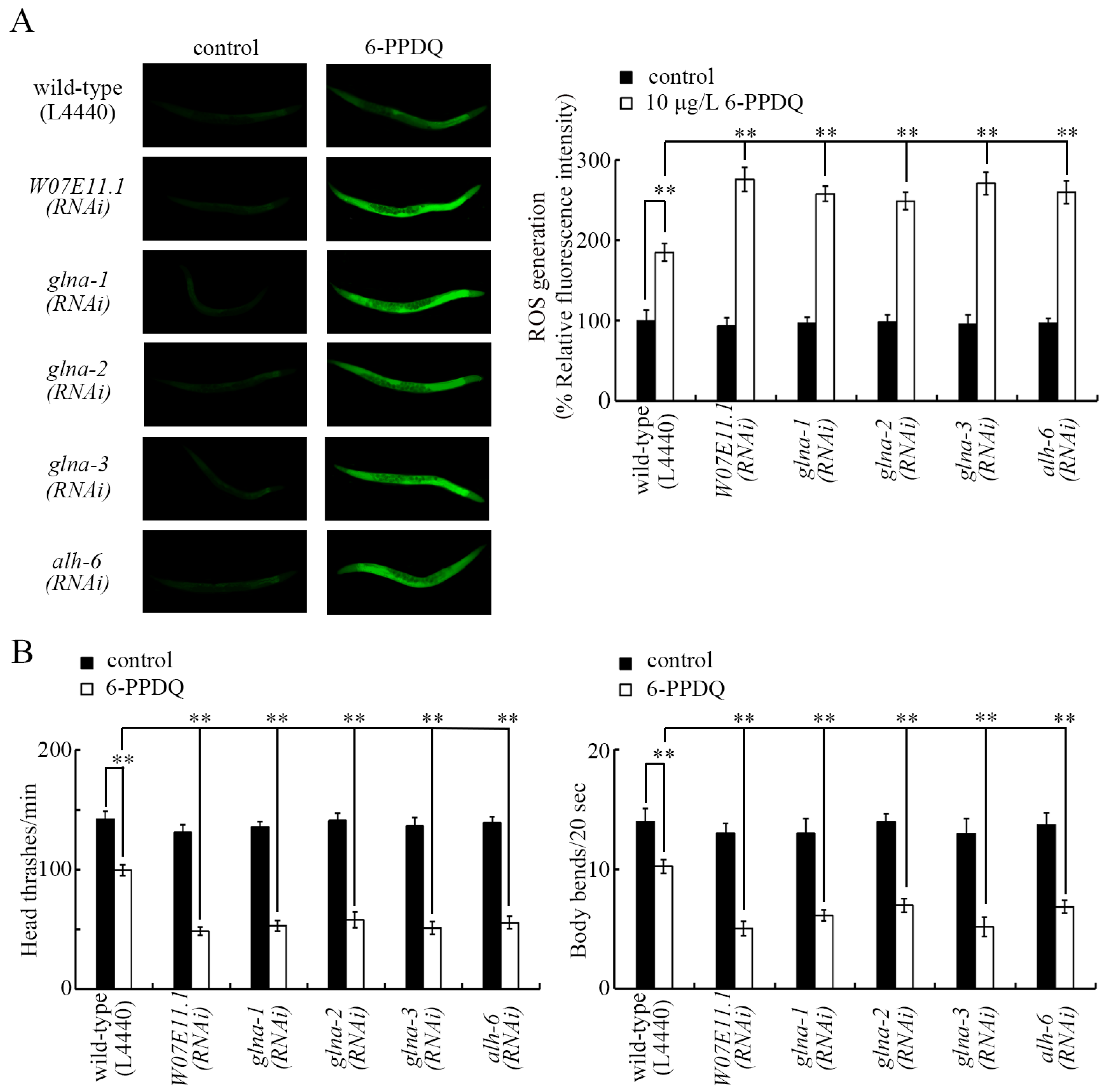
3.3. RNAI of W07E11.1, glna-1, glna-2, glna-3, and alh-6 Induced Susceptibility to 6-PPDQ Toxicity

We next investigated the association of an alteration in genes governing glutamate synthesis with 6-PPDQ toxicity induction. ROS generation and locomotion were used as endpoints. In 6-PPDQ-exposed nematodes, both ROS generation and decrease in locomotion were strengthened by W07E11.1, glna-1, glna-2, glna-3, and alh-6 RNAi (Figure 3A,B), suggesting susceptibility to 6-PPDQ in nematodes with RNAi of these genes.

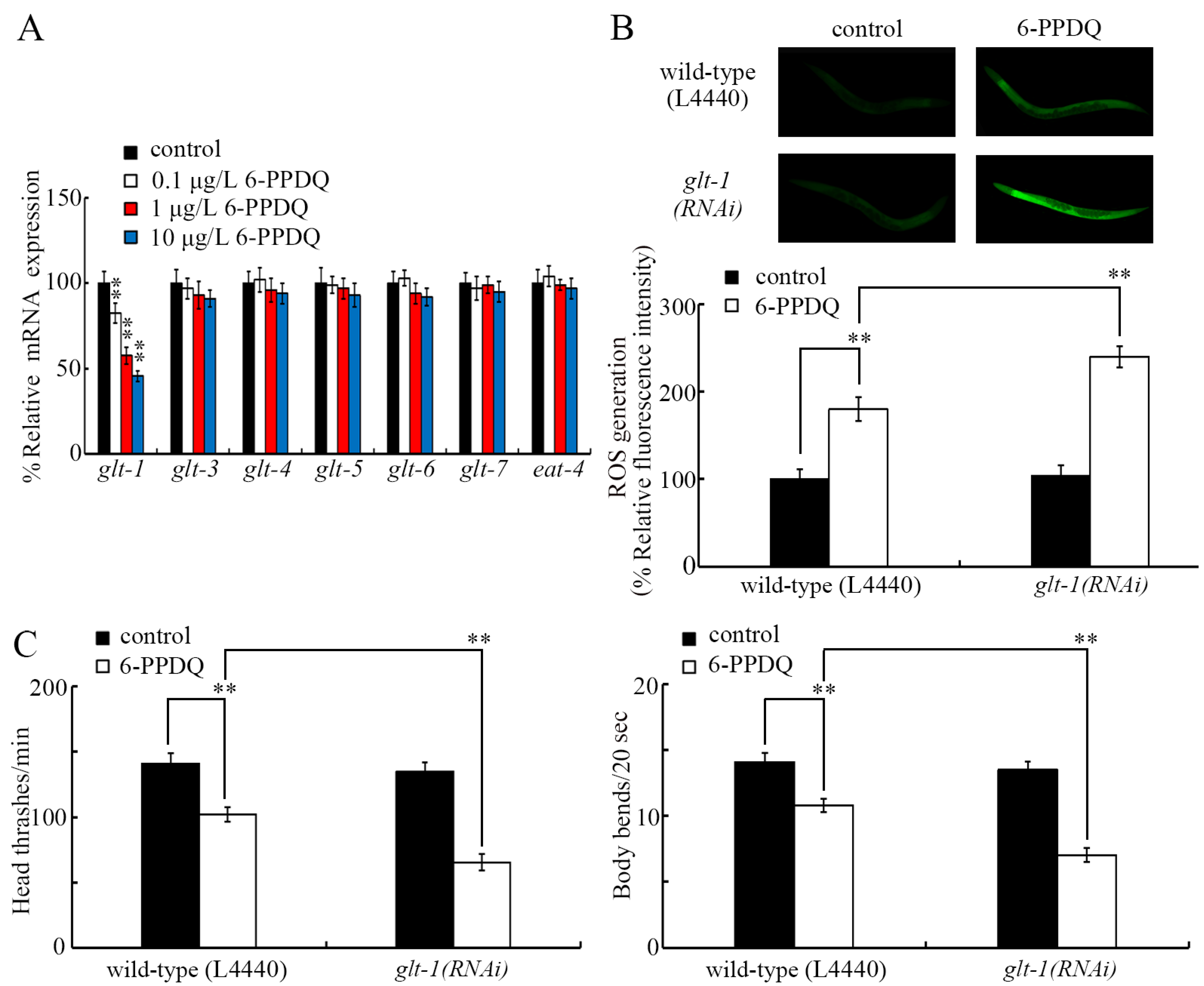
3.4. 6-PPDQ Decreased Expression of Glutamate Transporter Gene glt-1

Among the glutamate transporter genes, glt-1 expression was significantly decreased by 0.1–10 μg/L 6-PPDQ, but the expression of eat-4 and glt-3-7 was not changed by 0.1–10 μg/L 6-PPDQ (Figure 4A). RNAi of glt-1 resulted in susceptibility to 6-PPDQ toxicity (Figure 4B,C).

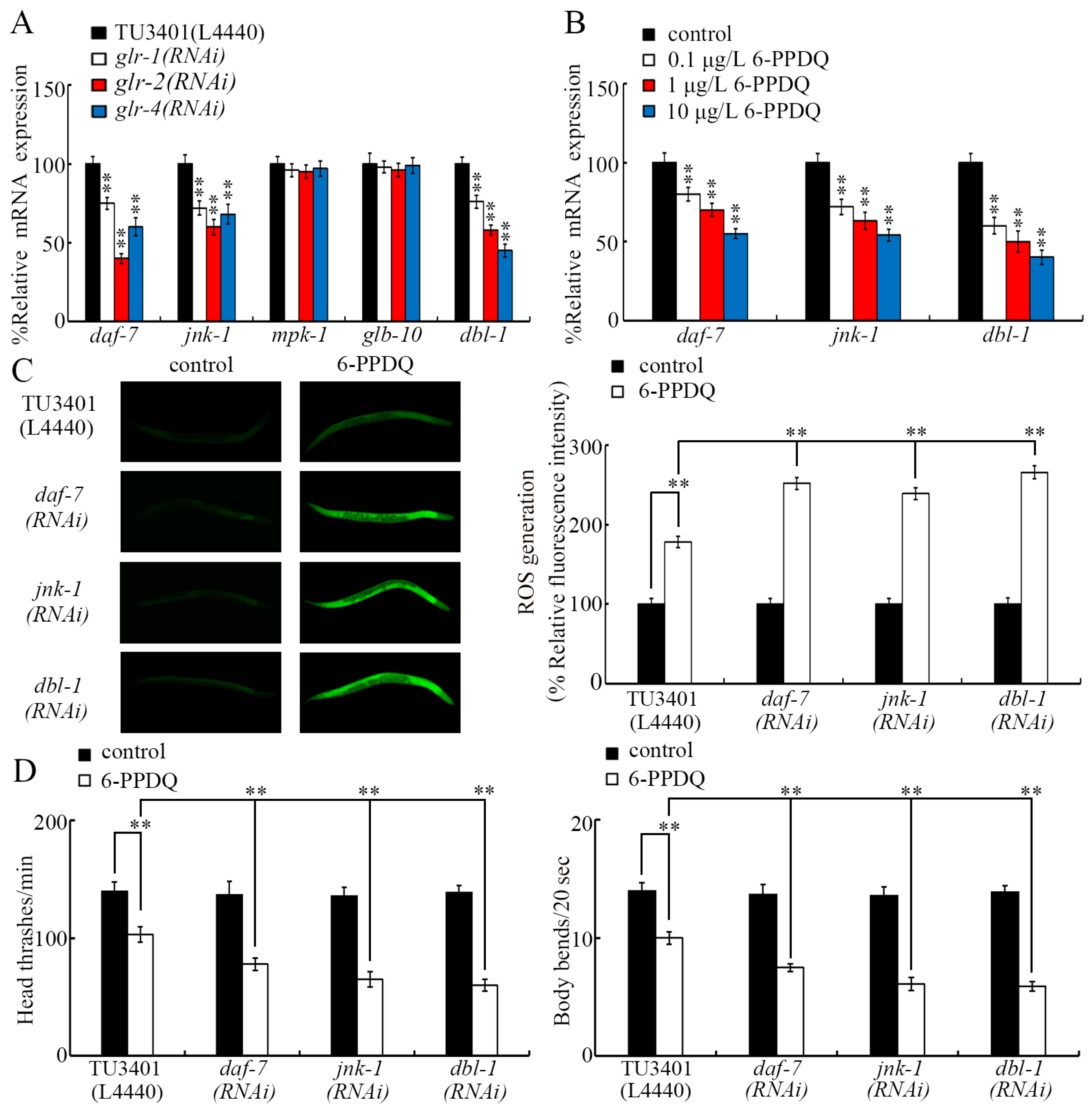
3.5. 6-PPDQ Decreased Expression of the Glutamate Receptor Genes glr-1, glr-2, and glr-4

Among the glutamate receptor genes, the expression of glr-1, glr-2, and glr-4 was inhibited by 0.1–10 μg/L 6-PPDQ, but the expression of glr-3, glr-5, glr-6, glr-7, and glr-8 was not altered by 0.1–10 μg/L 6-PPDQ (Figure 5A). After 6-PPDQ exposure, the expression of glr-1, glr-2, and glr-4 was suppressed by RNAi of genes governing glutamate synthesis (W07E11.1, glna-1/2/3, and alh-6), as well as RNAi of glt-1 (Figure 5B).
The ROS generation induced by 6-PPDQ was enhanced by glr-1, glr-2, and glr-4 RNAi (Figure 5C). Similarly, 6-PPDQ-induced locomotion inhibition was strengthened by glr-1, glr-2, and glr-4 RNAi (Figure 5D).

3.6. GLR-1, GLR-2, and GLR-4 Regulated 6-PPDQ Toxicity by Affecting DAF-7, JNK-1, and DBL-1

GLR-1, GLR-2, and GLR-4 are predominantly expressed in neurons (https://wormbase.org). Regarding pollutant toxicity, some molecular signals (DAF-7, JNK-1, MPK-1, GLB-10, and DBL-1) act in the neurons to carry out their functions [48]. After 6-PPDQ exposure, the expression of daf-7, jnk-1, and dbl-1 was suppressed by neuronal RNAi of glr-1, glr-2, and glr-4, but the expression of mpk-1 and glb-10 was not changed by neuronal RNAi of glr-1, glr-2, and glr-4 (Figure 6A). Meanwhile, 6-PPDQ (0.1–10 μg/L) decreased daf-7, jnk-1, and dbl-1 expression, and this decrease in their expression was concentration-dependent (Figure 6B). Moreover, the ROS generation and inhibition of locomotion induced by 6-PPDQ were strengthened by neuronal RNAi of daf-7, jnk-1, and dbl-1 (Figure 6C,D).

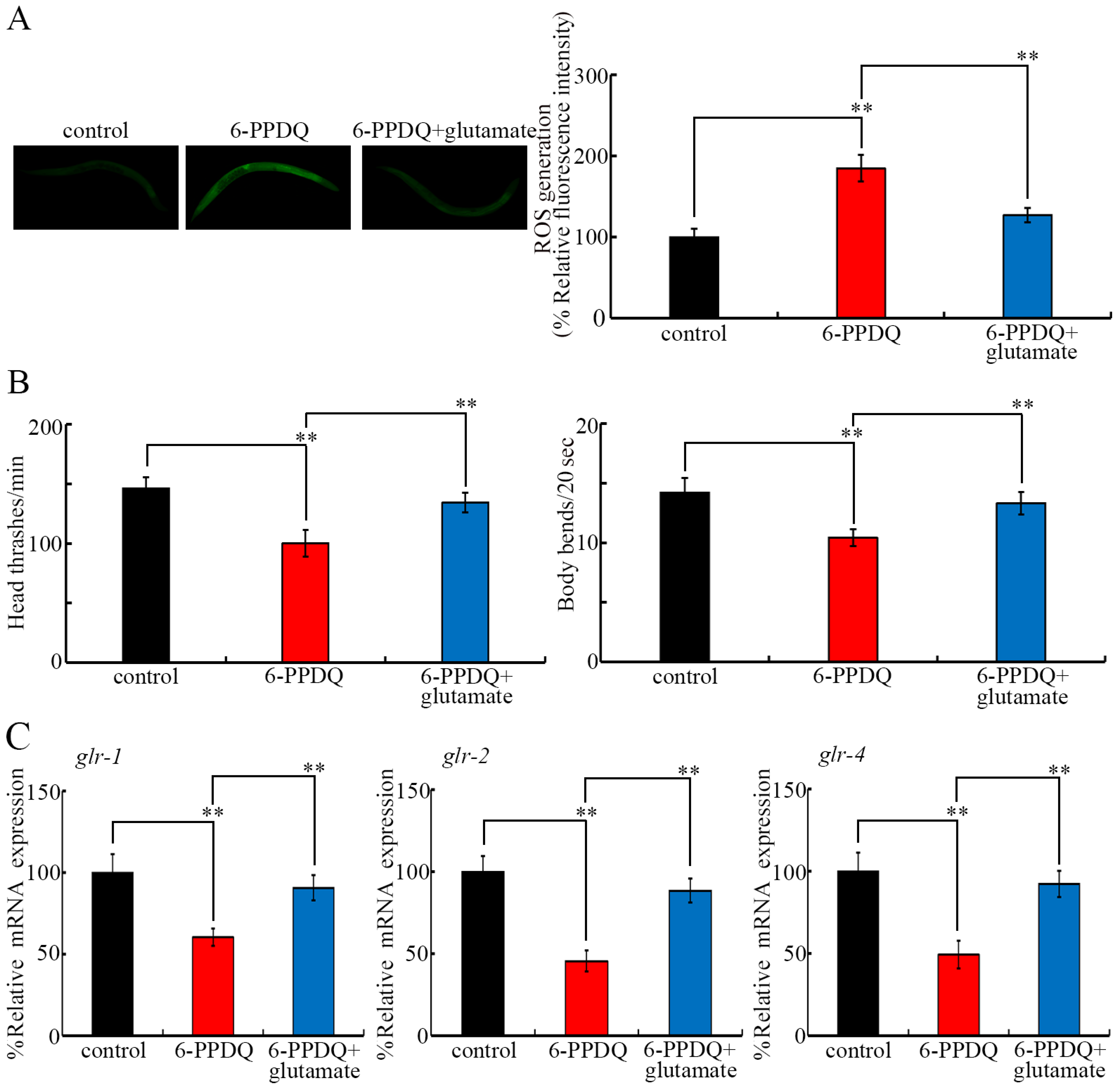
3.7. Pharmacological Effect of Glutamate Treatment on 6-PPDQ Toxicity

ROS generation induced by 10 μg/L 6-PPDQ was inhibited following 5 mM glutamate treatment (Figure 7A). Similarly, the decrease in locomotion induced by 10 μg/L 6-PPDQ was suppressed by treatment with 5 mM glutamate (Figure 7B). Moreover, the decrease in glr-1, glr-2, and glr-4 expression in 6-PPDQ-exposed nematodes was further inhibited by treatment with 5 mM glutamate (Figure 7C).

4. Discussion

C. elegans can be used to determine mechanisms of biochemical metabolism, such as glucose and lipid metabolism [27,49]. 6-PPDQ caused an accumulation of lipids, glucose, and glycogen in C. elegans [28,29,50]. Additionally, dopamine metabolism in C. elegans has also been shown to be disrupted by 6-PPDQ [51]. Glutamate content was further reduced by 0.1–10 μg/L 6-PPDQ (Figure 1B). Thus, 6-PPDQ at ERCs potentially disrupts the biochemical metabolism of multiple compounds.
In nematodes, W07E11.1 is predicted to govern glutamate synthesis from α-ketoglutarate, and its expression was decreased by 6-PPDQ (Figure 2A). GLNA-1/2/3 govern glutamate synthesis from glutamine [35], and their expression was also inhibited by 6-PPDQ (Figure 2A). PRDH-1 and ALH-6 are required for glutamate synthesis from proline [36], and only alh-6 expression was suppressed by 6-PPDQ (Figure 2A). After 6-PPDQ exposure, glutamate content was reduced by W07E1.1, glna-1/2/3, and alh-6 RNAi (Figure 2B), which confirmed the functions of these genes in controlling glutamate synthesis. Therefore, the decrease in expression of W07E1.1, glna-1/2/3, and alh-6 provided an important biochemical basis for the reduction in glutamate content induced by 6-PPDQ exposure.
Susceptibility to 6-PPDQ toxicity could be caused by RNAi of W07E1.1, glna-1/2/3, and alh-6 (Figure 3). This observation suggests that a decrease in the expression of genes of enzymes governing glutamate synthesis from α-ketoglutarate, glutamine, and proline contributes to 6-PPDQ toxicity induction. A mutation in alh-6 could cause premature reproductive senescence [52], and ALH-6 could be considered a biomarker of age-related muscle changes [53].
Among the glutamate transporter genes, only glt-1 expression showed sensitivity to 6-PPDQ exposure. The glt-1 expression was decreased by 0.1–10 μg/L 6-PPDQ (Figure 4A). Meanwhile, susceptibility to 6-PPDQ toxicity in causing ROS generation and locomotion inhibition was caused by glt-1 RNAi (Figure 4B,C). Similar to the function of the identified genes governing glutamate synthesis, this suggests that the inhibition of GLT-1 can also mediate 6-PPDQ toxicity.
Among eight glutamate receptor genes, three (glr-1, glr-2, and glr-4) were found to be involved in the control of 6-PPDQ toxicity. RNAi of these three glutamate receptor genes caused susceptibility to 6-PPDQ toxicity (Figure 5C,D). Meanwhile, the expression of these three glutamate receptor genes was inhibited by 6-PPDQ (Figure 5A). These observations suggest that the inhibition of these three glutamate receptors mediates the toxicity induction of 6-PPDQ. The expression of the other five glutamate receptor genes was not altered by 6-PPDQ (Figure 5A), implying that the expression of these receptor genes may not be sensitive to 6-PPDQ exposure. RNAi of glr-4 can cause susceptibility to nanoplastic toxicity [54]. The behavioral response of C. elegans to Al2O3-NPs exposure has been shown to be mediated by GLR-2 [40]. In nematodes, glr-1 expression has also been shown to be decreased by lindane exposure [55]. Moreover, after 6-PPDQ exposure, we found that the expression of glr-1, glr-2, and glr-4 was decreased by RNAi of W07E11.1, glna-1/2/3, alh-6, and glt-1 (Figure 5B), suggesting that a decrease in the expression of genes governing glutamate synthesis and transport can cause the further inhibition of receptors after 6-PPDQ exposure. These receptors are predominantly expressed in neurons (https://wormbase.org). In addition to neurons, W07E11.1, GLNA-1/2/3, ALH-6, and GLT-1 are also expressed in the intestine, gonads, muscle, and/or epidermis (https://wormbase.org), which suggests that the glutamate is synthesized and further transported from different tissues to the neuron to activate the GLR-1, GLR-2, and GLR-4 receptors, thereby exerting its effects.
Considering that these glutamate receptors are predominantly expressed in the neurons of C. elegans, we identified targets of these three glutamate receptors in the neurons to control 6-PPDQ toxicity. DAF-7, JNK-1, and DBL-1 were identified as targets for neuronal GLR-1, GLR-2, and GLR-4 in controlling 6-PPDQ toxicity. Three aspects of evidence were raised to support this. Firstly, the expression of daf-7, jnk-1, and dbl-1 was decreased by neuronal RNAi of glr-1, glr-2, and glr-4 in 6-PPDQ-exposed nematodes (Figure 6A). Secondly, the expression of daf-7, jnk-1, and dbl-1 was decreased by 6-PPDQ (Figure 6B). Thirdly, neuronal RNAi of daf-7, jnk-1, and dbl-1 caused susceptibility to 6-PPDQ (Figure 6C,D). DAF-7 and DBL-1 are two TGF-β ligands [56,57]. JNK-1 is a JNK MAPK [54]. RNAi of daf-7, jnk-1, and dbl-1 has also been shown to result in susceptibility to the toxicity of other pollutants, such as nanoplastics and CDDP quinone [48,58,59]. In nematodes, it remains unclear whether GLR-1, GLR-2, and GLR-4 regulate 6-PPDQ toxicity by activating or inhibiting other downstream targets; thus, this needs further determination.
In the current study, ROS generation and locomotion inhibition, which are two aspects of toxicity induced by 6-PPDQ, were suppressed following glutamate treatment (Figure 7A,B). Similarly, glutamate treatment inhibited the toxicity of heavy metals such as cadmium [30]. These observations suggest that glutamate treatment can potentially be used as a pharmacological intervention against damage caused by 6-PPDQ. Nevertheless, more studies are needed to further confirm this interventional strategy in mammals. Moreover, the 6-PPDQ-induced inhibition of the expression of glutamate receptor genes was suppressed following glutamate treatment (Figure 7C). This further supports the role of glutamate in inhibiting 6-PPDQ toxicity by activating these glutamate receptor genes. After 6-PPDQ exposure, glutamate synthesis was suppressed, and the resulting reduction in glutamate content may lead to the inhibition of glutamate receptors, which, in turn, causes susceptibility to 6-PPDQ toxicity.

5. Conclusions

In conclusion, the reduction in glutamate content by 6-PPDQ was observed in nematodes, which was due to the inhibition of expression of genes governing glutamate synthesis from α-ketoglutarate, glutamine, and proline. The expression of glutamate transporter gene glt-1 and receptor genes (glr-1, glr-2, and glr-4) was also decreased by 6-PPDQ, and susceptibility to 6-PPDQ toxicity was observed in nematodes after RNAi of genes governing glutamate synthesis, transporter gene glt-1, and receptor genes. Some molecular signals (DAF-7, JNK-1, and DBL-1) were identified as downstream targets of neuronal glutamate receptors (GLR-1, GLR-2, and GLR-4) to control 6-PPDQ toxicity. Pharmacological treatment with glutamate further supported the role of glutamate in modulating 6-PPDQ toxicity by affecting corresponding receptor genes. Nevertheless, considering the simple developmental structure of C. elegans, additional studies in mammals still need to be carried out.

Supplementary Materials

The following supporting information can be downloaded at: https://www.mdpi.com/article/10.3390/toxics13060434/s1. Figure S1: RNAi efficiency of W07E11.1, glna-1, glna-2, glna-3, glt-1, glr-1, glr-2, and glr-4. ** p < 0.01 vs. wild-type (L4440); Table S1. Primer information for qRT-PCR.

Author Contributions

Investigation and writing of the draft, W.W.; supervision and review of the draft, Y.L. and D.W. All authors have read and agreed to the published version of the manuscript.

Funding

This research received no external funding.

Institutional Review Board Statement

Not applicable.

Informed Consent Statement

Not applicable.

Data Availability Statement

The original data presented in this study are included in the article/Supplementary Materials; further inquiries can be directed to the corresponding author.

Conflicts of Interest

The authors declare that they have no known competing financial interests or personal relationships that could appear to have influenced the work reported in this paper.

References

  1. Huntink, N.M.; Datta, R.N.; Noordermeer, J.W.M. Addressing durability of rubber compounds. Rubber Chem. Technol. 2004, 77, 476–511. [Google Scholar] [CrossRef]
  2. Hua, X.; Wang, D.-Y. Tire-rubber related pollutant 6-PPD quinone: A review of its transformation, environmental distribution, bioavailability, and toxicity. J. Hazard. Mater. 2023, 459, 132265. [Google Scholar] [CrossRef] [PubMed]
  3. Chen, X.; He, T.; Yang, X.; Gan, Y.; Qing, X.; Wang, J.; Huang, Y. Analysis, environmental occurrence, fate and potential toxicity of tire wear compounds 6PPD and 6PPD-quinone. J. Hazard. Mater. 2023, 452, 131245. [Google Scholar] [CrossRef]
  4. Wan, X.; Liang, G.-Y.; Wang, D.-Y. Potential human health risk of the emerging environmental contaminant 6-PPD quinone. Sci. Total Environ. 2024, 949, 175057. [Google Scholar] [CrossRef] [PubMed]
  5. Tian, Z.; Zhao, H.; Peter, K.T.; Gonzalez, M.; Wetzel, J.; Wu, C.; Hu, X.; Prat, J.; Mudrock, E.; Hettinger, R.; et al. A ubiquitous tire rubber–derived chemical induces acute mortality in coho salmon. Science 2021, 371, 185–189. [Google Scholar] [CrossRef]
  6. Liang, Y.; Zhu, F.; Li, J.; Wan, X.; Ge, Y.; Liang, G.; Zhou, Y. P-phenylenediamine antioxidants and their quinone derivatives: A review of their environmental occurrence, accessibility, potential toxicity, and human exposure. Sci. Total Environ. 2024, 948, 174449. [Google Scholar] [CrossRef]
  7. Kazmi, S.S.U.H.; Xu, Q.; Tayyab, M.; Pastorino, P.; Barcelò, D.; Yaseen, Z.M.; Khan, Z.H.; Li, G. Navigating the environmental dynamics, toxicity to aquatic organisms and human associated risks of an emerging tire wear contaminant 6PPD-quinone. Environ. Pollut. 2024, 356, 124313. [Google Scholar] [CrossRef]
  8. Zhang, Y.Y.; Huang, J.W.; Liu, Y.H.; Zhang, J.N.; Huang, Z.; Liu, Y.S.; Zhao, J.L.; Ying, G.G. In vitro metabolism of the emerging contaminant 6PPD-quinone in human and rat liver microsomes: Kinetics, pathways, and mechanism. Environ. Pollut. 2024, 345, 123514. [Google Scholar] [CrossRef]
  9. He, W.-M.; Chao, J.; Gu, A.-H.; Wang, D.-Y. Evaluation of 6-PPD quinone toxicity on lung of male BALB/c mice by quantitative proteomics. Sci. Total Environ. 2024, 922, 171220. [Google Scholar] [CrossRef]
  10. Yao, K.; Kang, Q.; Liu, W.; Chen, D.; Wang, L.; Li, S. Chronic exposure to tire rubber-derived contaminant 6PPD-quinone impairs sperm quality and induces the damage of reproductive capacity in male mice. J. Hazard. Mater. 2024, 470, 134165. [Google Scholar] [CrossRef]
  11. Fang, J.; Wang, X.; Cao, G.; Wang, F.; Ru, Y.; Wang, B.; Zhang, Y.; Zhang, D.; Yan, J.; Xu, J.; et al. 6PPD-quinone exposure induces neuronal mitochondrial dysfunction to exacerbate Lewy neurites formation induced by alpha-synuclein preformed fibrils seeding. J. Hazard. Mater. 2024, 465, 133312. [Google Scholar] [CrossRef] [PubMed]
  12. Jiang, Y.; Wang, C.; Ma, L.; Gao, T.; Wang, Y. Environmental profiles, hazard identification, and toxicological hallmarks of emerging tire rubber-related contaminants 6PPD and 6PPD-quinone. Environ. Int. 2024, 187, 108677. [Google Scholar] [CrossRef] [PubMed]
  13. Deng, M.; Ji, X.; Peng, B.; Fang, M. In vitro and in vivo biotransformation profiling of 6PPD-quinone toward their detection in human urine. Environ. Sci. Technol. 2024, 58, 9113–9124. [Google Scholar] [CrossRef] [PubMed]
  14. Wang, D.-Y. Exposure Toxicology in Caenorhabditis elegans; Springer Nature Pte Ltd.: Singapore, 2020. [Google Scholar]
  15. Li, H.; Zeng, L.; Wang, C.; Shi, C.; Li, Y.; Peng, Y.; Chen, H.; Zhang, J.; Cheng, B.; Chen, C.; et al. Review of the toxicity and potential molecular mechanisms of parental or successive exposure to environmental pollutants in the model organism Caenorhabditis elegans. Environ. Pollut. 2022, 311, 119927. [Google Scholar] [CrossRef]
  16. Wang, Y.-X.; Wang, D.-Y. Effect of disruption in intestinal barrier function during transgenerational process on nanoplastic toxicity induction in Caenorhabditis elegans. Environ. Sci. Nano 2025, 12, 2741–2749. [Google Scholar] [CrossRef]
  17. Shao, Y.-T.; Li, Y.-H.; Wang, D.-Y. Polylactic acid microplastics cause transgenerational reproductive toxicity associated with activation of insulin and hedgehog ligands in C. elegans. Sci. Total Environ. 2024, 942, 173746. [Google Scholar] [CrossRef]
  18. Wu, J.-W.; Shao, Y.-T.; Hua, X.; Wang, Y.-X.; Wang, D.-Y. Nanoplastic at environmentally relevant concentrations induces toxicity across multiple generations associated with inhibition in germline G protein-coupled receptor CED-1 in Caenorhabditis elegans. Chemosphere 2024, 364, 143011. [Google Scholar] [CrossRef]
  19. Song, M.-X.; Ruan, Q.-L.; Wang, D.-Y. Comparison of transgenerational toxicity on locomotion behavior and development of D-type motor neurons between pristine and amino modified nanoplastics at environmentally relevant doses in C. elegans. Toxics 2024, 12, 555. [Google Scholar]
  20. Liu, Z.-Y.; Wang, Y.-X.; Bian, Q.; Wang, D.-Y. Transgenerational response of germline nuclear hormone receptor genes to nanoplastics at predicted environmental doses in Caenorhabditis elegans. Toxics 2024, 12, 420. [Google Scholar] [CrossRef]
  21. Wang, Y.-X.; Wang, D.-Y. Transgenerational intestinal toxicity of 6-PPD quinone in causing ROS production, enhancement in intestinal permeability and suppression in innate immunity in C. elegans. Environ. Pollut. 2024, 363, 125208. [Google Scholar] [CrossRef]
  22. Song, M.-X.; Ruan, Q.-L.; Wang, D.-Y. Paeoniflorin alleviates toxicity and accumulation of 6-PPD quinone by activating ACS-22 in Caenorhabditis elegans. Ecotoxicol. Environ. Saf. 2024, 286, 117226. [Google Scholar] [CrossRef] [PubMed]
  23. Hua, X.; Wang, D.-Y. 6-PPD quinone at environmentally relevant concentrations induced damage on longevity in C. elegans: Mechanistic insight from inhibition in mitochondrial UPR response. Sci. Total Environ. 2024, 954, 176275. [Google Scholar] [CrossRef] [PubMed]
  24. Wu, J.-W.; Shen, S.-H.; Wang, D.-Y. 6-PPD quinone at environmentally relevant concentrations induces immunosenescenece by causing immunosuppression during the aging process. Chemosphere 2024, 368, 143719. [Google Scholar] [CrossRef] [PubMed]
  25. Hua, X.; Liang, G.-Y.; Chao, J.; Wang, D.-Y. Exposure to 6-PPD quinone causes damage on mitochondrial complex I/II associated with lifespan reduction in Caenorhabditis elegans. J. Hazard. Mater. 2024, 472, 134598. [Google Scholar] [CrossRef]
  26. Hua, X.; Wang, D.-Y. 6-PPD quinone causes alteration in ubiquinone-mediated complex III associated with toxicity on mitochondrial function and longevity in Caenorhabditis elegans. J. Environ. Chem. Eng. 2025, 13, 116571. [Google Scholar] [CrossRef]
  27. Watts, J.L.; Ristow, M. Lipid and carbohydrate metabolism in Caenorhabditis elegans. Genetics 2017, 207, 413–446. [Google Scholar]
  28. Wang, Y.-X.; Wang, D.-Y. Exposure to 6-PPD quinone enhances glycogen accumulation in Caenorhabditis elegans. Environ. Pollut. 2024, 359, 124600. [Google Scholar] [CrossRef]
  29. Liu, Z.-J.; Li, Y.-H.; Wang, D.-Y. Exposure to 6-PPD quinone disrupts glucose metabolism associated with lifespan reduction by affecting insulin and AMPK signals in Caenorhabditis elegans. Chemosphere 2024, 363, 142975. [Google Scholar] [CrossRef]
  30. Wang, Y.-X.; Wu, J.-W.; Wang, D.-Y. 6-PPD quinone causes lipid accumulation across multiple generations differentially affected by metabolic sensors and components of COMPASS complex in Caenorhabditis elegans. Environ. Pollut. 2025, 366, 125539. [Google Scholar] [CrossRef]
  31. Liu, Z.-J.; Li, Y.-H.; Wang, D.-Y. Transgenerational response of glucose metabolism in Caenorhabditis elegans exposed to 6-PPD quinone. Chemosphere 2024, 367, 143653. [Google Scholar] [CrossRef]
  32. Monné, M.; Vozza, A.; Lasorsa, F.M.; Porcelli, V.; Palmieri, F. Mitochondrial carriers for aspartate, glutamate and other amino acids: A review. Int. J. Mol. Sci. 2019, 20, 4456. [Google Scholar] [CrossRef] [PubMed]
  33. Chen, Q.Y.; Zhuo, M. Glutamate acts as a key neurotransmitter for itch in the mammalian spinal cord. Mol. Pain 2023, 19, 17448069231152101. [Google Scholar] [CrossRef] [PubMed]
  34. Jiang, M.; Jiang, J.; Li, S.; Li, M.; Tan, Y.; Song, S.; Shu, Q.; Huang, J. Glutamate alleviates cadmium toxicity in rice via suppressing cadmium uptake and translocation. J. Hazard. Mater. 2020, 384, 121319. [Google Scholar] [CrossRef] [PubMed]
  35. Liang, Q.; Zhao, G. The effect of glna loss on the physiological and pathological phenotype of Parkinson‘s disease C. elegans. J. Clin. Lab. Anal. 2024, 38, e25129. [Google Scholar] [CrossRef]
  36. Pang, S.; Curran, S.P. Adaptive capacity to bacterial diet modulates aging in C. elegans. Cell Metab. 2014, 19, 221–231. [Google Scholar] [CrossRef]
  37. Mano, I.; Straud, S.; Driscoll, M. Caenorhabditis elegans glutamate transporters influence synaptic function and behavior at sites distant from the synapse. J. Biol. Chem. 2007, 282, 34412–34419. [Google Scholar] [CrossRef]
  38. da Silveira, T.L.; Machado, M.L.; Arantes, L.P.; Zamberlan, D.C.; Cordeiro, L.M.; Obetine, F.B.B.; da Silva, A.F.; Tassi, C.L.; Soares, F.A.A. Guanosine prevents against glutamatergic excitotoxicity in C. elegans. Neuroscience 2019, 414, 265–272. [Google Scholar] [CrossRef]
  39. Bronstone, G.J.; Harton, M.; Muldowney, M.; Reigle, J.; Funk, A.J.; O‘Donovan, S.M.; McCullumsmith, R.E.; Bauer, D.E. The C. elegans glutamate transporters GLT-4 and GLT-5 regulate protein expression, behavior, and lifespan. Neurochem. Int. 2025, 186, 105966. [Google Scholar] [CrossRef]
  40. Li, Y.; Yu, S.; Wu, Q.; Tang, M.; Wang, D. Transmissions of serotonin, dopamine, and glutamate are required for the formation of neurotoxicity from Al2O3-NPs in nematode Caenorhabditis elegans. Nanotoxicology 2013, 7, 1004–1013. [Google Scholar] [CrossRef]
  41. Brenner, S. The genetics of Caenorhabditis elegans. Genetics 1974, 77, 71–94. [Google Scholar] [CrossRef]
  42. Yuan, C.-Y.; Wang, Y.-X.; Zhang, L.; Wang, D.-Y. Procatechuic acid and protocatechuic aldehyde increase survival of Caenorhabditis elegans after fungal infection and inhibit fungal virulence. Front. Pharmacol. 2024, 15, 1396733. [Google Scholar] [CrossRef]
  43. Wu, J.-W.; Shao, Y.-T.; Hua, X.; Wang, D.-Y. Activated hedgehog and insulin ligands by decreased transcriptional factor DAF-16 mediate transgenerational nanoplastic toxicity in Caenorhabditis elegans. J. Hazard. Mater. 2024, 480, 135909. [Google Scholar] [CrossRef]
  44. Hua, X.; Zhang, L.; Wang, D.-Y. Nanoplastic at environmentally relevant concentrations activates germline mir-240-rab-5 signaling cascade to affect secreted ligands associated with transgenerational toxicity induction in C. elegans. Environ. Sci. Nano 2024, 11, 3615. [Google Scholar] [CrossRef]
  45. Hua, X.; Wang, D.-Y. Transgenerational response of germline histone acetyltransferases and deacetylases to nanoplastics at predicted environmental doses in Caenorhabditis elegans. Sci. Total Environ. 2024, 952, 175903. [Google Scholar] [CrossRef]
  46. Wu, T.-S.; He, J.; Ye, Z.-J.; Xia, J.-Y.; Chen, M.; Chen, S.-Y.; Liu, K.-H.; Xing, P.-C.; Yang, J.-F.; Qian, Y.-J.; et al. Aged biodegradable nanoplastics enhance body accumulation associated with worse neuronal damage in Caenorhabditis elegans. Environ. Sci. Technol. 2025, 59, 4352–4363. [Google Scholar] [CrossRef]
  47. Edwards, C.; Canfield, J.; Copes, N.; Brito, A.; Rehan, M.; Lipps, D.; Brunquell, J.; Westerheide, S.D.; Bradshaw, P.C. Mechanisms of amino acid-mediated lifespan extension in Caenorhabditis elegans. BMC Genet. 2015, 16, 8. [Google Scholar] [CrossRef]
  48. Wang, D.-Y. Toxicology at Environmentally Relevant Concentrations in Caenorhabditis elegans; Springer Nature Pte Ltd.: Singapore, 2022. [Google Scholar]
  49. An, L.; Fu, X.; Chen, J.; Ma, J. Application of Caenorhabditis elegans in lipid metabolism research. Int. J. Mol. Sci. 2023, 24, 1173. [Google Scholar] [CrossRef]
  50. Wang, Y.-X.; Hua, X.; Wang, D.-Y. Exposure to 6-PPD quinone enhances lipid accumulation through activating metabolic sensors of SBP-1 and MDT-15 in Caenorhabditis elegans. Environ. Pollut. 2023, 333, 121937. [Google Scholar] [CrossRef]
  51. Hua, X.; Wang, D.-Y. Disruption of dopamine metabolism by exposure to 6-PPD quinone in Caenorhabditis elegans. Environ. Pollut. 2023, 337, 122649. [Google Scholar] [CrossRef]
  52. Yen, C.A.; Curran, S.P. Incomplete proline catabolism drives premature sperm aging. Aging Cell 2021, 20, e13308. [Google Scholar] [CrossRef]
  53. Villa, O.; Stuhr, N.L.; Yen, C.A.; Crimmins, E.M.; Arpawong, T.E.; Curran, S.P. Genetic variation in ALDH4A1 is associated with muscle health over the lifespan and across species. Elife 2022, 11, e74308. [Google Scholar] [CrossRef] [PubMed]
  54. Wang, S.; Liu, H.; Qu, M.; Wang, D. Response of tyramine and glutamate related signals to nanoplastic exposure in Caenorhabditis elegans. Ecotoxicol. Environ. Saf. 2021, 217, 112239. [Google Scholar] [CrossRef] [PubMed]
  55. Yu, Y.; Hua, X.; Chen, H.; Wang, Z.; Han, Y.; Chen, X.; Yang, Y.; Xiang, M. Glutamatergic transmission associated with locomotion-related neurotoxicity to lindane over generations in Caenorhabditis elegans. Chemosphere 2022, 290, 133360. [Google Scholar] [CrossRef]
  56. Ren, P.; Lim, C.S.; Johnsen, R.; Albert, P.S.; Pilgrim, D.; Riddle, D.L. Control of C. elegans larval development by neuronal expression of a TGF-beta homolog. Science 1996, 274, 1389–1391. [Google Scholar] [CrossRef]
  57. Suzuki, Y.; Yandell, M.D.; Roy, P.J.; Krishna, S.; Savage-Dunn, C.; Ross, R.M.; Padgett, R.W.; Wood, W.B. A BMP homolog acts as a dose-dependent regulator of body size and male tail patterning in Caenorhabditis elegans. Development 1999, 126, 241–250. [Google Scholar] [CrossRef]
  58. Kawasaki, M.; Hisamoto, N.; Iino, Y.; Yamamoto, M.; Ninomiya-Tsuji, J.; Matsumoto, K. A Caenorhabditis elegans JNK signal transduction pathway regulates coordinated movement via type-D GABAergic motor neurons. EMBO J. 1999, 18, 3604–3615. [Google Scholar] [CrossRef]
  59. Wan, X.; Liang, G.; Wang, D. Neurotoxicity and accumulation of CPPD quinone at environmentally relevant concentrations in Caenorhabditis elegans. Chemosphere 2024, 361, 142499. [Google Scholar] [CrossRef]
Figure 1. Effects of 6-PPDQ exposure on glutamate content. (A) Diagram showing biochemical basis for synthesis of glutamate in nematodes. (B) Effect of 6-PPDQ exposure on glutamate content. ** p < 0.01 vs. control.
Toxics 13 00434 g001
Figure 2. Effect of 6-PPDQ exposure on glutamate synthesis. (A) Effect of 6-PPDQ exposure on expression of genes governing glutamate synthesis. ** p < 0.01 vs. control. (B) Effect of RNAi of W07E11.1, glna-1, glna-2, glna-3, and alh-6 on glutamate content in 6-PPDQ-exposed nematodes. Exposure concentration of 6-PPDQ was 10 μg/L. ** p < 0.01.
Toxics 13 00434 g002
Figure 3. Effect of RNAi of W07E11.1, glna-1, glna-2, glna-3, and alh-6 on 6-PPDQ toxicity in causing ROS generation (A) and decrease in locomotion (B). Exposure concentration of 6-PPDQ was 10 μg/L. ** p < 0.01.
Toxics 13 00434 g003
Figure 4. Effect of 6-PPDQ exposure on expression of glutamate transporter genes. (A) Effect of 6-PPDQ exposure on expression of eat-4, glt-1, glt-3, glt-4, glt-5, glt-6, and glt-7. ** p < 0.01 vs. control. (B) Effect of RNAi of glt-1 on 6-PPDQ toxicity in causing ROS generation. Exposure concentration of 6-PPDQ was 10 μg/L. ** p < 0.01. (C) Effect of RNAi of glt-1 on 6-PPDQ toxicity in decreasing locomotion. Exposure concentration of 6-PPDQ was 10 μg/L. ** p < 0.01.
Toxics 13 00434 g004
Figure 5. Effect of 6-PPDQ exposure on expression of glutamate receptor genes. (A) Effect of 6-PPDQ exposure on expression of glr-1, glr-2, glr-3, glr-4, glr-5, glr-6, glr-7, and glr-8. ** p < 0.01 vs. control. (B) Effect of RNAi of W07E11.1, glna-1, glna-2, glna-3, alh-6, and glt-1 on expression of glr-1, glr-2, and glr-4 in 6-PPDQ-exposed nematodes. Exposure concentration of 6-PPDQ was 10 μg/L. ** p < 0.01 vs. wild-type (L4440). (C) Effect of RNAi of glr-1, glr-2, and glr-4 on 6-PPDQ toxicity in causing ROS generation. Exposure concentration of 6-PPDQ was 10 μg/L. ** p < 0.01. (D) Effect of RNAi of glr-1, glr-2, and glr-4 on 6-PPDQ toxicity in decreasing locomotion. Exposure concentration of 6-PPDQ was 10 μg/L. ** p < 0.01.
Toxics 13 00434 g005
Figure 6. Neuronal RNAi of glr-1, glr-2, and glr-4 affected expression of daf-7, jnk-1, and dbl-1 in 6-PPDQ-exposed nematodes. (A) Effect of neuronal RNAi of glr-1, glr-2, and glr-4 on expression of daf-7, jnk-1, mpk-1, glb-10, and dbl-1 in 6-PPDQ-exposed nematodes. Exposure concentration of 6-PPDQ was 10 μg/L. ** p < 0.01 vs. TU3401 (L4440). (B) Effect of 6-PPDQ exposure on expression of daf-7, jnk-1, and dbl-1. ** p < 0.01 vs. control. (C) Effect of neuronal RNAi of daf-7, jnk-1, and dbl-1 on 6-PPDQ toxicity in causing ROS generation. Exposure concentration of 6-PPDQ was 10 μg/L. ** p < 0.01. (D) Effect of neuronal RNAi of daf-7, jnk-1, and dbl-1 on 6-PPDQ toxicity in decreasing locomotion. Exposure concentration of 6-PPDQ was 10 μg/L. ** p < 0.01.
Toxics 13 00434 g006
Figure 7. Effect of glutamate (5 mM) treatment on toxicity induced by 6-PPDQ (10 μg/L). (A) Effect of glutamate treatment on ROS generation in 6-PPDQ-exposed nematodes. (B) Effect of glutamate treatment on locomotion in 6-PPDQ-exposed nematodes. (C) Effect of glutamate treatment on expression of glr-1, glr-2, and glr-4 in 6-PPDQ-exposed nematodes. ** p < 0.01.
Toxics 13 00434 g007
Disclaimer/Publisher’s Note: The statements, opinions and data contained in all publications are solely those of the individual author(s) and contributor(s) and not of MDPI and/or the editor(s). MDPI and/or the editor(s) disclaim responsibility for any injury to people or property resulting from any ideas, methods, instructions or products referred to in the content.

