
Ecotoxicity of Polyvinylidene Difluoride (PVDF) and Polylactic Acid (PLA) Microplastics in Marine Zooplankton


 ,
,
Abstract
:1. Introduction
2. Materials and Methods
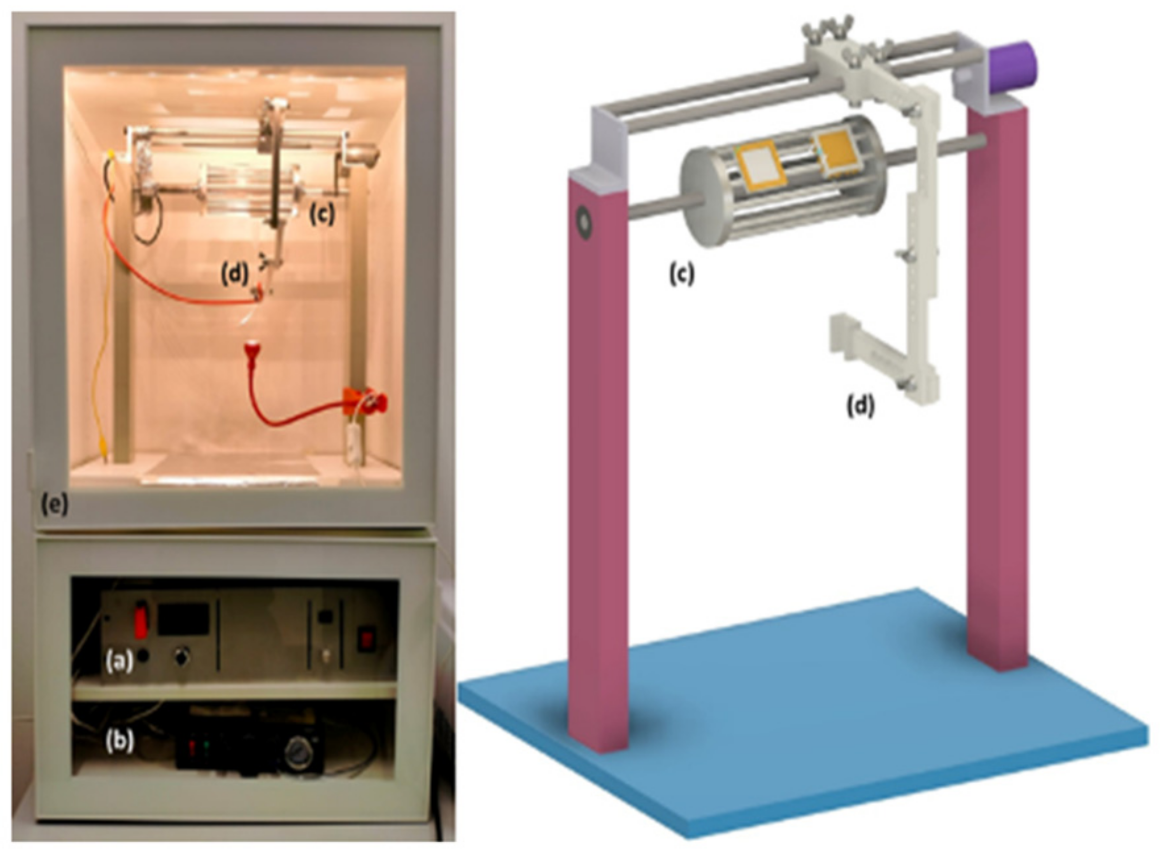
2.1. Plastic Materials and Grinding Process
2.1.1. Morphological Characterization
2.1.2. Material Staining with Nile Red
2.2. Organisms
2.2.1. Artemia franciscana
2.2.2. Aurelia sp.
2.3. PVDF and PLA Uptake
2.4. Toxicity Tests
3. Results
3.1. Material Characterization
3.2. MPs Uptake
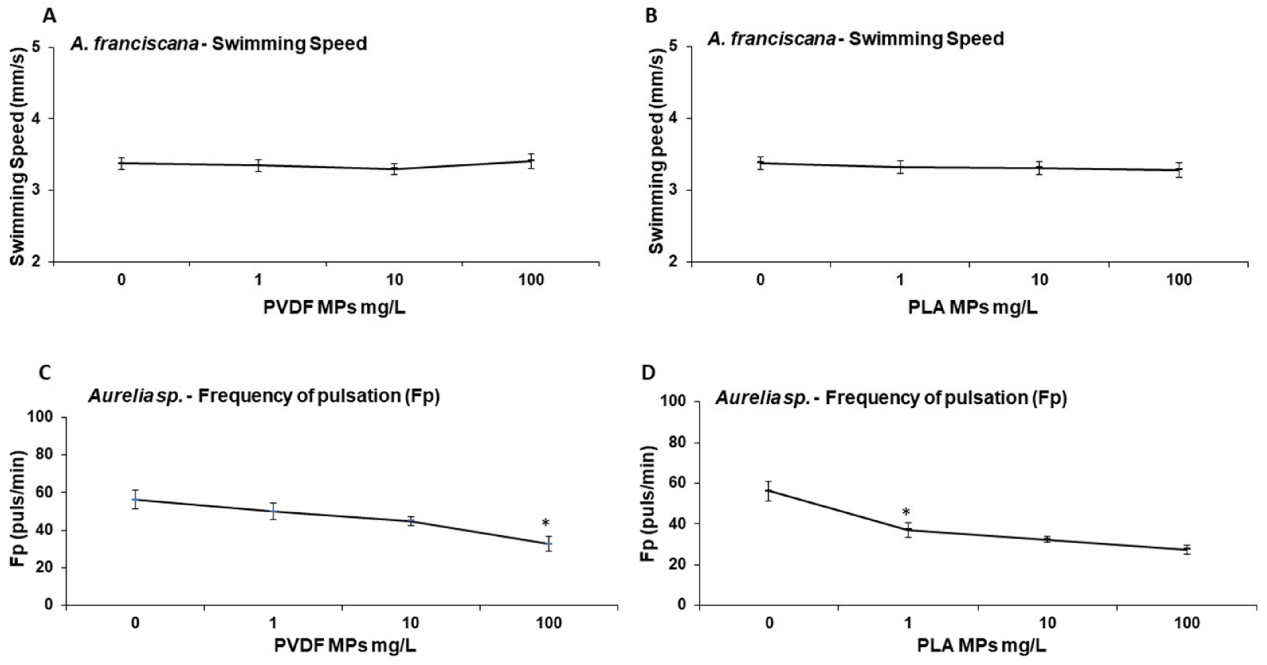
3.3. Ecotoxicology
4. Discussion
5. Conclusions
Supplementary Materials
Author Contributions
Funding
Institutional Review Board Statement
Data Availability Statement
Acknowledgments
Conflicts of Interest
References
- Duis, K.; Coors, A. Microplastics in the Aquatic and Terrestrial Environment: Sources (with a Specific Focus on Personal Care Products), Fate and Effects. Environ. Sci. Eur. 2016, 28, 2. [Google Scholar] [CrossRef] [PubMed]
- Filho, W.L.; Salvia, A.L.; Bonoli, A.; Saari, U.A.; Voronova, V.; Klõga, M.; Kumbhar, S.S.; Olszewski, K.; de Quevedo, D.M.; Barbir, J. An Assessment of Attitudes towards Plastics and Bioplastics in Europe. Sci. Total Environ. 2021, 755, 142732. [Google Scholar] [CrossRef] [PubMed]
- Parashar, N.; Hait, S. Plastics in the Time of COVID-19 Pandemic: Protector or Polluter? Sci. Total Environ. 2021, 759, 144274. [Google Scholar] [CrossRef] [PubMed]
- Azevedo-Santos, V.M.; Brito, M.F.G.; Manoel, P.S.; Perroca, J.F.; Rodrigues-Filho, J.L.; Paschoal, L.R.P.; Goncavales, G.R.L.; Wolf, M.R.; Blettler, M.C.M.; Andrade, M.C.; et al. Plastic pollution: A focus on freshwater biodiversity. Ambio 2021, 50, 1313–1324. [Google Scholar] [CrossRef] [PubMed]
- Derraik, J.G.B. The pollution of the marine environment by plastic debris: A review. Mar. Pollut. Bull. 2002, 44, 842–852. [Google Scholar] [CrossRef]
- Jambeck, J.R.; Geyer, R.; Wilcox, C.; Siegler, T.R.; Perryman, M.; Andrady, A.; Narayan, R.; Law, K.L. Plastic waste inputs from land into the ocean. Science 2015, 347, 768–771. [Google Scholar] [CrossRef]
- Abeynayaka, A.; Kojima, F.; Miwa, Y.; Ito, N.; Nihei, Y.; Fukunaga, Y.; Yashima, Y.; Itsubo, N. Rapid Sampling of Suspended and Floating Microplastics in Challenging Riverine and Coastal Water Environments in Japan. Water 2020, 12, 1903. [Google Scholar] [CrossRef]
- Luo, H.; Liu, C.; He, D.; Xu, J.; Sun, J.; Li, J.; Pan, X. Environmental Behaviors of Microplastics in Aquatic Systems: A Systematic Review on Degradation, Adsorption, Toxicity and Biofilm under Aging Conditions. J. Hazard. Mater. 2022, 423, 126915. [Google Scholar] [CrossRef]
- Miller, T.H.; Ng, K.T.; Bury, S.T.; Bury, S.E.; Bury, N.R.; Barron, L.P. Biomonitoring of Pesticides, Pharmaceuticals and Illicit Drugs in a Freshwater Invertebrate to Estimate Toxic or Effect Pressure. Environ. Int. 2019, 129, 595–606. [Google Scholar] [CrossRef]
- Cole, M.; Lindeque, P.; Fileman, E.; Halsband, C.; Goodhead, R.; Moger, J.; Galloway, T.S. Microplastic Ingestion by Zooplankton. Environ. Sci. Technol. 2013, 47, 6646–6655. [Google Scholar] [CrossRef]
- Savoca, M.S.; McInturf, A.G.; Hazen, E.L. Plastic Ingestion by Marine Fish Is Widespread and Increasing. Glob. Change Biol. 2021, 27, 2188–2199. [Google Scholar] [CrossRef] [PubMed]
- Zantis, L.J.; Carroll, E.L.; Nelms, S.E.; Bosker, T. Marine Mammals and Microplastics: A Systematic Review and Call for Standardisation. Environ. Pollut. 2021, 269, 116142. [Google Scholar] [CrossRef]
- Wilcox, C.; Puckridge, M.; Schuyler, Q.A.; Townsend, K.; Hardesty, B.D. A quantitative analysis linking sea turtle mortality and plastic debris ingestion. Sci. Rep. 2018, 8, 12536. [Google Scholar] [CrossRef]
- Azevedo-Santos, V.M.; Gonçalves, G.R.L.; Manoel, P.S.; Andrade, M.C.; Lima, F.P.; Pelicice, F.M. Plastic ingestion by fish: A global assessment. Environ. Pollut. 2019, 255, 112994. [Google Scholar] [CrossRef] [PubMed]
- Wilcox, C.; Van Sebille, E.; Hardesty, B.D. Threat of plastic pollution to seabirds is global, pervasive, and increasing. Proc. Natl. Acad. Sci. USA 2015, 12, 11899–11904. [Google Scholar] [CrossRef] [PubMed]
- Botterell, Z.L.R.; Beaumont, N.; Dorrington, T.; Steinke, M.; Thompson, R.C.; Lindeque, P.K. Bioavailability and Effects of Microplastics on Marine Zooplankton: A Review. Environ. Pollut. 2019, 245, 98–110. [Google Scholar] [CrossRef]
- Cózar, A.; Echevarría, F.; González-Gordillo, J.I.; Irigoien, X.; Úbeda, B.; Hernández-León, S.; Palma, Á.T.; Navarro, S.; García-de-Lomas, J.; Ruiz, A.; et al. Plastic Debris in the Open Ocean. Proc. Natl. Acad. Sci. USA 2014, 111, 10239–10244. [Google Scholar] [CrossRef]
- Murray, F.; Cowie, P.R. Plastic Contamination in the Decapod Crustacean Nephrops norvegicus (Linnaeus, 1758). Mar. Pollut. Bull. 2011, 62, 1207–1217. [Google Scholar] [CrossRef]
- Setälä, O.; Fleming-Lehtinen, V.; Lehtiniemi, M. Ingestion and Transfer of Microplastics in the Planktonic Food Web. Environ. Pollut. 2014, 185, 77–83. [Google Scholar] [CrossRef]
- Batel, A.; Linti, F.; Scherer, M.; Erdinger, L.; Braunbeck, T. Transfer of Benzo[a]pyrene from Microplastics to Artemia Nauplii and Further to Zebrafish via a Trophic Food Web Experiment: CYP1A Induction and Visual Tracking of Persistent Organic Pollutants. Environ. Toxicol. Chem. 2016, 35, 1656–1666. [Google Scholar] [CrossRef]
- Macali, A.; Semenov, A.; Venuti, V.; Crupi, V.; D’Amico, F.; Rossi, B.; Corsi, I.; Bergami, E. Episodic Records of Jellyfish Ingestion of Plastic Items Reveal a Novel Pathway for Trophic Transference of Marine Litter. Sci. Rep. 2018, 8, 6105. [Google Scholar] [CrossRef] [PubMed]
- Sucharitakul, P.; Pitt, K.A.; Welsh, D.T. Limited Ingestion, Rapid Egestion and No Detectable Impacts of Microbeads on the Moon Jellyfish, Aurelia aurita. Mar. Pollut. Bull. 2020, 156, 111208. [Google Scholar] [CrossRef] [PubMed]
- Cole, M.; Galloway, T.S. Ingestion of Nanoplastics and Microplastics by Pacific Oyster Larvae. Environ. Sci. Technol. 2015, 49, 14625–14632. [Google Scholar] [CrossRef] [PubMed]
- Gambardella, C.; Morgana, S.; Ferrando, S.; Bramini, M.; Piazza, V.; Costa, E.; Garaventa, F.; Faimali, M. Effects of Polystyrene Microbeads in Marine Planktonic Crustaceans. Ecotoxicol. Environ. Saf. 2017, 145, 250–257. [Google Scholar] [CrossRef]
- Wu, W.; Wang, W.; Li, J. Star Polymers: Advances in Biomedical Applications. Prog. Polym. Sci. 2015, 46, 55–85. [Google Scholar] [CrossRef]
- Costa, E.; Piazza, V.; Lavorano, S.; Faimali, M.; Garaventa, F.; Gambardella, C. Trophic Transfer of Microplastics From Copepods to Jellyfish in the Marine Environment. Front. Environ. Sci. 2020, 8, 571732. [Google Scholar] [CrossRef]
- Costa, E.; Gambardella, C.; Piazza, V.; Vassalli, M.; Sbrana, F.; Lavorano, S.; Garaventa, F.; Faimali, M. Microplastics Ingestion in the Ephyra Stage of Aurelia Sp. Triggers Acute and Behavioral Responses. Ecotoxicol. Environ. Saf. 2020, 189, 109983. [Google Scholar] [CrossRef]
- Rapp, J.; Herrera, A.; Bondyale-Juez, D.R.; González-Pleiter, M.; Reinold, S.; Asensio, M.; Martínez, I.; Gómez, M. Microplastic Ingestion in Jellyfish Pelagia noctiluca (Forsskal, 1775) in the North Atlantic Ocean. Mar. Pollut. Bull. 2021, 166, 112266. [Google Scholar] [CrossRef]
- Boero, F. Review of Jellyfish Blooms in the Mediterranean and Black Sea. General Fisheries Commission for the Mediterranean. Stud. Rev. Gen. Fish. Comm. Mediterr. 2013, 92, 53. [Google Scholar]
- Faimali, M.; Garaventa, F.; Piazza, V.; Costa, E.; Greco, G.; Mazzola, V.; Beltrandi, M.; Bongiovanni, E.; Lavorano, S.; Gnone, G. Ephyra Jellyfish as a New Model for Ecotoxicological Bioassays. Mar. Environ. Res. 2014, 93, 93–101. [Google Scholar] [CrossRef]
- Richardson, A.J.; Bakun, A.; Hays, G.C.; Gibbons, M.J. The Jellyfish Joyride: Causes, Consequences and Management Responses to a More Gelatinous Future. Trends Ecol. Evol. 2009, 24, 312–322. [Google Scholar] [CrossRef] [PubMed]
- Båmstedt, U.; Wild, B.; Martinussen, M. Significance of food type for growth of ephyrae Aurelia aurita (Scyphozoa). Mar. Biol. 2001, 139, 641–650. [Google Scholar] [CrossRef]
- Essa, W.K.; Yeasin, S.A.; Saeed, I.A.; Ali, G.A.M. Nanofiber-based face masks and respirators as COVID-19 protection: A review. Membranes 2021, 11, 250. [Google Scholar] [CrossRef]
- Ikada, Y.; Tsuji, H. Biodegradable polyesters for medical and ecological applications. Macromol. Rapid Commun. 2000, 21, 117–132. [Google Scholar] [CrossRef]
- Montalvão, G.R.; Moshrefi-Torbati, M.; Hamilton, A.; Machado, R.; João, A. Behavior of 3D printed PLA and PLA-PHA in marine environments. IOP Conf. Ser. Earth Environ. Sci. 2020, 424, 012013. [Google Scholar] [CrossRef]
- Jem, K.J.; Tan, B. The development and challenges of poly (lactic acid) and poly (glycolic acid). Adv. Ind. Eng. Polym. Res. 2020, 3, 60–70. [Google Scholar] [CrossRef]
- Market Insight. Polyvinylidene Fluoride (PVDF) Market Report. In By Application (Coatings, Pipes, Sheets, Tubes, Films, Membrane, Cables and Others), by End-Use Industry (Oil & Gas, Chemical Processing, Construction, and Others), and By Region (North America, Europe, Asia Pacific, Latin America, Middle East, and Africa)—Size, Share, Trends, and Forecast to 2025; Market Insight: Seattle, WC, USA, 2018; p. 120. [Google Scholar]
- González-Henríquez, C.M.; Sarabia-Vallejos, M.A.; Rodriguez-Hernandez, J. Polymers for Additive Manufacturing and 4D-Printing: Materials, Methodologies, and Biomedical Applications. Prog. Polym. Sci. 2019, 94, 57–116. [Google Scholar] [CrossRef]
- Sin, L.T.; Rahmat, A.R.; Rahman, W.A.W.A. Applications of Poly(Lactic Acid). In Handbook of Biopolymers and Biodegradable Plastics; Elsevier: Amsterdam, The Netherlands, 2013; pp. 55–69. [Google Scholar]
- Castro-Aguirre, E.; Iñiguez-Franco, F.; Samsudin, H.; Fang, X.; Auras, R. Poly(Lactic Acid)—Mass Production, Processing, Industrial Applications, and End of Life. Adv. Drug Deliv. Rev. 2016, 107, 333–366. [Google Scholar] [CrossRef]
- Nagarajan, V.; Mohanty, A.K.; Misra, M. Perspective on Polylactic Acid (PLA) Based Sustainable Materials for Durable Applications: Focus on Toughness and Heat Resistance. ACS Sustain. Chem. Eng. 2016, 4, 2899–2916. [Google Scholar] [CrossRef]
- Roether, J.A.; Boccaccini, A.R.; Hench, L.L.; Maquet, V.; Gautier, S.; Jérôme, R. Development and In Vitro Characterisation of Novel Bioresorbable and Bioactive Composite Materials Based on Polylactide Foams and Bioglass® for Tissue Engineering Applications. Biomaterials 2002, 23, 3871–3878. [Google Scholar] [CrossRef]
- Balla, E.; Daniilidis, V.; Karlioti, G.; Kalamas, T.; Stefanidou, M.; Bikiaris, N.D.; Vlachopoulos, A.; Koumentakou, I.; Bikiaris, D.N. Poly(Lactic Acid): A Versatile Biobased Polymer for the Future with Multifunctional Properties-from Monomer Synthesis, Polymerization Techniques and Molecular Weight Increase to PLA Applications. Polymers 2021, 13, 1822. [Google Scholar] [CrossRef] [PubMed]
- Höhnemann, T.; Steinmann, M.; Schindler, S.; Hoss, M.; König, S.; Ota, A.; Dauner, M.; Buchmeiser, M.R. Poly(Ethylene furanoate) along its life-cycle from a polycondensation approach to high-performance yarn and its recyclate. Materials 2021, 14, 1044. [Google Scholar] [CrossRef] [PubMed]
- Loos, K.; Zhang, R.; Pereira, I.; Agostinho, B.; Hu, H.; Maniar, D.; Sbirrazzuoli, N.; Silvestre, A.J.D.; Guigo, N.; Sousa, A.F. A Perspective on PEF Synthesis, Properties, and End-Life. Front. Chem. 2020, 8, 585. [Google Scholar] [CrossRef] [PubMed]
- Reichert, C.L.; Bugnicourt, E.; Coltelli, M.B.; Cinelli, P.; Lazzeri, A.; Canesi, I.; Braca, F.; Martínez, B.M.; Alonso, R.; Agostinis, L.; et al. Bio-based packaging: Materials, modifications, industrial applications and sustainability. Polymers 2020, 12, 1588. [Google Scholar] [CrossRef]
- Achmad, F.; Yamane, K.; Quan, S.; Kokugan, T. Synthesis of polylactic acid by direct polycondensation under vacuum without catalysts, solvents and initiators. Chem. Eng. J. 2009, 151, 342–350. [Google Scholar] [CrossRef]
- Kang, B.; Li, Y.-D.; Liang, J.; Yan, X.; Chen, J.; Lang, W.-Z. Novel PVDF Hollow Fiber Ultrafiltration Membranes with Antibacterial and Antifouling Properties by Embedding N-Halamine Functionalized Multi-Walled Carbon Nanotubes (MWNTs). RSC Adv. 2016, 6, 1710–1721. [Google Scholar] [CrossRef]
- Ullah, S.; Ullah, A.; Lee, J.; Jeong, Y.; Hashmi, M.; Zhu, C.; Joo, K.I.; Cha, H.J.; Kim, I.S. Reusability Comparison of Melt-Blown vs. Nanofiber Face Mask Filters for Use in the Coronavirus Pandemic. ACS Appl. Nano Mater. 2020, 3, 7231–7241. [Google Scholar] [CrossRef]
- Li, X.; Luo, J.; Zeng, H.; Zhu, L.; Lu, X. Microplastics Decrease the Toxicity of Sulfamethoxazole to Marine Algae (Skeletonema costatum) at the Cellular and Molecular Levels. Sci. Total Environ. 2022, 824, 153855. [Google Scholar] [CrossRef]
- Pagter, E.; Frias, J.; Kavanagh, F.; Nash, R. Differences in Microplastic Abundances within Demersal Communities Highlight the Importance of an Ecosystem-Based Approach to Microplastic Monitoring. Mar. Pollut. Bull. 2020, 160, 111644. [Google Scholar] [CrossRef]
- Expósito, N.; Rovira, J.; Sierra, J.; Gimenez, G.; Domingo, J.L.; Schuhmacher, M. Levels of microplastics and their characteristics in molluscs from North-West Mediterranean Sea: Human intake. Mar. Pollut. Bull. 2022, 181, 113843. [Google Scholar] [CrossRef]
- Echeverría, T.B.R. Acute Toxicity of Bioplastic Leachates to Paracentrotus lividus Sea Urchin Larvae. Mar. Environ. Res. 2022, 176, 105605. [Google Scholar] [CrossRef] [PubMed]
- Baczewska, M.; Eder, K.; Ketelhut, S.; Kemper, B.; Kujawińska, M. Refractive Index Changes of Cells and Cellular Compartments Upon Paraformaldehyde Fixation Acquired by Tomographic Phase Microscopy. Cytom. Part A 2021, 99, 388–398. [Google Scholar] [CrossRef] [PubMed]
- Cacace, T.; Bianco, V.; Mandracchia, B.; Pagliarulo, V.; Oleandro, E.; Paturzo, M.; Ferraro, P. Compact Off-Axis Holographic Slide Microscope: Design Guidelines. Biomed. Opt. Express 2020, 11, 2511. [Google Scholar] [CrossRef] [PubMed]
- Lauer, V. New Approach to Optical Diffraction Tomography Yielding a Vector Equation of Diffraction Tomography and a Novel Tomographic Microscope. J. Microsc. 2002, 205, 165–176. [Google Scholar] [CrossRef] [PubMed]
- Libralato, G.; Prato, E.; Migliore, L.; Cicero, A.M.; Manfra, L. A review of toxicity testing protocols and endpoints with Artemia spp. Ecol. Indic. 2016, 69, 35–49. [Google Scholar] [CrossRef]
- Costa, E.; Gambardella, C.; Piazza, V.; Greco, G.; Lavorano, S.; Beltrandi, M.; Bongiovanni, E.; Gnone, G.; Faimali, M.; Garaventa, F. Effect of Neurotoxic Compounds on Ephyrae of Aurelia aurita Jellyfish. Hydrobiologia 2015, 759, 75–84. [Google Scholar] [CrossRef]
- Gambardella, C.; Costa, E.; Piazza, V.; Fabbrocini, A.; Magi, E.; Faimali, M.; Garaventa, F. Effect of silver nanoparticles on marine organisms belonging to different trophic levels. Mar. Environ. Res. 2015, 111, 41–49. [Google Scholar] [CrossRef]
- Cormier, B.; Gambardella, C.; Tato, T.; Perdriat, Q.; Costa, E.; Veclin, C.; le Bihanic, F.; Grassl, B.; Dubocq, F.; Kärrman, A.; et al. Chemicals Sorbed to Environmental Microplastics Are Toxic to Early Life Stages of Aquatic Organisms. Ecotoxicol. Environ. Saf. 2021, 208, 111665. [Google Scholar] [CrossRef]
- Russo, F.; Ursino, C.; Avruscio, E.; Desiderio, G.; Perrone, A.; Santoro, S.; Galiano, F.; Figoli, A. Innovative Poly (Vinylidene Fluoride) (PVDF) Electrospun Nanofiber Membrane Preparation Using DMSO as a Low Toxicity Solvent. Membranes 2020, 10, 36. [Google Scholar] [CrossRef]
- Ding, B. Electrospinning, Fibers and Textiles: A New Driving Force for Global Development. E-Polymers 2017, 17, 209–210. [Google Scholar] [CrossRef]
- Karakolis, E.G.; Nguyen, B.; You, J.B.; Rochman, C.M.; Sinton, D. Fluorescent Dyes for Visualizing Microplastic Particles and Fibers in Laboratory-Based Studies. Environ. Sci. Technol. Lett. 2019, 6, 334–340. [Google Scholar] [CrossRef]
- Nasser, F.; Lynch, I. Secreted Protein Eco-Corona Mediates Uptake and Impacts of Polystyrene Nanoparticles on Daphnia magna. J. Proteom. 2016, 137, 45–51. [Google Scholar] [CrossRef] [PubMed]
- Garaventa, F.; Gambardella, C.; di Fino, A.; Pittore, M.; Faimali, M. Swimming Speed Alteration of Artemia sp. and Brachionus plicatilis as a Sub-Lethal Behavioral End-Point for Ecotoxicological Surveys. Ecotoxicology 2010, 19, 512–519. [Google Scholar] [CrossRef]
- Faimali, M.; Garaventa, F.; Piazza, V.; Greco, G.; Corrà, C.; Magillo, F.; Pittore, M.; Giacco, E.; Gallus, L.; Falugi, C.; et al. Swimming Speed Alteration of Larvae of Balanus amphitrite as a Behavioral End-Point for Laboratory Toxicological Bioassays. Mar. Biol. 2006, 149, 87–96. [Google Scholar] [CrossRef]
- Finney, D.J. Statistical Method In Biological Assay; Charles Griffin & Co., Ltd.: London, UK, 1978. [Google Scholar]
- Abdullah, I.Y.; Yahaya, M.; Jumali, M.H.H.; Shanshool, H.M. Influence of the Substrate on the Crystalline Phase and Morphology of Poly (Vinylidene Fluoride) (Pvdf) Thin Film. Surf. Rev. Lett. 2016, 23, 1650005. [Google Scholar] [CrossRef]
- Mofokeng, J.P.; Luyt, A.S.; Tábi, T.; Kovács, J. Comparison of Injection Moulded, Natural Fibre-Reinforced Composites with PP and PLA as Matrices. J. Thermoplast. Compos. Mater. 2012, 25, 927–948. [Google Scholar] [CrossRef]
- Rajhans, A.; Gore, P.M.; Siddique, S.K.; Kandasubramanian, B. Ion-Imprinted Nanofibers of PVDF/1-Butyl-3-Methylimidazolium Tetrafluoroborate for Dynamic Recovery of Europium (III) Ions from Mimicked Effluent. J. Environ. Chem. Eng. 2019, 7, 103068. [Google Scholar] [CrossRef]
- Zhang, M.; Gao, Y.; Zhan, Y.; Ding, X.; Wang, M.; Wang, X. Preparing the Degradable, Flame-Retardant and Low Dielectric Constant Nanocomposites for Flexible and Miniaturized Electronics with Poly(Lactic Acid), Nano ZIF-8@GO and Resorcinol Di(Phenyl Phosphate). Materials 2018, 11, 1756. [Google Scholar] [CrossRef]
- Naser, A.Z.; Deiab, I.; Darras, B.M. Poly(Lactic Acid) (PLA) and Polyhydroxyalkanoates (PHAs), Green Alternatives to Petroleum-Based Plastics: A Review. RSC Adv. 2021, 11, 17151–17196. [Google Scholar] [CrossRef]
- Beiras, R.; Bellas, J.; Cachot, J.; Cormier, B.; Cousin, X.; Engwall, M.; Gambardella, C.; Garaventa, F.; Keiter, S.; le Bihanic, F.; et al. Ingestion and Contact with Polyethylene Microplastics Does Not Cause Acute Toxicity on Marine Zooplankton. J. Hazard. Mater. 2018, 360, 452–460. [Google Scholar] [CrossRef]
- Bergami, E.; Bocci, E.; Vannuccini, M.L.; Monopoli, M.; Salvati, A.; Dawson, K.A.; Corsi, I. Nano-Sized Polystyrene Affects Feeding, Behavior and Physiology of Brine Shrimp Artemia franciscana Larvae. Ecotoxicol. Environ. Saf. 2016, 123, 18–25. [Google Scholar] [CrossRef]
- Mitchell, D.G.; Edgar, A.; Martindale, M.Q. Improved histological fixation of gelatinous marine invertebrates. Front. Zool. 2021, 18, 29. [Google Scholar] [CrossRef] [PubMed]
- Haddoq, S.H.D. A golden age of gelata: Past and future research on planktonic ctenophores and cnidarians. Hydrobiologia 2004, 530–531, 549–556. [Google Scholar]
- Hanaoka, K.; Ohno, H.; Wada, N.; Ueno, S.; Goessler, W.; Kuehnelt, D.; Schlagenhaufen, C.; Kaise, T.; Irgolic, K.J. Occurrence of Organo-Arsenicals in Jellyfishes and Their Mucus. Chemosphere 2001, 44, 743–749. [Google Scholar] [CrossRef]
- Patwa, A.; Thiéry, A.; Lombard, F.; Lilley, M.K.S.; Boisset, C.; Bramard, J.-F.; Bottero, J.-Y.; Barthélémy, P. Accumulation of Nanoparticles in “Jellyfish” Mucus: A Bio-Inspired Route to Decontamination of Nano-Waste. Sci. Rep. 2015, 5, 11387. [Google Scholar] [CrossRef]
- Shim, W.J.; Song, Y.K.; Hong, S.H.; Jang, M. Identification and Quantification of Microplastics Using Nile Red Staining. Mar. Pollut. Bull. 2016, 113, 469–476. [Google Scholar] [CrossRef]
- Sturm, M.T.; Horn, H.; Schuhen, K. The Potential of Fluorescent Dyes—Comparative Study of Nile Red and Three Derivatives for the Detection of Microplastics. Anal. Bioanal. Chem. 2021, 413, 1059–1071. [Google Scholar] [CrossRef]
- Khattab, Y.; Mohammadein, A.; al Malki, J.S.; Hussien, N.A.; Tantawy, E.M. Preliminary Screening of Microplastic Contamination in Different Marine Fish Species of Taif Market, Saudi Arabia. Open Life Sci. 2022, 17, 333–343. [Google Scholar] [CrossRef]
- Anderson, G.; Shenkar, N. Potential effects of biodegradable single-use items in the sea: Polylactic acid (PLA) and solitary ascidians. Environ. Pollut. 2021, 268, 115364. [Google Scholar] [CrossRef]
- Mesarič, T.; Gambardella, C.; Milivojević, T.; Faimali, M.; Drobne, D.; Falugi, C.; Makovec, D.; Jemec, A.; Sepčić, K. High Surface Adsorption Properties of Carbon-Based Nanomaterials Are Responsible for Mortality, Swimming Inhibition, and Biochemical Responses in Artemia salina Larvae. Aquat. Toxicol. 2015, 163, 121–129. [Google Scholar] [CrossRef]
- Green, D.S.; Colgan, T.J.; Thompson, R.C.; Carolan, J.C. Exposure to Microplastics Reduces Attachment Strength and Alters the Haemolymph Proteome of Blue Mussels (Mytilus edulis). Environ. Pollut. 2019, 246, 423–434. [Google Scholar] [CrossRef] [PubMed]
- Green, D.S. Effects of microplastics on European flat oysters, Ostrea edulis and their associated benthic communities. Environ. Pollut. 2016, 216, 95–103. [Google Scholar] [CrossRef] [PubMed]
- Nakanishi, N.; Hartenstein, V.; Jacobs, D.K. Development of the rhopalial nervous system in Aurelia sp.1 (Cnidaria, Scyphozoa). Dev. Gene Evol. 2009, 219, 301–317. [Google Scholar] [CrossRef]
- Ivar Do Sul, J.A.; Costa, M.F.; Fillmann, G. Microplastics in the pelagic environment around oceanic islands of the western Tropical Atlantic Ocean. Water Air Soil Pollut. 2014, 225, 2004. [Google Scholar] [CrossRef]
- Fossette, S.; Gleiss, A.C.; Chalumeau, J.; Bastian, T.; Armstrong, C.D.; Vandenabeele, S.; Karpytchev, M.; Hays, G.C. Current-Oriented swimming by jellyfish and its role in bloom manteinance. Curr. Biol. 2015, 25, 342–347. [Google Scholar] [CrossRef] [PubMed]
- Gibbons, M.J.; Boero, F.; Brotz, L. Food for Thought. We should not assume that fishing jellyfish will solve our jellyfish problem. ICES J. Mar. Sci. 2016, 73, 1012–1018. [Google Scholar] [CrossRef]
- Rochman, C.M.; Hentschel, B.T.; The, S.J. Long-Term Sorption of Metals Is Similar among Plastic Types: Implications for Plastic Debris in Aquatic Environments. PLoS ONE 2014, 9, e85433. [Google Scholar] [CrossRef] [PubMed]
- Kedzierski, M.; D’Almeida, M.; Magueresse, A.; le Grand, A.; Duval, H.; César, G.; Sire, O.; Bruzaud, S.; le Tilly, V. Threat of Plastic Ageing in Marine Environment. Adsorption/Desorption of Micropollutants. Mar. Pollut. Bull. 2018, 127, 684–694. [Google Scholar] [CrossRef]
- Spoerk, M.; Holzer, C.; Gonzalez-Gutierrez, J. Material extrusion-based additive manufacturing of polypropylene: A review on how to improve dimensional inaccuracy and warpage. J. Appl. Polym. Sci. 2019, 136, 48545. [Google Scholar] [CrossRef]
- Taylor, K.C.; Nasr-El-Din, H.A. Water-Soluble Hydrophobically Associating Polymers for Improved Oil Recovery: A Literature Review. J. Pet. Sci. Eng. 1998, 19, 265–280. [Google Scholar] [CrossRef]
- Morgana, S.; Gambardella, C.; Costa, E.; Piazza, V.; Garaventa, F.; Faimali, M. Ecotoxicological Effects of Microplastics in Marine Zooplankton. In Proceedings of the 2nd International Conference on Microplastic Pollution in the Mediterranean Sea, Capri, Italy, 15–18 September 2019; Springer Water: Cham, Switzerland, 2020. [Google Scholar] [CrossRef]


| Organisms | Species | Endpoint | Polymer | LOEC (mg/L) | EC50 (mg/L) | References |
|---|---|---|---|---|---|---|
| Cnidarians | Aurelia sp. | Immobility | PLA | >100 | >100 | This study |
| Cnidarians | Aurelia sp. | Frequency of pulsations | PLA | 1 | 77.43 (7.83–100) | This study |
| Cnidarians | Aurelia sp. | Frequency of pulsations | PE | 0.1 | 3.16 (1.73–5.79) | [27] |
| Cnidarians | Aurelia sp. | Frequency of pulsations | PE | 0.01 | <0.01 | [27] |
| Crustaceans | Artemia franciscana | Immobility | PLA | >100 | >100 | This study |
| Crustaceans | Artemia franciscana | Swimming Speed | PE | >100 | >100 | This study |
| Crustaceans | Artemia franciscana | Immobility | PE | >10 | >10 | [94] |
| Crustaceans | Artemia franciscana | Swimming Speed | PE | 0.01 | >10 | [94] |
| Microalgae | Skeletonema costatum | Growth inhibition | PLA | 10 | >50 | [50] |
| Microalgae | Skeletonema costatum | Growth inhibition | PE | 5 | >50 | [50] |
Publisher’s Note: MDPI stays neutral with regard to jurisdictional claims in published maps and institutional affiliations. |
© 2022 by the authors. Licensee MDPI, Basel, Switzerland. This article is an open access article distributed under the terms and conditions of the Creative Commons Attribution (CC BY) license (https://creativecommons.org/licenses/by/4.0/).
Share and Cite
Di Giannantonio, M.; Gambardella, C.; Miroglio, R.; Costa, E.; Sbrana, F.; Smerieri, M.; Carraro, G.; Utzeri, R.; Faimali, M.; Garaventa, F. Ecotoxicity of Polyvinylidene Difluoride (PVDF) and Polylactic Acid (PLA) Microplastics in Marine Zooplankton. Toxics 2022, 10, 479. https://doi.org/10.3390/toxics10080479
Di Giannantonio M, Gambardella C, Miroglio R, Costa E, Sbrana F, Smerieri M, Carraro G, Utzeri R, Faimali M, Garaventa F. Ecotoxicity of Polyvinylidene Difluoride (PVDF) and Polylactic Acid (PLA) Microplastics in Marine Zooplankton. Toxics. 2022; 10(8):479. https://doi.org/10.3390/toxics10080479
Chicago/Turabian StyleDi Giannantonio, Michela, Chiara Gambardella, Roberta Miroglio, Elisa Costa, Francesca Sbrana, Marco Smerieri, Giovanni Carraro, Roberto Utzeri, Marco Faimali, and Francesca Garaventa. 2022. "Ecotoxicity of Polyvinylidene Difluoride (PVDF) and Polylactic Acid (PLA) Microplastics in Marine Zooplankton" Toxics 10, no. 8: 479. https://doi.org/10.3390/toxics10080479
APA StyleDi Giannantonio, M., Gambardella, C., Miroglio, R., Costa, E., Sbrana, F., Smerieri, M., Carraro, G., Utzeri, R., Faimali, M., & Garaventa, F. (2022). Ecotoxicity of Polyvinylidene Difluoride (PVDF) and Polylactic Acid (PLA) Microplastics in Marine Zooplankton. Toxics, 10(8), 479. https://doi.org/10.3390/toxics10080479