
Food-Grade Microemulsion for High-Loading Octacosanol: Formulation Optimization, Characterization, and Biological Evaluation
Abstract
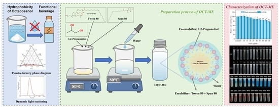
1. Introduction
2. Materials and Methods
2.1. Raw Materials and Reagents
2.2. Solubility of OCT in Different Oil Phases
2.3. Construction of Pseudo-Ternary Phase Diagrams
2.4. Optimization of O/W ME Formulation
2.5. OCT Loading Capacity of O/W ME
2.6. Characterization of OCT-ME
2.6.1. Droplet Size and PDI
2.6.2. Transmission Electron Microscopy (TEM)
2.6.3. OCT-ME Type Determination
2.6.4. Viscosity Measurement
2.6.5. Fourier Transform Infrared (FT-IR) Spectroscopy
2.7. Stability of OCT-ME
2.7.1. Environmental Stress Stability Evaluation
2.7.2. Storage Stability Evaluation
2.8. In Vitro Digestion
2.9. Biocompatibility Evaluation
2.9.1. Cell Viability Assay
2.9.2. Live/Dead Cell Staining
2.10. Statistical Analysis
3. Results and Discussion
3.1. Analysis of OCT Solubility Behavior in Oil Phase and Oil Phase Selection
3.2. Construction and Formulation Optimization of OCT-ME
3.2.1. Determination of the Oil Phase
3.2.2. Determination of Emulsifier
3.2.3. Determination of the HLB Value
3.2.4. Determination of Co-Emulsifier
3.2.5. Determination of the Km Value
3.3. Determination of OCT Loading
3.4. Configuration Analysis of OCT-ME
3.5. Transmission Electron Microscopy (TEM) Analysis
3.6. Rheological Properties Analysis
3.7. FT-IR Analysis
3.8. Stability Study of OCT-ME
3.8.1. Centrifugal Stability
3.8.2. Temperature Stability
3.8.3. pH Stability
3.8.4. Salinity Stability
3.8.5. Storage Stability
3.9. In Vitro Digestion Characteristics of OCT-ME
3.10. Biocompatibility Analysis of OCT-ME
4. Conclusions
Supplementary Materials
Author Contributions
Funding
Institutional Review Board Statement
Informed Consent Statement
Data Availability Statement
Acknowledgments
Conflicts of Interest
Abbreviations
| OCT | Octacosanol |
| O/W ME | Oil-in-Water Microemulsion |
| OCT-ME | Octacosanol-loaded Microemulsion |
| HLB | Hydrophilic-Lipophilic Balance |
| PDI | Polydispersity Index |
| FT-IR | Fourier Transform Infrared Spectroscopy |
| GC-MS | Gas Chromatography-Mass Spectrometry |
| SSF | Simulated Saliva Fluid |
| SGF | Simulated Gastric Fluid |
| SIF | Simulated Intestinal Fluid |
| Smix | emulsifier and co-emulsifier mixture |
| TEM | Transmission Electron Microscopy |
References
- Ma, Z.F.; Liu, S.; Fu, C.; Zhou, S.; Lee, Y.Y. Functional foods in health promotion and disease prevention: Innovations, evidence and challenges. Foods 2026, 15, 764. [Google Scholar] [CrossRef]
- Shan, F.; Liu, L.; Li, L.; Wang, W.; Bi, Y.; Li, M. Management, Safety, and Efficacy Evaluation of Nutraceutical and Functional Food: A Global Perspective. Compr. Rev. Food Sci. Food Saf. 2025, 24, e70222. [Google Scholar] [CrossRef]
- Wang, T.; Soyama, S.; Luo, Y. Development of a novel functional drink from all natural ingredients using nanotechnology. LWT 2016, 73, 458–466. [Google Scholar] [CrossRef]
- Gruenwald, J. Novel botanical ingredients for beverages. Clin. Dermatol. 2009, 27, 210–216. [Google Scholar] [CrossRef]
- Ou, S.; Zhao, J.; Wang, Y.; Tian, Y.; Wang, J. Preparation of octacosanol from filter mud produced after sugarcane juice clarification. LWT-Food Sci. Technol. 2012, 45, 295–298. [Google Scholar] [CrossRef]
- Zhou, Y.; Cao, F.; Luo, F.; Lin, Q. Octacosanol and health benefits: Biological functions and mechanisms of action. Food Biosci. 2022, 47, 101632. [Google Scholar] [CrossRef]
- Flanagan, J.; Singh, H. Microemulsions: A potential delivery system for bioactives in food. Crit. Rev. Food Sci. Nutr. 2006, 46, 221–237. [Google Scholar] [CrossRef]
- Bergonzi, M.C.; Hamdouch, R.; Mazzacuva, F.; Isacchi, B.; Bilia, A.R. Optimization, characterization and in vitro evaluation of curcumin microemulsions. LWT-Food Sci. Technol. 2014, 59, 148–155. [Google Scholar] [CrossRef]
- Aboudzadeh, M.A.; Mehravar, E.; Fernandez, M.; Lezama, L.; Tomovska, R. Low-energy encapsulation of α-tocopherol using fully food grade oil-in-water microemulsions. ACS Omega 2018, 3, 10999–11008. [Google Scholar] [CrossRef] [PubMed]
- Uchiyama, H.; Chae, J.; Kadota, K.; Tozuka, Y. Formation of food grade microemulsion with rice glycosphingolipids to enhance the oral absorption of coenzyme Q10. Foods 2019, 8, 502. [Google Scholar] [CrossRef]
- Jia, M.; Bai, W.; Deng, J.; Li, W.; Lin, Q.; Zhong, F.; Luo, F. Enhancing solubility and bioavailability of octacosanol: Development of a green O/W nanoemulsion synthesis process. Int. J. Pharm. 2024, 651, 123726. [Google Scholar] [CrossRef] [PubMed]
- Zhu, H.; Xu, T.; Tan, H.; Wang, M.; Wang, J. O/W nanoemulsions encapsulated octacosanol: Preparation, characterization and anti-fatigue activity. Colloids Surf. B Biointerfaces 2024, 241, 114066. [Google Scholar] [CrossRef]
- Wang, M.; Li, H.; Yang, J.; Hu, W.; Wang, J. Preparation, Characterization, and In Vivo Anti-Fatigue Evaluation of Octacosanol Nanoemulsions. J. Food Process Eng. 2024, 47, e70000. [Google Scholar] [CrossRef]
- Jalali-Jivan, M.; Rostamabadi, H.; Assadpour, E.; Tomas, M.; Capanoglu, E.; Alizadeh-Sani, M.; Kharazmi, M.S.; Jafari, S.M. Recent progresses in the delivery of β-carotene: From nano/microencapsulation to bioaccessibility. Adv. Colloid Interface Sci. 2022, 307, 102750. [Google Scholar] [CrossRef] [PubMed]
- Zhou, X.; Wang, H.; Wang, C.; Zhao, C.; Peng, Q.; Zhang, T.; Zhao, C. Stability and in vitro digestibility of beta-carotene in nanoemulsions fabricated with different carrier oils. Food Sci. Nutr. 2018, 6, 2537–2544. [Google Scholar] [CrossRef]
- Gunarto, C.; Ju, Y.-H.; Putro, J.N.; Tran-Nguyen, P.L.; Soetaredjo, F.E.; Santoso, S.P.; Ayucitra, A.; Angkawijaya, A.E.; Ismadji, S. Effect of a Nonionic Surfactant on the Pseudoternary Phase Diagram and Stability of Microemulsion. J. Chem. Eng. Data 2020, 65, 4024–4033. [Google Scholar] [CrossRef]
- Pant, A.; Jha, K.; Singh, M. Role of Excipient’s HLB Values in Microemulsion System. J. Pharm. Biol. Sci. 2019, 14, 1–6. [Google Scholar]
- Spernath, A.; Yaghmur, A.; Aserin, A.; Hoffman, R.E.; Garti, N. Food-Grade Microemulsions Based on Nonionic Emulsifiers: Media To Enhance Lycopene Solubilization. J. Agric. Food Chem. 2002, 50, 6917–6922. [Google Scholar] [CrossRef]
- Stauffer, C.E. Emulsifiers for the food industry. In Bailey’s Industrial Oil and Fat Products; John Wiley & Sons Inc.: Hoboken, NJ, USA, 2005; Volume 6. [Google Scholar]
- Kamba, E.; Itodo, A.; Ogah, E. Utilization of different emulsifying agents in the preparation and stabilization of emulsions. Int. J. Mater. Chem. 2013, 3, 69–74. [Google Scholar]
- Choudhary, A.; Desai, S.; Kamruzzaman, M.; Landera, A.; Ghosh, K.; Poorey, K. Predicting the critical micelle concentration of binary surfactant mixtures using machine learning. J. Cheminform. 2025, 17, 170. [Google Scholar] [CrossRef]
- Holland, P.M.; Rubingh, D.N. Mixed Surfactant Systems. In Mixed Surfactant Systems; ACS Symposium Series: Washington, DC, USA; American Chemical Society: Washington, DC, USA, 1992; Volume 501, pp. 2–30. [Google Scholar]
- Zhou, L.; Cai, Y.; Li, Y.; Zhou, Y.; Xiao, Z. Cinnamon Essential Oil Delivery Systems: Preparation Processes, Packaging Forms, and Industrialization Potentials. Food Sci. Nutr. 2025, 13, e70814. [Google Scholar] [CrossRef] [PubMed]
- Yu, F.; Miao, Y.; Wang, M.; Liu, Q.; Yuan, L.; Geng, R.; Qiu, Q.; Ni, C.; Kay, M. Predicting nanoemulsion formulation and studying the synergism mechanism between surfactant and cosurfactant: A combined computational and experimental approach. Int. J. Pharm. 2022, 615, 121473. [Google Scholar] [CrossRef]
- Vlaia, L.; Coneac, G.; Muţ, A.M.; Olariu, I.; Vlaia, V.; Anghel, D.F.; Maxim, M.E.; Dobrescu, A.; Hîrjău, M.; Lupuleasa, D. Topical biocompatible fluconazole-loaded microemulsions based on essential oils and sucrose esters: Formulation design based on pseudo-ternary phase diagrams and physicochemical characterization. Processes 2021, 9, 144. [Google Scholar] [CrossRef]
- Mousa, A.M. Development and Assessment of a Microemulsion-Based Itraconazole Gel as Topical Antifungal Therapy. Kron. J. 2025, 25, 254–267. [Google Scholar]
- Minekus, M.; Alminger, M.; Alvito, P.; Ballance, S.; Bohn, T.; Bourlieu, C.; Carrière, F.; Boutrou, R.; Corredig, M.; Dupont, D. A standardised static in vitro digestion method suitable for food–an international consensus. Food Funct. 2014, 5, 1113–1124. [Google Scholar] [CrossRef]
- Wei, S.; Huang, J.; Zhang, L.; Sun, Q.; Sun, X.; Jin, L.; Wang, Q. Physicochemical properties and stabilities of crude and purified oil bodies extracted from high oleic peanuts. Eur. J. Lipid Sci. Technol. 2020, 122, 1900183. [Google Scholar] [CrossRef]
- Wang, L.; Liu, L.; Pang, N.; Li, W.; Guo, W.; Zhang, R.; Wei, G.; Dai, L.; Sun, Q.; Dong, X. Effect of ultrasound-assisted aqueous enzymatic extraction on the interfacial properties of high-oleic peanut oil bodies. Grain Oil Sci. Technol. 2025, 8, 100–108. [Google Scholar] [CrossRef]
- Lundgren, S.M.; Ruths, M.; Danerlöv, K.; Persson, K. Effects of unsaturation on film structure and friction of fatty acids in a model base oil. J. Colloid Interface Sci. 2008, 326, 530–536. [Google Scholar] [CrossRef]
- Cong, Y.; Zhang, W.; Liu, C.; Huang, F. Composition and Oil-Water Interfacial Tension Studies in Different Vegetable Oils. Food Biophys. 2020, 15, 229–239. [Google Scholar] [CrossRef]
- Duranova, H.; Kuzelova, L.; Fialkova, V.; Simora, V.; Kovacikova, E.; Joanidis, P.; Borotova, P.; Straka, D.; Hoskin, R.T.; Moncada, M. Coconut-sourced MCT oil: Its potential health benefits beyond traditional coconut oil. Phytochem. Rev. 2025, 24, 659–700. [Google Scholar] [CrossRef]
- Jintapattanakit, A.; Hasan, H.M.; Junyaprasert, V.B. Vegetable oil-based nanoemulsions containing curcuminoids: Formation optimization by phase inversion temperature method. J. Drug Deliv. Sci. Technol. 2018, 44, 289–297. [Google Scholar] [CrossRef]
- Liu, H.; Li, H.; Gu, J.; Deng, L.; Ren, L.; Hong, Y.; Lu, Q.; Chen, X.; Liang, X. Identification of the candidate proteins related to oleic acid accumulation during peanut (Arachis hypogaea L.) seed development through comparative proteome analysis. Int. J. Mol. Sci. 2018, 19, 1235. [Google Scholar] [CrossRef]
- Farooq, S.; Abdullah; Zhang, H.; Weiss, J. A comprehensive review on polarity, partitioning, and interactions of phenolic antioxidants at oil–water interface of food emulsions. Compr. Rev. Food Sci. Food Saf. 2021, 20, 4250–4277. [Google Scholar] [CrossRef]
- Seçmeler, Ö.; Güçlü Üstündağ, Ö. Behavior of lipophilic bioactives during olive oil processing. Eur. J. Lipid Sci. Technol. 2017, 119, 1600404. [Google Scholar] [CrossRef]
- Nwankwo, J.A.; Liu, W.; Guo, X.; Lin, Y.; Hussain, M.; Khan, I.; Joshua, M.; Ibrahim, A.N.; Ngozi, O.J.; Ali, A. Microemulsion gel systems: Formulation, stability studies, biopolymer interactions, and functionality in food product development. Compr. Rev. Food Sci. Food Saf. 2025, 24, e70110. [Google Scholar] [CrossRef]
- Singh, A.K.; Chandra, A.; Kandpal, J. Octacosanol extraction, synthesis method and sources: A review. Carpathian J. Food Sci. Technol. 2020, 12, 27. [Google Scholar]
- Akula, S.; Gurram, A.K.; Devireddy, S.R. Self-Microemulsifying Drug Delivery Systems: An Attractive Strategy for Enhanced Therapeutic Profile. Int. Sch. Res. Not. 2014, 2014, 964051. [Google Scholar] [CrossRef] [PubMed]
- Subongkot, T.; Ngawhirunpat, T. Development of a novel microemulsion for oral absorption enhancement of all-trans retinoic acid. Int. J. Nanomed. 2017, 12, 5585–5599. [Google Scholar] [CrossRef]
- Wang, Z. Pseudo-ternary phase diagrams of a drug delivery system. In Proceedings of the Masters Abstracts International; University of Waterloo: Waterloo, ON, Canada, 2009. [Google Scholar]
- Yatheshappa, G.K.; Farooq, S.; Jiang, Q.; Chen, M.; Zhang, H. Investigating the effects of polar and non-polar polyphenols on the physicochemical properties and functional characteristics of camellia oil body emulsions. Food Chem. 2025, 481, 144033. [Google Scholar] [CrossRef]
- Trost, V.W. Characterization of corn oil, soybean oil and sunflowerseed oil nonpolar material. J. Am. Oil Chem. Soc. 1989, 66, 325–333. [Google Scholar] [CrossRef]
- Ghayour, A.; Acosta, E. Characterizing the oil-like and surfactant-like behavior of polar oils. Langmuir 2019, 35, 15038–15050. [Google Scholar] [CrossRef] [PubMed]
- Yatheshappa, G.K.; Farooq, S.; Zhang, H. Effects of polar quercetin and non-polar resveratrol on the functional properties, oxidative stability and interfacial behavior of camellia oil body emulsions. Food Chem. 2025, 497, 146998. [Google Scholar] [CrossRef] [PubMed]
- Li, Y.; Wu, C.; Wu, T.; Wang, L.; Chen, S.; Ding, T.; Hu, Y. Preparation and characterization of citrus essential oils loaded in chitosan microcapsules by using different emulsifiers. J. Food Eng. 2018, 217, 108–114. [Google Scholar] [CrossRef]
- Yan, G.S.; Li, Y.; Wang, H.; He, L.Q.; Li, Y.; Li, Y.; Zhang, L.B.; Yan, J.G. Effect of composition of emulsifier blends on aerated emulsions: Stability, thermodynamic, interfacial behavior and aeration properties. LWT Food Sci. Technol. 2023, 188, 115395. [Google Scholar] [CrossRef]
- Fu, Z.; Liu, M.; Xu, J.; Wang, Q.; Fan, Z. Stabilization of water-in-octane nano-emulsion. Part I: Stabilized by mixed surfactant systems. Fuel 2010, 89, 2838–2843. [Google Scholar] [CrossRef]
- Hong, I.K.; Kim, S.I.; Lee, S.B. Effects of HLB value on oil-in-water emulsions: Droplet size, rheological behavior, zeta-potential, and creaming index. J. Ind. Eng. Chem. 2018, 67, 123–131. [Google Scholar] [CrossRef]
- Graca, M.; Bongaerts, J.H.; Stokes, J.R.; Granick, S. Friction and adsorption of aqueous polyoxyethylene (Tween) surfactants at hydrophobic surfaces. J. Colloid Interface Sci. 2007, 315, 662–670. [Google Scholar] [CrossRef]
- Wang, Q.; Zhang, H.; Han, Y.; Cui, Y.; Han, X. Study on the relationships between the oil HLB value and emulsion stabilization. RSC Adv. 2023, 13, 24692–24698. [Google Scholar] [CrossRef]
- Chen, J.; Ma, X.-h.; Yao, G.-l.; Zhang, W.-t.; Zhao, Y. Microemulsion-based anthocyanin systems: Effect of surfactants, cosurfactants, and its stability. Int. J. Food Prop. 2018, 21, 1152–1165. [Google Scholar] [CrossRef]
- Smejkal, G.; Gross, V.; Lazarev, A. Theoretical and Experimental Determinations of the Hydrophilic–Lipophilic Balance (HLB) of Representative Oils and Lecithins. Colloids Interfaces 2024, 8, 21. [Google Scholar] [CrossRef]
- Acosta, E.J.; Le, M.A.; Harwell, J.H.; Sabatini, D.A. Coalescence and Solubilization Kinetics in Linker-Modified Microemulsions and Related Systems. Langmuir 2003, 19, 566–574. [Google Scholar] [CrossRef]
- Yan, G.; Wang, S.; Li, Y.; He, L.; Li, Y.; Zhang, L. Effect of emulsifier HLB on aerated emulsions: Stability, interfacial behavior, and aeration properties. J. Food Eng. 2023, 351, 111505. [Google Scholar] [CrossRef]
- Mehta, S.; Kaur, G.; Mutneja, R.; Bhasin, K. Solubilization, microstructure, and thermodynamics of fully dilutable U-type Brij microemulsion. J. Colloid Interface Sci. 2009, 338, 542–549. [Google Scholar] [CrossRef]
- Zeng, L.; Zhang, Y. Impact of short-chain alcohols on the formation and stability of nano-emulsions prepared by the spontaneous emulsification method. Colloids Surf. A Physicochem. Eng. Asp. 2016, 509, 591–600. [Google Scholar] [CrossRef]
- Gomes, A.; Costa, A.L.R.; do Amaral Sobral, P.J.; Cunha, R.L. Delivering β-carotene from O/W emulsion-based systems: Influence of phase ratio and carrier lipid composition. Food Hydrocoll. Health 2023, 3, 100125. [Google Scholar] [CrossRef]
- Abrar, I.; Bhaskarwar, A. Effect of alcohols on water solubilization in surfactant-free diesel microemulsions. Energy Rep. 2022, 8, 504–512. [Google Scholar] [CrossRef]
- Yuan, Y.; Li, S.-m.; Mo, F.-k.; Zhong, D.-f. Investigation of microemulsion system for transdermal delivery of meloxicam. Int. J. Pharm. 2006, 321, 117–123. [Google Scholar] [CrossRef]
- Patel, M.R.; Patel, R.B. Nanoemulsion for topical therapy of acne: Optimization and evaluation. Indian Drugs 2021, 58, 31. [Google Scholar] [CrossRef]
- Liang, Y.; Zou, J.; Zhang, X.; Shi, Y.; Tai, J.; Wang, Y.; Guo, D.; Yang, M. Preparation and quality evaluation of a volatile oil microemulsion from Flos magnoliae and Centipeda minima. Mol. Med. Rep. 2020, 22, 4531–4540. [Google Scholar] [CrossRef]
- Tang, H.; Xiang, S.; Li, X.; Zhou, J.; Kuang, C. Preparation and in vitro performance evaluation of resveratrol for oral self-microemulsion. PLoS ONE 2019, 14, e0214544. [Google Scholar] [CrossRef]
- Wang, M.; Li, N.; Guo, X.; Chen, Y.; Zhang, M.; Sun, J.; Farag, M.A.; Simal-Gandara, J.; Sun, S.; Liu, C. Development of microemulsions-filled sodium alginate-chitosan composite hydrogels: Preparation, characterization and release kinetics analysis. Food Hydrocoll. 2025, 163, 111059. [Google Scholar] [CrossRef]
- Zhu, S.; Kong, L.; Peng, Y.; Zeng, Q.; Feng, B.; Jian, O.; Zhao, P.; Zhang, W.; Li, Z. Long-chain alkyl emulsifiers induced asphalt particle dispersion: Lipophilicity-enhancement effect. Constr. Build. Mater. 2024, 449, 138275. [Google Scholar] [CrossRef]
- El Aoud, A.; Reboul, E.; Dupont, A.; Mériadec, C.; Artzner, F.; Marze, S. In vitro solubilization of fat-soluble vitamins in structurally defined mixed intestinal assemblies. J. Colloid Interface Sci. 2021, 589, 229–241. [Google Scholar] [CrossRef]
- Mou, L.; Li, J.; Lu, Y.; Wu, L.; Bu, G.; Li, J.; Li, G. Innovative cellulose-based porous matrix loaded with carvacrol microemulsion for sustainable mango preservation. Int. J. Food Microbiol. 2025, 443, 111403. [Google Scholar] [CrossRef] [PubMed]
- Yang, H.; Li, R.; Ma, H.; Tian, L.; Yan, J.; Chen, G.; Zhang, Z. Development of Forsythia Essential Oil Microemulsions: Effects of Surfactants on Stability and Antibacterial Activity. ACS Omega 2025, 10, 35540–35550. [Google Scholar] [CrossRef] [PubMed]
- Bumajdad, A.; Eastoe, J. Conductivity of water-in-oil microemulsions stabilized by mixed surfactants. J. Colloid Interface Sci. 2004, 274, 268–276. [Google Scholar] [CrossRef]
- Batool, A.; Asghar, M.N.; Rehman, R.; Sharif, M.S.; Mohyuddin, A.; Khan, A.M. Green microemulsion systems with choline-based SAILs: Formulation and piperine delivery. Chem. Phys. 2025, 595, 112724. [Google Scholar] [CrossRef]
- Kalaitzaki, A.; Xenakis, A.; Papadimitriou, V. Highly water dilutable microemulsions: A structural study. Colloid Polym. Sci. 2015, 293, 1111–1119. [Google Scholar] [CrossRef]
- Chen, B.; Hou, M.; Zhang, B.; Liu, T.; Guo, Y.; Dang, L.; Wang, Z. Enhancement of the solubility and antioxidant capacity of α-linolenic acid using an oil in water microemulsion. Food Funct. 2017, 8, 2792–2802. [Google Scholar] [CrossRef] [PubMed]
- Kogan, A.; Shalev, D.E.; Raviv, U.; Aserin, A.; Garti, N. Formation and Characterization of Ordered Bicontinuous Microemulsions. J. Phys. Chem. B 2009, 113, 10669–10678. [Google Scholar] [CrossRef]
- Talianu, M.-T.; Dinu-Pîrvu, C.-E.; Ghica, M.V.; Anuța, V.; Mihai, R.; Goia, R.-E.; Popa, L. Development and characterization of salicylic acid-based microemulsions for topical application. ICAMS 2024, 261, 261–268. [Google Scholar]
- Numin, M.S.; Jumbri, K.; Ramli, A.; Borhan, N. Microemulsion rheological analysis of alkaline, surfactant, and polymer in oil-water interface. Processes 2020, 8, 762. [Google Scholar] [CrossRef]
- Jähnigen, S.; Vuilleumier, R.; Zehnacker, A. The genesis of OH-stretching vibrational circular dichroism in chiral molecular crystals. Chem. Sci. 2025, 16, 9833–9842. [Google Scholar] [CrossRef]
- Dutta, C.; Svirida, A.; Mammetkuliyev, M.; Rukhadze, M.; Benderskii, A.V. Insight into water structure at the surfactant surfaces and in microemulsion confinement. J. Phys. Chem. B 2017, 121, 7447–7454. [Google Scholar] [CrossRef]
- Liu, W.; Pan, N.; Han, Y.; Li, D.; Chai, J. Solubilization, stability and antioxidant activity of curcumin in a novel surfactant-free microemulsion system. LWT 2021, 147, 111583. [Google Scholar] [CrossRef]
- Jintapattanakit, A. Preparation of nanoemulsions by phase inversion temperature (PIT) method. Pharm. Sci. Asia 2018, 45, 1–12. [Google Scholar] [CrossRef]
- Gao, Y.; Chen, C.; Li, M.; Sun, C. Ionic environment-modulated nucleation and stability of multiscale nanodomains in surfactant-free microemulsions. J. Colloid Interface Sci. 2025, 696, 137833. [Google Scholar] [CrossRef] [PubMed]
- Davis, C.R.; Martinez, C.J.; Howarter, J.A.; Erk, K.A. Impact of Saltwater Environments on the Coalescence of Oil-in-Water Emulsions Stabilized by an Anionic Surfactant. ACS EST Water 2021, 1, 1702–1713. [Google Scholar] [CrossRef]
- Shi, Y.; Wei, J.; Bai, W.; Zhao, Z.; Ayantobo, O.O.; Wang, G. Theoretical analysis of acoustic and turbulent agglomeration of droplet aerosols. Adv. Powder Technol. 2023, 34, 104145. [Google Scholar] [CrossRef]
- Madras, G.; McCoy, B.J. Temperature effects during Ostwald ripening. J. Chem. Phys. 2003, 119, 1683–1693. [Google Scholar] [CrossRef]
- Bera, B.; Khazal, R.; Schroën, K. Coalescence dynamics in oil-in-water emulsions at elevated temperatures. Sci. Rep. 2021, 11, 10990. [Google Scholar] [CrossRef] [PubMed]
- Harbottle, D.; Chen, Q.; Moorthy, K.; Wang, L.; Xu, S.; Liu, Q.; Sjoblom, J.; Xu, Z. Problematic stabilizing films in petroleum emulsions: Shear rheological response of viscoelastic asphaltene films and the effect on drop coalescence. Langmuir 2014, 30, 6730–6738. [Google Scholar] [CrossRef] [PubMed]
- Alqahtani, M.S.; Kazi, M.; Alsenaidy, M.A.; Ahmad, M.Z. Advances in oral drug delivery. Front. Pharmacol. 2021, 12, 618411. [Google Scholar] [CrossRef] [PubMed]
- Torres, O.; Murray, B.S.; Sarkar, A. Overcoming in vitro gastric destabilisation of emulsion droplets using emulsion microgel particles for targeted intestinal release of fatty acids. Food Hydrocoll. 2019, 89, 523–533. [Google Scholar] [CrossRef]
- Feng, X.; Wu, X.; Gao, T.; Geng, M.; Teng, F.; Li, Y. Revealing the interaction mechanism and emulsion properties of carboxymethyl cellulose on soy protein isolate at different pH. Food Hydrocoll. 2024, 150, 109739. [Google Scholar] [CrossRef]
- Birru, W.A.; Warren, D.B.; Ibrahim, A.; Williams, H.D.; Benameur, H.; Porter, C.J.; Chalmers, D.K.; Pouton, C.W. Digestion of phospholipids after secretion of bile into the duodenum changes the phase behavior of bile components. Mol. Pharm. 2014, 11, 2825–2834. [Google Scholar] [CrossRef]
- Macierzanka, A.; Torcello-Gómez, A.; Jungnickel, C.; Maldonado-Valderrama, J. Bile salts in digestion and transport of lipids. Adv. Colloid Interface Sci. 2019, 274, 102045. [Google Scholar] [CrossRef]
- Clulow, A.J.; Barber, B.; Salim, M.; Ryan, T.; Boyd, B.J. Synergistic and antagonistic effects of non-ionic surfactants with bile salt+ phospholipid mixed micelles on the solubility of poorly water-soluble drugs. Int. J. Pharm. 2020, 588, 119762. [Google Scholar] [CrossRef]
- Deguchi, S.; Ogata, F.; Watanabe, M.; Otake, H.; Yamamoto, N.; Kawasaki, N.; Nagai, N. Nanocrystalline suspensions of irbesartan enhance oral bioavailability by improving drug solubility and leading endocytosis uptake into the intestine. Pharmaceutics 2021, 13, 1404. [Google Scholar] [CrossRef]
- Liu, F.Y.; Zhou, Q.Q.; Cui, J.H.; Luo, Y.J.; Liao, W.G.; Wang, W.; Jiang, C.H.; Wang, H.Y.; Chen, L.; Gao, Y.Q. Field experiments of effects of octacosanol on military operation capacity at high altitude. Med. J. Nat. Defending Forces Southwest China 2009, 19, 670–671. [Google Scholar]
- Guardamagna, O.; Abello, F.; Baracco, V.; Stasiowska, B.; Martino, F. The treatment of hypercholesterolemic children: Efficacy and safety of a combination of red yeast rice extract and policosanols. Nutr. Metab. Cardiovasc. Dis. 2011, 21, 424–429. [Google Scholar] [CrossRef] [PubMed]
- Tang, M.; Wu, S.; Gong, X. Effects of policosanol combined with simvastatin on serum lipids and sex hormones in male patients with hyperlipidemia. Zhonghua Xin Xue Guan Bing Za Zhi 2013, 41, 488–492. [Google Scholar]
- Pilato, S.; Carradori, S.; Melfi, F.; Di Giacomo, S.; Ciavarella, S.; Ciulla, M.; Fontana, A.; Di Profio, P.; Aschi, M.; Moffa, S. Phenolic terpenes in liposomal bilayers: Unraveling physicochemical interactions and membrane perturbation via biophysical and computational approaches. J. Colloid Interface Sci. 2025, 700, 138358. [Google Scholar] [CrossRef] [PubMed]
- Hossain, S.I.; Saha, S.C.; Deplazes, E. Phenolic compounds alter the ion permeability of phospholipid bilayers via specific lipid interactions. Phys. Chem. Chem. Phys. 2021, 23, 22352–22366. [Google Scholar] [CrossRef] [PubMed]
- Gao, H.-X.; Chen, N.; He, Q.; Zeng, W.-C. A novel microemulsion loaded with Ligustrum robustum (Rxob.) Blume polyphenols: Preparation, characterization, and application. Food Chem. 2025, 476, 143495. [Google Scholar] [CrossRef]


| Oil Phase | Solubility (mg/L) |
|---|---|
| Peanut oil | 49.07 ± 0.07 a |
| Coconut oil | 46.32 ± 0.07 b |
| Corn oil | 39.67 ± 0.08 c |
| Soybean oil | 37.67 ± 0.10 d |
| Olive oil | 31.38 ± 0.24 e |
| Sample | OCT Retention Rate (%) | ||
|---|---|---|---|
| Oral | Gastric | Intestinal | |
| OCT suspension | 98.04 ± 0.56 | 33.60 ± 0.39 | 16.20 ± 0.05 |
| OCT microemulsion | 99.72 ± 0.17 | 54.81 ± 2.20 | 8.21 ± 0.06 |
Disclaimer/Publisher’s Note: The statements, opinions and data contained in all publications are solely those of the individual author(s) and contributor(s) and not of MDPI and/or the editor(s). MDPI and/or the editor(s) disclaim responsibility for any injury to people or property resulting from any ideas, methods, instructions or products referred to in the content. |
© 2026 by the authors. Licensee MDPI, Basel, Switzerland. This article is an open access article distributed under the terms and conditions of the Creative Commons Attribution (CC BY) license.
Share and Cite
Lin, J.; Yao, S.; Li, L.; Li, W.; Hang, F.; Li, K.; Xie, C. Food-Grade Microemulsion for High-Loading Octacosanol: Formulation Optimization, Characterization, and Biological Evaluation. Foods 2026, 15, 2154. https://doi.org/10.3390/foods15122154
Lin J, Yao S, Li L, Li W, Hang F, Li K, Xie C. Food-Grade Microemulsion for High-Loading Octacosanol: Formulation Optimization, Characterization, and Biological Evaluation. Foods. 2026; 15(12):2154. https://doi.org/10.3390/foods15122154
Chicago/Turabian StyleLin, Jiayi, Shengang Yao, Lanlan Li, Wanrong Li, Fangxue Hang, Kai Li, and Caifeng Xie. 2026. "Food-Grade Microemulsion for High-Loading Octacosanol: Formulation Optimization, Characterization, and Biological Evaluation" Foods 15, no. 12: 2154. https://doi.org/10.3390/foods15122154
APA StyleLin, J., Yao, S., Li, L., Li, W., Hang, F., Li, K., & Xie, C. (2026). Food-Grade Microemulsion for High-Loading Octacosanol: Formulation Optimization, Characterization, and Biological Evaluation. Foods, 15(12), 2154. https://doi.org/10.3390/foods15122154