Article

Ozone-Assisted Green Upgrading of Lactuca sativa Oil: Characterization and Bioactivity for Clean-Label Functional Applications

by
Abdulrahman S. Bazaid
1,*,
Sulaiman A. Alsalamah
2,
Waleed Hakami
3,
Mohammed Ibrahim Alghonaim
2,
Amro Duhduh
3 and
Husam Qanash
1,4
1
Department of Medical Laboratory Science, College of Applied Medical Sciences, University of Ha’il, Hail 55476, Saudi Arabia
2
Department of Biology, College of Science, Imam Mohammad Ibn Saud Islamic University (IMSIU), Riyadh 11623, Saudi Arabia
3
Department of Medical Laboratory Technology, College of Nursing and Health Sciences, Jazan University, Jazan 45142, Saudi Arabia
4
Medical and Diagnostic Research Center, University of Ha’il, Hail 55473, Saudi Arabia
*
Author to whom correspondence should be addressed.
Foods 2025, 14(20), 3458; https://doi.org/10.3390/foods14203458
Submission received: 21 September 2025 / Revised: 4 October 2025 / Accepted: 8 October 2025 / Published: 10 October 2025

Abstract

Ozonation is an emergent green technology that modifies the chemical composition and bioactivity of natural oils, creating new opportunities for functional and biomedical use. In this study, the chemical changes and in vitro activities of lettuce (Lactuca sativa) oil before and after ozonation were evaluated. Gas chromatography–mass spectrometry (GC–MS) revealed an increase in both the number and diversity of constituents in ozonated oil, with (Z)-13-docosenamide and trans-13-octadecenoic acid as predominant components. Fourier-transform infrared (FTIR) spectra showed overall similarity between native and ozonated oils, but with three additional characteristic bands in the ozonated sample. Bioassays demonstrated that ozonation enhanced anti-Helicobacter pylori activity (inhibition zone 21.3 ± 0.3 mm), supported bactericidal effects, and improved antibiofilm and antihemolytic properties. The antioxidant capacity of ozonated oil was modestly increased (IC50 = 3.95 ± 0.4 µg/mL), while butyrylcholinesterase inhibition was more markedly enhanced (IC50 = 2.58 ± 0.6 µg/mL), compared to that of the non-ozonated oil (IC50 = 6.14 ± 0.3 µg/mL and IC50 = 4.38 ± 0.4 µg/mL, respectively). Molecular docking suggested strong interactions of major ozonation-derived compounds with human BuChE and H. pylori urease, providing mechanistic support for the observed activities. Overall, these results indicate that ozonation modestly but consistently enhances the biological potential of lettuce oil through compositional shifts, highlighting its promise for development as a safe functional food ingredient with possible biomedical applications.

1. Introduction

Most of the world’s botanical species offer immense potential for research and for the development and manufacture of novel therapeutics that benefit humanity. Numerous contemporary strategies are available to discover new biologically active plant-derived substances suitable for the production of safe medications [1,2,3]. Extensive scientific efforts have focused on analyzing and identifying antioxidants, antimicrobial, and antifungal compounds from diverse natural sources, including soil, microbes, animals, and plants. Traditional botanical remedies, in their various forms, remain among the most valuable resources in this endeavor [4,5]. Systematic evaluation of these conventional plants can yield previously unrecognized bioactive constituents for functional food development. In particular, essential oils and crude botanical extracts attract interest because of their favorable safety profiles and widespread consumer use; these preparations contain natural chemicals that frequently exhibit multipurpose activities, such as antibacterial and antioxidant effects [6,7].
L. sativa, commonly known as lettuce, is a member of the Asteraceae family and is widely cultivated as a leafy vegetable [8,9]. First domesticated by the ancient Egyptians, it later spread to Europe and other regions. Lettuce is now cultivated in tropical and subtropical areas and provides essential nutrients, including vitamins and minerals, important for human health [10,11].
Ozonation of organic oils involves reacting medical-grade ozone with the unsaturated fatty acids present in oils (e.g., olive oil) to generate ozonides and peroxides, which confer biological activity [12,13]. The process is governed by ozone concentration, flow rate, and oil type, yielding stable oxygenated molecules that gradually release ozone and have applications in medicine and aesthetics. The presence of water during ozonation promotes peroxide formation, potentially enhancing antibacterial properties. Oils with a higher degree of unsaturation react more rapidly with ozone [14,15,16]. Lettuce oil is an underutilized yet nutritionally valuable edible oil, characterized by a high content of polyunsaturated fatty acids, primarily linoleic and oleic acids, along with tocopherols, phytosterols, and phenolic compounds [8,11]. These bioactive constituents not only provide inherent antioxidant and health-promoting effects but also serve as reactive precursors for the formation of oxygenated derivatives upon ozonation. Compared with other oils commonly investigated for ozonation, such as black seed and pumpkin seed oils [12,13], lettuce oil offers distinct advantages. Its elevated degree of unsaturation makes it particularly susceptible to ozone attack, thereby facilitating the generation of a broader range of ozonides and peroxides with potentially enhanced antimicrobial and antioxidant activities.
H. pylori is a bacterium that causes gastritis and peptic ulcers of the gastrointestinal tract; infections may produce symptoms such as diarrhea, abdominal pain, and nausea or remain asymptomatic [17,18]. Standard H. pylori therapy typically includes at least two antibiotics and a proton-pump inhibitor (PPI) to control gastric acid and in some cases bismuth subsalicylate to protect the gastric lining [19]. Important limitations of current regimens include rising antibiotic resistance, the complexity of taking multiple drugs, and the risk of reinfection after therapy [20,21].
Docking studies of natural compounds employ computational techniques to predict how small molecules interact with biological targets, thereby aiding the identification of candidate drugs, elucidating potential mechanisms of action, and reducing the time and cost of discovery [22]. Typical workflows involve preparing protein structures and natural-product ligands, performing docking calculations to determine optimal binding poses, and analyzing scores that estimate binding affinity and key interactions, such as hydrogen bonds and hydrophobic contacts. This approach accelerates natural-product screening by rapidly pinpointing prospective bioactive phytochemicals from large databases [23].
Despite the wide use of plant-derived oils as natural therapeutics, limited information is available regarding the impact of ozonation on the chemical composition and biological activities of lettuce oil, particularly against H. pylori and in relation to neuroprotective potential. Unlike common oils that have been extensively studied, lettuce oil has received limited attention, particularly with respect to its potential in controlling H. pylori and Alzheimer’s disease. Furthermore, its functional enhancement through eco-friendly processing such as ozonation remains poorly documented. By focusing on this unconventional oil, our study introduces novelty, expands the knowledge of underexplored edible oils, and opens opportunities for developing niche, clean-label functional ingredients. Therefore, the present study specifically selects lettuce oil with the aim of characterizing its chemical profile before and after ozone exposure using GC–MS and FTIR analyses, while also evaluating the antibacterial, antibiofilm, and antihemolytic properties of ozonated and non-ozonated lettuce oils against H. pylori, as well as assessing their antioxidant and butyrylcholinesterase inhibition potential.

2. Materials and Methods

2.1. Chemicals and Ozonation Process

All chemicals and reagents were obtained from Sigma (Cairo, Egypt). Lettuce oil was sourced from a specialized, authenticated company (Code: 03652). The oil was extracted from seeds by cold pressing and used in its virgin (unrefined) form. The peroxide value was determined to be 3.8 meq O2/kg oil, and the p-anisidine value was 2.6. Ozone gas was generated using an electric boundary shockwave plasma generator. The plasma reactor outlet was connected to a chilling bath set at −9 °C and submerged in a 1.7 L Drechsel cylinder containing 1.0 L of lettuce oil. Ozone was bubbled through the oil for six hours at an average flow rate of 0–8 L min−1, during which the oil assumed a semisolid consistency. The ozonated oil was collected from the Drechsel vessel, transferred to a clean, empty glass container, and stored at 8 °C [24,25].

2.2. GC–MS Analysis

For analysis of the lettuce oil types, gas chromatography (Thermo Scientific, ISQ 7000 Quadrupole GC–MS, Waters, Milford, MA, USA) coupled to an ISQ Dual Quadrupole mass analyzer (GC–MS) was used with high entitativity detector (MS). Separations were performed on a TR-5MS capillary column (29 m × 0.35 mm i.d., 0.257 µm film). The oven program was initial 61 °C, ramp to 242 °C, then increase at 31 °C min−1 to 292 °C, with a 2.0 min isothermal hold. The injector temperature was 252 °C, and the MS transfer line was maintained at 265 °C. Helium (highest grade) was employed as the carrier gas at a constant flow of 1.0 mL min−1. One microliter of each test oil was injected in split mode using an autosampler (AS1301) connected to the GC. Electron-ionization spectra were acquired in full-scan mode over m/z 40–1000 at 70 eV. Components were identified by retention time (RT) and mass spectral matching against the National Institute of Standards and Technology (NIST) library [26].

2.3. FTIR Analysis

Oil samples were analyzed at ambient temperature after forming a thin film on a metallic mesh. Spectra were collected using a Bruker VERTEX 70 series FTIR spectrometer (Bruker Optics, Ettlingen, Germany) and corrected using the mesh-cell background. The optical path of the oil samples was standardized to 0.15 mm, and spectra were recorded over 400–3549 cm−1. Data processing was performed with OMNIC 7.3 and TQ Analyzer 7.2 software [27].

2.4. Anti-H. pylori Assay

The antibacterial was assessed using H. pylori (ATCC 26695). In vitro anti-H. pylori activity was determined by a standard agar diffusion (well) method. Briefly, 100 µL of a bacterial suspension (108 CFU/mL) was spread on Mueller–Hinton agar supplemented with 10% blood and blood products. After solidification, 6 mm wells were bored using a sterile cork borer, and test samples were added [28]. Clarithromycin was used as the standard antibiotic (positive control) in the anti-H. pylori assay at a concentration of 10 µg/mL.

2.5. Minimum Inhibitory Concentration (MIC) and Minimum Bactericidal Concentration (MBC)

MICs were determined by microbroth dilution in Mueller–Hinton broth against H. pylori. The tested oils were prepared in a dilution series ranging from 0.98 to 1000 µg/mL. An aliquot (200 µL) of each dilution was dispensed into 96-well plates. A fresh H. pylori inoculum was adjusted in sterile NaCl (0.86%) to McFarland 1.0. Then, 2.0 µL of the inoculum was added to each well to achieve a final concentration of 5 × 10³ CFU/mL. Plates were incubated at 37 °C for 72 h. MICs were visually recorded as the lowest concentrations that inhibited visible growth. Negative controls contained the tested oils without bacteria, and positive controls contained the H. pylori inoculum without oils were included [29].
MBCs were determined by taking 10 µL from wells showing no visible growth and spotted onto Mueller–Hinton agar. Plated were incubated at 37 °C for 72 h. The MBC was defined as the lowest concentration that completely prevented H. pylori growth on agar. The MBC/MIC ratio was used to classify activity; ratios < 4 were interpreted as bactericidal [30].

2.6. Anti-Biofilm Activity

Biofilm-formation inhibition was assessed in 96-well microplates. Each well contained 280 µL of freshly inoculated trypticase soy–yeast (TSY) broth, standardized to a final density of 106 CFU/mL. Microplates were then incubated with test oils at previously established sublethal doses corresponding to 75%, 50%, and 25% of the MBC. Wells containing medium only, vehicle (alcohol) or specimen served as controls. Plates were incubated at 37 °C for 48 h. Supernatants were removed and wells were gently rinsed with sterile distilled water to remove planktonic cells. After air-drying for 30 min, adherent biofilms were stained with 0.11% crystal violet for 15 min at room temperature. Excess stains were removed with three washes of sterile distilled water. Bound dye was then solubilized with 250 µL of 96% ethanol, and absorbance was read at 575 nm using a microplate reader [25].

2.7. Anti-Hemolytic Assay

Anti-hemolytic efficacy was evaluated at sub-MIC levels of 25%, 50% and 75% in H. pylori-exposed conditions. Following treatment, cultures were adjusted to OD600 = 0.40 and centrifuged at 20,000× g for 25 min. Supernatants (500 µL) were mixed with 0.8 mL saline containing a 2% erythrocyte suspension, incubated for 2 h at 37 °C, and centrifuged for 10 min at 12,000× g and 6 °C. Negative controls (NCs) comprised un-hemolyzed erythrocytes incubated in Luria–Bertani (LB) broth; positive controls (PCs) were prepared by adding 0.1% sodium dodecyl sulfate (SDS) to the erythrocyte mixture. Hemoglobin release was quantified by absorbance at 545 nm. For sub-MIC–treated bacteria, results were reported as the percent change in oil-induced hemolysis relative to untreated reference cultures [31].

2.8. Antioxidant Assay

Antioxidant capacity was measured using the 2,2-diphenyl-1-picrylhydrazyl (DPPH) radical-scavenging assay. Forms of lettuce oil were dissolved in DMSO, diluted to 1.9–1000 µg/mL, and mixed with 2 mL of 0.1 mM DPPH in methanol. After vortexing, mixtures were kept in the dark for 1 h. Negative control was prepared by combining 2 mL of DPPH solution with 1 mL methanol. Absorbance was recorded at 516 nm, and values represent the average of three independent measurements [32].

2.9. Butyrylcholinesterase Inhibition Assay

Butyrylcholinesterase (BChE) inactivation was assessed using a modified Ellman procedure. Fresh buffer and BChE solutions were prepared. An S-butyrylthiocholine iodide (BTChI) solution (0.02 M; 6.9 mg BTChI in 1.0 mL water) and a BChE solution (0.45 U mL−1; 2.9 mg BChE enzyme in 6.7 mL buffer, pH 7.9) were used. Each specimen was first dissolved in DMSO and then in distilled water to 44 mg/ mL, yielding a final test concentration of 1000 µg/mL. For each assay, 200.0 µL buffer, 5 µL BChE enzyme, 5.0 µL Ellman’s reagent [5,5′-dithiobis (2-nitrobenzoic acid), DTNB], and 5 µL of the specimen at 40 mg mL−1 were combined and incubated in a temperature-controlled water bath for 15 min at 30 °C. The reaction was initiated by adding 5 µL of the BTChI substrate solution. Absorbance at 420 nm was measured 13 times at 44 s intervals using a microplate reader maintained at 35 °C [33].

2.10. Molecular Docking Studies

Molecular docking was performed using Molecular Operating Environment (MOE) software version 2019 (Chemical Computing Group Inc., Montreal, QC, Canada) to evaluate binding interactions between the ligands [13-Docosenamide, (Z) and trans-13-Octadecenoic acid] and target proteins [34]. Crystal structures of human butyrylcholinesterase (BChE, PDB ID: 4TPK) and H. pylori urease (PDB ID: 1E9Z) were retrieved from the Protein Data Bank. Protein preparation included addition of hydrogen atoms, removal of water molecules, and energy minimization. Ligand structures were constructed and energy-minimized within MOE. Active binding sites were identified using MOE Site Finder and defined as dummy atoms for docking; the pocket was retained in MOE for predicting ligand–protein interactions. Ligands were docked into the defined sites using Triangle Matcher placement followed by Rigid Receptor refinement in MOE [35,36]. Poses were initially scored with the London dG function and rescored with GBVI/WSA dG. Multiple docking runs were conducted for each ligand to obtain the best binding conformations. Docking outcomes were evaluated using docking score (S), rmsd_refine, and energy terms (E_conf, E_place, E_score1, E_refine, E_score2). Interaction analyses were performed with MOE tools to identify hydrogen bonds, hydrophobic interactions, and π-interactions. Two-dimensional and three-dimensional interaction diagrams were generated to visualize ligand binding within the active sites of the target proteins [37].

2.11. Statistical Analysis

All tests were performed in triplicate. Data are presented as the mean ± SD. Differences between two groups were analyzed using the t-test, and studies involving multiple variances were analyzed by one-way ANOVA, both conducted in GraphPad Prism v8 (GraphPad Software, San Diego, CA, USA). Statistical significance was accepted at p < 0.05.

3. Results and Discussion

3.1. GC–MS Chemical Profile and Ozonation-Induced Shifts

The various chemical constituents of lettuce oils were readily detected by GC–MS (Table 1; Figure 1 and Figure 2). The raw lettuce oil contained 16 molecules spanning 10 chemical classes, whereas the ozonated oil contained 22 molecules across 12 classes. The predominant compounds present in both oils were (Z)-13-docosenamide and trans-13-octadecenoic acid, with notable differences in their relative abundances. Six additional constituents were shared by both oil forms: 7,9-di-tert-butyl-1-oxaspiro(4,5)deca-6,9-diene-2,8-dione (spirocyclic diketone); trans-13-octadecenoic acid (fatty acid); phenol, 2,2′-methylenebis[6-(1,1-dimethylethyl)-4-methyl-] (phenol); (Z)-13-docosenamide (fatty amide); ethyl iso-allocholate (steroid); and dotriacontane (alkane). Mechanistically, ozonation converts unsaturated fatty acids into oxygenated derivatives, including aldehydes, carboxylic acids, and ozonides, thereby reshaping the chemical profile and effectively decreasing overall unsaturation; consistent with this, ozonated oils typically show increased proportions of decomposition products, with several carboxylic acids and aldehydes (notably hexanal and nonanal) becoming prominent constituents of the chemical fraction [38,39]. Collectively, these compositional shifts highlight ozonation as a precise, green modification strategy that reprograms lettuce oil chemistry toward bioactive oxygenated species, an effect that dovetails with, and helps rationalize, the enhanced biological activities reported in this study.

3.2. FTIR Spectral Signatures and Ozonation Effects

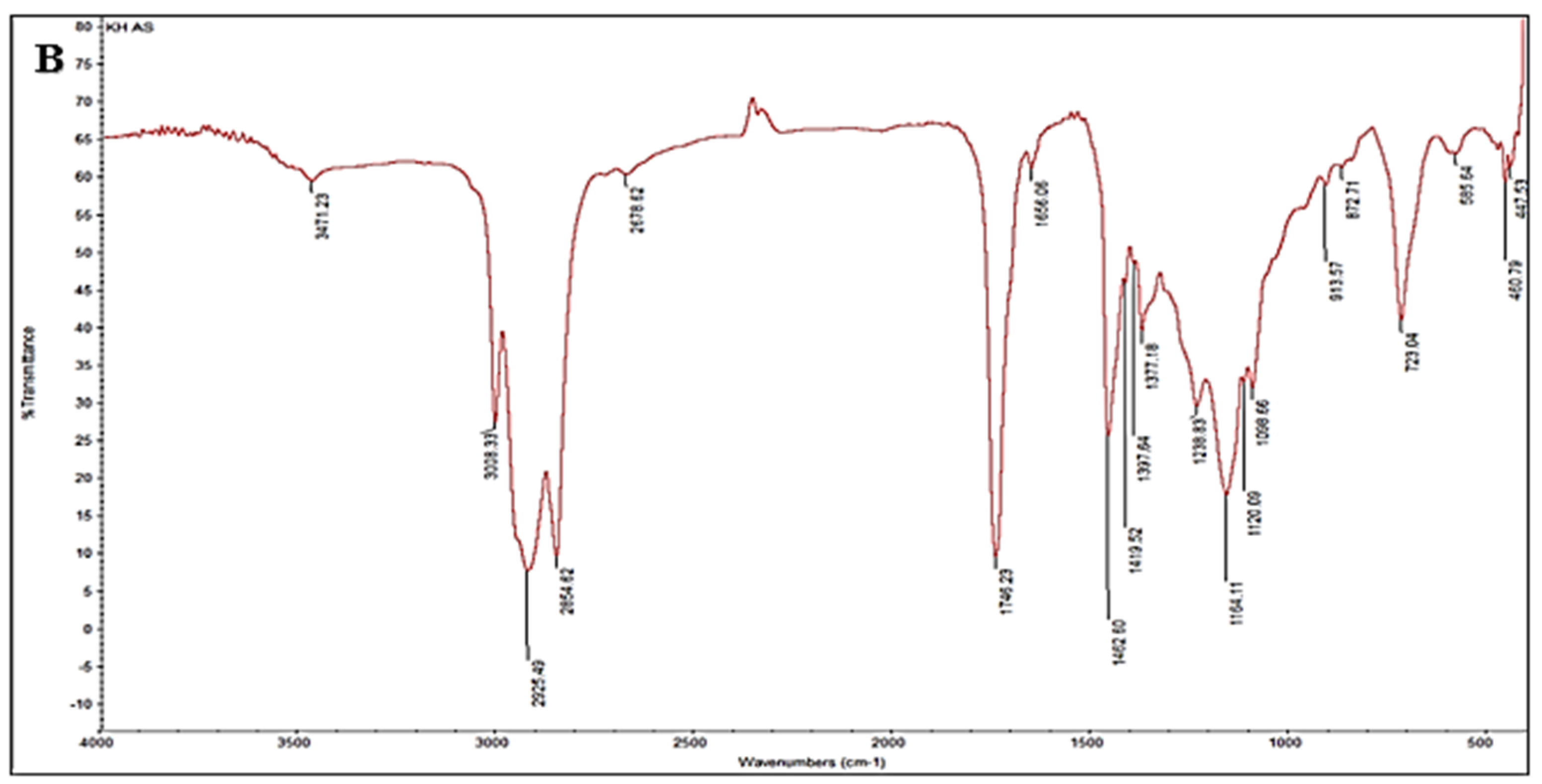
The FTIR pattern of native lettuce oil exhibited 23 characteristic bands at 3471.75, 3008.21, 2925.63, 2854.61, 2731.26, 2676.55, 1746.10, 1656.75, 1533.51, 1462.93, 1419.20, 1377.29, 1239.02, 1164.23, 1120.01, 1088.65, 914.22, 873.79, 722.84, 585.56, 477.83, 460.00, and 433.07 cm−1 (Figure 2A). In this spectrum, the 3500–3000 cm−1 region is taken to indicate features associated with unsaturation (C=C), the 1650–2000 cm−1 region is attributed to C–H bending, and the sharp band at 1746 cm−1 corresponds to C=O stretching of carbonyl groups in triacylglycerols, the principal constituents of vegetable oils. The 400–1650 cm−1 region is assigned to C–O and C–C skeletal vibrations in lettuce oil. Overall, FTIR shows promising utility for the classification of natural oils [40].
By comparison, the FTIR pattern of ozonated lettuce oil displayed 20 bands in analogous regions at 3471.23, 3008.31, 2925.49, 2854.62, 2676.62, 1746.23, 1656.06, 1462.60, 1419.52, 1377.18, 1238.83, 1164.11, 1120.09, 1096.66, 913.57, 872.71, 723.04, 585.64, 460.79, and 447.53 cm−1 (Figure 2B). Notably, the bands at 2731.26, 1533.51, and 477.83 cm−1 observed in the native oil were absent after ozonation. Mechanistically, ozonation disrupts double bonds in unsaturated fatty acids, reflected here by loss of C=C features (around 1500 cm−1) and the appearance of bands attributable to ozonides [41]. In addition, the band near 2700 cm−1 in oil FTIR spectra, typically associated with C–H stretching in aldehydes, was not evident post-ozonation, consistent with a decrease in aldehyde functional groups due to oxidative transformation (e.g., oxidation or epoxidation) into other functionalities [42]. Furthermore, the disappearance of a band near 400 cm−1 suggests the loss of a specific molecular feature, likely a chiral group, during the chemical process, a phenomenon used to monitor oxidative changes in oil composition [43].
Together, these spectral shifts furnish a coherent molecular fingerprint of ozonation in lettuce oil, reinforcing the compositional reprogramming toward oxygenated species that underpins the enhanced bioactivities documented in this study.

3.3. Anti-H. pylori Activity and Bactericidal Efficacy

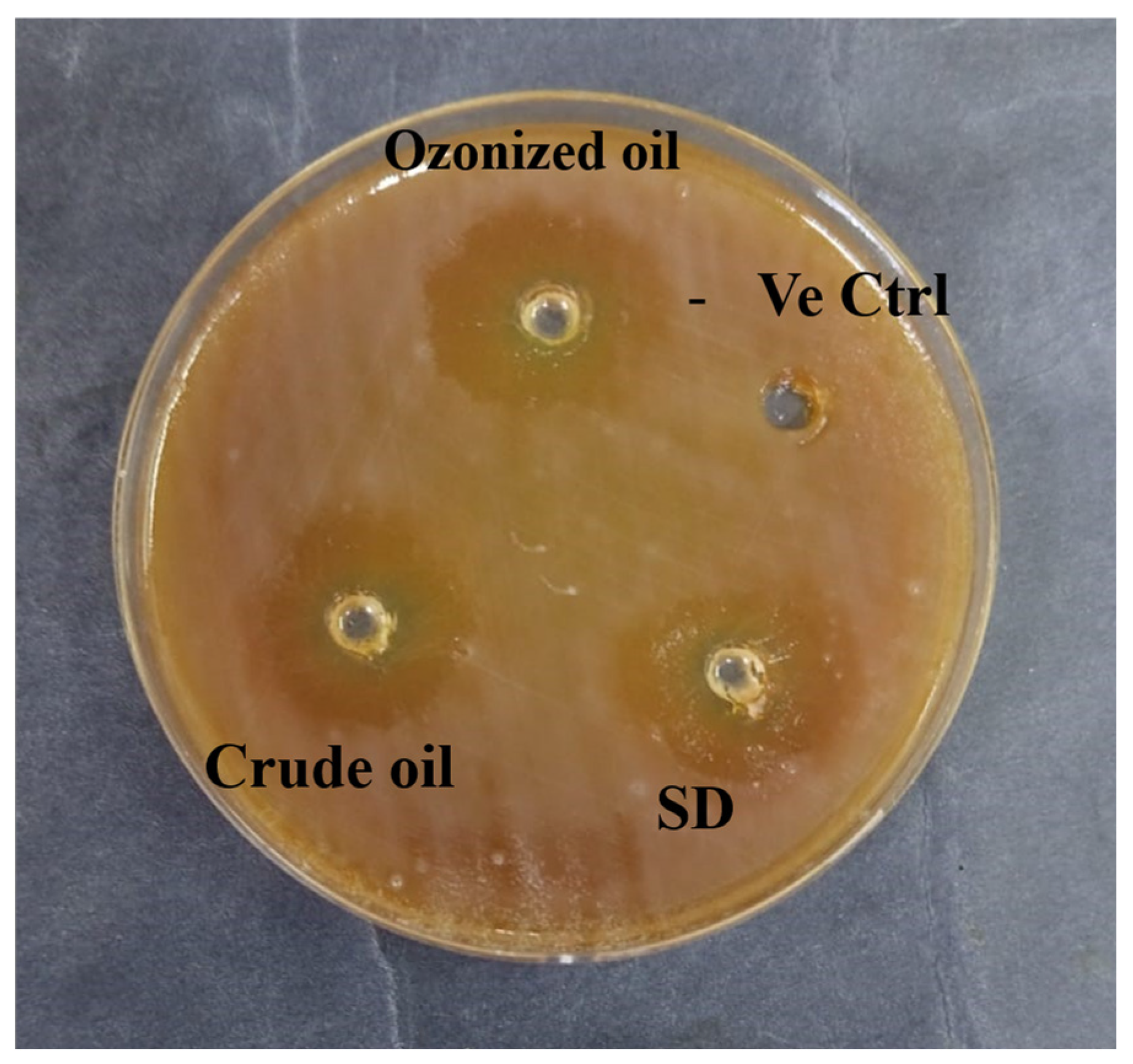
In this work, native lettuce oil exhibited appreciable anti-H. pylori activity with an inhibition zone of 13.7 ± 0.6 mm, compared with the standard drug (15.3 ± 0.4 mm). By contrast, ozonated lettuce oil produced a larger inhibition zone of 21.3 ± 0.3 mm. These findings are corroborated by minimum inhibitory concentration (MIC) and minimum bactericidal concentration (MBC) analyses, where ozonated lettuce oil displayed significantly lower MIC values (31.25 ± 0.5 µg/mL) and MBC values (62.5 ± 0.3 and 125.5 ± 0.8 µg/mL) compared with the native oil and both oil forms demonstrated bactericidal action against H. pylori (Figure 3; Table 2). The superior efficacy of ozonated lettuce oil can be attributed to its enriched content of oxygenated derivatives that arise from ozonolysis of unsaturated fatty acids which have been shown to disrupt bacterial cell membranes, interfere with protein functions, and induce oxidative stress within microbial cells [24]. In the case of H. pylori, which colonizes the gastric mucosa by producing urease and adapting to oxidative stress, these oxygen-enriched metabolites may synergistically impair bacterial survival mechanisms, thereby explaining the observed reduction in MIC and MBC values.
The present findings align with prior studies demonstrating the antimicrobial activity of oils following ozonation. For example, ozonated sunflower and olive oils have been reported to exhibit enhanced antibacterial effects against Gram-positive and Gram-negative pathogens [38]. Ozonated pumpkin seed oil was recently shown to produce larger inhibition zones and superior bactericidal action compared with its native counterpart (Alsalamah et al., 2025) [13]. This suggests that ozonation represents a broadly applicable green biotransformation strategy for augmenting the antimicrobial potential of plant-derived oils.
The therapeutic management of H. pylori infections is becoming increasingly challenging due to the global surge in resistance to antibiotics [44]. This growing resistance crisis necessitates the exploration of novel, cost-effective, and sustainable alternatives. Ozonated lettuce oil is a compelling candidate, capable of exerting potent bactericidal activity at relatively low concentrations. Its natural origin, favorable safety profile, and eco-friendly production process further strengthen its appeal for translational evaluation.

3.4. Antibiofilm Activity Against H. pylori

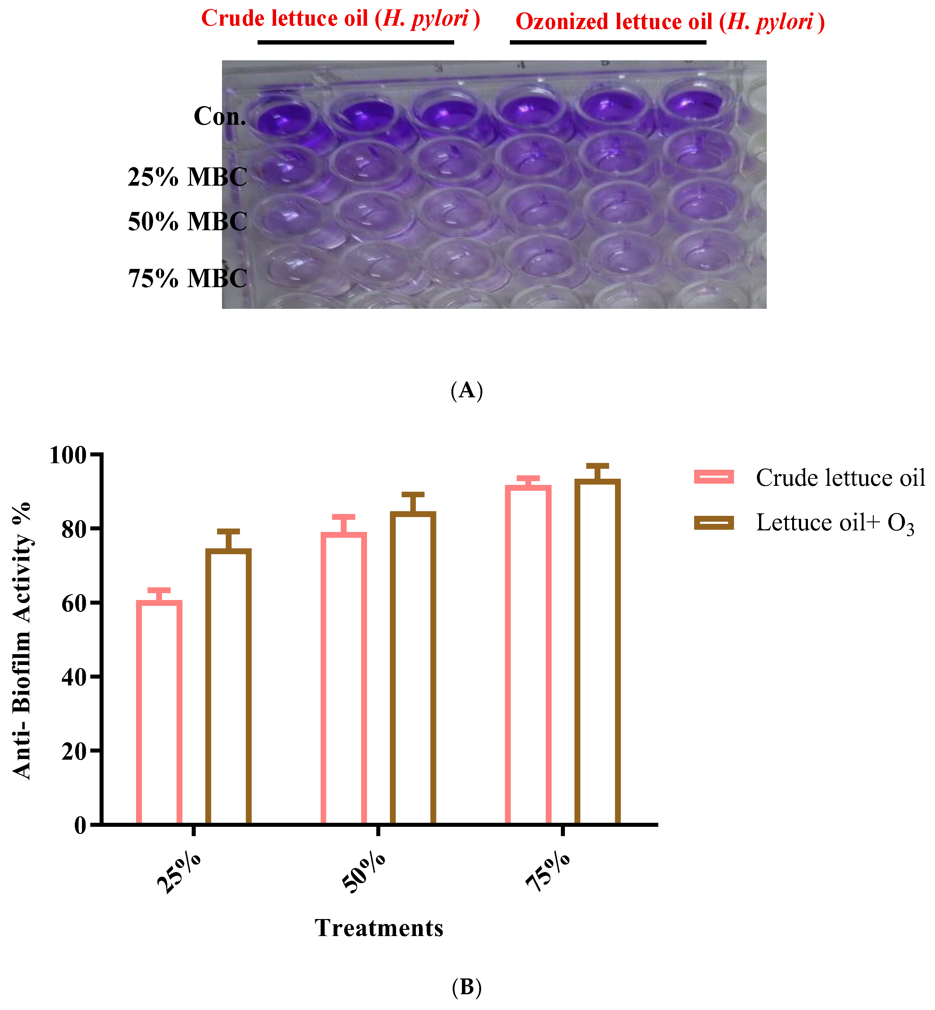
Exposure of lettuce oil to ozone produced a slight improvement in antibiofilm activity against H. pylori, with no statistically significant differences (p > 0.05) across the tested sublethal concentrations corresponding to 25–75% of the MBC (Figure 4). While the magnitude of improvement was modest, the consistent directional aligned with the broader evidence suggesting that ozonation of vegetable oils can subtly modulate antibiofilm properties [45]. However, several vegetable oils possess intrinsic antibiofilm effects, disruption of bacterial cell-to-cell communication (quorum sensing), impeding biofilm formation and facilitating biofilm removal thereby offering complementary or alternative options to conventional antibiotics [46]. Literature further indicates that ozonation can augment these effects, with enhanced antibiofilm performance contributing to the overall anti-H. pylori efficacy of ozonated oils [14,45]. Although lettuce oil showed mild antibiofilm activity, the ability of ozonated lettuce oil to act simultaneously as a bactericidal agent and a biofilm-disrupting adjunct positions it as a biofilm-conscious therapeutic candidate. Integration of ozonated oils with standard eradication regimens may improve treatment efficacy by weakening biofilm resilience, thereby enhancing antibiotic penetration among H. pylori communities.

3.5. Antihemolytic Activity

Lettuce oil exhibited good antihemolytic activity against H. pylori–induced hemolysis, and ozonation produced a slight improvement in antihemolytic effects across various sub-MIC levels (Figure 5). Although direct prior evidence for antihemolytic properties of lettuce oil is limited, research indicates that essential oils from diverse plants can inhibit H. pylori by restraining bacterial growth, and certain lettuce constituents may likewise help counter the infection [47]. Additional studies are needed to evaluate specific lettuce types and to define their efficacy in managing H. pylori, particularly with respect to antihemolytic actions, despite promising activity reported for some oils and botanical extracts. Notably, altering the oil’s phytochemical profile (e.g., via ozonation) may enhance both suppression of H. pylori growth and stabilization of red blood cells, hallmarks of antihemolytic activity. In line with this rationale, ozonated oils (e.g., ozonated coconut oil) have shown enhanced effectiveness and promise as natural therapeutic options for infectious diseases, including viral illnesses [14,48]. Together, these observations nominate ozonated lettuce oil as an attractive, mechanism-informed adjunct for mitigating H. pylori–associated hemolysis and warrant focused mechanistic and in vivo evaluation.

3.6. Antioxidant Capacity (DPPH Assay)

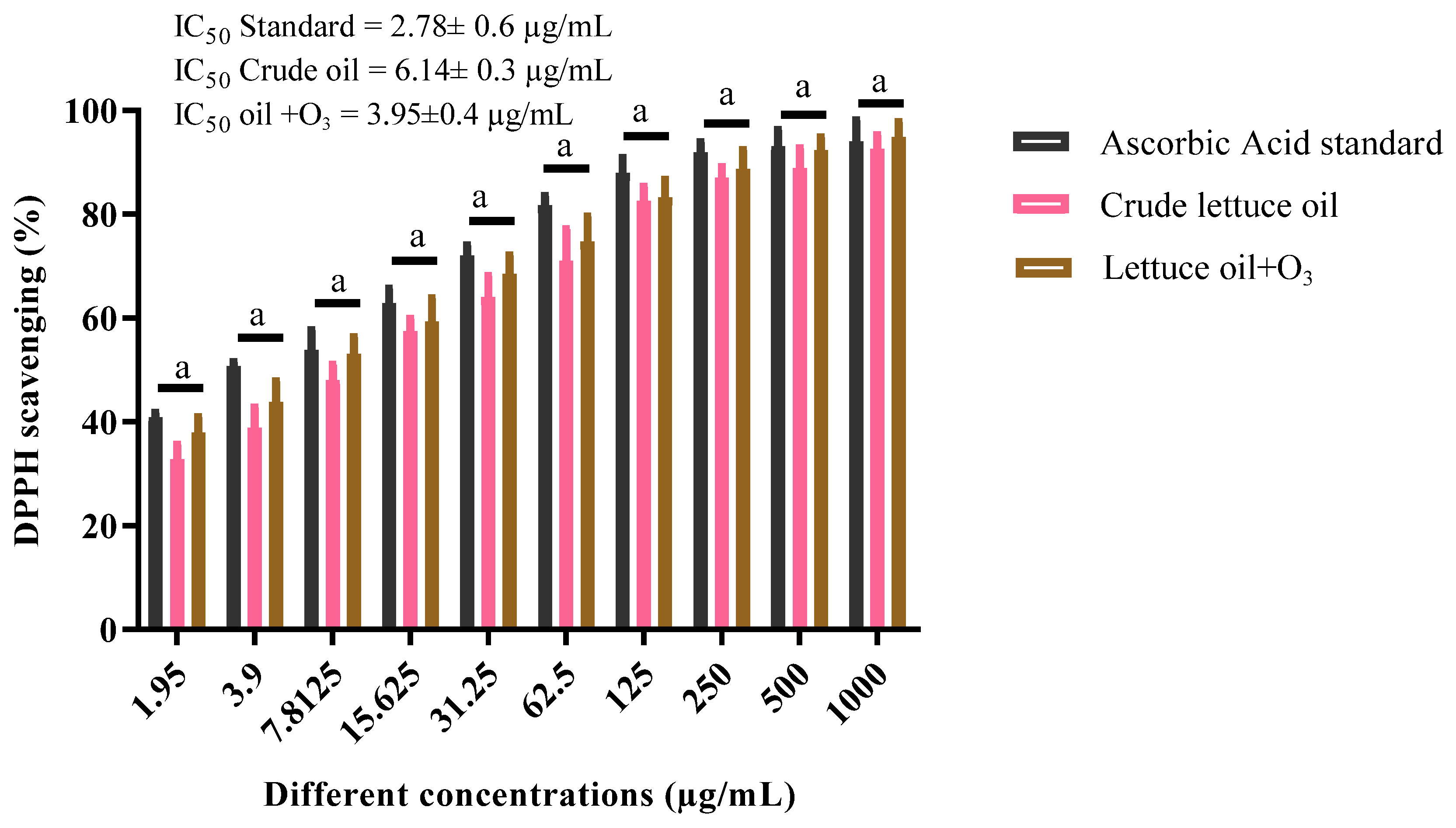
In this work, crude lettuce oil exhibited appreciable antioxidant activity with an IC50 of 6.14 ± 0.3 µg/mL. Ozonation yielded a notable enhancement, reducing the IC50 to 3.95 ± 0.4 µg/mL, in contrast to ascorbic acid used as a reference standard (IC50 = 2.78 ± 0.6 µg/mL). There is a statistically significant difference (p ≤ 0.05) between the IC50 value of the ascorbic acid standard and the IC50 values for both crude and ozonized lettuce oils, with a modest increase in antioxidant capacity observed in the ozonized oil relative to the crude form (Figure 6). Reports indicate that ozonation can enhance the antioxidant properties of several oils [49]. This improvement is largely attributed to the formation of new antioxidant species, with the magnitude of the effect governed by the oil type, inherent composition, and the ozonation dose and duration [24,50]. Collectively, these findings show that controlled ozonation tunes the redox-active constituents of lettuce oil toward stronger radical scavenging, narrowing the gap with ascorbic acid and underscoring its promise as a green, bioactive antioxidant.

3.7. Butyrylcholinesterase Inhibition

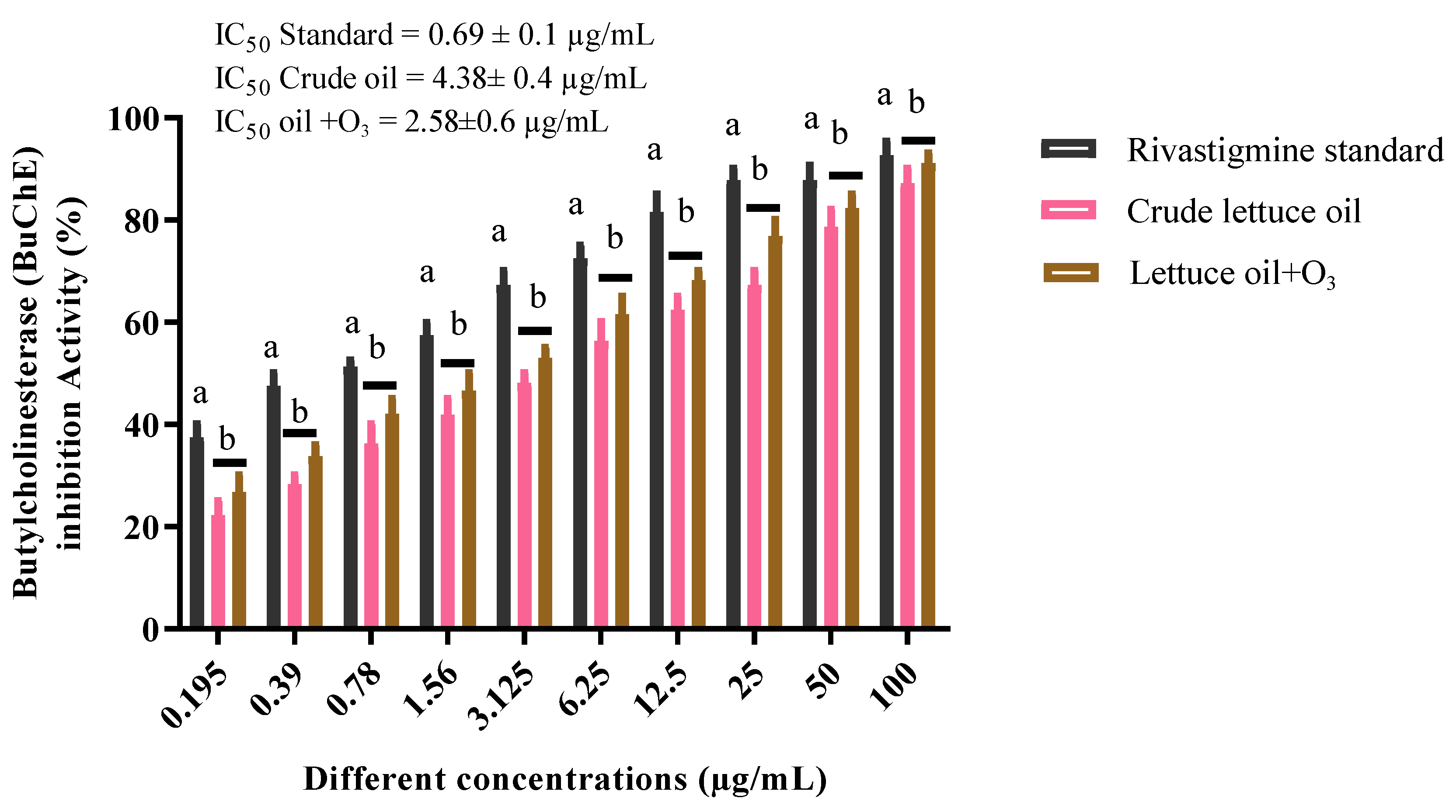
Native lettuce oil exhibited good butyrylcholinesterase (BChE) inhibitory activity with an IC50 of 4.38 ± 0.4 µg/mL. Upon ozonation, inhibition improved markedly, achieving an IC50 of 2.58 ± 0.6 µg/mL, compared with the rivastigmine reference standard (IC50 = 0.96 ± 0.6 µg/mL). There is a statistically significant difference (p ≤ 0.05) between the IC50 value of the rivastigmine standard and the IC50 values for both crude and ozonized lettuce oils, with a slight increase in inhibitory activity for the ozonized oil compared to the crude form (Figure 7). Cognitive dysfunction is a hallmark of Alzheimer’s disease, and BChE inhibition is a validated therapeutic strategy in its management. In support of this pharmacological axis, essential oils from Citrus aurantifolia and Salvia species contain constituents with strong BChE inhibitory potential [51]. Additionally, research indicates that specific fatty acids in vegetable oils can modulate BChE activity, aligning with the present observations. Consistent with previous reports, ozonation enhanced the butyrylcholinesterase inhibitory potential of natural oils, as evidenced by the stronger inhibition observed following ozonation. [48]. Taken together, these results show that ozonated lettuce oil narrows the gap to the reference inhibitor and merits targeted translational evaluation as a green, mechanism aligned BChE modulator.

3.8. Molecular Docking (BChE and H. pylori Urease)

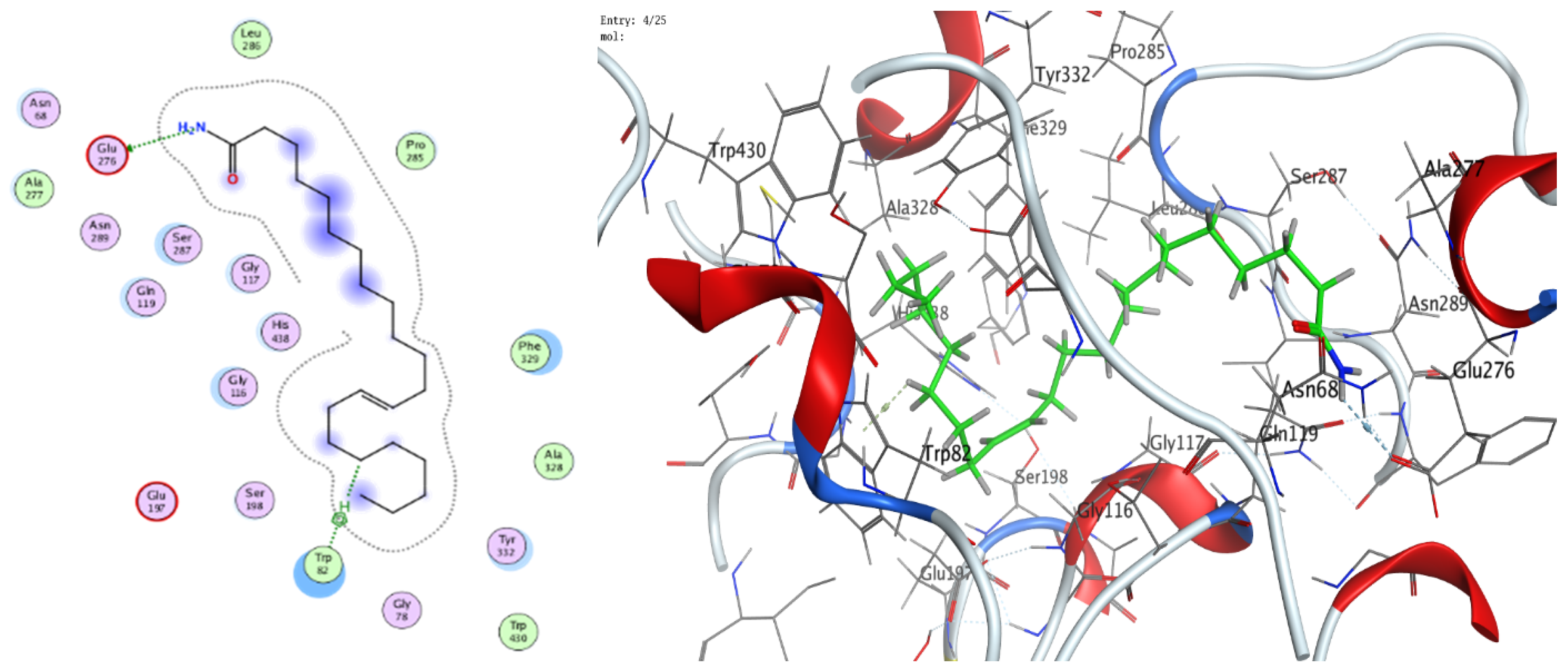
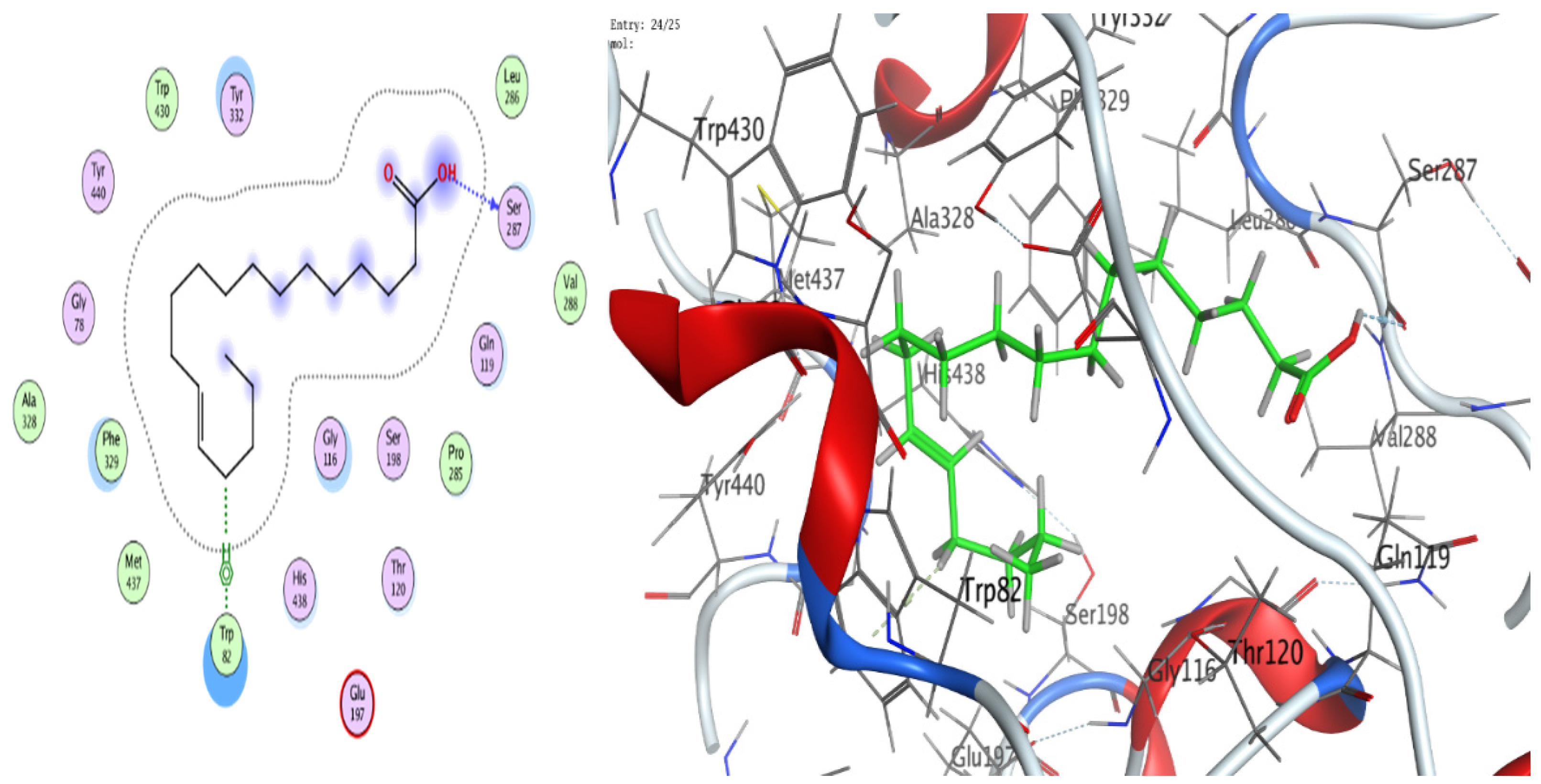
The docking analyses showed that both ligands effectively engage the target proteins. For human BChE, 13-docosenamide displayed superior affinity (best docking score −8.13092 kcal/mol) relative to trans-13-octadecenoic acid (−7.31024 kcal/mol) (Table 3). Interaction analysis indicated that 13-docosenamide formed a hydrogen bond with GLU 276 (distance 3.42 Å, energy −0.6 kcal/mol) and an H–π interaction with TRP 82 (distance 3.72 Å, energy −0.7 kcal/mol) (Table 3). In contrast, trans-13-octadecenoic acid established a hydrogen bond with SER 287 (distance 2.97 Å, energy −1.3 kcal/mol) and an H–π interaction with TRP 82 (distance 3.69 Å, energy −0.8 kcal/mol) (Table 4).
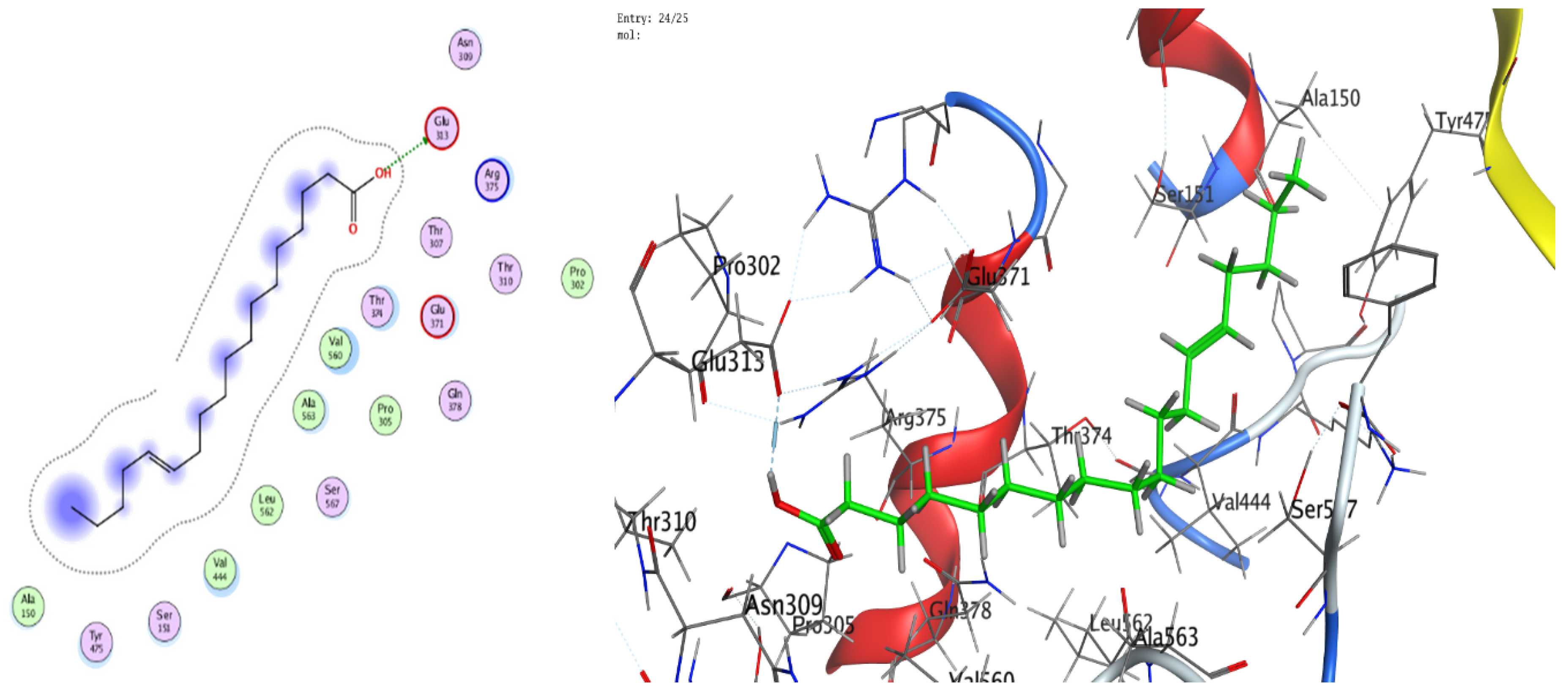
For H. pylori urease, trans-13-octadecenoic acid exhibited stronger binding (best docking score −7.33015 kcal/mol) than 13-docosenamide (−6.00058 kcal/mol) (Table 5). The interaction profile showed a hydrogen bond between 13-docosenamide and ARG 375 (distance 3.05 Å, energy −0.7 kcal/mol) (Table 6), while trans-13-octadecenoic acid formed a particularly strong hydrogen bond with GLU 313 (distance 2.83 Å, energy −6.4 kcal/mol) (Table 5), likely contributing substantially to complex stability. Two-dimensional and three-dimensional interaction diagrams corroborated these binding modes within the active sites of both targets (Figure 8, Figure 9, Figure 10, Figure 11 and Figure 12).
Collectively, the docking results underscore 13-docosenamide as the more promising BChE binder, consistent with the enzyme’s relevance to neurological disorders and the therapeutic value of BChE inhibition in Alzheimer’s disease [52,53]. Conversely, trans-13-octadecenoic acid emerged as the stronger urease ligand, aligning with the enzyme’s pivotal role in H. pylori survival and pathogenicity and highlighting its potential as a lead for anti-H. pylori development [36,54]. These differences are readily rationalized by structural features: the amide functionality in 13-docosenamide supports distinct hydrogen-bonding patterns compared with the carboxylic acid group in trans-13-octadecenoic acid [55], while chain length and saturation modulate hydrophobic contacts and overall pose geometry [56]. Together, these in silico findings mirror the experimental bioactivity trends and nominate both molecules, via complementary target preferences, as tractable scaffolds for optimization and translational advancement.

4. Conclusions

Ozonation of lettuce oil, achieved by bubbling ozone at a flow rate of 0–8 L min−1, altered its chemical composition and generated new molecular classes. Compared with crude oil, the ozonated preparation exhibited higher in vitro anti-H. pylori, antibiofilm, antihemolytic, antioxidant, and butyrylcholinesterase inhibitory activities. Molecular docking supported these experimental findings and revealed complementary target preferences. (Z)-13-Docosenamide bound human butyrylcholinesterase with higher affinity, supporting its candidacy as an inhibitor relevant to neurological disorders including Alzheimer’s disease. Trans-13-octadecenoic acid displayed stronger binding to H. pylori urease, driven in part by a robust hydrogen bond with GLU313, indicating potential as an anti-H. pylori agent. Taken together, these results provide coherent in vitro evidence and a mechanistic foundation showing that ozonation transforms lettuce oil into a richer source of candidate therapeutics, and they motivate formulation, safety, and in vivo efficacy studies to advance translation. Future toxicology studies are needed to highlight the safety of ozonated lettuce oil using animal experiments.

Author Contributions

Conceptualization, A.S.B.; formal analysis, W.H. and A.D.; formal analysis—antibacterial and anti-biofilm activities, A.S.B. and A.D.; formal analysis—antioxidant activity, S.A.A. and M.I.A.; formal analysis—antihemolytic activity, H.Q. and W.H.; formal analysis—anti-Alzheimer’s activity, S.A.A. investigation, A.S.B., S.A.A., M.I.A., W.H., A.D. and H.Q.; visualization and validation, A.S.B., S.A.A., M.I.A., W.H., A.D. and H.Q.; supervision, A.S.B.; funding acquisition, S.A.A. and M.I.A.; writing—original draft preparation, A.S.B. and H.Q.; writing—review and editing; A.S.B., S.A.A., M.I.A., W.H., A.D. and H.Q. All authors have read and agreed to the published version of the manuscript.

Funding

This work was supported and funded by the Deanship of Scientific Research at Imam Mohammad Ibn Saud Islamic University (IMSIU) (grant number IMSIU-DDRSP2501).

Institutional Review Board Statement

Not applicable.

Informed Consent Statement

Not applicable.

Data Availability Statement

The original contributions presented in the study are included in the article, further inquiries can be directed to the corresponding author.

Acknowledgments

All authors thank the Deanship of Scientific Research at Imam Mohammad Ibn Saud Islamic University (IMSIU) for their fund through a grant number of IMSIU-DDRSP2501.

Conflicts of Interest

The authors declare no conflicts of interest.

References

  1. Abdel Ghany, T.M.; Ganash, M.; Alawlaqi, M.M.; Al-Rajhi, A.M.H. Antioxidant, Antitumor, Antimicrobial Activities Evaluation of Musa paradisiaca L. Pseudostem Exudate Cultivated in Saudi Arabia. BioNanoScience 2019, 9, 172–178. [Google Scholar] [CrossRef]
  2. Al-Rajhi, A.M.H.; Abdel Ghany, T.M. Nanoemulsions of some edible oils and their antimicrobial, antioxidant, and anti-hemolytic activities. BioResources 2023, 18, 1465–1481. [Google Scholar] [CrossRef]
  3. Selim, S.; Alruwaili, Y.; Ejaz, H.; Abdalla, A.; Almuhayawi, M.; Nagshabandi, M.; Tarabulsi, M.; Al Jaouni, S.; Bazuhair, M.; Abdelghany, T. Estimation and action mechanisms of cinnamon bark via oxidative enzymes and ultrastructures as antimicrobial, anti-biofilm, antioxidant, anti-Diabetic, and anticancer agents. BioResources 2024, 19, 7019–7041. [Google Scholar] [CrossRef]
  4. Abdelghany, T.M.; Hassan, M.M.; El-Naggar, M.A.; El-Mongy, M.A. GC/MS analysis of Juniperus procera extract and its activity with silver nanoparticles against Aspergillus flavus growth and aflatoxins production. Biotechnol. Rep. 2020, 27, e00496. [Google Scholar] [CrossRef]
  5. Alawlaqi, M.M.; Al-Rajhi, A.M.H.; Abdelghany, T.M.; Ganash, M.; Moawad, H. Evaluation of Biomedical Applications for Linseed Extract: Antimicrobial, Antioxidant, Anti-Diabetic, and Anti-Inflammatory Activities In Vitro. J. Funct. Biomater. 2023, 14, 300. [Google Scholar] [CrossRef]
  6. Almehayawi, M.; Almuhayawi, M.; El-Fadl, S.; Nagshabandi, M.; Tarabulsi, M.; Selim, S.; Alruwaili, Y.; Mostafa, E.; Al Jaouni, S.; Abdelghany, T. Evaluating the anti-yeast, anti-diabetic, wound healing activities of Moringa oleifera extracted at different conditions of pressure via supercritical fluid extraction. BioResources 2024, 19, 5961–5977. [Google Scholar] [CrossRef]
  7. Bazaid, A.S.; Binsaleh, N.K.; Barnawi, H.; Alharbi, B.; Alsolami, A.; Selim, S.; Al Jaouni, S.K.; Saddiq, A.A.; Ganash, M.; Abdelghany, T.M.; et al. Unveiling the in vitro activity of extracted Euphorbia trigona via Supercritical Fluid Extraction against pathogenic yeasts, obesity, cancer, and its wound healing properties. Bioresour. Bioprocess. 2025, 12, 28. [Google Scholar] [CrossRef] [PubMed]
  8. Ahmed, Z.F.R.; Alnuaimi, A.K.H.; Askri, A.; Tzortzakis, N. Evaluation of Lettuce (Lactuca sativa L.) Production under Hydroponic System: Nutrient Solution Derived from Fish Waste vs. Inorganic Nutrient Solution. Horticulturae 2021, 7, 292. [Google Scholar] [CrossRef]
  9. Shi, M.; Gu, J.; Wu, H.; Rauf, A.; Emran, T.B.; Khan, Z.; Mitra, S.; Aljohani, A.S.M.; Alhumaydhi, F.A.; Al-Awthan, Y.S.; et al. Phytochemicals, Nutrition, Metabolism, Bioavailability, and Health Benefits in Lettuce—A Comprehensive Review. Antioxidants 2022, 11, 1158. [Google Scholar] [CrossRef]
  10. Al Nomaani, R.S.; Hossain, M.A.; Weli, A.M.; Al-Riyami, Q.; Al-Sabahi, J.N. Chemical composition of essential oils and in vitro antioxidant activity of fresh and dry leaves crude extracts of medicinal plant of Lactuca Sativa L. native to Sultanate of Oman. Asian Pac. J. Trop. Biomed. 2013, 3, 353–357. [Google Scholar] [CrossRef]
  11. Dyląg, A.; Smoleń, S.; Wisła-Świder, A.; Kowalska, I.; Sularz, O.; Krzemińska, J.; Pitala, J.; Koronowicz, A. Evaluation of the chemical composition and nutritional value of lettuce (Lactuca sativa L.) biofortified in hydroponics with iodine in the form of iodoquinolines. Front. Plant Sci. 2023, 14, 1288773. [Google Scholar] [CrossRef] [PubMed]
  12. Al-Rajhi, A.M.H.; Abdelghany, T.M.; Almuhayawi, M.S.; Alruhaili, M.H.; Saddiq, A.A.; Baghdadi, A.M.; Al Jaouni, S.K.; Albasri, H.M.; Waznah, M.S.; Alraddadi, F.A.; et al. Effect of ozonation on the phytochemicals of black seed oil and its anti-microbial, anti-oxidant, anti-inflammatory, and anti-neoplastic activities in vitro. Sci. Rep. 2024, 14, 30445. [Google Scholar] [CrossRef]
  13. Alsalamah, S.A.; Alghonaim, M.I.; Mohammad, A.M.; Khalel, A.F.; Alqahtani, F.S.; Abdelghany, T.M. Ozone-modified properties of pumpkin seed oil as anti-H. pylori, anticancer, anti-diabetic and anti-obesity agent. Sci. Rep. 2025, 15, 25959. [Google Scholar] [CrossRef]
  14. Silva, V.; Peirone, C.; Amaral, J.S.; Capita, R.; Alonso-Calleja, C.; Marques-Magallanes, J.A.; Martins, Â.; Carvalho, Á.; Maltez, L.; Pereira, J.E.; et al. High Efficacy of Ozonated Oils on the Removal of Biofilms Produced by Methicillin-Resistant Staphylococcus aureus (MRSA) from Infected Diabetic Foot Ulcers. Molecules 2020, 25, 3601. [Google Scholar] [CrossRef]
  15. Repciuc, C.C.; Vișan, G.-A.-M.; Teleky, B.-E.; Pintea, A.; Novac, C.Ș.; Oros, N.V. Physico-Chemical and Antimicrobial Evaluation of Ozonated Olive Oil Produced with a Medical-Grade Generator for Veterinary Purposes. Microorganisms 2025, 13, 1932. [Google Scholar]
  16. Sathwik, M.; Komarraju, S.; D, S.; Muralidharan, S. Technical Considerations of Ozonated Oils in Medical Applications: A Narrative Review. Cureus 2025, 17, e83185. [Google Scholar] [CrossRef]
  17. Aguilera Matos, I.; Diaz Oliva, S.E.; Escobedo, A.A.; Villa Jiménez, O.M.; Velazco Villaurrutia, Y.d.C. Helicobacter pylori infection in children. BMJ Paediatr. Open 2020, 4, e000679. [Google Scholar] [CrossRef]
  18. Malfertheiner, P.; Camargo, M.C.; El-Omar, E.; Liou, J.M.; Peek, R.; Schulz, C.; Smith, S.I.; Suerbaum, S. Helicobacter pylori infection. Nat. Rev. Dis. Primers 2023, 9, 19. [Google Scholar] [CrossRef] [PubMed]
  19. Scott, D.R.; Sachs, G.; Marcus, E.A. The role of acid inhibition in Helicobacter pylori eradication. F1000Research 2016, 5, 1747. [Google Scholar] [CrossRef]
  20. Elbehiry, A.; Abalkhail, A.; Anajirih, N.; Alkhamisi, F.; Aldamegh, M.; Alramzi, A.; AlShaqi, R.; Alotaibi, N.; Aljuaid, A.; Alzahrani, H.; et al. Helicobacter pylori: Routes of Infection, Antimicrobial Resistance, and Alternative Therapies as a Means to Develop Infection Control. Diseases 2024, 12, 311. [Google Scholar] [CrossRef]
  21. Cho, J.-H.; Jin, S.-Y. Efficacy and Safety of Modified Bismuth Quadruple Therapy for First-Line Helicobacter pylori Eradication: A Systematic Review and Meta-Analysis of Randomized Controlled Trials. Microorganisms 2025, 13, 519. [Google Scholar] [CrossRef]
  22. Muhammed, M.T.; Aki-Yalcin, E. Molecular Docking: Principles, Advances, and its Applications in Drug Discovery. Lett. Drug Des. Discov. 2024, 21, 480–495. [Google Scholar] [CrossRef]
  23. Qanash, H.; Alotaibi, K.; Aldarhami, A.; Bazaid, A.S.; Ganash, M.; Saeedi, N.H.; Ghany, T.A. Effectiveness of Oil-based Nanoemulsions with Molecular Docking of its Antimicrobial Potential. BioResources 2023, 18, 1554–1576. [Google Scholar] [CrossRef]
  24. Ugazio, E.; Tullio, V.; Binello, A.; Tagliapietra, S.; Dosio, F. Ozonated Oils as Antimicrobial Systems in Topical Applications. Their Characterization, Current Applications, and Advances in Improved Delivery Techniques. Molecules 2020, 25, 334. [Google Scholar] [CrossRef] [PubMed]
  25. Al-Rajhi, A.M.H.; Abdelghany, T.M.; Selim, S.; Almuhayawi, M.S.; Alruhaili, M.H.; Alharbi, M.T.; Al Jaouni, S.K. Phytochemical characterization of peanut oil and its ozonized form to explore biological activities in vitro. AMB Express 2025, 15, 76. [Google Scholar] [CrossRef]
  26. Muráriková, A.; Ťažký, A.; Neugebauerová, J.; Planková, A.; Jampílek, J.; Mučaji, P.; Mikuš, P. Characterization of Essential Oil Composition in Different Basil Species and Pot Cultures by a GC-MS Method. Molecules 2017, 22, 1221. [Google Scholar] [CrossRef]
  27. Mao, X.; Chen, W.; Huyan, Z.; Sherazi, S.T.H.; Yu, X. Impact of linolenic acid on oxidative stability of rapeseed oils. J. Food Sci. Technol. 2020, 57, 3184–3192. [Google Scholar] [CrossRef] [PubMed]
  28. Castillo-Juárez, I.; Rivero-Cruz, F.; Celis, H.; Romero, I. Anti-Helicobacter pylori activity of anacardic acids from Amphipterygium adstringens. J. Ethnopharmacol. 2007, 114, 72–77. [Google Scholar] [CrossRef] [PubMed]
  29. Shakir, S.M.; Otiso, J.; Keller, G.; Heule, H.V.; Osborn, L.J.; Cole, N.; Schuetz, A.N.; Richter, S.S.; Couturier, M.R. Multicenter Evaluation of a Gradient Diffusion Method for Antimicrobial Susceptibility Testing of Helicobacter pylori. Microbiol. Spectr. 2022, 10, e0211121. [Google Scholar] [CrossRef] [PubMed]
  30. Huang, X.; Liu, Y.; Lin, Z.; Wu, B.; Nong, G.; Chen, Y.; Lu, Y.; Ji, X.; Zhou, X.; Suo, B.; et al. Minimum inhibitory concentrations of commonly used antibiotics against Helicobacter Pylori: A multicenter study in South China. PLoS ONE 2021, 16, e0256225. [Google Scholar] [CrossRef]
  31. Selim, S.; Abdelghany, T.M.; Almuhayawi, M.S.; Nagshabandi, M.K.; Tarabulsi, M.K.; Elamir, M.Y.M.; Alharbi, A.A.; Al Jaouni, S.K. Biosynthesis and activity of Zn-MnO nanocomposite in vitro with molecular docking studies against multidrug resistance bacteria and inflammatory activators. Sci. Rep. 2025, 15, 2032. [Google Scholar] [CrossRef]
  32. Brand-Williams, W.; Cuvelier, M.E.; Berset, C. Use of a free radical method to evaluate antioxidant activity. LWT—Food Sci. Technol. 1995, 28, 25–30. [Google Scholar] [CrossRef]
  33. Al-Rajhi, A.M.H.; Qanash, H.; Almashjary, M.N.; Hazzazi, M.S.; Felemban, H.R.; Abdelghany, T.M. Anti-Helicobacter pylori, Antioxidant, Antidiabetic, and Anti-Alzheimer’s Activities of Laurel Leaf Extract Treated by Moist Heat and Molecular Docking of Its Flavonoid Constituent, Naringenin, against Acetylcholinesterase and Butyrylcholinesterase. Life 2023, 13, 1512. [Google Scholar] [CrossRef]
  34. Boittier, E.D.; Tang, Y.Y.; Buckley, M.E.; Schuurs, Z.P.; Richard, D.J.; Gandhi, N.S. Assessing Molecular Docking Tools to Guide Targeted Drug Discovery of CD38 Inhibitors. Int. J. Mol. Sci. 2020, 21, 5183. [Google Scholar] [CrossRef]
  35. Atatreh, N.; Al Rawashdah, S.; Al Neyadi, S.S.; Abuhamdah, S.M.; Ghattas, M.A. Discovery of new butyrylcholinesterase inhibitors via structure-based virtual screening. J. Enzym. Inhib. Med. Chem. 2019, 34, 1373–1379. [Google Scholar] [CrossRef]
  36. Masetti, M.; Falchi, F.; Gioia, D.; Recanatini, M.; Ciurli, S.; Musiani, F. Targeting the Protein Tunnels of the Urease Accessory Complex: A Theoretical Investigation. Molecules 2020, 25, 2911. [Google Scholar] [CrossRef] [PubMed]
  37. Iqbal, D.; Alsaweed, M.; Jamal, Q.M.S.; Asad, M.R.; Rizvi, S.M.D.; Rizvi, M.R.; Albadrani, H.M.; Hamed, M.; Jahan, S.; Alyenbaawi, H. Pharmacophore-Based Screening, Molecular Docking, and Dynamic Simulation of Fungal Metabolites as Inhibitors of Multi-Targets in Neurodegenerative Disorders. Biomolecules 2023, 13, 1613. [Google Scholar] [CrossRef] [PubMed]
  38. Radzimierska-Kaźmierczak, M.; Śmigielski, K.; Sikora, M.; Nowak, A.; Plucińska, A.; Kunicka-Styczyńska, A.; Czarnecka-Chrebelska, K.H. Olive Oil with Ozone-Modified Properties and Its Application. Molecules 2021, 26, 3074. [Google Scholar] [CrossRef]
  39. Gutarowska, B.; Szulc, J.; Jastrząbek, K.; Kręgiel, D.; Śmigielski, K.; Cieciura-Włoch, W.; Mroczyńska-Florczak, M.; Liszkowska, W.; Rygała, A.; Berłowska, J. Effectiveness of Ozonation for Improving the Microbiological Safety of Fresh-Cut Parsley (Petroselinum crispum) Leaves. Appl. Sci. 2023, 13, 8946. [Google Scholar] [CrossRef]
  40. Gardeli, C.; Sykioti, S.; Exarchos, G.; Koliatsou, M.; Andritsos, P.; Panagou, E.Z. The Differentiation of Extra Virgin Olive Oil from Other Olive Oil Categories Based on FTIR Spectroscopy and Random Forest. Appl. Sci. 2025, 15, 1061. [Google Scholar] [CrossRef]
  41. Matwijczuk, A.; Oniszczuk, T.; Matwijczuk, A.; Chruściel, E.; Kocira, A.; Niemczynowicz, A.; Wójtowicz, A.; Combrzyński, M.; Wiącek, D. Use of FTIR Spectroscopy and Chemometrics with Respect to Storage Conditions of Moldavian Dragonhead Oil. Sustainability 2019, 11, 6414. [Google Scholar] [CrossRef]
  42. Koczoń, P.; Hołaj-Krzak, J.T.; Palani, B.K.; Bolewski, T.; Dąbrowski, J.; Bartyzel, B.J.; Gruczyńska-Sękowska, E. The Analytical Possibilities of FT-IR Spectroscopy Powered by Vibrating Molecules. Int. J. Mol. Sci. 2023, 24, 1013. [Google Scholar] [CrossRef]
  43. Ye, Q.; Meng, X.; Pang, L. D2O assisted FTIR spectroscopic analysis of moisture in edible oil. Food Chem. X 2023, 18, 100679. [Google Scholar] [CrossRef]
  44. Puxeddu, S.; Scano, A.; Scorciapino, M.A.; Delogu, I.; Vascellari, S.; Ennas, G.; Manzin, A.; Angius, F. Physico-Chemical Investigation and Antimicrobial Efficacy of Ozonated Oils: The Case Study of Commercial Ozonated Olive and Sunflower Seed Refined Oils. Molecules 2024, 29, 679. [Google Scholar] [CrossRef] [PubMed]
  45. Hidalgo, A.; Bravo, D.; Soto, C.; Maturana, G.; Cordero-Machuca, J.; Zúñiga-López, M.C.; Oyarzun-Ampuero, F.; Quest, A.F.G. The Anti-Oxidant Curcumin Solubilized as Oil-in-Water Nanoemulsions or Chitosan Nanocapsules Effectively Reduces Helicobacter pylori Growth, Bacterial Biofilm Formation, Gastric Cell Adhesion and Internalization. Antioxidants 2023, 12, 1866. [Google Scholar] [CrossRef]
  46. Elbestawy, M.K.M.; El-Sherbiny, G.M.; Moghannem, S.A. Antibacterial, Antibiofilm and Anti-Inflammatory Activities of Eugenol Clove Essential Oil against Resistant Helicobacter pylori. Molecules 2023, 28, 2448. [Google Scholar] [CrossRef] [PubMed]
  47. Alharbi, B.; Qanash, H.; Almashjary, M.N.; Barnawi, H.; Aldarhami, A.; Alsaif, G.; Alsamaan, F.; Monjed, M.K.; Al Shmrany, H.; Bazaid, A.S. Watercress oil loaded with gel: Evaluation of hemolysis inhibition, antioxidant, antimicrobial, and healing properties. Front. Pharmacol. 2024, 15, 1424369. [Google Scholar] [CrossRef]
  48. Almuhayawi, M.S.; Alruhaili, M.H.; Tarabulsi, M.K.; Al Jaouni, S.K.; Alqurashi, A.A.; Alraddadi, F.A.; Bukhari, D.A.; Albasri, H.M.; Waznah, M.S.; Selim, S. Pharmacological activities and phytochemical evaluation of coconut crude oil and upon exposure to ozone. AMB Express 2025, 15, 3. [Google Scholar] [CrossRef]
  49. Cho, K.H.; Kang, D.J.; Nam, H.S.; Kim, J.H.; Kim, S.Y.; Lee, J.O.; Kim, B.J. Ozonated Sunflower Oil Exerted Protective Effect for Embryo and Cell Survival via Potent Reduction Power and Antioxidant Activity in HDL with Strong Antimicrobial Activity. Antioxidants 2021, 10, 1651. [Google Scholar] [CrossRef]
  50. Mohammad, A.A.; Salem, S.H.; Amer, H.M.; Hussein, M.S. Ozone as a postharvest treatment to maintain the quality characteristics of fresh-cut plants. J. Food Meas. Charact. 2025, 19, 4325–4336. [Google Scholar] [CrossRef]
  51. Calva, J.; Ludeña, C.; Bec, N.; Larroque, C.; Salinas, M.; Vidari, G.; Armijos, C. Constituents and Selective BuChE Inhibitory Activity of the Essential Oil from Hypericum aciculare Kunth. Plants 2023, 12, 2621. [Google Scholar] [CrossRef]
  52. Duran, H.E. Pyrimidines: Molecular docking and inhibition studies on carbonic anhydrase and cholinesterases. Biotechnol. Appl. Biochem. 2023, 70, 68–82. [Google Scholar] [CrossRef] [PubMed]
  53. Li, L.; Zhao, P.; Yang, C.; Yin, Q.; Wang, N.; Liu, Y.; Li, Y. Discovery, Biological Evaluation and Binding Mode Investigation of Novel Butyrylcholinesterase Inhibitors Through Hybrid Virtual Screening. Molecules 2025, 30, 2093. [Google Scholar] [CrossRef] [PubMed]
  54. Almarmouri, C.; El-Gamal, M.I.; Haider, M.; Hamad, M.; Qumar, S.; Sebastian, M.; Ghemrawi, R.; Muhammad, J.S.; Burucoa, C.; Khoder, G. Anti-urease therapy: A targeted approach to mitigating antibiotic resistance in Helicobacter pylori while preserving the gut microflora. Gut Pathog. 2025, 17, 37. [Google Scholar] [CrossRef] [PubMed]
  55. El-Gazzar, N.; Said, L.; Al-Otibi, F.O.; AbdelGawwad, M.R.; Rabie, G. Antimicrobial and cytotoxic activities of natural (Z)-13-docosenamide derived from Penicillium chrysogenum. Front. Cell. Infect. Microbiol. 2025, 15, 1529104. [Google Scholar] [CrossRef]
  56. Sun, Q. The Hydrophobic Effects: Our Current Understanding. Molecules 2022, 27, 7009. [Google Scholar] [CrossRef]
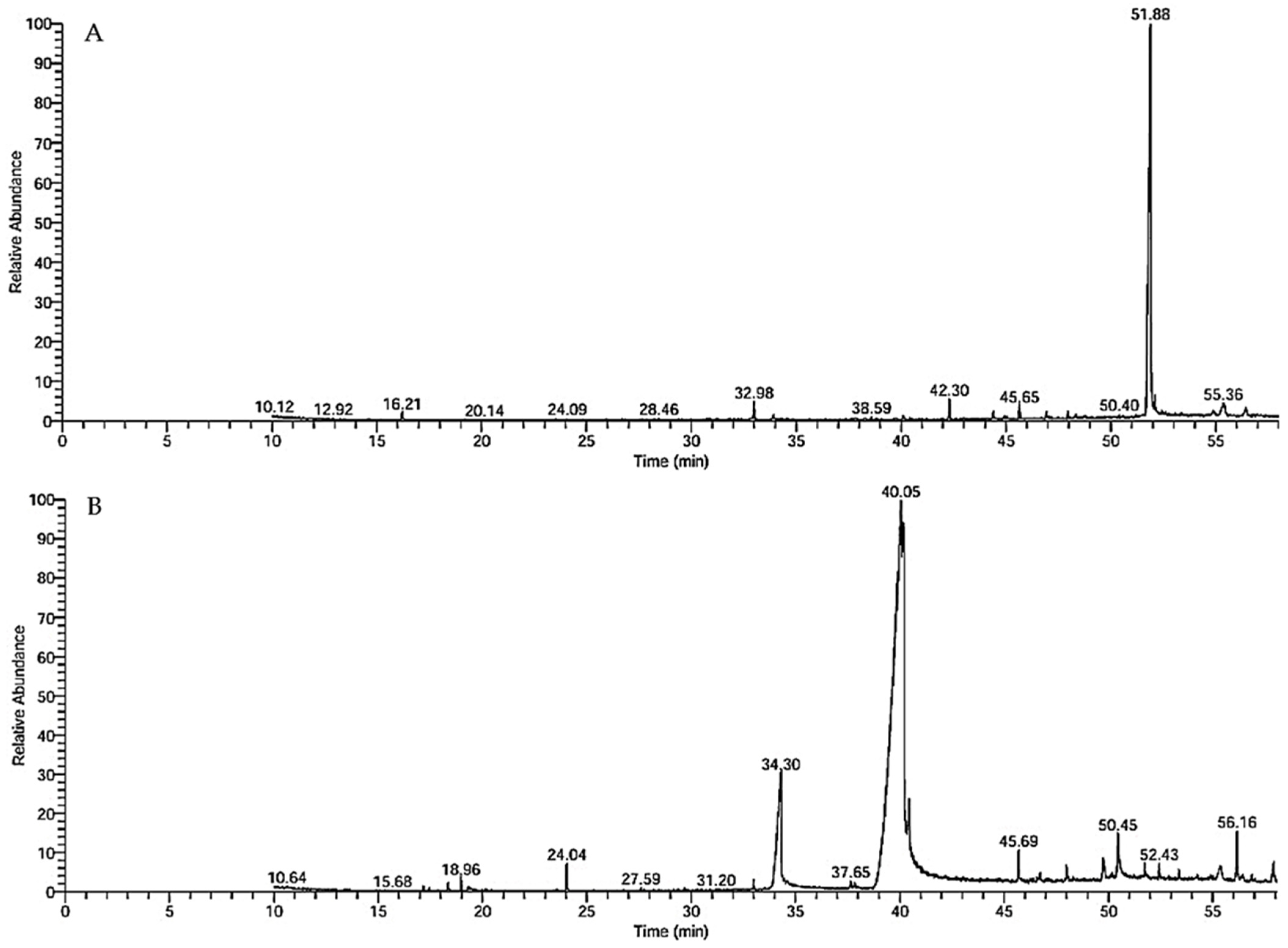
Figure 1. GC–MS chromatograms of (A) crude lettuce oil and (B) ozonated lettuce oil.
Foods 14 03458 g001
Figure 2. FTIR spectra of (A) crude lettuce oil and (B) ozonated lettuce oil.
Foods 14 03458 g002aFoods 14 03458 g002b
Figure 3. Anti-H. pylori activity of crude and ozonated lettuce oils assessed by the agar well-diffusion assay.
Foods 14 03458 g003
Figure 4. Antibiofilm activity against H. pylori of crude and ozonated lettuce oils. (A) Representative 96-well plate at various percentages of the MBC. (B) Bar graphs depicting differences between the oil forms. “Con.” in panel A denotes the control.
Foods 14 03458 g004
Figure 5. Inhibition of hemolysis in the presence of H. pylori by crude (A) and ozonated (B) lettuce oils. For both panels, conditions are arranged as follows: (1) H. pylori only, (2) 25% MIC, (3) 50% MIC, (4) 75% MIC, and (5) untreated; the SDS-induced complete-hemolysis control is also designated as 5 in the original panel. (C) Bar graphs illustrate differences between the oil forms; similar letters above columns indicate non-significant differences (p > 0.05).
Foods 14 03458 g005
Figure 6. Antioxidant capacity of crude lettuce oil and ozonated lettuce oil relative to the ascorbic acid standard. Data are presented as the mean ± SD; similar letters above columns indicate non-significant differences (R2 value = 0.9859; p > 0.05).
Foods 14 03458 g006
Figure 7. Butyrylcholinesterase (BChE) inhibition by crude lettuce oil and ozonated lettuce oil relative to the rivastigmine standard. Data are expressed as mean ± SD; different letters above the columns indicate significant differences among treatments (R2 value = 0.9555; p ≤ 0.05).
Foods 14 03458 g007
Figure 8. Two- and three-dimensional diagrams illustrating the interactions between (Z)-13-docosenamide and the active site of human butyrylcholinesterase (PDB: 4TPK).
Foods 14 03458 g008
Figure 9. Two- and three-dimensional diagrams illustrating the interactions between trans-13-octadecenoic acid and the active site of human butyrylcholinesterase (PDB: 4TPK).
Foods 14 03458 g009
Figure 10. Two- and three-dimensional diagrams illustrating the interactions between (Z)-13-docosenamide and the active site of H. pylori urease (PDB: 1E9Z).
Foods 14 03458 g010
Figure 11. Two-dimensional (2D) and three-dimensional (3D) diagrams illustrate the interactions between trans-13-octadecenoic acid and the active sites of H. pylori urease (PDB: 1E9Z).
Foods 14 03458 g011
Figure 12. Representative key illustrating the interaction types between ligands and selected protein receptors.
Foods 14 03458 g012
Table 1. Various chemical constituents identified in crude and ozonated lettuce oils by GC-MS.
Lettuce OilsOzonated Lettuce Oils
Compound NameMolecular
Formula
Molecular WeightRT (Raw)Area (%)ClassCompound NameMolecular
Formula
Molecular WeightRT (O3)Area (%)Class
Benzaldehyde, 2,5-dimethyl-C9H10O13416.210.97Aromatic aldehyde2-Decenal, (Z)-C10H18O15417.450.16Aldehyde
7,9-Di-tert-butyl-1-ox aspiro(4,5) deca-6,9-d iene-2,8-dioneC17H24O327632 982.38Spirocyclic diketone2,4-Decadienal, (E, E)-C10H16O15218.961.04Aldehyde
n-Hexadecanoic acidC16H32O225633.900.94Fatty acid1,3-Benzodioxol-5-olC7H6O313819.320.24Benzodioxole
Heptadecanoic acid, 9-methyl-, methyl esterC19H38O229838.580.44Fatty acid esterMethyl 4,4,7-trimethyl- 4,7-dihydroindan-6-carboxylateC14H20O222024.041.36Indane
HexadecanamideC16H33NO25540.110.57Fatty amideE-7-TetradecenolC14H28O21227.590.11Fatty alcohol
trans-13-Octadecenoic acidC18H34O228242.304.04Fatty acid7,9-Di-tert-butyl-1-ox aspiro(4,5) deca-6,9-d iene-2,8-dioneC17H24O327632.980.53Sspirocyclic diketone
9-Octadecenamide, (Z)-C18H35NO28144.391.4Fatty amidetrans-13-Octadecenoic acidC18H34O228234.2915.39Fatty acid
Eicosanoic acid, ethyl esterC22H44O234045.050.36Fatty acid ester9,12-Octadecadienoic acid, methyl ester, (E, E)-C19H34O229437.654.86Fatty acid ester
Phenol, 2,2′-methylenebis[6-(1,1-dimethylethyl)-4-methyl-]C23H32O234045.641.94Phenol9-Octadecenoic acid, methyl ester, (E)-C19H36O229637.840.70Fatty acid ester
Methyl erucate C23H44O235246.931.71Fatty acid
ester
13-Docosenamide, (Z)C22H43NO28240.0555.96Fatty amide
Isochiapin BC19H22O634647.031.10Sesquiterpene lactoneOctadecanoic acidC18H36O228440.53.74Fatty acid
1,2-Benzenedicarboxylic acidC24H38O439047.960.83Carboxylic acidPhenol, 2,2′-methylenebis[6-(1,1-dimethylethyl)-4-methyl-]C23H32O234045.691.83Phenol
13-Docosenamide, (Z)-C22H43NO33751.8678.41Fatty amideLinoleic acid ethyl esterC20H36O230846.590.81Fatty acid
ester
Ethyl iso-allocholateC26H44O543652.430.44Steroid12-Methyl-E, E-2,13 octadecadien-1-olC19H36O28047.060.17Fatty alcohol
Oleic acid, 3-(octadecyloxy)prop yl esterC39H76O359254.882.89Fatty acid estern-Propyl 9,12-octadecadienoateC21H38O232248.010.68Fatty acid
ester
DotriacontaneC32H6645056.431.58AlkaneOleic AcidC18H34O228249.731.48Fatty acid
      trans-9-Octadecenoic acid, pentyl esterC23H44O235250.453.74Fatty acid
ester
      Ethyl iso-allocholateC26H44O543651.731.03Steroid
      n-Hexadecanoic acidC16H32O225652.430.76Fatty acid
      9-Octadecenoic acid (Z)-, oxiranylmethyl esterC21H38O333854.270.95Fatty acid
ester
      DotriacontaneC32H6645055.732.57Alkane
      ç-SitosterolC29H50O41457.011.89Phytosterol
Table 2. Anti-H. pylori activity, MIC, and MBC of the tested oil types. Data are presented as the mean ± SD. Different letters above numbers in the same column indicate a significant difference (p ≤ 0.05).
Sample CodeInhibition Zone (mm)MIC
µg/mL
MBC
µg/mL
MBC/MIC
Crude lettuce oil13.7 ± 0.6 a62.5 ± 0.3 a125 ± 0.8 a2 a
Lettuce oil + O321.3 ± 0.3 a31.25 ± 0.5 b62.5 ± 0.2 b2 a
Standard drug15.3 ± 0.4 a31.25 ± 0.6 b13.25 ± 0.6 c1 b
Table 3. Docking scores and energy terms for (Z)-13-docosenamide and trans-13-octadecenoic acid docked into the crystal structure of human butyrylcholinesterase (BChE) (PDB: 4TPK).
MolSrmsd_refineE_confE_placeE_score1E_refineE_score2
13-Docosenamide−8.130921.5926156−19.6867−72.8444−9.60851−39.1181−8.13092
13-Docosenamide−8.048351.7194235−20.1995−74.5038−9.65519−41.7513−8.04835
13-Docosenamide−7.629812.3539269−19.1456−40.5661−10.4494−41.0256−7.62981
13-Docosenamide−7.547241.1664486−7.69996−69.2003−9.77358−36.6556−7.54724
13-Docosenamide−7.522641.743559−12.6639−74.552−9.41631−32.9138−7.52264
trans-13-Octadecenoic acid−7.310242.458077−13.897−58.9793−9.9934−40.7344−7.31024
trans-13-Octadecenoic acid−7.199111.197924−13.8079−67.8339−9.94211−32.6767−7.19911
trans-13-Octadecenoic acid−7.190941.1504909−16.0386−58.7043−9.75688−39.1273−7.19094
trans-13-Octadecenoic acid−7.100333.2766879−16.3373−62.915−10.9963−36.7538−7.10033
trans-13-Octadecenoic acid−7.09481.0196184−11.9226−62.6675−10.2318−34.4483−7.0948
Table 4. Interactions of (Z)-13-docosenamide and trans-13-octadecenoic acid with the structure of human butyrylcholinesterase (BChE) (PDB: 4TPK).
MolLigandReceptorInteractionDistance (Å)E (kcal/mol)
13-DocosenamideN  2OE1  GLU 276 (A)H-donor3.42−0.6
  C  355-ring TRP   82 (A)H-pi3.72−0.7
trans-13-Octadecenoic acidO  1O   SER 287 (A)H-donor2.97−1.3
  C  396-ring TRP  82 (A)H-pi3.69−0.8
Table 5. Docking scores and energy terms for (Z)-13-docosenamide and trans-13-octadecenoic acid docked into the crystal structure of H. pylori urease (PDB: 1E9Z).
MolSrmsd_refineE_confE_placeE_score1E_refineE_score2
13-Docosenamide−6.000582.5410826−21.2019−42.7297−9.22741−32.8763−6.00058
13-Docosenamide−5.96662.395309−19.3768−88.8088−9.60097−32.495−5.9666
13-Docosenamide−5.94151.8721349−17.2656−71.2884−9.55546−31.9705−5.9415
13-Docosenamide−5.889332.118561−21.9907−57.6622−9.80536−31.4298−5.88933
13-Docosenamide−5.888962.0570378−15.985−69.9133−8.96797−29.6238−5.88896
trans-13-Octadecenoic acid−7.330151.6620129−19.1551−54.2166−9.41694−36.3137−7.33015
trans-13-Octadecenoic acid−7.154661.249053−20.4189−52.7527−9.34551−35.3725−7.15466
trans-13-Octadecenoic acid−7.025361.0522252−13.2661−42.4143−9.99574−26.2253−7.02536
trans-13-Octadecenoic acid−6.939171.3046346−18.3703−59.747−9.90554−33.8415−6.93917
trans-13-Octadecenoic acid−6.865412.0998428−19.6161−42.7127−9.0778−34.6881−6.86541
Table 6. Interactions of (Z)-13-docosenamide and trans-13-octadecenoic acid with the structure of H. pylori urease (PDB: 1E9Z).
MolLigandReceptorInteractionDistanceE (kcal/mol)
13-Docosenamide, (z)O  1NH1  ARG 375 (B)H-acceptor3.05−0.7
trans-13-Octadecenoic acidO  1OE1   GLU 313 (B)H-donor2.83−6.4
Disclaimer/Publisher’s Note: The statements, opinions and data contained in all publications are solely those of the individual author(s) and contributor(s) and not of MDPI and/or the editor(s). MDPI and/or the editor(s) disclaim responsibility for any injury to people or property resulting from any ideas, methods, instructions or products referred to in the content.