Article

Isolation and Characterization of a Cold-Adapted Bacteriophage for Biocontrol of Vibrio parahaemolyticus in Seafood

1
School of Food and Biological Engineering, Key Laboratory for Animal Food Green Manufacturing and Resource Mining of Anhui Province, Engineering Research Center of Bio-Process, Ministry of Education, Hefei University of Technology, Hefei 230009, China
2
Guangdong Provincial Key Laboratory of Microbial Safety and Health, State Key Laboratory of Applied Microbiology Southern China, Institute of Microbiology, Guangdong Academy of Sciences, Guangzhou 510070, China
*
Authors to whom correspondence should be addressed.
These authors contributed equally to this work.
Foods 2025, 14(15), 2660; https://doi.org/10.3390/foods14152660
Submission received: 29 June 2025 / Revised: 20 July 2025 / Accepted: 27 July 2025 / Published: 29 July 2025

Abstract

Vibrio parahaemolyticus (V. parahaemolyticus) is a preeminent seafood-borne pathogen, imposing significant economic burdens on global aquaculture. The escalating prevalence of multidrug-resistant strains has accentuated the critical urgency for developing sustainable biocontrol strategies. In this study, a bacteriophage designated vB_VPAP_XY75 (XY75) was isolated and biologically characterized to establish an effective control against V. parahaemolyticus. XY75 exhibited remarkable specificity toward V. parahaemolyticus, effectively lysing 46.2% of the target strains while showing no lytic activity against non-target bacterial species. Morphological characterization confirmed its taxonomic assignment to the Myoviridae family, featuring an icosahedral head (40 ± 2 nm) and contractile tail (60 ± 2 nm). XY75 demonstrated strong environmental tolerance, remaining stable at pH 4–11 and temperatures as high as 50 °C. At an optimal multiplicity of infection (MOI = 0.01), XY75 achieved a peak titer of 8.1 × 10¹⁰ PFU/mL, a 5 min latent period, and burst size of 118 PFU/cell. Critically, XY75 reduced V. parahaemolyticus in salmon by more than 5.98 log CFU/g (99.9%) within 6 h at 4 °C, demonstrating exceptional cold tolerance and lytic activity. Genomic analysis confirmed that no virulence or antibiotic resistance genes were present. These results establish XY75 as a safe and efficacious biocontrol candidate for seafood preservation, with particular utility under refrigerated storage conditions.

1. Introduction

V. parahaemolyticus is a Gram-negative, non-spore-forming, halophilic bacterium of the genus Vibrio [1,2]. Vibrio parahaemolyticus is a common food-borne pathogen causing acute gastroenteritis, stomach cramping, and gastrointestinal discomfort, primarily caused by contaminated seafood [3,4]. The Centers for Disease Control and Prevention (CDC) have estimated that the bacterium is responsible for over 45,000 cases of acute gastroenteritis in the United States on an annual basis [5]. Although V. parahaemolyticus has historically been thought of as a coastal disease, recent research indicated that it may increasingly colonize freshwater settings inland [6]. Increased demand for seafood in inland areas and long cycles of cold chain transportation might be the causes of this phenomenon. Furthermore, V. parahaemolyticus is a prevalent pathogenic bacterium causing inflammation, ulceration, and mortality in aquatic animals [7]. Consequently, safeguards against V. parahaemolyticus are essential for maintaining the robust growth of aquaculture as well as for ensuring the safety of seafood.
V. parahaemolyticus is primarily characterized by acute clinical symptoms, leading to extensive administration of antibiotics for early treatment. However, the overuse of antibiotics has accelerated the emergence of “super-resistant bacteria” posing a serious threat to human health [8,9]. The widespread use of biocides and antimicrobial preservatives in agriculture and food processing may impose selective pressure on bacteria at sublethal concentrations. This may drive the emergence of broad-spectrum resistance mechanisms, or co-select antibiotic resistance genes through shared mobile genetic elements, ultimately conferring cross-resistance to clinically relevant antibiotics [10]. Studies confirm multidrug resistance in V. parahaemolyticus against antibiotics including ampicillin, streptomycin, amikacin, penicillin, and ceftazidime [11]. Additionally, the persistent accumulation of antibiotic residues in water environments exacerbates ecological risks by facilitating antibiotic resistance dissemination, with potential indirect implications on food safety through environmental contamination pathways [12].
Therefore, developing safe, efficient, and side-effect-free antibiotic alternatives is imperative. Compared to chemical agents, biocontrol offers an efficient strategy for suppressing pathogenic organisms. Bacteriophages, as kinds of environmentally friendly biocontrol agents that are abundant on the Earth, have broad prospects in the prevention and control of bacterial infections [13,14]. Phage therapy utilizes the bactericidal activity of bacteriophages to inhibit or eliminate pathogenic bacteria [15]. Specifically, bacteriophages offer distinct advantages over conventional chemical approaches, exhibiting high safety and specificity, minimally disrupting normal flora, and negligibly impacting food nutrients and flavor [16]. Recently, the U.S. Food and Drug Administration (FDA) has approved several commercial phage preparations, targeting Escherichia coli, Listeria, and Salmonella, which have demonstrated efficacy in controlling the contamination of common food-borne pathogens in food processing [17,18,19]. Currently, research on phages targeting V. parahaemolyticus remains limited, with a paucity of mature application-oriented studies.
In this study, we isolated phage XY75, with broad-spectrum and specific lytic activity against V. parahaemolyticus. Environmental tolerance, genomic characteristics, and inhibitory efficacy in salmon of phage XY75 were systematically analyzed. The results demonstrated that phage XY75 exhibited promising potential for mitigating V. parahaemolyticus contamination and suppressing its proliferation in salmon, thereby offering a novel biocontrol strategy for food-borne pathogen management in the safety of seafood and aquaculture.

2. Materials and Methods

2.1. Bacterial Strains and Growth Conditions

This study utilized a total of 26 V. parahaemolyticus strains and 8 strains of other non-Vibrio bacteria provided by Hefei University of Technology, China, which were stored at −80 °C in 50% (v/v) glycerol (Table 1). All V. parahaemolyticus strains were cultured in 5 mL of tryptic soy broth (TSB, HuanKai Microbial, Guangzhou, China) and incubated at 37 °C for 12 h.

2.2. Isolation, Purification, and Propagation of Bacteriophages

The isolation of phage XY75 was conducted from sewage samples collected from an effluent treatment plant in Hefei, China as described previously, with minor modifications [20]. Most bacteria were removed by vacuum filtration of sewage through a 0.45 µm filter membrane (Jin Jing, Shanghai, China) using a recirculating water vacuum pump (Great Wall, Zhengzhou, China); subsequently, the mixture was vacuum-filtered through a 0.22 µm filter membrane (Jin Jing, Shanghai, China) to collect phage particles, which were then eluted via ultrasonication. The suspension was subjected to filtration through a 0.45 µm filter (Jin Teng, Tianjin, China). An equal volume of Luria–Bertani (LB) broth (Haibo Microbial, Qingdao, China) supplemented with 2 mM CaCl2 was added to the phage suspension, followed by inoculation with 50 µL of the bacterial strain described in Section 2.1 of the log phase. The culture was then incubated with shaking at 37 °C for 8 h. After centrifugation (4000× g, 5 min), the supernatant was collected for phage titer determination using the double-layer agar method (the bottom layer was 1.5% TSA (HuanKai Microbial, Guangzhou, China) solid medium and the upper layer was 0.4% TSB soft agar layer containing bacteria). A single translucent phage plaque was picked, resuspended in SM buffer (50 mM Tris-HCl, 100 mM NaCl, 10 mM MgSO4, pH 7.5), and mixed thoroughly. Homogeneous plaque morphology was observed following 3–4 rounds of purification using the double-layer agar technique.

2.3. Transmission Electron Microscopy (TEM)

Phage morphology was analyzed by TEM using a modified protocol [21]. Briefly, purified phage particles were adsorbed onto carbon-coated copper grids for 4 min and then excess suspension was removed by filter paper adsorption. Copper grids were stained with 2% (w/v) phosphotungstic acid for 3 min, blotted to remove residual stain, and air-dried. The morphology of the phage was observed by a field-emission TEM (JEM-2100F, JEOL Ltd., Tokyo, Japan), at an acceleration voltage of 200 kV.

2.4. Phage Lytic Range

The host range was assessed based on their ability to form plaques on bacterial lawns using the double-layer plate spreading method, based on the clarity of the zone of inhibition [22]. Then, 100 μL of different strains in log phase were mixed with 10 mL of TSB (0.4% agar) and poured onto plates containing 1.5% of TSA bottom layer. A volume of 10 uL of phage suspension was dropped onto the coagulated solid medium. After that, the plates were incubated at 37 °C for 3 h and observed.

2.5. Multiplicity of Infection (MOI)

To determine the optimal MOI, V. parahaemolyticus strain GD75 was grown to logarithmic phase and diluted to 108 CFU/mL, and then mixed with phage based on ratios of MOIs of 1000, 100, 10, 1, 0.1, 0.01, and 0.001 [23]. The mixture was incubated at 37 °C and 200 rpm for 4 h, and the supernatant was collected by centrifugation for phage titer determination. The experiment was repeated three times.

2.6. One-Step Growth Assay

The one-step growth of XY75 was determined based on previous reports, with some modifications [24]. Bacterial cultures were grown to a pre-logarithmic phase at a concentration of 108 CFU/mL. Host bacteria were mixed with phage suspension at an optimal MOI of 1:100 and preincubated for 2 min at 37 °C. The mixture was then centrifuged at 10,000 rpm/min for 1 min, and the pellet was collected. The pellet was washed twice and resuspended in TSB medium preheated at 37 °C and incubated at 37 °C and 200 rpm. Samples were collected at 0 min, at 5 min intervals for the first 20 min, and at 10 min intervals thereafter. Each sample was centrifuged at 12,000 rpm for 30 s. The potency was determined at each time point by extraction with a 0.45 µm filter tip. A one-step growth curve was plotted with infection time as the horizontal coordinate and phage potency as the vertical coordinate.

2.7. Stability Tests of Bacteriophage XY75

A slight modification of the previous method [25] was used to test the stability of XY75. To assess pH stability, HCl and NaOH were used to adjust the different pHs (3, 4, 5, 6, 7, 8, 9, 10, 11, and 12) of TSB. Phages were equally added to this TSB and placed for 1 h at 37 °C, followed by determination of phage titers. To assess thermal stability, phages (109 PFU/mL) were exposed to various temperatures (25 °C, 30 °C, 37 °C, 40 °C, 50 °C, 60 °C, 65 °C, 70 °C, 75 °C, and 80 °C) for 1 h.

2.8. Extraction of DNA

The Sodium Dodecyl Sulfate (SDS)-Proteinase K protocol was used to extract the genomic DNA of phage XY75 [26]. Briefly, the phage was precipitated overnight with 15% (w/v) PEG 8000 and 0.5 M NaCl at 4 °C. Subsequently, the phage was centrifuged at 12,000× g for 20 min and then resuspended in SM buffer. The concentrated phage particles were treated with 1% (w/v) SDS and 100 μg/mL proteinase K and incubated at 56 °C for 2 h. Thereafter, an equal volume of phenol–chloroform–isoamyl alcohol (25:24:1) was added. The aqueous upper phase was collected and was precipitated with 2 volumes of ethanol and 0.1 volumes of 3 M sodium acetate (pH 5.2) at −20 °C overnight. Finally, the DNA pellet was collected by centrifugation (12,000 rpm, 15 min), washed twice with 70% ethanol, and resuspended in TE buffer (10 mM Tris-HCl, 1 mM EDTA, pH 8.0) for storage at −20 °C.

2.9. Genome-Wide Analysis and Phylogenetic Analysis

The extracted DNA was sequenced using the Illumina HiSeq X Ten platform (Illumina, San Diego, CA, USA). Reads were QCed by Cutadapt 1.9.1, then assembled using Velvet 1.2.10, and gaps were filled with SSPACE 3.0 and GapFiller 1–10. Whole genome sequences were compared using the National Center for Biotechnology Information (NCBI) database (https://blast.ncbi.nlm.nih.gov/Blast.cgi, accessed on 18 November 2024) [27]. A heatmap of the genome sequences was constructed of the 19 phage genomes with the highest sequence similarity to the XY75 phage using the Virus Intergenomic Distance Calculator (VIRIDIC) online website (https://rhea.icbm.uni-oldenburg.de/VIRIDIC/, accessed on 18 November 2024) [28]. Whole genome sequences of phages were compared to host virulence genes in the Virulence Factor Database (https://www.mgc.ac.cn/VFs/, accessed on 18 November 2024) using the Basic Local Alignment Search Tool for Nucleotide (BLASTn) (https://blast.ncbi.nlm.nih.gov/Blast.cgi, accessed on 18 November 2024) (coverage ≥ 95%, identity ≥ 90%) to determine whether phages carry virulence genes [20]. Manual annotation was performed using XY75 proteins predicted with the NCBI Conserved Domain Database (NCBI CDD) Hpred tool. The Circular Genome Viewer (CGView) (v1.5.0) [29] (https://www.bioinformatics.org/cgview/gallery.html, accessed on 18 November 2024) was used to generate XY75 genome maps. Whole genome sequences of phages were visualized using EasyFigure (2.2.4). A phylogenetic tree of RNA polymerase was constructed using MEGA X by the neighbor-joining method and 1000 bootstrap replicates. The Newick format was used in the creation of a phylogenetic tree by iTOL (v6.8.1) (https://itol.embl.de).

2.10. Prevention and Control Applications for Salmon

Fresh-cut salmon was purchased from a local fish market. The fish was rinsed twice with sterilized physiological saline, aseptically cut into equal-weight portions (10 g), and surface-decontaminated using a sterilizing UV lamp for 2 h. Following this treatment, no detectable bacteria were recovered from the samples. The samples were then artificially spiked with V. parahaemolyticus GD80 (1 × 10⁶ CFU/g fish fillet) and dried under laminar airflow for 10 min to simulate cross-contamination likely encountered during seafood processing. Subsequently, 100 μL of SM buffer containing bacteriophage XY75 (at MOIs of 10, 1, 0.1, and 0.01) was added dropwise onto the contaminated fish portions. The resulting samples were incubated at 4 °C and 37 °C for 6 h. Every 2 h, 10 mL of normal saline (0.9% NaCl, sterile) was added to 1 g of sample to make the dilutions. Each sample was homogenized for 2 min using a stomacher, and viable V. parahaemolyticus cells were enumerated by plating the homogenate onto TCBS agar.

3. Results

3.1. Isolation and Morphological Analysis of Phage XY75

Phage XY75 was isolated from Hefei sewage using strain GD75 as host. It formed transparent plaques (0.5–1 mm diameter; Figure 1A). TEM analysis (Figure 1B) revealed XY75 belongs to the Myoviridae family, with an icosahedral head (40 ± 2 nm nm diameter) and a contractile tail (60 ± 2 nm length).

3.2. Host Range of XY75

In this study, the host range of XY75 was determined using 26 V. parahaemolyticus strains and 8 strains representing other bacterial species (Table 1). The complete resistance of non-V. parahaemolyticus strains to XY75 infection underscored its high specificity for V. parahaemolyticus. Notably, 12 of the 26 V. parahaemolyticus strains were susceptible to XY75, indicating a relatively broad host range within the species. These findings highlight substantial potential of XY75 as a targeted bactericidal agent against V. parahaemolyticus.

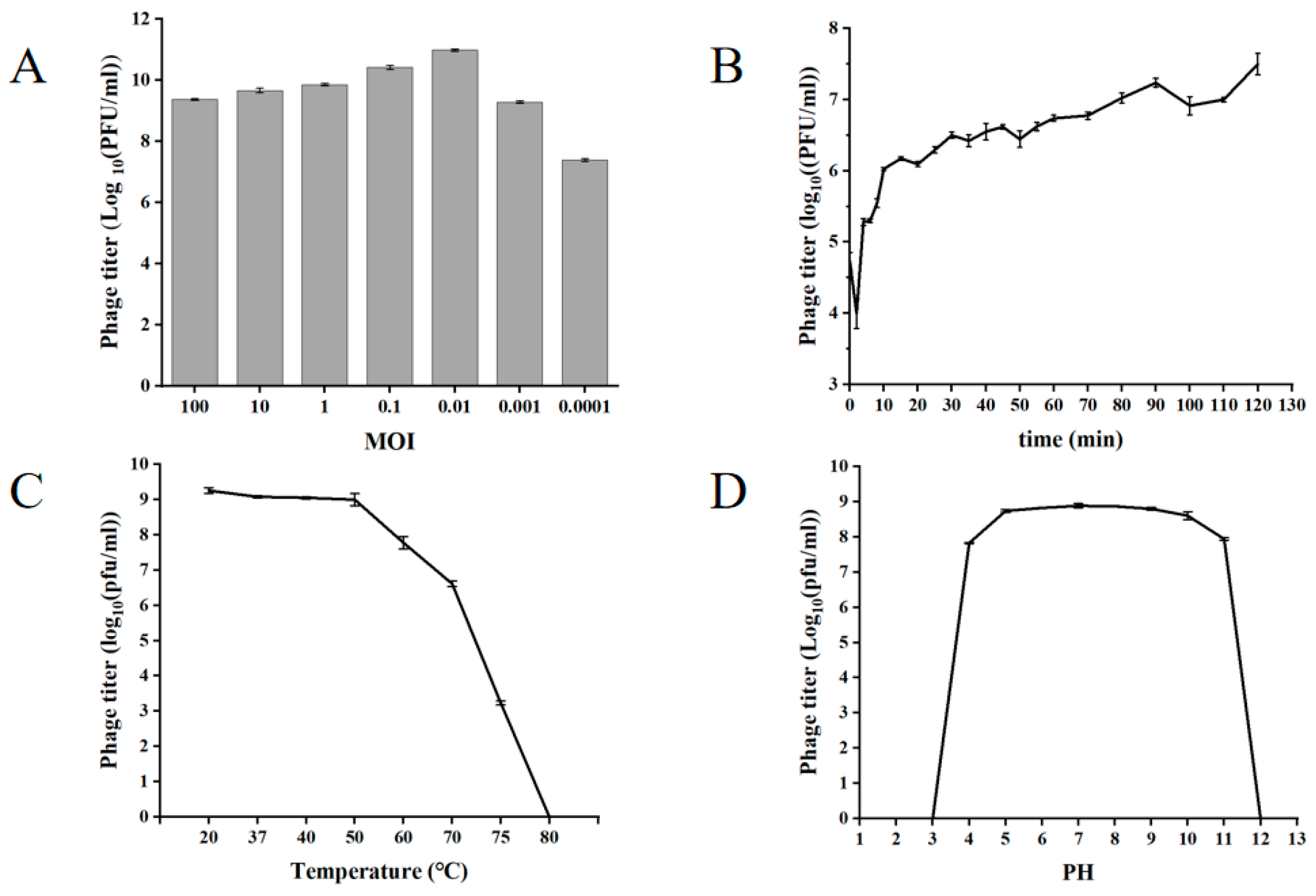
3.3. Biological Characteristics of XY75

The ratio of the number of phages to the number of indicator cells during infection is called the infection complex. The optimal multiplicity of infection is the one that yields the highest titer. At an optimal multiplicity of infection (MOI = 0.01), XY75 achieved a peak titer of 8.1 × 10¹⁰ PFU/mL (Figure 2A). The optimal MOI for phage XY75 was favorable for practical applications. The one-step growth curve of phage XY75 showed (Figure 2B) that 0–5 min was the adsorption period of phage XY75, 10–20 min was the lysis period of phage XY75, and 30–120 min was the plateau period of phage XY75. There was no obvious change in the activity of phage XY75 below 50 °C; the phage activity was severely decreased after 1 h of treatment at 60–70 °C; and the phage was completely inactivated after 1 h of treatment at 80 °C (Figure 2C). Phage XY75 exhibited remarkable resistance to acid–base fluctuations and thermal stress. However, exposure to extreme pH conditions (strong acid/alkali) or high temperatures led to disruption of its protein spatial conformation, specifically impairing the receptor-binding proteins and abrogating bacterial lytic activity. Phage XY75 was completely inactivated for pH ≤ 3 or pH ≥ 12 (Figure 2D).

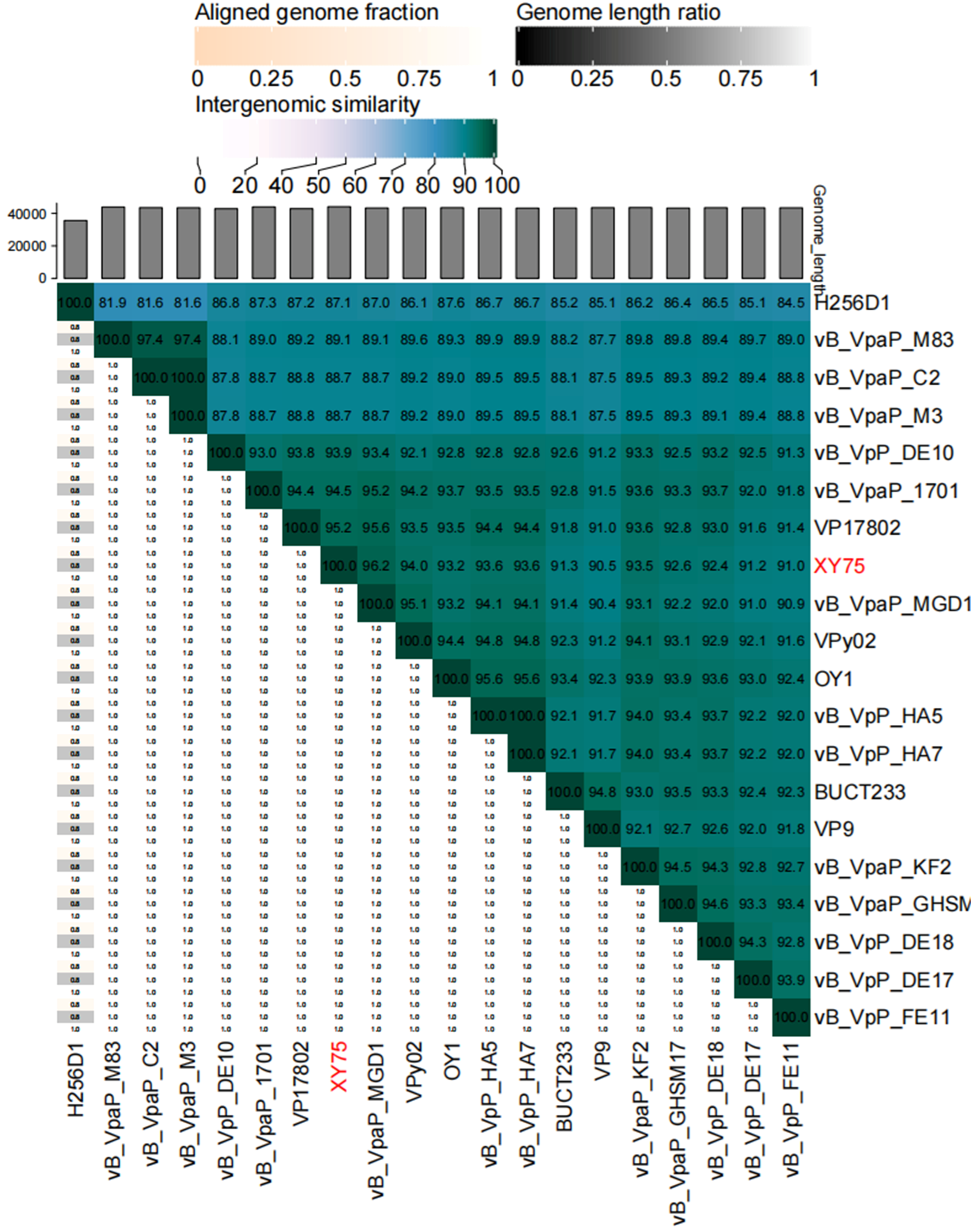
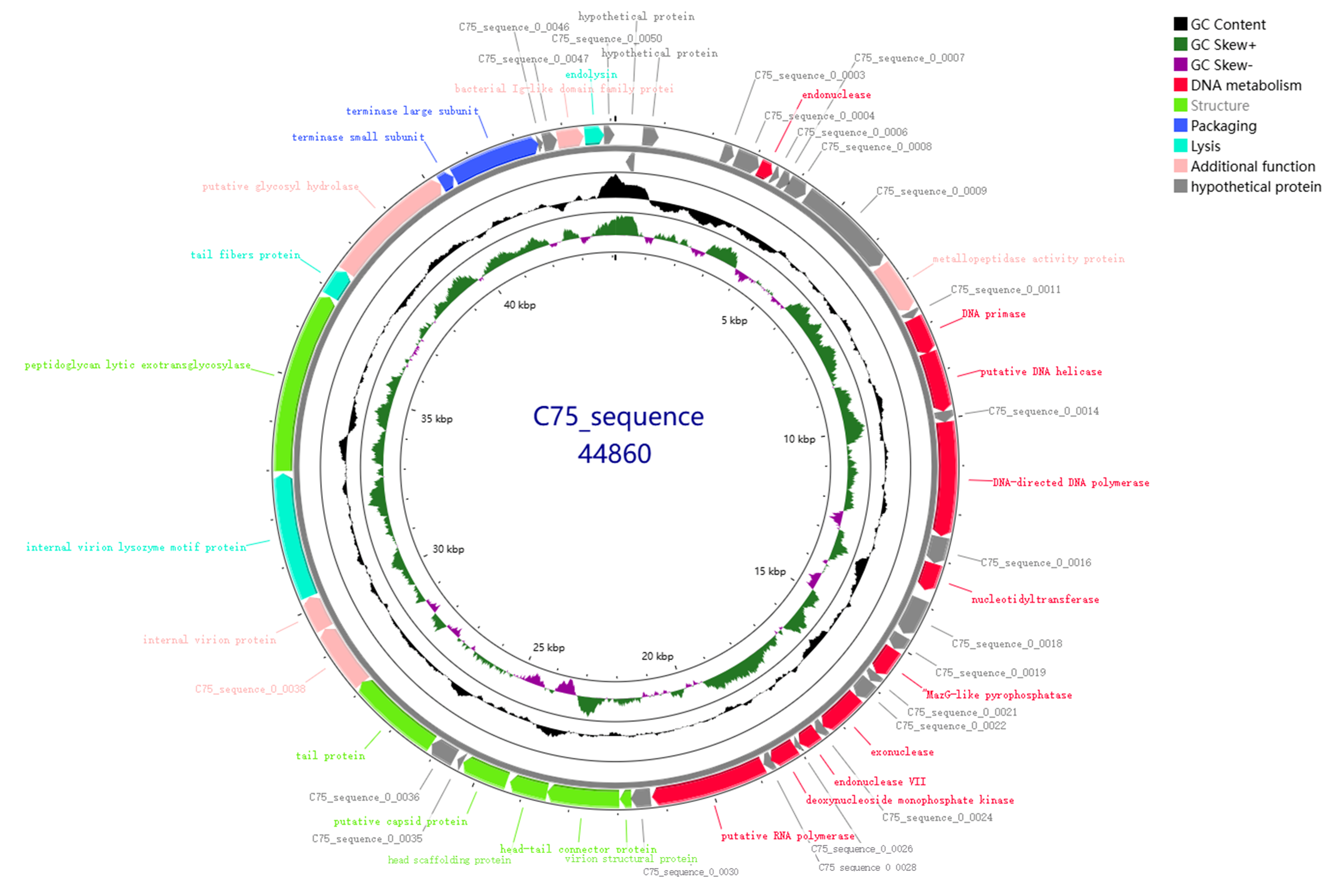
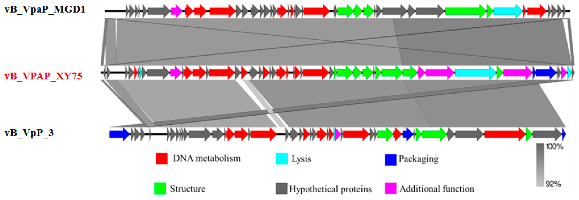
3.4. Genome Sequence Analysis

Illumina sequencing determined the double-stranded DNA (dsDNA) genome of bacteriophage XY75 to be 44,860 bp in length, with a GC content of 48.68%. The results of BLASTn comparison showed that the similarity between phage XY75 and Vibrio phage vB_VpaP_MGD1 (MT501516.1) was 100% (96.03% coverage); the similarity between phage XY75 and Vibrio phage vB_VpaP_1701 (ON872379) was 98% (95.28% coverage). The VIRIDIC heatmap (Figure 3) showed that phage XY75 exhibited significant intergenomic similarity with other vibrio phages. Furthermore, no antibiotic resistance genes (ARGs) or virulence-associated genes (VAGs) were detected in the genome of phage XY75. The morphology of phage XY75 particles and functional analysis of its predicted gene products suggested that it is a characteristic member of the Myoviridae family. Possessing its own RNA polymerase (ORF 29), ICTV classification places this phage within the Autographiviridae subfamily (Figure 4). Consequently, the genomic sequences of 43 Autographivirina Vibrio phages were downloaded from the ICTV database for the purpose of phylogenetic-tree analysis. Phylogenetic-tree analysis revealed that XY75 clustered within the branch of the Maculvirus genus, consistent with the whole-genome similarity result.
As demonstrated in Figure 5, the genome of phage XY75 is 44,860 bp in length, with a GC content of 48.68%. Genomic annotation analysis predicted 50 open reading frames (ORFs) without tRNA. Of these, 27 ORFs were annotated as functional proteins, and the rest were annotated as hypothetical protein. The functional proteins of XY75 were divided into six modules, including DNA metabolism; lysis; packaging; structure; additional function; and the hypothetical proteins module. In order to interpret the relationships of the phages at the genomic level, the complete genome sequence of XY75 was compared, vB_VpaP_MGD1 and Vibrio phage vB_VpP_3 (PP834370). As shown in Table 2, the phage genome carries multiple tail proteins, including tail tube protein (ORF 36, ORF 37), tail protein (ORF 38), and tail fiber protein (ORF 42). Moreover, it also contains lytic enzymes (ORF 41, ORF 49), bacterial Ig-like domain family protein (ORF 48), and other functional proteins. As shown in Figure 6, XY75 was homologous to vB_VpaP_MGD1 and vB_VpP_3 in a very large part of the genome, and the nucleotide identities were both not less than 92%. It has been suggested that phages exhibiting more than 40% protein similarity are likely to belong to a specific genus. Based on this method, we determined that phage XY75 belongs to the same genus as vB_VpaP_MGD1 and vB_VpP_3.

3.5. Food Applications of Phage XY75

Salmon contaminated with Vibrio parahaemolyticus were treated with phage XY75 at different MOIs. After being stored at 37 °C for 6 h, the survival numbers of Vibrio parahaemolyticus increased by 1.78 log CFU/mL (MOI = 10), 1.87 log CFU/mL (MOI = 1), 2.2 log CFU/mL (MOI = 0.1), and 2.14 log CFU/mL (MOI = 0.01) compared to the control group. However, when treated at 4 °C for 6 h, the survival numbers of Vibrio parahaemolyticus reduced by 5.98 log CFU/mL (MOI = 10), 2.01 log CFU/mL (MOI = 1), and 1.10 log CFU/mL (MOI = 0.01), while at MOI = 0.01, the survival numbers increased by 0.08 log CFU/mL (Figure 7A,B). The experiment demonstrated that high-titer phages (107 PFU/mL, MOI = 10) exhibited excellent bactericidal performance at both 37 °C and 4 °C. Notably, the 4 °C group achieved a 99.9% bactericidal rate after 6 h.

4. Discussion

One of the main causes of illnesses linked to seafood is the bacterium V. parahaemolyticus, which affects a variety of aquatic creatures, including fish, shellfish, and salmon [30,31]. This bacterium has the ability to develop biofilms in food processing environments, posing a significant threat to food safety within the seafood and aquaculture sectors [32]. Antibiotic-resistant V. parahaemolyticus strains have become more common in recent years [33,34], highlighting the urgent need for effective antimicrobial solutions. Lytic bacteriophages utilize the cellular machinery of host bacteria for replication, causing the bacteria to lyse and release the phage progeny into the surrounding environment, and this capability offers a promising approach for combating bacterial infections [35,36,37]. In this study, bacteriophage XY75, which specifically lyses V. parahaemolyticus, was isolated and biologically characterized to establish an effective control against V. parahaemolyticus.
Morphological analysis revealed an icosahedral head with a diameter of 40 ± 2 nm and a contractile tail measuring 60 ± 2 nm in length. These characteristics are typical of Myoviridae family members and suggest effective host infectivity. Host range assays confirmed a high degree of specificity for V. parahaemolyticus, which showed that the phage lysed 12 out of the 26 strains examined (46.2% coverage). The host range of the phage was determined principally by phage tail fibronectin [38]. Research findings have demonstrated the successful transformation of the fibronectin-associated cell-to-cell adhesion zone in Mycoplasma T3, which has enabled the expansion of its host range [39]. The one-step growth curve of our phage XY75 exhibited a peak titer of 8.1 × 10¹⁰ PFU/mL at an MOI of 0.01, a short latent period of 5 min, and robust lytic capacity, facilitating future industrial production of phage formulations. Seafood processing environments are inherently complex and variable, necessitating a robust environmental tolerance for phages to sustain their bioactivity. The shelf-life of salmon at 2 °C was determined to be approximately 10 days [40]. The XY75 phage exhibited excellent tolerance to temperature (20–50 °C) and pH (4–11). Therefore, compared to other phages, XY75 exhibits relatively broad temperature and pH stability, collectively supporting its capability for rapid bacterial eradication.
Illumina sequencing identified a 44,860 bp dsDNA genome with a GC content of 48.68%. VIRIDIC and BLASTN analyses showed 100% nucleotide similarity (96.03% coverage) with Vibrio phage vB_VpaP_MGD1 and more than 92% homology with conserved regions of vB_VpP_3. Phages XY75 and H256D1 were both located in the same evolutionary branch [41]. Among 50 predicted ORFs, 26 were functionally annotated, including DNA metabolism, structural assembly, packaging, and lysis modules. Practical application of this phage ultimately hinges on its safety profile and antimicrobial efficacy. Genomic analysis confirmed XY75 carries no genes associated with antibiotic resistance, toxins, lysogeny, or virulence factors, indicating that this bacteriophage may be harmless to humans and could be used in the food industry. Identification of an RNA polymerase (ORF 29) classified XY75 of the genus Maculvirus within the Autographiviridae subfamily. The most common structures for tail phages to recognize bacterial receptors are the tail spikes, tail fibers, and tail membrane penetrating proteins [42]. Among these, the tail fibers mediate adsorption by binding to receptors, including LPS, flagella, type 4 pili, and outer membrane porins (ompC, ompF) [43]. The phage genome encodes multiple tail proteins, including tail tube protein A/B (TTPA/TTPB, ORF36/37), tail protein (ORF 38), and tail fiber protein (ORF 42), which may serve as ligands to recognize conserved Vibrio receptors, thereby mediating phage adsorption and facilitating efficient recognition of host receptors. However, the fact that bacteriophages rely on receptor recognition to achieve infection implies that resistance to bacteriophages can arise through receptor mutations [39]. Critical lytic enzymes include an endolysin (ORF 49) and a peptidoglycan lytic exotransglycosylase (ORF 41), essential for adsorption, invasion, lysis, and biofilm disruption. An immunoglobulin-like domain protein (ORF 48) might facilitate mucosal surface binding for cell attachment or pathogen clearance [21,44,45].
The popularity of ready-to-eat (RTE) raw fish products, such as sashimi and sushi, has grown significantly in Asia, the Americas, and Europe [46,47,48,49]. However, despite stringent requirements for cold chain maintenance and hygiene during processing, distribution, and retail, these foods present a high risk of microbial contamination. This risk is exemplified by the relatively short shelf life of vacuum-packed salmon stored at 5 °C, which is typically only about 2 weeks [50]. Therefore, RTE raw fish slices serve as an appropriate model to investigate the potential of phage XY57 for the biocontrol of pathogenic Vibrio in RTE seafood. In this study, the degree and rate of pathogen reduction is significantly higher when samples were maintained at 37 °C, mimicking improper storage or serving conditions for RTE seafood. When XY75 was applied to pathogen-spiked samples at an MOI of 10, the V. parahaemolyticus count decreased by up to 1.0 log unit. The quantity of V. parahaemolyticus began to dramatically decline when XY75 was used to treat pathogen-spiked samples (4 °C). In 6 h, this reduction reached up to a 3.0 log reduction from the starting time point, achieving a sterilization rate of 99.9%. Recent studies have demonstrated that, when phages vB_VpaS_1601 and vB_VpaP_1701 were applied at an MOI of 10 to salmon samples, the numbers of V. parahaemolyticus were reduced by 1.51 and 1.39 CFU/cm3, respectively, with a sterilization rate not exceeding 30% within 6 h [51]. In comparison, phage XY75 exhibited a higher efficacy in eliminating Vibrio parahaemolyticus under the same conditions and significantly decreased the bacterial survival rate. Furthermore, phage OMN was applied to oyster samples, resulting in the inactivation of approximately 99% of V. parahaemolyticus on the surface of the oyster meat [52]. Our findings are consistent with those reported in these studies. The fast growth of bacteria at 37 °C may hinder the phage–bacteria interaction to some degree. Low-temperature conditions are typically employed in practical environmental applications to inhibit bacterial growth and reproduction, particularly during the shipping of seafood and other items. As a result, phage XY75 has more potential and benefits for use in this sector due to its enhanced bactericidal efficacy at low temperatures. The application of phage XY75 can effectively enhance the safety and quality of products during the transportation and storage of perishable products such as seafood. However, a major challenge for such phage-based biocontrol is the emergence of phage-resistant mutants [53]. The employment of bacteriophage cocktails has been demonstrated to enhance the efficacy of controlling and inactivating the growth of V. parahaemolyticus in comparison to the utilization of individual bacteriophage suspensions [54,55]. Therefore, in the future, the application of phage XY75 is expected to be utilized in phage cocktail mixtures as a sterilization system targeting as many Vibrio parahaemolyticus as possible.

5. Conclusions

Current global climate change has led to an increase in seawater surface temperatures, thereby expanding the geographical distribution and seasonal prevalence of pathogenic Vibrio infections. The emergence of antibiotic-resistant V. parahaemolyticus underscores the urgent need for sustainable, effective control strategies that can be effectively deployed in food and aquaculture industries. We isolated and characterized the lytic bacteriophage XY75 and evaluated its potential for the prevention and control of V. parahaemolyticus in seafood. Phage XY75 is characterized by a short latency and large release, thus facilitating ease of preparation and the subsequent industrial production of phage formulations. In addition, XY75 demonstrates robust functionality across a broad range of temperature (20–50 °C) and pH (4–11), indicating that the phage has a wider range of environmental applications. XY75 could reduce V. parahaemolyticus in salmon by more than 5.98 log CFU/g (99.9%) within 6 h at 4 °C, demonstrating superior efficacy to that observed at 37 °C. XY75 demonstrated exceptional cold tolerance and lytic activity, making it an excellent biological control agent for managing V. parahaemolyticus in the seafood cold chain. This study provides an innovative strategy and a valuable resource for combating V. parahaemolyticus contamination.

Author Contributions

Data curation, Validation, Writing—original draft, Writing—review and editing, Z.N.; Data curation, Validation, Resources, Methodology, X.C.; Investigation, Software, S.J. and Z.Z.; Formal analysis, Investigation, D.Z.; Software, H.C.; Conceptualization, Methodology, Funding acquisition, Supervision, Resources, Writing—review and editing, N.L.; Formal analysis, Conceptualization, Funding acquisition, Supervision, Y.Y. All authors have read and agreed to the published version of the manuscript.

Funding

We gratefully acknowledge the financial support of the National Key Research and Development Program, China (Grant No. 2022YFF1100703, Grant No. 2022YFF1100702), the National Natural Science Foundation of China (Grant No. 32230084), the Engineering Research Center of Bio-Process of Ministry of Education supported by “the Fundamental Research Funds for the Central Universities” (Grant No. PA2025GDGP0026).

Institutional Review Board Statement

Not applicable.

Informed Consent Statement

Not applicable.

Data Availability Statement

The original contributions presented in the study are included in the article, further inquiries can be directed to the corresponding authors.

Conflicts of Interest

The authors declare no conflict of interest.

References

  1. Lian, X.Q.; Liu, G.D.; Huang, M.F.; Fan, Q.H.; Lin, Z.D. Antimicrobial Resistance, Virulence Factors and Phylogenetic Profiles of Vibrio parahaemolyticus in the Eastern Coast of Shenzhen. Front. Microbiol. 2024, 15, 1452942. [Google Scholar] [CrossRef]
  2. Shinoda, S. Sixty Years from the Discovery of Vibrio parahaemolyticus and Some Recollections. Biocontrol Sci. 2011, 16, 129–137. [Google Scholar] [CrossRef]
  3. Baker-Austin, C.; Trinanes, J.; Gonzalez-Escalona, N.; Martinez-Urtaza, J. Non-Cholera Vibrios: The Microbial Barometer of Climate Change. Trends Microbiol. 2017, 25, 76–84. [Google Scholar] [CrossRef] [PubMed]
  4. Pang, R.; Li, Y.; Chen, M.; Zeng, H.; Lei, T.; Zhang, J.; Ding, Y.; Wang, J.; Wu, S.; Ye, Q.; et al. A Database for Risk Assessment and Comparative Genomic Analysis of Foodborne Vibrio parahaemolyticus in China. Sci. Data 2020, 7, 321. [Google Scholar] [CrossRef] [PubMed]
  5. Chimalapati, S.; Lafrance, A.E.; Chen, L.; Orth, K. Vibrio parahaemolyticus: Basic Techniques for Growth, Genetic Manipulation, and Analysis of Virulence Factors. Curr. Protoc. Microbiol. 2020, 59, e131. [Google Scholar] [CrossRef] [PubMed]
  6. Sorée, M.; Lozach, S.; Kéomurdjian, N.; Richard, D.; Hughes, A.; Delbarre-Ladrat, C.; Verrez-Bagnis, V.; Rincé, A.; Passerini, D.; Ritchie, J.M.; et al. Virulence Phenotypes Differ between Toxigenic Vibrio parahaemolyticus Isolated from Western Coasts of Europe. Microbiol. Res. 2024, 285, 127744. [Google Scholar] [CrossRef]
  7. Hong, X.; Lu, L.; Xu, D. Progress in Research on Acute Hepatopancreatic Necrosis Disease (AHPND). Aquacult. Int. 2016, 24, 577–593. [Google Scholar] [CrossRef]
  8. Cabello, F.C.; Godfrey, H.P.; Tomova, A.; Ivanova, L.; Dölz, H.; Millanao, A.; Buschmann, A.H. Antimicrobial Use in Aquaculture Re-Examined: Its Relevance to Antimicrobial Resistance and to Animal and Human Health. Environ. Microbiol. 2013, 15, 1917–1942. [Google Scholar] [CrossRef]
  9. Yano, Y.; Hamano, K.; Satomi, M.; Tsutsui, I.; Ban, M.; Aue-umneoy, D. Prevalence and Antimicrobial Susceptibility of Vibrio Species Related to Food Safety Isolated from Shrimp Cultured at Inland Ponds in Thailand. Food Control 2014, 38, 30–36. [Google Scholar] [CrossRef]
  10. Oniciuc, E.-A.; Likotrafiti, E.; Alvarez-Molina, A.; Prieto, M.; López, M.; Alvarez-Ordóñez, A. Food Processing as a Risk Factor for Antimicrobial Resistance Spread along the Food Chain. Curr. Opin. Food Sci. 2019, 30, 21–26. [Google Scholar] [CrossRef]
  11. Zaafrane, S.; Maatouk, K.; Alibi, S.; Ben Mansour, H. Occurrence and Antibiotic Resistance of Vibrio parahaemolyticus Isolated from the Tunisian Coastal Seawater. J. Water Health 2022, 20, 369–384. [Google Scholar] [CrossRef]
  12. Voigt, A.M.; Zacharias, N.; Timm, C.; Wasser, F.; Sib, E.; Skutlarek, D.; Parcina, M.; Schmithausen, R.M.; Schwartz, T.; Hembach, N.; et al. Association between Antibiotic Residues, Antibiotic Resistant Bacteria and Antibiotic Resistance Genes in Anthropogenic Wastewater—An Evaluation of Clinical Influences. Chemosphere 2020, 241, 125032. [Google Scholar] [CrossRef]
  13. Brüssow, H. Phage Therapy: The Escherichia coli Experience. Microbiology 2005, 151, 2133–2140. [Google Scholar] [CrossRef]
  14. Gibb, B.; Hyman, P.; Schneider, C.L. The Many Applications of Engineered Bacteriophages—An Overview. Pharmaceuticals 2021, 14, 634. [Google Scholar] [CrossRef]
  15. Myelnikov, D. An Alternative Cure: The Adoption and Survival of Bacteriophage Therapy in the USSR, 1922–1955. J. Hist. Med. Allied Sci. 2018, 73, 385–411. [Google Scholar] [CrossRef]
  16. Chen, C.; Yuan, L.; Zhou, W.; Mgomi, F.C.; Zhang, Y.; Wang, Y.; Zheng, X.; Hu, Q.; Gao, L.; Rao, S.; et al. Isolation and Genomic Characterization of P.A-5, a Novel Virulent Bacteriophage against Enterobacter hormaechei. Microb. Pathog. 2021, 152, 104767. [Google Scholar] [CrossRef] [PubMed]
  17. Carter, C.D.; Parks, A.; Abuladze, T.; Li, M.; Woolston, J.; Magnone, J.; Senecal, A.; Kropinski, A.M.; Sulakvelidze, A. Bacteriophage Cocktail Significantly Reduces Escherichia Coli O157: H7 Contamination of Lettuce and Beef, but Does Not Protect against Recontamination. Bacteriophage 2012, 2, 178–185. [Google Scholar] [CrossRef] [PubMed]
  18. Yang, S.; Sadekuzzaman, M.; Ha, S.-D. Reduction of Listeria monocytogenes on Chicken Breasts by Combined Treatment with UV-C Light and Bacteriophage ListShield. LWT 2017, 86, 193–200. [Google Scholar] [CrossRef]
  19. Zhang, X.; Niu, Y.D.; Nan, Y.; Stanford, K.; Holley, R.; McAllister, T.; Narváez-Bravo, C. SalmoFreshTM Effectiveness in Controlling Salmonella on Romaine Lettuce, Mung Bean Sprouts and Seeds. Int. J. Food Microbiol. 2019, 305, 108250. [Google Scholar] [CrossRef]
  20. Cheng, B.; Zhang, D.; Wang, T.; Chen, H.; Wang, Y.; Wang, Y.; Li, Z.; Ling, N.; Ye, Y. Characterization of the Broad-Spectrum Phage vB_CsaM_CBT2 and Its Inhibition of Multidrug-Resistant Cronobacter sakazakii in Powered Infant Formula. Food Control 2024, 158, 110262. [Google Scholar] [CrossRef]
  21. Vo Thi Tu, A.; Pham-Khanh, N.H.; Nguyen, S.H.; Sunahara, H.; Dai Thi Xuan, T.; Kamei, K. Isolation, Characterization, and Complete Genome Sequence of Vibrio Phage KIT04, a Novel Lytic Phage of the Subfamily Ermolyevavirinae. Virology 2023, 579, 148–155. [Google Scholar] [CrossRef] [PubMed]
  22. Peña-Rodríguez, A.; Ramírez-Sánchez, I.; Leptihn, S.; Quiroz-Guzmán, E. A Novel Vibrio phage vB_Vp_PvVp04 against Pathogenic Vibrio parahaemolyticus and Its Formulation for Inclusion in Shrimp Feed. Aquac. Int. 2025, 33, 117. [Google Scholar] [CrossRef]
  23. Yilmaz, N.; Yildirim, Z.; Sahin, T.S.; Karaoğlan, M. Isolation and Characterization of Lytic Bacteriophages Specific to Foodborne Pathogenic Pseudomonas aeruginosa and Use as a Biopreservative in Chicken Meat. Food Bioprocess Technol. 2024, 18, 2545–2560. [Google Scholar] [CrossRef]
  24. Azari, R.; Yousefi, M.H.; Taghipour, Z.; Wagemans, J.; Lavigne, R.; Hosseinzadeh, S.; Mazloomi, S.M.; Vallino, M.; Khalatbari-Limaki, S.; Berizi, E. Application of the Lytic Bacteriophage Rostam to Control Salmonella enteritidis in Eggs. Int. J. Food Microbiol. 2023, 389, 110097. [Google Scholar] [CrossRef]
  25. Zeng, H.; Li, C.; Luo, D.; Zhang, J.; Ding, Y.; Chen, M.; Yang, X.; Lei, T.; Wu, S.; Ye, Q.; et al. Novel Phage vB_CtuP_B1 for Controlling Cronobacter malonaticus and Cronobacter turicensis in Ready-to-Eat Lettuce and Powered Infant Formula. Food Res. Int. 2021, 143, 110255. [Google Scholar] [CrossRef]
  26. Kot, W. Genome Sequencing of dsDNA-Containing Bacteriophages Directly from a Single Plaque. Methods Mol. Biol. 2017, 1681, 179–184. [Google Scholar] [CrossRef]
  27. Johnson, M.; Zaretskaya, I.; Raytselis, Y.; Merezhuk, Y.; McGinnis, S.; Madden, T.L. NCBI BLAST: A Better Web Interface. Nucleic Acids Res. 2008, 36, W5–W9. [Google Scholar] [CrossRef]
  28. Moraru, C.; Varsani, A.; Kropinski, A.M. VIRIDIC—A Novel Tool to Calculate the Intergenomic Similarities of Prokaryote-Infecting Viruses. Viruses 2020, 12, 1268. [Google Scholar] [CrossRef] [PubMed]
  29. Stothard, P.; Grant, J.R.; Domselaar, G.V. Visualizing and Comparing Circular Genomes Using the CGView Family of Tools. Brief. Bioinform. 2017, 20, 1576–1582. [Google Scholar] [CrossRef]
  30. Lomelí-Ortega, C.O.; Martínez-Díaz, S.F. Phage Therapy against Vibrio Parahaemolyticus Infection in the Whiteleg Shrimp (Litopenaeus vannamei) Larvae. Aquaculture 2014, 434, 208–211. [Google Scholar] [CrossRef]
  31. Stalin, N.; Srinivasan, P. Characterization of Vibrio parahaemolyticus and Its Specific Phage from Shrimp Pond in Palk Strait, South East Coast of India. Biologicals 2016, 44, 526–533. [Google Scholar] [CrossRef]
  32. Han, N.; Mizan, M.F.R.; Jahid, I.K.; Ha, S.-D. Biofilm Formation by Vibrio parahaemolyticus on Food and Food Contact Surfaces Increases with Rise in Temperature. Food Control 2016, 70, 161–166. [Google Scholar] [CrossRef]
  33. Lopatek, M.; Wieczorek, K.; Osek, J. Antimicrobial Resistance, Virulence Factors, and Genetic Profiles of Vibrio parahaemolyticus from Seafood. Appl. Environ. Microbiol. 2018, 84, e00537-18. [Google Scholar] [CrossRef]
  34. Lee, L.-H.; Mutalib, N.-S.A.; Law, J.W.-F.; Wong, S.H.; Letchumanan, V. Discovery on Antibiotic Resistance Patterns of Vibrio Parahaemolyticus in Selangor Reveals Carbapenemase Producing Vibrio parahaemolyticus in Marine and Freshwater Fish. Front. Microbiol. 2018, 9, 2513. [Google Scholar] [CrossRef]
  35. Popova, K.B.; Valsamatzi-Panagiotou, A.; Penchovsky, R. New Drug Discovery Strategies for Targeting Drug-Resistant Bacteria. Environ. Chem. Lett. 2021, 19, 1995–2004. [Google Scholar] [CrossRef]
  36. Pereira, C.; Costa, P.; Duarte, J.; Balcão, V.M.; Almeida, A. Phage Therapy as a Potential Approach in the Biocontrol of Pathogenic Bacteria Associated with Shellfish Consumption. Int. J. Food Microbiol. 2021, 338, 108995. [Google Scholar] [CrossRef] [PubMed]
  37. Tian, F.; Li, J.; Nazir, A.; Tong, Y. Bacteriophage—A Promising Alternative Measure for Bacterial Biofilm Control. Infect. Drug Resist. 2021, 14, 205–217. [Google Scholar] [CrossRef]
  38. Harada, L.K.; Silva, E.C.; Campos, W.F.; Fiol, F.S.D.; Vila, M.; Dąbrowska, K.; Krylov, V.N.; Balcão, V.M. Biotechnological Applications of Bacteriophages: State of the Art. Microbiol. Res. 2018, 212–213, 38–58. [Google Scholar] [CrossRef] [PubMed]
  39. Yehl, K.; Lemire, S.; Yang, A.C.; Ando, H.; Mimee, M.; Torres, M.D.T.; de la Fuente-Nunez, C.; Lu, T.K. Engineering Phage Host-Range and Suppressing Bacterial Resistance through Phage Tail Fiber Mutagenesis. Cell 2019, 179, 459–469.e9. [Google Scholar] [CrossRef] [PubMed]
  40. Fogarty, C.; Whyte, P.; Brunton, N.; Lyng, J.; Smyth, C.; Fagan, J.; Bolton, D. Spoilage Indicator Bacteria in Farmed Atlantic Salmon (Salmo Salar) Stored on Ice for 10 Days. Food Microbiol. 2019, 77, 38–42. [Google Scholar] [CrossRef] [PubMed]
  41. Liu, W.; Hao, L.; Xia, H.; Wang, H.; Hou, W.; Wang, H.; Zhou, M. Inhibitory Effect of Two Closely Related Phages on Vibrio Parahaemolyticus. Foodborne Pathog. Dis. 2023, 20, 149–157. [Google Scholar] [CrossRef]
  42. Nobrega, F.L.; Vlot, M.; de Jonge, P.A.; Dreesens, L.L.; Beaumont, H.J.E.; Lavigne, R.; Dutilh, B.E.; Brouns, S.J.J. Targeting Mechanisms of Tailed Bacteriophages. Nat. Rev. Microbiol. 2018, 16, 760–773. [Google Scholar] [CrossRef]
  43. Hu, M.; Zhang, H.; Gu, D.; Ma, Y.; Zhou, X. Identification of a Novel Bacterial Receptor That Binds Tail Tubular Proteins and Mediates Phage Infection of Vibrio parahaemolyticus. Emerg. Microbes Infect. 2020, 9, 855–867. [Google Scholar] [CrossRef]
  44. Grabowski, Ł.; Łepek, K.; Stasiłojć, M.; Kosznik-Kwaśnicka, K.; Zdrojewska, K.; Maciąg-Dorszyńska, M.; Węgrzyn, G.; Węgrzyn, A. Bacteriophage-Encoded Enzymes Destroying Bacterial Cell Membranes and Walls, and Their Potential Use as Antimicrobial Agents. Microbiol. Res. 2021, 248, 126746. [Google Scholar] [CrossRef]
  45. Ge, H.; Fu, S.; Guo, H.; Hu, M.; Xu, Z.; Zhou, X.; Chen, X.; Jiao, X. Application and Challenge of Bacteriophage in the Food Protection. Int. J. Food Microbiol. 2022, 380, 109872. [Google Scholar] [CrossRef] [PubMed]
  46. Mudiyanselage, C.S.S.S.; Farouk, M.M.; Realini, C.E.; Kantono, K.; Hamid, N. Determinants of Purchase Intention for Meat-Based Chilled Ready Meals in New Zealand: A Consumer Behaviour Perspective. Foods 2025, 14, 1038. [Google Scholar] [CrossRef] [PubMed]
  47. Altintzoglou, T.; Heide, M.; Wien, A.H.; Honkanen, P. Traditional Sushi for Modern Consumers: A Comparison Between Sushi Consumption Behavior in Japan and Norway. J. Food Prod. Mark. 2016, 22, 717–732. [Google Scholar] [CrossRef]
  48. Kim, S.; Kim, S. Recent Surge of Ready Meals in South Korea: Can They Be Healthy Alternatives? Public Health Nutr. 2019, 23, 711–720. [Google Scholar] [CrossRef]
  49. Kulawik, P.; Dordević, D. Sushi Processing: Microbiological Hazards and the Use of Emerging Technologies. Crit. Rev. Food Sci. Nutr. 2020, 62, 1270–1283. [Google Scholar] [CrossRef] [PubMed]
  50. Giannoglou, M.; Evangelopoulou, A.-M.; Perikleous, N.; Baclori, C.; Tsironi, T.; Taoukis, P. Time Temperature Integrators for Monitoring the Shelf Life of Ready-to-Eat Chilled Smoked Fish Products. Food Packag. Shelf Life 2019, 22, 100403. [Google Scholar] [CrossRef]
  51. Tao, Z.; Sun, Y.; Zhou, M.; Li, T.; Lan, W.; Zhao, Y.; Sun, X. Two novel Vibrio parahaemolyticus phages (vB_VpaS_1601 and vB_VpaP_1701) can serve as potential biocontrol agents against Vibrio parahaemolyticus infection in seafood. Int. J. Food Microbiol. 2025, 441, 111303. [Google Scholar] [CrossRef]
  52. Zhang, H.; Yang, Z.; Zhou, Y.; Bao, H.; Wang, R.; Li, T.; Pang, M.; Sun, L.; Zhou, X. Application of a phage in decontaminating Vibrio parahaemolyticus in oysters. Int. J. Food Microbiol. 2018, 275, 24–31. [Google Scholar] [CrossRef]
  53. Pires, D.P.; Costa, A.R.; Pinto, G.; Meneses, L.; Azeredo, J. Current challenges and future opportunities of phage therapy. FEMS Microbiol. Rev. 2020, 44, 684–700. [Google Scholar] [CrossRef]
  54. Malik, S.; Nehra, K.; Rana, J.S. Bacteriophage cocktail and phage antibiotic synergism as promising alternatives to conventional antibiotics for the control of multi-drug-resistant uropathogenic Escherichia coli. Virus Res. 2021, 302, 198496. [Google Scholar] [CrossRef] [PubMed]
  55. You, H.J.; Lee, J.H.; Oh, M.; Hong, S.Y.; Kim, D.; Noh, J.; Kim, M.; Kim, B.S. Tackling Vibrio parahaemolyticus in Ready-to-Eat Raw Fish Flesh Slices Using Lytic Phage VPT02 Isolated from Market Oyster. Food Res. Int. 2021, 150, 110779. [Google Scholar] [CrossRef] [PubMed]
Figure 1. Morphological characterization of phage XY75. (A) Phage XY75 spots on TSA agar plates. (B) Morphology of phage XY75. Scale bar, 50 nm.
Foods 14 02660 g001
Figure 2. Biological characterization of phage XY75. (A) MOI. (B) One-step growth curve. (C) Temperature stability. (D) pH stability.
Foods 14 02660 g002
Figure 3. Heatmap of VIRIDIC. The values of the identity percentages range from 0 (white) to 100 (dark green).
Foods 14 02660 g003
Figure 4. Phylogenetic tree of phage XY75 based on the RNA polymerase sequence.
Foods 14 02660 g004
Figure 5. Genome-wide mapping of XY75. Circles from outside to inside: (1) ORFs are indicated in the first and second circles. The direction of the arrow, whether clockwise or counterclockwise, is indicative of the direction of the forward (or reverse) chain coding. For the open reading frames, different colors represent functional categories: hypothetical proteins (grey), DNA metabolism (red), structure (green), packaging (blue), lysis (cyan), and additional function (pink). (2) The third circle is indicative of the GC content of the genome. (3) The fourth circle is indicative of the value of (G − C)/(G + C), which is representative of the GC skew plot that is used to measure the relative contents of G and C (green indicates high GC content; purple indicates low GC content). (4) The innermost circle represents the total length of the genome, expressed in kilobase pairs (Kb).
Foods 14 02660 g005
Figure 6. Genome comparison of vB_VPAP_XY75, vB_VpaP_MGD1, vB_VpP_3, and EBPL using Easyfig. The different color arrows represent predicted open reading frames with different functions: hypothetical proteins (grey), DNA metabolism (red), structure (green), packaging (blue), lysis (cyan), and additional function (pink). Shading indicates nucleotide identity between sequences (92–100%).
Foods 14 02660 g006
Figure 7. Bacteriostatic effects of phage XY75 on V. parahaemolyticus in salmon. (A) Salmon, 37 °C; (B) salmon, 4 °C. (*) indicates p < 0.05, (**) indicates p < 0.01, (****) indicates p < 0.0001.
Foods 14 02660 g007
Table 1. Host range of phage XY75.
StrainSpeciesPlaque Formation aSource b
ATCC 17802Vibrio parahaemolyticusATCC
GD91Vibrio parahaemolyticus+Laboratory collection
GD103Vibrio parahaemolyticus+Laboratory collection
GD71Vibrio parahaemolyticus+Laboratory collection
GD78Vibrio parahaemolyticusLaboratory collection
GD81Vibrio parahaemolyticusLaboratory collection
GD62Vibrio parahaemolyticus+Laboratory collection
GD75Vibrio parahaemolyticus+Laboratory collection
GD74Vibrio parahaemolyticus+Laboratory collection
GD89Vibrio parahaemolyticus+Laboratory collection
GD80Vibrio parahaemolyticus+Laboratory collection
GD83Vibrio parahaemolyticus+Laboratory collection
GD73Vibrio parahaemolyticusLaboratory collection
99-1Vibrio parahaemolyticusFood
66-1Vibrio parahaemolyticusSeawater
124-1Vibrio parahaemolyticusFood
84-1Vibrio parahaemolyticus+Food
163-1Vibrio parahaemolyticusFreshwater
240-1Vibrio parahaemolyticusFood
79-1Vibrio parahaemolyticusFood
87-2Vibrio parahaemolyticusFreshwater
84-2Vibrio parahaemolyticusFood
139-1Vibrio parahaemolyticusFood
227-2Vibrio parahaemolyticusFood
51-1Vibrio parahaemolyticusFood
75-2Vibrio parahaemolyticusFood
ATCC 13076Salmonella enterica subsp. entericaATCC
ATCC 19111Listeria monocytogenesATCC
ATCC 51329Cronobacter muytjensiiATCC
ATCC 19433Enterococcus faecalisATCC
ATCC 14028Salmonella typhimuriumATCC
ATCC 49128Pseudomonas putidaATCC
ATCC 27583Pseudomonas aeruginosaATCC
ATCC 6538Staphylococcus aureusATCC
Note: A total of 34 strains were used as the host to determine the host spectrum of the phage using a spot test. a Plaque formation; +, clear plaques or slightly turbidity plaques; −, no plaques formed. b ATCC, American Type Culture Collection.
Table 2. Gene annotations of phage VB_VPAP_XY75.
ORFPredicted FunctionBest BLAST HitFunctional Module
5endonucleaseVibrio phage VP46DNA metabolism
10metallopeptidase activity proteinVibrio phage VP46Additional function
12DNA primaseVibrio phage vB_VpaP_GHSM17DNA metabolism
13putative DNA helicaseVibrio phage vB_VpaP_KF1DNA metabolism
15DNA-directed DNA polymeraseVibrio phage vB_VpP_HA1DNA metabolism
17nucleotidyltransferaseVibrio phage VP9DNA metabolism
20MazG-like pyrophosphataseVibrio phage F23s2DNA metabolism
23exonucleaseVibrio phage vB_VpP_NS8DNA metabolism
25endonuclease VIIVibrio phage VP48DNA metabolism
27putative deoxynucleoside monophosphate kinaseVibrio phage vB_VpP_DE18DNA metabolism
29putative RNA polymeraseVibrio phage H256D1DNA metabolism
31virion structural proteinVibrio phage VP48Structure
32head–tail connector proteinVibrio phage OY1Structure
33head scaffolding proteinVibrio phage vB_VpaP_KF1Structure
34putative capsid proteinVibrio phage vB_VpP_FE11Structure
36putative tail tubular AVibrio phage vB_VpaP_KF2Structure
37putative tail tubular protein BVibrio phage VP46DNA metabolism
38tail proteinVibrio phage vB_VpP_HA5Structure
39internal virion proteinVibrio phage vB_VpP_DE10Additional function
40internal virion lysozyme motif proteinVibrio phage vB_VpP_DE10Additional function
41peptidoglycan lytic exotransglycosylaseVibrio phage vB_VpP_DE10Lysis
42tail fiber proteinVibrio phage VP46Structure
43putative glycosyl hydrolaseVibrio phage vB_VpaP_KF1Additional function
44terminase small subunitVibrio phage vB_VpaP_KF1Additional function
45terminase large subunitVibrio phage BUCT233Additional function
48bacterial Ig-like domain family proteinVibrio phage vB_VpP_DE10Additional function
49endolysinVibrio phage VP46Lysis
Disclaimer/Publisher’s Note: The statements, opinions and data contained in all publications are solely those of the individual author(s) and contributor(s) and not of MDPI and/or the editor(s). MDPI and/or the editor(s) disclaim responsibility for any injury to people or property resulting from any ideas, methods, instructions or products referred to in the content.

