Article

Biocontrol Ability Against Harmful Microbial Contamination of Vegan Mortadella with an Ingredient of Oat Fermented by Lactiplantibacillus plantarum

1
Laboratory of Food Chemistry and Toxicology, Faculty of Pharmacy, University of Valencia, Av. Vicent Andrés Estellés s/n, 46100 Burjassot, Spain
2
Graduate Program in Animal Science, Pontifícia Universidade Católica do Paraná, Rua Imaculada Conceição, 1155 Prado Velho, Curitiba 80215-901, PR, Brazil
3
Department of Agriculture, Food and Environment (Di3A), University of Catania, Via Santa Sofia 100, 95123 Catania, Italy
*
Author to whom correspondence should be addressed.
Foods 2025, 14(13), 2195; https://doi.org/10.3390/foods14132195
Submission received: 15 May 2025 / Revised: 19 June 2025 / Accepted: 19 June 2025 / Published: 23 June 2025
(This article belongs to the Section Food Microbiology)

Abstract

The rising demand for vegan products calls for new plant-based antimicrobial preservation methods. This study evaluates an antifungal ingredient obtained by fermenting oat drink with lactic acid bacteria to extend vegan mortadella’s shelf life. In vitro tests showed antimicrobial effects against Aspergillus flavus, Penicillium commune, and Listeria monocytogenes (inhibition zones: 2–5 mm). The enrichment of the oat drink culture medium with additional nutrients enhanced fermentation performance and increased antifungal activity. The fermented culture medium with the highest antimicrobial activity was used to develop a bioactive ingredient for the preservation of vegan mortadella conservation. Adding 3% of this ingredient to vegan mortadella improved microbial stability, reducing mesophilic bacteria by 2.5 Log10 CFU/g and increasing lactic acid bacteria. Lower pH and water activity changes were observed but remained within quality standards. Contamination assays showed a consistent reduction of A. flavus over 7 days, while P. commune and L. monocytogenes dropped below detection within 2 days. In contrast, control samples maintained contamination levels near 3.0 Log10 CFU/g. These findings support the potential of fermented oat-based ingredients as effective, natural preservatives for vegan foods.

1. Introduction

The growth of vegan meat alternatives reflects a significant transformation in dietary patterns, driven by ethical, environmental, and health motivations. These plant-based products aim to replicate the sensory and nutritional characteristics of conventional meats while avoiding animal-derived ingredients [1]. Notably, efforts to mimic traditional sausages have extended to mortadella, resulting in novel vegan mortadella-type products designed to closely resemble the sensory and structural characteristics of the Italian original [2]. Among the various components used in their formulation, oats have gained prominence due to their nutritional profile, sustainability, and functional properties [3]. Oats provide abundant dietary fiber, protein, and bioactive compounds like β-glucans, making them a powerful addition to vegan formulations including beverages, fermented products, and meat analogues [4,5].
However, like all minimally processed or ready-to-eat foods, vegan meats are susceptible to microbial contamination throughout their production chain [6]. This includes pathogenic bacteria, such as Listeria monocytogenes, and spoilage molds like Aspergillus and Penicillium species [7]. L. monocytogenes is particularly problematic due to its ability to grow at refrigeration temperatures and cause severe illness in vulnerable populations [8]. Additionally, filamentous fungi degrade sensory quality and present a toxicological hazard through mycotoxin production [9,10]. These concerns are compounded by the clean-label expectations of consumers, which limit the use of synthetic preservatives and emphasize the need for natural preservation strategies [11]. In addition, oat fermentation by lactic acid bacteria has been shown to enhance the prebiotic qualities of oats, broadening their spectrum of bioactivities. This further establishes oats as an excellent medium for LAB fermentation, enhancing their antimicrobial potential [12].
Biopreservation using lactic acid bacteria (LAB) has emerged as a promising alternative to synthetic additives [13]. LAB produces antimicrobial effects through multiple mechanisms, including acidification, competition for nutrients, and the production of bioactive metabolites such as bacteriocins, reuterin, and phenyllactic acid [14]. In foods, LAB can be incorporated during production or applied as protective cultures in packaging [15,16]. These compounds are effective against a broad spectrum of foodborne pathogens and spoilage organisms, including L. monocytogenes and molds [17,18]. Additionally, the antimicrobial properties persist despite industrial processes such as increased temperature and enzyme presence [19].
Moreover, the application of LAB fermentation aligns with sustainable practices. It can utilize plant-based byproducts such as citrus peels to create bioactive formulations [20]. This dual function, not only enhancing food quality but also minimizing waste, directly addresses the rising demand for sustainable food alternatives [21]. Based on this evidence, the objectives of this study were to: (a) develop and assess the antimicrobial potential of a bioactive ingredient produced through lactic acid bacterial fermentation of vegetable sources; (b) investigate how different concentrations of the antifungal ingredient affect the technological properties of a vegan mortadella; and (c) evaluate the antimicrobial properties of this ingredient in vegan mortadella.

2. Materials and Methods

2.1. Chemicals and Microorganisms

Milli-Q water with a resistivity of <18 MΩ·cm was obtained using a Milli-Q purification system (Millipore, Bedford, MA, USA). Buffered peptone water, potato dextrose agar (PDA), Man, Rogosa, Sharpe Agar (MRSa), Man Rogosa Sharpe Broth (MRSb), brain heart infusion (BHI) and plate count agar (PCA) were purchased from Liofilchem (Teramo, Italy). Agar, glucose (>99.9), yeast extract, soy protein, ammonium sulfate (>99.9), sodium chloride (>99.9), magnesium sulfate (>99.9), potassium phosphate (>99.9), and manganese sulfate (>99.9) were purchased from Sigma-Aldrich (Dublin, Ireland). Beans and food colorant were obtained from Consum Cooperativa (Valencia, Spain).
The LAB strain isolated from vegan products used in this study was previously analysed by our group and demonstrated its potential to ferment oat drink, producing bioactive antimicrobial agents in vitro screening tests performed on several bacterial isolates. This strain was identified at the species level as Lactiplantibacillus plantarum (L. plantarum) through microbial identification and characterization by MALDI-TOF-MS (matrix assisted laser desorption/ionization time-of-flight mass spectrometry) in the Spanish Type Culture Collection (CECT), Valencia, Spain [22].
Microbial isolates including Aspergillus fumigatus CECT 20827, Aspergillus niger CECT 2088, Aspergillus steynii CECT 20510, Penicillium commune CECT 20767, Penicillium digitatum CECT 2954, Penicillium expansum CECT 2278, Penicillium camemberti CECT 2267, Penicillium italicum CECT 20332, Penicillium nordicum CECT 2320, Penicillium roqueforti CECT 2905, and Listeria monocytogenes CECT 935 were obtained from the CECT collection. Penicillium verrucosum D-01847 VTT was acquired from the VTT Technical Research Centre of Finland (Espoo, Finland), and Aspergillus flavus ITEM 8111 from the ISPA Culture Collection (Bari, Italy).
Oat drink was produced according to the method described by Demïr et al. (2021) with minor modifications [23]. Oat was combined with distilled water (1:5 w/v), heated to 95 °C, and soaked for 15 min to enhance extraction. The mix was homogenized with a blender (John Oster Manufacturing Company, Racine, WI, USA) and filtered through a cheesecloth to obtain the oat drink.

2.2. Preparation, Fermentation and Characterization of the Oat Drink Culture Mediums

A total of 3 different culture mediums for the LAB fermentation were prepared following the steps described in Dopazo et al. (2023) with adaptations for this study [24]. The mediums produced for this study were: (1) Culture medium oat drink (OD). (2) Culture medium oat drink enriched with glucose (ODG): oat drink complemented with 10.0 g/L of glucose. (3) Culture medium oat drink enriched with nutrients (ODN): oat drink with 10.0 g/L of glucose, 10.0 g/L of yeast extract, 10.0 g/L of soy protein, 6.5 g/L of sodium chloride, 2.5 g/L of dipotassium phosphate, 0.25 g/L of magnesium sulphate, and 62.5 mg/L of manganese sulphate. After homogenization and pasteurization for 30 min at 80 °C, the culture mediums were cooled to 30 °C. Finally, they were inoculated at a concentration of 5% (v/v) with a LAB suspension, from a previous incubation of 24 h at 37 °C in MRSb and then washed twice with a PBS solution 0.1 M. Afterwards, the culture mediums were incubated at 37 °C for 48 h.
For a better evaluation of the fermentation of the culture a comprehensive assessment of key parameters for effective LAB fermentation and antifungal activity was conducted. The pH of the samples was analysed using a XS pH 7 Vio pH-meter with a 2 Pore Steel T electrochemical sensor (XS Instruments, Carpi, Italy). Bacterial count (performed in MRS), lactic acid and phenyllactic acid measurement was performed according to the steps described in Dopazo et al. (2023) [24].
The resultant fermented culture medium was frozen at −20 °C. Subsequently, the samples underwent lyophilization in a FreeZone 2.5 L freeze-drying equipment (Labconco, Kansas City, MO, USA) for further analysis [25].

2.3. Evaluation of the Antimicrobial Properties of the LAB Fermented Oat Drink Culture Mediums

The qualitative antifungal properties of the oat drink culture mediums after the fermentation were evaluated by the agar diffusion method [26]. First, a suspension of 400 g/L of the samples was prepared using sterilized distilled water. For the antifungal assays, PDA plates were used for the assays against fungi, while BHI agar plates were used for Listeria monocytogenes. Microbial suspensions were spread on the agar plates with a sterile swab, and wells were pierced in the agar using sterile 1 mL micropipette tips. Subsequently, 100 µL of the sample suspension was added to each well. After a 48-h incubation at 30 °C, inhibition zones were measured to evaluate antifungal activity. Each sample was compared to a control containing a non-fermented culture medium to assess relative antifungal effects. Assays were performed per triplicate and the mean was used as a result.

2.4. Production of the Vegan Mortadella Incorporating the Bioactive Ingredient

The culture medium ODN fermented by the LAB isolated evidenced the best results in the antimicrobial assays and was selected for the production of the vegan mortadella bioactive ingredient. To prepare the mortadella, beans were boiled for 40 min until soft, then blended with salt, thyme and garlic into a mash. Simultaneously, an agar suspension in distilled water was heated to boiling and gently stirred for 5 min. The agar was quickly combined with the bean mash, a total of 0.150 mL of pink food colorant, and the mixture was poured into a mold and refrigerated at 5 °C for 8 h.
The final composition of the mortadella included the following ingredients: bean mash, water, agar, salt, thyme and garlic. Four variations of mortadella were prepared: a control and 3 sample versions containing the lyophilized powder from the bioactive ingredient at 0.5% (M 0.5%), 1.0% (M 1%), and 3.0% (M 3%) concentration (w/w). The bioactive ingredient was incorporated by replacing an equivalent portion of the bean mash. All mortadellas reached a final weight of 750 g. The ingredient concentrations are detailed in Table 1 for reference.

2.5. Evaluation of the Effect of the Bioactive Ingredient in the Vegan Mortadella Quality

To investigate the influence of the fermented ODN ingredient in vegan mortadella, several analyses were conducted through the 7-day study. The pH of the samples was measured using a XS pH 7 Vio pH-meter with a 2 Pore Steel T electrochemical sensor (XS Instruments, Carpi, Italy) [25]. Water activity (Aw) was assessed using a Humimeter RH2 water activity meter (Max-Schaller-Straße, Styria, Austria) [27]. Moisture content was determined by an indirect gravimetric method, where approximately 10 g of each sample was weighed before and after drying in an oven at 105 °C for 24 h [28]. To analyze microbial populations due to natural contamination, 10 g of vegan mortadella were homogenized in 0.1% (w/v) peptone water using a Masticator Classic Stomacher (IUL S.A., Barcelona, Spain). The homogenized samples were then serially diluted and plated on PCA, incubated at 30 °C for 48 h, for the total aerobic mesophilic bacteria count, and on MRSa under anaerobic conditions at 37 °C for 48 h, for the LAB count [29]. The color of the vegan mortadella was measured at different locations using a ColorQuest® XE (HunterLab, Birstall, Leicestershire, UK) to determine hue, saturation, and intensity (HSI), comparing any differences [30]. All assays were performed in triplicate.

2.6. Preparation of Microbial Inoculum for In Situ Antimicrobial Activity Assays

The Aspergillus and Penicillium isolates were cultured on PDA at 25 ± 1 °C in the dark until the mycelium completely covered the surface of the Petri dish. For each isolate, a conidial suspension (107 spore/mL) was prepared in sterile distilled water. The conidial concentration was determined using a Neubauer counting chamber (BLAUBRAND®, Merck KGaA, Darmstadt, Germany). The bacterial suspension of Listeria monocytogenes was prepared by cultivating the strain on BHI medium, at 37 °C for 24 h. After centrifugation at 5000× g for 10 min at 4 °C, bacterial cells were resuspended in sterile distilled water to achieve a final concentration of 108 CFU/mL, verified using a Neubauer counting chamber. Finally, solutions were adjusted to a concentration of 2 × 10⁶ CFU/mL for the assays [31,32].

2.7. Assessment of the Antimicrobial Properties of the Bioactive Ingredient in the Vegan Mortadellas

The in situ antimicrobial activity of the bioactive ingredient was evaluated by monitoring fungal and bacterial contamination in mortadella samples, using both spoilage and pathogenic microorganisms throughout the storage period. The experimental procedure followed the methodology described by Dopazo et al. (2022), with adaptations specific to this study [33]. Mortadella slices of approximately 20 g were cut, and 10 µL of the microbial suspension (2.5 × 10⁶ CFU/mL) of the contaminating microorganisms was applied to four separate spots on each slice. The slices were then incubated under sterile conditions at 10 °C.
Microbial population dynamics were monitored after 0, 2, 4, and 7 days of incubation. Quantification was performed by homogenizing the samples in 0.1% peptone water using a stomacher, followed by serial dilutions and plating on agar. For Aspergillus niger and Penicillium commune, the samples were plated on PDA and incubated at 25 °C for 48 h. For Listeria monocytogenes, the dilutions were plated on BHI agar and incubated at 37 °C for 48 h. All assays were conducted in quadruplicate to ensure reproducibility.

2.8. Statistical Analysis

Statistical analyses were performed using a one-way ANOVA to evaluate differences among the experimental groups. When significant differences were detected (p < 0.05), Tukey’s HSD post hoc test was applied to identify specific pairwise differences. For the culture assays, six groups were compared: the three non-fermented culture media and the three fermented culture media. The culture media tested were OD, ODG, and ODN. For the vegan mortadella samples, four groups were analyzed: the control (mortadella without the bioactive ingredient) and three treatments containing 0.5%, 1.0%, and 3.0% of the bioactive ingredient (M 0.5%, M 1.0%, and M 3.0%). All statistical analyses were performed using InfoStat 2019 software (Universidad Nacional de Córdoba, Córdoba, Argentina).

3. Results and Discussion

3.1. Assessment of Fermentation Parameters and Antimicrobial Properties of Bioactive Ingredients

The data obtained from the analysis of the fermented oat mediums can be appreciated in the Table 2. The pH evaluation clearly indicated active bacterial fermentation in the LAB fermented samples. In the non-fermented control, the pH remained significantly higher, with values close to pH 7. In contrast, fermentation reduced the pH to an average of 3.46 in the inoculated samples. These results were consistent with the data obtained from other analyses. No bacterial growth was detected in the control samples based on CFS counting, and both lactic acid and phenyllactic acid were below the limit of detection. In the fermented samples, LAB counts performed in MRSa reached 7.37, 8.20, and 8.32 log10 CFU/mL in OD, ODG, and ODN, respectively. Lactic acid concentrations were 14.84, 17.27, and 17.34 g/L in fermented OD, ODG, and ODN, respectively. When comparing these values, both bacterial counts and lactic acid production were significantly higher in ODG and ODN than in OD, suggesting an improved fermentation performance in matrices with enhanced nutrient composition. Phenyllactic acid concentrations also increased significantly with the nutrient complexity of the matrix. Fermented OD reached 2.61 mg/L, ODG 5.42 mg/L, and ODN 9.23 mg/L, highlighting the positive correlation between nutrient availability and production of this antimicrobial metabolite.
The results of the qualitative evaluation of antimicrobial activity from fermented oat drink culture mediums are presented in Table 3. The data show that fermentation of the oat drink culture mediums by Lactiplantibacillus plantarum VC7 exhibited varying degrees of antimicrobial activity against both fungi and bacteria. ODN demonstrated the highest antimicrobial activity, followed by ODG, indicating that increased nutrient availability promotes antifungal metabolite production. Fermentation of the OD showed inhibition zones of less than 2 mm against certain Penicillium species (P. commune, P. digitatum, P. expansum, and P. verrucosum) and Listeria monocytogenes. This indicates that OD serves as a valuable nutrient source for microbial growth and antimicrobial metabolite production, but its antimicrobial efficacy is limited without nutrient enrichment. The ODN exhibited a significantly increased antimicrobial activity, in comparison to the other treatments, particularly against Penicillium species and L. monocytogenes, with inhibition zones exceeding 5 mm for Aspergillus fumigatus, P. expansum, P. italicum, and P. nordicum. None of the non-fermented control samples exhibited any antimicrobial activity against the tested microorganisms.
Vegan antimicrobial bioactive ingredients studied are limited in the literature. Among these, essential oils have been the most extensively studied [34]. For instance, clove oil has been shown to significantly reduce populations of E. coli strains evidencing minimal inhibitory concentrations of 2.5 mL/L [35]. Additionally, Ocimum basilicum (basil) essential oil has demonstrated inhibition zones up to 20 mm at concentrations near 1 g/L against fungal species like Aspergillus niger and Fusarium solani, as well as bacteria such as Escherichia coli and Bacillus subtilis [36]. However, not all essential oils exhibit effective activity against foodborne pathogens. Moreover, the use of this essential oils carries the problematic of undesired odors to the food applied, the extraction is related to high cost in organic solvents for the extraction and economically in expensive techniques [37]. The method described in this study not only yields comparable results but also offers a simpler production process. The application of fermented oat drink as a bioactive ingredient offers a promising, novel alternative to conventional additives used in the food industry for protecting against microbial contaminants.
Unlike essential oils, the antimicrobial activity of ODN is attributed to the bioactive compounds generated during fermentation by Lactiplantibacillus plantarum. These include organic acids, such as lactic acid, acetic acid, or phenyllactic acid, and other fermentation-derived metabolites that lower pH and disrupt microbial membranes [38]. In particular, phenyllactic acid has been associated with antifungal and antibacterial properties in cereal-based matrices [39]. Moreover, the fermentation process may enhance the release or transformation of oat phenolics and fibers, contributing further to antimicrobial activity through synergistic effects [40]. This suggests that the combination of oat and lactic acid bacteria fermentation contributes directly to the antimicrobial activity observed.
Previous studies have shown that nutrient enrichment alone does not explain the increased antimicrobial activity observed in ODN, highlighting the importance of the oat-based matrix. While nutrient-rich media such as MRS broth can support LAB growth, they often lack the structural or biochemical components required for substantial antimicrobial metabolite production [24]. Similarly, certain food matrices may not function as effective substrates for antimicrobial synthesis during short fermentation periods, likely due to limited nutrient availability that slows microbial growth. However, the addition of targeted nutrients can accelerate LAB growth and enzyme activity, enhancing the breakdown and utilization of the food matrix for metabolite production [41]. This likely explains the higher antimicrobial activity observed in the present results. Nevertheless, exploring the potential of using food matrices alone remains valuable, as reducing the need for nutrient supplementation could lower production costs and improve the scalability of antimicrobial ingredient development for industrial applications [42].
This phenomenon was also evident in the assessment of fermentation parameters, where the use of more complex matrices significantly enhanced key indicators relevant to the objectives of this study, such as bacterial growth and the presence of antimicrobial metabolites. The lack of significant differences in lactic acid production between fermented ODG and ODN can be attributed to the similar glucose content in both matrices, as lactic acid production by LAB is closely linked to glucose metabolism [43]. In contrast, the increasing complexity of the culture medium led to a higher production of secondary antifungal metabolites, particularly phenyllactic acid. The literature highlights the importance of a broad range of minor antimicrobial compounds as key contributors to the overall antifungal activity of LAB. The synthesis of these compounds is associated with the utilization of diverse nutrients, not solely disaccharides [14]. Therefore, based on the fermentation performance and antimicrobial profile, the LAB-fermented ODN demonstrated the most promising results and appears to be the most suitable candidate for use as a bioactive ingredient.

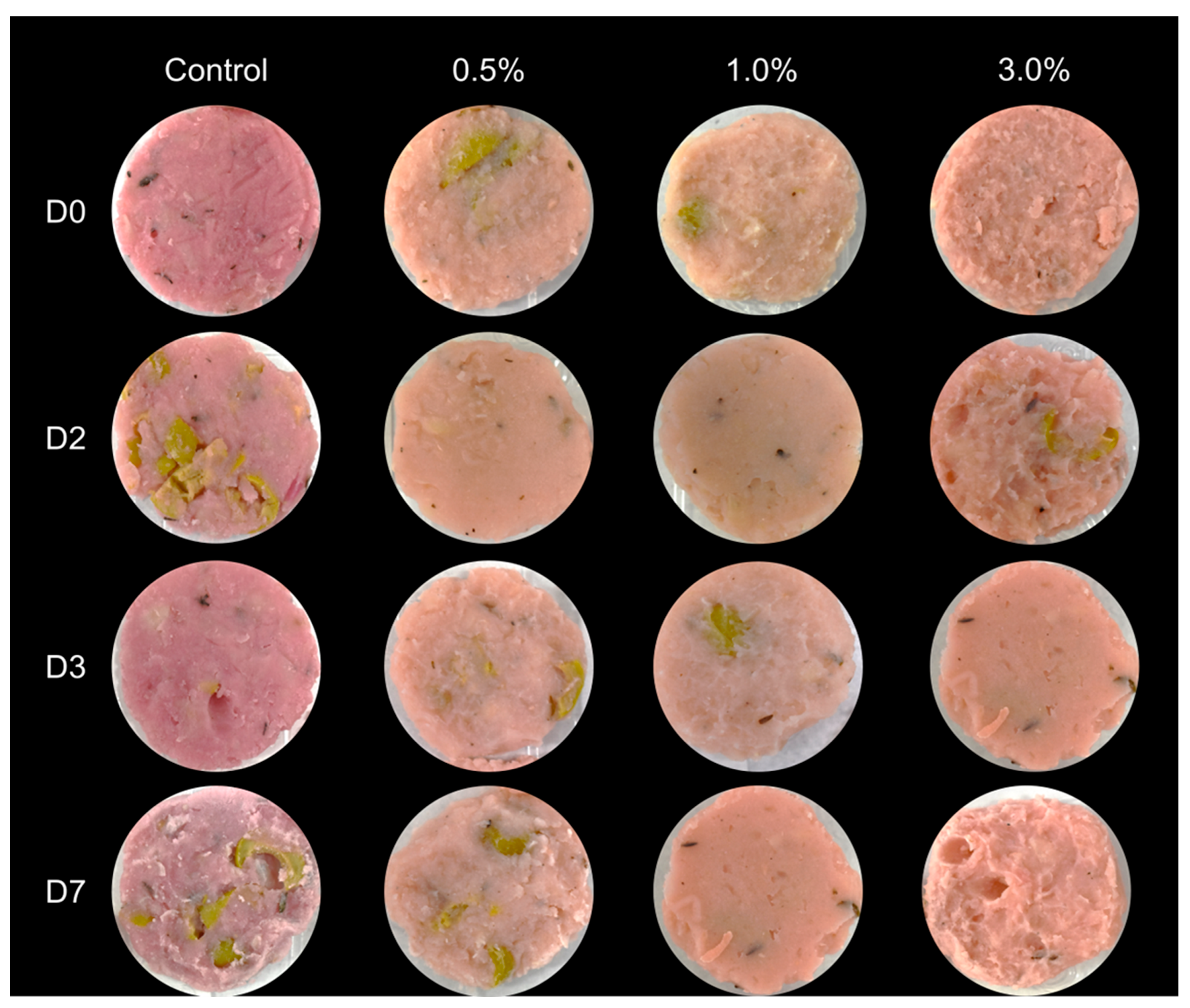
3.2. Assessment of the Effect of the Bioactive Ingredient in the Vegan Mortadella Quality

The results of how the bioactive ingredient affected the mortadella quality are shown in Table 4. The control consisted of mortadella without the bioactive ingredient, while M 0.5%, M 1.0%, and M 3.0% corresponded to mortadella samples formulated with 0.5%, 1.0%, and 3.0% of the bioactive ingredient derived from the ODN medium fermented by L. plantarum VC7, respectively. The pH displayed a consistent trend throughout the study. Following mortadella preparation, all treatments initially exhibited a rise in pH for two days, followed by a sharp decline after day 2, returning to levelssimilar to day 0. As expected, among the treatment samples, pH values declinedas the concentration of the bioactive ingredient in the mortadella increased. It can be observedthat the values from the control sample fluctuated within 5.93 to 6.22, while in the sample with a 3% of the bioactive ingredient values were between 4.60 and 4.85 (Table 4). The water activity of the samples was affected differently. In general, the greatest values were found at day 0 and 3, while lower values were recorded on days 2 and 7. The comparison of the samples evidenced a decrease in the Aw value as the concentration of the ingredient increased. The samples reached average Aw of 0.95, 0.91 and 0.90 in the M 0.5%, M 1.0%, M 3.0%, respectively, while in the control the value was 0.95 on average (Table 4). The moisture content was significantly greater in the samples with a higher concentration of the ingredient, starting at moistures of 30 g/100 g in sample M 3.0%, a total of 5 g/100 g greater than in the control. Nevertheless, after day 3 the moisture concentration in the samples began to significantly decline in the treatment samples losing 2.7, 2.3 and 4.1 g/100 g when comparing the initial measures with the ones after a 7 day observation. Meanwhile, the control samples increased the moisture content 3.3 g/100 g over the same period of time (Table 4). Finally, the HSI values were lower in the control in comparison to the samples, the difference among samples were minimal. Average values for sample treatments were above 48 while in the control the average HSI was 26. During incubation, HSI values decreased consistently from day 0 to day 7 for all samples. The highest decrease was observed in the samples M 1.0%, of 11 units of HSI, and the lowest in the sample M 3.0%, of 4 units of HSI (Table 4).
Quantification of Log10 colony forming units per gram of food (CFU/g) can be appreciated in Figure 1. It indicated that lactic acid bacterial populations growing under anaerobic conditions at 37 °C increased significantly in both the control and treated samples during the 7-day incubation. The final concentration in the control samples was initially of 3.31 Log10 CFU/g and ended at 5.93 Log10 CFU/g. The initial concentration of the samples M 0.5%, M 1.0%, and M 3.0% were 3.76, 4.19, and 5.80 Log10 CFU/g, initially, and ended at 6.10, 6.46, and 7.29 Log10 CFU/g, respectively. The average microbial growths during the days of the analysis indicated that the highest rise in bacterial concentration was observed in the control, 0.91 Log10 CFU/g, while the lowest was detected at the sample M 3.0%, of 0.50 Log10 CFU/g (Figure 1A). Throughout the MRSa study, samples with a higher concentration of the bioactive ingredient exhibited higher bacterial populations, likely due to the presence of lactic acid bacteria associated with the ingredient. In contrast, mesophilic aerobic bacterial counts on PCA showed a decrease in bacterial populations when the concentration of the antimicrobial ingredient increased. Specifically, samples containing 3.0% of the ingredient maintained a close to stable cell counts over the 7-day period, between 5.36 to 5.87 Log10 CFU/g, while lower ingredient concentrations were associated with an increase in Log10 CFU over time. The biggest increase was detected in the control samples rising from the 5.78 to 8.32 Log10 CFU/g (Figure 1B).
Fluctuations in pH and water content can be attributed to the relationship between these parameters, with changes in pH likely driven by LAB originally present in the ingredient, which affect both pH and Aw [44]. The increase in LAB, as suggested by cell counts on MRSa, is an advantageous outcome, as these vegan meat-like products, similar to their meat counterparts, often contain high levels of LAB. LAB presence in foods is generally associated with protective effects against foodborne contaminants and, in some cases, enhanced flavor profiles [45,46]. This increase in LAB concentration may be a key factor in controlling mesophilic microbial populations, typically associated with food spoilage, which might explain the minimal increase in mesophilic microorganisms in the treatment when compared to the control [47]. Despite differences in HSI values, the minimal variation suggests that treated samples and the control retained the characteristic pink hue of meat-based mortadella, as shown in Figure 2 [30]. In addition, these changes can be addressed with minimal adjustments to the recipe using low concentrations of food colorant [48]. However, since the focus of this work was to improve the microbiological stability of the mortadella using the ingredient, these modifications were not implemented. Overall, the quality of this treatment can be considered comparable to that of regular mortadella, the main goal of meat-like vegan products [49].

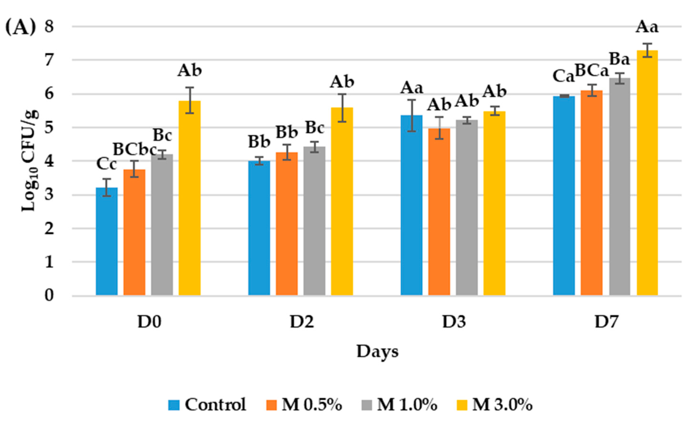
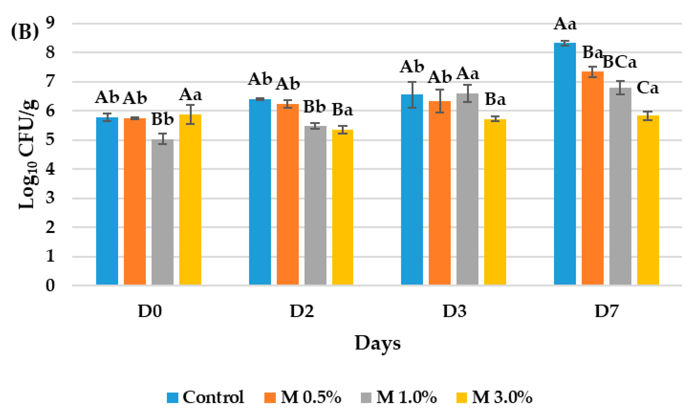
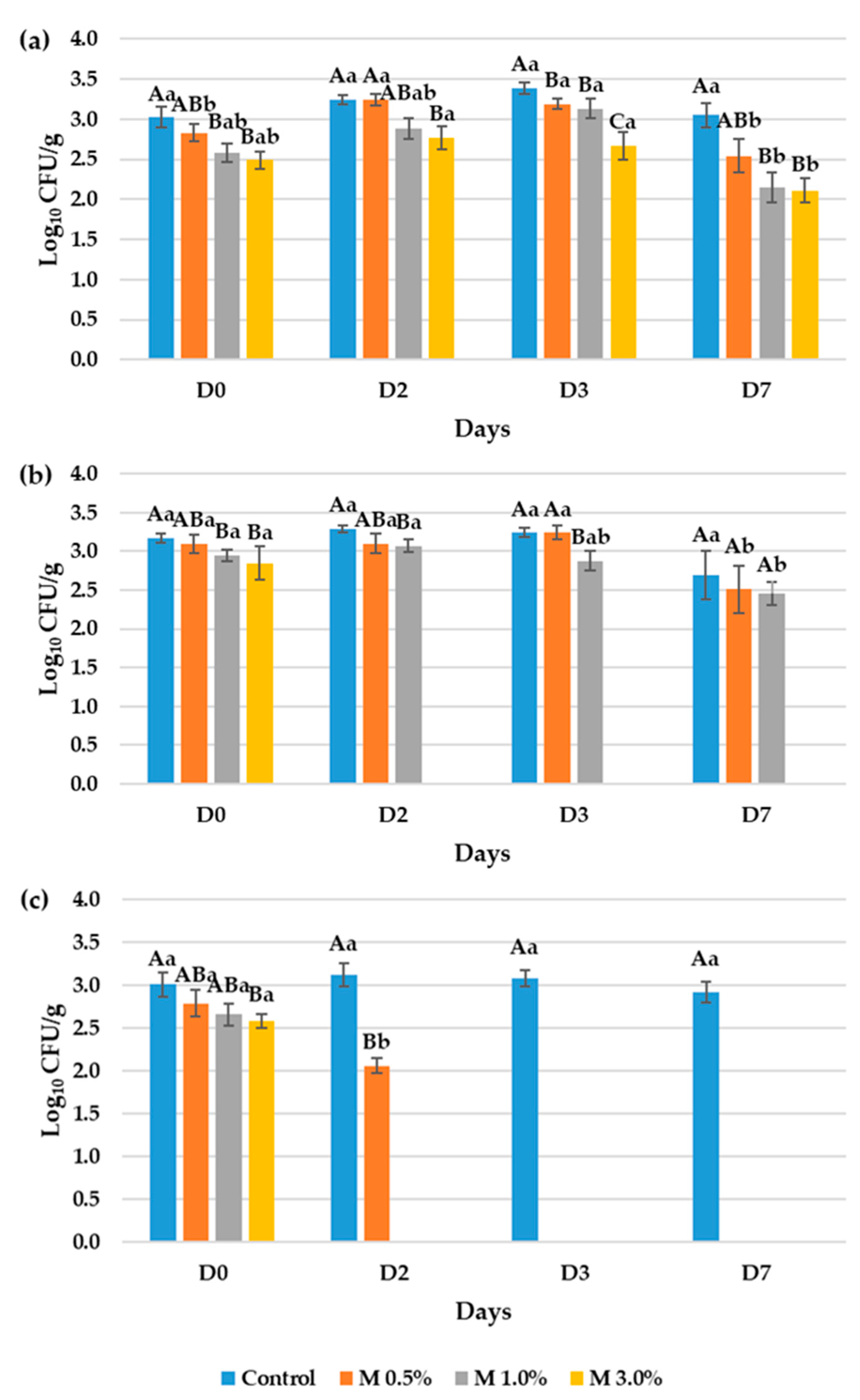
3.3. Evaluation of the Antimicrobial Properties of the Bioactive Ingredient in the Vegan Mortadellas

Figure 3 presents the results of the antimicrobial ingredient effectiveness in reducing the occurrence and proliferation of foodborne contaminants. At day 0, a significant decrease in Log10 CFU/g was observed across all pathogens with increasing concentrations of the bioactive ingredient. This effect was particularly notable against Aspergillus niger and Listeria monocytogenes, where the use of a 0.5% concentration of the bioactive ingredient demonstrated significant efficacy in comparison to the control. The observed reduction at day 0 in the M 0.5% samples when compared to the control was 0.19 and 0.22 Log10 CFU/g in the Aspergillus niger and Listeria monocytogenes assays, respectively. However, in samples contaminated with Penicillium commune, the significant reduction in Log10 CFU/g was only observed in mortadella containing 3.0% of the ingredient, where this initial decrease was of 0.32 Log10 CFU/g (Figure 3).
In samples contaminated with Aspergillus niger, fungal counts did not exceed 3.0 Log10 CFU/g in mortadella containing 3.0%, whereas in the rest of the samples and control fungal counts remained steady at over 3.0 Log10 CFU/g. Aspergillus niger presence at the day sevenwas 3.05, 2.54, 2.15 and 2.11 in the control sample, M 0.5%, M 1.0% and M 3.0%, respectively. The statistic alanalysis determined that samples M 1.0% and M 3.0% had significant reduction in the population of the microorganism population at the end of the assays (Figure 3a).
Assays against Penicillium commune showed that mortadella with higher concentrations of the ingredient reduced fungal populations below detection limits after two days of incubation, and this reduction persisted forseven days. Moreover, samples with 1.0% of the ingredient showed significant reductions in fungal populations compared to the control over a three-dayperiod of 0.36 Log10 CFU/g (Figure 3b).
For samples contaminated with Listeria monocytogenes, the treatment led to a significant decrease in microbial counts. After seven days of incubation, the pathogen was found only in the control samples. In samples treated with 0.5% of the ingredient, L. monocytogenes levels dropped below detection limits by day 3, and in samples with 1.0% and 3.0% of the ingredient, by day 2, while in the control samples the population of the bacterial pathogen remained on an 3.03 Log10 CFU/g (Figure 3c).
This data demonstrates the effectiveness of this bioactive ingredient in the biopreservation of mortadella against three common contaminants in vegan foods. The low treatment concentrations used in these assays (0.5% to 3.0%) are comparable to the typically employed in the industry for similar purposes. Companies typically use spices to prevent the contamination of vegan foods. Spices such as turmeric, mustard, ground paprika, fenugreek, basil, rosemary, and oregano, commonly used at concentrations of 0.1% to 2% [50,51]. Also, synthetic additives are the second choice for the preservation of vegan meat-like products. Nevertheless, synthetic additives are only used on limited occasions due to the strong relationship between consumer demand for vegan foods and the reduction of synthetic compounds in foods [52]. When synthetic additives are used in vegan foods, they are generally added in concentrations recommended by regulatory authorities, such as those outlined by the European Food Safety Authority. For instance, sorbic acid is permitted up to 0.2% in vegan meat- or cheese-like products, while propionic acid and its salts may be applied to cheese-like products or incorporated up to 2% [53]. Meanwhile, research increasingly focuses on novel applications of essential oils as natural antimicrobials. Extensive literature details the antimicrobial mechanisms of essential oils against foodborne pathogens and spoilage organisms. Schelz et al. (2006) provides evidence of the ability of essential oils from several plant sources to disrupt with the proper functions of gram-positive microorganism such as L. monocytogenes [54]. Several studies have developed direct applications of essential oils, including their use as ingredients, coatings, or external methods such as in packaging or bags that release volatile oil compounds into various food matrices [55,56,57].
The addition of spices and essential oils is more widely accepted by consumers compared to synthetic compounds of origin [58] However, these natural additives significantly impact the product taste. While consumers sometimes appreciate the flavor changes, this effect also limits their use. For example, the spiciness or garlic flavor imparted by ingredients like garlic or pepper is often unsuitable for bakery products [59,60]. In contrast, synthetic additives typically do not alter the food flavor profile when used at regulated concentrations [61,62]. Nevertheless, health concerns associated with synthetic additives have increased consumer demand for reducing their presence in foods [63] Therefore, ongoing research is needed into food additives from safe sources that have minimal impact on the flavor profile.
The bioactive ingredients developed in this study meet these requirements. LAB and oat drink are considered natural by both regulators and consumers, while the literature indicates they generate a volatile profile with fewer impactful aroma compounds compared to spices and essential oils [64,65,66]. Furthermore, there has been limited research on the development of bioactive ingredients specifically for vegan foods. Therefore, the ingredient developed here represents a novel application for the biopreservation of vegan mortadella.

4. Conclusions

This study demonstrated that an antifungal ingredient developed through lactic acid bacterial fermentation of oat drink has potential as an effective bioactive ingredient for vegan foods. In vitro assays showed that adding simple nutrients to the oat drink enhanced the fermentation performance, increasing the bacterial growth and antimicrobial metabolite occurrence. Additionally its antimicrobial activity, resulted in inhibition zones of 2 to over 5 mm against tested food-contaminant microorganisms, leading to its selection as a bioactive ingredient. Quality analysis of vegan mortadella containing this ingredient revealed significant differences in pH and water activity compared to the control. Microbial profiling indicated that the ingredient increased lactic acid bacterial counts while reducing natural mesophilic bacterial contamination. In shelf-life extension assays, mortadella samples with 3% of the ingredient showed significant reductions in all three contaminant microorganisms at initial measurements, suggesting that the ingredient effectively inhibits contaminant viability. In samples contaminated with Aspergillus flavus, fungal levels remained stable at significantly lower levels than those in the control. Additionally, assays with Penicillium commune and Listeria monocytogenes demonstrated that mortadella with 3% of the antimicrobial ingredient reduced contaminant populations below detectable limits within two days of incubation, while in control samples, contaminant levels exceeded 2.5 Log10 CFU/g. These findings highlight the potential of this novel ingredient for biopreserving vegan mortadella and suggest its applicability across other food matrices with similar preservation benefits.

Author Contributions

Conceptualization: V.D., J.C. and G.M. Data curation: A.M., A.G. and M.R. Investigation: A.M., A.G. and M.R. Methodology: A.M., A.G. and M.R. Project administration: J.C. and G.M. Resources: G.M. Supervision: V.D., J.C. and G.M. Writing—original draft preparation: A.M. and V.D. Writing—review and editing: V.D., J.C. and G.M. All authors have read and agreed to the published version of the manuscript.

Funding

This research was funded by Ministerio de Ciencia e Innovación, by the project REVALORIZACIÓN DE RESIDUOS DE LA INDUSTRIA AGROALIMENTARIA PARA EL DESARROLLO DE INGREDIENTES ANTIFÚNGICOS Y ANTIMICOTOXIGÉNICOS (MYCOFOODWASTE).

Institutional Review Board Statement

Not applicable.

Informed Consent Statement

Not applicable.

Data Availability Statement

The original contributions presented in this study are included in the article. Further inquiries can be directed to the corresponding author.

Acknowledgments

The authors would like to thank the pre-Ph.D. Grant PRE2020-093996 funded by MCIN/AEI/10.13039/501100011033 and by the European Union.

Conflicts of Interest

The authors declare that they have no known competing financial interests or personal relationships that could have appeared to influence the work reported in this paper.

Abbreviations

The following abbreviations are used in this manuscript:
AwWater activity
BHIBrain heart infusion
CFU/gColony forming units per gram of food
HSIHue, saturation, and intensity
LABLactic acid bacteria
M 0.5%Vegan mortadella with a 0.5% of the bioactive ingredient
M 1.0%Vegan mortadella with a 1.0% of the bioactive ingredient
M 3.0%Vegan mortadella with a 3.0% of the bioactive ingredient
MRSaMan, Rogosa and Sharpe agar
MRSbMan, Rogosa and Sharpe broth
PCAPlate count agar
PDAPotato dextrose agar
ODCulture medium oat drink
ODGCulture medium oat drink enriched with glucose
ODNCulture medium oat drink enriched with nutrients

References

  1. de Oliveira, L.F.R.; de Medeiros, N.S.; de Assis, C.F.; de Sousa, F.C., Jr. Simultaneous vehiculation of probiotics and yellow mombin (Spondias mombin L.) seed extract to develop a new vegan multifunctional ingredient. LWT 2024, 198, 116018. [Google Scholar] [CrossRef]
  2. Mazumder, M.A.R.; Panpipat, W.; Chaijan, M.; Shetty, K.; Rawdkuen, S. Role of plant protein on the quality and structure of meat analogs: A new perspective for vegetarian foods. Future Foods 2023, 8, 100280. [Google Scholar] [CrossRef]
  3. McLauchlan, J.; Tyler, A.I.I.; Chakrabarti, B.; Orfila, C.; Sarkar, A. Oat protein: Review of structure-function synergies with other plant proteins. Food Hydrocoll. 2024, 154, 110139. [Google Scholar] [CrossRef]
  4. Mel, R.; Malalgoda, M. Oat protein as a novel protein ingredient: Structure, functionality, and factors impacting utilization. Cereal Chem. 2022, 99, 21–36. [Google Scholar] [CrossRef]
  5. Holopainen-Mantila, U.; Vanhatalo, S.; Lehtinen, P.; Sozer, N. Oats as a source of nutritious alternative protein. J. Cereal Sci. 2024, 116, 103862. [Google Scholar] [CrossRef]
  6. Tkaczewska, J.; Jamróz, E.; Zając, M.; Guzik, P.; Derbew Gedif, H.; Turek, K.; Kopeć, M. Antioxidant edible double-layered film based on waste from soybean production as a vegan active packaging for perishable food products. Food Chem. 2023, 400, 134009. [Google Scholar] [CrossRef]
  7. Tóth, A.J.; Dunay, A.; Battay, M.; Illés, C.B.; Bittsánszky, A.; Süth, M. Microbial spoilage of plant-based meat analogues. Appl. Sci. 2021, 11, 8309. [Google Scholar] [CrossRef]
  8. Beamonte Vela, B.N.; Garcia-Carretero, R.; Carrasco-Fernandez, B.; Gil-Romero, Y.; Perez-Pomata, M.T. Listeria monocytogenes infections: Analysis of 41 patients. Med. Clin. 2020, 155, 57–62. [Google Scholar] [CrossRef]
  9. Pitt, J.I.; Hocking, A.D. Fungi and Food Spoilage, 4th ed.; Springer US: New York, NY, USA, 2022; ISBN 9783030856403. [Google Scholar]
  10. Wei, S.; Xu, Q.; Pei, S.; Lv, Y.; Lei, Y.; Zhang, S.; Zhai, H.; Hu, Y. Unraveling the antifungal and anti-aflatoxin B1 mechanisms of piperitone on Aspergillus flavus. Food Microbiol. 2024, 123, 104588. [Google Scholar] [CrossRef]
  11. Ebrahimi, M.; Sadeghi, A.; Mortazavi, S.A. The use of cyclic dipeptide producing lab with potent anti-aflatoxigenic capability to improve techno-functional properties of clean-label bread. Ann. Microbiol. 2020, 70, 24. [Google Scholar] [CrossRef]
  12. Abdelshafy, A.M.; Mustafa, M.A.; Hassan, M.A.; Al-Asmari, F. Probiotic-fermentation of oat: Safety, strategies for improving quality, potential food applications and biological activities. Trends Food Sci. Technol. 2024, 151, 104640. [Google Scholar] [CrossRef]
  13. Zang, J.; Yan, B.; Liu, Z.; Tang, D.; Liu, Y.; Chen, J.; Yin, Z. Current state, challenges and future orientations of the applications of lactic acid bacteria exopolysaccharide in foods. Food Microbiol. 2025, 126, 104678. [Google Scholar] [CrossRef] [PubMed]
  14. Rajanikar, R.V.; Nataraj, B.H.; Naithani, H.; Ali, S.A.; Panjagari, N.R.; Behare, P.V. Phenyllactic acid: A green compound for food biopreservation. Food Control 2021, 128, 108184. [Google Scholar] [CrossRef]
  15. Nasrollahzadeh, A.; Mokhtari, S.; Khomeiri, M.; Saris, P.E.J. antifungal preservation of food by lactic acid bacteria. Foods 2022, 11, 395. [Google Scholar] [CrossRef]
  16. Zapaśnik, A.; Sokołowska, B.; Bryła, M. Role of lactic acid bacteria in food preservation and safety. Foods 2022, 11, 1283. [Google Scholar] [CrossRef]
  17. Schnürer, J.; Magnusson, J. Antifungal lactic acid bacteria as biopreservatives. Trends Food Sci. Technol. 2005, 16, 70–78. [Google Scholar] [CrossRef]
  18. Jamuna, M.; Babusha, S.T.; Jeevaratnam, K. Inhibitory efficacy of nisin and bacteriocins from Lactobacillus isolates against food spoilage and pathogenic organisms in model and food systems. Food Microbiol. 2005, 22, 449–454. [Google Scholar] [CrossRef]
  19. Peng, Q.; Yang, J.; Wang, Q.; Suo, H.; Hamdy, A.M.; Song, J. Antifungal effect of metabolites from a new strain Lactiplantibacillus plantarum LPP703 isolated from naturally fermented yak yogurt. Foods 2023, 12, 181. [Google Scholar] [CrossRef] [PubMed]
  20. Riolo, M.; Luz, C.; Santilli, E.; Meca, G.; Cacciola, S.O. Antifungal activity of selected lactic acid bacteria from olive drupes. Food Biosci. 2023, 52, 102422. [Google Scholar] [CrossRef]
  21. Ortiz de Elguea-Culebras, G.; Bourbon, A.I.; Costa, M.J.; Muñoz-Tebar, N.; Carmona, M.; Molina, A.; Sánchez-Vioque, R.; Berruga, M.I.; Vicente, A.A. Optimization of a chitosan solution as potential carrier for the incorporation of Santolina chamaecyparissus L. solid by-product in an edible vegetal coating on ‘Manchego’ cheese. Food Hydrocoll. 2019, 89, 272–282. [Google Scholar] [CrossRef]
  22. Nacef, M.; Chevalier, M.; Chollet, S.; Drider, D.; Flahaut, C. MALDI-TOF mass spectrometry for the identification of lactic acid bacteria isolated from a french cheese: The maroilles. Int. J. Food Microbiol. 2017, 247, 2–8. [Google Scholar] [CrossRef]
  23. Demir, H.; Simsek, M.; Yıldırım, G. Effect of oat milk pasteurization type on the characteristics of yogurt. LWT 2021, 135, 110271. [Google Scholar] [CrossRef]
  24. Dopazo, V.; Illueca, F.; Luz, C.; Musto, L.; Moreno, A.; Calpe, J.; Meca, G. Revalorization by lactic acid bacterial fermentation of goat whey from cheese industry as a potential antifungal agent. Food Biosci. 2023, 53, 102586. [Google Scholar] [CrossRef]
  25. Dopazo, V.; Illueca, F.; Luz, C.; Musto, L.; Moreno, A.; Calpe, J.; Meca, G. Evaluation of shelf life and technological properties of bread elaborated with lactic acid bacteria fermented whey as a bio-preservation ingredient. LWT 2023, 174, 114427. [Google Scholar] [CrossRef]
  26. De Muynck, C.; Leroy, A.I.J.; De Maeseneire, S.; Arnaut, F.; Soetaert, W.; Vandamme, E.J. Potential of Selected lactic acid bacteria to produce food compatible antifungal metabolites. Microbiol. Res. 2004, 159, 339–346. [Google Scholar] [CrossRef]
  27. Illueca, F.; Moreno, A.; Calpe, J.; Nazareth, T.d.M.; Dopazo, V.; Meca, G.; Quiles, J.M.; Luz, C. Bread biopreservation through the addition of lactic acid bacteria in sourdough. Foods 2023, 12, 864. [Google Scholar] [CrossRef] [PubMed]
  28. Mendonça, J.C.F.; Franca, A.S.; Oliveira, L.S. A Comparative evaluation of methodologies for water content determination in green coffee. LWT 2007, 40, 1300–1303. [Google Scholar] [CrossRef]
  29. Luz, C.; Quiles, J.M.; Romano, R.; Blaiotta, G.; Rodríguez, L.; Meca, G. Application of whey of mozzarella di bufala campana fermented by lactic acid bacteria as a bread biopreservative agent. Int. J. Food Sci. Technol. 2021, 56, 4585–4593. [Google Scholar] [CrossRef]
  30. Wang, B.; Yang, H.; Yang, C.; Lu, F.; Wang, X.; Liu, D. Prediction of total volatile basic nitrogen (TVB-N) and 2-thiobarbituric acid (TBA) of smoked chicken thighs using computer vision during storage at 4 °C. Comput. Electron. Agric. 2022, 199, 107170. [Google Scholar] [CrossRef]
  31. Riolo, M.; Villena, A.M.; Calpe, J.; Luz, C.; Meca, G.; Tuccitto, N.; Cacciola, S.O. A Circular economy approach: A new formulation based on a lemon peel medium activated with Lactobacilli for sustainable control of post-harvest fungal rots in fresh citrus fruit. Biol. Control 2024, 189, 105443. [Google Scholar] [CrossRef]
  32. Rovetto, E.I.; La Spada, F.; El boumlasy, S.; Conti Taguali, S.; Riolo, M.; Pane, A.; Cacciola, S.O. Biological control of green mold in simulated post-harvest chain of citrus fruit: Efficacy of Candida oleophila strain o and molecular insight into elicitation of host immune system. Biol. Control 2024, 193, 105531. [Google Scholar] [CrossRef]
  33. Dopazo, V.; Luz, C.; Calpe, J.; Vila-Donat, P.; Rodríguez, L.; Meca, G. Antifungal Properties of whey fermented by lactic acid bacteria in films for the preservation of cheese slices. Int. J. Dairy. Technol. 2022, 75, 619–629. [Google Scholar] [CrossRef]
  34. Tongnuanchan, P.; Benjakul, S. Essential Oils: Extraction, bioactivities, and their uses for food preservation. J. Food Sci. 2014, 79, R1231–R1249. [Google Scholar] [CrossRef] [PubMed]
  35. Moreira, M.R.; Ponce, A.G.; Del Valle, C.E.; Roura, S.I. Inhibitory parameters of essential oils to reduce a foodborne pathogen. LWT 2005, 38, 565–570. [Google Scholar] [CrossRef]
  36. Hussain, A.I.; Anwar, F.; Hussain Sherazi, S.T.; Przybylski, R. Chemical composition, antioxidant and antimicrobial activities of basil (Ocimum basilicum) essential oils depends on seasonal variations. Food Chem. 2008, 108, 986–995. [Google Scholar] [CrossRef]
  37. Mariod, A.A. Effect of essential oils on organoleptic (smell, taste, and texture) properties of food essential oils in food preservation. In Flavor and Safety, 1st ed.; Elsevier: Amsterdam, The Netherlands, 2016; pp. 131–137. [Google Scholar] [CrossRef]
  38. Sorathiya, K.B.; Melo, A.; Hogg, M.C.; Pintado, M. Organic acids in food preservation: Exploring synergies, molecular insights, and sustainable applications. Sustainability 2025, 17, 3434. [Google Scholar] [CrossRef]
  39. Russo, P.; Fares, C.; Longo, A.; Spano, G.; Capozzi, V. Lactobacillus plantarum with broad antifungal activity as a protective starter culture for bread production. Foods 2017, 6, 110. [Google Scholar] [CrossRef]
  40. De Rossi, L.; Rocchetti, G.; Lucini, L.; Rebecchi, A. Antimicrobial potential of polyphenols: Mechanisms of action and microbial responses—A narrative review. Antioxidants 2025, 14, 200. [Google Scholar] [CrossRef]
  41. Nkhata, S.G.; Ayua, E.; Kamau, E.H.; Shingiro, J.B. Fermentation and germination improve nutritional value of cereals and legumes through activation of endogenous enzymes. Food Sci. Nutr. 2018, 6, 2446. [Google Scholar] [CrossRef]
  42. Chen, G.Q.; Liu, X. On the future fermentation. Microb. Biotechnol. 2021, 14, 18–21. [Google Scholar] [CrossRef]
  43. Ojo, A.O.; de Smidt, O. Lactic acid: A comprehensive review of production to purification. Processes 2023, 11, 688. [Google Scholar] [CrossRef]
  44. Gabriel, A.A. Estimation of water activity from pH and °Brix values of some food products. Food Chem. 2008, 108, 1106–1113. [Google Scholar] [CrossRef] [PubMed]
  45. Tangyu, M.; Fritz, M.; Tan, J.P.; Ye, L.; Bolten, C.J.; Bogicevic, B.; Wittmann, C. Flavour by design: Food-grade lactic acid bacteria improve the volatile aroma spectrum of oat milk, sunflower seed milk, pea milk, and faba milk towards improved flavour and sensory perception. Microb. Cell Factories 2023, 22, 133. [Google Scholar] [CrossRef]
  46. Dagnas, S.; Gauvry, E.; Onno, B.; Membré, J.M. Quantifying effect of lactic, acetic, and propionic acids on growth of molds isolated from spoiled bakery products. J. Food Prot. 2015, 78, 1689–1698. [Google Scholar] [CrossRef] [PubMed]
  47. Ibrahim, S.A.; Ayivi, R.D.; Zimmerman, T.; Siddiqui, S.A.; Altemimi, A.B.; Fidan, H.; Esatbeyoglu, T.; Bakhshayesh, R.V. Lactic acid bacteria as antimicrobial agents: Food safety and microbial food spoilage prevention. Foods 2021, 10, 3131. [Google Scholar] [CrossRef]
  48. Landim Neves, M.I. Color solutions in plant-based foods. In Handbook of Plant-Based Food and Drinks Design, 1st ed.; Elsevier: Amsterdam, The Netherlands, 2024; pp. 319–334. [Google Scholar] [CrossRef]
  49. Kale, P.; Mishra, A.; Annapure, U.S. development of vegan meat flavour: A review on sources and techniques. Future Foods 2022, 5, 100149. [Google Scholar] [CrossRef]
  50. Peters, J.C.; Breen, J.A.; Pan, Z. Effects of Culinary spices on liking and consumption of protein rich foods in community-dwelling older adults. Nutrients 2023, 15, 1172. [Google Scholar] [CrossRef]
  51. Gajowniczek-Ałasa, D.; Baranowska-Wójcik, E.; Szwajgier, D. Vegan and vegetarian soups are excellent sources of cholinesterase inhibitors. Nutrients 2024, 16, 2025. [Google Scholar] [CrossRef]
  52. Craig, W.J. Nutrition concerns and health effects of vegetarian diets. Nutr. Clin. Pract. 2010, 25, 613–620. [Google Scholar] [CrossRef]
  53. EFSA Regulation (EC) No 1333/2008 of the European Parliament and of the Council of 16 December 2008 on Food Additives. Off. J. Eur. Union 2008, 354, 16–33.
  54. Schelz, Z.; Molnar, J.; Hohmann, J. Antimicrobial and antiplasmid activities of essential oils. Fitoterapia 2006, 77, 279–285. [Google Scholar] [CrossRef] [PubMed]
  55. Quiles, J.M.; Nazareth, T.d.M.; Luz, C.; Luciano, F.B.; Mañes, J.; Meca, G. Development of an antifungal and antimycotoxigenic device containing allyl isothiocyanate for silo fumigation. Toxins 2019, 11, 137. [Google Scholar] [CrossRef] [PubMed]
  56. Hayouni, E.A.; Chraief, I.; Abedrabba, M.; Bouix, M.; Leveau, J.Y.; Mohammed, H.; Hamdi, M. Tunisian Salvia officinalis L. and Schinus molle L. Essential oils: Their chemical compositions and their preservative effects against Salmonella inoculated in minced beef meat. Int. J. Food Microbiol. 2008, 125, 242–251. [Google Scholar] [CrossRef]
  57. Lu, Y.; Joerger, R.; Wu, C. Similar reduction of Salmonella enterica Typhimurium on grape tomatoes and its cross-contamination in wash water by washing with natural antimicrobials as compared with chlorine treatment. Food Bioprocess Technol. 2014, 7, 661–670. [Google Scholar] [CrossRef]
  58. Lima, G.; Spina, A.M.; Castoria, R.; De Curtis, F.; De Cicco, V. Integration of Biocontrol agents and food-grade additives for enhancing protection of stored apples from Penicillium expansum. J. Food Prot. 2005, 68, 2100–2106. [Google Scholar] [CrossRef] [PubMed]
  59. Ríos, J.L. Essential Oils: What they are and how the terms are used and defined essential oils in food preservation. In Flavor and Safety, 1st ed.; Elsevier: Amsterdam, The Netherlands, 2016; pp. 3–10. [Google Scholar] [CrossRef]
  60. Jackson-Davis, A.; White, S.; Kassama, L.S.; Coleman, S.; Shaw, A.; Mendonca, A.; Cooper, B.; Thomas-Popo, E.; Gordon, K.; London, L. A review of regulatory standards and advances in essential oils as antimicrobials in foods. J. Food Prot. 2023, 86, 100025. [Google Scholar] [CrossRef]
  61. Aguilar, F.; Crebelli, R.; Domenico, A.; Dusemund, B.; Frutos, M.; Galtier, P.; Gott, D.; Gundert-Remy, U.; Lambré, C.; Leblanc, J.; et al. Scientific Opinion on the re-evaluation of propionic acid (E 280), sodium propionate (E 281), calcium propionate (E 282) and potassium propionate (E 283) as food additives. EFSA J. 2014, 12, 3779. [Google Scholar] [CrossRef]
  62. Sutoh, M.; Imura, T.; Tsukada, H.; Yamada, A. Flavour preference conditioned by postabsorptive propionate and acetate in wethers. Appl. Anim. Behav. Sci. 2008, 111, 274–285. [Google Scholar] [CrossRef]
  63. El-Nezami, H.S.; Chrevatidis, A.; Auriola, S.; Salminen, S.; Mykkänen, H. Removal of common Fusarium toxins in vitro by strains of Lactobacillus and Propionibacterium. Food Addit. Contam. 2002, 19, 680–686. [Google Scholar] [CrossRef]
  64. Sadgrove, N.J.; Padilla-González, G.F.; Phumthum, M. Fundamental chemistry of essential oils and volatile organic compounds, methods of analysis and authentication. Plants 2022, 11, 789. [Google Scholar] [CrossRef]
  65. Dąbrowski, G.; Paulauskienė, A.; Baltušnikienė, A.; Kłębukowska, L.; Czaplicki, S.; Konopka, I. Changes in selected quality indices in microbially fermented commercial almond and oat drinks. Appl. Sci. 2022, 12, 9983. [Google Scholar] [CrossRef]
  66. Pisoschi, A.M.; Pop, A.; Georgescu, C.; Turcuş, V.; Olah, N.K.; Mathe, E. An overview of natural antimicrobials role in food. Eur. J. Med. Chem. 2018, 143, 922–935. [Google Scholar] [CrossRef] [PubMed]
Figure 1. Results from the quantification of the Log10 CFU/g of food in (A) lactic acid bacteria count and (B) total aerobic mesophilic bacteria count of the mortadella samples thought the 7-day incubation. The control consisted of mortadella without the bioactive ingredient, while M 0.5%, M 1.0%, and M 3.0% corresponded to mortadella samples formulated with 0.5%, 1.0%, and 3.0% of the bioactive ingredient derived from the ODN medium fermented by L. plantarum VC7, respectively. Different letters were used to signal significative differences of the studied parameters. Capital letters were used for the comparation among different days and case letters for the comparation among samples the same day.
Foods 14 02195 g001aFoods 14 02195 g001b
Figure 2. Mortadella control and mortadella samples with 0.5%, 1.0% and 3.0% of the bioactive ingredient at days 0, 2, 3 and 7 after the elaboration.
Foods 14 02195 g002
Figure 3. Results from the in vitro assays to evaluate the capacity of the bioactive ingredient to reduce the microbial contamination of mortadella against (a) Aspergillus niger, (b) Penicillium commune and (c) Listeria monocytogenes. The control consisted of mortadella without the bioactive ingredient, while M 0.5%, M 1.0%, and M 3.0% corresponded to mortadella samples formulated with 0.5%, 1.0%, and 3.0% of the bioactive ingredient derived from the ODN medium fermented by L. plantarum VC7, respectively. Different letters were used to signal significative differences of the studied parameters. Capital letters were used for the comparation among different days and case letters for the comparation among samples the same day. Results under the limit of detection (2.00 Log10 CFU/g) were marked as 0.00 Log10 CFU/g.
Foods 14 02195 g003
Table 1. Recipe of the different vegan mortadella developed in this study. Data was expressed in grams.
IngredientsSamples
ControlM 0.5%M 1.0%M 3.0%
Bean mash538.0534.5531.0517.0
Water135.0135.0135.0135.0
Olive50.050.050.050.0
Agar24.424.424.424.4
Salt2.02.02.02.0
Thyme0.40.40.40.4
Garlic0.20.20.20.2
Bioactive ingredient-3.57.021.0
Table 2. Results from the analysis of the fermentation parameters of the oat mediums after the 48 h incubation. The pH, bacterial count (Log10 LAB CFU/mL of sample), lactic acid (g/L), phenyllactic acid (mg/L).
SamplespHBacterial CountLactic AcidPhenyllactic Acid
ControlOD7.39 ± 0.01 andndnd
ODG6.58 ± 0.01 bndndnd
ODN6.41 ± 0.01 cndndnd
L. plantarum VC7OD3.44 ± 0.01 d7.37 ± 0.20 b14.84 ± 0.01 b2.61 ± 0.09 c
ODG3.41 ± 0.02 e8.20 ± 0.09 a17.27 ± 0.01 a5.42 ± 0.21 b
ODN3.52 ± 0.02 e8.32 ± 0.12 a17.34 ± 0.01 a9.23 ± 0.13 a
Data was displayed as mean ± SD. Different letters were used to signal significative differences among each studied parameter. Results below the limit of detection were marked as nd.
Table 3. Results from the qualitative antimicrobial evaluation of the oat drink fermented by the L. plantarum VC7 isolate against different vegan mortadella contaminants.
Contaminant
Microorganism
ControlL. plantarum VC7
ODODGODNODODGODN
A. flavus----+++++
A. fumigatus----++++++
A. niger-----++
A. steynii----+++++
P. camemberti-----+++
P. commune---++++++
P. digitatum---++++++
P. expansum---++++++++
P. italicum----+++++++
P. nordicum----+++++++
P. roqueforti---++++
P. verrucosum---+++++++
L. monocytogenes---++++++++
Results were expressed as “++++” when the inhibition halo was superior to 5 mm, “+++”, when the halos were among 2 to 5 mm, “++” from 1 to 2 mm, “+” lower than 1 mm and “-“ when no inhibition was observed.
Table 4. Results from the measurement of the (a) pH, (b) Aw, (c) moisture (g/100 g) and (d) HSI of the different mortadella samples thought the 7-day incubation.
(a) pH
SamplesDays
D0D2D3D7
Control5.98 ± 0.03 BCa6.22 ± 0.06 Aa6.03 ± 0.01 Ba5.93 ± 0.04 Ca
M 0.5%5.70 ± 0.02 ABb5.90 ± 0.09 Ab5.68 ± 0.04 ABb5.65 ± 0.12 Bb
M 1.0%5.10 ± 0.06 Cc5.50 ± 0.13 Ac5.41 ± 0.05 Bc5.40 ± 0.03 Bc
M 3.0%4.60 ± 0.03 Bd4.85 ± 0.09 Ad4.73 ± 0.02 ABd4.74 ± 0.03 ABd
(b) Aw
SamplesDays
D0D2D3D7
Control0.975 ± 0.001 Ba0.905 ± 0.001 Da0.985 ± 0.001 Aa0.954 ± 0.001 Ca
M 0.5%0.975 ± 0.002 Aa0.907 ± 0.001 Da0.968 ± 0.002 Bb0.935 ± 0.001 Cb
M 1.0%0.925 ± 0.001 Bb0.858 ± 0.001 Db0.948 ± 0.001 Ad0.918 ± 0.001 Cc
M 3.0%0.874 ± 0.001 Cc0.862 ± 0.001 Db0.958 ± 0.001 Ac0.908 ± 0.001 Bd
(c) Moisture
SamplesDays
D0D2D3D7
Control25.22 ± 0.76 Bb25.61 ± 0.66 Bc26.34 ± 0.40 ABb28.53 ± 0.52 Aa
M 0.5%27.34 ± 0.38 Ab26.34 ± 0.31 Aab23.76 ± 1.25 Bb24.69 ± 0.97 Bc
M 1.0%27.31 ± 0.76 Ab27.74 ± 0.66 Aba24.46 ± 0.30 Bb25.01 ± 0.31 Ab
M 3.0%30.57 ± 0.19 Aa29.55 ± 0.18 Aa28.76 ± 0.96 Ba26.46 ± 0.25 Cb
(d) HSI
SamplesDays
D0D2D3D7
Control25.39 ± 4.67 ABc26.33 ± 3.85 Ab20.21 ± 0.53 Bc19.23 ± 6.54 ABb
M 0.5%49.86 ± 0.93 Ab53.09 ± 5.10 Aa43.36 ± 6.68 Aa44.17 ± 3.67 Aa
M 1.0%58.27 ± 2.91 Aa59.89 ± 4.95 Aa48.27 ± 0.46 Ba47.31 ± 5.32 Ba
M 3.0%48.13 ± 1.97 Ab52.79 ± 5.68 Aa47.01 ± 1.50 Aa44.38 ± 3.70 Aa
Data was displayed as mean ± SD. Different letters were used to signal significative differences of the studied parameters. Capital letters for the comparation among different days (rows) and case letter for the comparation among samples the same day (columns).
Disclaimer/Publisher’s Note: The statements, opinions and data contained in all publications are solely those of the individual author(s) and contributor(s) and not of MDPI and/or the editor(s). MDPI and/or the editor(s) disclaim responsibility for any injury to people or property resulting from any ideas, methods, instructions or products referred to in the content.

