Article

Undescribed Cyclohexene and Benzofuran Alkenyl Derivatives from Choerospondias axillaris, a Potential Hypoglycemic Fruit

1
State Key Laboratory of Phytochemistry and Plant Resources of West China, Kunming Institute of Botany, Chinese Academy of Sciences, Kunming 650201, China
2
University of Chinese Academy of Sciences, Beijing 100049, China
3
Yunnan Key Laboratory of Natural Medicinal Chemistry, Kunming Institute of Botany, Chinese Academy of Sciences, Kunming 650201, China
*
Author to whom correspondence should be addressed.
Foods 2024, 13(10), 1495; https://doi.org/10.3390/foods13101495
Submission received: 4 April 2024 / Revised: 7 May 2024 / Accepted: 7 May 2024 / Published: 11 May 2024
(This article belongs to the Section Nutraceuticals, Functional Foods, and Novel Foods)

Abstract

:
The fruit of Choerospondias axillaris (Anacardiaceae), known as south wild jujube in China, has been consumed widely in several regions of the world to produce fruit pastille and leathers, juice, jam, and candy. A comprehensive chemical study on the fresh fruits led to the isolation and identification of 18 compounds, including 7 new (17) and 11 known (818) comprised of 5 alkenyl (cyclohexenols and cyclohexenones) derivatives (15), 3 benzofuran derivatives (68), 6 flavonoids (914) and 4 lignans (1518). Their structures were elucidated by extensive spectroscopic analysis. The known lignans 1518 were isolated from the genus Choerospondias for the first time. Most of the isolates exhibited significant inhibitory activity on α-glucosidase with IC50 values from 2.26 ± 0.06 to 43.9 ± 0.96 μM. Molecular docking experiments strongly supported the potent α-glucosidase inhibitory activity. The results indicated that C. axillaris fruits could be an excellent source of functional foods that acquire potential hypoglycemic bioactive components.

1. Introduction

Choerospondias axillaris (Roxb.) B.L. Burtt. & A.W. Hill (Anacardiaceae), a tall deciduous tree bearing edible fruits, is distributed in slopes, hills or valleys at altitudes ranging from 300 to 2000 m in India, China, Japan, Bhutan, Laos, Vietnam, and Thailand [1,2,3]. The fruit, known as south wild jujube in China, with a light yellow flesh and sour–sweet taste, is valued for its substantial nutritional and therapeutic values in several regions of the world [4,5]. In Traditional Chinese Medicine (TCM), the fruit of C. axillaris has been historically utilized for its potential therapeutic properties to improve cardiac function as well as to regulate blood sugar levels and alleviate symptoms linked to diabetes [1,3,4,5,6,7]. Furthermore, it is used widely in China’s food industry for producing fruit pastilles and juice [8,9] and is utilized in Nepal for processing local products, e.g., pickles, fruit leathers, jam, and candy [1,10]. Regardless of insufficient reports on global sales data and socio-economic purposes, the market value of C. axillaris was found to be approximately USD 0.65 million in Kathmandu, Nepal [1,8,11]. C. axillaris as a functional food provides health benefits beyond basic nutrition due to its physiologically active components, which play an active role in promoting overall health and regulating disease progression [1,2,3,4,5,6,7,8,9,10].
So far, 26 compounds consisting of flavonoids, phenolic acids, organic acids, fatty acids, and sterols have been isolated from C. axillaris fruits [12,13,14,15], among which flavonoids represent a class of the most important and effective components that proven to have various biological activities, such as antioxidant [9,10,16], antiproliferative and cytotoxic [17,18], as well as anti-diabetic effects [19].
α-glucosidase is a kind of oligosaccharide hydrolase located in the small intestinal epithelial cell brush border responsible for the cleavage of α-1,4 bonds from the non-reducing end of α-linked oligosaccharide or α-glucan substrates to liberate glucose [20,21,22,23,24,25], which increases postprandial blood glucose, thereby causing obesity or diabetes [26]. Inhibiting the activity of α-glucosidase has been an effective therapeutic approach for hyperglycemia linked to type 2 diabetes mellitus. Rich in flavonoids which showed potent α-glucosidase inhibitory activity [27,28,29,30,31,32], C. axillaris fruit might be a potential source of hypoglycemic functional food.
Therefore, the present study was undertaken with the objective of isolating, identifying, and investigating the in vitro anti-diabetic potential of bioactive compounds from the fresh fruit of C. axillaris. Furthermore, aimed to reveal the possible inhibition mechanisms and binding mode of these compounds on α-glucosidase through molecular docking simulations.

2. Materials and Methods

2.1. General Procedure

UV spectra were measured on a Shimadzu UV-2401PC spectrophotometer (Shimadzu Co., Kyoto, Japan) with methanol as solvent. The optical rotations were recorded with a P-1020 polarimeter (JASCO, Tokyo, Japan). JASCO J-810 was used to obtain Experimental CD spectra, Bruker Av (600 MHz) spectrometers (Bruker Co., Karlsruhe, Germany) were used to measure 1D and 2D NMR spectra in methanol-d4 (CD3OD). Chemical shifts were denoted by δ (ppm) and coupling constants were indicated as J (Hz), with TMS as an internal standard. IR spectra were measured using BioRad FtS-135 spectrophotometer (Bio-Rad, Richmond, CA, USA). Semi-preparative HPLC was performed on Waters 600 controller equipped with waters 2487 dual absorbance detector HPLC (Waters, Milford, USA) using COSMOSIL 5C18-MS-II (5 μm, 10 × 250 mm), Thermo scientific Hypersil GOLD aQ (5 μm, 10 × 250 mm) and Agilent ZORBAX SB-C18 (5 μm, 9.4 × 150 mm) packed columns. ESIMS and HRESIMS data were recorded using an Agilent 1290 UPLC/6540 Q-TOF instrument (Agilent Technologies, Palo Alto, CA, USA).

2.2. Chemicals and Reagents

LiChroprep Rp-18 (40–63 μm, Merck, Darmstadt, Germany), silica gel (200–300 mesh, Qingdao Haiyang Chemical Co. Ltd., Qingdao, China) and Sephadex LH-20 (25–100 μm, GE Healthcare Bio-Science AB, Uppsala, Sweden) were used as a stationary phase for column chromatography (CC). Precoated silica gel GF254 plates, 0.2–0.25 mm thick (Qingdao Haiyang Chemical Co., Qingdao, China), were used for thin layer chromatography (TLC) analysis, spots were visualized under a UV radiation by spraying with a sulfuric acid: ethanol (1:9, v/v) solution followed by heating. 4-Nitrophenyl-α-D-glucopyranoside (pNPG), α-glucosidase, quercetin and acarbose were purchased from sigma chemical (Merck KGaA, Darmstadt, Germany).

2.3. Materials

The fresh fruits of Choerospondias axillaris were collected in Kunming Botanical Garden, Kunming Institute of Botany (KIB), Chinese Academy of Sciences (CAS), Yunnan Province, China, on September 2020, and authenticated by Dr. En-De Liu from KIB, CAS. A voucher specimen (KIB-ZL-20200903) was stored in the State Key Laboratory of Phytochemistry and Plant Resources in West China, KIB, CAS.

2.4. Extraction and Isolation

The fresh fruits (95 kg) of C. axillaris were extracted with 80% aqueous acetone (4 × 80 L). The resulting solutions were filtered and concentrated under reduced pressure to give the acetone residue (5.79 kg). This was suspended into H2O and partitioned with EtOAc to obtain the EtOAc (732 g) extract and H2O residue. The EtOAc fraction (300 g) was subjected to CC using Sephadex LH-20 assisted with TLC, and gradient elution with MeOH/H2O (0:1–1:0, v/v) to give six fractions (Fr. 1–Fr. 6). Fr. 5 (54 g) underwent separation over silica gel CC, eluting with MeOH/CHCl3 (0:1–1:0, v/v) gradient system to obtain Fr. 5-1–Fr. 5-4. Fr. 5-4 was chromatographed with Sephadex LH-20 column, eluting with MeOH/H2O (0:1–1:0, v/v) and further purified with semi-preparative HPLC using Agilent ZORBAX SB-C18 (5 μm, 9.4 × 150 mm) packed column (CH3CN/H2O 16:84, v/v) to yield compounds 9 (25 mg, tr = 18.9 min, CH3CN/H2O 17:83, v/v), 10 (15 mg, tr = 20.9 min, CH3CN/H2O 16:84, v/v), 11 (18 mg, tr = 20.9 min, CH3CN/H2O 20:80, v/v), 12 (22 mg, tr = 40.7 min, CH3CN/H2O 20:80, v/v), 13 (20 mg, tr = 23.4 min, CH3CN/H2O 28:72, v/v), 14 (10 mg, tr = 12.2 min, CH3CN/H2O 13:87, v/v). Fr. 6 (43 g) was chromatographed over a silica gel column, employing MeOH/CHCl3 (0:1–1:0, v/v) gradient system as the eluent, to yield sub-fractions (Fr. 6-1–Fr. 6-5). Fr. 6-4 was fractionated on a column packed with RP-18 eluted with MeOH/H2O (0:1–1:0, v/v) to afford Fr. 6-4-1–Fr. 6-4-4. Fr. 6-4-4 was purified with semi-preparative HPLC using Thermo scientific Hypersil GOLD aQ (5 μm, 10 × 250 mm) packed column (CH3CN/H2O 82:18, v/v) to yield compounds 6 (6 mg, tr = 50.5 min), 7 (5 mg, tr = 35.3 min) and 8 (6.5 mg, tr = 99 min). Fr. 6-5 was transferred to a column packed with silica gel, eluting with MeOH/CHCl3 (0:1–1:0, v/v), to afford sub-fractions Fr. 6-5-1–Fr. 6-5-4. Fr. 6-5-1 was repeatedly applied to CC over RP-18 with aqueous MeOH (40–100%) as eluent and further semi-preparative HPLC purification using Agilent ZORBAX SB-C18 (5 μm, 9.4 × 150 mm) packed column, yielded compounds 16 (8 mg, tr = 11.3 min, CH3CN/H2O 17:83, v/v), 15 (5.3 mg, tr = 16.05 min, CH3CN/H2O 17:83, v/v), 17 (7.4 mg, tr = 30.2 min, CH3CN/H2O 17:83, v/v) and 18 (10 mg, tr = 22.9 min, CH3CN/H2O 24:76, v/v). Silica gel CC separation of Fr. 6-5-1-6 eluting with MeOH/CHCl3 (1:9–2:8, v/v) and further semi-preparative HPLC purification using Thermo scientific Hypersil GOLD aQ column yield compounds 2 (7.3 mg, tr = 21.8 min, CH3CN/H2O 79:21, v/v), 3 (3.3 mg, tr = 18.2 min, CH3CN/H2O 81:19, v/v). Further purification of fraction Fr. 6-5-1-5 via semi-preparative HPLC applying COSMOSIL 5C18-MS-II (5 μm, 10 × 250 mm) packed column (CH3CN/H2O 80:20, v/v) yeilded compound 4 (8 mg, tr = 36.0 min). Fr. 6-5-2 was successively subjected to RP-18 column, MeOH/H2O gradient elution of 50–100% and further resolved by semi-preparative HPLC employing a COSMOSIL 5C18-MS-II column (CH3CN/H2O 78:22, v/v) to afford compounds 1 (3.5 mg, tr = 19.7 min) and 5 (4 mg, tr = 23.5 min).

2.4.1. Choerosponol F

Colorless solid, α D 20 8.86 (c 0.07, MeOH); UV (MeOH) λmax (log ε): 202.0 (3.34), 217.5 (2.97), 278.5 (2.13) nm; IR (KBr) νmax 3404, 2926, 2854, 1061 cm−1; molecular formula: C21H40O3; HRESIMS m/z 385.2957 [M + HCOO] (calcd. for C21H40O3COOH, 385.2959); 1H and 13C NMR (600 and 150 MHz, in CD3OD) spectroscopic data, presented in Table 1.

2.4.2. Choerosponol G

Colorless oil, α D 20 65.11 (c 0.09, MeOH); UV (MeOH) λmax (log ε): 195.0 (3.16) nm; IR (KBr) νmax 3414, 2924, 2853 cm−1; molecular formula: C23H42O3; HRESIMS m/z 389.3029 [M + Na]+ (calcd. for C23H42O3Na+, 389.3026); 1H and 13C NMR (600 and 150 MHz, in CD3OD) spectroscopic data, presented in Table 1.

2.4.3. Choerosponol H

Colorless oil, α D 20 49.60 (c 0.05, MeOH); UV (MeOH) λmax (log ε): 195.0 (2.91) nm; IR (KBr) νmax 3405, 2923, 2853 cm−1; molecular formula: C25H46O3; HRESIMS m/z 417.3337 [M + Na]+ (calcd. for C25H46O3Na+, 417.3339); 1H and 13C NMR (600 and 150 MHz, in CD3OD) spectroscopic data, presented in Table 1.

2.4.4. Choerosponol I

Colorless oil, α D 20 19.45 (c 0.11, MeOH); UV (MeOH) λmax (log ε): 195.0 (3.04), 234.0 (2.97) nm; IR (KBr) νmax 3394, 2924, 2853, 1658 cm−1; molecular formula: C23H40O3; HRESIMS m/z 387.2868 [M + Na]+ (calcd. for C23H40O3Na+, 387.2870); 1H and 13C NMR (600 and 150 MHz, in CD3OD) spectroscopic data, presented in Table 2.

2.4.5. Choerosponol J

Colorless amorphous solid, α D 20 41.60 (c 0.05, MeOH); UV (MeOH) λmax (log ε): 195.0 (2.95), 234.0 (2.89) nm; IR (KBr) νmax 3406, 2925, 2853, 1661 cm−1; molecular formula: C23H40O4; HRESIMS m/z 425.2909 [M + HCOO] (calcd. for C23H40O4COOH, 425.2909). 1H and 13C NMR (600 and 150 MHz, in CD3OD) spectroscopic data, presented in Table 2.

2.4.6. Choerosponol K

Colorless amorphous solid, α D 20 7.17 (c 0.12, MeOH); UV (MeOH) λmax (log ε): 204.0 (4.37), 251.0 (4.03), 294.0 (3.56) nm; IR (KBr) νmax 3413, 2922, 2851, 1605, 1468, 1198 cm−1; molecular formula: C23H34O2; HRESIMS m/z 341.2479 [M − H] (calcd. for C23H34O2 341.2486); 1H and 13C NMR (600 and 150 MHz, in CD3OD) spectroscopic data, presented in Table 2.

2.4.7. Choerosponol L

Colorless oil, α D 20 6.00 (c 0.05, MeOH); UV (MeOH) λmax (log ε): 205.0 (4.27), 251.0 (3.98), 294.0 (3.52) nm; IR (KBr) νmax 3427, 2924, 2853, 1607, 1468, 1197 cm−1; molecular formula: C21H30O2; HRESIMS m/z 337.2137 [M + Na]+ (calcd. for C21H30O2Na+ 337.2138); 1H and 13C NMR (600 and 150 MHz, in CD3OD) spectroscopic data, presented in Table 2.

2.5. Quantum Chemistry ECD Calculation

Conformational analysis was performed with the MMFF forcefield (Merck Molecular forcefield) using the Monte Carlo algorithm implemented in Spartan’10 software (Wavefunction, Irvine, CA, USA, 2018). ECD calculations were carried out, following optimizing the conformers at B3LYP/6-31+g (d, p) level in MeOH using the CPCM polarizable conductor calculation model, the conformers with a Boltzmann population of over 5% were chosen. The theoretical calculation of ECD was conducted in MeOH using time-dependent density functional theory (TD-DFT) at the B3LYP/6-311+g (d, p) level for all conformers of compound 18b. Rotatory strengths for a total of 50 excited states were calculated. ECD spectra were generated using the program. SpecDis 1.6 (University of Würzburg, Würzburg, Germany) and GraphPad Prism 5 (University of California, San Diego, CA, USA) from dipole-length rotational strengths by applying Gaussian band shapes with sigma = 0.3 eV.

2.6. α-Glucosidase Inhibitory Assay

The α-glucosidase inhibitory activity was evaluated according to a previous method [33]. In brief, α-glucosidase solution (0.025 U/mL, dissolved in 0.1 M phosphate-buffered saline (PBS) solution with a pH of 6.8), was mixed with 50 μM inhibitor and incubated at 37 °C for 10 min. pNPG solution (1 μM, dissolved in 0.1 M PBS solution) was added to the mixed solution and incubated at 37 °C for 40 min. Sodium carbonate (1 M) solution was added to terminate the reaction. Quercetin and acarbose were used as a positive control. Quercetin is selected as a reference in α-glucosidase assay because of its efficacy as a therapeutic inhibitor; it binds strongly within the binding region (active site) of the protein and forms a compact structure, whereas acarbose is a common medication utilized in clinical practice to manage postprandial hyperglycemia [30,34,35]. The absorbance was recorded at 405 nm usin a microplate reader. The α-glucosidase inhibitory rate was calculated as follows:
Inhibitory rate% = [(A0 − A1)/A0)] × 100%, where:
A0 and A1 represent the absorbance of the blank group (containing enzyme, pNPG, PBS buffer, and DMSO), the sample group (containing enzyme, pNPG, PBS buffer, and samples), respectively. IC50 values were computed based on the Reed–Muench method [36].

2.7. Molecular Docking Analysis

The X-ray crystallographic structures of α-glucosidase from Saccharomyces cerevisiae (PDB ID: 3A4A) at a resolution of 1.6 Å were downloaded from the RCSB Protein Data Bank (https://www.rcsb.org (accessed on 7 February 2024)); prior to docking, water molecules and hetero atoms of the protein were removed, missing atoms were repaired, polar hydrogens and kollman charges were added. The 3D structure of the ligands was prepared in Chem3D by energy minimization using the MM2 force field, saved in Sybyl Mol2 format and converted to AutoDock PDBQT format with Open Babel 3.1.1 chemical toolbox [37]. Molecular docking simulations were performed using AutoDockTools-1.5.7 with Lamarckian genetic algorithm 4.2 to understand the interactions and binding of isolated compounds with selected proteins following previously described protocols with minor modification [38,39]. The docking results were visualized and analyzed using the PyMOL molecular graphics system and open-source visualization tools [40,41,42].

2.8. Statistical Analysis

All data were analyzed and presented as mean values ± SD from three independent assays (n = 3). The IC50 values were determined by Graph Pad Prism version 5 (University of California, San Diego, CA, USA).

3. Results and Discussion

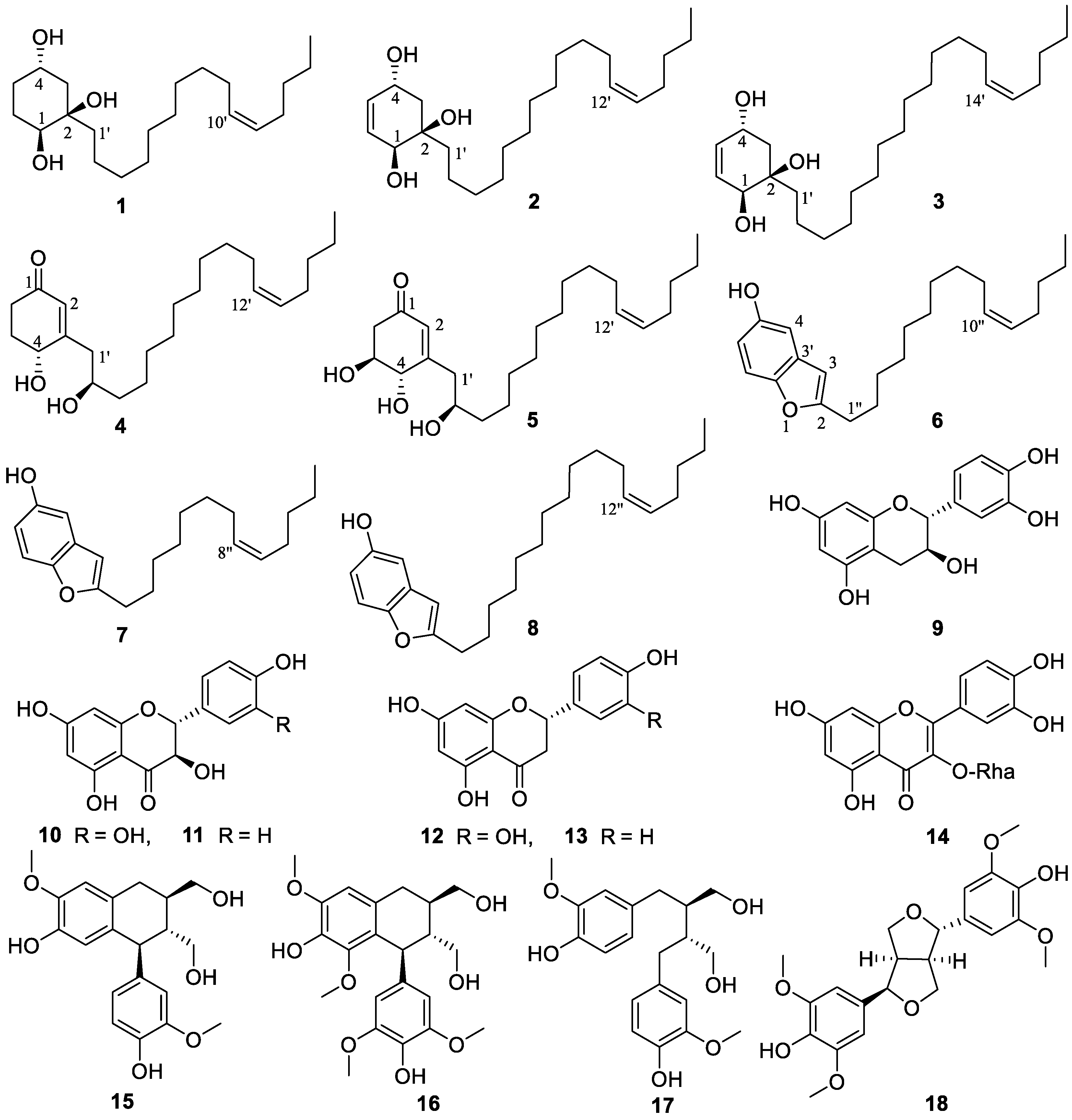
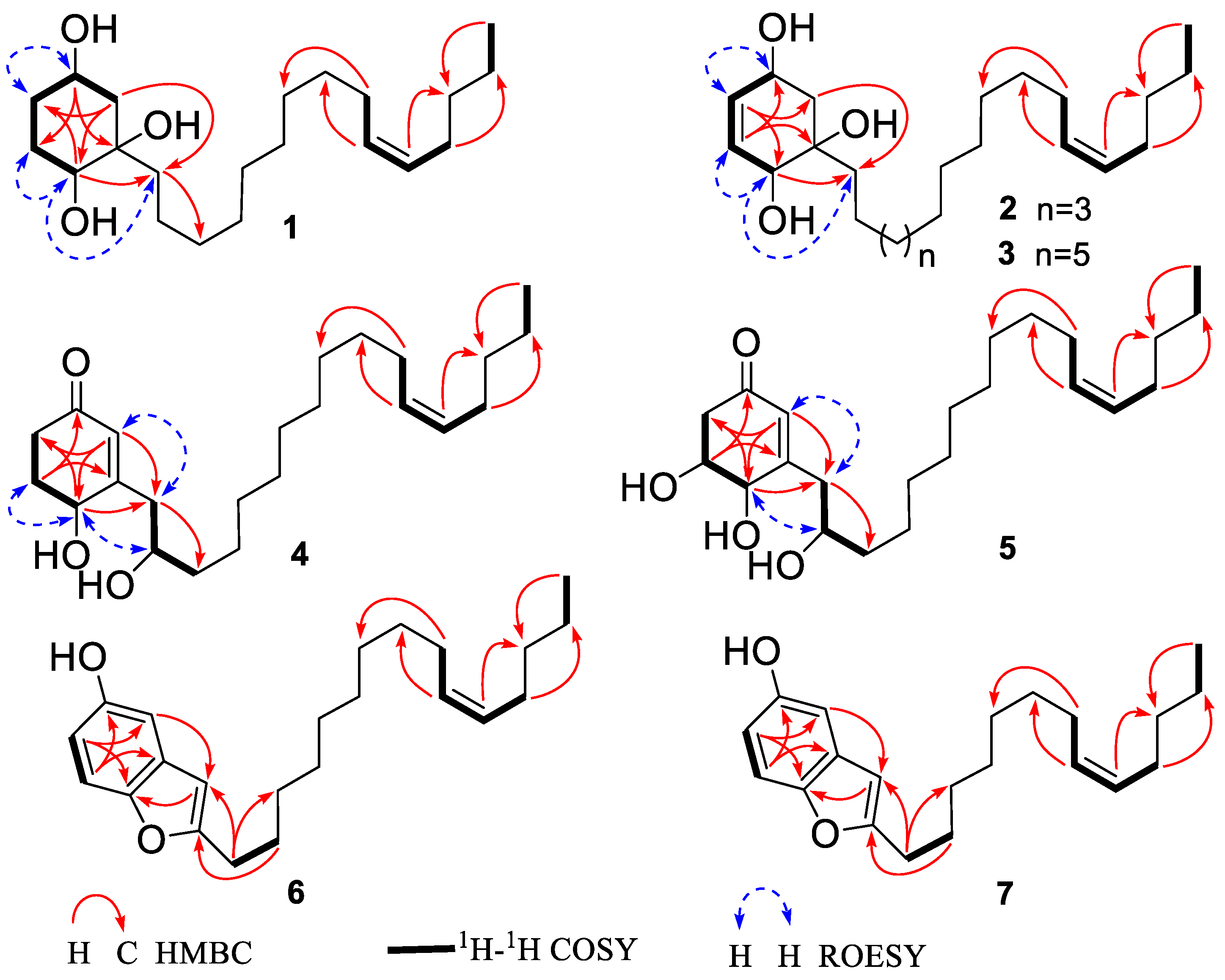
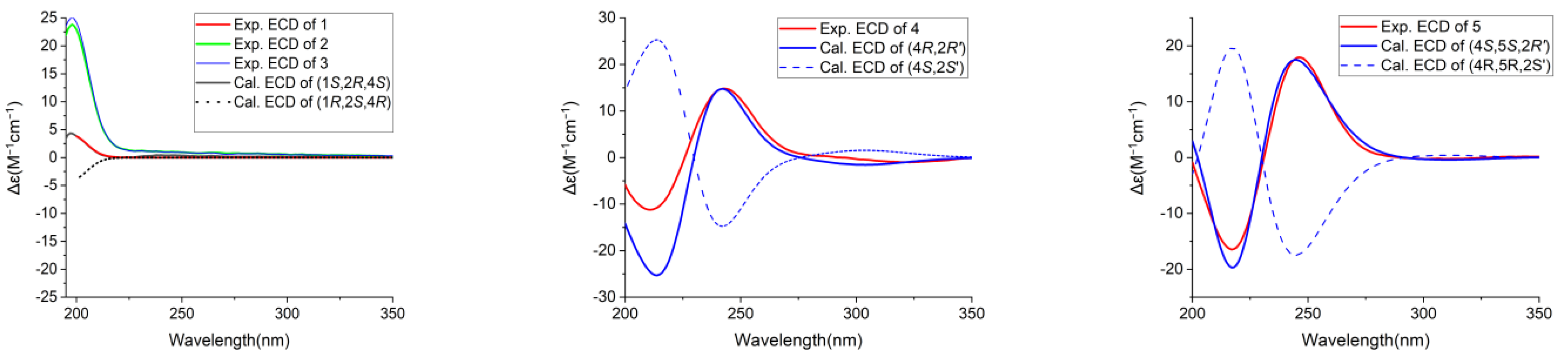
3.1. Structural Elucidation

Eighteen compounds (118) (Figure 1) were isolated from the 80% aqueous acetone extract of C. axillaris fresh fruits by repeated CC over Sephadex LH-20, RP-18, and silica gel, followed by semi-preparative HPLC purification. Seven of them are new (17), while the known ones 818 were identified by comparison of their spectroscopic data with those reported in the literature, as one benzofuran derivative, choerosponol A (8) [18], six flavonoids, catechin (9) [43], dihydroquercetin (10) [44], dihydrokaempferol (11) [44], eriodictyol (12) [45], naringenin (13) [46], quercetin-3-O-α-L-rhamnoside (14) [47], and four lignans, (+)-isolariciresinol (15) [48], (−)-lyoniresinol (16) [49], (−)-secoisolariciresinol (17) [50], and lirioresinol (18) [51]. The known lignans 1518 were isolated from the genus Choerospondias for the first time.
Compound 1 was obtained as a colorless solid. Its molecular formula was established as C21H40O3 based on HRESIMS data, which provided a [M + HCOO] ion at m/z 385.2957 (calcd. 385.2959). The IR band at 3404 cm−1 revealed the presence of a hydroxyl group. The 1H NMR and 13C NMR spectra of 1 displayed the presence of two oxymethine protons resonating at δH 3.84 (1H, tt, J = 11.1, 4.4 Hz) and δH 3.36 (1H, dd, J = 10.7, 5.4 Hz) with the corresponding carbon resonances at δC 67.4 (C-4) and δC 73.9 (C-1), respectively, in HSQC experiment and a quaternary oxygenated carbon at δC 75.4 (C-2). Moreover, proton signals ascribed to methylene groups at δH 1.95 (1H, ddd, J = 13.3, 4.4, 2.8 Hz, H-3a), 1.90 (1H, ddd, J = 12.5, 4.4, 2.9 Hz, H-5a), 1.70 (2H, m, H-6) and overlapping peaks at δH 1.23 (2H, m, H-3b and H-5b) along with carbon shifts at δC 43.7 (C-3), 29.1 (C-6) and 34.3 (C-5) were observed (Table 1). The 1,2,4-trihydroxysubstituted cyclohexane ring were established by 1H-1H COSY correlations between H-3/H-4, H-1/H-6, H-5/H-6 and H-4/H-5 as well as, HMBC correlations of H-4 (δH 3.84) to C-6 and C-2, H-1 (δH 3.36) to C-1′ (δC 40.7) and C-5. The location of Δ10′,11′ double bond was deduced from the HMBC correlation of terminal methyl proton H-15′ (δH 0.92) to C-13′ (δC 33.1), overlapping vinylic protons H-10′/H-11′ to C-8′ (δC 30.3) and to C-13′, and protons H-9′/H-12′ (δH 2.04) to C-7′ (δC 30.6) and C-14′ (δC 23.4), respectively. The assignment of the (Z)-stereochemistry to the side-chain double bond in the alkenyl chain was established by comparison of the chemical shifts observed in the 13C NMR for C-9′ and C-12′ with the literature data, instead of a higher chemical shift value of δC 32.6 exhibited for the (E)-configuration; compound 1 possesses chemical shift of C-9′ (δC 28.1) and C-12′ (δC 27.9) [52,53,54,55,56]. The ROESY cross-peak correlation observed between H-1/H-1′, H-4/H-5, and H-5/H-6 suggested OH-1, and OH-2 to be β-oriented and OH-4 to be α-oriented. The experimental ECD spectra aligned with the calculated ECD spectra bearing a 1S,2R,4S configuration (Figure 1). Thus, the absolute configurations were assigned, the structure of 1 was established as (1S,2R,4S)-2-[10′(Z)-pentadecenyl]- cyclohexane-1,2,4-triol and named as choerosponol F.
Based on HRESIMS analysis, compound 2 was determined to have a molecular formula of C23H42O3, with a molecular ion peak at m/z 389.3029 [M + Na]+ (calcd. for 389.3026). The 1H NMR (Table 1) spectrum showed a pair of olefinic protons resonances at δH 5.75 (1H, dd, J = 10.2, 2.0 Hz) and 5.54 (1H, dt, J = 10.2, 2.6 Hz), two oxymethine proton resonates at δH 4.39 (1H, ddd, J = 9.5, 5.5, 2.0 Hz), and 4.00 (1H, d, J = 2.6 Hz) together with methylene protons at δH 2.17 (1H, dd, J = 13.2, 5.5 Hz, H-3a) and δH 1.42 (1H, dd, J = 13.2, 9.5 Hz, H-3b). The 13C NMR data coupled with the HSQC spectra showed that the cyclohexene ring is substituted with three hydroxy groups, at C-4 (δC 66.1), C-2 (δC 75.1) and C-1 (δC 70.8) along with olefinic carbon shifts at C-5 (δC 133.3) and C-6 (δC 130.7) (Table 1), which is in consistence with correlations observed (as depicted in Figure 2) in the HMBC spectrum from H-4 (δH 4.39) to C-6, H-3a (δH 1.42)/H-3b (δH 2.17) to C-5, C-1 and C-1′ (δC 40.4), and from olefinic protons H-5 (δH 5.75) to C-1 and C-3, H-6 (δH 5.54) to C-4 and C-2. In comparison with the 1D/2D NMR spectra in the reported literature, compound 2 was similar to the previously identified compound from Lannea schimperi (Anacardiaceae) [57]; the only differences in the structure were due to the location and geometry of the double bond within the alkenyl chain, which were supported by the 1H-1H COSY correlations of H-11′/H-12′, H-13′/H-14′ and HMBC correlation of terminal methyl proton H-17′ (δH 0.92) to C-15′ (δC 33.1), overlapping vinylic protons H-12′/H-13′ (δH 5.35) to C-10′ (δC 30.9) and to C-15′ (δC 33.1), permitted the position of the double bond Δ12′,13′. The experimental ECD spectrum of 2 matched the calculated ECD curve of 1; consequently, (1S,2R,4S) absolute configuration was established for choerosponol G. Therefore, compound 2 was identified as (1S,2R,4S)-2- [12′(Z)-heptadecenyl]-cyclohex-5-en-1,2,4-triol.
Compound 3 exhibited a [M + Na]+ molecular ion peak at m/z 417.3337 (calcd. for 417.3339) in the HRESIMS experiment, verifying the molecular formula as C25H46O3. The 13C NMR and HSQC experiment of compound 3 revealed the presence of 25 carbon resonances attributed to one methyl (δC 14.4), 17 methylenes (δC 41.5–23.4), 4 methines (δC 133.3, 130.8, 130.8 and 130.8), 2 oxygenated methines (δC 66.2 and 70.9) and 1 quaternary oxygenated carbon (δC 75.2). Proton shifts at δH 5.74 (1H, dd, J = 10.2, 2.0 Hz) and 5.53 (1H, dt, J = 10.2, 2.5 Hz) in the 1H NMR spectrum (Table 1) deduce a pair of olefinic protons with corresponding carbon signals in the 13C NMR that were detected at δC 133.3 and 130.8, respectively. Two oxymethine protons resonating at δH 4.38 (1H, ddd, J = 9.5, 3.9, 2.0 Hz) and 4.00 (1H, d, J = 2.5 Hz), methylene protons at δH 2.15 (1H, dd, J = 13.2, 3.9 Hz) and δH 1.42 (1H, dd, J = 13.2, 9.5 Hz) were also observed. The location of three hydroxyl groups on the cyclohexene moiety was determined from the correlations observed in the HMBC spectrum from oxymethine proton H-4 (δH 4.38) to C-6 (δC 130.8) and C-2 (δC 75.2), methylene proton signals H-3a (δH 1.42) and H-3b (δH 2.15) to C-5 (δC 133.3), C-1 (δC 70.9) and C-1′ (δC 40.4), and from olefinic protons H-5 (δH 5.74) to C-3 (δC 41.5) and C-1, H-6 (δH 5.53) to C-4 (δC 66.2) and C-2. ROESY correlations observed between H-1/H-1′, H-1/H-6, and H-4/H-5 suggested the cis orientation of OH-1 and OH-2; subsequently, from the established absolute configuration of compound 1, the absolute configuration of compound 3 was determined as (1S,2R,4S). The structure of 3 was therefore established as (1S,2R,4S)-2-[14′(Z)-nonadecenyl]-cyclohex-5-en-1,2,4-triol and given the trivial name choerosponol H.
HRESIMS data of 4 provided a [M + Na]+ ion peak at m/z 387.2868 (calcd. for 387.2870) confirming its molecular formula as C23H40O3. The IR spectrum showed a broad OH absorption band at 3394 cm−1 and the absorption of a carbonyl group at 1658 cm−1, indicating an α,β-unsaturated ketone. The 1H NMR spectrum (Table 2) showed olefinic proton resonance at δH 5.88 (1H, s), a triplet signal of vinaylic protons at δH 5.34 (2H, t, J = 5.6 Hz), two oxymethine proton resonates at δH 4.39 (1H, dd, J = 8.2, 4.6 Hz) and 3.86 (1H, hept, J = 4.5 Hz). The 13C NMR and HSQC spectra of the α,β-unsaturated cyclohexanone ring displayed chemical shift value of δC 201.7 ascribed for C=O, monohydroxy substituted at C-4 (δC 68.7)/δH 4.39, besides exhibited olefinic carbon shifts at C-2 (δC 128.0)/δH 5.88 and C-3 (δC 167.6), whereby their positions on the cyclohexanone ring were established via 1H-1H COSY correlations between H-4/H-5 and H-5/H-6 as well as the HMBC correlation of H-2 to C-6 (δC 35.5), C-4, and C-1′ (δC 43.0), H-4 to C-2, C-6 and C-1′, H-6a/H-6b (δH 2.37, 2.54) to C-1 and C-4, H-1′a/H-1′b (δH 2.44, 2.59) to C-3′ (δC 38.7), C-4, and C-2, H-5a/H-5b (δH 1.97, 2.23) to C-1 and C-3. The calculated ECD spectrum of 4 matched well with the experimental ECD curve of 4 (Figure 3), permitting the assignment of the (4R,2′R) absolute configuration; hence, compound 4 determined as (4R,2′R)-dihydroxy-3-[12′(Z)-heptadecenyl]-2-cyclohexenone given the trivial name choerosponol I.
Compound 5 was acquired as a colorless amorphous solid, and its molecular formula C23H40O4 was determined based on its HRESIMS data, which provided a [M + HCOO] ion at m/z 425.2909 (calcd. for 425.2909). The 1H and 13C NMR data for 5 (Table 2) were comparable to those obtained for 4; however, signals for an additional oxygenated methine δC 71.0 (C-5) group were observed. Its 1H NMR and 13C NMR spectrum indicated the presence of an α,β-unsaturated carbonyl group δC 199.9 (C-1), olefinic proton at δH 5.91 (1H, s)/δC 128.4 (C-2), δC 164.8 (C-3), three oxymethine protons resonating at δH 4.17 (1H, dd, J = 6.8, 1.4 Hz), 3.95 (1H, ddd, J = 9.5, 6.8, 4.3 Hz) and 3.87 (1H, hept, J = 4.5 Hz) with the corresponding carbon resonance at δC 74.4, δC 71.0 and δC 72.7, respectively, showed in the HSQC experiment. The exact location in the cyclohexanone ring was feature from 1H-1H COSY correlation between H-4/H-5, H-5/H-6, H-1′/H-2′, H-2′/H-3′ and the HMBC spectrum correlation of H-2 (δH 5.91) to C-6 (δC 44.4), C-1′ (δC 42.8), C-4 (δC 74.4), H-4 (δH 4.17) to C-6, C-1′, H-5 (δH 3.95) to C-3, C-1 (δC 199.9), and nonequivalent methylene protons H-6a/6b (δH 2.42, 2.73) to C-4, C-2, H-1′ (δH 2.46, 2.61) to C-3′ (δC 38.5), C-4, C-2. The calculated ECD spectrum of (4S,5S,2′R)-5 matched the experimental ECD curve (Figure 3). Consequently, the absolute configuration of 5 was determined as (4S,5S,2′R)-trihydroxy-3-[12′(Z)-heptadecenyl]-2-cyclohexenone given the trivial name choerosponol J.
The molecular formula of 6 was assigned as C23H34O2, based on HRESIMS data revealing a [M − H] ion at m/z 341.2479 (calcd 341.2486). The 1H NMR (Table 2) spectrum showed aromatic protons at δH 7.15 (1H, d, J = 8.7 Hz), 6.84 (1H, d, J = 2.5 Hz), 6.66 (1H, dd, J = 8.7, 2.5 Hz) and 6.25 (1H, s). The HMBC correlation signals from H-7 (δH 7.15) to C-5 (δC 153.9) and C-3′ (δC 131.1), H-6 (δH 6.66) to C-4 (δC 106.1) and C-7′ (150.6), H-4 (δH 6.84) to C-7′, C-6 (112.5) and long-range correlations H-3 (δH 6.25) to C-4, C-7′, H-1′′ (δH 2.67) to C-3 (δC 102.8), C-4, H-2′′ (δH 1.68) to C-2 (δC 161.4), besides the 1H-1H COSY correlation between aromatic protons H-6/H-7, and H-1′′/H-2′′, reveal the benzofuran arene moiety. Signals referable to methylene protons at δH 2.67 (2H, t, J = 7.6 Hz), 1.68 (2H, p, J = 7.4 Hz), a series of overlapping peaks at δH 1.24–1.35 (16H, m), olefinic proton resonance at 5.32 (2H, m) and terminal methyl proton at δH 0.89 (3H, t, J = 7.0 Hz) indicated the presence of a long alkenyl chain in 6. 1H and 13C NMR data of 6 were closely related to those of 8 [18]. However, compound 6 was missing signals corresponding to two methylene carbons occurring in the unsaturated hydrocarbon chain which was confirmed by the HRESIMS experiment. Therefore, the structure of compound 6 was established (Figure 1) which is attributed to be 2-[10′(Z)-pentadecenyl]-benzofuran-5-ol and named as choerosponol K.
HRESIMS data of 7 revealed a [M + Na]+ ion at m/z 337.2137 (calcd. for C21H30O2Na+ 337.2138) indicating the molecular formula of C21H30O2. The 13C-NMR spectra (Table 2) displayed 21 carbon resonances, attributed to one methyl, ten methylenes, six methines (two olefinic, four aromatic), and four quaternary carbons (one carbonyl oxygenated). Aromatic protons at δH 7.16 (1H, d, J = 8.7 Hz), 6.83 (1H, d, J = 2.5 Hz), 6.65 (1H, dd, J = 8.7, 2.5 Hz) and 6.27 (1H, s) were observed in the 1H NMR (Table 2) spectrum. The presence of benzofuran arene moiety was confirmed by the HMBC correlations from H-7 (δH 7.16) to C-5 (δC 153.9) and C-3′ (δC 131.1), H-6 (δH 6.65) to C-4 (δC 106.1) and C-7′ (150.6), H-4 (δH 6.83) to C-7′, C-6 (112.5) and H-3 (δH 6.27) to C-4, C-7′, H-1′′ (δH 2.69) to C-3 (δC 102.8), C-4, H-2′′ (δH 1.70) to C-2 (δC161.5). The 1D and 2D NMR data for 7 were comparable to those obtained for compound 6, which showed that the structure of the cyclic moiety was identical, and the differences consisted of the absence of two methylene groups in the alkenyl chain; these observations were supported by HRESIMS data of 7 [M + Na]+ ion at m/z 337.2137, indicating a molecular formula of C21H30O2. Hence, compound 7 was established (Figure 1) as 2-[8′(Z)-tridecenyl]-benzofuran-5-ol and named as choerosponol L.

3.2. α-Glucosidase Inhibition

The α-glucosidase inhibitory activity of compounds isolated from C. axillaris was evaluated. As shown in Table 3, most compounds were demonstrated with potential α-glucosidase inhibitory activity. Benzofuran derivatives 6, 7, and 8 exert superior inhibitory activity than quercetin and acarbose, with IC50 values of 3.5 ± 0.12, 2.26 ± 0.06, 4.5 ± 0.10 μM, respectively. Out of the tested flavonoids, 9, 12, and 13 were proved to demonstrate excellent α-glucosidase potency with IC50 values of 43.9 ± 0.96, 38.3 ± 0.53, 36.3 ± 1.3, respectively. Compounds 10, 11 and 14, exhibit comparable α-glucosidase inhibitory, whereas weak activity was noted for lignans 15, and 17. At a concentration of 50 μM, compounds 15 showed no inhibitory effects on α-glucosidase.

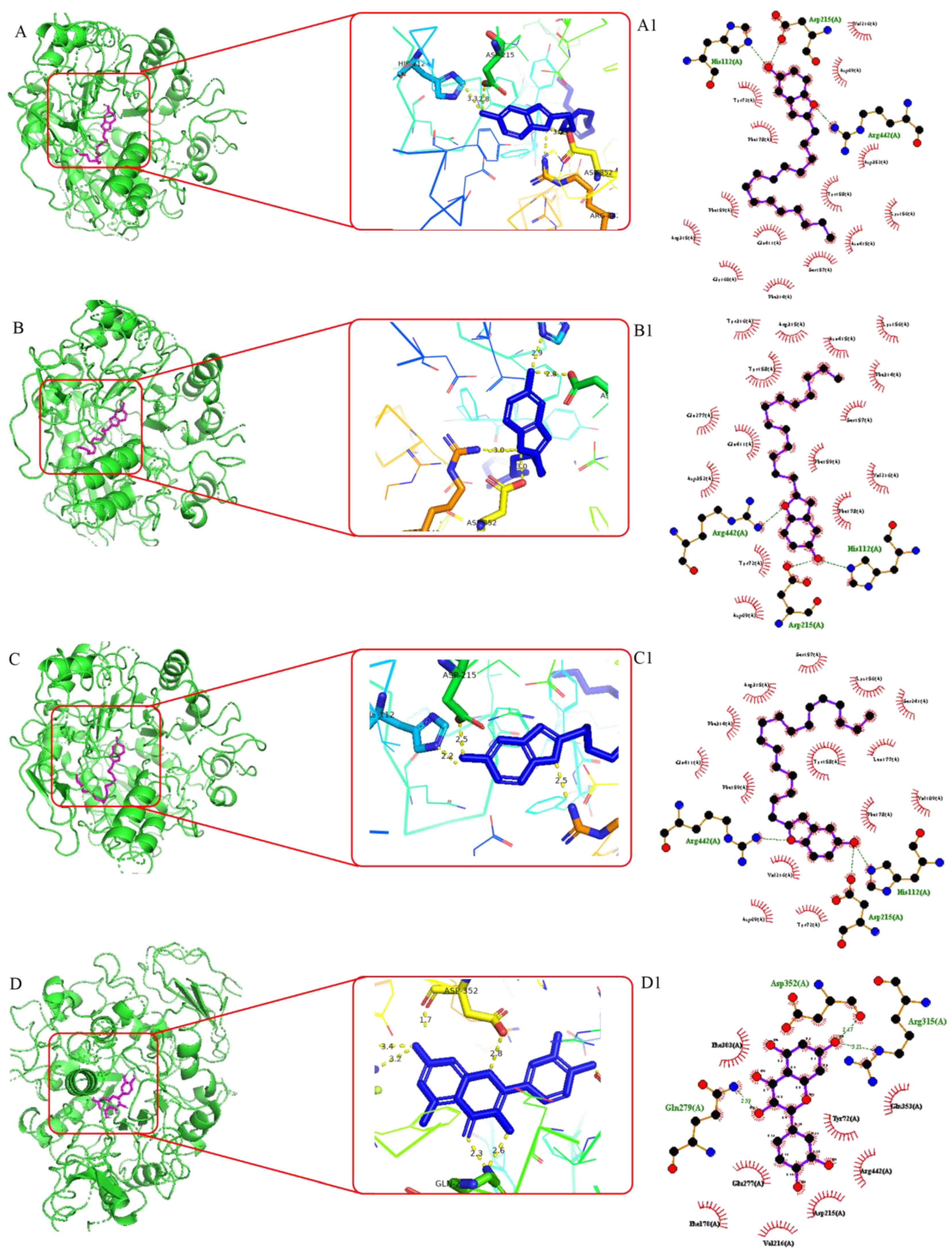
3.3. Molecular Docking Studies

To rationalize the significant in vitro α-glucosidase inhibition results achieved, molecular docking studies were designed [58,59,60,61], thereby illustrating the docking affinities and the docking conformations of the isolated compounds with α-glucosidase from Saccharomyces cerevisiae isomaltase (PDB ID: 3A4A), a principal diabetes-related enzyme located in the small intestine, which catalyzes the cleavage of α-glucopyranoside bond in oligosaccharides and disaccharides, leading to increases in blood glucose concentration.
The molecular docking results, including the binding affinities of ligands (6, 7, 8 and quercetin) that exhibited potent α-glucosidase inhibition and their interactions (hydrogen bonds and hydrophobic interactions), are shown in Figure 4. The highest docking score indicates greater affinity of the compound towards the protein which finds well fit inside the binding pocket of the protein. This is consistent with the results of the α-glucosidase inhibitory assay.
Compound 6 demonstrated the highest binding affinity of −9.77 kcal/mol which is attributed to the hydrogen bonding interactions observed with Arg 442A (2.8 Å), Asp 215A (2.8 Å), and His112A (3.3 Å), as well as established hydrophobic bond interactions with Arg 315A (3.8 Å), Phe 178A (3.2 Å), Val 216A (3.8 Å), Tyr 158A (3.4 Å), Lys 156A (3.2 Å), Tyr 72A (3.1 Å), and Phe 314A (3.4 Å), implying that the hydroxyl group and the pi-electrons on the arene moiety may play an important role in the inhibition of the α-glucosidase and justify its interesting in vitro inhibition rate.
Compound 7 scored enhanced docking score of −9.49 kcal/mol where amino acid residues Arg 442A (2.3 Å), Asp 215A (2.8 Å), and His112A (2.9 Å), forming hydrogen bond interactions with benzofuran ring, and interactions with Lys 156A (3.6 Å), Arg 315A (3.7 Å), Tyr 158A (3.2 Å), Tyr 316A (3.7 Å), Val 216A (3.3 Å), Tyr 72A (3.3 Å), Phe 159A (3.8 Å), Phe 178A (3.8 Å) and Phe 314A (3.5 Å), residues through hydrophobic interaction, were also illustrated. Evidently, this compound exhibited potent α-glucosidase inhibition, which translated into analogous in vitro IC50 values of 2.26 ± 0.06 μM.
Compound 8 exhibits excellent binding affinities of −9.62 kcal/mol, and hydrogen bond interactions were executed between the hydroxyl group and the key amino acid residue Arg 442A (2.5 Å), Asp 215A (2.5 Å), and His112A (2.2 Å). The interactions with Tyr 72A (3.1 Å), Lys 156A (3.9 Å), Val 216A (3.1 Å), Tyr 158A (3.3 Å), Phe 159A (3.7 Å), Leu 177A (3.7 Å), Phe 178A (3.8 Å), Phe 178A (3.2 Å), Phe 314A (3.6 Å) and Arg 315A (3.2 Å) residues through hydrophobic interaction were also demonstrated, which are extrapolated to its impressive analogous in vitro IC50 values.
The positive control quercetin showed a binding affinity of −7.73 kcal/mol, which is consistent with the previous study by Qin et al. [28], who proved the reason why it possesses less α-glucosidase inhibition compared to compounds 6, 7 and 8. Notably, it formed a hydrogen bond interaction with amino acid residues Asp 352A, Arg 315A, Asp 215A and Gln 279.

3.4. Structural Activity Relationship

The new compounds possess analogous alkenyl side chains; nonetheless, the inhibitory effect significantly changed when a heterocyclic aromatic ring was attached instead of a non-aromatic cyclic ring. The results showed that the compounds with benzofuran arene moiety tend to exert remarkable α-glucosidase inhibitory activity than alkenyl cyclohexenol and cyclohexenone derivatives. These significant differences in activity indicate that the presence of a heterocyclic aromatic ring undoubtedly plays a crucial role in the compounds exerting α-glucosidase inhibitory activity, which is attributed to the high docking score and hydrogen, hydrophobic interactions in the docking study.
The flavanonols dihydroquercetin (10), dihydrokaempferol (11) and flavanones eriodictyol (12) and naringenin (13) possess carbonyl groups; however, they differ in the number of OH groups, resulting in the observed differences in inhibition of α-glucosidase. Flavan-3-ol (9), with the absence of the carbonyl group, displayed a notable α-glucosidase inhibition; furthermore, the presence of the rhamnosyl group at C-3 on quercitrin (14) altered the enzyme inhibition activity, which is consistent with previous research findings by Li et al., [29].
Lignans (+)-isolariciresinol (15), (−)-lyoniresinol (16), (−)-secoisolariciresinol (17) and lirioresinol (18) displayed various weak inhibitory activity; a methoxy group in the aromatic ring could be responsible for the differences observed in α-glucosidase inhibition.

4. Conclusions

In this study, chemical investigation of the fresh fruits of C. axillaris led to the isolation and structural elucidation of 18 compounds including 7 new (17) and 11 known (818), comprised of 5 alkenyl (cyclohexenols and cyclohexenones) derivatives (15), 3 benzofuran derivatives (68), 6 known flavonoids (914), and 4 known lignans (1518). Compounds 1518 were isolated from the genus Choerospondias for the first time. Compounds 69, 12, and 13 exerted strong α-glucosidase inhibitory activity with IC50 values from 2.26 to 43.9 μM. The results of molecular docking simulations illustrate good coherence with in vitro α-glucosidase inhibition. Interestingly, benzofuran derivatives were proved to have potential hypoglycemic activity, suggesting these compounds may also play a substantial role in the hypoglycemic activity of C. axillaris fruit. The results suggested that C. axillaris fruit could be an ideal source of hypoglycemic functional foods.

Supplementary Materials

The following supporting information can be downloaded at: https://www.mdpi.com/article/10.3390/foods13101495/s1, Figure S1.1–Figure S8.4. 1H NMR and 13C NMR spectrum of compounds 18 in CD3OD; 1H-1H COSY, HSQC, HMBC, ROESY, HRESIMS, UV and IR spectrum of compounds 17 in CD3OD.

Author Contributions

E.T.W.: Conceptualization, Writing—original draft, Writing—review and editing, Investigation. N.L.: Conceptualization, Formal analysis, Methodology, Writing—review and editing. J.L.: Methodology. J.S.: Methodology. H.Z.: Resources. Y.Z.: Supervision, Conceptualization, Funding acquisition, Writing—review and editing. All authors have read and agreed to the published version of the manuscript.

Funding

This work was supported by the National Natural Science Foundation of China (82074124, 32270426), the Key Project of Basic Research Plan of Yunnan Province, China (202001AS070017), University of Chinese Academy of Science and CAS-TWAS President’s Fellowship Programme that provides the funding for the Ph.D. Scholarship (No. 2019A8010642001).

Institutional Review Board Statement

Not applicable.

Informed Consent Statement

Not applicable.

Data Availability Statement

The original contributions presented in the study are included in the article/Supplementary Material, further inquiries can be directed to the corresponding author.

Acknowledgments

The authors are grateful to the staff of the analytical and bioactivity screening group at the State Key Laboratory of Phytochemistry and Plant Resources in West China, KIB, CAS.

Conflicts of Interest

The authors declare no conflicts of interest.

References

  1. Dangal, A.; Timsina, P.; Dahal, S. A comprehensive review on study of physical, nutritional, and phytochemical characteristics as well as therapeutic activities of Choerospondias axillaris (lapsi). Food Biosci. 2023, 53, 102713. [Google Scholar] [CrossRef]
  2. Surayothee, W.; Buajan, S.; Fu, P.; Pumijumnong, N.; Fan, Z.; Panthi, S.; Finnegan, P.M.; Zhang, Y.; Chen, Y.; Tor-ngern, P.; et al. Growth-climate relationships and long-Term growth trends of the tropical forest tree Choerospondias axillaris (Anacardiaceae) in east-Central Thailand. Forests 2021, 12, 1655. [Google Scholar] [CrossRef]
  3. Mann, S.; Chakraborty, D.; Biswas, S. An alternative perspective of an underutilized fruit tree Choerospondias axillaris in health promotion and disease prevention: A review. Food Biosci. 2022, 47, 101609. [Google Scholar] [CrossRef]
  4. Wang, C.; Li, J.; Cao, Y.; Huang, J.; Lin, H.; Zhao, T.; Liu, L.; Shen, P.; Julian McClements, D.; Chen, J.; et al. Extraction and characterization of pectic polysaccharides from Choerospondias axillaris peels: Comparison of hot water and ultrasound-assisted extraction methods. Food Chem. 2023, 401, 134156. [Google Scholar] [CrossRef] [PubMed]
  5. Rong, W.; Shi, Q.; Yang, Y.; Su, W.; Li, M.; Qin, M.; Bai, S.; Zhu, Q.; Wang, A. Fructus choerospondiatis: A comprehensive review of its traditional uses, chemical composition, pharmacological activities, and clinical studies. J. Ethnopharmacol. 2024, 323, 117696. [Google Scholar] [CrossRef] [PubMed]
  6. Li, Q.; Chen, J.; Li, T.; Liu, C.; Wang, X.; Dai, T.T.; McClements, D.J.; Liu, J. Impact of in vitro simulated digestion on the potential health benefits of proanthocyanidins from Choerospondias axillaris peels. Food Res. Int. 2015, 78, 378–387. [Google Scholar] [CrossRef] [PubMed]
  7. Zeng, Z.; Huang, K.; McClements, D.J.; Hu, X.; Luo, S.; Liu, C. Phenolics, antioxidant activity, and in vitro starch digestibility of extruded brown rice influenced by Choerospondias axillaris fruit peels addition. Starch 2019, 71, 1800346. [Google Scholar] [CrossRef]
  8. Li, Q.; Wang, X.; Chen, J.; Liu, C.; Li, T.; McClements, D.J.; Dai, T.; Liu, J. Antioxidant activity of proanthocyanidins-rich fractions from Choerospondias axillaris peels using a combination of chemical-based methods and cellular-based assay. Food Chem. 2016, 208, 309–317. [Google Scholar] [CrossRef] [PubMed]
  9. Li, Q.; Wang, X.; Dai, T.; Liu, C.; Li, T.; McClements, D.J.; Chen, J.; Liu, J. Proanthocyanidins, isolated from Choerospondias axillaris fruit peels, exhibit potent antioxidant activities in vitro and a novel anti-angiogenic property in vitro and in vivo. J. Agric. Food Chem. 2016, 64, 3546–3556. [Google Scholar] [CrossRef]
  10. Mann, S.; Sarkar, A.; Sharma, A.; Gupta, R.K.; Biswas, S. Antitumor Activity of Choerospondias axillaris Fruit Extract by Regulating the Expression of SNCAIP and SNCA on MDA-MB-231 Cells. Asian Pac. J. Cancer Prev. 2022, 23, 1577–1586. [Google Scholar] [CrossRef]
  11. Labh, S.N.; Shakya, S.R. Medicinal importance of Choerospondias axillaris (Roxb.) Burtt & Hill fruits in Nepal. Trop. Plant Res. 2016, 3, 463–469. [Google Scholar]
  12. Tang, L.; Li, G.Y.; Yang, B.Y.; Hai, H.X. Chemical constituents from Choerospondias axillaris (Roxb.) Burtt et Hill. Chin. Tradit. Herb. 2009, 40, 541–543. (In Chinese) [Google Scholar]
  13. Shen, X.J.; Ge, R.L.; Wang, J.H. Chemical constituents from Choerospondias axillaris (Roxb.) Burtt et Hill. J. Hunan Univ. Med. Sci. 2009, 28, 196–199. (In Chinese) [Google Scholar]
  14. Lian, Z.; Zhang, C.; Li, C.; Zhou, Y. Studies on Chemical constituents of Choerospondias axillaris. Chin. Med. Mat. 2003, 26, 23–24. (In Chinese) [Google Scholar]
  15. Qian, H.; Hu, Q.L. Studies on the chemical constituents of Choerospondias axillaris. Chin. J. Mod. Appl. Pharm. 1992, 9, 212–213. (In Chinese) [Google Scholar]
  16. Wang, H.; Gao, X.D.; Zhou, G.C.; Cai, L.; Yao, W.B. In vitro and in vivo antioxidant activity of aqueous extract from Choerospondias axillaris fruit. Food Chem. 2008, 106, 888–895. [Google Scholar] [CrossRef]
  17. Li, Q.; Liu, C.; Li, T.; McClements, D.J.; Fu, Y.; Liu, J. Comparison of phytochemical profiles and antiproliferative activities of different proanthocyanidins fractions from Choerospondias axillaris fruit peels. Food Res. Int. 2018, 113, 298–308. [Google Scholar] [CrossRef] [PubMed]
  18. Kil, Y.S.; Risinger, A.L.; Petersen, C.L.; Liang, H.; Grkovic, T.; O’Keefe, B.R.; Mooberry, S.L.; Cichewicz, R.H. Using the cancer dependency map to identify the mechanism of action of a cytotoxic alkenyl derivative from the Fruit of Choerospondias axillaris. J. Nat. Prod. 2020, 83, 584–592. [Google Scholar] [CrossRef]
  19. Li, Q.; Chen, J.; Li, T.; Liu, C.; Zhai, Y.; McClements, D.J.; Liu, J. Separation and characterization of polyphenolics from underutilized byproducts of fruit production (Choerospondias axillaris peels): Inhibitory activity of proanthocyanidins against glycolysis enzymes. Food Funct. 2015, 6, 3693–3701. [Google Scholar] [CrossRef]
  20. Ghani, U. Re-exploring promising α-glucosidase inhibitors for potential development into oral anti-diabetic drugs: Finding needle in the haystack. Eur. J. Med. Chem. 2015, 103, 133–162. [Google Scholar] [CrossRef]
  21. Dhameja, M.; Gupta, P. Synthetic heterocyclic candidates as promising α-glucosidase inhibitors: An overview. Eur. J. Med. Chem. 2019, 176, 343–377. [Google Scholar] [CrossRef] [PubMed]
  22. Ye, C.; Zhang, R.; Dong, L.; Chi, J.; Huang, F.; Dong, L.; Zhang, M.; Jia, X. α-Glucosidase inhibitors from brown rice bound phenolics extracts (BRBPE): Identification and mechanism. Food Chem. 2022, 372, 131306. [Google Scholar] [CrossRef] [PubMed]
  23. Wei, B.; Wang, L.; Su, L.; Tao, X.; Chen, S.; Wu, J.; Xia, W. Structural characterization of slow digestion dextrin synthesized by a combination of α-Glucosidase and cyclodextrin glucosyltransferase and its prebiotic potential on the gut microbiota in vitro. Food Chem. 2023, 426, 136554. [Google Scholar] [CrossRef] [PubMed]
  24. Sunagar, R.R.; Sreerama, Y.N. Implication of solvent polarities on browntop millet (Urochloa ramosa) phenolic antioxidants and their ability to protect oxidative DNA damage and inhibit α-amylase and α-glucosidase enzymes. Food Chem. 2023, 411, 135474. [Google Scholar] [CrossRef] [PubMed]
  25. Song, Y.B.; Lamothe, L.M.; Esmeralda Nava Rodriguez, N.; Rose, D.R.; Lee, B.H. New insights suggest isomaltooligosaccharides are slowly digestible carbohydrates, rather than dietary fibers, at constitutive mammalian α-glucosidase levels. Food Chem. 2022, 383, 132456. [Google Scholar] [CrossRef]
  26. Kobayashi, K.; Yokoh, H.; Sato, Y.; Takemoto, M.; Uchida, D.; Kanatsuka, A.; Kuribayashi, N.; Terano, T.; Hashimoto, N.; Sakurai, K.; et al. Efficacy and safety of the dipeptidyl peptidase-4 inhibitor sitagliptin compared with α-glucosidase inhibitor in Japanese patients with type 2 diabetes inadequately controlled on sulfonylurea alone (SUCCESS-2): A multicenter, randomized, open-label, non-inferiority trial. Diabetes Obes. Metab. 2014, 16, 761–765. [Google Scholar] [PubMed]
  27. Lim, J.; Zhang, X.; Ferruzzi, M.G.; Hamaker, B.R. Starch digested product analysis by HPAEC reveals structural specificity of flavonoids in the inhibition of mammalian α-amylase and α-glucosidases. Food Chem. 2019, 288, 413–421. [Google Scholar] [CrossRef]
  28. Qin, Y.; Chen, X.; Xu, F.; Gu, C.; Zhu, K.; Zhang, Y.; Wu, G.; Wang, P.; Tan, L. Effects of hydroxylation at C3′ on the B ring and diglycosylation at C3 on the C ring on flavonols inhibition of α-glucosidase activity. Food Chem. 2023, 406, 135057. [Google Scholar] [CrossRef] [PubMed]
  29. Li, N.; Chen, M.; Zhu, H.T.; Zhang, M.; Wanga, D.; Yang, C.R.; Zhang, Y.J. Theaflavoids A-C, new flavan-3-ols with potent α-glucosidase inhibitory activity from Yunnan black tea ‘Jin-Ya’. LWT Food Sci. Technol. 2022, 168, 113918. [Google Scholar] [CrossRef]
  30. Lim, J.; Ferruzzi, M.G.; Hamaker, B.R. Structural requirements of flavonoids for the selective inhibition of α-amylase versus α-glucosidase. Food Chem. 2022, 370, 130981. [Google Scholar] [CrossRef]
  31. Wang, L.; Liu, Y.; Luo, Y.; Huang, K.; Wu, Z. Quickly screening for potential α-glucosidase inhibitors from guava leaves tea by bioaffinity ultrafiltration coupled with HPLC-ESI-TOF/MS method. J. Agric. Food Chem. 2018, 66, 1576–1582. [Google Scholar] [CrossRef]
  32. Ma, Y.Y.; Zhao, D.G.; Zhou, A.Y.; Zhang, Y.; Du, Z.; Zhang, K. α-Glucosidase inhibition and antihyperglycemic activity of phenolics from the flowers of Edgeworthia gardneri. J. Agric. Food Chem. 2015, 63, 8162–8169. [Google Scholar] [CrossRef] [PubMed]
  33. Li, N.; Zhu, H.T.; Wang, D.; Zhang, M.; Yang, C.R.; Zhang, Y.J. New flavoalkaloids with potent α-Glucosidase and acetylcholinesterase inhibitory activities from Yunnan black tea ‘Jin-Ya’. J. Agric. Food Chem. 2020, 68, 7955–7963. [Google Scholar] [CrossRef]
  34. Yang, J.; Wanga, X.; Zhang, C.; Ma, L.; Weib, T.; Zhao, Y.; Peng, X. A Comparative study of inhibition mechanisms of structurally different flavonoid compounds on α-glucosidase and synergistic effect with acarbose. Food Chem. 2021, 347, 129056. [Google Scholar] [CrossRef] [PubMed]
  35. Li, Y.Q.; Zhou, F.C.; Gao, F.; Bian, J.S.; Shan, F. Comparative Evaluation of Quercetin, Isoquercetin and Rutin as Inhibitors of α-Glucosidase. J. Agric. Food Chem. 2009, 57, 11463–11468. [Google Scholar] [CrossRef] [PubMed]
  36. Haggett, P.; Gunawardena, K.A. Determination of population thresholds for settlement functions by the reed-muench method. Prof. Geogr. 1964, 16, 6–9. [Google Scholar] [CrossRef]
  37. O’Boyle, N.M.; Banck, M.; James, C.A.; Morley, C.; Vandermeersch, T.; Hutchison, G.R. Open Babel: An open chemical toolbox. J. Cheminform. 2011, 3, 33. [Google Scholar] [CrossRef] [PubMed]
  38. Wang, G.; Peng, Z.; Wang, J.; Li, J.; Li, X. Synthesis, biological evaluation, and molecular docking study of N-arylbenzo[d]oxazol-2-amines as potential α-glucosidase inhibitors. Bioorg. Med. Chem. 2016, 24, 5374–5379. [Google Scholar] [CrossRef] [PubMed]
  39. Hu, G.; Peng, X.; Dong, D.; Nian, Y.; Gao, Y.; Wang, X.; Hong, D.; Qiu, M. New ent-kaurane diterpenes from the roasted arabica coffee beans and molecular docking to α-glucosidase. Food Chem. 2021, 345, 128823. [Google Scholar] [CrossRef]
  40. Schrödinger LLC. The PyMOL Molecular Graphics System, Version 2.6.0; Schrödinger LLC: New York, NY, USA, 2015.
  41. Adasme, M.F.; Linnemann, K.L.; Bolz, S.N.; Kaiser, F.; Salentin, S.; Haupt, V.J.; Schroeder, M. PLIP 2021: Expanding the scope of the protein-ligand interaction profiler to DNA and RNA. Nucleic Acids Res. 2021, 49, W530–W534. [Google Scholar] [CrossRef]
  42. Graef, J.; Ehrt, C.; Rarey, M. Binding site detection remastered: Enabling fast, robust, and reliable binding site detection and descriptor calculation with DoGSite3. J. Chem. Inf. Model. 2023, 63, 3128–3137. [Google Scholar] [CrossRef]
  43. Fortune, M.; Berhanu, A.G.; Runner, R.T.M. A new flavan from Elephantorrhiza goetzei. Fitoterapia 1999, 70, 412–416. [Google Scholar]
  44. Lee, E.H.; Kim, H.J.; Song, Y.S.; Jin, C.; Lee, K.T.; Cho, J.; Lee, Y.S. Constituents of the stems and fruits of Opuntia ficus-indica var. saboten. Arch. Pharm. Res. 2003, 26, 1018–1023. [Google Scholar] [CrossRef] [PubMed]
  45. Zhou, L.; Li, D.; Wang, J.; Liu, Y.; Wu, J. Antibacterial phenolic compounds from the spines of Gleditsia sinensis Lam. Nat. Prod. Res. 2007, 21, 283–291. [Google Scholar] [CrossRef] [PubMed]
  46. Din, S.; Hamid, S.; Yaseen, A.; Yatoo, A.M.; Ali, S.; Shamim, K.; Mahdi, W.A.; Alshehri, S.; Rehman, M.U.; Shah, W.A. Isolation and characterization of flavonoid naringenin and evaluation of cytotoxic and biological efficacy of water lilly (Nymphaea mexicana Zucc.). Plants 2022, 11, 3588. [Google Scholar] [CrossRef]
  47. Chen, Z.; Liu, Y.M.; Yang, S.; Song, B.A.; Xu, G.F.; Bhadury, P.S.; Jin, L.H.; Hu, D.Y.; Liu, F.; Xue, W.; et al. Studies on the chemical constituents and anticancer activity of Saxifraga stolonifera (L.) Meeb. Bioorg. Med. Chem. 2008, 16, 1337–1344. [Google Scholar] [CrossRef]
  48. Zhu, J.X.; Ren, J.; Qin, J.J.; Cheng, X.R.; Zeng, Q.; Zhang, F.; Yan, S.K.; Jin, H.Z.; Zhang, W.D. Phenylpropanoids and lignanoids from Euonymus acanthocarpus. Arch. Pharm. Res. 2012, 35, 1739–1747. [Google Scholar] [CrossRef] [PubMed]
  49. Lu, C.L.; Zhu, W.; Wang, D.M.; Chen, W.L.; Hu, M.M.; Wang, M.; Xu, X.J.; Lu, C.J. Inhibitory effects of chemical compounds isolated from the rhizome of Smilax glabra on nitric oxide and tumor necrosis factor—α production in lipopolysaccharide-induced RAW264.7 cell. Evid. Based Complement. Alternat. Med. 2015, 2015, 602425. [Google Scholar] [CrossRef] [PubMed]
  50. Xie, L.H.; Akao, T.; Hamasaki, K.; Deyama, T.; Hattori, M. Biotransformation of pinoresinol diglucoside to mammalian lignans by human intestinal microflora, and isolation of Enterococcus faecalis strain PDG-1 responsible for the transformation of (+)-pinoresinol to (+)-lariciresinol. Chem. Pharm. Bull. 2003, 51, 508–515. [Google Scholar] [CrossRef]
  51. Ma, Y.; Bao, Y.; Zhang, W.; Ying, X.; Stien, D. Four lignans from Portulaca oleracea L. and its antioxidant activities. Nat. Prod. Res. 2020, 34, 2276–2282. [Google Scholar] [CrossRef]
  52. Queiroz, E.F.; Kuhl, C.; Terreaux, C.; Mavi, S.; Hostettmann, K. New dihydroalkylhexenones from Lannea edulis. J. Nat. Prod. 2003, 66, 578–580. [Google Scholar] [CrossRef]
  53. Roumy, V.; Fabre, N.; Portet, B.; Bourdy, G.; Acebey, L.; Vigor, C.; Valentin, A.; Moulis, C. Four anti-protozoal and anti-bacterial compounds from Tapirira guianensis. Phytochemistry 2009, 70, 305–311. [Google Scholar] [CrossRef] [PubMed]
  54. Ledoux, A.; St-Gelais, A.; Cieckiewicz, E.; Jansen, O.; Bordignon, A.; Illien, B.; Di Giovanni, N.; Marvilliers, A.; Hoareau, F.; Pendeville, H.; et al. Antimalarial activities of alkyl cyclohexenone derivatives isolated from the leaves of Poupartia borbonica. J. Nat. Prod. 2017, 80, 1750–1757. [Google Scholar] [CrossRef] [PubMed]
  55. Correia, S.D.; David, J.M.; David, J.P.; Chai, H.B.; Pezzuto, J.M.; Cordell, G.A. Alkyl phenols and derivatives from Tapirira obtusa. Phytochemistry 2001, 56, 781–784. [Google Scholar] [CrossRef] [PubMed]
  56. Jie, M.S.F.L.K.; Pasha, M.K.; Alam, M.S. Synthesis and nuclear magnetic resonance properties of all geometrical isomers of conjugated linoleic acids. Lipids 1997, 32, 1041–1044. [Google Scholar] [PubMed]
  57. Okoth, D.A.; Koorbanally, N.A. Cardanols, long chain cyclohexenones and cyclohexenols from Lannea schimperi (Anacardiaceae). Nat. Prod. Commun. 2015, 10, 103–106. [Google Scholar] [CrossRef] [PubMed]
  58. Li, S.; Zhang, W.; Wang, R.; Li, C.; Lin, X.; Wang, L. Screening and identification of natural α-glucosidase and α-amylase inhibitors from partridge tea (Mallotus furetianus Muell-Arg) and in silico analysis. Food Chem. 2022, 388, 133004. [Google Scholar] [CrossRef] [PubMed]
  59. Chen, J.; Tang, Y.; Zhou, H.; Shao, J.; Ji, W.; Wang, Z.; Liang, D.; Zhao, C. Lignan constituents with α-amylase and α-glucosidase inhibitory activities from the fruits of Viburnum urceolatum. Phytochemistry 2023, 216, 113895. [Google Scholar] [CrossRef] [PubMed]
  60. Al-Romaima, A.; Hu, G.; Wang, Y.; Quan, C.; Dai, H.; Qiu, M. Identification of new diterpenoids from the pulp of Coffea arabica and their α-glucosidase inhibition activity. J. Agric. Food Chem. 2024, 72, 1683–1694. [Google Scholar] [CrossRef]
  61. Yin, H.; Dan, W.J.; Fan, B.Y.; Guo, C.; Wu, K.; Li, D.; Xian, K.F.; Pescitelli, G.; Gao, J.M. Anti-inflammatory and α-Glucosidase Inhibitory Activities of Labdane and Norlabdane Diterpenoids from the Rhizomes of Amomum villosum. J. Nat. Prod. 2019, 82, 2963–2971. [Google Scholar] [CrossRef]
Figure 1. Compounds 118 isolated from the fruits of Choerospondias axillaris.
Foods 13 01495 g001
Figure 2. Key HMBC, 1H-1H COSY and ROESY correlations of compounds 17.
Foods 13 01495 g002
Figure 3. Calculated and experimental ECD curves of compounds 15.
Foods 13 01495 g003
Figure 4. Molecular docking study of compounds binding to α-glucosidase protein. (AD): 3D ligand interaction diagram of 6, 7, 8 and quercetin with α-glucosidase and a close view of the binding active site with key residues that interacted to ligands (right); (A1D1): 2D protein–ligand interaction diagram of 6, 7, 8, quercetin and α-glucosidase complex.
Foods 13 01495 g004
Table 1. 13C (150 MHz) and 1H (600 MHz) NMR spectroscopic data of compounds 13 in CD3OD (δ in ppm, J in Hz).
No.1No.2No.3
δC, TypeδH (J in Hz)δC, TypeδH (J in Hz)δC, TypeδH (J in Hz)
173.93.36 (dd, 10.7, 5.4)170.84.00 (d, 2.6)170.94.00 (d, 2.5)
275.4 275.1 275.2
343.71.23 (dd, 13.3, 11.1) a
1.95 (ddd, 13.3, 4.4, 2.8)
341.41.42 (dd, 13.2, 9.5)
2.17 (dd, 13.2, 5.5)
341.51.42 (dd, 13.2, 9.5)
2.15 (dd, 13.2, 3.9)
467.43.84 (tt, 11.1, 4.4)466.1 4.39 (ddd, 9.5, 5.5, 2.0) 466.24.38 (ddd, 9.5, 3.9, 2.0)
534.31.23 (m) a
1.90 (ddd, 12.5, 4.4, 2.9)
5133.3 5.75 (dd, 10.2, 2.0)5133.35.74 (dd, 10.2, 2.0)
629.11.70 (m)6130.75.54 (dt, 10.2, 2.6)6130.85.53 (dt, 10.2, 2.5)
1′40.71.51 (m)
1.58 (m)
1′40.41.60 (m)1′40.41.59 (m)
2′24.51.29–1.36 (m) a2′24.71.28–1.39 (m) a2′24.71.28–1.37 (m) a
3′31.51.29–1.36 (m) a3′31.51.28–1.39 (m) a3′31.51.28–1.37 (m) a
4′–8′30.3–30.91.29–1.36 (m) a 4′–10′30.4–30.91.28–1.39 (m) a4′–12′30.3–30.91.28–1.37 (m) a
9′28.12.04 (m) a11′28.22.04 (m) a13′28.12.04 (m) a
10′130.85.34 (t, 5.1) a12′130.75.35 (t, 5.1) a14′130.85.35 (t, 5.1) a
11′130.85.34 (t, 5.1) a13′130.75.35 (t, 5.1) a15′130.85.35 (t, 5.1) a
12′27.92.04 (m) a14′27.92.04 (m) a16′27.92.04 (m) a
13′33.1 1.29–1.36 (m) a15′33.11.28–1.39 (m) a17′33.11.28–1.37 (m) a
14′23.41.29–1.36 (m) a16′23.41.28–1.39 (m) a18′23.41.28–1.37 (m) a
15′14.40.92 (t, 7.1)17′14.50.92 (t, 7.2)19′14.40.91 (t, 7.1)
a Overlapping signals; chemical shifts were determined from HSQC and HMBC correlations.
Table 2. 13C (150 MHz) and 1H (600 MHz) NMR spectroscopic data of compounds 47 in CD3OD (δ in ppm, J in Hz).
No.4No.5No.6No.7
δC, TypeδH (J in Hz)δC, TypeδH (J in Hz)δC, TypeδH (J in Hz)δC, TypeδH (J in Hz)
1201.7 1199.9 2161.4 2161.5
2128.05.88 (s)2128.45.91 (s)3102.86.25 (s)3102.86.27 (s)
3167.6 3164.8 4106.16.84 (d, 2.5)4106.16.83 (d, 2.5)
468.74.39 (dd, 8.2, 4.6) 474.44.17 (dd, 6.8, 1.4) 5153.9 5153.9
532.71.97 (dtd, 12.8, 8.2, 4.0)
2.23 (ddt, 12.8, 6.8, 4.6)
571.03.95 (ddd, 9.5, 6.8, 4.3) 6112.56.66 (dd, 8.7, 2.5)6112.56.65 (dd, 8.7, 2.5)
635.52.37 (m)
2.54 (dt, 10.8, 6.8)
644.42.42 (dd, 16.4, 9.5)
2.73 (dd, 16.4, 4.3)
7111.57.15 (d, 8.7)7111.57.16 (d, 8.7)
1′43.02.44 (dd, 14.2, 4.6)
2.59 (dd, 14.2, 8.0)
1′42.82.46 (dd, 13.8, 4.8)
2.61 (dd, 13.9, 7.8)
3′131.1 3′131.1
2′71.13.86 (hept, 4.5)2′72.73.87 (hept, 4.5)7′150.6 7′150.6
3′38.71.50 (m) a3′38.51.50 (m) a1″29.42.67 (t, 7.6)1″29.42.69 (t, 7.6)
4′26.7 1.50 (m) a 4′26.71.50 (m) a2″28.81.68 (p, 7.4)2″28.81.70 (p, 7.4)
5′–
10′
30.3–
30.9
1.28–1.37 (m) a5′–
12′
30.3–
30.8
1.27–1.35 (m) a3″–
8″
30.3–
30.8
1.24–1.35 (m) a3″–
6″
30.3–
30.8
1.26–1.36 (m) a
11′28.1 2.04 (m) a13′28.12.03 (m) a9″28.1 2.00 (m) a7″28.12.02 (m) a
12′130.85.34 (t, 5.6) a 14′130.85.34 (t, 5.6) a10″130.85.32 (t, 5.7) a8″130.85.33 (t, 5.4) a
13′130.85.34 (t, 5.6) a15′130.85.34 (t, 5.6) a11″130.85.32 (t, 5.7) a9″130.85.33 (t, 5.4) a
14′27.92.04 (m) a16′27.92.03 (m) a12″27.92.00 (m) a10″27.92.02 (m) a
15′33.11.28–1.37 (m) a17′33.11.27–1.35 (m) a13″33.11.24–1.35 (m) a11″33.11.26–1.36 (m) a
16′23.41.28–1.37 (m) a18′23.41.27–1.35 (m) a14″23.41.24–1.35 (m) a12″23.41.26–1.36 (m) a
17′14.40.91 (t, 7.1)19′14.40.90 (t, 7.1)15″14.40.89 (t, 7.0)13″14.40.90 (t, 7.1)
a Overlapping signals; chemical shifts were determined from HSQC and HMBC correlations.
Table 3. In vitro α-glucosidase inhibitory activity (%) of compounds 118 (50 μM) a.
Compd.Inhibition Rate bIC50 (μM) cCompd.Inhibition Rate bIC50 (μM) c
1−8.1 ± 2.4-1143.8 ± 1.3-
2−12.4 ± 0.66-1266.6 ± 0.9238.3 ± 0.53
3−13.6 ± 1.7-1365.1 ± 2.036.3 ± 1.3
411.1 ± 1.1-1426.2 ± 1.3-
5−11.2 ± 0.94-1516.6 ± 0.57-
697.7 ± 0.193.5 ± 0.12167.5 ± 1.4-
796.7 ± 0.512.26 ± 0.061737.6 ± 0.12-
898.7 ± 0.204.5 ± 0.10184.8 ± 0.38-
961.1 ± 1.643.9 ± 0.96Quercetin d-5.2 ± 0.25
1036.4 ± 0.72-Acarbose d-229.0 ± 0.4
a Data expressed as means ± SD (n = 3); b Inhibition ratio (%) at a concentration of 50 μM; c IC50 = half-maximal inhibitory concentration to α-glucosidase; d Positive control.
Disclaimer/Publisher’s Note: The statements, opinions and data contained in all publications are solely those of the individual author(s) and contributor(s) and not of MDPI and/or the editor(s). MDPI and/or the editor(s) disclaim responsibility for any injury to people or property resulting from any ideas, methods, instructions or products referred to in the content.

