Article

Isolation of a Novel Anti-Diabetic α-Glucosidase Oligo-Peptide Inhibitor from Fermented Rice Bran

1
Jiangsu Key Laboratory of Marine Bioresources and Environment/Jiangsu Key Laboratory of Marine Biotechnology, Jiangsu Ocean University, Lianyungang 222005, China
2
Co-Innovation Center of Jiangsu Marine Bio-Industry Technology, Jiangsu Ocean University, Lianyungang 222005, China
*
Author to whom correspondence should be addressed.
Foods 2023, 12(1), 183; https://doi.org/10.3390/foods12010183
Submission received: 4 October 2022 / Revised: 22 December 2022 / Accepted: 27 December 2022 / Published: 1 January 2023
(This article belongs to the Section Food Nutrition)

Abstract

:
At present, the incidence rate of diabetes is increasing gradually, and inhibiting α-glucosidase is one of the effective methods used to control blood sugar. This study identified new peptides from rice bran fermentation broth and evaluated their inhibitory activity and mechanism against α-glucosidase. Rice bran was fermented with Bacillus subtilis MK15 and the polypeptides of <3 kDa were isolated by ultrafiltration and chromatographic column, and were then subjected to LC-MS/MS mass spectrometry analysis. The results revealed that the oligopeptide GLLGY showed the greatest inhibitory activity in vitro. Docking studies with GLLGY on human α-glucosidase (PDB ID 5NN8) suggested a binding energy of −7.1 kcal/mol. GLLGY acts as a non-competitive inhibitor and forms five hydrogen bonds with Asp282, Ser523, Asp616, and His674 of α-glucosidase. Moreover, it retained its inhibitory activity even in a simulated digestion environment in vitro. The oligopeptide GLLGY could be developed into a potential anti-diabetic agent.

Graphical Abstract

1. Introduction

Diabetes is a complex multifactorial disease. Type 2 diabetes mellitus (T2DM) has become a serious health concern in developing countries [1], and is caused by insulin resistance due to altered β-insulin secretion or deficiency [2]. Worldwide, the incidence rate of diabetes is increasing rapidly, including in China. The development of diabetes is a very long process which is often accompanied by other complications such as diabetic foot, diabetic nephropathy, and cerebral thrombosis [2,3]. Delaying glucose absorption by inhibiting the activity of carbohydrate digestive enzymes such as α-glucosidase is one of the effective ways to control blood glucose levels [4]. To date, acarbose, voglibose, and miglitol are the three main α-glucosidase inhibitors used in the clinic [4]. These show good inhibitory effects but have certain side effects. Also, because of their long synthetic pathway it is necessary to find other α-glucosidase inhibitors with acceptable side effects. Natural products are rich in peptides, polysaccharides, phenolic compounds, terpenoids, and other bioactive substances that have a variety of functional properties [5,6]. The bioactive peptides extracted from Spirulina platensis [7], oat protein [8], canary seeds [9], quinoa [10], and other natural substances have inhibitory effects on α-glucosidase. In recent years, bioactive peptides among bioactive compounds have become the most important compounds for the health and food industry, and can be used as substitutes for drug therapy to control lifestyle-related diseases [11]. Therefore, the inhibition of α-glucosidase by bioactive peptides is a good candidate for further research.
China, a large producer of rice, produces ample rice bran from rice processing. However, rice bran utilization is low, as it is mainly used as feed. This resource is being seriously overlooked [12]. Rice bran contains various bioactive substances that are beneficial to human health, such as amino acids, phenolic acids, flavonoids, vitamin E, γ- oryzanol, and pigment compounds. Bacillus subtilis, a popular biological starter, can produce protease, lipase, and cellulase during its growth process. It is often used for the hydrolysis of beans to produce amino acids, peptides, and amines [13]. Microorganisms can make complete use of rice bran nutrients for their growth and biochemical metabolism. They can metabolically modify, recombine, transfer, or rearrange active groups to improve the functional and processing characteristics of rice bran peptides [14]. The rice bran fermentation broth contains high-quality proteins [15]. The bioactive peptides produced from rice bran proteins are easy to absorb and have non-toxic side effects. Studies suggest that these bioactive peptides have inhibitory qualities and work against cancer, α-glucosidase, β-glucosidase, α-amylase, and angiotensin-converting enzymes [16]. Therefore, they are widely used in the food and pharmaceutical industry and have become a popular research topic [17].
Molecular docking has been used for the development of health products and drug research. In vitro and in vivo tests for ligand screening are quite time-consuming and expensive [18,19,20]. On the other hand, molecular docking can help screen ligands more quickly. This method has been extensively used in the research of health products and drugs [21,22]. Molecular docking can be of three types: rigid docking, semi-flexible docking, and flexible docking. Most docking programs are based on the semi-flexible type of docking. In this method, the macromolecular protein is fixed, and the conformations of the small molecule ligand are changed during the docking process to simulate the ligand docking on protein [23]. The docking predictions made possible by molecular docking can greatly shorten research time, improving research efficiency.
This study aimed to isolate bioactive peptides from the fermentation rice bran that inhibits α-glucosidase. The potential peptides that inhibited α-glucosidase were further purified by ultrafiltration and chromatography. Next, the peptides were identified by LC-MS/MS. Furthermore, molecular docking simulation studies were performed and enzyme inhibition mechanisms on α-glucosidase were investigated. Finally, the best oligopeptide obtained from the fermented rice bran was tested in a simulated GI digestion system in vitro. Our results may improve the economic benefits of rice bran.

2. Materials and Methods

2.1. Materials

Rice bran was provided by Yancheng Kanglinda Biotechnology Co., Ltd. (Yancheng, China). Bacillus subtilis MK15 was provided by the Provincial Institute of Marine Resources Development (Lianyungang, China) and cultured in Luria–Bertani (LB) medium. α-glucosidase, α-amylase, porcine pepsin, pancreatin, bile extract porcine, and 4-Nitrophenyl α-D-glucopyranoside (pNPG) were purchased from Sigma Co., St. Louis, Missouri, USA. Fish meal peptone, glucose, NaCl, urea, beef extract, sodium carbonate, Na2HPO4·12H20, NaH2PO4·2H20, absolute ethanol, and trichloroacetic acid (TCA) were purchased from Aladdin, Shanghai, China. DEAE Sepharose fast flow ion exchange resin and Sephadex G-25 were acquired from Auyoo Biotechnology Co., Ltd. (Shanghai, China).

2.2. Preparation of Seed and Fermentation Broth

B. subtilis MK15 was inoculated into a 100 mL seed culture medium (NA: beef extract 0.3%, peptone 1%, NaCl 0.5%, pH 7.0, and tap water; sterilized at 121 °C for 20 min), and then incubated in a shaking table at 180 rpm and 37 °C for 12 h [24]. The 60 mL of seed culture was inoculated (3%) into 2 L of rice bran fermentation medium (rice bran 5%, glucose 0.1%, NaCl 0.5%, urea 0.5%, pH 8.0, and tap water; sterilized at 115 °C for 20 min), and then incubated in a shaking table at 35 °C and 180 rpm for 48 h. The fermented broth was centrifuged at 8000 rpm and 4 °C for 30 min to collect the supernatant [25]. The initial volume of fermentation medium was 2 L, and the supernatant obtained was 1.82 L.

2.3. Fermentation Product Extraction

The supernatant was added to four times the amount of absolute ethanol for precipitation at 4 °C for 24 h and the mixture was centrifuged at 8000 rpm for 30 min. Next, the obtained supernatant was evaporated in a rotary evaporator at 45 °C to reduce the mixture volume and paused every 10 min (to prevent protein denaturation). This was repeated approximately 10 times. 1.82 L supernatant, evaporation time was 2 h, and the remaining volume of the final supernatant was 150 mL. Subsequently, an equal volume of 10% TCA was added and the mixture was rotated at 4 °C for 1 h with a magnetic stirrer. After centrifugation at 8000 rpm, the obtained supernatant was subjected to rotary evaporation until there was no flowing liquid. Next, 50 mL of water was added for re-dissolution. Finally, after centrifugation, the obtained precipitate was freeze-dried (FreeZon®2.5 L, Labconco, Kansas City, MO, USA) [26].

2.4. Determination of α-Glucosidase Inhibitory Activity

The activity assay was improved based on an in vitro assay model [27]. 50 μL (0.5 mol/L) pH 6.7 phosphate solution, 30 μL (0.5 mM) pNPG substrate solution, 50 μL rice bran fermentation extract, and 20 μL α-glucosidase solution (0.3 U/mL in phosphate solution) were mixed in a 96-well plate. The mixture was incubated at 37 °C for 30 min and then added to a 50 μL (0.67 mol/L) Na2CO3 solution to terminate the reaction. The sample absorbance was measured at 405 nm (Multiskan GO, Thermo Scientific, Waltham, MA, USA). Acarbose was used as a positive control. The blank group used phosphate solution instead of pNPG substrate and enzyme solutions; the control group used phosphate solution instead of the sample solution. The α-glucosidase inhibition rate of fermentation products was calculated as follows:
α-glucosidase-inhibitory activity (%) = [1 − ((Aa − Ab)/Ac)] × 100
where: Aa, Ab, and Ac are the absorbance values of the sample, blank, and control groups, respectively.

2.5. Separation and Purification

2.5.1. Ultrafiltration

The concentration of crude rice bran peptide solution was 30 mg/mL, which was separated by ultrafiltration membranes with a molecular weight cutoff of 10 and 3 kDa in a serial manner to collect various components such as >10 kDa, 3–10 kDa, and <3 kDa. The three fractions were prepared with the same concentration (50 mg/mL) to determine their α-glucosidase inhibitory activity [28].

2.5.2. DEAE Sepharose Fast Flow Ion Column Chromatography

10 mL (50 mg/mL) of bioactive peptide with molecular weight less than 3 kDa was filtered with a 0.22 μM membrane before separation by DEAE Sepharose fast flow ion column chromatography (column size: 20 cm × 4 cm, one-time elution volume was ~230 mL). The column was successively passed through with distilled water, 0.1 M NaCl, 0.2 M NaCl, 0.3 M NaCl, 0.7 M NaCl, and 1 M NaCl at a flow rate of 1 mL/min. Different collected fractions were detected at 280 nm and lyophilized after elution [28,29].

2.5.3. Sephadex G-25 Column Chromatography

As seen earlier, after filtration with 0.22 μm membrane, 10 mL (50 mg/mL) of the sample purified by the ion column was passed through a Sephadex G-25 column (column size: 100 cm × 1.6 cm, one-time elution volume was ~190 mL) and then eluted. The flow rate was 1 mL/min, and the components were monitored at 280 nm and lyophilized after elution [28,29].

2.6. Identification of Rice Bran Peptides

The respective samples were sent to the DG peptides company Dgpeptides Co., Ltd., Hang Zhou, China for LC-MS/MS mass spectrometry analysis.
Sample Preparation: after reduction by 10 mM DL-dithiothreitol (DTT) at 56 °C for 1 h and alkylation by 50 mM iodoacetamide (IAA) at room temperature for 40 min in the dark, the extracted peptides were lyophilized. The peptides were re-suspended in 2–20 μL of 0.1% formic acid for LC-MS/MS analysis.
Nano-LC details were as follows: Nanoflow UPLC: Easy-nLC 1200 (Thermo Fisher Scientific, MA, USA); Nanocolumn: 150 μm × 15 cm in-house-made column packed with a reversed-phase ReproSil-Pur C18-AQ resin (1.9 μm, 100 Å, Dr. Maisch GmbH, Germany); loaded sample volume: 5 μL; mobile phase: A: 0.1% formic acid in water; B: 20% 0.1% formic acid in water −80% acetonitrile; total flow rate: 600 nL/min; LC linear gradient: 6 to 9% B for 5 min, 9 to 14% B for 15 min, 14 to 30% B for 30 min, 30 to 40% B for 8 min, and 40 to 95% B for 2 min.
Select up to 20 strongest peptide ions from the preview scan of Orbitrap. Data analysis: The raw MS files were analyzed and searched against the protein database based on the species of the samples using Byonic. The parameters were set as follows: the protein modifications were carbamidomethylation (C) (fixed), oxidation (M) (variable); the enzyme specificity was set to non-specific; the maximum missed cleavages were set to 3; the precursor ion mass tolerance was set to 20 ppm, and MS/MS tolerance was 0.02 Da. Only the peptides with high confidence were selected for downstream protein identification analysis.

2.7. Molecular Docking Studies

Docking with Autodock Vina 1.1.2.The 3D X-ray crystal structure of the N-terminal domain of human lysosomal α-glucosidase (PDB ID 5NN8; resolution 2.45 Å) was retrieved in PDB format from the protein database (www.rcsb.org, accessed on 6 March 2022). Subsequently, the cocrystallized ligands and water molecules were removed from the protein structure using PyMOL version 2.5.0. The 2D structure of the polypeptide was drawn by KingDraw software and converted into a 3D structure in Chem3D 20.0 software. Jiang’s butt joint size method [30] was used with little improvement. The search grid of the α-glucosidase was identified as center x −13.389, center y −38.216, and center z 95.021 with dimension sizes x 27.0, y 23.25, and z 23.25, spacing (angstrom) 0.375. The value of exhaustiveness was set to 9. The other parameters were set to default values. The binding energy of each active peptide was recorded. The hydrogen bond interactions between the receptor and ligand were analyzed by PyMOL version 2.5.0.

2.8. α-Glucosidase Inhibition by Synthetic Peptides In Vitro

The peptides with good binding ability in molecular docking studies were synthesized and tested for in vitro α-glucosidase inhibitory activity as in Section 2.4. The peptides were purchased from DGpeptides Co., Ltd. (Hangzhou City, Zhejiang Province, China) and purified to >98%. Their identity was confirmed by LC-MS/MS.

2.9. Mechanism of α-Glucosidase Inhibition

The inhibition kinetics of the oligopeptide GLLGY were tested to determine its efficiency. The sample GLLGY concentration was 1 mg/mL and the substrate pNPG concentrations were 0.482, 0.241, 0.121, and 0.060 mM. The Lineweaver–Burke diagram plots were constructed by initial velocity data to determine the enzyme Km (Michaelis constant), Vmax (maximum velocity), and Ki (inhibition binding constant) [24].

2.10. In Vitro Simulated Gastrointestinal (GI) Digestion System

According to the experimental method of Minekus et al. [31], 5 mL of sample (1 mg/mL) was mixed with 3.5 mL of simulated salivary fluid (SSF), 0.5 mL of α-amylase prepared by SSF (1500 U/mL), 25 μL of CaCl2 (0.3 M), and 975 μL of ionic water, and reacted at pH 7 and 37 °C for 2 min. 1 M HCl regulated the orally digested sample to pH 3.0 to terminate the oral digestion. 3.75 mL simulated gastric juice (SGF) was added for mixing, as well as 0.8 mL porcine pepsin prepared by SGF (25,000 U/mL), 2.5 L CaCl2 (0.3 M), and 0.348 μL ionized water, reacted at 37 °C for 2 h. 1 M NaOH adjusted the stomach digested sample to pH 7.0 to terminate the gastric digestion experiment, 2.75 mL simulated intestinal fluid (SIF) was added for mixing, as well as 1.25 mL of pancreatin prepared by SIF (800 U/mL), 0.625 mL of bile extract porcine (160 mM), 10 μL of CaCl2 (0.3 M), and 0.328 mL of deionized water, reacted at 37 °C for 2 h. The sample was boiled in water for 5 min, and centrifuged to collect the supernatant.

2.11. Statistical Analysis

All experiments were conducted in three parallel groups. Data are presented as means ± SD and analyzed (SPSS 17.0). The threshold for statistical significance was p < 0.05.

3. Results

3.1. Isolation, Purification, and Identification of Rice Bran Peptides

3.1.1. Inhibition Rate of Fermentation Products against α-Glucosidase

The crude peptides extracted from the rice bran fermentation broth showed a lower inhibitory effect on α-glucosidase compared with acarbose (Figure 1a); the inhibition rate increased slowly at 20–50 mg/mL and rapidly at 50–80 mg/mL. This is because a higher polypeptide concentration improved binding to the substrate pNPG. The inhibition rate became stable at 80–100 mg/mL, suggesting saturated binding between the substrate and polypeptide. A further increase in the concentration showed no improvement in the inhibitory effect [32].

3.1.2. α-Glucosidase Inhibitory Activity of Ultrafiltered Rice Bran Peptides

As shown in Figure 1b, peptides in the ultrafiltration component <3 kDa showed the highest inhibition rate (86%), and peptides of the 3–10 kDa component showed the lowest inhibition rate (44%). The inhibition rate of peptides >10 kDa was 50%. These results are consistent with Ibrahim et al. They showed that the inhibitory peptides of α-glucosidase were <2 kDa [33]. Therefore, peptides <3 kDa were selected for further analysis.
The inhibition rate of ultrafiltration component <3 kDa was determined at different concentrations (Figure 1c); the inhibition rate increased significantly at 45–60 mg/mL. This showed the inhibition of α-glucosidase is a concentration-dependent phenomenon.

3.1.3. Purification by DEAE Sepharose Fast Flow Ion Column

The purification chromatogram of the polypeptide by DEAE Sepharose fast flow ion column is shown in Figure 2a. Two peaks were detected at 280 nm. Peak 1 components RB1 (neutral peptides; Xu et al., 2020) were eluted (peak fractions) with ultrapure water; peak 2 components RB2 (negatively charged) were eluted (peak fractions) with 0.1 M NaCl. Notably, the α-glucosidase inhibitory effect of RB1 was higher than that of RB2. Accordingly, RB1 components were further purified (Figure 2a).

3.1.4. Purification by Sephadex G-25 Column and Identification

The RB1 components were eluted with pure water through a Sephadex G-25 chromatographic column, and 5 peaks were observed in the chromatogram (Figure 2b). The α-glucosidase inhibitory effect of 4 peaks was measured, while peak 5 was not tested due to very small amounts of testable material available. The inhibitory effect of the peak 4 components was the best-performing at 40% (Figure 2b), and was lyophilized for peptide component identification by LC-MS/MS mass spectrometry.

3.2. Identification and Molecular Docking of Rice Bran Peptides

The LC-MS/MS of the rice bran peptide is shown in Figure S1. The identified peptides were docked with Autodock Vina 1.1.2 software to understand their binding ability and mechanism with α-glucosidase. The molecular docking results are shown in Table 1. The docking binding energy of acarbose is −7.4 kcal/mol, with 11 hydrogen bond interactions with Asp282, Asp281, Asp404, Arg600, Asp616, and His674 of α-glucosidase. Among the identified peptides, LFSGF, HWP, QSFF, FPF, FSGF, VYGF, GLIGY, and GLLGY showed binding energy close to that of acarbose (>−7.0 kcal/mol). These peptides have <6 amino acid residues [34]. This suggests that short-chain peptides have a better inhibitory effect against α-glucosidase [34]. Oligopeptide PSR had three amino acids and 10 hydrogen bonds, which were synthesized for later experiments. Oligopeptide KGDPY and polypeptides QSFLQRYYFLFRILPL, QSFLQRYYFLFRILPL, ISIFLSFFFGLIAGT were not synthesized for the next experiment due to their low docking score.

3.3. In Vitro α-Glucosidase Inhibition Activity

Several peptides with excellent docking results were selected for an in vitro inhibition test against α-glucosidase. As shown in Figure 3a, GLLGY had the best inhibition effect (15%) at the equivalent concentration of 1 mg/mL. The α-glucosidase inhibition rates of FPF, VYGF, and LFSGF were <6%, and the inhibition rate of GLIGY and PSR were <3%. The inhibitory effect of the GLLGY is modest. Meanwhile, the inhibition rate against α-glucosidase increased with the increase in the concentration of GLLGY (Figure 3b). The LC-MS/MS of the GLLGY sample is shown in Figure S2a and the most abundant peptide (M/Z 522.292) is shown in Figure S2b.

3.4. Characteristics of Oligopeptide GLLGY

The features of GLLGY were as follows: isoelectric point, 6.2; hydrophilic residues, 0%; average hydrophilicity, −1.1; net charge, 0.0 at pH = 7.0 (https://pepcalc.com/ accessed on 10 May 2022); the solubility of oligopeptide GLLGY in pH 3 and pH 7, 100%. Figure 4a depicts the 3D structure of α-glucosidase. Figure 4b shows the structure of oligopeptide GLLGY. The interactions between oligopeptide GLLGY and α-glucosidase were simulated by molecular docking. Oligopeptide GLLGY binds to the active site and has five hydrogen bond interactions with α-glucosidase: Asp282, 3.1 Å; Ser523, 2.3 Å; Asp616, 2.2 Å and 3.2 Å; His674, 2.9 Å (Figure 4c, Table 1). The binding energy of GLLGY is −7.1 kcal/mol.

3.5. α-Glucosidase Inhibitory Mechanism of Oligopeptide GLLGY

Figure 5 showed that GLLGY is a non-competitive inhibitor of α-glucosidase. Vmax decreased and Km remained unchanged. For α-glucosidase, the calculated Ki of GLLGY is 0.724 mM pNPG. GLLGY is linked to the α-glucosidase binding sites, which reduces the activity of α-glucosidase [35]. The increase in substrate concentration does not reduce the inhibitory effect. Non-competitive inhibitors usually have reversible binding properties and do not affect the substrate binding process as they can act on various sites, inducing conformational changes affecting enzyme activity [9].

3.6. Effect of Simulated Gastrointestinal (GI) Digestion on the α-Glucosidase Activity of GLLGY

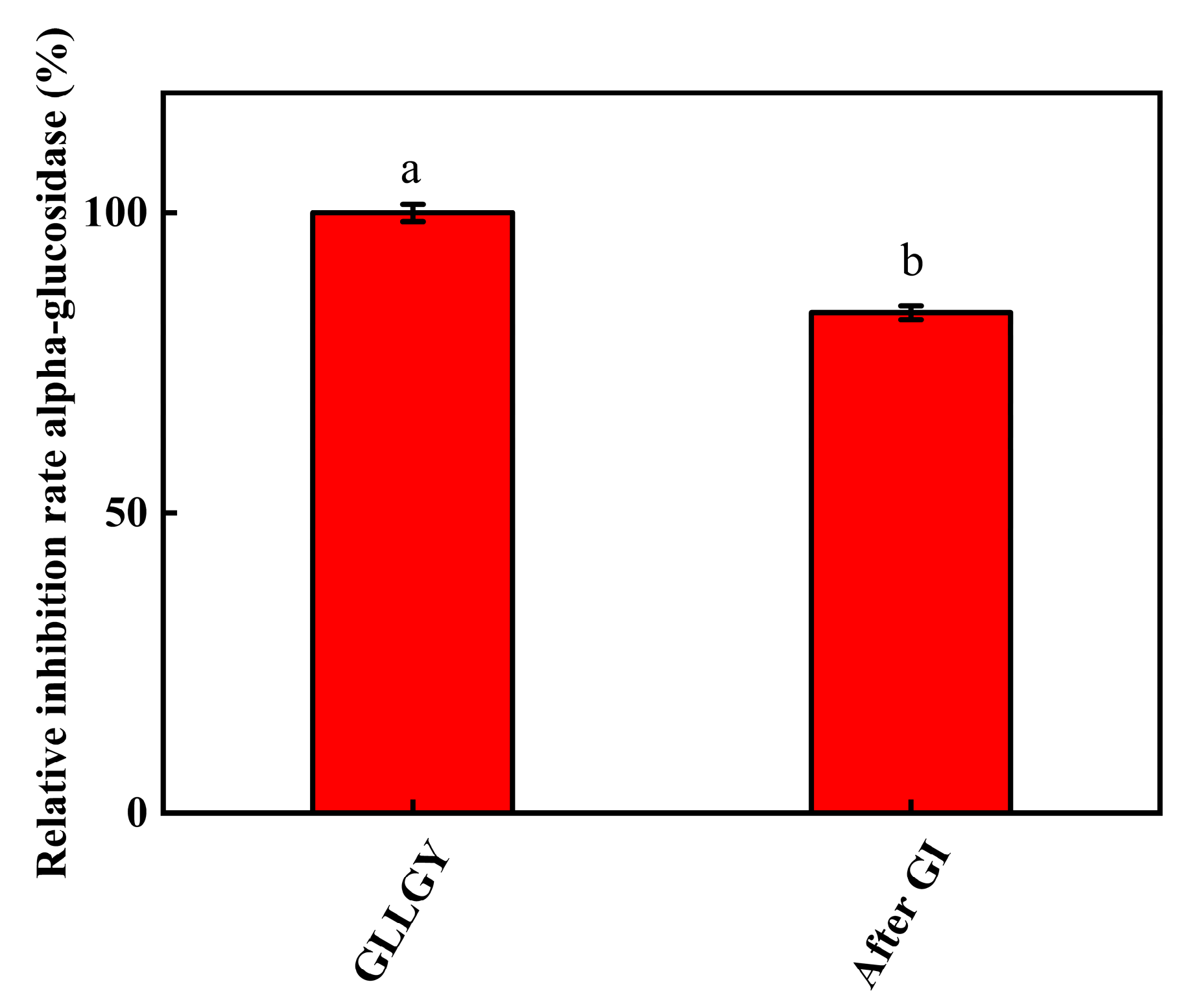
The results of in vitro GI simulation digestion are shown in Figure 6. After gastric and gastrointestinal digestion, the oligopeptide GLLGY still remained active, showing 83.37% inhibition rates against α-glucosidase. Statistically, though the α-glucosidase inhibitory effect of GLLGY decreased significantly, it retained strong anti-digestion ability in both the stomach and intestine.

4. Discussion

The extraction of active substances by microbial fermentation is emerging as a promising method [36]. Natural antidiabetic drugs have been extracted from microbial fermented food substances [37]. Rice bran is rich in proteins [5]. Like the α-glucosidase inhibitor peptides obtained from fermented rubing cheese [38], Chinese pickled chili peppers [39], and soybean condiments [40], the bioactive peptides extracted from rice bran fermentation broth can inhibit α-glucosidase. Bioactive peptides inhibit α-glucosidase by inhibiting the competition between oligosaccharides and α-glucosidase activity in small intestinal parietal cells, which slows down the intestinal absorption of glucose, reducing blood glucose levels [41]. The short-chain peptides not only maintain their activity during gastrointestinal digestion and hydrolysis, but they are also suitable for design and chemical synthesis [33]. Several studies showed that peptides resistant to GI digestion have lower hydrophobicity, and more positive net charge at pH 7.0. [42,43]. The average chain length and molecular weight of stable peptides are 4.5 ± 2.0 amino acid residues and 547.78 ± 233.17 g/mol, respectively, with a slightly positive net charge. Peptides with lower molecular weight may possess fewer protease recognition and cleavage sites [43]. The oligopeptide GLLGY is consistent with the peptide stability requirements of the in vitro GI digestion system. Moreover, the oligopeptide GLLGY maintained a good inhibitory effect on α-glucosidase in the simulated GI digestion system in vitro.
After a series of purifications, we found that the bioactive peptides were mainly <3 kDa. Thus, directly purifying components <3 kDa can save a significant amount of time. This approach is preferably applicable to the anti-diabetic peptides derived by fermentation from rice bran. A higher molecular weight peptide can have steric hindrance while binding to α-glucosidase [34]. The docking results of the purified peptide and alpha glucosidase molecule by ion exchange and Sephadex gel chromatography (Figure 2) proved that the peptide with lower molecular weight has a better inhibitory effect. GLLGY forms five hydrogen bonds with Asp282, Ser523, Asp616, and His674 residues of α-glucosidase (Figure 4c). It binds into the active site of α-glucosidase to prevent the entry of substrate-altering enzyme conformation that is no longer suitable for activity [44]. Jiang et al. [30] reported four hydrogen bonds between GSR and Asp282, Asp518, and Asp616 of α-glucosidase (PDB ID 5NN8), and five hydrogen bonds between EAK and His674, Asp518, Arg600, Asp616, and Asp282 of α-glucosidase. GLLGY binds to Asp282, Ser523, Asp616, and His674 residues to produce five hydrogen bonds. This shows that even with different amino acids, GLLGY binds to α-glucosidase like oligopeptide Gly-Ser-Arg and Glu-Ala-Lys.
In vitro activity assays showed that GLLGY inhibited the activity of α-glucosidase more than the other four peptides. The inhibition rate of GLLGY at 1 mg/mL was 15%, which is lower than that of hemp seed protein PLMLP (IC50 at 0.024 mg/mL) [45]. However, the α-glucosidase inhibition rate of GLLGY (1 mg/mL) is comparable to HNKPEVEVR (1.25 mg/mL), found in soft-shelled turtle eggs [34]. Notably, acarbose is a well-known competitive inhibitor of α-glucosidase. On the other hand, we found that GLLGY is a non-competitive inhibitor. Studies showed that the active site of α-glucosidase is mainly formed by acidic (Asp518, Glu521, and Asp616) and basic (Arg600 and His674) residues [46,47]. Asp518 acts as a nucleophile, while Glu521 and Asp616 stabilize the transition state through acid–base catalysis with Arg600 and His674. Past studies have shown that molecular docking is a reliable method to screen inhibitors of α-glucosidase [4]. In molecular docking studies, acarbose binds to Arg600, Asp616, and His674 residues, while GLLGY binds to Asp616 and His674 residues. According to the Lineweaver–Burk plots, the GLLGY is a non-competitive inhibitor. Compared with Acarbose, GLLGY has a different hydrogen bond distance with Asp616 and His674. Based on the structure of α-glucosidase, Asp616 and His674 are the key amino acid residues of the catalyst domain. They might be set as targets of molecule docking of α-glucosidase inhibititors. Leucine was considered important for enhancing insulin secretion through metabolic variable structure activation or membrane depolarization [45,48]. GLLGY binds to α-glucosidase via five hydrogen bonds, which may be the reason for its higher inhibition rate compared to other oligopeptides. Overall, GLLGY is a novel anti-diabetic peptide and the exact mechanism of its inhibition of α-glucosidase activity remains unclear and should be explored further.

5. Conclusions

Rice bran was fermented by Bacillus subtilis MK15 to extract the peptide components. The rice bran peptides were identified by LC-MS/MS. Oligopeptide GLLGY showed the best inhibitory effect against α-glucosidase. Molecular docking results suggested that it forms 5 hydrogen bonds and van der Waals interactions involving the second Leu (GLLGY) with α-glucosidase. GLLGY is a non-competitive inhibitor of α-glucosidase. In addition, GLLGY maintained an excellent α-glucosidase inhibitory effect in a GI digestion system. GLLGY is a promising anti-diabetic candidate and must be researched further. So far, the inhibitory effect was only studied in vitro; the effect in vivo still needs further exploration.

Supplementary Materials

The following supporting information can be downloaded at: https://www.mdpi.com/article/10.3390/foods12010183/s1, Figure S1. LC-MS/MS profile for rice bran peptide; Figure S2. (a) LC-MS/MS chromatogram of GLLGY obtained from preparative RP-HPLC. (b) ESI-MS/MS spectrum of the most intense peptide at m/z 522.292.

Author Contributions

Conceptualization, S.W., M.L. and J.H.; methodology, M.L.; software, J.H.; validation, J.H. and H.W.; formal analysis, J.H.; investigation, N.W.; resources, J.L. and S.W.; data curation, X.L. and X.W.; writing—original draft preparation, J.H.; writing—review and editing, M.L.; visualization, J.H.; supervision, S.W.; project administration, S.W. and M.L.; funding acquisition, S.W. All authors have read and agreed to the published version of the manuscript.

Funding

This study was supported by the National Natural Science Foundation of China (Grant no. 32172154); 521 Program Grant No. LYG06521202107; The Priority Academic Program Development of Jiangsu Higher Education Institutions (PAPD); The Research & Practice Innovation Program of Jiangsu (SJCX20_1321).

Data Availability Statement

The interaction data used to support the findings of this study are included within the article and the supporting information file. Also, all the data used to support the findings of this study are available from the corresponding author upon request.

Conflicts of Interest

The authors declare no conflict of interest.

References

  1. Aamir, A.H.; Ul-Haq, Z.; Mahar, S.A.; Qureshi, F.M.; Ahmad, I.; Jawa, A.; Sheikh, A.; Raza, A.; Fazid, S.; Jadoon, Z.; et al. Diabetes Prevalence Survey of Pakistan (DPS-PAK): Prevalence of type 2 diabetes mellitus and prediabetes using HbA1c: A population-based survey from Pakistan. BMJ Open 2019, 9, e025300. [Google Scholar] [CrossRef] [PubMed]
  2. Støy, J.; De Franco, E.; Ye, H.; Park, S.Y.; Bell, G.I.; Hattersley, A.T. In celebration of a century with insulin—Update of insulin gene mutations in diabetes. Mol. Metab. 2021, 52, 101280. [Google Scholar] [CrossRef] [PubMed]
  3. Helfer, G.; Wu, Q.F. Chemerin: A multifaceted adipokine involved in metabolic disorders. J. Endocrinol. 2018, 238, R79–R94. [Google Scholar] [CrossRef] [Green Version]
  4. Xie, X.; Chen, C.; Fu, X. Screening α-glucosidase inhibitors from four edible brown seaweed extracts by ultra-filtration and molecular docking. LWT 2021, 138, 110654. [Google Scholar] [CrossRef]
  5. Hossain, U.; Das, A.K.; Ghosh, S.; Sil, P.C. An overview on the role of bioactive α-glucosidase inhibitors in ameliorating diabetic complications. Food Chem. Toxicol. 2020, 145, 111738. [Google Scholar] [CrossRef]
  6. Di Stefano, E.; Oliviero, T.; Udenigwe, C.C. Functional significance and structure–activity relationship of food-derived α-glucosidase inhibitors. Curr. Opin. Food Sci. 2018, 20, 7–12. [Google Scholar] [CrossRef]
  7. Hu, S.; Fan, X.; Qi, P.; Zhang, X. Identification of anti-diabetes peptides from Spirulina platensis. J. Funct. Foods 2019, 56, 333–341. [Google Scholar] [CrossRef]
  8. Ramírez Fuentes, L.; Richard, C.; Chen, L. Sequential alcalase and flavourzyme treatment for preparation of α-amylase, α-glucosidase, and dipeptidyl peptidase (DPP)-IV inhibitory peptides from oat protein. J. Funct. Foods 2021, 87, 104829. [Google Scholar] [CrossRef]
  9. Urbizo-Reyes, U.; Liceaga, A.M.; Reddivari, L.; Kim, K.-H.; Anderson, J.M. Enzyme kinetics, molecular docking, and in silico characterization of canary seed (Phalaris canariensis L.) peptides with ACE and pancreatic lipase inhibitory activity. J. Funct. Foods 2022, 88, 104892. [Google Scholar] [CrossRef]
  10. Abbasi, S.; Moslehishad, M.; Salami, M. Antioxidant and alpha-glucosidase enzyme inhibitory properties of hydrolyzed protein and bioactive peptides of quinoa. Int. J. Biol. Macromol. 2022, 213, 602–609. [Google Scholar] [CrossRef]
  11. Elam, E.; Feng, J.; Lv, Y.-M.; Ni, Z.-J.; Sun, P.; Thakur, K.; Zhang, J.-G.; Ma, Y.-L.; Wei, Z.-J. Recent advances on bioactive food derived anti-diabetic hydrolysates and peptides from natural resources. J. Funct. Foods 2021, 86, 104674. [Google Scholar] [CrossRef]
  12. Park, H.Y.; Lee, K.W.; Choi, H.D. Rice bran constituents: Immunomodulatory and therapeutic activities. Food Funct. 2017, 8, 935–943. [Google Scholar] [CrossRef] [PubMed]
  13. Singh, B.; Kumar, G.; Kumar, V.; Singh, D. Enhanced Phytase Production by Bacillus subtilis subsp. subtilis in Solid State Fermentation and its Utility in Improving Food Nutrition. Protein Pept. Lett. 2021, 28, 1083–1089. [Google Scholar] [CrossRef] [PubMed]
  14. Anu; Kumar, S.; Kumar, A.; Kumar, V.; Singh, B. Optimization of cellulase production by Bacillus subtilis subsp. subtilis JJBS300 and biocatalytic potential in saccharification of alkaline-pretreated rice straw. Prep. Biochem. Biotechnol. 2021, 51, 697–704. [Google Scholar] [CrossRef] [PubMed]
  15. Shobako, N.; Ohinata, K. Anti-Hypertensive Effects of Peptides Derived from Rice Bran Protein. Nutrients 2020, 12, 3060. [Google Scholar] [CrossRef] [PubMed]
  16. Uraipong, C.; Zhao, J. Rice bran protein hydrolysates exhibit strong in vitro α-amylase, β-glucosidase and ACE-inhibition activities. J. Sci. Food Agric. 2016, 96, 1101–1110. [Google Scholar] [CrossRef]
  17. Manzoor, M.; Singh, J.; Gani, A. Exploration of bioactive peptides from various origin as promising nutraceutical treasures: In vitro, in silico and in vivo studies. Food Chem. 2022, 373, 131395. [Google Scholar] [CrossRef]
  18. Kumar, S.; Bhardwaj, V.K.; Singh, R.; Das, P.; Purohit, R. Identification of acridinedione scaffolds as potential inhibitor of DENV-2 C protein: An in silico strategy to combat dengue. J. Cell Biochem. 2022, 123, 935–946. [Google Scholar] [CrossRef]
  19. Kumar Bhardwaj, V.; Purohit, R.; Kumar, S. Himalayan bioactive molecules as potential entry inhibitors for the human immunodeficiency virus. Food Chem. 2021, 347, 128932. [Google Scholar] [CrossRef]
  20. Singh, R.; Bhardwaj, V.K.; Sharma, J.; Purohit, R.; Kumar, S. In-silico evaluation of bioactive compounds from tea as potential SARS-CoV-2 nonstructural protein 16 inhibitors. J. Tradit. Complement. Med. 2022, 12, 35–43. [Google Scholar] [CrossRef]
  21. Rajendran, V.; Sethumadhavan, R. Drug resistance mechanism of PncA in Mycobacterium tuberculosis. J. Biomol. Struct. Dyn. 2014, 32, 209–221. [Google Scholar] [CrossRef] [PubMed]
  22. Rajendran, V. Structural analysis of oncogenic mutation of isocitrate dehydrogenase 1. Mol. Biosyst. 2016, 12, 2276–2287. [Google Scholar] [CrossRef] [PubMed]
  23. Rajendran, V.; Gopalakrishnan, C.; Sethumadhavan, R. Pathological role of a point mutation (T315I) in BCR-ABL1 protein-A computational insight. J. Cell. Biochem. 2018, 119, 918–925. [Google Scholar] [CrossRef] [PubMed]
  24. Chen, X.; Lu, Y.; Zhao, A.; Wu, Y.; Zhang, Y.; Yang, X. Quantitative analyses for several nutrients and volatile components during fermentation of soybean by Bacillus subtilis natto. Food Chem. 2022, 374, 131725. [Google Scholar] [CrossRef]
  25. Su, W.; Jiang, Z.; Wang, C.; Xu, B.; Lu, Z.; Wang, F.; Zong, X.; Jin, M.; Wang, Y. Dynamics of defatted rice bran in physicochemical characteristics, microbiota and metabolic functions during two-stage co-fermentation. Int. J. Food Microbiol. 2022, 362, 109489. [Google Scholar] [CrossRef]
  26. Zhang, H.; Chen, Y.; Guo, Y.; Xu, W.; Wang, W.; Wu, S.; Chen, W.; Huang, Y. Label-free quantification proteomics reveals the active peptides from protein degradation during anaerobic fermentation of tea. LWT 2021, 150, 111950. [Google Scholar] [CrossRef]
  27. Cecile Urbain Marie, G.; Perreault, V.; Henaux, L.; Carnovale, V.; Aluko, R.E.; Marette, A.; Doyen, A.; Bazinet, L. Impact of a high hydrostatic pressure pretreatment on the separation of bioactive peptides from flaxseed protein hydrolysates by electrodialysis with ultrafiltration membranes. Sep. Purif. Technol. 2019, 211, 242–251. [Google Scholar] [CrossRef]
  28. Zhao, Q.; Wei, G.; Li, K.; Duan, S.; Ye, R.; Huang, A. Identification and molecular docking of novel α-glucosidase inhibitory peptides from hydrolysates of Binglangjiang buffalo casein. LWT 2022, 156, 113062. [Google Scholar] [CrossRef]
  29. Liu, L.; Chen, J.; Li, X. Novel peptides with α-glucosidase inhibitory activity from Changii Radix hydrolysates. Process Biochem. 2021, 111, 200–206. [Google Scholar] [CrossRef]
  30. Jiang, M.; Hui, Y.; He, R.; Ma, Y. Purification and a molecular docking study of α-glucosidase-inhibitory peptides from a soybean protein hydrolysate with ultrasonic pretreatment. Eur. Food Res. Technol. 2018, 244, 1995–2005. [Google Scholar] [CrossRef]
  31. Minekus, M.; Alminger, M.; Alvito, P.; Ballance, S.; Bohn, T.; Bourlieu, C.; Carrière, F.; Boutrou, R.; Corredig, M.; Dupont, D.; et al. A standardised static in vitro digestion method suitable for food—An international consensus. Food Funct. 2014, 5, 1113–1124. [Google Scholar] [CrossRef] [PubMed] [Green Version]
  32. Bougatef, A.; Nedjar-Arroume, N.; Manni, L.; Ravallec, R.; Barkia, A.; Guillochon, D.; Nasri, M. Purification and identification of novel antioxidant peptides from enzymatic hydrolysates of sardinelle (Sardinella aurita) by-products proteins. Food Chem. 2010, 118, 559–565. [Google Scholar] [CrossRef]
  33. Ibrahim, M.A.; Bester, M.J.; Neitz, A.W.H.; Gaspar, A.R.M. Structural properties of bioactive peptides with α-glucosidase inhibitory activity. Chem. Biol. Drug Des. 2018, 91, 370–379. [Google Scholar] [CrossRef] [PubMed] [Green Version]
  34. Qiu, L.; Deng, Z.; Zhao, C.; Xiao, T.; Weng, C.; Li, J.; Zheng, L. Nutritional composition and proteomic analysis of soft-shelled turtle (Pelodiscus sinensis) egg and identification of oligopeptides with alpha-glucosidase inhibitory activity. Food Res. Int. 2021, 145, 110414. [Google Scholar] [CrossRef]
  35. Ibrahim, M.A.; Bester, M.J.; Neitz, A.W.; Gaspar, A.R.M. Rational in silico design of novel α-glucosidase inhibitory peptides and in vitro evaluation of promising candidates. Biomed Pharm. 2018, 107, 234–242. [Google Scholar] [CrossRef] [Green Version]
  36. Yang, L.; Zeng, X.; Qiao, S. Advances in research on solid-state fermented feed and its utilization: The pioneer of private customization for intestinal microorganisms. Anim. Nutr. 2021, 7, 905–916. [Google Scholar] [CrossRef]
  37. Rocchetti, G.; Miragoli, F.; Zacconi, C.; Lucini, L.; Rebecchi, A. Impact of cooking and fermentation by lactic acid bacteria on phenolic profile of quinoa and buckwheat seeds. Food Res. Int. 2019, 119, 886–894. [Google Scholar] [CrossRef]
  38. Wei, G.; Zhao, Q.; Wang, D.; Fan, Y.; Shi, Y.; Huang, A. Novel ACE inhibitory, antioxidant and α-glucosidase inhibitory peptides identified from fermented rubing cheese through peptidomic and molecular docking. LWT 2022, 159, 113196. [Google Scholar] [CrossRef]
  39. Li, M.; Bao, X.; Zhang, X.; Ren, H.; Cai, S.; Hu, X.; Yi, J. Exploring the phytochemicals and inhibitory effects against α-glucosidase and dipeptidyl peptidase-IV in Chinese pickled chili pepper: Insights into mechanisms by molecular docking analysis. LWT 2022, 162, 113467. [Google Scholar] [CrossRef]
  40. Ademiluyi, A.O.; Oboh, G.; Boligon, A.A.; Athayde, M.L. Effect of fermented soybean condiment supplemented diet on α-amylase and α-glucosidase activities in Streptozotocin-induced diabetic rats. J. Funct. Foods 2014, 9, 1–9. [Google Scholar] [CrossRef]
  41. Gu, X.; Gao, T.; Hou, Y.; Li, D.; Fu, L. Identification and characterization of two novel α-glucosidase inhibitory peptides from almond (Armeniaca sibirica) oil manufacture residue. LWT-Food Sci. Technol. 2020, 134, 110215. [Google Scholar] [CrossRef]
  42. You, L.; Zhao, M.; Regenstein, J.M.; Ren, J. Changes in the antioxidant activity of loach (Misgurnus anguillicaudatus) protein hydrolysates during a simulated gastrointestinal digestion. Food Chem. 2010, 120, 810–816. [Google Scholar] [CrossRef]
  43. Ahmed, T.; Sun, X.; Udenigwe, C.C. Role of structural properties of bioactive peptides in their stability during simulated gastrointestinal digestion: A systematic review. Trends Food Sci. Technol. 2022, 120, 265–273. [Google Scholar] [CrossRef]
  44. Ju, H.; Chen, H.; Xiang, A.; Wang, Y.; Yue, T.; Yuan, Y. Identification and characterization of Lactobacillus paracasei strain MRS-4 antibacterial activity against Alicyclobacillus acidoterrestris. LWT 2021, 150, 111991. [Google Scholar] [CrossRef]
  45. Ren, Y.; Liang, K.; Jin, Y.; Zhang, M.; Chen, Y.; Wu, H.; Lai, F. Identification and characterization of two novel α-glucosidase inhibitory oligopeptides from hemp (Cannabis sativa L.) seed protein. J. Funct. Foods 2016, 26, 439–450. [Google Scholar] [CrossRef]
  46. Ur Rehman, N.; Rafiq, K.; Khan, A.; Ahsan Halim, S.; Ali, L.; Al-Saady, N.; Hilal Al-Balushi, A.; Al-Busaidi, H.K.; Al-Harrasi, A. α-Glucosidase Inhibition and Molecular Docking Studies of Natural Brominated Metabolites from Marine Macro Brown Alga Dictyopteris hoytii. Mar. Drugs 2019, 17, 666. [Google Scholar] [CrossRef] [Green Version]
  47. Roig-Zamboni, V.; Cobucci-Ponzano, B.; Iacono, R.; Ferrara, M.C.; Germany, S.; Bourne, Y.; Parenti, G.; Moracci, M.; Sulzenbacher, G. Structure of human lysosomal acid α-glucosidase-a guide for the treatment of Pompe disease. Nat. Commun. 2017, 8, 1111. [Google Scholar] [CrossRef] [PubMed] [Green Version]
  48. Yu, Z.; Yin, Y.; Zhao, W.; Yu, Y.; Liu, B.; Liu, J.; Chen, F. Novel peptides derived from egg white protein inhibiting alpha-glucosidase. Food Chem. 2011, 129, 1376–1382. [Google Scholar] [CrossRef]
Figure 1. α-glucosidase inhibition activity of (a) crude rice bran peptides, (b) bioactive peptides after ultrafiltration, and (c) bioactive peptides of <3 kDa.
Foods 12 00183 g001
Figure 2. (a) Chromatogram (insert) and α-glucosidase inhibitory activity of bioactive peptides purified with a DEAE Sepharose fast flow ion column. (b) Chromatogram (insert) and α-glucosidase inhibitory activity of bioactive peptides purified with a Sephadex G-25 column.
Foods 12 00183 g002
Figure 3. The α-glucosidase inhibitory activity of (a) peptides and (b) oligopeptide GLLGY.
Foods 12 00183 g003
Figure 4. The binding interactions between oligopeptide GLLGY and human α-glucosidase. (a) Three-dimensional structure of α-glucosidase. (b) The structure of oligopeptide GLLGY. (c) Interaction diagram of GLLGY with α-glucosidase. The yellow dotted lines indicate hydrogen bonds.
Foods 12 00183 g004
Figure 5. Lineweaver−Burk plots of α-glucosidase catalyzed reactions in the presence of GLLGY.
Foods 12 00183 g005
Figure 6. GLLGY α-glucosidase inhibition activity in a simulated gastrointestinal digestion system.
Foods 12 00183 g006
Table 1. Identification of peptides in rice bran fermentation products by LC-MS/MS and molecular docking analysis of their potential binding sites with α-glucosidase.
PeptidesScan Time
(min)
Accurate Mass (Da)Protein FamilyScoreIntensityBinding Energy
(kcal/mol)
Binding Residues of α-GlucosidaseHydrogen Bond Distance (Å)Number of Hydrogen Bonds
Acarbose −7.4Asp282
Asp281
Asp404
Arg600
Asp616
His674
2.3/1.9
3.4/2.9
2.8
3.4/3.0
2.0/1.7
3.2/3.4
11
LFSGF60.4865570.292ORYSJ Cellulose synthase-like protein79.70−8.0Asp282
Ala284
Asp616
3.5
3.0
2.1/2.4/2.6/3.2
6
HWP54.567439.209ORYSJ Mixed-linked glucan synthase193.6193.6−7.8Arg600
Asp616
His674
3.1
1.9/2.1/3.2
3.2
5
QSFF55.3714528.248ORYSJ protein286.6336,170−7.7Asp282
Ser523
Asn524
Asp616
2.3/3.0/3.5
3.0
2.3/3.5
2.6
7
FPF61.4677410.207BACSU PTS system oligo-beta-mannoside-specific EIIC component85.30−7.6Asp6161.9/3.42
FSGF51.4358457.209ORYSJ Cellulose synthase-like protein249.9534,630−7.6Asp282
Asp284
Ser523
Asn524
Ala555
2.9
3.5
2.9/2.4/3.1
3.3
3.3
7
VYGF50.7739485.241ORYSJ Cellulose synthase-like protein219.3982,140−7.6Asp282
Asp518
Asp616
2.4/3.5
2.5
2.3
4
GLIGY50.3365522.292BACIU Phage tail length tape-measure protein171.7594,040−7.2Asp282
Asp404
Ser523
Ala555
Asp616
His674
2.0/2.9
2.1
1.8/3.3
2.5
1.9/3.3
2.9
9
GLLGY50.3365522.292ORYSJ Phospholipase A1-II171.7594,040−7.1Asp282
Ser523
Asp616
His674
3.1
2.3
2.2/3.2
2.9
5
KGDPY51.7567579.293BACSU Aspartokinase 188.80−6.4Asp282
Asp404
Asp616
His674
2.1/2.2
2.0
2.1/3.4
2.9
6
QSFLQRYYFLFRILPL55.37142104.154ORYSJ Protein kinase domain-containing protein142.1336,170−6.4Arg281
Asp282
Ala284
Pro285
Phe525
Arg527
Trp618
Glu622
Gln623
3.4/3.0/3.1
2.3/2.4
3.5
3.2
3.4
3.4
3.4
2.3
3.0
12
PSR59.1375359.218ORYSA DNA-directed RNA polymerase subunit α8.02,1243,000−6.3Asp282
Ser523
Arg600
Asp616
2.4/3.1/3.4/3.6
2.1/2.2/2.3
3.2
3.3/3.4
10
ISIFLSFFFGLIAGT60.69461632.906BACIU Amino acid ABC transporter permease82.82,322,000−6.2Arg281
Asp282
Asp518
Asn524
Arg600
2.8/3.3
1.9
2.4/3.2
3.3/3.3
3.2
8
Disclaimer/Publisher’s Note: The statements, opinions and data contained in all publications are solely those of the individual author(s) and contributor(s) and not of MDPI and/or the editor(s). MDPI and/or the editor(s) disclaim responsibility for any injury to people or property resulting from any ideas, methods, instructions or products referred to in the content.