
Screening of Bioactive Properties in Brown Algae from the Northwest Iberian Peninsula

 ,
,  ,
,  ,
,  , , , ,
 ,
,  and
and 
Abstract
: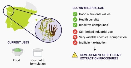
1. Introduction
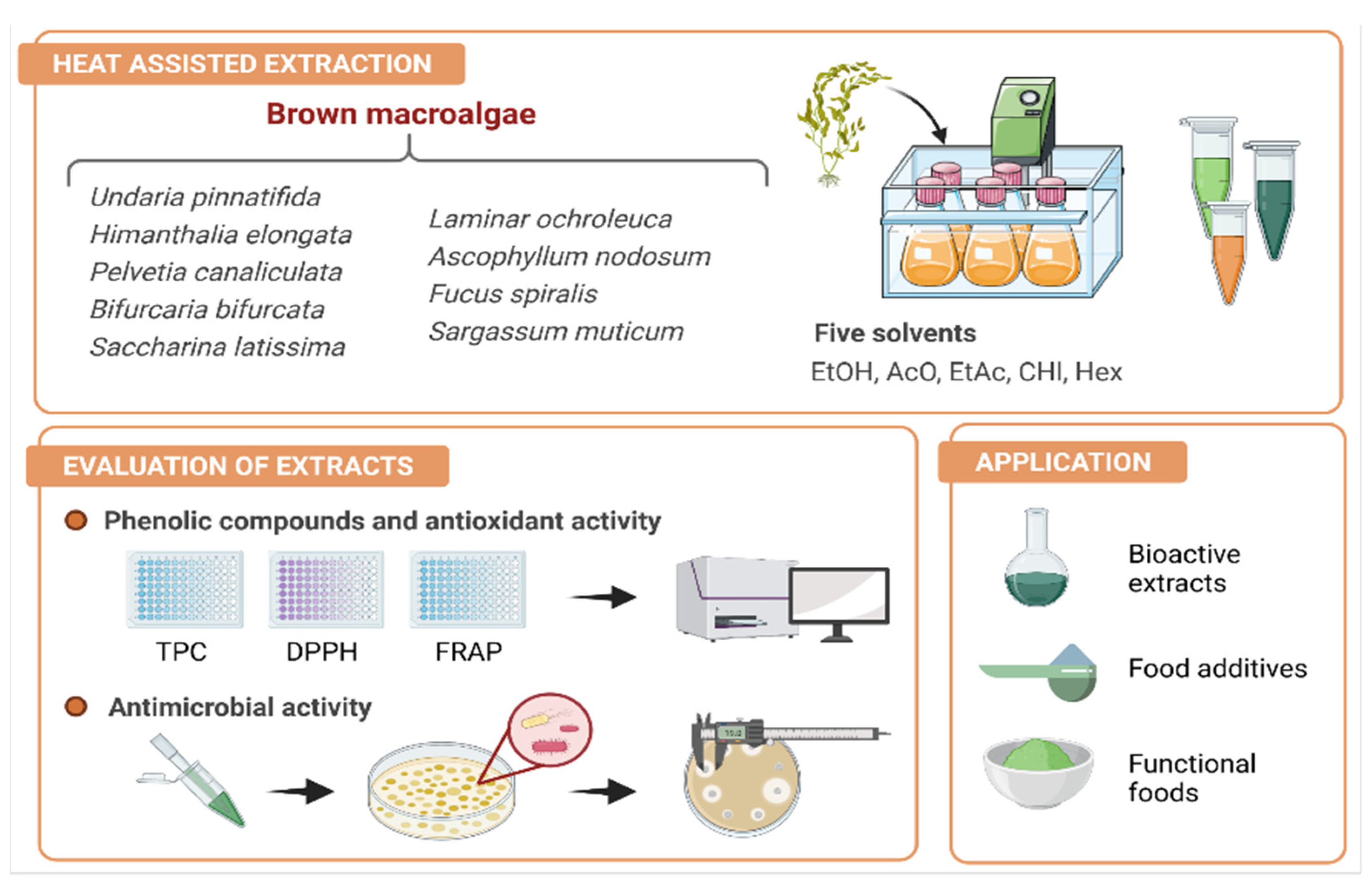
2. Materials and Methods
2.1. Chemicals and Reagents
2.2. Algae Sampling and Preparation
2.3. Extraction Procedure
2.4. Total Phenolic Content Determination
2.5. Antioxidant Activity Determination
2.5.1. DPPH-Radical Scavenging Activity (DPPH-RSA) Assay
2.5.2. Ferric Reduction Activity Power (FRAP) Assay
2.6. Antibacterial Tests
2.6.1. Microorganisms and Culture Conditions
2.6.2. Agar Diffusion Assay
2.7. Statistical Analysis
3. Results and Discussion
3.1. Extraction Efficiency, Phenolic Content, and Antioxidant Activity
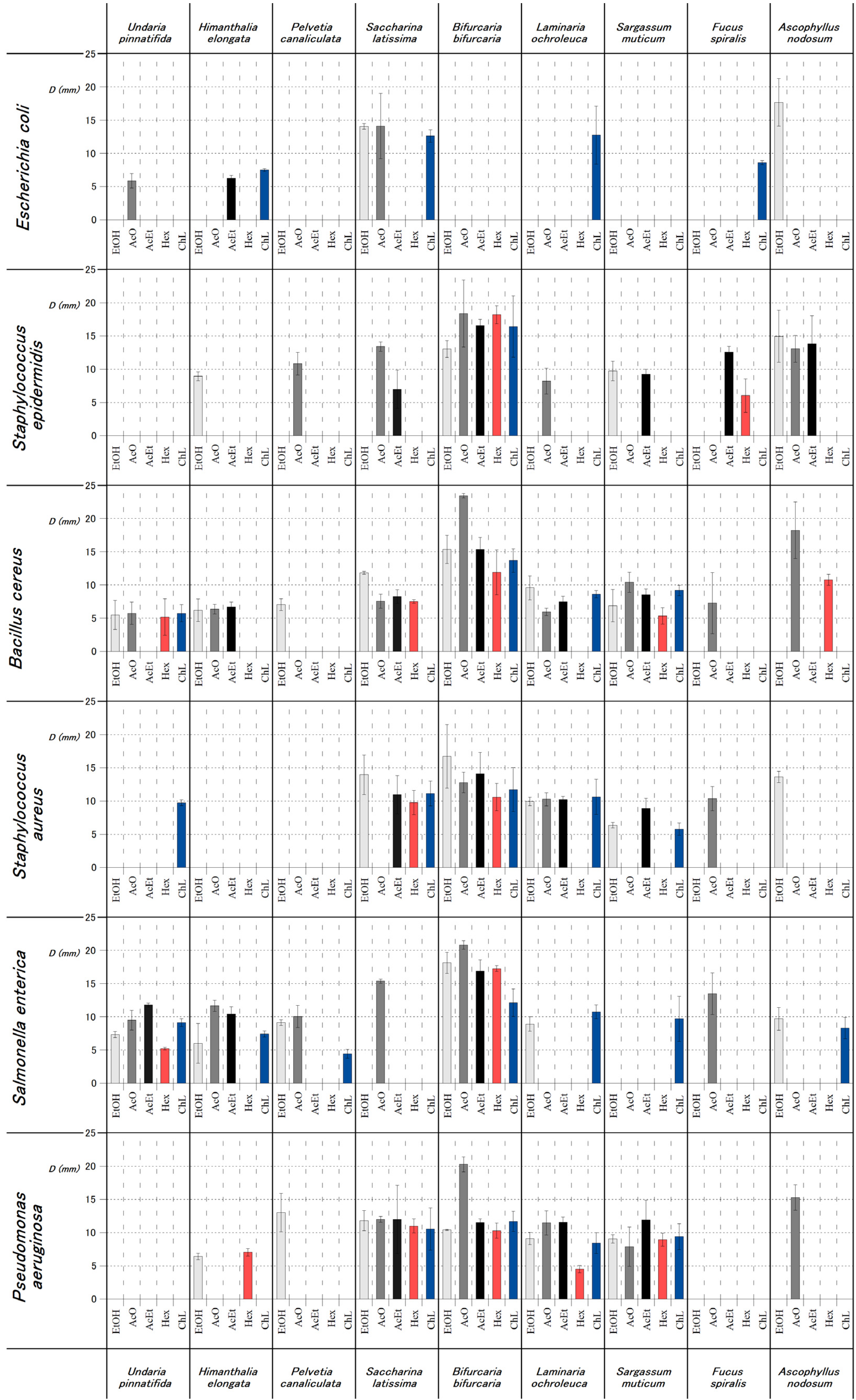
3.2. Antimicrobial Activity
4. Conclusions
Author Contributions
Funding
Institutional Review Board Statement
Informed Consent Statement
Data Availability Statement
Acknowledgments
Conflicts of Interest
Abbreviations
| AAE | Ascorbic acid equivalents |
| AcO | Acetone |
| ATCC | American type culture collection |
| CFU | Colony formation units |
| ChL | Chloroform |
| DMSO | Dimethyl Sulfoxide |
| DPPH | 1,1-diphenyl-2-picryl hydrazyl |
| DW | Dry weight |
| EtAc | Ethyl acetate |
| EtOH | Ethanol |
| FRAP | Ferric reduction activity power |
| FW | Fresh weight |
| GAE | Gallic acid equivalents |
| Hex | Hexane |
| MHB | Mueller–Hinton broth |
| PI | Polarity index |
| TPC | Total phenolic content |
| TPTZ | Fe3+-2,4,6-tri(2-pyridyl)-S-triazine |
| Trolox | 6-hydroxy-2,5,7,8-tetramethylchroman-2-carboxylic acid |
References
- The European Food Safety Authority and European Centre for Disease Prevention and Control (EFSA and ECDC). The European Union summary report on trends and sources of zoonoses, zoonotic agents and food-borne outbreaks in 2017. EFSA J. 2018, 16, e05500. [Google Scholar] [CrossRef]
- Stenfors Arnesen, L.P.; Fagerlund, A.; Granum, P.E. From soil to gut: Bacillus cereus and its food poisoning toxins. FEMS Microbiol. Rev. 2008, 32, 579–606. [Google Scholar] [CrossRef] [Green Version]
- Parsaeimehr, A.; Lutzu, G.A. Algae as a Novel Source of Antimicrobial Compounds: Current and Future Perspectives. Current and Future Perspectives; Elsevier: Amsterdam, The Netherlands, 2016; ISBN 978-0-12803-668-6. [Google Scholar]
- Cardoso, S.M.; Carvalho, L.G.; Silva, P.J.; Rodrigues, M.S.; Pereira, O.R. Bioproducts from Seaweeds: A Review with Special Focus on the Iberian Peninsula. Curr. Org. Chem. 2014, 896–917. [Google Scholar] [CrossRef]
- Yuan, Y.; Zhang, J.; Fan, J.; Clark, J.; Shen, P.; Li, Y.; Zhang, C. Microwave assisted extraction of phenolic compounds from four economic brown macroalgae species and evaluation of their antioxidant activities and inhibitory effects on α-amylase, α-glucosidase, pancreatic lipase and tyrosinase. Food Res. Int. 2018, 113, 288–297. [Google Scholar] [CrossRef]
- Afonso, N.C.; Catarino, M.D.; Silva, A.M.S.; Cardoso, S.M. Brown macroalgae as valuable food ingredients. Antioxidants 2019, 8, 365. [Google Scholar] [CrossRef] [Green Version]
- López-Hortas, L.; Domínguez, H.; Torres, M.D. Valorisation of edible brown seaweeds by the recovery of bioactive compounds from aqueous phase using MHG to develop innovative hydrogels. Process Biochem. 2019, 78, 100–107. [Google Scholar] [CrossRef]
- Leandro, A.; Pereira, L.; Gonçalves, A.M.M. Diverse applications of marine macroalgae. Mar. Drugs 2020, 18, 17. [Google Scholar] [CrossRef] [PubMed] [Green Version]
- García-Vaquero, M.; Ummat, V.; Tiwari, B.; Rajauria, G. Exploring Ultrasound, Microwave and Ultrasound—Microwave Assisted Extraction Technologies to Increase the Extraction of Bioactive Compounds and Antioxidants from Brown Macroalgae. Mar. Drugs 2020, 18, 172. [Google Scholar] [CrossRef] [Green Version]
- Sornsiri, J.; Srisook, K.; Pornngam, P.; Sootanan, P. Prediction of biochemical mechanism of anti-inflammation explained from two marine-derived bioactive compounds. Agric. Nat. Resour. 2018, 52, 588–595. [Google Scholar] [CrossRef]
- Tenorio-Rodríguez, P.A.; Esquivel-Solis, H.; Murillo-Álvarez, J.I.; Ascencio, F.; Campa-Córdova, Á.I.; Angulo, C. Biosprospecting potential of kelp (Laminariales, Phaeophyceae) from Baja California Peninsula: Phenolic content, antioxidant properties, anti-inflammatory, and cell viability. J. Appl. Phycol. 2019, 31, 3115–3129. [Google Scholar] [CrossRef]
- Ford, L.; Stratakos, A.C.; Theodoridou, K.; Dick, J.T.A.; Sheldrake, G.N.; Linton, M.; Corcionivoschi, N.; Walsh, P.J. Polyphenols from Brown Seaweeds as a Potential Antimicrobial Agent in Animal Feeds. ACS Omega 2020. [Google Scholar] [CrossRef] [Green Version]
- Buedenbender, L.; Astone, F.A.; Tasdemir, D. Bioactive Molecular Networking for Mapping the Antimicrobial Constituents of the Baltic Brown Alga Fucus vesiculosus. Mar. Drugs 2020, 18, 311. [Google Scholar] [CrossRef] [PubMed]
- Santos, S.A.O.; Félix, R.; Pais, A.C.S.; Rocha, S.M.; Silvestre, A.J.D. The quest for phenolic compounds from macroalgae: A review of extraction and identification methodologies. Biomolecules 2019, 9, 847. [Google Scholar] [CrossRef] [PubMed] [Green Version]
- Fernández-Segovia, I.; Lerma-García, M.J.; Fuentes, A.; Barat, J.M. Characterization of Spanish powdered seaweeds: Composition, antioxidant capacity and technological properties. Food Res. Int. 2018, 111, 212–219. [Google Scholar] [CrossRef]
- Martínez–Hernández, G.B.; Castillejo, N.; Carrión–Monteagudo, M.d.M.; Artés, F.; Artés-Hernández, F. Nutritional and bioactive compounds of commercialized algae powders used as food supplements. Food Sci. Technol. Int. 2018, 24, 172–182. [Google Scholar] [CrossRef]
- Buschmann, A.H.; Camus, C.; Infante, J.; Neori, A.; Israel, Á.; Hernández-González, M.C.; Pereda, S.V.; Gomez-Pinchetti, J.L.; Golberg, A.; Tadmor-Shalev, N.; et al. Seaweed production: Overview of the global state of exploitation, farming and emerging research activity. Eur. J. Phycol. 2017, 52, 391–406. [Google Scholar] [CrossRef]
- García-Pérez, P.; Lozano-Milo, E.; Landín, M.; Gallego, P.P. Combining Medicinal Plant In Vitro Culture with Machine Learning Technologies for Maximizing the Production of Phenolic Compounds. Antioxidants 2020, 9, 210. [Google Scholar] [CrossRef] [Green Version]
- López, C.J.; Caleja, C.; Prieto, M.A.; Sokovic, M.; Calhelha, R.C.; Barros, L.; Ferreira, I.C.F.R. Stability of a cyanidin-3-O-glucoside extract obtained from Arbutus unedo L. and incorporation into wafers for colouring purposes. Food Chem. 2019, 275, 426–438. [Google Scholar] [CrossRef] [PubMed] [Green Version]
- Singleton, V.L.; Rossi, J.A. Colorimetry of Total Phenolics with Phosphomolybdic-Phosphotungstic Acid Reagents. Am. J. Enol. Vitic. 1965, 16, 144–158. [Google Scholar]
- Barroso, M.F.; Ramalhosa, M.J.; Alves, R.C.; Dias, A.; Soares, C.M.D.; Oliva-Teles, M.T.; Delerue-Matos, C. Total antioxidant capacity of plant infusions: Assessment using electrochemical DNA-based biosensor and spectrophotometric methods. Food Control 2016, 68, 153–161. [Google Scholar] [CrossRef]
- Paz, M.; Gúllon, P.; Barroso, M.F.; Carvalho, A.P.; Domingues, V.F.; Gomes, A.M.; Becker, H.; Longhinotti, E.; Delerue-Matos, C. Brazilian fruit pulps as functional foods and additives: Evaluation of bioactive compounds. Food Chem. 2015, 172, 462–468. [Google Scholar] [CrossRef] [PubMed] [Green Version]
- Clinical and Laboratory Standards Institute. Performance Standards for Antimicrobial Disk Susceptibility Tests, 11th ed.; Approved Standard; Clinical and Laboratory Standards Institute: Wayne, PA, USA, 2012; Volume 32, ISBN 156-238-7812. [Google Scholar]
- Cox, S.; Abu-Ghannam, N.; Gupta, S. An assessment of the antioxidant and antimicrobial activity of six species of edible Irish seaweeds. Int. Food Res. J. 2010, 17, 205–220. [Google Scholar] [CrossRef]
- Montero, L.; Herrero, M.; Ibáñez, E.; Cifuentes, A. Separation and characterization of phlorotannins from brown algae Cystoseira abies-marina by comprehensive two-dimensional liquid chromatography. Electrophoresis 2014, 35, 1644–1651. [Google Scholar] [CrossRef] [Green Version]
- Generalić Mekinić, I.; Skroza, D.; Šimat, V.; Hamed, I.; Čagalj, M.; Popović Perković, Z. Phenolic Content of Brown Algae (Pheophyceae) Species: Extraction, Identification, and Quantification. Biomolecules 2019, 9, 244. [Google Scholar] [CrossRef] [Green Version]
- Cikos, A.M.; Jokic, S.; Subaric, D.; Jerkovic, I. Overview on the Application of Modern Methods for the Extraction of Bioactive Compounds from Marine Macroalgae. Mar. Drugs 2018, 16, 348. [Google Scholar] [CrossRef] [Green Version]
- Sánchez-Camargo, A.D.P.; Montero, L.; Stiger-Pouvreau, V.; Tanniou, A.; Cifuentes, A.; Herrero, M.; Ibáñez, E. Considerations on the use of enzyme-assisted extraction in combination with pressurized liquids to recover bioactive compounds from algae. Food Chem. 2016, 192, 67–74. [Google Scholar] [CrossRef] [PubMed]
- Otero, P.; López-Martínez, M.I.; García-Risco, M.R. Application of pressurized liquid extraction (PLE) to obtain bioactive fatty acids and phenols from Laminaria ochroleuca collected in Galicia (NW Spain). J. Pharm. Biomed. Anal. 2019, 164, 86–92. [Google Scholar] [CrossRef] [PubMed]
- Kuda, T.; Tsunekawa, M.; Goto, H.; Araki, Y. Antioxidant properties of four edible algae harvested in the Noto Peninsula, Japan. J. Food Compos. Anal. 2005, 18, 625–633. [Google Scholar] [CrossRef]
- López, A.; Rico, M.; Rivero, A.; Suárez de Tangil, M. The effects of solvents on the phenolic contents and antioxidant activity of Stypocaulon scoparium algae extracts. Food Chem. 2011, 125, 1104–1109. [Google Scholar] [CrossRef]
- Galali, Y.; Omar, Z.A.; Sajadi, S.M. Biologically active components in by-products of food processing. Food Sci. Nutr. 2020, 8, 3004–3022. [Google Scholar] [CrossRef] [PubMed]
- Fernandes de Oliveira, A.; Sousa Pinheiro, L.; Souto Pereira, C.; Neves Matias, W.; Albuquerque Gomes, R.; Souza Chaves, O.; Vanderlei de Souza, M.; Nóbrega de Almeida, R.; Simões de Assis, T. Total Phenolic Content and Antioxidant Activity of Some Malvaceae Family Species. Antioxidants 2012, 1, 33. [Google Scholar] [CrossRef] [PubMed]
- Wong, S.; Leong, L.; Williamkoh, J. Antioxidant activities of aqueous extracts of selected plants. Food Chem. 2006, 99, 775–783. [Google Scholar] [CrossRef]
- Silva, A.; Silva, S.A.; Lourenço-Lopes, C.; Jimenez-Lopez, C.; Carpena, M.; Gullón, P.; Fraga-Corral, M.; Domingues, V.F.; Barroso, M.F.; Simal-Gandara, J.; et al. Antibacterial Use of Macroalgae Compounds against Foodborne Pathogens. Antibiotics 2020, 9, 712. [Google Scholar] [CrossRef] [PubMed]
- Akremi, N.; Cappoen, D.; Anthonissen, R.; Verschaeve, L.; Bouraoui, A. Phytochemical and in vitro antimicrobial and genotoxic activity in the brown algae Dictyopteris membranacea. S. Afr. J. Bot. 2017, 108, 308–314. [Google Scholar] [CrossRef]
- Barros, L.; Calhelha, R.C.; Vaz, J.A.; Ferreira, I.C.F.R.; Baptista, P.; Estevinho, L.M. Antimicrobial activity and bioactive compounds of Portuguese wild edible mushrooms methanolic extracts. Eur. Food Res. Technol. 2007, 225, 151–156. [Google Scholar] [CrossRef]
- Joshi, D.R.; Adhikari, N. An Overview on Common Organic Solvents and Their Toxicity. J. Pharm. Res. Int. 2019, 1–18. [Google Scholar] [CrossRef]
- Alzeer, J.; Abou Hadeed, K. Ethanol and its Halal status in food industries. Trends Food Sci. Technol. 2016, 58, 14–20. [Google Scholar] [CrossRef]
- Seaweed, M.; Pérez, M.J.; Falqué, E.; Domínguez, H.; Seaweed, M. Antimicrobial action of compounds from marine seaweed. Mar. Drugs 2016, 14, 52. [Google Scholar] [CrossRef] [Green Version]


| Species | Solvent | Extraction Yield (%) | TPC | DPPH-RSA | FRAP |
|---|---|---|---|---|---|
| mg GAE/g Dry E | mg TE/g Dry E | mg AAE/g Dry E | |||
| Undaria pinnatifida | EtOH | 38.8 | 3.68 ± 0.35 efD | -±- | 1.50 ± 0.02 cD |
| AcO | 3.40 | 41.5 ± 3.95 dA | 13.79 ± 0.92 dC | 15.89 ± 1.23 eA | |
| EtAc | 2.40 | 16.53 ± 1.46 fB | 46.55 ± 0.97 cA | 9.51 ± 0.62 fgB | |
| Hex | 2.20 | 3.46 ± 0.30 dD | 33.92 ± 2.86 dB | 1.78 ± 0.17 gD | |
| ChL | 3.20 | 10.29 ± 1.07 deC | 36.32 ± 1.71 cB | 8.05 ± 0.47 bC | |
| Himanthalia elongata | EtOH | 27.0 | 30.26 ± 2.28 dC | -± | 10.26 ± 1.83 bC |
| AcO | 3.6 | 162.22 ± 5.98 bA | 5.19 ± 0.51 eD | 62.98 ± 2.27 aA | |
| EtAc | 0.20 | 53.34 ± 4.45 cB | 54.24 ± 3.26 cB | 28.12 ± 2.45 cB | |
| Hex | 2.10 | 6.60 ± 0.22 cD | 75.33 ± 8.52 bA | 2.76 ± 0.10 deD | |
| ChL | 3.20 | 13.07 ± 0.45 cdD | 16.52 ± 2.09 dC | 4.87 ± 0.58 deD | |
| Pelvetia canaliculata | EtOH | 15.6 | 49.49 ± 3.32 cB | -± | 12.88 ± 0.71 bB |
| AcO | 12.2 | 87.32 ± 2.90 cA | 2.46 ± 0.16 efD | 21.14 ± 0.52 dA | |
| EtAc | 6.90 | 35.61 ± 3.25 deC | 16.06 ± 0.84 eA | 13.07 ± 0.20 efB | |
| Hex | 7.60 | 2.07 ± 0.17 eD | 5.98 ± 0.57 efC | 0.87 ± 0.08 hD | |
| ChL | 7.7 | 7.66 ± 0.43 efD | 7.58 ± 0.79 deB | 5.39 ± 0.29 dC | |
| Saccharina latissima | EtOH | 18.0 | 2.44 ± 0.17 fD | 1.23 ± 0.02 dE | 1.97 ± 0.03 cD |
| AcO | 2.90 | 16.53 ± 0.63 eA | 21.59 ± 1.8 cB | 8.56 ± 0.27 fA | |
| EtAc | 1.40 | 12.36 ± 0.89 fB | 24.87 ± 2.29 dA | 6.88 ± 0.37 ghB | |
| Hex | 1.70 | 3.69 ± 0.27 dD | 12.59 ± 0.00 eC | 2.48 ± 0.25 efCD | |
| ChL | 0.60 | 7.36 ± 0.32 fC | 5.97 ± 0.53 eD | 3.02 ± 0.11 fC | |
| Bifurcaria bifurcata | EtOH | 24.1 | 11.39 ± 0.32 eC | 73.54 ± 2.95 aC | 2.76 ± 0.15 cB |
| AcO | 10.8 | 86.08 ± 5.54 cA | 0.50 ± 0.03 fD | 35.38 ± 2.16 cA | |
| EtAc | 4.40 | 21.88 ± 0.94 efB | 110.58 ± 5.09 aB | 3.46 ± 0.28 hB | |
| Hex | 1.60 | 18.82 ± 0.85 aB | 145.61 ± 4.6 aA | 3.60 ± 0.31 cB | |
| ChL | 4.40 | 16.81 ± 0.77 bBC | 107.28 ± 8.95 aB | 2.86 ± 0.17 fB | |
| Laminaria ochroleuca | EtOH | 19.2 | 2.81 ± 0.25 fC | 5.38 ± 0.18 dD | 1.24 ± 0.70 cD |
| AcO | 0.80 | 14.59 ± 0.75 eB | 22.83 ± 1.59 cC | 11.86 ± 0.53 fB | |
| EtAc | 1.40 | 32.46 ± 2.54 deA | 72.73 ± 4.01 dA | 18.33 ± 1.27 deA | |
| Hex | 0.60 | 3.78 ± 0.24 dC | 56.95 ± 3.09 dB | 2.21 ± 0.14 fgD | |
| ChL | 2.10 | 11.82 ± 1.01 dB | 6.13 ± 0.13 eD | 7.14 ± 0.56 bcC | |
| Sargassum muticum | EtOH | 17.9 | 8.31 ± 0.33 efC | 4.01 ± 0.23 dD | 4.18 ± 0.1 cB |
| AcO | 3.40 | 25.89 ± 2.54 eB | 5.48 ± 0.21 eBC | 17.46 ± 1.09 deA | |
| EtAc | 0.30 | 41.63 ± 3.97 cdA | 16.19 ± 1.08 eA | 18.33 ± 1.51 dA | |
| Hex | 2.00 | 10.38 ± 0.41 bC | 6.06 ± 0.62 efB | 5.99 ± 0.29 bB | |
| ChL | 2.10 | 15.35 ± 0.97 bcC | 4.65 ± 0.39 eCD | 6.16 ± 0.65 cdB | |
| Fucus spiralis | EtOH | 14.6 | 95.75 ± 5.28 bC | 60.34 ± 3.07 bA | 46.27 ± 2.28 aB |
| AcO | 7.80 | 184.22 ± 12.82 aA | 57.48 ± 2.08 bA | 48.18 ± 1.00 bB | |
| EtAc | 5.30 | 123.67 ± 4.50 bB | 52.87 ± 3.14 cA | 68.97 ± 4.54 bA | |
| Hex | 5.70 | 10.91 ± 0.49 bD | 56.35 ± 2.43 cA | 3.00 ± 0.07 dC | |
| ChL | 7.00 | 17.15 ± 1.24 bD | 53.20 ± 3.86 bA | 3.93 ± 0.27 efC | |
| Ascophyllum nodosum | EtOH | 18.2 | 117.2 ± 6.33 aC | 52.52 ± 2.27 cC | 43.69 ± 1.03 aC |
| AcO | 15.4 | 183.13 ± 5.30 aB | 73.3 ± 2.26 aB | 59.97 ± 2.03 aB | |
| EtAc | 10.3 | 211.83 ± 18.22 aA | 89.8 ± 4.28 bA | 79.97 ± 1.11 aA | |
| Hex | 8.00 | 6.18 ± 0.36 cD | 3.76 ± 0.18 fE | 8.09 ± 0.01 aE | |
| ChL | 9.40 | 23.73 ± 0.80 aD | 29.05 ± 1.26 cD | 14.360.92 aD |
| Species | Solvent | Inhibition Zone (mm) | |||||
|---|---|---|---|---|---|---|---|
| Gram (+) | Gram (−) | ||||||
| S. aureus | S. epidermidis | B. cereus | E. coli | S. enteritidis | P. aeruginosa | ||
| Undaria pinnatifida | EtOH | - | - | 5.49 ± 2.19 cA | - | 7.34 ± 0.47 bcC | - |
| AcO | - | - | 5.74 ± 1.68 bA | 5.86 ±1.08 b | 9.51 ± 1.47 dB | - | |
| EtAc | 9.75 ± 0.44 b | - | - | - | 11.81 ± 0.30 bA | - | |
| Hex | - | - | 5.19 ± 2.75 cA | - | 5.21 ± 0.20 bD | - | |
| ChL | - | - | 5.74 ± 1.30 cA | - | 9.11 ± 0.60 abBC | - | |
| Himanthalia elongata | EtOH | - | 8.94 ± 0.67 b | 6.20 ± 1.68 cA | - | 5.65 ± 2.50 cC | 6.42 ± 0.49 cA |
| AcO | - | - | 6.36 ± 0.71 bA | - | 11.67 ± 0.85 bcdA | - | |
| EtAc | - | - | 6.71 ± 0.71 bA | 6.28 ± 0.42 A | 10.42 ± 1.14 bAB | 7.06 ± 0.58 bA | |
| Hex | - | - | - | - | - | - | |
| ChL | - | - | - | 7.52 ± 0.22 bA | 7.44 ± 0.46 bcBC | - | |
| Pelvetia canaliculata | EtOH | - | - | 7.05 ± 0.88 c | - | 9.14 ± 0.42 bA | 13.01 ± 2.90 a |
| AcO | - | - | - | - | 10.06 ± 1.65 cdA | - | |
| EtAc | - | 10.84 ± 1.70 bc | - | - | - | - | |
| Hex | - | - | - | - | - | - | |
| ChL | - | - | - | - | 4.43 ± 0.67 cB | - | |
| Saccharina latissima | EtOH | 13.96 ± 2.98 abA | - | 11.83 ± 0.22 abA | 14.08 ± 0.43 bA | - | 11.83 ± 1.52 abA |
| AcO | - | 13.41 ± 0.69 abA | 7.56 ± 1.06 bB | 14.12 ± 4.92 aA | 15.38 ± 0.31 b | 12.00 ± 0.45 bA | |
| EtAc | 10.98 ± 2.88 abA | 6.98 ± 2.88 cB | 8.28 ± 1.01 bB | - | - | 11.98 ± 5.14 aA | |
| Hex | 9.78 ± 1.83 aA | - | 7.52 ± 0.28 bcB | - | - | 10.99 ± 1.06 aA | |
| ChL | 11.13 ± 1.86 aA | - | - | 12.65 ± 0.94 aA | - | 10.55 ± 3.18 aA | |
| Bifurcaria bifurcata | EtOH | 16.74 ± 4.79 aA | 13.05 ± 1.28 abA | 15.35 ± 2.13 aB | - | 18.14 ± 1.60 aAB | 10.43 ± 0.08 abB |
| AcO | 12.81 ± 1.56 aA | 18.39 ± 5.03 aA | 23.45 ± 0.35 aA | - | 20.84 ± 0.63 aA | 20.29 ± 1.13 aA | |
| EtAc | 14.12 ± 3.22 aA | 16.55 ± 0.95 aA | 15.37 ± 1.79 aB | - | 16.88 ± 1.71 aB | 11.53 ± 0.55 abB | |
| Hex | 10.60 ± 2.05 aA | 18.22 ± 1.36 aA | 11.90 ± 3.39 aB | - | 17.28 ± 0.43 aAB | 10.03 ± 1.13 abB | |
| ChL | 11.73 ± 3.29 aA | 16.44 ± 4.63 A | 13.68 ± 1.74 aB | - | 12.14 ± 2.07 aC | 11.74 ± 1.49 aB | |
| Laminaria ochroleuca | EtOH | 9.94 ± 0.64 bcA | - | 9.57 ± 1.81 bcA | - | 8.93 ± 1.07 bcB | 9.12 ± 0.90 bcA |
| AcO | 10.27 ± 0.99 bA | 8.20 ± 1.95 b | 5.95 ± 0.54 bB | - | - | 11.49 ± 1.82 bcA | |
| EtAc | 10.24 ± 0.47 abA | - | 7.48 ± 0.85 bAB | - | - | 11.55 ± 0.81 abA | |
| Hex | - | - | - | - | - | 4.53 ± 0.55 cB | |
| ChL | 10.64 ± 2.65 aA | - | 8.61 ± 0.56 bA | 12.76 ± 4.38 a | 10.75 ± 1.05 abA | 8.43 ± 1.58 aA | |
| Sargassum muticum | EtOH | 6.36 ± 0.41 cB | 9.73 ± 1.48 bA | 6.89 ± 2.44 cAB | - | - | 9.06 ± 0.64 bcA |
| AcO | - | - | 10.42 ± 1.50 bA | - | - | 7.89 ± 2.94 cA | |
| EtAc | 8.90 ± 1.50 bA | 9.22 ± 0.77 bcA | 8.53 ± 0.89 bAB | - | - | 11.89 ± 3.04 aA | |
| Hex | - | - | 5.35 ± 1.22 cB | - | - | 8.94 ± 0.97 bA | |
| ChL | 5.76 ± 0.91 bA | - | 9.19 ± 0.78 bA | - | 9.71 ± 3.38 ab | 9.39 ± 1.95 aA | |
| Fucus spiralis | EtOH | - | - | - | - | - | - |
| AcO | 10.38 ± 1.81 b | - | 7.28 ± 4.60 b | - | 13.49 ± 3.12 bc | - | |
| EtAc | - | 12.57 ± 0.86 abA | - | - | - | - | |
| Hex | - | 6.04 ± 2.52 bB | - | - | - | - | |
| ChL | - | - | - | 8.60 ± 0.29 b | - | - | |
| Ascophyllum nodosum | EtOH | 13.63 ± 0.85 ab | 14.97 ± 3.92 aA | - | 17.70 ± 3.58 a | 9.71 ± 1.72 bA | - |
| AcO | - | 13.07 ± 2.01 abA | 18.24 ± 4.26 aA | - | - | 15.28 ± 1.91 b | |
| EtAc | - | 13.85 ± 4.21 abA | - | - | - | - | |
| Hex | - | - | 10.79 ± 0.84 abB | - | - | - | |
| ChL | - | - | - | - | 8.30 ± 1.61 bA | - | |
| DMSO | - | - | - | - | - | - | |
| Lactic acid | 18.55 ± 3.75 | 17.20 ± 3.83 | 16.75 ± 2.98 | 18.52 ± 3.63 | 19.19 ± 3.23 | 18.70 ± 2.64 | |
Publisher’s Note: MDPI stays neutral with regard to jurisdictional claims in published maps and institutional affiliations. |
© 2021 by the authors. Licensee MDPI, Basel, Switzerland. This article is an open access article distributed under the terms and conditions of the Creative Commons Attribution (CC BY) license (https://creativecommons.org/licenses/by/4.0/).
Share and Cite
Silva, A.; Rodrigues, C.; Garcia-Oliveira, P.; Lourenço-Lopes, C.; Silva, S.A.; Garcia-Perez, P.; Carvalho, A.P.; Domingues, V.F.; Barroso, M.F.; Delerue-Matos, C.; et al. Screening of Bioactive Properties in Brown Algae from the Northwest Iberian Peninsula. Foods 2021, 10, 1915. https://doi.org/10.3390/foods10081915
Silva A, Rodrigues C, Garcia-Oliveira P, Lourenço-Lopes C, Silva SA, Garcia-Perez P, Carvalho AP, Domingues VF, Barroso MF, Delerue-Matos C, et al. Screening of Bioactive Properties in Brown Algae from the Northwest Iberian Peninsula. Foods. 2021; 10(8):1915. https://doi.org/10.3390/foods10081915
Chicago/Turabian StyleSilva, Aurora, Carla Rodrigues, Paula Garcia-Oliveira, Catarina Lourenço-Lopes, Sofia A. Silva, Pascual Garcia-Perez, Ana P. Carvalho, Valentina F. Domingues, M. Fátima Barroso, Cristina Delerue-Matos, and et al. 2021. "Screening of Bioactive Properties in Brown Algae from the Northwest Iberian Peninsula" Foods 10, no. 8: 1915. https://doi.org/10.3390/foods10081915
APA StyleSilva, A., Rodrigues, C., Garcia-Oliveira, P., Lourenço-Lopes, C., Silva, S. A., Garcia-Perez, P., Carvalho, A. P., Domingues, V. F., Barroso, M. F., Delerue-Matos, C., Simal-Gandara, J., & Prieto, M. A. (2021). Screening of Bioactive Properties in Brown Algae from the Northwest Iberian Peninsula. Foods, 10(8), 1915. https://doi.org/10.3390/foods10081915