
Metabolic Profiling of the Oil of Sesame of the Egyptian Cultivar ‘Giza 32’ Employing LC-MS and Tandem MS-Based Untargeted Method

 ,
,  ,
, 

Abstract
1. Introduction
2. Materials and Methods
2.1. Chemicals
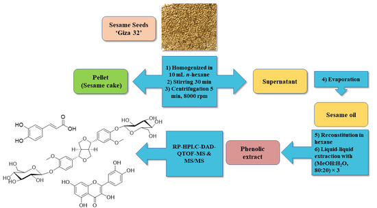
2.2. Samples Procurement and Oil Extraction Procedures
2.3. Analysis by RP-HPLC-DAD-ESI-QTOF-MS and -Tandem MS
3. Results and Discussion
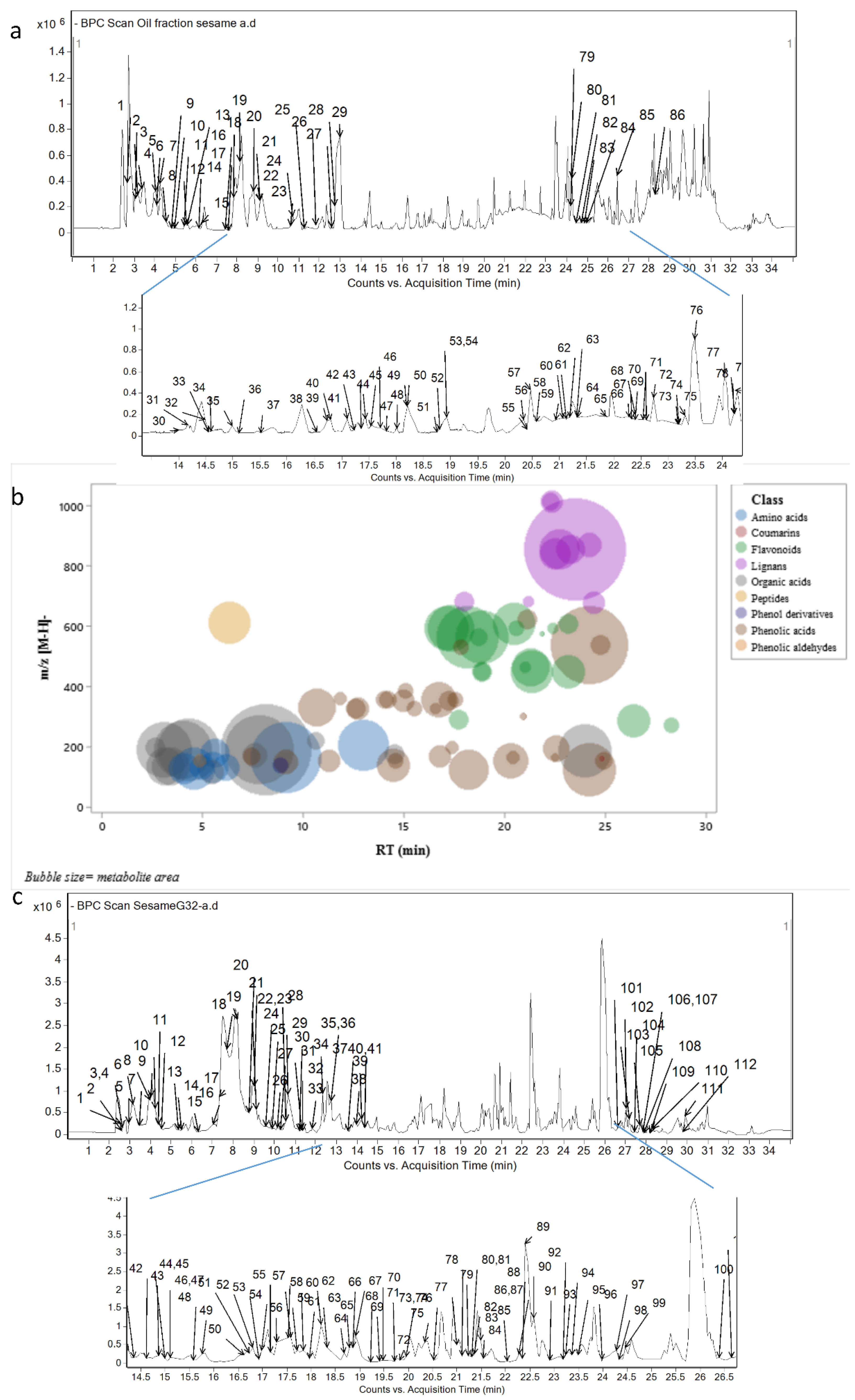
3.1. RP-HPLC-DAD-ESI-QTOF-MS and Tandem-MS of SG32 Oil
3.1.1. Phenolic Compounds
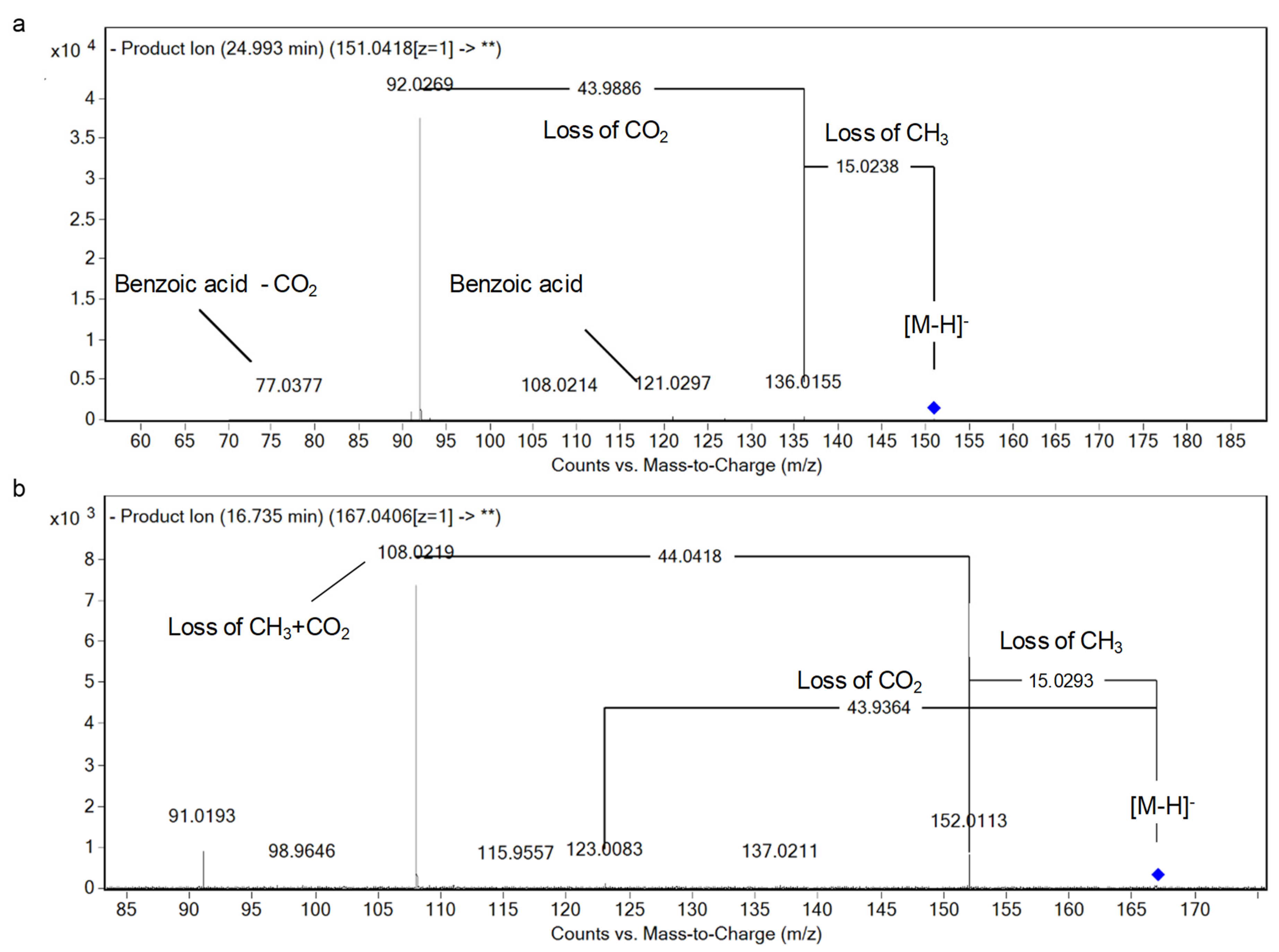
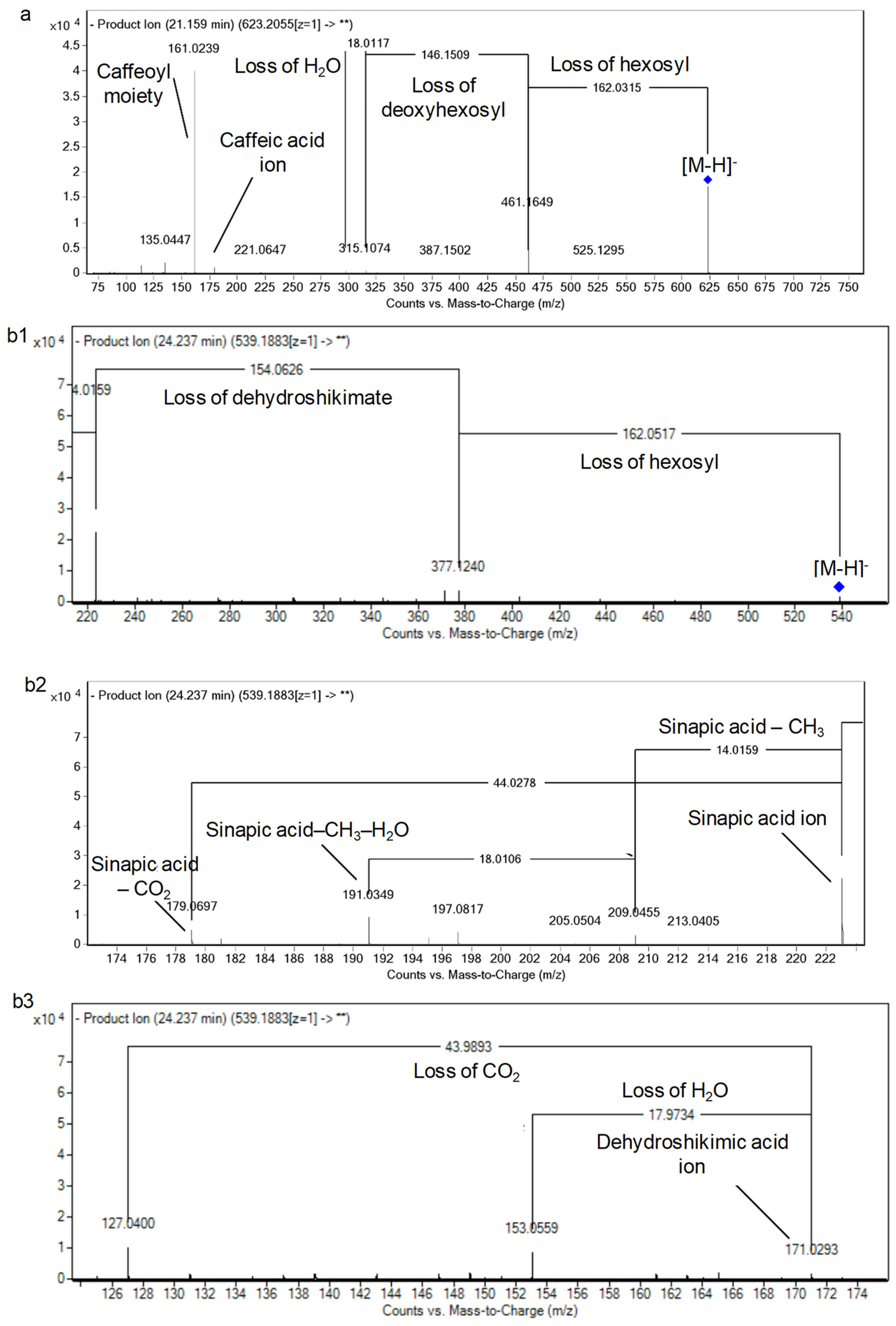
Phenolic Acids
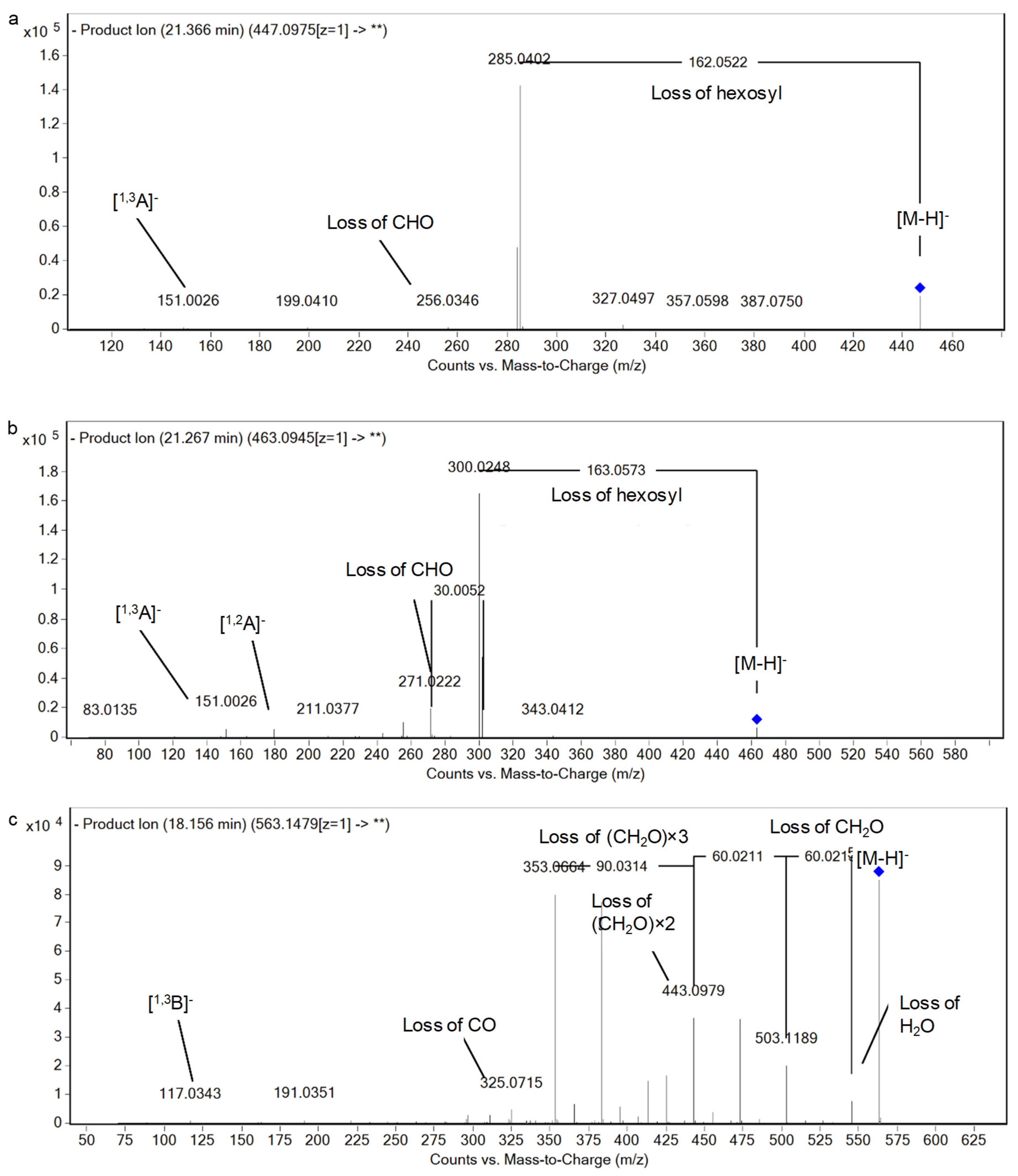
Flavonoids
Lignans
Coumarins, Phenol Aldehydes, and Derivatives
3.1.2. Non-Phenolic Compounds
Nitrogenous Compounds
Organic Acids
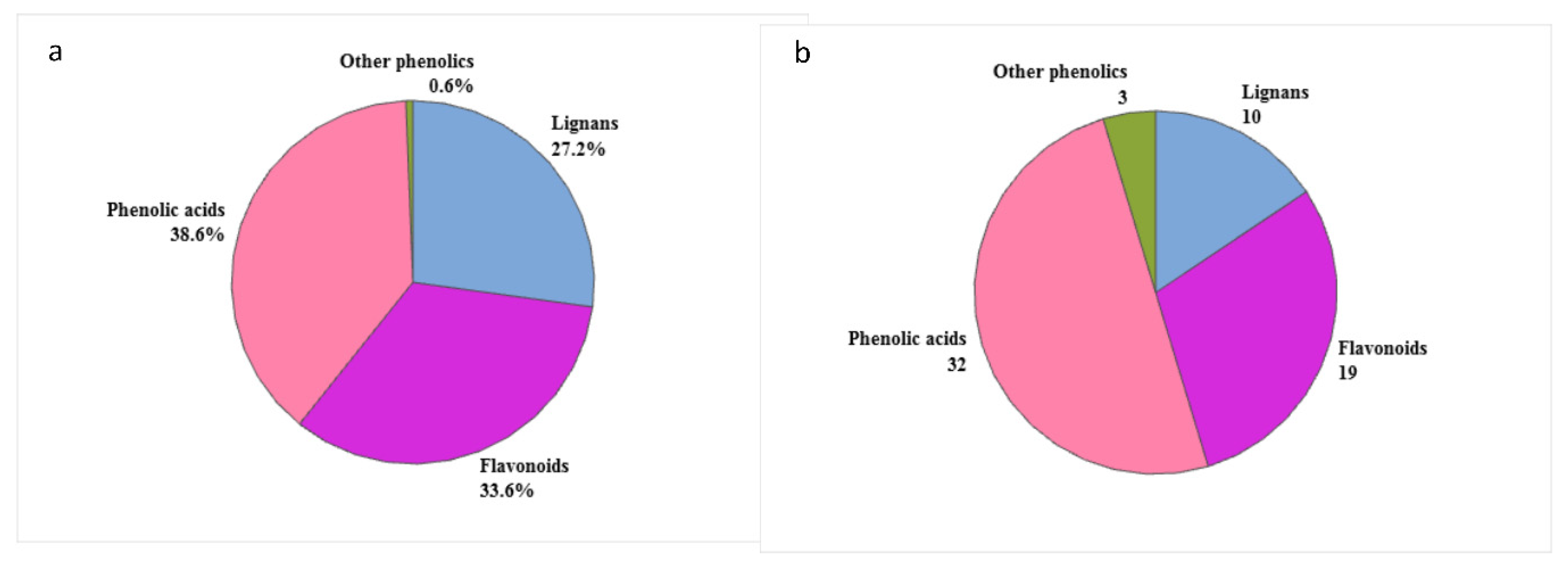
3.2. Semi-Quantitative Analysis
3.3. Comparison between Sesame Seed Oil and Cake
4. Conclusions
Supplementary Materials
Author Contributions
Funding
Data Availability Statement
Acknowledgments
Conflicts of Interest
References
- Berry, P.E. Lamiales. In Encyclopædia Britannica; Encyclopædia Britannica Inc.: Chicago, IL, USA, 2017. [Google Scholar]
- FAO-Statistics. Productions, Crops; FAO: Rome, Italy, 2018. [Google Scholar]
- Aboelsoud, N.H. Herbal medicine in ancient Egypt. J. Med. Plants Res. 2010, 4, 082–086. [Google Scholar]
- Bedigian, D.; Harlan, J.R. Evidence for cultivation of sesame in the ancient world. Econ. Bot. 1986, 40, 137–154. [Google Scholar] [CrossRef]
- Lim, T. Sesamum indicum. In Edible Medicinal and Non-Medicinal Plants; Springer: Dordrecht, The Netherlands, 2012; Volume 4, pp. 187–219. [Google Scholar]
- Hassan, M.A. Studies on Egyptian Sesame Seeds (Sesamum indicum L.) and Its Products 1-Physicochemical Analysis and Phenolic Acids of Roasted Egyptian Sesame seeds (Sesamum indicum L.). World J. Dairy Food Sci. 2012, 7, 195–201. [Google Scholar]
- Dravie, E.E.; Kortei, N.K.; Essuman, E.K.; Tettey, C.O.; Boakye, A.A.; Hunkpe, G. Antioxidant, phytochemical and physicochemical properties of sesame seed (Sesamum indicum L). Sci. Afr. 2020, 8, e00349. [Google Scholar] [CrossRef]
- Kim, Y.H.; Kim, E.Y.; Rodriguez, I.; Nam, Y.H.; Jeong, S.Y.; Hong, B.N.; Choung, S.-Y.; Kang, T.H. Sesamum indicum L. Oil and sesamin induce auditory-protective effects through changes in hearing loss-related gene expression. J. Med. Food 2020, 23, 491–498. [Google Scholar] [CrossRef]
- Ma’mag, L.K.; Zintchem, A.A.A.; Atchadé, A.d.T.; Kopa Kowa, T.; Tchinda Tiabou, A.; Bikobo, D.S.N.; Pegnyemb, D.E. Antileishmanial activity of long chain alkyl benzene and other constituents from seeds of Sesamum indicum. L (Pedaliaceae). Nat. Prod. Res. 2020, 1–5. [Google Scholar] [CrossRef]
- Mekky, R.H.; Abdel-Sattar, E.; Segura-Carretero, A.; Contreras, M.d.M. Phenolic Compounds from Sesame Cake and Antioxidant Activity: A New Insight for Agri-Food Residues’ Significance for Sustainable Development. Foods 2019, 8, 432. [Google Scholar] [CrossRef]
- Namiki, M. Nutraceutical Functions of Sesame: A Review. Crit. Rev. Food Sci. Nutr. 2007, 47, 651–673. [Google Scholar] [CrossRef]
- Hsu, C.-C.; Huang, H.-C.; Wu, P.-T.; Tai, T.-W.; Jou, I.M. Sesame oil improves functional recovery by attenuating nerve oxidative stress in a mouse model of acute peripheral nerve injury: Role of Nrf-2. J. Nutr. Biochem. 2016, 38, 102–106. [Google Scholar] [CrossRef]
- Zeb, A.; Muhammad, B.; Ullah, F. Characterization of sesame (Sesamum indicum L.) seed oil from Pakistan for phenolic composition, quality characteristics and potential beneficial properties. J. Food Meas. Charact. 2017, 11, 1362–1369. [Google Scholar] [CrossRef]
- Khaleel, A.E.S.; Gonaid, M.H.; El-Bagry, R.I.; Sleem, A.A.; Shabana, M. Chemical and biological study of the residual aerial parts of Sesamum indicum L. J. Food Drug Anal. 2007, 15, 249–257. [Google Scholar] [CrossRef]
- Dachtler, M.; van de Put, F.H.M.; Stijn, F.; Beindorff, C.M.; Fritsche, J. On-line LC-NMR-MS characterization of sesame oil extracts and assessment of their antioxidant activity. Eur. J. Lipid Sci. Technol. 2003, 105, 488–496. [Google Scholar] [CrossRef]
- Wu, R.; Ma, F.; Zhang, L.; Li, P.; Li, G.; Zhang, Q.; Zhang, W.; Wang, X. Simultaneous determination of phenolic compounds in sesame oil using LC–MS/MS combined with magnetic carboxylated multi-walled carbon nanotubes. Food Chem. 2016, 204, 334–342. [Google Scholar] [CrossRef] [PubMed]
- Görgüç, A.; Özer, P.; Yılmaz, F.M. Simultaneous effect of vacuum and ultrasound assisted enzymatic extraction on the recovery of plant protein and bioactive compounds from sesame bran. J. Food Compost. Anal. 2020, 87, 103424. [Google Scholar] [CrossRef]
- Görgüç, A.; Bircan, C.; Yılmaz, F.M. Sesame bran as an unexploited by-product: Effect of enzyme and ultrasound-assisted extraction on the recovery of protein and antioxidant compounds. Food Chem. 2019, 283, 637–645. [Google Scholar] [CrossRef]
- Shyu, Y.-S.; Hwang, L.S. Antioxidative activity of the crude extract of lignan glycosides from unroasted Burma black sesame meal. Food Res. Int. 2002, 35, 357–365. [Google Scholar] [CrossRef]
- Ishtiaque, S.; Khan, N.; Siddiqui, M.A.; Siddiqi, R.; Naz, S. Antioxidant potential of the extracts, fractions and oils derived from oilseeds. Antioxidants 2013, 2, 246–256. [Google Scholar] [CrossRef]
- Mekky, R.H.; Contreras, M.d.M.; El-Gindi, M.R.; Abdel-Monem, A.R.; Abdel-Sattar, E.; Segura-Carretero, A. Profiling of phenolic and other compounds from Egyptian cultivars of chickpea (Cicer arietinum L.) and antioxidant activity: A comparative study. RSC Adv. 2015, 5, 17751–17767. [Google Scholar] [CrossRef]
- Mekky, R.H.; Thabet, M.M.; Rodríguez-Pérez, C.; Elnaggar, D.M.Y.; Mahrous, E.A.; Segura-Carretero, A.; Abdel-Sattar, E. Comparative metabolite profiling and antioxidant potentials of seeds and sprouts of three Egyptian cultivars of Vicia faba L. Food Res. Int. 2020, 136, 109537. [Google Scholar] [CrossRef]
- Abouzed, T.K.; Contreras, M.d.M.; Sadek, K.M.; Shukry, M.H.; Abdelhady, D.; Gouda, W.M.; Abdo, W.; Nasr, N.E.; Mekky, R.H.; Segura-Carretero, A.; et al. Red onion scales ameliorated streptozotocin-induced diabetes and diabetic nephropathy in Wistar rats in relation to their metabolite fingerprint. Diabetes Res. Clin. Pract. 2018, 140, 253–264. [Google Scholar] [CrossRef]
- Ammar, S.; Contreras, M.d.M.; Belguith-Hadrich, O.; Segura-Carretero, A.; Bouaziz, M. Assessment of the distribution of phenolic compounds and contribution to the antioxidant activity in Tunisian fig leaves, fruits, skins and pulps using mass spectrometry-based analysis. Food Funct. 2015, 6, 3663–3677. [Google Scholar] [CrossRef] [PubMed]
- Reaxys. Available online: http://www.reaxys.com (accessed on 1 December 2020).
- KNApSAcK-Core-System. Available online: http://www.knapsackfamily.com/knapsack_jsp/top.html (accessed on 1 December 2020).
- SciFinder-Scholar. Available online: https://scifinder.cas.org (accessed on 1 December 2020).
- PubChem. Available online: http://pubchem.ncbi.nlm.nih.gov (accessed on 1 December 2020).
- ChemSpider. Available online: http://www.chemspider.com (accessed on 1 December 2020).
- METLIN-Metabolite-Database. Available online: http://metlin.scripps.edu (accessed on 1 December 2020).
- Phenol-Explorer. Available online: www.phenol-explorer.eu (accessed on 1 December 2020).
- Dictionary-of-Natural-Products. Available online: http://dnp.chemnetbase.com (accessed on 1 December 2020).
- Phytochemical-Dictionary-of-Natural-Products. Available online: https://www.crcpress.com/go/the_dictionary_of_natural_products (accessed on 1 December 2020).
- Egyptian-Knowledge-Bank. Available online: https://www.ekb.eg/ (accessed on 1 December 2020).
- Abdel-Sattar, E.; Mahrous, E.A.; Thabet, M.M.; Elnaggar, D.M.Y.; Youssef, A.M.; Elhawary, R.; Zaitone, S.A.; Rodríguez-Pérez, C.; Segura-Carretero, A.; Mekky, R.H. Methanolic extracts of a selected Egyptian Vicia faba cultivar mitigate the oxidative/inflammatory burden and afford neuroprotection in a mouse model of Parkinson’s disease. Inflammopharmacology 2020. [Google Scholar] [CrossRef] [PubMed]
- Ammar, S.; Contreras, M.d.M.; Belguith-Hadrich, O.; Bouaziz, M.; Segura-Carretero, A. New insights into the qualitative phenolic profile of Ficus carica L. fruits and leaves from Tunisia using ultra-high-performance liquid chromatography coupled to quadrupole-time-of-flight mass spectrometry and their antioxidant activity. RSC Adv. 2015, 5, 20035–20050. [Google Scholar] [CrossRef]
- Contreras, M.d.M.; Arráez-Román, D.; Fernández-Gutiérrez, A.; Segura-Carretero, A. Nano-Liquid chromatography coupled to time-of-flight mass spectrometry for phenolic profiling: A case study in cranberry syrups. Talanta 2015, 132, 929–938. [Google Scholar] [CrossRef]
- Mekky, R.H. A Comparative Phytochemical and Biological Studies on Certain Egyptian Varieties of Cicer arietinum Linn., Family Fabaceae. Ph.D. Thesis, Cairo University, Cairo, Egypt, 2016. [Google Scholar]
- Tsimogiannis, D.; Samiotaki, M.; Panayotou, G.; Oreopoulou, V. Characterization of Flavonoid Subgroups and Hydroxy Substitution by HPLC-MS/MS. Molecules 2007, 12, 593–606. [Google Scholar] [CrossRef]
- De Rijke, E.; Out, P.; Niessen, W.M.A.; Ariese, F.; Gooijer, C.; Brinkman, U.A.T. Analytical separation and detection methods for flavonoids. J. Chromatogr. A 2006, 1112, 31–63. [Google Scholar] [CrossRef]
- Materska, M. Flavone C-glycosides from Capsicum annuum L.: Relationships between antioxidant activity and lipophilicity. Eur. Food Res. Technol. 2015, 240, 549–557. [Google Scholar] [CrossRef]
- Abu-Reidah, I.M.; Arráez-Román, D.; Quirantes-Piné, R.; Fernández-Arroyo, S.; Segura-Carretero, A.; Fernández-Gutiérrez, A. HPLC–ESI-Q-TOF-MS for a comprehensive characterization of bioactive phenolic compounds in cucumber whole fruit extract. Food Res. Int. 2012, 46, 108–117. [Google Scholar] [CrossRef]
- Eklund, P.C.; Backman, M.J.; Kronberg, L.Å.; Smeds, A.I.; Sjöholm, R.E. Identification of lignans by liquid chromatography-electrospray ionization ion-trap mass spectrometry. J. Mass Spectrom. 2008, 43, 97–107. [Google Scholar] [CrossRef]
- Guo, H.; Liu, A.-H.; Ye, M.; Yang, M.; Guo, D.-A. Characterization of phenolic compounds in the fruits of Forsythia suspensa by high-performance liquid chromatography coupled with electrospray ionization tandem mass spectrometry. RCM 2007, 21, 715–729. [Google Scholar]
- Abu-Reidah, I.M.; Arráez-Román, D.; Segura-Carretero, A.; Fernández-Gutiérrez, A. Extensive characterisation of bioactive phenolic constituents from globe artichoke (Cynara scolymus L.) by HPLC–DAD-ESI-QTOF-MS. Food Chem. 2013, 141, 2269–2277. [Google Scholar] [CrossRef] [PubMed]
- Lee, J.; Choe, E. Extraction of lignan compounds from roasted sesame oil and their effects on the autoxidation of methyl linoleate. J. Food Sci. 2006, 71, C430–C436. [Google Scholar] [CrossRef]
- Ammar, S.; Contreras, M.d.M.; Gargouri, B.; Segura-Carretero, A.; Bouaziz, M. RP-HPLC-DAD-ESI-QTOF-MS based metabolic profiling of the potential Olea europaea by-product “wood” and its comparison with leaf counterpart. Phytochem. Anal. 2017, 28, 217–229. [Google Scholar] [CrossRef]
- Herzog, K.; IJlst, L.; van Cruchten, A.G.; van Roermund, C.W.T.; Kulik, W.; Wanders, R.J.A.; Waterham, H.R. An UPLC-MS/MS Assay to Measure Glutathione as Marker for Oxidative Stress in Cultured Cells. Metabolites 2019, 9, 45. [Google Scholar] [CrossRef] [PubMed]
- Gómez-Romero, M.; Segura-Carretero, A.; Fernández-Gutiérrez, A. Metabolite profiling and quantification of phenolic compounds in methanol extracts of tomato fruit. Phytochemistry 2010, 71, 1848–1864. [Google Scholar] [CrossRef]
- Kozukue, E.; Kozukue, N.; Tsuchida, H. Identification and changes of phenolic compounds in bamboo shoots during storage at 20 °C. J. Jpn. Soc. Hortic. Sci. 1998, 67, 805–811. [Google Scholar] [CrossRef][Green Version]
- Talhaoui, N.; Gómez-Caravaca, A.M.; León, L.; De la Rosa, R.; Fernández-Gutiérrez, A.; Segura-Carretero, A. From Olive Fruits to Olive Oil: Phenolic Compound Transfer in Six Different Olive Cultivars Grown under the Same Agronomical Conditions. Int. J. Mol. Sci. 2016, 17, 337. [Google Scholar] [CrossRef]
- De Torres, A.; Espínola, F.; Moya, M.; Alcalá, S.; Vidal, A.M.; Castro, E. Assessment of phenolic compounds in virgin olive oil by response surface methodology with particular focus on flavonoids and lignans. LWT 2018, 90, 22–30. [Google Scholar] [CrossRef]
- Miho, H.; Moral, J.; López-González, M.A.; Díez, C.M.; Priego-Capote, F. The phenolic profile of virgin olive oil is influenced by malaxation conditions and determines the oxidative stability. Food Chem. 2020, 314, 126183. [Google Scholar]
- Wang, X.; Zeng, Q.; Contreras, M.d.M.; Wang, L. Profiling and quantification of phenolic compounds in Camellia seed oils: Natural tea polyphenols in vegetable oil. Food Res. Int. 2017, 102, 184–194. [Google Scholar] [CrossRef]
- Wang, X.; Contreras, M.d.M.; Xu, D.; Xing, C.; Wang, L.; Yang, D. Different distribution of free and bound phenolic compounds affects the oxidative stability of tea seed oil: A novel perspective on lipid antioxidation. LWT 2020, 129, 109389. [Google Scholar] [CrossRef]
- Kim, I.; Choe, E. Effects of Bleaching on the Properties of Roasted Sesame Oil. J. Food Sci. 2005, 70, C48–C52. [Google Scholar] [CrossRef]
- Wu, S.; Wang, L.; Shu, F.; Cao, W.; Chen, F.; Wang, X. Effect of refining on the lignan content and oxidative stability of oil pressed from roasted sesame seed. Int. J. Food Sci. Technol. 2013, 48, 1187–1192. [Google Scholar] [CrossRef]
- Olmo-García, L.; Polari, J.J.; Li, X.; Bajoub, A.; Fernández-Gutiérrez, A.; Wang, S.C.; Carrasco-Pancorbo, A. Deep insight into the minor fraction of virgin olive oil by using LC-MS and GC-MS multi-class methodologies. Food Chem. 2018, 261, 184–193. [Google Scholar] [CrossRef] [PubMed]
- Wan, Y.; Li, H.; Fu, G.; Chen, X.; Chen, F.; Xie, M. The relationship of antioxidant components and antioxidant activity of sesame seed oil. J. Sci. Food Agric. 2015, 95, 2571–2578. [Google Scholar] [CrossRef] [PubMed]


| Peak No. | RT (min) | Experimental m/z a [M-H]− | Theoretical Mass (M) | Molecular Formula | Score | Error (ppm) | Error (mDa) | Main Fragments | DBE | UV (nm) | Proposed Compound | Area (Response × RT) | % |
|---|---|---|---|---|---|---|---|---|---|---|---|---|---|
| 9 | 4.7 | 151.0402 | 152.0473 | C8H8O3 | 98.66 | −0.3 | 0 | 123.0450, 122.0372 | 5 | 267 | Vanillin | 1.14 × 10⁵ | 0.26% |
| 16 | 7.31 | 169.014 | 170.0215 | C7H6O5 | 86.57 | 1.25 | 0.2 | 125.0247 | 5 | N.D. | Gallic acid * | 2.25 × 10⁵ | 0.51% |
| 20 | 8.74 | 137.0243 | 138.0316 | C7H6O3 | 94.24 | 0.34 | 0.05 | N.D. | 5 | N.D. | Sesamol | 1.43 × 10⁵ | 0.33% |
| 21 | 9.19 | 147.0447 | 148.0524 | C9H8O2 | 96.29 | 3 | 0.5 | 103.0556 | 6 | N.D. | Cinnamic acid | 3.97 × 10⁵ | 0.90% |
| 24 | 10.64 | 329.0879 | 330.0951 | C14H18O9 | 99.34 | −0.2 | −0.1 | 167.0347, 152.0114, 123.0450. 108.0218 | 6 | 255, 286 | Vanillic acid hexoside | 1.06 × 10⁶ | 2.40% |
| 25 | 11.33 | 153.0192 | 154.0266 | C7H6O4 | 98.75 | 1.02 | 0.16 | 137.0244, 109.0293, 108.0213 | 5 | 258, 288sh | Protocatechuic acid * | 3.27 × 10⁵ | 0.74% |
| 26 | 11.826 | 359.0978 | 360.1057 | C15H20O10 | 96.98 | 1.2 | 0.4 | 197.0474, 182.0213, 153.0544, 138.0318 | 6 | N.D. | Syringic acid hexoside | 1.18 × 10⁵ | 0.27% |
| 27 | 12.47 | 325.0932 | 326.1002 | C15H18O8 | 93.93 | 0.4 | 0.1 | 163.0398, 119.0502 | 7 | 287 | p-Coumaric acid hexoside I | 2.86 × 10⁵ | 0.65% |
| 28 | 12.72 | 325.0931 | 326.1002 | C15H18O8 | 99.12 | −0.5 | −0.2 | 163.0400, 119.0502 | 7 | 288 | p-Coumaric acid hexoside II | 3.28 × 10⁵ | 0.74% |
| 30 | 13.95 | 355.1035 | 356.1107 | C16H20O9 | 99.4 | −0.1 | −0.03 | 193.0505, 178.0271, 149.0607 | 7 | 279 | Ferulic acid hexoside I | 2.31 × 10⁵ | 0.52% |
| 31 | 14.01 | 355.1037 | 356.1107 | C16H20O9 | 99.5 | −0.7 | −0.25 | N.D. | 7 | 282 | Ferulic acid hexoside II | 2.31 × 10⁵ | 0.52% |
| 32 | 14.36 | 137.024 | 138.0317 | C7H6O3 | 99.24 | −1.1 | −0.2 | 119.0142, 109.0293, 108.0218, 93.0344, 92.0269 | 5 | 273 | Hydroxybenzoic acid * | 7.59 × 10⁵ | 1.72% |
| 33 | 14.49 | 151.0401 | 152.0473 | C8H8O3 | 97.18 | −0.9 | −0.1 | 137.0245, 107.0506 | 5 | 237, 274 | Hydroxybenzoic acid methyl ester | 1.52 × 10⁵ | 0.34% |
| 35 | 14.99 | 353.0879 | 354.0951 | C16H18O9 | 99.38 | −0.5 | −0.2 | 191.0559,179.0344, 173.0457, 161.0246, 135.0452 | 8 | 238, 279 | Caffeoylquinic acid I * (chlorogenic acid) | 3.75 × 10⁵ | 0.85% |
| 36 | 15.1 | 385.1137 | 386.1213 | C17H22O10 | 87.7 | −0.6 | −0.23 | 223.0611, 208.0373, 193.0141, 179.0703, 164.0472, 149.0242 | 7 | 290 | Sinapic acid hexoside | 1.38 × 10⁵ | 0.31% |
| 37 | 15.53 | 325.0928 | 326.1002 | C15H18O8 | 97.52 | −0.2 | −0.1 | 163.0397, 119.0503 | 7 | 277 | p-Coumaric acid hexoside III | 1.60 × 10⁵ | 0.36% |
| 38 | 16.61 | 325.0929 | 326.1002 | C15H18O8 | 89.25 | −1.1 | −0.4 | N.D. | 7 | N.D. | p-Coumaric acid hexoside IV | 9.37 × 10⁴ | 0.21% |
| 39 | 16.68 | 355.1033 | 356.1107 | C16H20O9 | 99.7 | 0.5 | 0.18 | 193.0500, 178.0272, 149.0609 | 7 | 282 | Ferulic acid hexoside III | 9.21 × 10⁵ | 2.09% |
| 40 | 16.73 | 167.0346 | 168.0426 | C8H8O4 | 97.64 | 1.29 | 0.22 | 152.0144, 123.0449, 108.0251 | 5 | 258 | Vanillic acid * | 3.11 × 10⁵ | 0.71% |
| 41 | 17.11 | 593.151 | 594.1585 | C27H30O15 | 99.3 | 0.3 | 0.15 | 503.1188, 473.1085, 443.0981, 413.0872, 383.0767, 353.0667, 135.0457, 119.0357, 117.0367 | 13 | 267, 320 | Luteolin C-deoxyhexoside C-hexoside I | 1.42 × 10⁶ | 3.22% |
| 42 | 17.23 | 353.0876 | 354.0951 | C16H18O9 | 96.29 | 0.3 | 0.1 | 191.0556, 179.0327, 173.0450, 161.0233, 135.0445 | 8 | 274 | Caffeoylquinic acid II | 2.52 × 10⁵ | 0.57% |
| 43 | 17.36 | 593.159 | 594.1585 | C27H30O15 | 99.3 | 0.3 | 0.15 | 533.1285, 503.1193, 473.1085, 443.0989, 413.0873, 383.0771, 353.0665 | 13 | 270, 325 | Luteolin C-deoxyhexoside C-hexoside II | 1.59 × 10⁶ | 3.60% |
| 44 | 17.34 | 197.0453 | 198.05282 | C9H10O5 | 96.97 | 0.6 | 0.1 | N.D. | 5 | Syringic acid * | 1.12 × 10⁵ | 0.25% | |
| 45 | 17.47 | 355.1034 | 356.1107 | C16H20O9 | 96.4 | 0.4 | 0.16 | N.D. | 7 | 287 | Ferulic acid hexoside IV | 1.64 × 10⁵ | 0.37% |
| 46 | 17.78 | 289.0718 | 290.079 | C15H14O6 | 92.99 | 0.79 | 0.02 | 253.0334, 245.1390, 217.0027, 131.0712, 123.0450 | 9 | N.D. | (−)-Epicatechin * | 2.52 × 10⁵ | 0.57% |
| 47 | 17.78 | 531.1719 | 532.1792 | C23H32O14 | 89.5 | −0.5 | −0.28 | 179.0140, 165.0554, 150.0317 | 8 | 283 | Sinapic acid deoxyhexoside hexoside | 1.46 × 10⁵ | 0.33% |
| 48 | 17.96 | 681.24 | 682.2473 | C32H42O16 | 99.63 | 0.54 | 0.37 | 357.1302, 151.0384 | 12 | 242, 275 | Pinoresinol dihexoside I | 2.96 × 10⁵ | 0.67% |
| 49 | 18.21 | 563.1411 | 564.1479 | C26H28O14 | 99.03 | −0.57 | −0.32 | 545.1297, 503.1189, 473.1087, 443.0979, 413.0872, 383.0771, 353.0664, 117.0343 | 13 | 274, 330 | Apigenin C-pentoside C-hexoside I | 2.93 × 10⁶ | 6.64% |
| 50 | 18.27 | 121.0293 | 122.0368 | C7H6O2 | 99.78 | 1.2 | 0.15 | 92.0269 | 5 | 278 | Benzoic acid | 1.22 × 10⁶ | 2.76% |
| 51 | 18.63 | 563.1405 | 564.1479 | C26H28O14 | 98.83 | 0.08 | 0.05 | 545.1323, 503.1179, 473.1083, 443.0976, 413.0871, 383.0767, 353.0666, 117.0335 | 13 | 272, 327 | Apigenin C-pentoside C-hexoside II | 2.20 × 10⁵ | 0.50% |
| 52 | 18.89 | 563.1408 | 564.1479 | C26H28O14 | 98.83 | 0.08 | 0.05 | 545.1298, 503.1191, 473.1087, 443.0976, 413.0878, 383.0769, 353.0666, 117.0336 | 13 | 269, 331 | Apigenin C-pentoside C-hexoside III | 2.06 × 10⁶ | 4.67% |
| 53 | 18.87 | 447.0935 | 448.1006 | C21H20O11 | 99.41 | −0.46 | −0.21 | 327.0515, 179.0141, 135.0447 | 12 | 268, 325 | Luteolin C-hexoside I | 2.94 × 10⁵ | 0.67% |
| 54 | 19.5 | 447.093 | 448.1006 | C21H20O11 | 91.66 | 0.28 | 0.21 | 327.0539, 179.0138, 135.0450 | 12 | N.D. | Luteolin C-hexoside II | 1.92 × 10⁵ | 0.44% |
| 55 | 20.34 | 151.04 | 152.0473 | C8H8O3 | 98.04 | −0.3 | 0 | 136.0167, 92.0269 | 5 | 256 | Methoxybenzoic acid I | 9.28 × 10⁵ | 2.11% |
| 56 | 20.4 | 163.0399 | 164.0473 | C9H8O3 | 97.35 | 0.47 | 0.08 | N.D. | 6 | N.D. | p-Coumaric acid * | 1.17 × 10⁵ | 0.27% |
| 57 | 20.46 | 609.1472 | 610.1534 | C27H30O16 | 96.31 | −1.41 | −0.86 | 300.0267, 151.0033 | 13 | 255, 355 | Quercetin 3-O-rutinoside (rutin) * | 1.27 × 10⁶ | 2.88% |
| 58 | 20.59 | 593.151 | 594.1585 | C27H30O15 | 99.5 | 0.3 | 0.16 | 447.0897, 285.0396, 133.0281 | 13 | 267, 320 | Luteolin deoxyhexoside hexoside | 1.65 × 10⁵ | 0.37% |
| 59 | 20.93 | 300.9988 | 302.00627 | C14H6O8 | 94.92 | 0.82 | 0.25 | N.D. | 12 | N.D. | Ellagic acid * | 2.68 × 10⁴ | 0.06% |
| 60 | 21.06 | 463.0874 | 464.09548 | C21H20O12 | 84.99 | 0.25 | 0.12 | 300.9980, 151.0030 | 12 | N.D. | Quercetin 3-O-β-D-galactopyranoside * | 5.82 × 10⁴ | 0.13% |
| 61 | 21.18 | 623.1972 | 624.2054 | C29H36O15 | 94.2 | 1.52 | 0.95 | 461.1649, 387.1502, 315.1074, 297.0957, 179.0347, 161.0239, 153.0543, 135.0447, 113.0233 | 12 | N.D. | Verbascoside | 2.80 × 10⁵ | 0.64% |
| 62 | 21.18 | 681.2402 | 682.2473 | C32H42O16 | 99.38 | −0.12 | −0.08 | 519.1525, 357.1333, 179.0529,151.0382, 149.0467 | 12 | N.D. | Pinoresinol dihexoside II | 7.45 × 10⁴ | 0.17% |
| 63 | 21.3 | 463.0883 | 464.09548 | C21H20O12 | 98.82 | −0.02 | −0.01 | 301.0324, 300.0248 271.02228, 255.0276, 178.9974, 151.0027, 136.0172, 135.0447 | 12 | 250, 352 | Quercetin 3-O-β-D-glucopyranoside * | 9.70 × 10⁵ | 2.20% |
| 64 | 21.36 | 447.0936 | 448.1006 | C21H20O11 | 97.26 | −0.84 | −0.38 | 285.0402,151.0026, 133.0284 | 12 | N.D. | Luteolin 7-O-β-D-glucopyranoside * | 1.27 × 10⁶ | 2.89% |
| 65 | 21.87 | 575.1189 | 576.1268 | C30H24O12 | 92.55 | 0.51 | 0.3 | N.D. | 19 | N.D. | Procyanidin A2 * | 1.22 × 10⁴ | 0.03% |
| 66 | 21.79 | 1017.3111 | 1018.3165 | C44H58O27 | 96.98 | −1.71 | −1.74 | 855.2573, 693.2019, 369.0973, 323.0977, 221.0642, 219.0663, 179.0559, 161.0452, 149.0451, 143.0349 | 16 | 280 | Sesaminol tetrahexoside I | 1.86 × 10⁵ | 0.42% |
| 67 | 22.27 | 1017.3095 | 1018.3165 | C44H58O27 | 98.45 | −0.15 | −0.15 | 855.2556, 693.2026, 369.0971, 323.0973, 221.0682, 179.0555, 161.0459, 149.0443, 143.0342 | 16 | 280 | Sesaminol tetrahexoside II | 3.13 × 10⁵ | 0.71% |
| 68 | 22.39 | 593.1504 | 594.1585 | C27 H30 O15 | 96.05 | 0.84 | 0.5 | N.D. | 13 | N.D. | Kaempferol 3-O-rutinoside * | 7.10 × 10⁴ | 0.16% |
| 69 | 22.52 | 841.2775 | 842.2857 | C38H50O21 | 99.4 | −0.04 | -0.04 | 679.2225, 485.1504, 355.1176, 323.0978, 221.0665, 179.0548, 161.0454, 149.0450, 143.0352, 121.0288, 89.0245 | 14 | 286 | Xanthoxylol trihexoside | 7.38 × 10⁵ | 1.68% |
| 70 | 22.54 | 163.0404 | 164.0473 | C9H8O3 | 84.16 | −1.97 | -0.32 | N.D. | 6 | 284 | m-Coumaric acid * | 3.22 × 10⁴ | 0.07% |
| 71 | 22.6 | 193.0505 | 194.0579 | C10H10O4 | 99.6 | 0.5 | 0.1 | 178.0270, 134.0371, 119.0503 | 6 | 230, 282, 310 | Ferulic acid * | 5.09 × 10⁵ | 1.16% |
| 72 | 22.73 | 855.2582 | 856.2637 | C38H48O22 | 96.7 | −1.2 | −1.55 | 693.2036, 485.1494, 369.0963, 323.0999, 221.0663, 179.0556, 161.0456, 149.0446, 143.0346, 119.0348 | 15 | 278 | Sesaminol trihexoside I | 1.14 × 10⁶ | 2.59% |
| 73 | 23.18 | 609.1822 | 610.1898 | C28H34O15 | 98.33 | 0.49 | 0.3 | 447.1293, 301.0713, 259.0811, 175.0023, 151.0031 | 12 | 281 | Hesperetin hexoside deoxyhexoside | 2.63 × 10⁵ | 0.60% |
| 74 | 23.2 | 447.0943 | 448.1006 | C21H20O11 | 96.91 | −2.07 | −0.93 | 285.0404, 135.0452, 127.0764 | 12 | N.D. | Kaempferol 3-O-β-D-glucopyranoside * | 7.60 × 10⁵ | 1.72% |
| 75 | 23.3 | 855.2572 | 856.2637 | C38H48O22 | 98.3 | −0.6 | −0.53 | 693.2060, 485.1514, 369.0970, 323.0983, 221.0667, 179.0560, 161.0453, 149.0452, 143.0352, 119.0351 | 15 | 277 | Sesaminol trihexoside II | 5.46 × 10⁵ | 1.24% |
| 76 | 23.49 | 855.2568 | 856.2637 | C38H48O22 | 99.7 | −0.3 | −0.24 | 693.2029, 485.1508, 369.0981, 323.0980, 221.0660, 179.0563, 161.0457, 149.0451, 143.0351, 119.0349 | 15 | 287 | Sesaminol trihexoside III | 7.90 × 10⁶ | 17.94% |
| 78 | 24.21 | 871.2521 | 872.2586 | C38H48O23 | 94.5 | −0.7 | −0.61 | 709.1983, 691.1938, 485.1527, 385.0925, 323.0985, 221.0664, 179.0556, 161.0447, 143.0354, 137.0245, 119.0354, 89.0245 | 15 | 277 | Hydroxysesamolin trihexoside | 4.42 × 10⁵ | 1.00% |
| 79 | 24.27 | 539.1775 | 540.1843 | C25H32O13 | 99.11 | −0.8 | −0.43 | 377.1240, 359.1192, 333.0842, 327.0880, 275.0918, 223.0641, 209.0455, 191.0349, 179.0697, 171.0293, 161.0473, 153.0559, 127.0400 | 10 | 235, 273 | Sinapoyl-3-dehydroshikimic acid hexoside I | 4.56 × 10⁶ | 10.36% |
| 80 | 24.24 | 135.0451 | 136.0525 | C8H8O2 | 85.4 | −0.17 | −0.02 | 77.04 | 5 | 273 | Methyl benzoic acid | 2.07 × 10⁶ | 4.69% |
| 81 | 24.43 | 679.2248 | 680.2319 | C32H40O16 | 98.53 | −0.35 | −0.24 | 517.2748, 485.1429, 355.1176, 323.0964, 221.0661, 179.0566, 161.0453, 149.0449, 143.0342, 121.0288, 89.0244 | 13 | 282 | Xanthoxylol dihexoside | 3.27 × 10⁵ | 0.74% |
| 82 | 24.76 | 539.1775 | 540.1843 | C25H32O13 | 99.09 | 0.52 | 0.28 | 333.0854, 327.0889, 223.0609 209.0446, 191.0340, 171.0299, 161.0482, 153.0537, 127.0399 | 10 | 230, 277 | Sinapoyl-3-dehydroshikimic acid hexoside II | 2.46 × 10⁵ | 0.56% |
| 83 | 24.82 | 161.0249 | 162.0322 | C9H6O3 | 75.63 | −3.38 | −0.19 | N.D. | 7 | N.D. | 7-hydroxycoumarin (umbelliferone) * | 1.60 × 10⁴ | 0.04% |
| 84 | 24.97 | 151.04 | 152.0473 | C8H8O3 | 99.76 | −0.1 | 0 | 136.0167, 92.0270 | 5 | N.D. | Methoxybenzoic acid II | 2.41 × 10⁵ | 0.55% |
| 85 | 26.45 | 285.0408 | 286.0477 | C15H10O6 | 99.42 | −1.2 | −0.3 | 227.1288, 135.0450 | 11 | 287, 325 | Luteolin * | 8.39 × 10⁵ | 1.91% |
| 86 | 28.32 | 271.0614 | 272.0685 | C15H12O5 | 99.03 | −0.7 | −0.2 | N.D. | 10 | N.D. | Naringenin * | 1.68 × 10⁵ | 0.38% |
 Highest value.
| Peak No. | RT (min) | Experimental m/z a [M-H]− | Theoretical Mass (M) | Molecular Formula | Error (ppm) | Error (mDa) | Score | Main Fragments | DBE | UV (nm) | Proposed Compound | Area (Response × RT) | % |
|---|---|---|---|---|---|---|---|---|---|---|---|---|---|
| 1 | 2.65 | 195.0506 | 196.0583 | C6H12O7 | 2.33 | 0.046 | 98.38 | 165.0398 | 1 | N.D. | Gluconic/galactonic acid | 2.56 × 10⁵ | 0.8 |
| 2 | 3.09 | 191.0201 | 192.027 | C6H8O7 | −1.72 | −0.33 | 99.05 | 173.0094, 111.0089 | 3 | N.D. | Citric acid I | 2.19 × 10⁶ | 6.5 |
| 3 | 3.15 | 133.0142 | 134.0215 | C4H6O5 | 0.29 | 0.04 | 98.8 | 115.0039 | 2 | N.D. | Malic acid I | 9.99 × 10⁵ | 2.9 |
| 4 | 3.40 | 133.0141 | 134.0215 | C4H6O5 | 1.37 | 0.18 | 99.11 | 115.004 | 2 | N.D. | Malic acid II | 9.83 × 10⁵ | 2.9 |
| 5 | 4.02 | 191.0203 | 192.027 | C6H8O7 | −2.91 | −0.56 | 97.87 | 173.0096, 111.0092 | 3 | N.D. | Citric acid II | 2.46 × 10⁶ | 7.3 |
| 6 | 4.08 | 128.0358 | 129.0426 | C5H7NO3 | −2.98 | −0.38 | 95.55 | 111.0092 | 3 | N.D. | Pyroglutamic acid I | 6.80 × 10⁵ | 2.0 |
| 7 | 4.27 | 191.0191 | 192.027 | C6H8O7 | 3.25 | 0.62 | 96.75 | 173.0081, 111.0088 | 3 | N.D. | Citric acid III | 2.82 × 10⁶ | 8.3 |
| 8 | 4.58 | 128.0353 | 129.0426 | C5H7NO3 | −0.83 | −0.11 | 98.45 | 111.0092 | 3 | N.D. | Pyroglutamic acid II | 1.36 × 10⁶ | 4.0 |
| 10 | 5.01 | 130.0869 | 131.0949 | C6H13NO2 | 3.76 | 0.49 | 95.92 | 112.9856 | 1 | N.D. | Leucine/Isoleucine I | 4.05 × 10⁵ | 1.2 |
| 11 | 5.45 | 117.0193 | 118.0266 | C4H6O4 | 0 | 0 | 98.47 | 73.0298 | 2 | N.D. | Succinic acid | 4.22 × 10⁵ | 1.2 |
| 12 | 5.51 | 130.087 | 131.0949 | C6H13NO2 | 2.59 | 0.34 | 99.11 | 112.9856 | 1 | N.D. | Leucine/Isoleucine II | 6.60 × 10⁵ | 1.9 |
| 13 | 5.82 | 180.0668 | 181.0745 | C9H11NO3 | 1.73 | 0.31 | 95.85 | 163.097 | 5 | 265 | Tyrosine * | 6.00 × 10⁵ | 1.8 |
| 14 | 6.01 | 130.087 | 131.0949 | C6H13NO2 | 2.98 | 0.39 | 85.93 | 112.9856 | 1 | N.D. | Leucine/Isoleucine III | 5.14 × 10⁵ | 1.5 |
| 15 | 6.01 | 611.1444 | 612.152 | C20H32N6O12S2 | 0.25 | 0.016 | 98.41 | 306.0731, 305.0671, 128.0366 | 8 | N.D. | Oxidized Glutathione (glutathione disulfide) | 1.25 × 10⁶ | 3.7 |
| 17 | 7.75 | 171.0293 | 172.0372 | C7H8O5 | 3.2 | 0.6 | 83.75 | 127.0402 | 4 | 230 | (-)-3-dehydroshikimic acid | 4.07 × 10⁵ | 1.2 |
| 18 | 7.87 | 191.0563 | 192.0634 | C7H12O6 | −0.8 | −0.2 | 99.72 | 147.0665, 129.0556, 101.0608 | 2 | N.D. | Quinic acid I | 3.49 × 10⁶ | 10.3 |
| 19 | 8.18 | 191.0562 | 192.0634 | C7H12O6 | −0.3 | −0.1 | 99.45 | 147.0660, 129.0556, 101.0607 | 2 | N.D. | Quinic acid II | 6.29 × 10⁶ | 18.5 |
| 22 | 9.20 | 164.0718 | 165.0790 | C9H11NO2 | −0.3 | 0.0 | 99.72 | 147.049 | 5 | N.D. | Phenylalanine * | 3.64 × 10⁶ | 10.7 |
| 23 | 10.61 | 218.1031 | 219.1107 | C9H17NO5 | 1.33 | 0.3 | 97.8 | 146.0819 | 2 | N.D. | Pantothenic acid (Vit B5) | 2.09 × 10⁵ | 0.6 |
| 29 | 13.05 | 203.0834 | 204.0906 | C11H12N2O2 | −1.9 | −0.4 | 86.6 | 142.0663, 116.0507 | 7 | 277 | Tryptophan * | 1.96 × 10⁶ | 5.8 |
| 34 | 14.49 | 175.0612 | 176.0685 | C7H12O5 | 0.19 | 0.03 | 99.66 | 115.0402 | 2 | N.D. | Isopropylmalic acid | 2.48 × 10⁵ | 0.7 |
| 77 | 24.05 | 187.0979 | 188.1049 | C9H16O4 | −1.7 | −0.3 | 99 | 125.097 | 2 | N.D. | Azelaic acid | 2.06 × 10⁶ | 6.1 |
 Highest value.
Publisher’s Note: MDPI stays neutral with regard to jurisdictional claims in published maps and institutional affiliations. |
© 2021 by the authors. Licensee MDPI, Basel, Switzerland. This article is an open access article distributed under the terms and conditions of the Creative Commons Attribution (CC BY) license (http://creativecommons.org/licenses/by/4.0/).
Share and Cite
Mekky, R.H.; Abdel-Sattar, E.; Segura-Carretero, A.; Contreras, M.d.M. Metabolic Profiling of the Oil of Sesame of the Egyptian Cultivar ‘Giza 32’ Employing LC-MS and Tandem MS-Based Untargeted Method. Foods 2021, 10, 298. https://doi.org/10.3390/foods10020298
Mekky RH, Abdel-Sattar E, Segura-Carretero A, Contreras MdM. Metabolic Profiling of the Oil of Sesame of the Egyptian Cultivar ‘Giza 32’ Employing LC-MS and Tandem MS-Based Untargeted Method. Foods. 2021; 10(2):298. https://doi.org/10.3390/foods10020298
Chicago/Turabian StyleMekky, Reham Hassan, Essam Abdel-Sattar, Antonio Segura-Carretero, and María del Mar Contreras. 2021. "Metabolic Profiling of the Oil of Sesame of the Egyptian Cultivar ‘Giza 32’ Employing LC-MS and Tandem MS-Based Untargeted Method" Foods 10, no. 2: 298. https://doi.org/10.3390/foods10020298
APA StyleMekky, R. H., Abdel-Sattar, E., Segura-Carretero, A., & Contreras, M. d. M. (2021). Metabolic Profiling of the Oil of Sesame of the Egyptian Cultivar ‘Giza 32’ Employing LC-MS and Tandem MS-Based Untargeted Method. Foods, 10(2), 298. https://doi.org/10.3390/foods10020298