Article

New Mixed Y0.5R0.5VO4 and RVO4:Bi Materials: Synthesis, Crystal Structure and Some Luminescence Properties

1
Semiconductor Electronics Department, Lviv Polytechnic National University, 12 Bandera Str., 79013 Lviv, Ukraine
2
Natural Sciences College, Ivan Franko National University of Lviv, 79016 Lviv, Ukraine
3
Deparment of Experimental Physics, Ivan Franko National University of Lviv, 79016 Lviv, Ukraine
4
Institute of Physics, Polish Academy of Sciences, Al. Lotników 32/46, 02 668 Warsaw, Poland
*
Author to whom correspondence should be addressed.
Inorganics 2018, 6(3), 94; https://doi.org/10.3390/inorganics6030094
Submission received: 25 July 2018 / Revised: 10 August 2018 / Accepted: 7 September 2018 / Published: 10 September 2018
(This article belongs to the Special Issue Mixed Metal Oxides)

Abstract

:
The results are reported on a precise crystal structure and microstructure determination of new mixed YVO4-based orthovanadates of Y0.5R0.5VO4 (R = Sm, Tb, Dy, Ho, Tm, Yb, Lu) as well as some Bi3+-doped RVO4 (R = La, Gd, Y, Lu) nano- (submicro-) materials. The formation of continuous solid solutions in the YVO4RVO4 pseudo-binary systems (R = Sm, Tb, Dy, Ho, Tm, Yb, Lu) has been proved. The lattice constants and unit cell volumes of the new mixed orthovanadates were analyzed as a function of R3+ cation radius. The impact of crystal structure parameters on the energy band gap of the materials was studied by means of photoluminescence studies of the Bi3+-doped compounds.

1. Introduction

Rare earth (RE) orthovanadates RVO4 with monazite and zircon type structures have been widely studied in the last decades due to their important properties. They are used as laser host materials and optical polarizers, gas sensors, thin film phosphors and catalysts (see [1,2,3] and references therein). Recently, mixed RE orthovanadates were proposed as new class of stimulated Raman scattering (SRS)-active crystals [4]. Nowadays, the rare earth orthovanadates doped with Bi3+ ions are of great importance in modern electronics because of their unique luminescent properties. Moreover, co-doping with other RE causes the change in color of luminescence, e.g., from red to green [5]. Recently, yttrium–scandium–niobium vanadates doped with Bi3+ ions were proposed as phosphors perspective for white LEDs due to the possibility of tuning of their photoluminescence color over the whole visible range [6]. Besides, these kinds of solid solutions were proved to be effective photocatalysts for overall water splitting under UV light [7] and UV-absorbing luminescent converters to enhance the power conversion efficiency and photochemical stability of solar cells [8]. Just recently an extensive review article, which includes a comprehensive survey of the diverse application fields of RVO4-based materials, was published in Reference [9].
Among all RVO4 orthovanadates, the YVO4-based materials are undoubtedly the most studied, substantially due to its two principal applications as active laser medium used in diode-pumped solid-state lasers and the dominant red phosphor used in cathode ray tubes. However, in spite of increasing number of publications on the mixed orthovanadates in the last decades, detailed structural information for the mixed Y1−xRxVO4 materials is rather limited. To the best of our knowledge, full structural data are available only for the compositions with “light” rare earths (La, Ce, Pr, Nd, Sm, Eu and Gd) [10,11]. Among corresponding representatives of the “heavy” RE, structural data are reported only for Y0.25Er0.75VO4 [12].
Therefore, the main aim of the present study is the synthesis and precise crystal structural characterization of new mixed YVO4-based orthovanadates Y0.5R0.5VO4 (R = Sm, Tb, Dy, Ho, Tm, Yb, Lu), as well as some Bi3+-doped RVO4 materials (R = Gd, Y, Y0.5Lu0.5, Lu), which were studied in order to reveal an impact of structural parameters on energy band gap and photoluminescence properties.

2. Results and Discussion

2.1. Crystal Structure Results

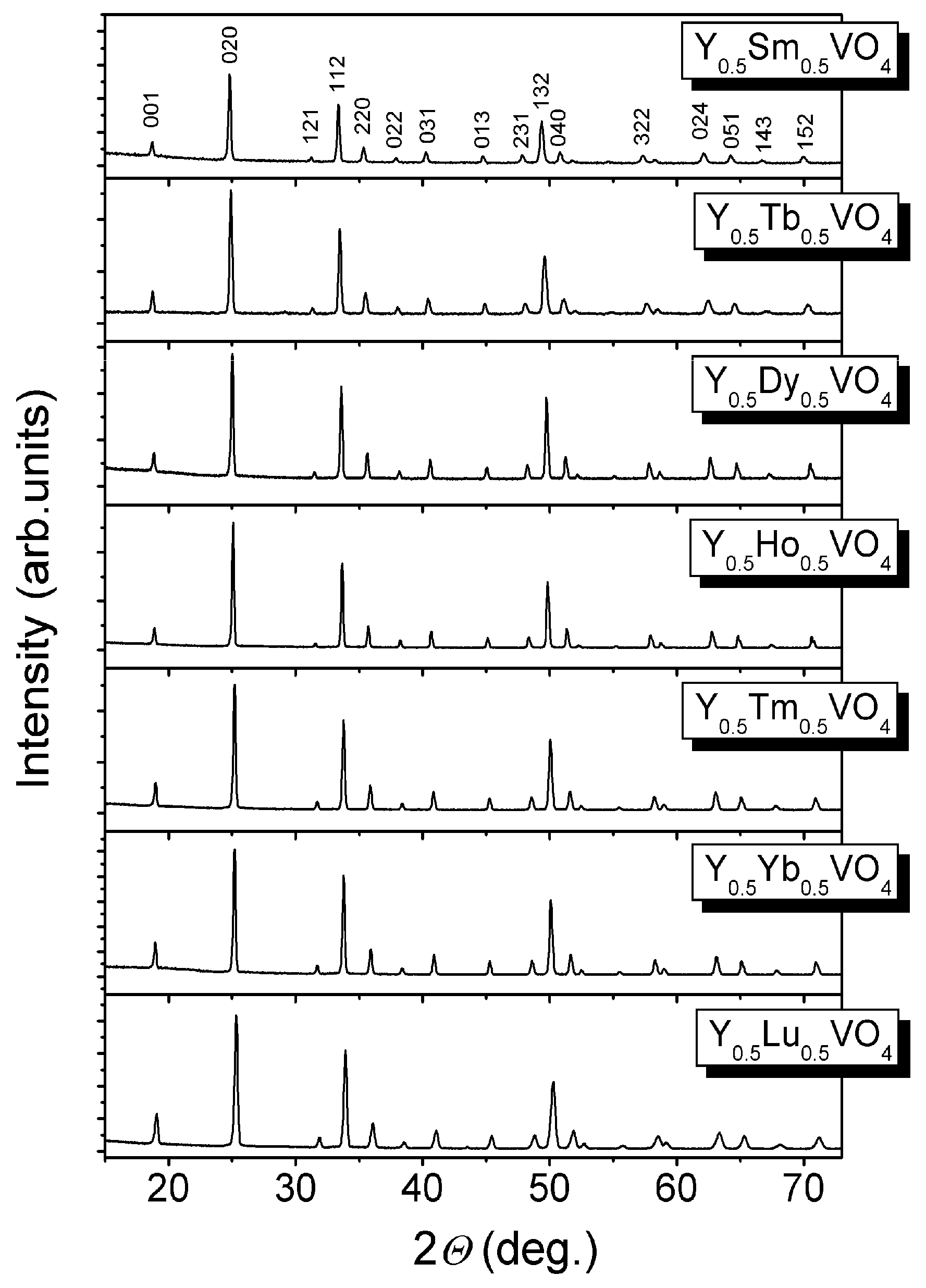
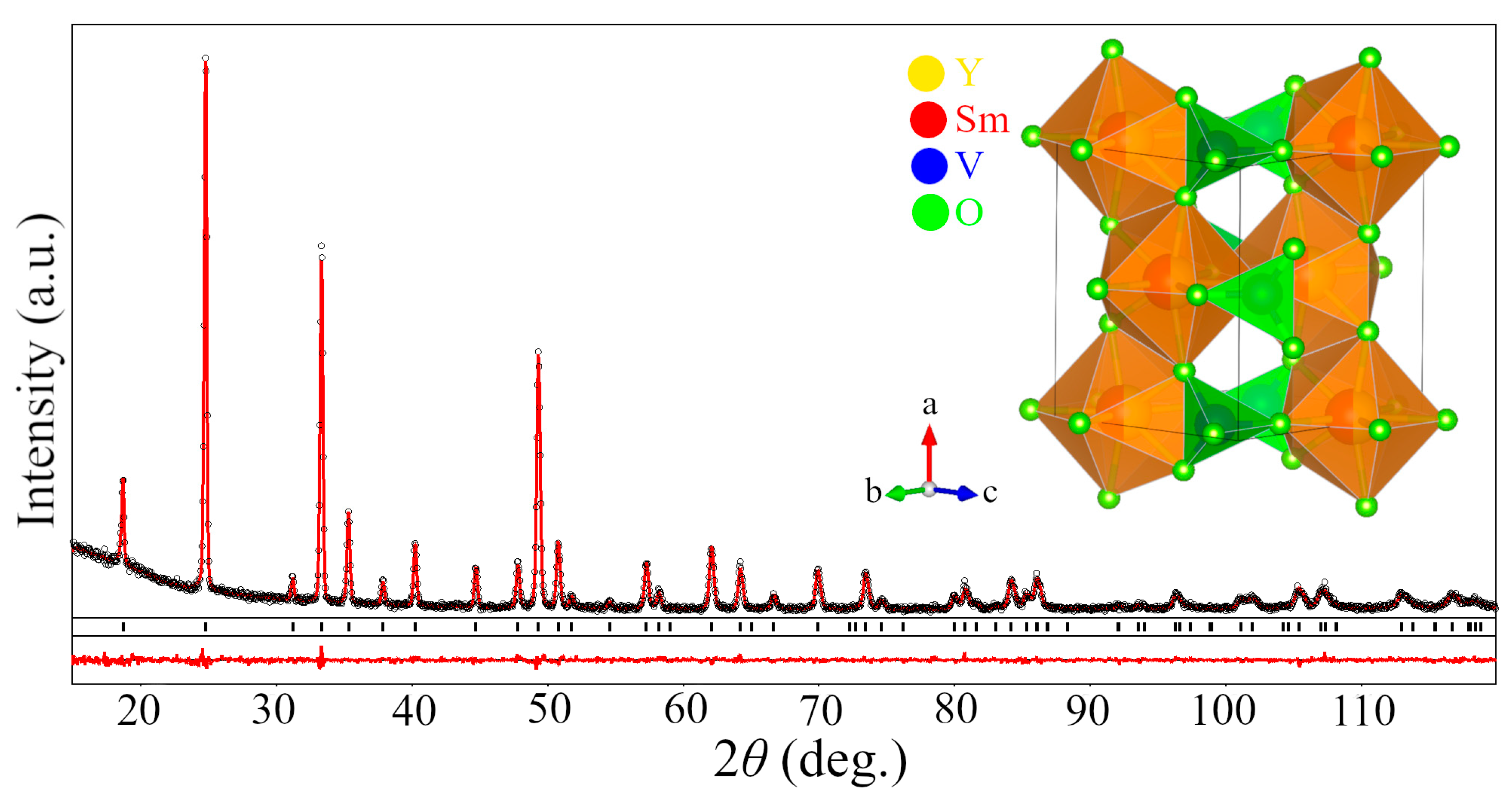
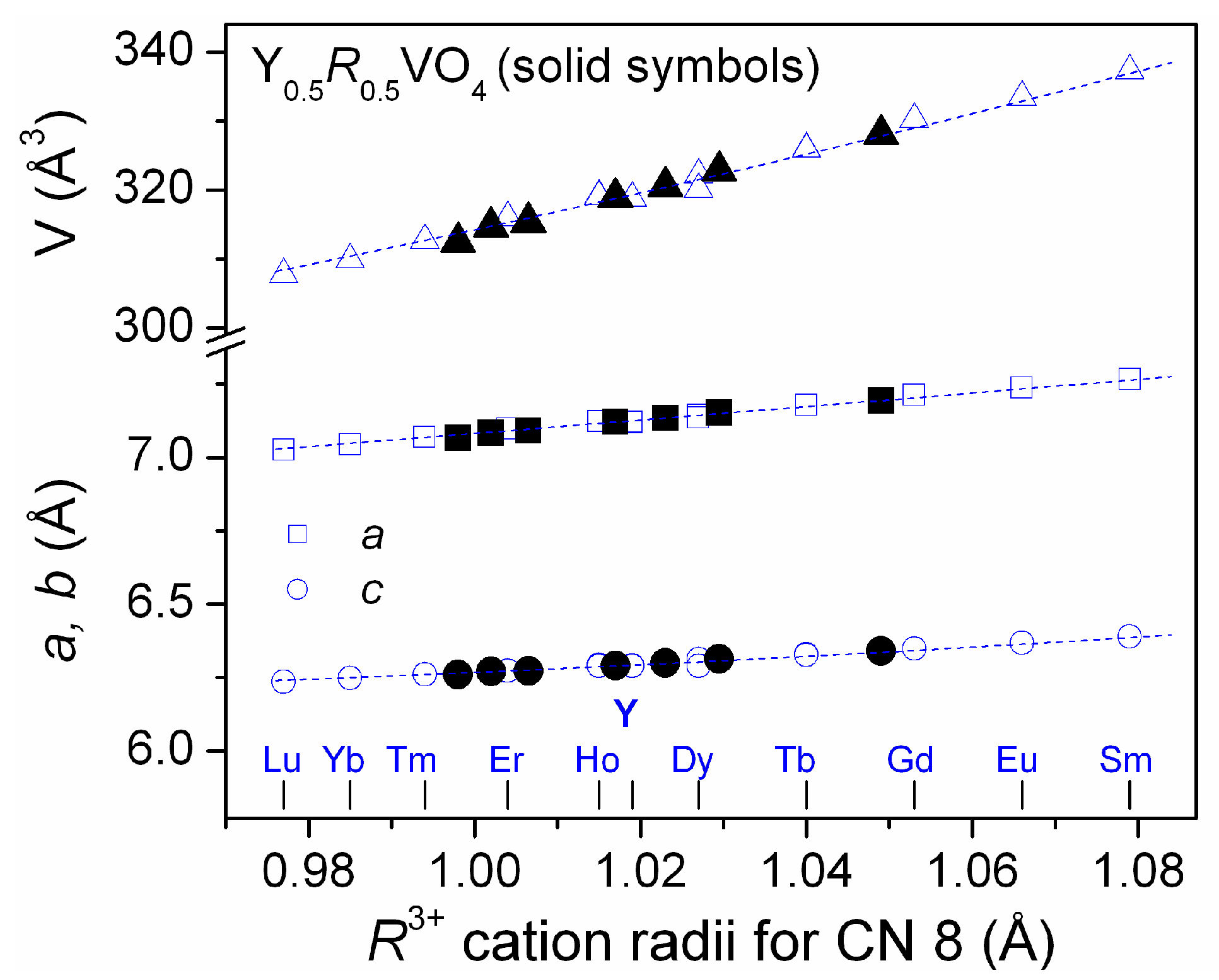
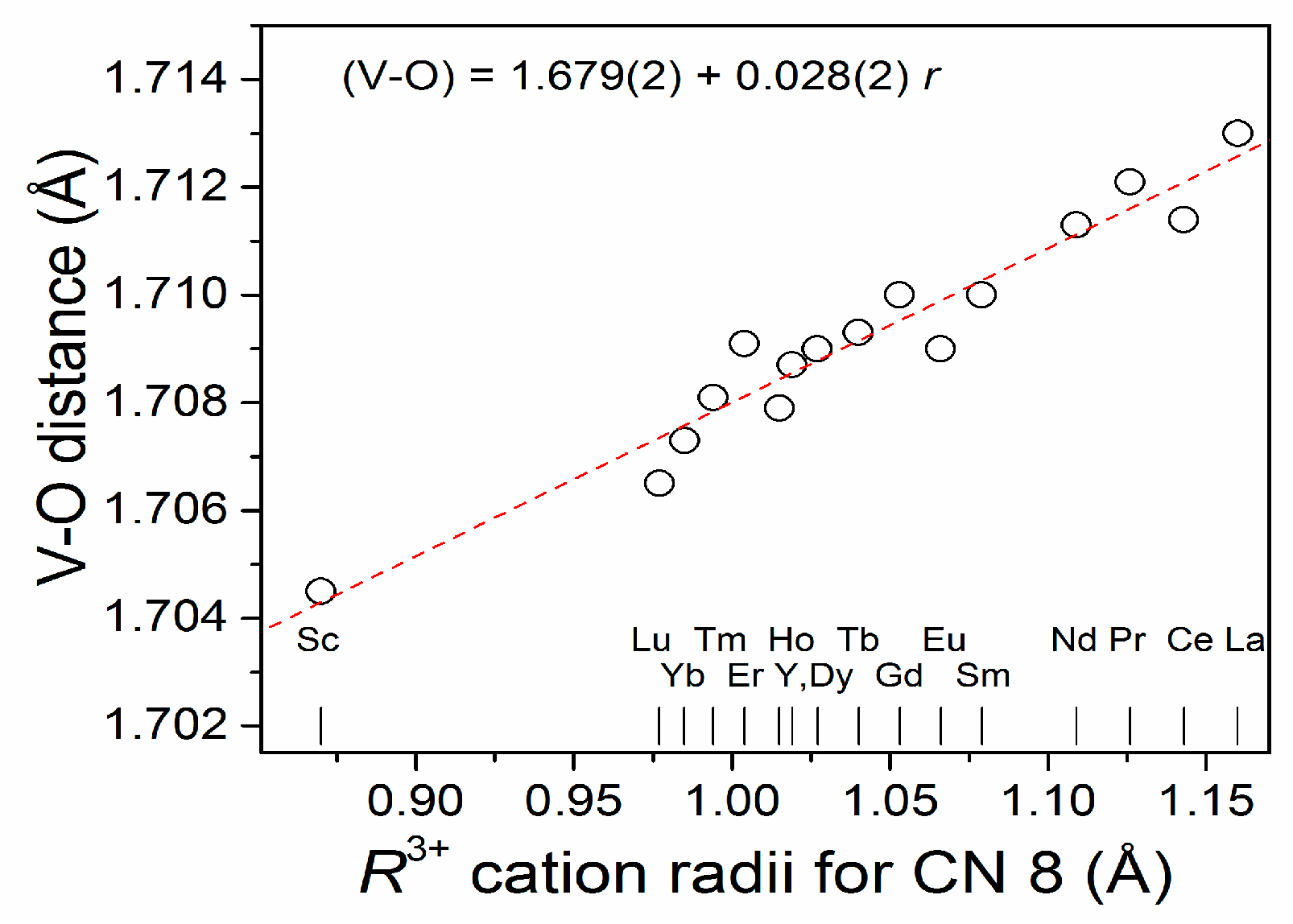
2.1.1. Mixed Vanadates Y0.5R0.5VO4

According to X-ray powder diffraction examination, all the Y0.5R0.5VO4 (R = Sm, Tb, Dy, Ho, Tm, Yb, Lu) vanadates adopt tetragonal zircon-type structures (Figure 1). No traces of the impurity phases were detected. Detectable broadening of the diffraction maxima is evidently associated with the microstrains <ε> caused by dispersion of interplanar distances <Δd>/d and possible nano-crystalline nature of the powders obtained. The colour of the Y0.5R0.5VO4 samples synthesized at 1273 K varied between tan or beige, depending on composition.
Phase purity and crystal structure of the materials synthesized were further proved by the full profile Rietveld refinement technique. As a starting model for the refinement, the atomic positions in the YVO4 structure obtained from neutron powder diffraction data were used [13]. The Y and R atoms are assuming to occupy the same 4a atomic positions in Y0.5R0.5VO4 structures in a random way. In the refinement procedure, the lattice constants, positions of oxygens and displacement parameters of all atoms (adp’s) were refined together with profile parameters and corrections for the adsorption and instrumental sample shift. In all cases, an excellent fit between calculated and experimental diffraction profiles was obtained. As an example, Figure 2 demonstrates graphical results of Rietveld refinement of Y0.5Sm0.5VO4 structure in space group I41/amd.
The refined structural parameters of the Y0.5R0.5VO4 (R = Sm, Tb, Dy, Ho, Tm, Yb, Lu) solid solutions and corresponding interatomic distances and angles in the zircon-type Y0.5R0.5VO4 structures are summarized in Table 1 and Table 2, respectively.
Table 1 contains also the values of average grain size, D, and microstrains <ε> = <Δd>/d, evaluated from the analysis of angular dependence of XRD peak broadening by using full profile refinement procedure. The smallest grain size (92–93 nm) was detected in the Y0.5Dy0.5VO4 and Y0.5Lu0.5VO4 materials, whereas the biggest one (202 nm)—in Y0.5Tb0.5VO4 sample. No obvious dependence of the grain size on the sample composition was observed. The obtained microstrain values <Δd>/d are varied over the range of 0.032–0.207%, predictable being lower for the Y0.5Dy0.5VO4 and Y0.5Ho0.5VO4 materials due to minor mismatch in ionic radii between Y3+ ions and Dy3+ or Ho3+ species.
The obtained structural parameters of the Y0.5R0.5VO4 (R = Sm, Tb, Dy, Ho, Tm, Yb, Lu) solid solutions agree well with the literature data for the parent YVO4 and RVO4 compounds [13,14]. Figure 3 demonstrates evolution of the unit cell dimensions of the Y0.5R0.5VO4 materials synthesized in comparison with the known data for pure RVO4 compounds proving the formation of continuous solid solutions in the YVO4RVO4 pseudo-binary systems.

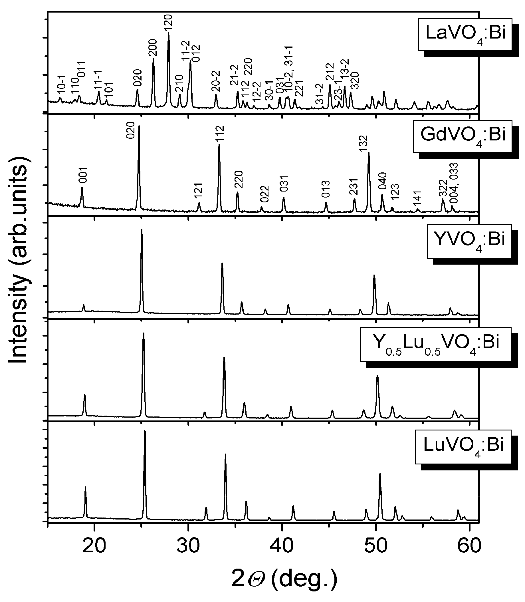
2.1.2. Bi3+-Doped Vanadates RVO4 (R = La, Gd, Y, Y0.5Lu0.5, Lu)

The analysis of XRD patterns of the synthesized Bi-doped vanadates revealed a monazite-type monoclinic structure of LaVO4:Bi and zircon-type tetragonal structure of all other RVO4:Bi (R = Gd, Y, Y0.5Lu0.5, Lu) materials (Figure 4). No detectable amount of the foreign phase(s) was detected within the sensitivity limits of the technique used.
The full profile Rietveld refinement confirms phase purity and crystal structure of the synthesized RVO4:Bi materials (Figure 5). Atomic positions in the monoclinic LaVO4 [16] and tetragonal YVO4 [13] structures were used as starting models for the refinement of corresponding Bi3+-doped orthovanadates. In the refinement procedure it was assumed that Bi3+ species partially substitute R3+ cations in the corresponding structures. Refinement of the lattice parameters, positional and displacement parameters of atoms together with profile parameters in space groups P21/n for LaVO4:Bi and I41/amd for other RVO4:Bi materials shows excellent agreement between calculated and experimental diffraction profiles (see Figure 5 as an example) and led to final structural parameters and residuals presented in Table 3.
Evaluation of microstructural parameters of Bi-doped materials lead to the average grain size, D, of 145, 138, 207 and 251 nm for the LaVO4:Bi, GdVO4:Bi, YVO4:Bi and LuVO4:Bi samples (Table 3). The smaller D values of 98 nm is observed for the mixed yttrium-lutetium vanadate (Y,Lu)VO4:Bi. In comparison with “single rare earth” vanadates RVO4:Bi, the latest material also shows considerably higher values of microstrains <ε> (Table 3) due to significant dispersion of interplanar distances <Δd>/d.

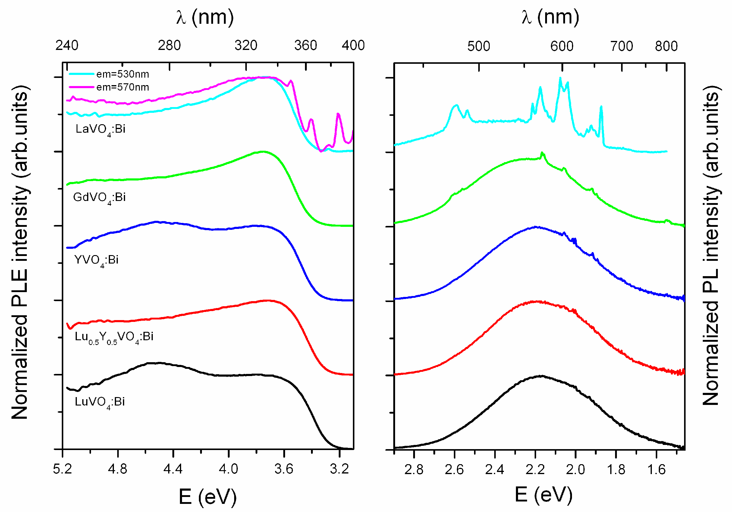
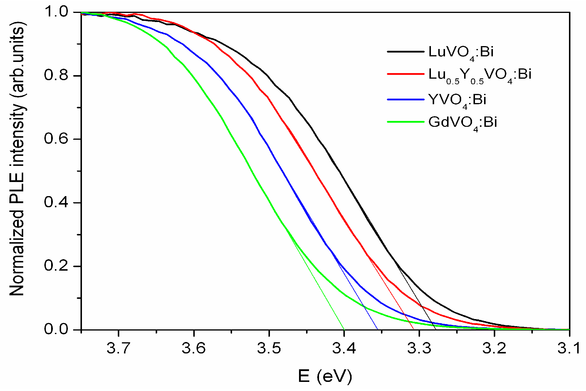
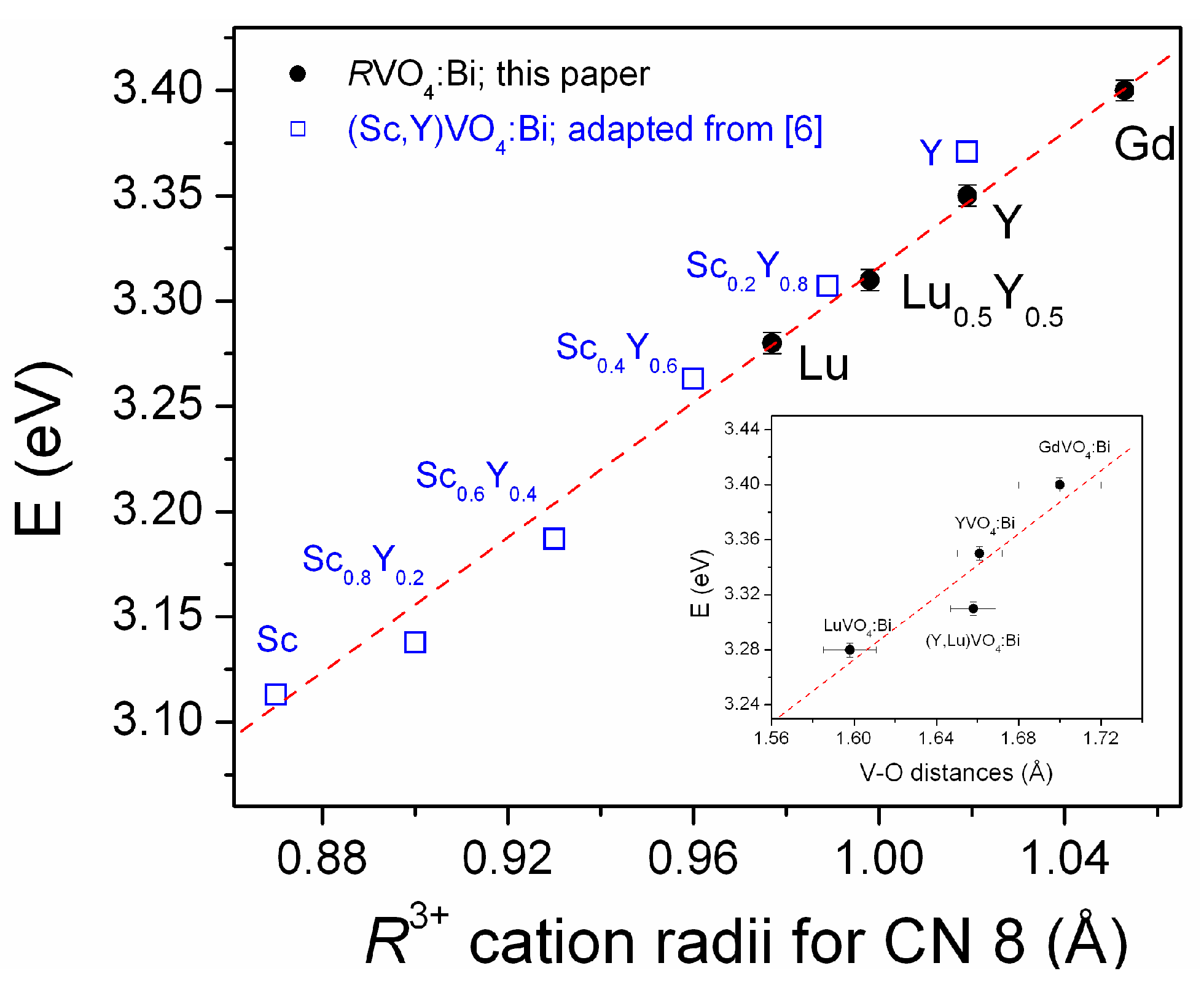
2.2. Luminescence and Optical Studies of the Bi-Doped Materials

The colour of the RVO4:Bi materials obtained depends on the sample composition and annealing temperature and varies between white or slightly yellowish to tan. The Y- and Lu-based RVO4:Bi vanadates prepared at 1073 K were white or slightly yellowish, whereas LaVO4:Bi and GdVO4:Bi materials synthesized at 1273 K were brownish evidently due to the partial formation of mixed-valence compounds [17]. It was reported [18] that there is a linear dependence between the intensity of yellow coloration of YVO4 and the EPR signal of V4+ ion adjacent to an oxygen vacancy. The increase of V4+ ion content quenches the luminescence of the prepared samples. Therefore before the luminescence measuring the samples were washed with 0.1 M water solution of NaOH as it is recommended in the literature [19] to eliminate these adverse phenomena.
The room temperature photoluminescence (PL) and photoluminescence excitation (PLE) spectra of RVO4:Bi3+ samples are shown in Figure 6. These spectra match well with the results reported earlier for YVO4:Bi3+ (see e.g., [20]) and reveal a broad emission band in visible range with the maximum near 570 nm. The excitation spectrum represents a complex broad band, in which the maxima near 275 and 330 nm can be distinguished, with the excitation edge located at 350–380 nm. The emission band with maximum at 570 nm, which is independent of excitation wavelength, is believed to be caused by excitons localized near isolated Bi3+ ions (see [21] and references herein). Similar emission of self-trapped excitons was also observed in other complex oxide compounds (see e.g., [22,23]). The excitation band at 275 nm most probably is caused by the spin-forbidden transition of VO43− groups (1A11T1) [21,24,25], while the 330 nm band can be attributed to the metal-to-metal charge transfer transition in Bi3+–V5+ complex [21,26]. The transitions from the 1S0 ground state to the 3P1 excited state of Bi3+ ions should also contribute to the excitation band below 330 nm [27].
Besides the broad emission band at 570 nm related to Bi3+, the photoluminescence spectra of the studied (Gd,Y,Lu)VO4:Bi3+ samples contains also some minor lines attributed to traces of Sm3+ ions in the studied samples (see e.g., [28,29] for comparison). The Sm3+ traces most evidently came as a contamination of the equipment after earlier syntheses of some Sm-based materials. At the same time, for the LaVO4:Bi3+ sample, the Bi-related emission was revealed to be of low intensity, so the unwanted emission of Sm3+ impurity dominates (see Figure 6). Nevertheless, the PLE spectrum for LaVO4:Bi3+ is similar to these observed in other (Gd,Y,Lu)VO4:Bi3+ samples.
The washing in NaOH changes the color of the as-synthesized samples from yellowish-brown to snow-white as a result of bleaching of the broad absorption at 370–550 nm (see Figure 7 for details). Our photoluminescence quantum yield (QY) measurements testify that the Bi-doped samples washed in NaOH have got also a higher QY in comparison with the corresponding as-synthesized (yellowish) samples. For example, the QY of the YVO4:Bi at 330 nm excitation was revealed to be 36 ± 1% and 70 ± 3% for the as-synthesized and washed samples, respectively.
One can notice that the excitation edge position of the Bi-doped samples shifts towards higher energies with increasing of ionic radius of the rare-earth ion (from Lu3+ to Gd3+). The precise estimation of the excitation edge position was done, as shown in Figure 8. In the case of LaVO4:Bi, the excitation edge position is distorted by ff transitions of Sm3+ impurity observed in this particular sample (see PLE spectrum for LaVO4:Bi in Figure 6).
The electronic structure calculations suggested that the valence band of the vanadates is formed mainly by 2p oxygen states and the bottom of the conduction band is formed by 3d states of vanadium [30,31]. At the same time, the 6s states of the bismuth ions are located slightly above the valence band of YVO4, while the 6p states of bismuth ions are included inside conduction band and are mixed with vanadium 3d states [26,32,33]. Therefore, the luminescence excitation edge of RVO4:Bi3+ can be formed by the charge transfer transitions from 6s2 states of Bi3+ to 3d0 states of V5+ ions.
The excitation edge position for the studied RVO4:Bi samples as a function of the RE ionic radius is presented in Figure 9 together with the literature data for the isostructural (Sc,Y)VO4:Bi materials. The excitation edge positions for Bi-doped scandium-yttrium orthovanadates were estimated similarly as in Figure 7 from the excitation spectra of (Y1−yScy)VO4:Bi compounds presented in Reference [6]. These results testify that the excitation edge position of the Bi-related emission, which should correlate with the energy band gap of the material, depends linearly on the RE ionic radius as well as from the V–O distance in the zircon-type RVO4 structures. Knowing that the energy band gap in the vanadates is determined by O and V, and the V–O distances in the zircon-type vanadates are increasing with the RE ionic radius (see Figure 10), it can be assumed that the energy band gap in RVO4 series increases gradually with the RE ionic radius increase.

3. Materials and Methods

The studied materials were obtained in the form of nano-and submicro-crystalline powders by a standard solid-state reaction technique. For the preparation of mixed YVO4-based orthovanadates with nominal compositions Y0.5R0.5VO4 (R = Sm, Tb, Dy, Ho, Tm, Yb, Lu), stoichiometric amounts of yttrium oxide Y2O3 (99.9%, Alfa Aesar, Ward Hill, MA, USA), ammonium metavanadate NH4VO3 (99%, LLC Sfera Sim, Lviv, Ukraine) and RE oxides (R2O3 and Tb4O7, all 99.9%, Alfa Aesar) were carefully mixed in agate mortar, placed on alumina crucibles and heat treated in air subsequently at 1123 K for 4 h, at 1173 K for 3 h and at 1273 K for 6 h with intermediate regrinding of the product.
Similar experimental technique has been applied for a synthesis of the selected Bi3+-doped vanadates. Bismuth oxide Bi2O3 was used as a doping precursor for preparation of the samples with nominal compositions La0.99Bi0.01VO4, Gd0.99Bi0.01VO4, Y0.99Bi0.01VO4, Lu0.99Bi0.01VO4 and Y0.49Lu0.50Bi0.01VO4 assuming that Bi substitutes RE ions. The temperature regime for synthesis of the LaVO4:Bi and GdVO4:Bi materials was the same as described above, whereas the Y- and Lu-based vanadates were prepared by heat treatment of corresponding precursors in air at 1073 K for 14 h with one intermediate regrinding of the products.
Phase composition, crystal structure and microstructural parameters of the samples were studied by means of the X-ray powder diffraction (XRD) technique. The experimental diffraction data were collected at the modernized DRON-3M diffractometer (Bourevestnik, Saint Petersburg, Russia) (Cu Kα-radiation) in the 2θ range 15–120° with the 2θ step of 0.02° and typical exposition 5–8 s per step. The refinement of crystal structure parameters, as well as evaluation of microstructural parameters of the powders from angular dependence of the Bragg’s maxima broadening, has been performed by full profile Rietveld method using the WinCSD program package [36]. The LaB6 external standard was used for the correction of instrumental broadening. The observed peak shapes were modelled using the pseudo-Voigt function which is a convolution of Gaussian profile and Lorentzian profile functions.
The photoluminescence (PL) and photoluminescence excitation (PLE) spectra were measured at room temperature using a Horiba/Jobin-Yvon Fluorolog-3 spectrofluorometer (Edison, NJ, USA) with a 450 W continuous xenon lamp as an excitation source and a Hamamatsu R928P PMT detector (Hamamatsu Photonics K.K., Hamamatsu City, Shizuoka, Japan) in photon counting mode. The measured PLE spectra were corrected for the xenon lamp emission spectrum. The PL spectra were corrected for the spectral response of the spectrometer system used. The photoluminescence quantum yield (QY) was evaluated as the ratio of the number of emitted photons to that of the absorbed photons similarly as it was described elsewhere [37]. The optical absorption spectra were derived from the diffuse reflectance spectra using the Kubelka-Munk transformation. The diffuse reflectance spectra of the powder samples were measured using a Varian Cary 5000 spectrophotometer (Palo Alto, CA, USA.) with external diffuse reflectance accessory DRA-2500.

4. Conclusions

The phase-pure nano- and submicro-crystalline powders of new mixed orthovanadates Y0.5R0.5VO4 (R = Sm, Tb, Dy, Ho, Tm, Yb, Lu) and some Bi3+-doped RVO4 (R = La, Gd, Y, Lu) materials have been prepared by a facile solid state reaction route at 1073–1273 K from the rare earth oxides and ammonium metavanadate as initial reagents and Bi2O3 oxide as a doping precursor. The precise crystal structure parameters including unit cell dimensions, positional and displacement parameters of atoms, as well as microstructural parameters of the powders such as average grain size and microstrains, were derived from the experimental X-ray powder diffraction data by using full-profile Rietveld refinement technique. Based on comparison of the crystal structure parameters of the new mixed orthovanadates Y0.5R0.5VO4 with the parent YVO4 and RVO4 compounds, a formation of continuous solid solutions Y1−xRxVO4 in the YVO4RVO4 pseudo-binary systems (R = Sm, Tb, Dy, Ho, Tm, Yb, Lu) has been proved.
It was shown that the excitation edge position of the Bi-related emission in RVO4:Bi orthovanadates, which correlates with the energy band gap of the materials, increases linearly with increment of the V–O distance as a result of the increasing RE ionic radius in the series from Sc3+ to Gd3+.

Author Contributions

A.T., V.H. and I.L. synthesized the samples by solid state reactions technique, contributed to the data evaluation and to the manuscript writing. L.V. performed the laboratory X-ray powder diffraction measurements, made the structural characterization of the samples and wrote the manuscript. V.T. and Y.Z. carried out the luminescence studies of the Bi-doped materials and contributed to the manuscript writing. All authors read and approved the final manuscript.

Funding

This research was funded by the Ministry of Education and Science of Ukraine (project DB/Feryt, N 0118U000264), and partially by the Polish National Science Center (project 2015/17/B/ST5/01658).

Conflicts of Interest

The authors declare no conflict of interest.

References

  1. Kolitsch, U.; Holtstam, D. Crystal chemistry of REEXO4 compounds (X = P, As, V). II. Review of REEXO4 compounds and their stability fields. Eur. J. Mineral. 2004, 16, 117–126. [Google Scholar] [CrossRef]
  2. Koechner, W. Solid-State Laser Engineering; Springer: Berlin/Heidelberg, Germany, 2006; p. 69. ISBN 978-0-387-29094-2. [Google Scholar]
  3. Kalai Selvan, R.; Gedanken, A.; Anilkumar, P.; Manikandan, G.; Karunakaran, C. Synthesis and characterization of rare earth orthovanadate (RVO4; R = La, Ce, Nd, Sm, Eu & Gd) Nanorods/Nanocrystals/Nanospindles by a facile sonochemical method and their catalytic properties. J. Clust. Sci. 2009, 20, 291–305. [Google Scholar] [CrossRef]
  4. Kaminskii, A.A. Tetragonal vanadates REVO4 (RE = Ln (Ce–Lu), Y)—A novel class of SRS-active crystals. Dokl. Phys. 2013, 58, 165–168. [Google Scholar] [CrossRef]
  5. Chen, L.; Chen, K.J.; Lin, C.C.; Chu, C.I.; Hu, S.F.; Lee, M.H.; Liu, R.S. Combinatorial approach to the development of a single mass YVO4:Bi3+, Eu3+ Phosphor with red and green dual colors for high color rendering white light–emitting diodes. J. Comb. Chem. 2010, 12, 587–594. [Google Scholar] [CrossRef] [PubMed]
  6. Kang, F.; Zhang, H.; Wondraczek, L.; Yang, X.; Zhang, Y.; Lei, D.Y.; Peng, M. Band-gap modulation in single Bi3+-doped yttrium–scandium–niobium vanadates for color tuning over the whole visible spectrum. Chem. Mater. 2016, 28, 2692–2703. [Google Scholar] [CrossRef]
  7. Liu, H.; Yuan, J.; Jiang, Z.; Shangguan, W.; Einaga, H.; Teroaka, Y. Novel photocatalyst of V–based solid solutions for overall water splitting. J. Mater. Chem. 2011, 21, 16535–16543. [Google Scholar] [CrossRef]
  8. Huang, X.Y.; Wang, J.X.; Yu, D.C.; Ye, S.; Zhang, Q.Y. Spectral conversion for solar cell efficiency enhancement using YVO4:Bi3+, Ln3+ (Ln = Dy, Er, Ho, Eu, Sm, and Yb) phosphors. J. Appl. Phys. 2011, 109, 113526. [Google Scholar] [CrossRef]
  9. Errandonea, D.; Garg, A.B. Recent progress on the characterization of the high-pressure behaviour of AVO4 orthovanadates. Prog. Mater. Sci. 2018, 97, 123–169. [Google Scholar] [CrossRef]
  10. International Centre for Diffraction Data (ICDD). PDF-4+ 2010 (Database); Kabekkodu, S., Ed.; International Centre for Diffraction Data: Newtown Square, PA, USA, 2010. [Google Scholar]
  11. Isasi, J.; Veiga, M.L.; Laureiro, Y.; Saez-Puche, R.; Pico, C. Synthesis, structural determination and magnetic behavior of YxGd1−xVO4 phases (x = 0.25, 0.50, 0.75). J. Alloys Compd. 1991, 177, 143–147. [Google Scholar] [CrossRef]
  12. Milligan, W.O.; Vernon, L.W. Crystal Structure of Heavy metal orthovanadates. J. Phys. Chem. 1952, 56, 145–148. [Google Scholar] [CrossRef]
  13. Chakoumakos, B.C.; Abraham, M.M.; Boatner, L.A. Crystal structure refinements of zircon-type MVO4 (M = Sc, Y, Ce, Pr, Nd, Tb, Ho, Er, Tm, Yb, Lu). J. Solid State Chem. 1994, 109, 197–202. [Google Scholar] [CrossRef]
  14. Aldred, A.T. Cell Volumes of APO4, AVO4 and ANbO4 Compounds, where A = Sc, Y, La–Lu*. Acta Crystallogr. B 1984, 40, 569–574. [Google Scholar] [CrossRef]
  15. Shannon, R.D. Revised effective ionic radii and systematic studies of interatomic distances in halides and chalcogenides. Acta Crystallogr. A 1976, 32, 751–757. [Google Scholar] [CrossRef]
  16. Rice, C.E.; Robinson, W.R. Lanthanum orthovanadate. Acta Crystallogr. B 1976, 32, 2232–2233. [Google Scholar] [CrossRef] [Green Version]
  17. Takeshita, S.; Ogata, H.; Isobe, T.; Sawayama, T.; Niikura, S. Effects of citrate additive on transparency and photostability properties of YVO4:Bi3+, Eu3+ nanophosphor. J. Electrochem. Soc. 2010, 157, J74. [Google Scholar] [CrossRef]
  18. Garces, N.Y.; Stevens, K.T.; Foundos, G.K.; Halliburton, L.E. Electron paramagnetic resonance and optical absorption study of V4+ centres in YVO4 crystals. J. Phys. Condens. Matter 2004, 16, 7095–7106. [Google Scholar] [CrossRef]
  19. Cooper, J.K.; Scott, S.B.; Ling, Y.; Yang, J.; Hao, S.; Li, Y.; Toma, F.M.; Stutzman, M.; Lakshmi, K.V.; Sharp, I.D. Role of Hydrogen in Defining the n-Type Character of BiVO4 Photoanodes. Chem. Mater. 2016, 28, 5761–5771. [Google Scholar] [CrossRef]
  20. Datta, R. Bismuth in yttrium vanadate and yttrium europium vanadate phosphors. J. Electrochem. Soc. 1967, 114, 1057–1063. [Google Scholar] [CrossRef]
  21. Cavalli, E.; Angiuli, F.; Mezzadri, F.; Trevisani, M.; Bettinelli, M.; Boutinaud, P.; Brik, M.G. Tunable luminescence of Bi3+-doped YPxV1−xO4 (0 ≤ x ≤1). J. Phys. Condens. Matter 2014, 26, 385503. [Google Scholar] [CrossRef] [PubMed]
  22. Zazubovich, S.; Krasnikov, A.; Zorenko, Y.; Gorbenko, V.; Babin, V.; Mihokova, E.; Nikl, M. Luminescence of Pb- and Bi-Related Centers in Aluminum Garnet, Perovskite, and Orthosilicate Single-Crystalline Films. In Nanocomposite, Ceram. Thin Film Scintill; Nikl, M., Ed.; Pan Stanford Publishing: Singapore, 2017; pp. 227–302. ISBN 978-981-4745-22-2. [Google Scholar]
  23. Krasnikov, A.; Luchechko, A.; Mihokova, E.; Nikl, M.; Syvorotka, I.I.; Zazubovich, S.; Zhydachevskii, Y. Origin of Bi3+-related luminescence in Gd3Ga5O12:Bi epitaxial films. J. Lumin. 2017, 190, 81–88. [Google Scholar] [CrossRef]
  24. Barendswaard, W.; Weber, R.T.; van der Waals, J.H. An EPR study of the luminescent triplet state of VO3−4 in YVO4 and YP0.96V0.04O4 single crystals at 1.2 K. J. Chem. Phys. 1987, 87, 3731. [Google Scholar] [CrossRef]
  25. Matos, M.; Rocha, L.; Nassar, E.; Verelst, M. Influence of Bi3+ ions on the excitation wavelength of the YVO4:Eu3+ matrix. Opt. Mater. 2016, 62, 12–18. [Google Scholar] [CrossRef]
  26. Boutinaud, P. Revisiting the spectroscopy of the Bi3+ ion in oxide compounds. Inorg. Chem. 2013, 52, 6028–6038. [Google Scholar] [CrossRef] [PubMed]
  27. Blasse, G.; Bril, A. Investigations on Bi3+-Activated Phosphors. J. Chem. Phys. 1968, 48, 217–222. [Google Scholar] [CrossRef]
  28. Atuchin, V.V.; Aleksandrovsky, A.S.; Chimitova, O.D.; Diao, C.P.; Gavrilova, T.A.; Kesler, V.G.; Molokeev, M.S.; Krylov, A.S.; Bazarov, B.G.; Bazarova, J.G.; et al. Electronic structure of β-RbSm(MoO4)2 and chemical bonding in molybdates. Dalton Trans. 2015, 44, 1805. [Google Scholar] [CrossRef] [PubMed]
  29. Atuchin, V.V.; Aleksandrovsky, A.S.; Molokeev, M.S.; Krylov, A.S.; Oreshonkov, A.S.; Zhou, D. Structural and spectroscopic properties of self-activated monoclinic molybdate BaSm2(MoO4)4. J. Alloys Compd. 2017, 729, 843–849. [Google Scholar] [CrossRef]
  30. Panchal, V.; Errandonea, D.; Segura, A.; Rodrguez-Hernandez, P.; Muoz, A.; Lopez-Moreno, S.; Bettinelli, M. The electronic structure of zircon-type orthovanadates: Effects of high-pressure and cation substitution. J. Appl. Phys. 2011, 110, 043723. [Google Scholar] [CrossRef]
  31. Garg, A.B.; Errandonea, D.; Rodrigues-Hernandez, P.; Munoz, A. ScVO4 under non-hydrostatic compression: A new metastable polymorph. J. Phys. Condens. Matter 2017, 29, 055401. [Google Scholar] [CrossRef] [PubMed]
  32. Dolgos, M.R.; Paraskos, A.M.; Stoltzfus, M.W.; Yarnell, S.C.; Woodward, P.M. The electronic structures of vanadate salts: Cation substitution as a tool for band gap manipulation. J. Solid State Chem. 2009, 182, 1964–1971. [Google Scholar] [CrossRef]
  33. Mahlik, S.; Amer, M.; Boutinaud, P. Energy level structure of Bi3+ in Zircon and Scheelite polymorphs of YVO4. J. Phys. Chem. C 2016, 120, 8261–8265. [Google Scholar] [CrossRef]
  34. Mullica, D.F.; Sappenfield, E.L.; Abraham, M.M.; Chakoumakos, B.C.; Boatner, L.A. Structural investigations of several LnVO4 compounds. Inorg. Chim. Acta 1996, 248, 85–88. [Google Scholar] [CrossRef]
  35. Oka, Y.; Yao, T.; Yamamoto, N.J. Hydrothermal synthesis of lanthanum vanadates: Synthesis and crystal structures of zircon-type LaVO4 and a new compound LaV3O9. Solid State Chem. 2000, 152, 486–491. [Google Scholar] [CrossRef]
  36. Akselrud, L.; Grin, Yu. WinCSD: Software package for crystallographic calculations (Version 4). J. Appl. Crystallogr. 2014, 47, 803–805. [Google Scholar] [CrossRef]
  37. Zhydachevskyy, Y.; Syvorotka, I.I.; Tsiumra, V.; Baran, M.; Lipińska, L.; Wierzbicka, A.; Suchocki, A. Quantum efficiency of the down-conversion process in Bi3+–Yb3+ and Ce3+–Yb3+ co-doped garnets. Sol. Energy Mater. Sol. Cells 2018, 185, 240–251. [Google Scholar] [CrossRef]
Figure 1. Representative parts of XRD patterns of the Y0.5R0.5VO4 (R = Sm, Tb, Dy, Ho, Tm, Yb, Lu) samples. The Miller indices are given for the zircon-type structure.
Inorganics 06 00094 g001
Figure 2. Graphical results of Rietveld refinement of Y0.5Sm0.5VO4 structure. Experimental XRD pattern (black circles) is shown in comparison with the calculated profile (red line). Short vertical bars indicate positions of Bragg’s maxima in space group I41/amd. Inset shows visualization of Y0.5Sm0.5VO4 zircon-type structure as chains of alternating VO4 tetrahedra and Y/SmO8 polyhedra.
Inorganics 06 00094 g002
Figure 3. Lattice parameters and unit cell volumes of the Y0.5R0.5VO4 (R = Sm, Tb, Dy, Ho, Tm, Yb, Lu) series (solid symbols) and the parent RVO4 compounds [13,14] (open symbols) as a function of eight-coordinated R3+ cations after Shannon [15].
Inorganics 06 00094 g003
Figure 4. Representative parts of XRD patterns of the Bi3+-doped orthovanadates RVO4. The Miller indices are given for the monazite-type LaVO4:Bi and zircon-type RVO4:Bi structures.
Inorganics 06 00094 g004
Figure 5. Graphical results of Rietveld refinement of Bi-doped LaVO4 (top) and Y0.5Lu0.5VO4 (bottom) structures. Experimental XRD patterns (black circles) are shown in comparison with the calculated profiles (red and blue lines, respectively). Short vertical bars on the top and bottom panels indicate positions of Bragg’s maxima in the monoclinic P21/n and tetragonal I41/amd structures, respectively. Insets shows polyhedral views of monazite-type LaVO4:Bi and zircon-type Y0.5Lu0.5VO4:Bi structures.
Inorganics 06 00094 g005
Figure 6. Normalized PLE (left) and PL (right) spectra of RVO4:Bi (1%) samples (R = La, Gd, Y, Y0.5Lu0.5 and Lu) measured at room temperature.
Inorganics 06 00094 g006
Figure 7. Comparison of the optical absorption spectra and color of YVO4:Bi, Y0.5Lu0.5VO4:Bi and LuVO4:Bi samples, as-synthesized and washed in NaOH.
Inorganics 06 00094 g007
Figure 8. Estimation of the excitation edge of the Bi-related emission (λem = 570 nm) registered in the studied RVO4:Bi(1%) samples at room temperature.
Inorganics 06 00094 g008
Figure 9. The room-temperature excitation edge position for the tetragonal zircon-type RVO4:Bi vanadates vs. the RE ionic radius. The solid line represents a linear fit of our data for the Bi-doped Lu, Lu0.5Y0.5, Y and Gd materials. The inset represents the excitation edge position as a function of the V–O distances in the RVO4:Bi samples studied. LaVO4:Bi drops out from this consideration because it belongs to the monoclinic monazite-type structure.
Inorganics 06 00094 g009
Figure 10. The V–O distances in the tetragonal zircon-type orthovanadate series as a function of eight-coordinated R3+ cations after Shannon [15]. Corresponding values were evaluated from the structural data of RVO4 compounds published in References [13,34,35].
Inorganics 06 00094 g010
Table 1. Lattice parameters, fractional coordinates and isotropic adp’s in Y0.5R0.5VO4 series and microstructural parameters of corresponding powders. Space group I41/amd; Y(R) in 4a (0 3/4 1/8); V in 4b (0 1/4 3/8), O in 16h (0 y z).
Parameters, ResidualsR in Y0.5R0.5VO4
SmTbDyHoTmYbLu
a, Å7.1933(4)7.1509(3)7.1337(2)7.1206(1)7.0907(2)7.0815(1)7.0655(2)
c, Å6.3398(4)6.3126(3)6.2991(2)6.2901(1)6.2724(2)6.2684(2)6.2588(2)
V, Å3328.05(5)322.79(5)320.56(2)318.92(2)315.36(2)314.35(2)312.45(3)
Biso(Y/R), Å20.59(4)0.62(3)0.78(3)1.17(3)0.88(2)0.84(2)0.67(2)
Biso(V), Å20.71(12)0.45(7)0.64(6)0.48(5)0.80(4)0.65(4)0.52(4)
y/b(O)0.4302(13)0.4278(7)0.4313(6)0.4308(5)0.4312(4)0.4320(5)0.4321(4)
z/c(O)0.2033(13)0.2071(7)0.2048(7)0.2054(5)0.2040(4)0.2062(5)0.2034(4)
Biso(O), Å21.1(3)1.42(15)1.55(14)1.29(10)1.48(8)1.38(9)1.51(9)
RI0.0300.0290.0210.0230.0220.0290.023
RP0.1240.1290.0950.0800.0820.0730.085
D, nm1082029215810612993
<ε>, %0.1210.1850.0320.0390.0900.0650.207
R0.0140.0450.0030.0420.0030.0060.044
Table 2. Selected interatomic distances (Å) and angles (o) in Y0.5R0.5VO4 structures.
SmTbDyHoTmYbLu
VO4 tetrahedra
V–O × 41.693(9)1.656(10)1.680(7)1.673(8)1.673(6)1.665(8)1.675(8)
O···O × 42.846(11)2.781(6)2.816(9)2.811(10)2.811(8)2.789(10)2.813(10)
O···O × 22.593(13)2.543(7)2.590(10)2.567(12)2.568(9)2.571(12)2.571(12)
O–V–O × 4114.41(3)114.22(2)113.93(3)114.30(3)114.27(3)113.81(3)114.26(3)
O–V–O × 299.99(3)100.34(2)100.88(3)100.20(3)100.25(3)101.11(3)100.27(3)
RO8 polyhedra
R–O × 42.353(9)2.361(11)2.328(7)2.331(8)2.315(7)2.312(8)2.300(8)
R–O × 42.453(9)2.452(10)2.450(7)2.437(8)2.430(6)2.442(8)2.425(8)
O···O × 22.593(13)2.543(7)2.590(10)2.567(12)2.568(9)2.571(12)2.571(12)
O···O × 42.767(11)2.810 (6)2.763(10)2.757(11)2.739(9)2.762(11)2.723(11)
O···O × 83.080(10)3.069(5)3.053(8)3.050(9)3.037(7)3.032(9)3.025(9)
O···O × 43.401(9)3.419(5)3.368(8)3.371(8)3.348(7)3.348(9)3.326(9)
O–R–O × 263.83(2)62.49(13)63.81(2)63.55(2)63.80(2)63.52(2)64.03(2)
O–R–O × 470.27(2)71.42(13)70.63(2)70.60(2)70.46(2)70.97(2)70.31(2)
O–R–O × 879.68(2)79.20(13)79.38(2)79.50(2)79.53(2)79.20(2)79.57(2)
O–R–O × 492.55(3)92.75(13)92.70(2)92.63(2)92.62(2)92.78(2)92.61(2)
Table 3. Lattice parameters, fractional coordinates and isotropic displacement parameters in RVO4:Bi structures and corresponding microstructural parameters of the powders.
Lattice Parameters, ResidualsAtoms, Sitesx/ay/bz/cBiso/eq, Å2
LaVO4:Bi, space group P21/n (D = 145 nm; <ε> = 0.048%)
a = 7.0396(2) Å
b = 7.2777(3) Å
c = 6.7212(2) Å
β = 104.856(2)°
La/Bi, 4e0.2768(3)0.1574(4)0.1037(4)0.97(3)
V, 4e0.3013(9)0.1643(11)0.6170(10)0.82(11)
O1, 4e0.253(3)0.008(3)0.431(3)0.5(5)
O2, 4e0.384(3)0.344(4)0.496(3)1.1(4)
O3, 4e0.478(3)0.099(3)0.826(3)1.4(5)
O4, 4e0.118(3)0.218(3)0.729(3)2.2(6)
RI = 0.031; Rp = 0.122
GdVO4:Bi, space group I41/amd (D = 138 nm; <ε> = 0.020%)
a = 7.2118(4) Å
c = 6.3484(4) Å
Gd/Bi, 4a03/41/80.95(9)
V, 4b01/43/81.0(3)
O1, 16h00.433(2)0.206(2)0.5(4)
RI = 0.029; Rp = 0.167
YVO4:Bi, space group I41/amd (D = 207 nm; <ε> = 0.032%)
a = 7.1188(1) Å
c = 6.2902(2) Å
Y/Bi, 4a03/41/81.20(4)
V, 4b01/43/80.47(7)
O1, 16h00.4319(7)0.2096(7)1.60(13)
RI = 0.035; Rp = 0.076
LuVO4:Bi, space group I41/amd (D = 251 nm; <ε> = 0.037%)
a = 7.0269(1) Å
c = 6.2341(1) Å
Lu/Bi, 4a03/41/81.47(2)
V, 4b01/43/80.40(6)
O1, 16h00.4252(8)0.2117(9)1.63(15)
RI = 0.035; Rp = 0.069
(Y,Lu)VO4:Bi, space group I41/amd (D = 98 nm; <ε> = 0.095%)
a = 7.0690(2) Å
c = 6.2595(3) Å
Y/Lu/Bi, 4a03/41/81.41(3)
V, 4b01/43/80.38(6)
O1, 16h00.4286(7)0.2035(7)1.69(14)
RI = 0.031; Rp = 0.077

