Article

Synthesis, Characterization, DFT Calculations, Biological Evaluation, and Molecular Docking of Cd(II) and Zn(II) Schiff Base Complexes: A Green Ball-Milling Approach


by
Hanan Alhussain
1 and
Rania R. Zaky
2,*
1
Department of Chemistry, Faculty of Science, Imam Mohammad Ibn Saud Islamic University (IMSIU), P.O. Box 90950, Riyadh 11623, Saudi Arabia
2
Department of Chemistry, Faculty of Science, Mansoura University, El-Gomhoria Street, Mansoura 35516, Egypt
*
Author to whom correspondence should be addressed.
Inorganics 2026, 14(7), 182; https://doi.org/10.3390/inorganics14070182
Submission received: 4 May 2026 / Revised: 24 June 2026 / Accepted: 29 June 2026 / Published: 8 July 2026

Abstract

A one-pot ball-milling chelation method was used to create Cd(II) and Zn(II) complexes of a 3-hydroxy-2-naphthoyl Schiff base derivative (H2L), which provided greater efficiency under milder reaction conditions. 1H NMR, 13C NMR, UV–Vis, IR, SEM, XRD, EDX, and elemental studies were used to characterize the isolated solid chelates. The optimized structures were confirmed by DFT theoretical calculations, which also yielded important energetic characteristics such as EHOMO and ELUMO. The three-dimensional crystal structures of HePG-2 (PDB ID: 5EQG), MCF-7 (PDB ID: 6NM0), and HeLa (PDB ID: 5IAE) were carefully analyzed after molecular docking experiments were carried out on the formed complexes utilizing Schrödinger’s LigPrep procedure with default parameters. Finally, the antibacterial, antioxidant, DNA-binding, and cytotoxic properties of the tested solid compounds were assessed.

1. Introduction

Schiff bases and their metal complexes have drawn increasing attention in current inorganic and bioinorganic research, owing to their varied biological activities and rich coordination chemistry [1,2]. These condensation products, which are formed when an active carbonyl compound reacts with a primary amine to produce an azomethine group, have been established as versatile active ligands for both transition metals and lanthanides since Hugo Schiff first labeled them in 1864 [3]. The donor set of Schiff bases, which is reasonably changeable, consists of imine nitrogen and other donor atoms (such as amine nitrogen, phenolic oxygen, and sulfur in thio-Schiff bases). This flexibility allows for the design of mono-, bi-, or polydentate ligands with a variety of geometries [4].
In coordination chemistry as well as materials science, Schiff bases exhibit strong affinity for a broad range of metals, forming mononuclear, binuclear and multinuclear chelates with various geometries from square-planar to octahedral [5,6]. Their rigid, conjugated backbones promote chelate stabilization, enabling applications in materials development, catalysis, and magnetism, including molecular ferromagnetism and spintronic materials [7]. The resulting metal-Schiff base frameworks often enhance the biological activity, involving fungicidal, antibacterial, anticancer, antidepressant-like, anti-inflammatory (antiphlogistic), nematicidal, and other enzymatic or catalytic properties [8,9,10]. Complexation can modify cellular uptake, pharmacokinetic properties, and interactions with biomolecular targets, thus tuning selectivity and efficacy [11]. In microbial systems, selected metal ions interact with membranes and cell walls and disrupt respiration as well as protein synthesis [12]. Schiff base ligands and their metal complexes can modify these interactions by influencing metal delivery, lipophilicity, and redox behavior, thus contributing to practical anticancer and antimicrobial activities [13,14,15,16].
In our current study, we use ball milling for inorganic synthesis to provide a clean, simple, solvent-free, and eco-friendly technique for creating Schiff base complexes [17,18]. In keeping with the principles of green chemistry, this mechanochemical approach provides brilliant yields with slight to no waste and does away with the need for substantial purification. Similarly, the present study reports the synthesis of 3-hydroxy-2-naphthoyl Schiff base derivative (H2L) and its Cd(II) and Zn(II) chelates via ball milling. Chelation modes were discussed based on various spectroscopic evidence from 1H NMR, 13C NMR, IR, UV–Vis, and solid-state analyses (XRD, EXD, SEM). Additionally, MIC values were determined and a colorimetric assay was used for the isolated compounds.

2. Experimental

2.1. Chemicals

All chemicals and reagents were of analytical grade and were used without further purification. Cadmium sulfate monohydrate (CdSO4·H2O), zinc sulfate heptahydrate (ZnSO4·7H2O), absolute ethanol, glacial acetic acid, 3-hydroxy-2-naphthohydrazide, and 2-formylpyrrole were purchased from Sigma-Aldrich (St. Louis, MO, USA).

2.2. Equipment

FT-IR spectra were determined using an FT-IR spectrophotometer FT-IR spectra were recorded using a Mattson 5000 FT-IR spectrophotometer (Mattson instruments Inc., Madison, WI, USA). 1H and 13C NMR spectra were recorded on a Varian Mercury-300 spectrometer (Varian Inc., Palo Alto, CA, USA). Elemental analyses (C, H, and N) were done using a PerkinElmer 2400 Series II elemental analyzer (PerkinElmer Inc., Waltham, MA, USA). Surface morphology was observed using a JSM-6510LV scanning electron microscope (JEOL Ltd., Tokyo, Japan). Powder X-ray diffraction (XRD) patterns were recorded using an X’Pert diffractometer (PANalytical B.V., Almelo, The Netherlands).

2.3. Preparation of Ligand and Its Metal Chelates

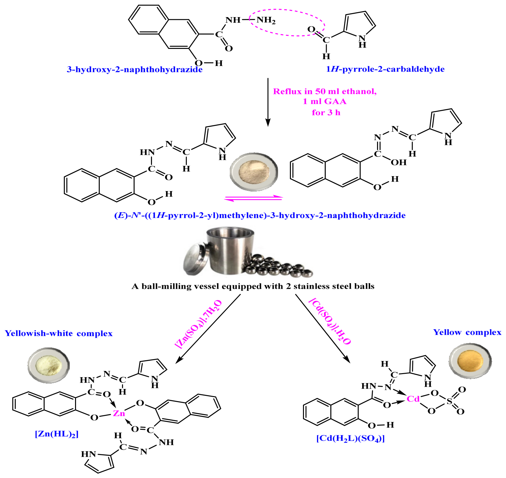
The ligand was synthesized by equimolar combination of 2-hydroxy-1-naphthohydrazide (0.01 mol; 3.06 g) and 2-formylpyrrole (0.01 mol; 0.95 g) in 50 mL ethanol under reflux for 3 h [19]. The reaction mixture was concentrated to half its volume, then cooled to precipitate the product, which was filtered, recrystallized from absolute ethanol, and dried under vacuum over anhydrous CaCl2. A waste-free, solid-state ball-milling method was used to synthesize Cd(II) and Zn(II) chelates, as shown in Scheme 1, providing a green chemistry methodology with a high yield of 98%.
Synthesis of H2L: H2L was obtained as a brown solid (yield 70%, m.p. 255 °C). Anal. Calc. for C16H13O2N3 (279.301): C, 68.80, and H, 4.69. Found: C, 68.86, and H, 4.66%.
Synthesis of [Cd(H2L)(SO4)]·2H2O: The chelate was isolated as a yellow solid (yield 98%, m.p. 290 °C). Anal. Calc. for CdC32H30O9N6S (803.034): C, 47.86; H, 7.76; N and Cd, 13.99; and SO4, 11.95. Found: C, 47.85; H, 3.82; Cd(II), 13.81; and SO4, 11.88%. Molar conductivity (Λm, DMSO): 6 Ω−1 cm2 mol−1.
Synthesis of [Zn(HL)2]: The chelate was obtained as a yellowish-white solid (yield 97%, m.p. 280 °C). Anal. Calc. for ZnC32H24O4N6 (621.956): C, 61.79; H, 3.89; and Zn, 10.51. Found: C, 61.88; H, 3.90; and Zn, 10.48%. Molar conductivity (Λm, DMSO): 3 Ω−1 cm2 mol−1.

2.4. Molecular Modeling of Isolated Complexes

Molecular modeling is a theoretical method used to mimic the complexation mode in prepared compounds. The computational studies for H2L along with its complexes were studied via DMOL3 in the Material Studio package by using the DFT method (density functional theory) and (DNP) as a basis set based on the generalized gradient approximation (GGA) and the PBEsol functional [20,21].

2.5. Molecular Docking with Receptors 6NM0, 5EQG, and 5IAE

Molecular docking simulation studies are a crucial methodology in computational drug discovery, enabling detailed interpretation of ligand interactions with target receptors. These computational analyses provide essential data on binding affinity and spatial conformation, thereby supporting the rational, structure-based development of new pharmaceutical agents.

2.5.1. Ligand and Protein Preparation

Molecular docking analyses were performed on the studied ligand and its complexes using LigPrep (Schrödinger Suite) with default parameters [22]. The Protein Data Bank (RCSB PDB) was the source of the three-dimensional crystal structures of MCF-7 (PDB ID: 6NM0) [23] https://www.rcsb.org/structure/6NM0, (accessed on 3 October 2025), HePG-2 (PDB ID: 5EQG) [24] https://www.rcsb.org/structure/5EQG, (accessed on 5 October 2025) and Hela (PDB ID: 5IAE) https://www.rcsb.org/structure/5IAE (accessed on 7 October 2025) [25]. Protein preparation was performed using the Protein Preparation Wizard (Maestro v. 14.1, Schrödinger Suite) [21], which involved a series of refinement steps, including correcting all structural errors, optimizing side chains, and energy minimization.
In the present study, all protonation states were designated using PROPKA, as implemented in Schrödinger Maestro v14.1 (Schrödinger, LLC, New York, NY, USA), was used for protonation state prediction, at a pH of 7.0 to maintain the needed physiological significance. This was successfully achieved by using the OPLS4 force field for energy minimization, with a convergence cut-off set at 0.30 Å for the heavy-atom root-mean-square deviation (RMSD). The settings of the designed study adjusted the bond orders carefully, and all hydrogen atoms were incorporated within a pH range of 7.0 ± 2.0 to guarantee suitable charge distribution and maintain structural stability.
The OPLS 2005 force field concluded a gentle energy minimization by applying an RMSD convergence threshold of 0.3 Å to guarantee the best molecular shape for the docking assessments. These initial actions are essential for achieving trustworthy binding affinity estimations and clarifying the ways in which drugs interact with their targets, thus aiding in the design of new therapeutic agents based on structure.

2.5.2. Induced Fit Docking

The molecular docking studies were conducted using Schrödinger’s Induced Fit Docking (IFD) protocol, a sophisticated approach that simultaneously accounts for both ligand conformational flexibility and receptor binding site adaptability to improve prediction accuracy. For each target protein, MCF-7 (PDB ID: 6NM0), HePG-2 (PDB ID: 5EQG), and Hela (PDB ID: 5IAE), a 10 Å3 docking grid centered on the crystallographic ligand coordinates was established to ensure comprehensive sampling of potential binding modes. Ligand conformational space was explored using Glide (version 10.4) with enhanced sampling parameters, including a 2.5 kcal/mol energy window specifically for ring system flexibility. Concurrently, receptor flexibility was modeled through Prime’s advanced algorithms, allowing for dynamic optimization of binding site residues during the docking simulation.
Induced Fit Docking (IFD) was performed using adjusted van der Waals and electrostatic parameters, incorporating transient side-chain flexibility to enhance the accuracy of protein-ligand conformational fitting. Energy-minimized ligands were docked utilizing standard sampling protocols, with a potential scaling factor of 0.5 applied to account for soft interactions. For each ligand, up to three top-ranked poses were retained. Ranking was guided by IFD scoring metrics that quantify binding interaction energies and the overall stability of the protein-ligand complex [26].

2.5.3. Docking Protocol Validation

To validate the molecular docking methodology, redocking experiments were performed as a benchmark strategy for assessing the precision of ligand binding predictions. Co-crystallized ligands namely, 4-[3-(2,4-difluorophenyl)-2-oxo-2,3-dihydro-1H-imidazol-1-yl]benzene-1-sulfonamide inhibitor from MCF-7 (PDB ID: 6NM0), (2~{S})-3-(4-fluorophenyl)-2-[2-(3-hydroxyphenyl)ethanoylamino]-~{N}-[(1~{S})-1-phenylethyl]propanamide inhibitor from HePG-2 (PDB ID: 5EQG), and Ac-Asp-Glu-Val-Asp-CMK Peptide-like inhibitor from Hela (PDB ID: 5IAE), were retrieved from their native complexes and redocked into their respective active sites using Schrödinger Maestro v14.1 (Schrödinger, LLC, New York, NY, USA). All docking parameters were kept unchanged during this process to uphold methodological consistency. The protocol enabled rigorous assessment of pose reproducibility and minimized deviations from the original crystallographic configurations, thereby reinforcing the credibility of the docking workflow.

2.6. Biological Efficacy

2.6.1. Screening of Antimicrobial Activity

The diffusion-disc assay, as outlined in Scheme S2, was employed to assess the antibacterial as well as antifungal activities of the free ligand and its solid chelates against Bacillus subtilis (Gram-positive), Escherichia coli (Gram-negative), and Candida albicans. Ampicillin and Clotrimazole functioned as reference antibiotics. Serial dilutions spanning (0.50–64) μg/mL were prepared from stock solutions (1 mg/mL) of the compounds and controls to establish the MIC values [27].

2.6.2. Free-Radical-Scavenging Activity

As a standard, Vitamin C is used in the ABTS (2,2-azino-bis(3-ethylbenzthiazoline-6-sulfonic acid)) radical-scavenging assay for the compounds under investigation. Scheme S3 shows how to prepare a blue-green ABTS+ solution by combining ABTS with potassium persulfate (K2S2O8) in a 1:1 ratio. Upon reaction with the test samples, the ABTS+ color fades. The decrease in absorbance at 734 nm, recorded with a spectrophotometer, yields the IC50 values (the concentration causing 50% inhibition) [28].

2.6.3. DNA Binding Activity

A colorimetric DNA-binding assay was performed to estimate a compound’s ability to interact with a DNA-protein complex. At neutral pH ≈ 7, methyl green forms a stable colored complex with highly polymerized DNA, while it dissociates from DNA at the same pH. As represented in Scheme S4, compounds that bind DNA displace methyl green from the complex, weakening the color. Absorbance was measured at 630 nm with a spectrophotometer, and IC50 values were calculated from the dilution data [29,30].

2.6.4. Cytotoxic Activity

The produced compounds were screened for antiproliferative activity against three cancer cell lines: HEPG-2 (ATCC HB-8065), MCF-7 (ATCC HTB-22), and HeLa (ATCC CCL-2). All cell lines were obtained from the American Type Culture Collection (ATCC, Manassas, VA, USA) and were authenticated by short tandem repeat (STR) profiling prior to use. Cells were maintained under recommended culture conditions to preserve genetic stability. In the MTT assay, mitochondrial succinate dehydrogenase reduces MTT to a purple formazan, which is subsequently dissolved in DMSO as shown in Scheme S5. Absorbance was recorded at 570 nm using a spectrophotometer, and IC50 values were calculated from the resulting data [31].
MCF-7 (human breast adenocarcinoma; ATCC HTB-22), HeLa (human cervical carcinoma; ATCC CCL-2), and HepG2 (human hepatocellular carcinoma; ATCC HB-8065) were used in this study. All cell lines were obtained from the American Type Culture Collection (ATCC, Manassas, VA, USA) and verified by short tandem repeat (STR) profiling. Cells were cultured according to appropriate guidelines to maintain genetic stability.

3. Results and Discussion

The isolated solid compounds demonstrated air stability at room temperature. Every synthesized metal complex was soluble in DMF or DMSO.

3.1. Infrared and 1H NMR Spectra of H2L and Its Metal Complexes

The most significant infrared bands of H2L and its metal complexes summarize in Table 1, while Figure S1 provides a graphic representation of these bands.
The υ(C=O) and υ(C=N) [32] vibrations are responsible for the bands at 1683 and 1626 cm−1 in the infrared spectrum of H2L, respectively. The υ(N–N) and υ(C–O)naphthoic [32] vibrations are attributed to the medium-intensity bands at 1052 and 1268 cm−1, respectively. The υ(OH)naphthoic, υ(NH)pyrrole, and υ(NH)amidic [32] are responsible for the bands at 3269, 3236, and 3045 cm−1, respectively. The appearance of υ(OH) vibrations at 3269 cm−1 indicates the presence of hydrogen bonds within the molecule [32]. The weak broad bands noticed in the regions between 1912 and 2047 and between 2120 and 2228 cm−1 can be regarded as further support for the existence of intramolecular hydrogen bonding (O–H··O) [32], as shown in Scheme 1.
The 1H NMR spectrum of H2L soluble in DMSO (Figure S2) displayed the protons of the (OH)naphthoic, (NH)amidic, and (NH)pyrrole groups, which are related to the three signals at 11.68, 8.68, and 8.67 ppm, respectively. The formation of an intramolecular hydrogen bond was confirmed by the shift in the (OH)naphthoic proton’s signal to a higher value downfield from TMS. Furthermore, the naphthyl and (–N=C–H) protons are attributed to the multiple bands appearing in the 7.52–8.65 ppm region. Additionally, the pyrrole ring’s protons are responsible for the signals seen in the 7.34–7.49 ppm range.
The 13C NMR spectrum of H2L (Figure S2) displayed the subsequent data: (i) aromatic carbons appeared in the range (δ) 111.22–136.33 ppm, and (ii) sharp peaks at (δ) 172.55, 162.25, 155.92, and 153.05 ppm were related to (C=O), (C–OH), (C=N), and (C–NH), respectively.
The behavior of H2L as a neutral or mononegative bidentate ligand is dependent on the metal salt and reaction conditions, which is reflected in the alteration of the ligand’s characteristic absorption bands upon complexation and the emergence of new vibrational bands.
H2L behaved as a neutral bidentate ligand in the [Cd(H2L)2(SO4)]·2H2O complex via the carbonyl oxygen (C=O) and (OH)naphthoic. The movements of both υ(OH)naphthoic and υ(C=O) to a lower wavenumber supported this method of chelation, while the υ(C=N) did not shift, suggesting it did not participate in the coordination process. Additionally, υ(Cd–O) is attributed to the newly detected band at 563 cm−1 [33]. Furthermore, the cadmium (II) complex’s infrared spectrum shows two bands at 1067 and 921 cm−1 assignable to the SO stretching vibrations, which suggest a bridging or bidentate bonding of the sulphate group [33].
Also, in the 1H NMR spectrum of the Cd(II) complex, the signals attributed to (OH)naphthoic, (NH)amidic, and (NH)pyrrole protons remain more or less at the same positions. In addition, the 13CNMR spectrum of the Cd(II) complex in DMSO, as depicted in Figure S2, exhibited a sharp signal at δ = 145 ppm, which corresponded to –C=N. Another sharp signal appeared at δ = 136 ppm, attributable to –C–NH. A sharp signal was observed at δ = 128, corresponding to –C=O. A sharp signal appeared at δ = 129 ppm, corresponding to –C–OH. Signals observed in (δ = 111–127) corresponded to aromatic carbon. A signal observed at 21 ppm was attributable to the carbon of the acetate group.
The H2L in the [Zn(HL)2] complex acted as a mononegative bidentate ligand, chelating through the carbonyl oxygen (C=O) and the deprotonated (OH)naphthoic. The following factors support this proposed mode of chelation: (i) the band that can be attributed to (OH)naphthoic disappears; (ii) the appearance of υ(C=O) at a lower wavenumber; (iii) the υ(C=N) shifts to a higher wavenumber; and (iv) the new band appeared at 559 cm−1, which is tentatively assigned to υ(Zn–O) [34].
Moreover, in the 1H NMR spectrum of the Zn(II) complex, the absence of the proton signal of (OH)naphthoic in the 1H NMR spectrum of the diamagnetic Zn(II) complex strongly supported the deprotonation of the (OH)naphthoic group. As well as this, the 13CNMR spectrum of the Zn(II) complex in DMSO, as represented in Figure S2, displayed a sharp signal at δ = 150 ppm, which corresponded to –C=N. Another sharp signal appeared at δ = 136 ppm, attributable to –C–NH. A sharp signal was observed at δ = 129, corresponding to –C=O. A sharp signal appeared at δ = 128 ppm, corresponding to –C–O. Signals observed in (δ = 111–127) corresponded to aromatic carbon.

3.2. Powder X-Ray Diffraction

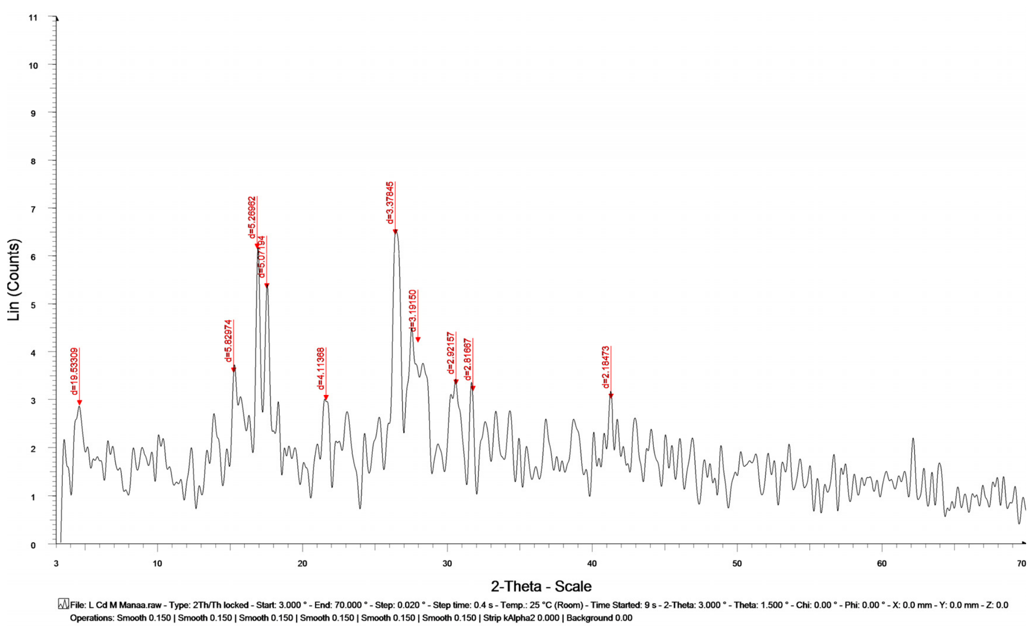
The Cu anode Kα radiation (λ = 1.5406 Å) was used to assess X-ray diffraction at room temperature in the region of (10° < 2θ < 80°). A crystalline phase found in the [Cd(H2L)(SO4)]·2H2O complex is displayed in Figure 1, indicating a semi-crystalline form or crystalline phase. The interplanar spacing was calculated using the Bragg equation, Equation (1), and was found to be d = 0.337 nm, the diffraction angle (2θ = 26.4°). The full width at half maximum (FWHM, β) of the most intense peak was determined to be 0.47° (0.0083 rad) after conversion to radians [35]. The crystallite size (S = 17.23 nm) was calculated using the Debye-Scherrer equation, Equation (2):
n λ = 2 d s i n ( θ ) a t n = 1
B = 0.9 λ / S c o s ( θ )

3.3. SEM and EDX Examinations

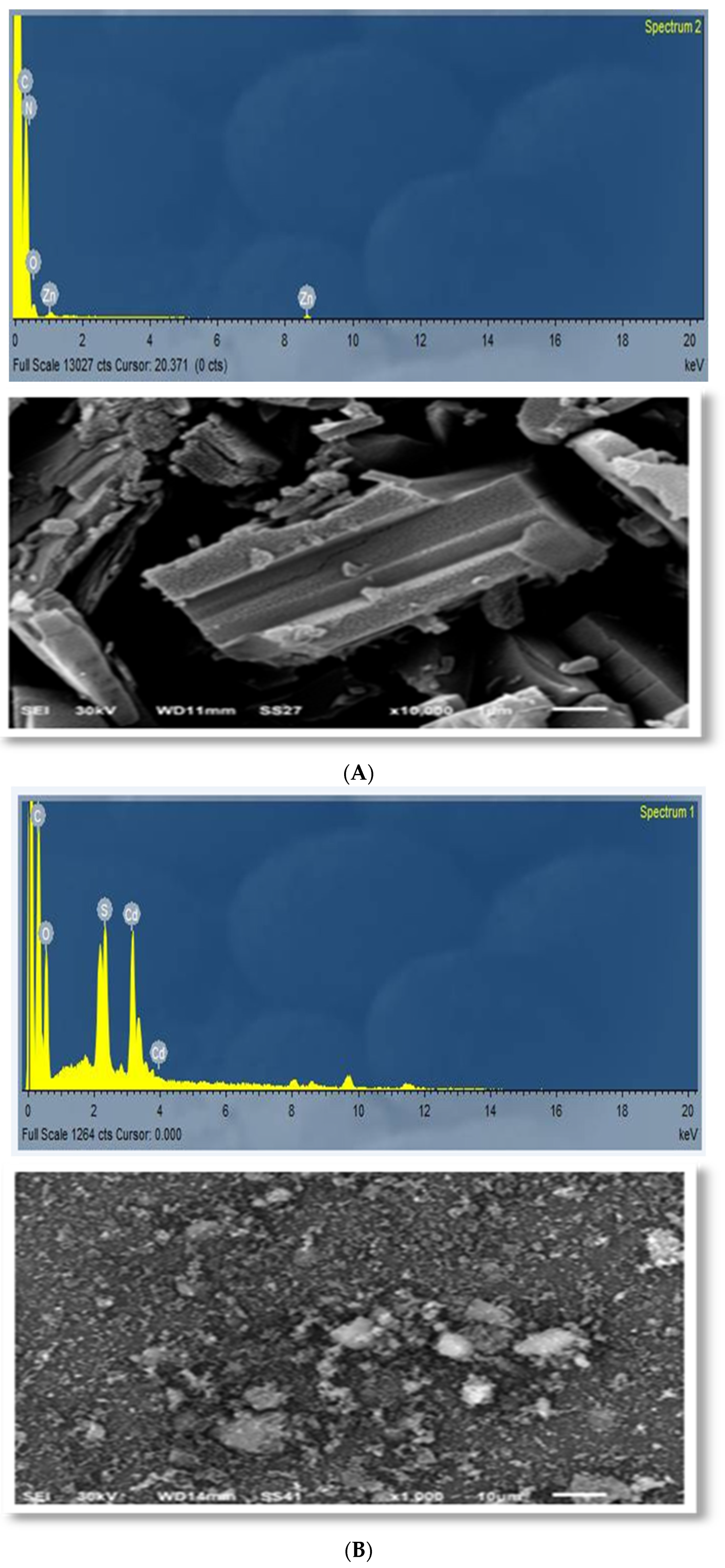
The micro-scale morphology of the isolated solid complexes was investigated using the SEM morphological assessment (SEM technique) in order to determine both the ligand’s coordination with the matching metal ions and their morphological composition. The [Zn(HL)2] complex’s SEM picture in Figure 2A displayed a distinctly varying shape. The structure is described by irregular needle-like domains rather than compact particle characteristics. Furthermore, the complex particles are observed to be randomly distributed throughout the solid matrix, forming an overall morphology consistent with a needle/rod-like growth tendency. The [Cd(H2L)(SO4)]·2H2O complex’s SEM picture shows micrometer-sized particles, as displayed in Figure 2B. These particles display a lack of uniform packing and are dispersed randomly through the cracked rock-like substrate, suggesting that the complicated formation outcomes result in the non-ordered deposition of solid particles. The conclusion that the ligand-metal coordination mode directly affects the final solid-state microstructure is reinforced by this morphology [36].
The solid complexes were further characterized by EDX, and the corresponding spectra are shown in Figure 2. The existence of the metal centers Zn(II) or Cd(II) and the anticipated constituent elements C, H, N, and O is exposed by the elemental signals, indicating that the isolated products contain both the organic ligand framework and the associated metal ions. Specifically, by matching the elemental composition predicted from the suggested coordination skeleton assumed in the solid complexes’ spectra, the EDX results validate the synthesis of the solid chelates [37]. The effective synthesis of the target complexes is often reinforced by the elemental analysis, and the experimental EDX results are consistent with the structural characteristics expected for their individual chelating coordination modes.

3.4. Computational Studies of Naphthohydrazide Ligand (H2L) and Its Metal Complexes

The molecular modeling studies provided a wide-ranging set of structural as well as electronic parameters that provided deeper insight into the chemical behavior of ligand H2L and its metal complexes. Key reactivity descriptors such as (EHOMO) and (ELUMO) were estimated to assess the electron-donating and electron-accepting abilities of the isolated compounds, from which global reactivity indices including the energy gap, softness, chemical hardness, electronegativity, and electrophilicity index were estimated. These parameters helped predict the reactivity, relative stability, and potential biological activity of the ligand and its metal complexes. Moreover, geometric optimization produced full structural information, including bond angles and bond lengths, which confirmed the coordination geometry around the central metal ions and the nature of the ligand-metal interactions.
The total energy components, as well as the kinetic energy contributions, were established to evaluate the energetic stability of the optimized structures. Furthermore, the binding energies of H2L and its metal complexes were estimated, providing a quantitative measure of the strength of interaction between the metal ions and the ligand framework. Together, these computational findings not only confirmed the proposed molecular structures but also offered a theoretical basis for the observed differences in biological activity across the series of compounds [38].

3.4.1. Structural Optimization Using the DFT Method

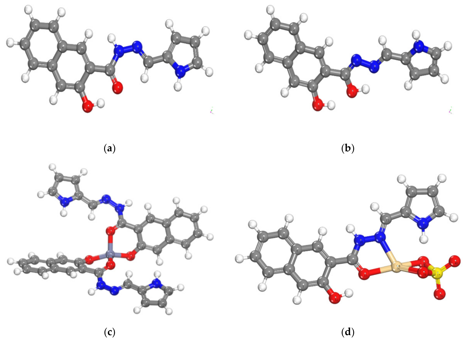
The geometry-optimized molecular structures of H2L and its metal complexes are represented in Figure 3. Also, the bond lengths and angles for the optimized molecular structures of H2L and its metal complexes were obtained and are represented in Tables S1–S8. These theoretical parameters are the key factor in understanding the structure of metal complexes and their expected binding interactions.
Upon complex formation, the bond angles of the free H2L ligand go through measurable variations at the coordination sites. The most important alterations were detected for C(17)–N(16)–N(15), N(16)–N(15)–C(11), N(15)–C(11)–O(13), N(15)–C(11)–C(9), O(13)–C(11)–C(9), C(11)–C(9)–C(8), and O(12)–C(8)–C(9), with each angle either increasing or decreasing comparative to the free ligand. These changes show that the geometric arrangement of atoms is rearranged upon coordination with metal ions. In both the Cd(II) and Zn(II) complexes, the measured bond angles were reliable with a tetrahedral geometry and sp3 hybridization, as previously reported [39].
Concerning bond lengths, the distances between several atom pairs including N(16)–C(17), N(15)–N(16), C(11)–N(15), C(11)–O(13), C(8)–O(12), C(9)–C(11), and C(8)–C(9) were establish to increase in the metal complexes compared to H2L alone. This elongation confirms the formation of M–N and M–O coordination bonds [40].
Based on the M–O(12) bond lengths, the complexes followed the order Cd–O > Zn–O, suggesting that the Zn–N bond is stronger than Cd–N. Following the same trend, the Zn–O bond was the shortest between the complexes, indicating the greatest bond strength. For the M–O(13) bond, the order was Cd–O < Zn–O, further confirming that the Cd–O bond is weaker than Zn–O. So, the observed difference in bond strength may contribute to the observed variation in antibacterial activity among the compounds [41].

3.4.2. Hypothetical Chemical Global Reactivity Descriptors of H2L and Its Metal(II) Complexes

The DFT studies present a useful perception of the electronic and structural properties of the prepared complexes. This perception is important to realize the stability and reactivity of these compounds. Molecular orbital studies are the basis for UV–Vis studies and electronic properties [42].
The HOMO and LUMO energies, which mean π acceptor and π donor, respectively, are considered significant parameters in quantum chemical calculations. The energy gap (∆E = EHOMOELUMO) and the reactivity of an electronic system are related. ∆E is a crucial metric for describing the compound’s chemical reactivity and kinetic stability. Electrons might readily migrate between these orbitals, promoting chemical reactivity and interactions with biological molecules, as indicated by the tiny energy gap value. This characteristic may improve complex antibacterial activity.
The negative (−)ve magnitudes of EHOMO, ELUMO, and their adjacent orbitals showed the good stability of the prepared metal(II) complexes (Table 2). The stability of complexes is a key parameter in their biological effectiveness because stable complexes have a low tendency for side reactions that could reduce their antimicrobial properties [38].
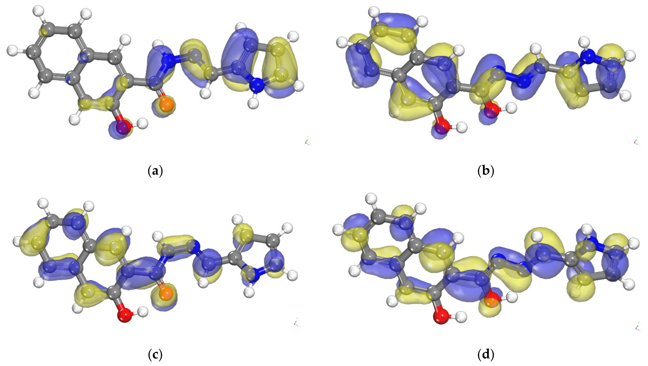
In the keto form of H2L, the C(11), O(13), N(15), N(16), and C(17) atoms, as well as the pyrrole ring, are where the HOMO is primarily found and they might be the most preferred positions concerning nucleophilic attack. While in enol form, the HOMO orbitals are located on the same atoms as in the keto form, alongside the naphthyl ring. According to this localization, these atoms are probably the best locations for the nucleophilic attacks that are crucial for the complexes’ reactivity in biological systems. The complexes’ ability to interact with electrophilic targets, including bacterial enzymes or nucleic acids, is increased when nucleophilic sites are present.
High overlapping between the HOMO of the ligand and the LUMO of the metal ion can enhance the stability of the ligand-metal complex (Figure 4, Figures S3 and S4). Significant overlap indicates that the ligand’s electrons can easily be transferred to the metal, promoting stronger bonding interactions. This means they are more effective in stabilizing the investigated complexes through strong interactions.
The energy gap was an essential stability index that assisted in the description of the chemical reactivity and kinetic stability of the prepared compounds [38].
The density functional theory (DFT) method elucidates molecular systems’ chemical reactivity and site selectivity. Critical parameters such as global hardness (ηG), global softness (SG), and the global electrophilicity index (ω) are essential in this study, as shown in Table 2. Additionally, the energies of the frontier molecular orbitals (EHOMO and ELUMO), the energy band gap (calculated as EHOMO − ELUMO), electronegativity (χ), and chemical potential (μ) [38,43] are calculated by applying the following equations, Equations (5)–(10).
η G = 1 2 E L U M O E H O M O
S G = 1 2 η G
ω = μ 2 2
μ = χ = 1 2 ( E L U M O + E H O M O )
χ = 1 2 ( E L U M O + E H O M O )
ϭ = 1 η
The ability of an atom to withstand charge transfer is measured by the hardness of complexes (ηG), which is equal to half of the energy gap (ηG = ∆E/2). The formula for calculating chemical softness (SG), which measures an atom or molecule’s ability to accept electrons, is SG = 1/2ηG. These measurements provided insight into the reactivity of the compounds, with higher softness and lower hardness values generally indicating more reactivity. Soft molecules are known to have smaller HOMO-LUMO gaps, whereas hard molecules are associated with higher energy gap values. Zn(II) and Cd(II) complexes were therefore regarded as hard complexes.

3.4.3. Molecular Electrostatic Potential (MEP)

Nucleophilic and electrophilic attack sites, in addition to hydrogen-bonding interactions, were determined by the molecular electrostatic potential [39]. The molecular electrostatic potential (MEP) of H2L and its metal (II) complexes is illustrated in Figure S5 in various colors, blue, green, and red. Areas of severe electron shortage, which are especially vulnerable to nucleophilic assaults, are indicated by red sections in this illustration. Because they seek out positively charged or electron-deficient centers for bonding, this property makes them attractive sites for reactants with lots of electrons. On the other hand, the blue spots draw attention to places with a high electron density, which are more suited for electrophilic interactions since they have a lot of electrons. Additionally, areas of neutral electrostatic potential are indicated by green zones, which show a balanced distribution of charge that is neither significantly deficient in electrons nor in excess of them [39].

3.4.4. Mulliken Population Analysis

The Mulliken mathematical pattern for calculating the atomic charge is a fundamental function in quantum studies of molecular systems. These calculations were done using the quantum mechanical density functional theory (DFT) program. The associated Mulliken plots shown in (Tables S9–S12) provide a visual representation of these computations. The distribution of atomic charges throughout the molecule is illustrated by these theoretical computations.
The electrical environment of H2L and how it affects its chemical activity and possible uses in medicinal chemistry can be better understood thanks to the analysis of these plots. All things considered, our thorough comprehension of the molecular properties and reactivity of H2L depends on the analysis of Mulliken atomic charges. The decrease or increase in bond length was determined mainly by the distribution of positive and negative charges [41].
In H2L some of atoms had positive charge, such as C(11), C (21), C(4), C(5), C(8), C(9), and C(18). Also, both C(11) and C(8) showed positive atomic charges that were possibly associated with the (C=O) and (C–OH) groups, respectively. This positive distribution is important because it reveals regions of the molecule that might serve as electrophilic sites and are susceptible to nucleophilic attacks from species with a high electron density. Because the presence of these positive charges suggested a potential for interactions with negatively charged or polar chemicals, these sites are crucial for understanding H2L reactivity in a variety of chemical settings.
Some other atoms had negative charges, such as C(1), C(10), C(2), N(20), C(3), O(12), O(13), C(22), C(23), C(6), N(15), C(7),N(16), and C(17). The O(12) and O(13) atoms had the greatest negative charges. These oxygen atoms might operate as nucleophilic sites, engaging with other molecules’ positive centers or creating hydrogen bonds, given their high negative charge levels. Therefore, H2L reactivity and binding affinities were largely dependent on the correct ratio of positive to negative charges inside the zone.

3.4.5. Molecular Parameters of H2L and Its Metal(II) Complexes

The DFT method calculates different energy components such as electrostatic energy, dipole moment, entire atomic energy, total energy, kinetic and binding energy, exchange-correlation energy, and spin polarization energy. According to the calculated values in Table 3, the formed metal (II) complexes are more significant than the parent ligand alone. It can be noticed that the computed complexes’ binding energy values are higher than those of the H2L, demonstrating that the metal complexes are more stable than the ligand. From a practical viewpoint, this means that the coordination with metal ions reinforced the complex’s structural integrity, and perhaps this will enhance its resistance in biological environments. Understanding the prolonged antibacterial action of produced complexes may be aided by the stability of these complexes, which are less likely to disintegrate and lose their effectiveness when interacting with pathogens. Meanwhile, as a stable complex is necessary to stop DNA replication and pathogens, the higher binding energy of the metal complexes under study emphasizes their ability to interact with DNA more effectively.
Inside the system, the separation of electrical charges can be measured via the electric dipole moment values. The dipole moment of the free Cd(II) complex is higher than that of the ligand, H2L, and the other complexes, which enhances the activities of the Cd(II) complex [44]. Therefore, the Cd(II) complex interaction with polar biological targets is often improved by its high dipole moment [45].

3.5. Biological Potency

3.5.1. Antimicrobial Activity

The antimicrobial activity index of (H2L) and its solid chelates was estimated versus antibacterial Ampicillin and antifungal Colitrimazole via applying Equation (9) [46], as shown in Table 4.
%   Activity   index = I n h i b i t o r y   z o n e   o f   e x a m i n e d   c o m p o u n d ,   d i a m e t e r I n h i b i t o r y   z o n e   o f   s t a n d a r d   c o m p o u n d ,   d i a m e t e r × 100
The antibacterial as well as antifungal activities of the tested compounds are varied according to the applied microorganism. Meanwhile, tests with E. coli, B. subtilis, and C. albicans showed that the Zn(II) complex was found to be the most active compound, followed by the Cd(II) complex, while H2L exhibited low antimicrobial activity, as shown in Table 4. Metal complexation increases activity when H2L coordinates to Zn(II) or Cd(II), as shown in Scheme 1.
Chelation of the H2L ligand with Zn(II) and Cd(II) metal ions improves lipophilicity relative to the free ligand, facilitating more efficient transport across microbial membranes, a factor of particular significance for bacterial cells. Once inside the cell, the metal center in the Cd(II) and Zn(II) chelates can coordinate with electron-donor atoms in microbial proteins and enzymes, for example, carbonyl or phenoxy oxygen atoms and amino nitrogen atoms, thus inactivating enzymes essential for cell survival. Furthermore, coordination changes the electron density of the free ligand, enabling stronger binding to microbial targets compared to the H2L. Among the two complexes, the Zn(II) analog displayed greater antibacterial activity than the Cd(II) complex.
This experimental result is due to several factors: (i) Zn(II) forms more stable interactions with donor atoms in enzymatic and biomolecular systems, leading to stronger inhibition of vital metabolic pathways; (ii) many energetic microbial enzymes trust on coordinated active sites, and Zn(II) has a stronger affinity for such donor environments, resulting in greater enzyme dysfunction; and (iii) even though Cd(II) is inherently more toxic in a biological context, its antimicrobial efficacy may be reduced depending on how readily it binds and its stability and reactivity within the assay medium—for example, competition with chloride or sulfate ions or binding to other medium components may limit target engagement compared to Zn(II).

3.5.2. Antioxidant Activity (ABTS Assay)

The ABTS examination of the synthesized compounds was detected versus Vitamin C as a positive control by applying Equation (10).
Inhibition % = A   b l a n k A   s a m p l e A   b l a n k × 100
where (A) sample represents the absorbance of the tested or standard compound, and (A) blank represents the absorbance of the blank sample, which contains phosphate buffer (1:1) without ABTS solution or any tested compound [47]. ABTS examination is expressed as IC50 values, as shown in Table 5.
The ABTS assay results displayed in Table 5 indicated that the [Zn(HL)2] complex exhibited the strongest radical-scavenging activity among the synthesized compounds, with an IC50 value of 42.4 ± 0.3 μg/mL, followed by the Cd(H2L)(SO4)]·2H2O complex (46.8 ± 0.2 μg/mL), however the free ligand displayed the weakest activity (69.4 ± 0.3 μg/mL). Obviously, the reference antioxidant vitamin C showed the lowest IC50 value (30.2 ± 0.2 μg/mL), indicating its superior antioxidant capacity. These results proposed that coordination of the ligand with Zn(II) or Cd(II) metal ions improves its radical-scavenging activity compared with the free ligand.
Structure-Activity Relationship
When the ligand coordinates to the metal (Zn/Cd), the electronic distribution and stability of the complex change. This often improves the compound’s ability to donate electrons/hydrogen to neutralize radicals or to stabilize the radical/oxidized form. Also, the metal ion can promote stronger interaction with reactive species through its Lewis acidity and altered redox/coordination behavior compared with the free ligand. This can make the Zn(II) complex more efficient than Cd(II) in the ABTS system. The free ligand lacks the metal center; therefore, its radical-scavenging relies mainly on the ligand’s inherent functional groups (weaker or less effective under the assay conditions), giving the highest IC50.

3.5.3. DNA-Binding Assay

The produced compound’s DNA-binding assay was performed using doxorubicin as the standard material. The results shown in Table 5 display the IC50 values of the isolated solid compounds under examination. The results indicate that: (i) the Zn(II) complex had a significant ability to bind with DNA protein with a value of 36.4, which was comparatively near to the standard value of 31.5; (ii) H2L displayed a moderate ability to bind with DNA with a value of 62.6; and (iii) the Cd(II) complex had a value of 41.0. Therefore, among the chemicals, the Zn(II) complex binds DNA more strongly and is closest to doxorubicin, but H2L exhibits weaker binding. The enhanced activity of the complexes can be attributed to a number of key factors. The coordination provided a positively charged metal center that facilitated stronger interactions with the negatively charged phosphate backbone of DNA, improving the capability of the complexes to bind biomolecular targets. Moreover, complexation changed the ligand conformation and the distribution of donor atoms (O/N/S), enabling more effective electrostatic and coordination-based interactions with DNA alone or DNA-associated proteins. In particular, Zn(II) complexes have a tendency to form stable coordination environments that help more organized interactions with biomolecular functional groups under biological assay conditions, resulting in lower IC50 values compared to Cd(II) complexes. In contrast, the free ligand (H2L) lacked a metal center and consequently had fewer strong binding modes available, making it less capable of successfully attracting or coordinating to DNA, which led to higher IC50 values.

3.5.4. Cytotoxicity Assay

Relative cell viability of the applied cell lines was estimated versus Doxorubicicn as positive control by using Equation (11) [48]:
Relative   cell   viability   %   = a b s o r b a n c e   o f   t r e a t e d   s a m p l e   a t   570   n m a b s o r b a n c e   o f   u n t r e a t e d   s a m p l e   a t   570   n m × 100
IC50 values necessary for a reduction in viable cells were measured as shown in Table 6. The cytotoxic effect of H2L and its solid Zn(II) and Cd(II) complexes was evaluated against three tumor cell lines (HePG-2, MCF-7, and Hela) using an MTT-based viability approach, with Doxorubicin as the positive control. Stronger anti-proliferative action was shown by lower IC20 values, according to the assumed IC50 categories. Generally, the findings proved a distinct cell-line-dependent cytotoxicity and demonstrated that, in comparison to the ligand alone, metal complexation increases cytotoxic efficacy. H2L showed the least amount of cytotoxicity in all tested cell lines, with moderate activity for HePG-2 (IC50 = 41.0 µM) and strong/moderate activity for MCF-7 (28.7 µM) and Hela (54.9 µM, i.e., weak activity range). The Zn(II) and Cd(II) complexes, on the other hand, consistently produced lower IC50 values, confirming improved anticancer performance. Notably, the complexes exhibited the highest cytotoxicity among the tested compounds, with Zn(II) ≈ Cd(II) > H2L under most conditions.
More specifically, the Cd(II) complex also exhibited substantial cytotoxicity in MCF-7 (19.3 µM) and HePG-2 (22.3 µM), whereas the Zn(II) complex was most effective in HePG-2 (IC50 = 19.0 µM, strong activity) and MCF-7 (IC50 = 16.8 µM, strong activity). Both complexes continued to be more active than the ligand alone for the Hela cell line, although the IC50 values showed that the Zn(II) complex (27.4 µM, moderate/strong) > Cd(II) complex (30.2 µM, moderate/strong), whereas H2L was relatively less powerful (54.9 µM, weak activity). All investigated compounds have higher IC50 values when compared to doxorubicin, suggesting that they are less effective than the usual medication. However, their activities are still evidently substantial in relation to the ligand alone.
Mechanistically, the metal center-ligand coordination effect, which may enhance interaction with cellular targets (such as DNA/protein binding), increase cellular uptake, and/or encourage the generation of reactive species inside cells, is responsible for the increased cytotoxicity upon coordination. Additionally, the distinctions between HePG-2, MCF-7, and Hela highlight how each cell line may react differently because of alterations in intracellular sensitivity to metal-ligand complexes, drug transport pathways, and membrane permeability.

3.6. Molecular Docking

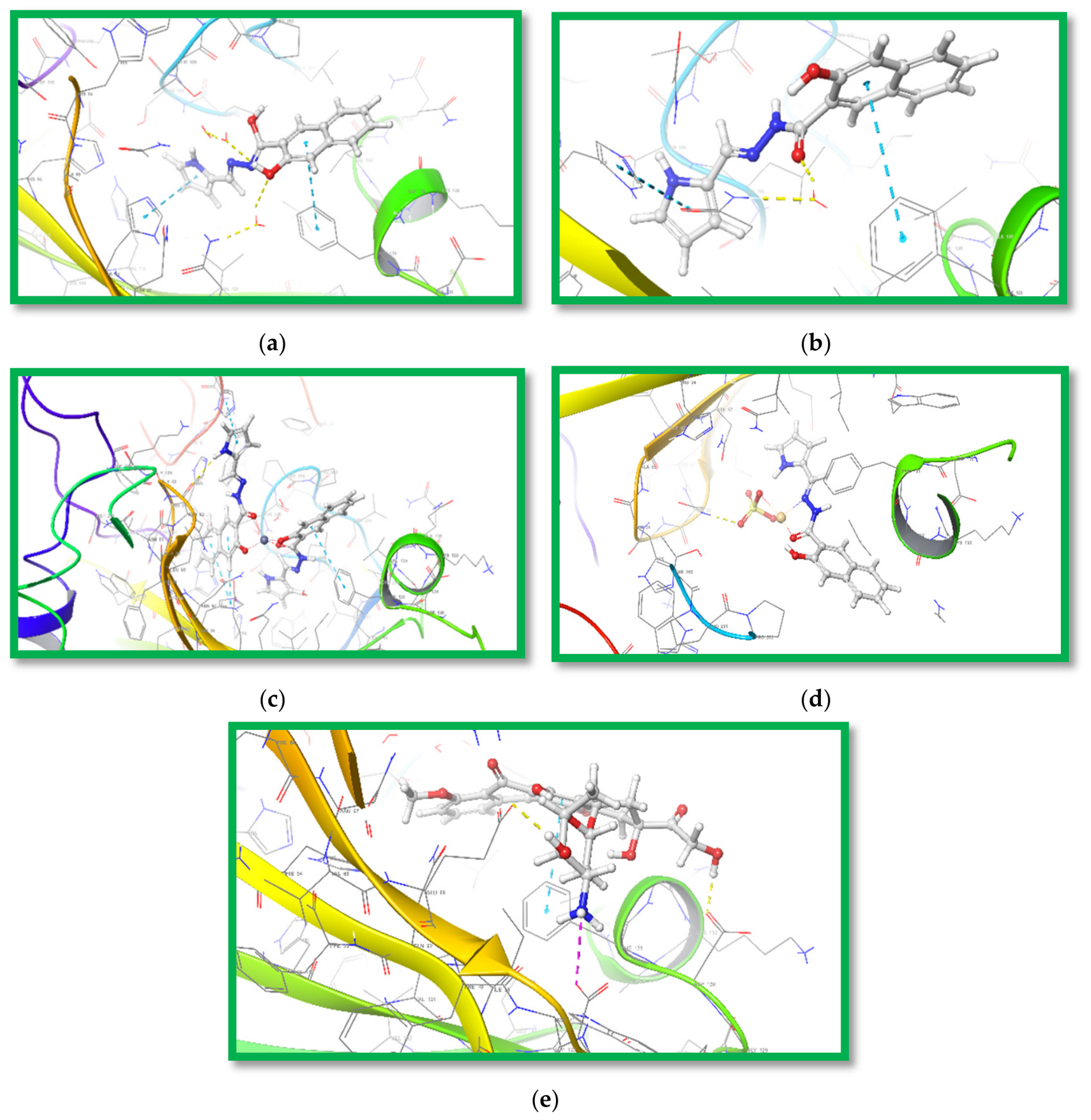
Molecular docking evaluations of the ligand H2L (in both keto and enol tautomeric forms), its corresponding metal complexes of Zn(II), and Cd(II) ions, and the control inhibitor Doxorubicin were conducted against MCF-7 (PDB ID: 6NM0), HepG-2 (PDB ID: 5EQG), and Hela (PDB ID: 5IAE) targets to elucidate binding affinity profiles and interaction dynamics. The analysis incorporated Induced Fit Docking (IFD) scores, root-mean-square deviation (RMSD) metrics, and ligand-protein interaction mapping, with detailed comparisons outlined illustrated in Figure 5, Figures S6 and S7, as well as Table 7, Tables S13–S15.
  • For MCF-7 (6NM0):
The enol form of H2L exhibited a slightly better binding strength (IFD:−560.34 kcal/mol) compared to the keto form (−559.16 kcal/mol), primarily stabilized by water-mediated hydrogen bonds with GLN92 and THR200. Among the metal complexes, the Cd(II) complex demonstrated the highest affinity (IFD: −564.67 kcal/mol), followed by the Zn(II) complex (−562.05 kcal/mol), both exceeding the original ligand (−560.25 kcal/mol) and Doxorubicin (−557.68 kcal/mol). The Zn(II) complex was anchored by a hydrogen bond with TRP388 (2.64 Å) and dual π–π stacking with HIS94.
  • For HepG-2 (5EQG):
The docking results revealed a significant increase in binding affinity for the metal complexes and the enol tautomer. The Zn(II) complex (−905.68 kcal/mol) surpassed Doxorubicin (−907.16 kcal/mol) and the original ligand (−586.44 kcal/mol). The enhanced stability of these complexes is attributed to robust π–π stacking interactions with TRP388 and PHE26. The enol form of H2L also showed a remarkable IFD score of −903.08 kcal/mol, suggesting a preferential binding orientation over the keto form (−578.03 kcal/mol).
  • For Hela (5IAE):
The original ligand exhibited the strongest binding affinity (IFD: −574.63 kcal/mol), supported by an extensive network of hydrogen bonds (GLY122, CYS163, GLN161, ARG207) and salt bridges. Doxorubicin followed with a score of −555.14 kcal/mol. Among the synthesized compounds, the Cd(II) complex (−553.92 kcal/mol) and the keto form of H2L (−548.7 kcal/mol) showed moderate activity. Notably, the Zn(II) complexes failed to achieve docking compatibility with the Hela target, indicating high selectivity or steric hindrance within the 5IAE binding pocket.
The superior IFD scores observed for the Zn(II) complex, particularly against HepG-2, suggest a synergistic effect where the metal center stabilizes the complex through enhanced lipophilic contacts Glipo and van der Waals forces. For instance, the Zn(II) complex against HepG-2 exhibited a Gevdw of −73.875 kcal/mol and a Glipo of −5.003 kcal/mol, significantly higher than those of the free ligand. Furthermore, the Gemodel energies for these complexes (ranging from −88.53 to −112.93 kcal/mol for HepG-2) indicate highly favorable energetic states upon binding.
While molecular docking provides valuable preliminary insights, it is inherently constrained by algorithmic assumptions, scoring biases, and limited representation of protein conformational dynamics. Furthermore, it does not fully integrate experimental conditions, cytotoxicity diversity, or pharmacokinetic behaviors, underscoring the need for complementary validation through in vitro and in vivo approaches [49].

Docking Validation

Redocking procedures were employed to assess the reliability of the docking protocol by reproducing native ligand poses within their crystallographic binding sites.
  • MCF-7 (PDB ID: 6NM0)
The redocking of the native ligand into the MCF-7 target (Figure S8) yielded an RMSD of 0.705 Å, indicating exceptional structural fidelity. The following interactions were preserved:
    • Hydrogen Bonding: The amino group (NH2) of the ligand formed three direct hydrogen bonds with HIE119 (2.31 Å), HIS96 (2.33 Å), and HIS94 (1.63 Å).
    • Water-Bridged Interaction: A critical water-mediated hydrogen bond was observed between the carbonyl group (C=O) and GLN92 (1.72, 1.76 Å).
    • π–π Stacking: The aromatic rings were stabilized by stacking interactions with PHE131 (5.31 Å) and HIS94 (5.29 Å).
B.
HepG-2 (PDB ID: 5EQG)
The validation for the HepG-2 target (Figure S9) resulted in an RMSD of 0.830 Å.
    • Surface Interaction: Unlike the other targets, the native ligand in this pocket exhibited high solvent exposure, meaning it rests in a configuration where few direct hydrogen bonds are formed, yet it maintains a stable IFD score of −586.44 kcal/mol.
    • Protocol Accuracy: The low RMSD confirms that even in the absence of dense specific bonds, the algorithm correctly identified the spatial orientation of the ligand within the hydrophobic cleft.
C.
Hela (PDB ID: 5IAE)
Validation against the Hela target was highly robust, producing an RMSD of 0.848 Å (Figure S10). This target featured the most complex interaction network:
    • Extensive Hydrogen Bonding: The ligand engaged multiple residues, including GLY122 (2.14 Å), CYS163 (2.34 Å), GLN161 (1.82 Å), ASN208 (2.60 Å), SER209 (1.78 Å), and TRP214 (2.10 Å).
    • Multiple Bonds with ARG207: This residue acted as a major anchor, forming three distinct hydrogen bonds (1.70 Å, 2.00 Å, and 2.59 Å).
    • Water-Mediated Bridging: Water molecules bridged interactions between the ligand and SER63, GLU248, PHE250, and SER209.
    • Salt Bridges: Electrostatic stability was provided by salt bridges with ARG207 (3.93 Å) and ARG64 (3.18 Å).
Overall, structural alignment revealed RMSD values well within the accepted threshold for all targets, affirming the accuracy of the docking strategy. Minor conformational variances likely stem from induced fit effects and water-mediated contacts that adjust the protein environment to the ligand’s presence.

4. Conclusions

A one-pot ball-milling chelation procedure was used in the present investigation to yield two isolated solid complexes of Cd(II) and Zn(II) derived from the 3-hydroxy-2-naphthoyl Schiff base ligand, which provided more efficacy under gentler conditions. Some spectroscopic techniques were used to characterize the isolated solid chelates, and the DFT method showed that H2L acted as a neutral/mononegative bidentate with different metal cations, resulting in tetrahedral geometries around core metal ions. The investigated solid compounds were prepared for docking studies using Schrödinger’s LigPrep workflow. For molecular docking analyses, the synthesized compounds were optimized using LigPrep (Schrödinger Suite) with default parameters. For MCF-7 (6NM0), the enol form of H2L had a slightly better binding strength compared to the keto form primarily, while the Cd(II) complex demonstrated the highest affinity, followed by the Zn(II) complex, both exceeding the original ligand and Doxorubicin. Finally, the antimicrobial activity of the tested compounds varied significantly with the tested microorganisms due to differences in cell wall/membrane architecture and the ability of the compounds to reach and disrupt intracellular targets. Due to its poor penetration into microbial cells and weak interactions with biomolecules through non-specific forces, the free ligand established modest antimicrobial efficacy. When compared to the ligand alone, coordination to metal ions greatly improves biological activity, as displayed by the ABTS and DNA binding experiments. The antioxidant activity is Zn(II) > Cd(II) > H2L, proposing that the metal center facilitates more efficient radical neutralization than the uncoordinated ligand. This may be because of the improved stability of reactive species and changed electrical characteristics. The same order was also observed in DNA binding.

Supplementary Materials

The supporting information can be downloaded at https://www.mdpi.com/article/10.3390/inorganics14070182/s1. Scheme S1: The equipment used to illustrate the isolated compounds; Scheme S2: The antibacterial and antifungal activities for investigated compounds; Scheme S3: ABTS Scavenging Assay of the examined compound; Scheme S4: DNA Binding Assay of the examined compounds; Scheme S5: The cytotoxicity using the MTT assay; Figure S1: Infrared spectra of (A) H2L, (B) [Cd(H2L)(SO4)]·2H2O, and (C) [Zn(HL)2] in KBr discs; Figure S2: 1H NMR and 13C NMR Spectra of (A) H2L, (B) [Cd(H2L)(SO4)]·2H2O, and (C) [Zn(HL)2] complexes; Figure S3: (a) HOMO and (b) LUMO for [Zn(HL)2] complex; Figure S4: (a) HOMO and (b) LUMO for [Cd(H2L)(SO4)]·2H2O complex; Figure S5: Molecular Electrostatic Potential of (a) H2L (keto form), (b) H2L (enol form), (c) [Zn(HL)2], and (f) [Cd(H2L)(SO4)]·2H2O; Figure S6: 3D poses for (a) H2L (enol form), (b) H2L (keto form), (c) Zn(II)-complex, (d) Cd(II)-complex, and (e) Doxorubicin with HepG-2 (PDB ID: 5EQG); Figure S7: 3D poses for (a) H2L (enol form), (b) H2L (keto form), (c) Cd(II)-complex, and (d) Doxorubicin with Hela (PDB ID: 5IAE); Figure S8: The overlaid of re-docked original ligand (grey) and the native co-crystallized (green) into the receptor HepG-2 (PDB ID: 6NM0) with RMSD = 0.705 Å; Figure S9: The overlaid of re-docked original ligand (grey) and the native co-crystallized (green) into the receptor HepG-2 (PDB ID: 5EQG) with RMSD = 0.830 Å; Figure S10: The overlaid of re-docked original ligand (grey) and the native co-crystallized (green) into the receptor Hela (PDB ID: 5IAE) with RMSD = 0.848 Å; Table S1: Selected bond lengths (Å) of H2L (keto form) using DFT-method from DMOL3 calculations; Table S2: Selected bond angles (°) of H2L (keto form) using the DFT-method from DMOL3; Table S3: Selected bond lengths (Å) of H2L (enol form) using DFT-method from DMOL3 calculations; Table S4: Selected bond angles (°) of H2L (enol form) using the DFT-method from DMOL3; Table S5: Selected bond lengths (Å) of [Zn(HL)2]complex using the DFT-method from DMOL3 calculations; Table S6: Selected bond angles (°) of [Zn(HL)2] complex using the DFT-method from DMOL3 calculations; Table S7: Selected bond lengths (Å) of [Cd(H2L)(SO4)]·2H2O complex using the DFT-method from DMOL3 calculations; Table S8: Selected bond angles (°) of [Cd(H2L)(SO4)]·2H2O complex using the DFT-method from DMOL3 calculations; Table S9: Mulliken atomic charges of H2L (keto form) using DFT-method from DMOL3 calculations; Table S10: Mulliken atomic charges of H2L (enol form) using DFT-method from DMOL3 calculations; Table S11: Mulliken atomic charges of [Zn(HL)2]complex using the DFT-method from DMOL3 calculations; Table S12: Mulliken atomic charges of [Cd(H2L)(SO4)]·2H2O complex using the DFT-method from DMOL3 calculations; Table S13: Induced fit Docking scores of investigated compounds against MCF-7 (PDB ID: 6NM0) target; Table S14: Induced fit Docking scores of investigated compounds against HepG-2 (PDB ID: 5EQG) target; Table S15: Induced fit Docking scores of investigated compounds against Hela (PDB ID: 5IAE) target.

Author Contributions

Conceptualization, R.R.Z. and H.A.; methodology, R.R.Z.; software, R.R.Z.; validation, R.R.Z., and H.A.; formal analysis, R.R.Z.; investigation, R.R.Z.; resources, R.R.Z.; data curation, R.R.Z.; writing original draft preparation, R.R.Z.; writing review and editing, R.R.Z.; visualization, R.R.Z.; supervision, R.R.Z.; project administration, R.R.Z.; funding acquisition, H.A. All authors have read and agreed to the published version of the manuscript.

Funding

This work was supported and funded by the Deanship of Scientific Research at Imam Mohammad Ibn Saud Islamic University (IMSIU) (grant number IMSIU-DDRSP2601).

Data Availability Statement

The original contributions presented in this study are included in the article/Supplementary Material. Further inquiries can be directed to the corresponding author.

Conflicts of Interest

The authors declare no conflict of interest.

References

  1. Nassar, M.Y.; Ahmed, I.S.; Dessouki, H.A.; Ali, S.S. Synthesis and characterization of some Schiff base complexes derived from 2, 5-dihydroxyacetophenone with transition metal ions and their biological activity. J. Basic Environ. Sci. 2018, 5, 60–71. [Google Scholar] [CrossRef]
  2. Thakkar, K.J.; Patel, R.J.; Chauhan, R.R.; Thakor, P.M.; Patel, J.D.; Patel, H.V.; Thakkar, A.B.; Mansuri, J.A.; Kunjadiya, A.P. Design and Bio Evaluation of Schiff Base and Their Metal Complexes: A Green Route to Potential Anticancer Agents. ACS Omega 2026, 11, 11721–11738. [Google Scholar] [CrossRef] [PubMed]
  3. Schiff, H. Untersuchungen über Metallhaltige Anilinderivate und über die Bildung des Anilinroths; Springer: Berlin/Heidelberg, Germany, 1864. [Google Scholar]
  4. Kumar, S.; Haider, M. Synthesis, Computational Studies, and Biological Evaluation of Sulphamethoxazole-based Schiff Bases as Antimicrobial Agents. Anti-Infect. Agents 2025, 23, e22113525321240. [Google Scholar] [CrossRef]
  5. Enamullah, M.; Haque, I.; Abdullah, G.; Islam, M.K.; Sourav, F.H.; Ferber, P.; Janiak, C. Molecular, supramolecular and Hirshfeld surface analyses of square-planar nickel(II)-Schiff base complexes with antibacterial activities. J. Mol. Struct. 2026, 1363, 145755. [Google Scholar] [CrossRef]
  6. Abd El-Lateef, H.M.; Almuqbil, R.M.; Khalaf, M.M.; Abdou, A. Octahedral Fe(III) and tetrahedral Ni(II) mixed-ligand complexes derived from Schiff base and nifuroxazide: Structural, DFT reactivity studies, FabH docking studies, and in vitro antimicrobial activity. Polyhedron 2026, 297, 118254. [Google Scholar] [CrossRef]
  7. Ibrahim, E.M.M.; Abdel-Rahman, L.H.; Abu-Dief, A.M.; Elshafaie, A.; Hamdan, S.K.; Ahmed, A.M. The synthesis of CuO and NiO nanoparticles by facile thermal decomposition of metal-Schiff base complexes and an examination of their electric, thermoelectric and magnetic Properties. Mater. Res. Bull. 2018, 107, 492–497. [Google Scholar] [CrossRef]
  8. Silva, Y.F.; Riga, B.A.; Deflon, V.M.; Souza, J.R.; Silva, L.H.F.; Machado, A.E.H.; Maia, P.I.S.; Valdemiro, P.C., Jr.; Goi, B.E. Organometallic-mediated radical polymerization using well-defined Schiff base cobalt(II) complexes. J. Coord. Chem. 2018, 71, 3776–3789. [Google Scholar] [CrossRef]
  9. Dalia, S.A.; Afsan, F.; Hossain, M.S.; Khan, M.N.; Zakaria, C.; Zahan, M.-E.; Ali, M. A short review on chemistry of schiff base metal complexes and their catalytic application. Int. J. Chem. Stud. 2018, 6, 2859–2867. [Google Scholar]
  10. Al-Zaidi, B.H.; Hasson, M.M.; Ismail, A.H. New complexes of chelating Schiff base: Synthesis, spectral investigation, antimicrobial, and thermal behavior studies. J. Appl. Pharm. Sci. 2019, 9, 45–57. [Google Scholar] [CrossRef]
  11. Ghosh, P.; Dey, S.; Ara, M.; Karim, K.; Islam, A.B.M.N. A review on synthesis and versatile applications of some selected Schiff bases with their transition metal complexes. Egypt J. Chem. 2019, 62, 523–547. [Google Scholar] [CrossRef]
  12. Sharma, B.; Shukla, S.; Rattan, R.; Fatima, M.; Goel, M.; Bhat, M.; Dutta, S.; Ranjan, R.K.; Sharma, M. Antimicrobial Agents Based on Metal Complexes: Present Situation and Future Prospects. Int. J. Biomater. 2022, 6819080, 1–21. [Google Scholar] [CrossRef] [PubMed]
  13. Shah, S.S.; Shah, D.; Khan, I.; Ahmad, S.; Ali, U.; Rahman, A.U. Synthesis and Antioxidant Activities of Schiff Bases and Their Complexes: An Updated Review. Biointerface Res. Appl. Chem. 2020, 10, 6936–6963. [Google Scholar] [CrossRef]
  14. Dasgupta, S.; Karim, S.; Banerjee, S.; Saha, M.; Das Saha, K.; Das, D. Das, Designing of novel zinc(II) Schiff base complexes having acyl hydrazone linkage: Study of phosphatase and anti-cancer activities. Dalton Trans. 2020, 49, 1232–1240. [Google Scholar] [CrossRef] [PubMed]
  15. Jasim, E.Q.; Alasadi, E.A.; Fayadh, R.H.; Muhamman-Ali, M.A. Synthesis and Antibacterial Evaluation of Some Azo-Schiff Base Ligands and Estimation the Cadmium Metal by Complexation. Syst. Rev. Pharm. 2020, 11, 677–687. [Google Scholar]
  16. Kaur, M.; Kumar, S.; Younis, S.A.; Yusuf, M.; Lee, J.; Weon, S.; Kim, K.-H.; Malik, A.K. Post-Synthesis modification of metal-organic frameworks using Schiff base complexes for various catalytic applications. Chem. Eng. J. 2021, 423, 130230. [Google Scholar] [CrossRef]
  17. Alharbi, A.; Alsoliemy, A.; Alzahrani, S.O.; Alkhamis, K.; Almehmadi, S.J.; Khalifa, M.E.; Zaky, R.; El-Metwaly, N.M. Green synthesis approach for new Schiff’s-base complexes; theoretical and spectral based characterization with in-vitro and in-silico screening. J. Mol. Liq. 2022, 345, 117803. [Google Scholar] [CrossRef]
  18. Almehmadi, S.J.; Alharbi, A.; Abualnaja, M.M.; Alkhamis, K.; Alhasani, M.; Abdel-Hafez, S.H.; Zaky, R.; El-Metwaly, N.M. Solvent free synthesis, characterization, DFT, cyclic voltammetry and biological assay of Cu(II), Hg(II) and UO2(II)-Schiff base complexes. Arab. J. Chem. 2022, 15, 103586. [Google Scholar] [CrossRef]
  19. Al-Qahtani, S.D.; Alsoliemy, A.; Almehmadi, S.J.; Alkhamis, K.; Alrefaei, A.F.; Zaky, R.; El-Metwaly, N. Green synthesis for new Co(II), Ni(II), Cu(II) and Cd(II) hydrazone-based complexes; characterization, biological activity and electrical conductance of nano-sized copper sulphate. J. Mol. Struct. 2021, 1244, 131238. [Google Scholar] [CrossRef]
  20. Abdullah, T.B.; Behjatmanesh-Ardakani, R.; Faihan, A.S.; Jirjes, H.M.; Abou-Krisha, M.M.; Yousef, T.A.; Kenawy, S.H.; Al-Janabi, A.S.M. Cd(II) and Pd(II) Mixed Ligand Complexes of Dithiocarbamate and Tertiary Phosphine Ligands—Spectroscopic, Anti-Microbial, and Computational Studies. Molecules 2023, 28, 2305. [Google Scholar] [CrossRef] [PubMed]
  21. Chai, L.-Q.; Zhang, X.-F.; Tang, L.-J. Crystallographic, spectroscopic, TD/DFT calculations and Hirshfeld surface analysis of cadmium(II) coordination polymer containing pyridine ring. J. Mol. Struct. 2021, 1245, 131028. [Google Scholar] [CrossRef]
  22. Schrödinger Suite, LigPrep, version 2024-3; Schrödinger, LLC: New York, NY, USA, 2024.
  23. Mboge, M.Y.; Combs, J.; Singh, S.; Andring, J.; Wolff, A.; Tu, C.; Zhang, Z.; McKenna, R.; Frost, S.C. Inhibition of Carbonic Anhydrase Using SLC-149: Support for a Noncatalytic Function of CAIX in Breast Cancer. J. Med. Chem. 2021, 64, 1713–1724. [Google Scholar] [CrossRef] [PubMed]
  24. Kapoor, K.; Finer-Moore, J.S.; Pedersen, B.P.; Caboni, L.; Waight, A.; Hillig, R.C.; Bringmann, P.; Heisler, I.; Müller, T.; Siebeneicher, H.; et al. Mechanism of inhibition of human glucose transporter GLUT1 is conserved between cytochalasin B and phenylalanine amides. Proc. Natl. Acad. Sci. USA 2016, 113, 4711–4716. [Google Scholar] [CrossRef] [PubMed]
  25. Maciag, J.J.; Mackenzie, S.H.; Tucker, M.B.; Schipper, J.L.; Swartz, P.; Clark, A.C. Tunable allosteric library of caspase-3 identifies coupling between conserved water molecules and conformational selection. Proc. Natl. Acad. Sci. USA 2016, 113, E6080–E6088. [Google Scholar] [CrossRef] [PubMed]
  26. Thanh, N.D.; Hai, D.S.; Huyen, L.T.; Giang, N.T.K.; Thu Ha, N.T.; Tung, D.T.; Le, C.T.; Van, H.T.K.; Toan, V.N. Synthesis and in vitro anticancer activity of 4H-pyrano[2,3-d]pyrimidine−1H-1,2,3-triazole hybrid compounds bearing D-glucose moiety with dual EGFR/HER2 inhibitory activity and induced fit docking study. J. Mol. Struct. 2023, 1271, 133932. [Google Scholar] [CrossRef]
  27. Stylianakis, I.; Kolocouris, A.; Kolocouris, N.; Fytas, G.; Foscolos, G.B.; Padalko, E.; Neyts, J.; De Clercq, E. Spiro[pyrrolidine-2,2′-adamantanes]: Synthesis, anti-influenza virus activity and conformational properties. Bioorg. Med. Chem. Lett. 2003, 13, 1699–1703. [Google Scholar] [CrossRef] [PubMed]
  28. Re, R.; Pellegrini, N.; Proteggente, A.; Pannala, A.; Yang, M.; Rice-Evans, C. Antioxidant activity applying an improved ABTS radical cation decolorization assay. Free Radic. Biol. Med. 1999, 26, 1231–1237. [Google Scholar] [CrossRef] [PubMed]
  29. Burres, N.S.; Frigo, A.; Rasmussen, R.R.; McAlpine, J.B. A Colorimetric Microassay for the Detection of Agents that Interact with DNA. J. Nat. Prod. 1992, 55, 1582–1587. [Google Scholar] [CrossRef] [PubMed]
  30. Gillespie, S. Medical Bacteriology–A Practical Approach; Oxford University Press: Oxford, UK, 1994. [Google Scholar]
  31. Mauceri, H.J.; Hanna, N.N.; Beckett, M.A.; Gorski, D.H.; Staba, M.-J.; Stellato, K.A.; Bigelow, K.; Heimann, R.; Gately, S.; Dhanabal, M.; et al. Combined effects of angiostatin and ionizing radiation in antitumour therapy. Nature 1998, 394, 287–291. [Google Scholar] [CrossRef] [PubMed]
  32. Ibrahim, K.; Gabr, I.; Zaky, R. Synthesis and magnetic, spectral and thermal eukaryotic DNA studies of some 2-acetylpyridine-[N-(3-hydroxy-2-naphthoyl)] hydrazone complexes. J. Coord. Chem. 2009, 62, 1100–1111. [Google Scholar] [CrossRef]
  33. Zaky, R.R.; Ibrahim, K.M.; Gabr, I.M. Bivalent transition metal complexes of o-hydroxyacetophenone [N-(3-hydroxy-2-naphthoyl)] hydrazone: Spectroscopic, antibacterial, antifungal activity and thermogravimetric studies. Spectrochim. Acta A 2011, 81, 28–34. [Google Scholar] [CrossRef] [PubMed]
  34. Alkhamis, K.; Alatawi, N.M.; Alsoliemy, A.; Qurban, J.; Alharbi, A.; Khalifa, M.E.; Zaky, R.; El-Metwaly, N.M. Synthesis and Investigation of Bivalent Thiosemicarbazone Complexes: Conformational Analysis, Methyl Green DNA Binding and In-silico Studies. Arab. J. Sci. Eng. 2022, 48, 273–290. [Google Scholar] [CrossRef]
  35. Geete, A. X-ray diffraction study of copper and cobalt complexes of chloroaniline dithiocarbamate. Int. J. Phys. Appl. 2024, 6, 25–29. [Google Scholar] [CrossRef]
  36. Shaaban, S.; Negm, A.; Sobh, M.A.; Wessjohann, L.A. Organoselenocyanates and symmetrical diselenides redox modulators: Design, synthesis and biological evaluation. Eur. J. Med. Chem. 2015, 97, 190–201. [Google Scholar] [CrossRef] [PubMed]
  37. Wang, S.; Yu, J.; Yu, J. Conformation and location of amorphous and semi-crystalline regions in C-type starch granules revealed by SEM, NMR and XRD. Food Chem. 2008, 110, 39–46. [Google Scholar] [CrossRef] [PubMed]
  38. Abou El-Reash, Y.G.; Al-Farraj, E.S.; Adam, F.A.; El-Moneim, A.A.; Abu El-Reash, G.M. Bleomycin-dependent DNA damage, erythrocyte hemolysis, antitumor MTT assay, and antimicrobial activity studies for Cd (II), Mn (II), Zn (II), Cr (III), and Fe (III) complexes of a multidentate carbohydrazone ligand. Appl. Organomet. Chem. 2024, 38, e7539. [Google Scholar] [CrossRef]
  39. Adam, F.A.; Abou El-Reash, Y.G.; Al-Farraj, E.S.; Abdelwahed, I.A.; El-Gamil, M.M.; Rashed, A.E.; El-Moneim, A.A.; Abu El-Reash, G.M. Structural, theoretical and biological studies on Cu2+ and Co2+ complexes of new thiosemicarbazone ligands. J. Mol. Struct. 2024, 1311, 138360. [Google Scholar] [CrossRef]
  40. Gorelsky, S.I. MO Description of Transition Metal Complexes by DFT and INDO/S. In Comprehensive Coordination Chemistry II; McCleverty, J.A., Meyer, T.J., Eds.; Elsevier: Amsterdam, The Netherlands, 2003; pp. 651–660. [Google Scholar]
  41. Arunagiri, C.; Arivazhagan, M.; Subashini, A.; Maruthaiveeran, N. Theoretical and experimental calculations, Mulliken charges and thermodynamic properties of 4-chloro-2-nitroanisole. Spectrochim. Acta A 2014, 131, 647–656. [Google Scholar] [CrossRef] [PubMed]
  42. Younis, A.M.; El-Gamil, M.M.; Rakha, T.H.; Abu El-Reash, G.M. Iron(III), copper(II), cadmium(II), and mercury(II) complexes of isatin carbohydrazone Schiff base ligand (H3L): Synthesis, characterization, X-ray diffraction, cyclic voltammetry, fluorescence, density functional theory, biological activity, and molecular docking studies. Appl. Organomet. Chem. 2021, 35, e6250. [Google Scholar] [CrossRef]
  43. Daina, A.; Michielin, O.; Zoete, V. SwissADME: A free web tool to evaluate pharmacokinetics, drug-likeness and medicinal chemistry friendliness of small molecules. Sci. Rep. 2017, 7, 42717. [Google Scholar] [CrossRef] [PubMed]
  44. Anacona, J.R.; Rincones, M. Tridentate hydrazone metal complexes derived from cephalexin and 2-hydrazinopyridine: Synthesis, characterization and antibacterial activity. Spectrochim. Acta A 2015, 141, 169–175. [Google Scholar] [CrossRef] [PubMed]
  45. Anacona, J.R.; Calvo, G.; Camus, J. Tetradentate hydrazone metal complexes derived from cefazolin and 2,6-diacetylpyridine hydrazide: Synthesis, characterization, and antibacterial activity. Monatshefte Für Chem. 2015, 147, 725–733. [Google Scholar] [CrossRef]
  46. Refat, H.M.; Fadda, A.A. Synthesis and antimicrobial activity of some novel hydrazide, benzochromenone, dihydropyridine, pyrrole, thiazole and thiophene derivatives. Eur. J. Med. Chem. 2013, 70, 419–426. [Google Scholar] [CrossRef] [PubMed]
  47. Dolatabadi, J.E.N.; Mokhtarzadeh, A.; Ghareghoran, S.M.; Dehghan, G. Synthesis, characterization and antioxidant property of quercetin-Tb (III) complex. Adv. Pharm. Bull. 2013, 4, 101. [Google Scholar] [CrossRef] [PubMed]
  48. Mitra, I.; Mukherjee, S.; Reddy B., V.P.; Dasgupta, S.; Bose K, J.C.; Mukherjee, S.; Linert, W.; Moi, S.C. Benzimidazole based Pt(ii) complexes with better normal cell viability than cisplatin: Synthesis, substitution behavior, cytotoxicity, DNA binding and DFT study. RSC Adv. 2016, 6, 76600–76613. [Google Scholar] [CrossRef]
  49. Younis, A.M.; Rakha, T.H.; El-Gamil, M.M.; El-Reash, G.M.A. Synthesis and Characterization of Some Complexes Derived from Isatin Dye Ligand and Study of their Biological Potency and Anticorrosive Behavior on Aluminum Metal in Acidic Medium. J. Inorg. Organomet. Polym. Mater. 2022, 32, 895–911. [Google Scholar] [CrossRef]
Scheme 1. The outline synthesis of the H2L ligand and its metal complexes.
Inorganics 14 00182 sch001
Figure 1. XRD of [Cd(H2L)(SO4)]·2H2O complex.
Inorganics 14 00182 g001
Figure 2. ERD and SEM investigations of (A) [Zn(HL)2], and (B) [Cd(H2L)(SO4)]·2H2O complexes.
Inorganics 14 00182 g002
Figure 3. Molecular modeling of (a) H2L (keto form), (b) H2L (enol form), (c) [Cd(H2L)(SO4)]·2H2O, and (d) [Zn(HL)2].
Inorganics 14 00182 g003
Figure 4. (a) HOMO and (b) LUMO for H2L (keto form); (c) HOMO and (d) LUMO for H2L (enol form).
Inorganics 14 00182 g004
Figure 5. 3D poses for (a) H2L (enol form), (b) H2L (keto form), (c) Zn(II) complex, (d) Cd(II) complex, and (e) Doxorubicin with MCF-7 (PDB ID: 6NM0).
Inorganics 14 00182 g005
Table 1. Most important IR spectral bands of H2L and its metal complexes.
Compoundυ(OH)
naphthoic
υ(NH)
amidic
υ(NH)
pyrrole
υ(C=O)υ(C=N)υ(C=N) *υ(C–O)
naphthoic
υ(C–O)
naphthoic
H2L326932363045168316261268
[Cd(H2L)2(SO4)]·2H2O339832423051166316421270
[Zn(HL)2]325030531650162512721174
(*) is related to the new (C=N) as a result of enolization.
Table 2. Calculated (EHOMO, ELUMO), energy band gap (EH − EL), electronegativity (χ), chemical potential (μ), global hardness (ηG), global softness (SG), global electrophilicity index (ω), and softness (ϭ) for CFP and its metal complexes.
Compound H2L(keto)H2(enol)Cd(H2L)(SO4)]·2H2O [Zn(HL)2]
EHOMO (eV)−4.607−4.560−5.035−4.575
ELUMO (eV)−2.348−2.298−3.782−2.586
E (eV)2.2592.2621.2531.989
χ (eV)3.4783.4294.4093.581
μ (eV)−3.478−3.429−4.409−3.581
ηG (eV)1.1301.1310.6270.995
SG (eV)0.5650.5660.3130.497
ϭ (eV−1)0.8850.8841.5961.006
ω (eV)5.3535.19815.5116.445
∆Nmax3.0793.0327.0373.600
Table 3. Energy characteristics of H2L and its metal (II) complexes.
CompoundH2L (keto)H2L (enol)Cd(H2L)(SO4)]·2H2O[Zn(HL)2]
Sum of atomic energies (kcal/mol)−5.78 × 105−5.78 × 10⁵−1.07 × 10⁶−1.31 × 10⁶
Kinetic energy
(kcal/mol)
−6.54 × 10³−6.30 × 10³−8.93 × 10³−1.09 × 10⁴
Electrostatic energy (kcal/mol)−8.45 × 102−1.07 × 1035.97 × 102−3.54 × 10³
Exchange-correlation (kcal/mol)1.58 × 1031.58 × 1031.96 × 1033.29 × 103
Spin polarization
(kcal/mol)
1.37 × 1031.37 × 1031.38 × 1032.39 × 103
Total energy
(kcal/mol)
−5.82 × 10⁵−5.82 × 10⁵−1.08 × 10⁶−1.32 × 10⁶
Binding energy
(kcal/mol)
−4.43 × 10³−4.43 × 10³−4.99 × 10³−8.71 × 10³
Dipole
(Debye)
2.33355.273117.21232.8462
Table 4. Antimicrobial activity of H2L and its solid chelates.
CompoundE. coliB. subtilisC. albicans
Diameter of
Inhibition
Zone (mm)
% Activity
Index
MIC
(µg/mL)
Diameter of
Inhibition
Zone (mm)
%
Activity
Index
MIC
(µg/mL)
Diameter of
Inhibition
Zone (mm)
%
Activity
Index
MI (µg/mL)
H2L4 ± 0.416.167 ± 06 ± 0.527.332 ± 05 ± 0.419.264 ± 0
Cd(H2L)(SO4)]·2H2O11 ± 0.544.212 ± 012 ± 0.654.68 ± 09 ± 0.434.627 ± 0
[Zn(HL)2]13 ± 0.553.97 ± 017 ± 0.677.410 ± 011 ± 0.542.317 ± 0
Ampicillin26 ± 0.61000.5 ± 023 ± 0.51001 ± 0
Colitrimazole27 ± 0.61001 ± 0
Table 5. ABTS radical-scavenging activity and DNA-binding assay of H2L and its solid chelates expressed as IC50 values (mean ± SD).
CompoundABTS AssayDNA Assay
IC50IC50
H2L69.4 ± 0.362.6 ± 2.7
Cd(H2L)(SO4)]·2H2O46.8 ± 0.241.0 ± 2.2
[Zn(HL)2]42.4 ± 0.336.4 ± 2.5
Vitamin C30.2 ± 0.2
Doxorubicicn31.5 ± 1.5
Table 6. Cytotoxic assay of H2L and its solid chelates.
CompoundHePG-2MCF-7Hela
IC50IC50IC50
H2L40.7 ± 2.628.7 ± 2.254.9 ± 3.6
Cd(H2L)(SO4)]·2H2O22.3 ± 2.019.3 ± 1.830.2 ± 2.2
[Zn(HL)2]19.0 ± 2.516.8 ± 1.927.4 ± 2.4
Doxorubicicn4.5 ± 0.24.2 ± 0.25.6 ± 0.4
IC50 (µM): (1–10) very strong, (11–20) strong, (21–50) moderate, (51–100) weak and (>100) non–cytotoxic.
Table 7. Induced Fit Docking score between the selected most active investigated compounds against MCF-7 (PDB ID: 6NM0), HePG-2 (PDB ID: 5EQG), and Hela (PDB ID: 5IAE) targets (kcal/mol).
CompoundMCF-7 (PDB ID: 6NM0)
Gscore aGevdw bGecoul cGenergy dGemodel eGhbond fGlipo gIFDScore h
Original Ligand−7.317−25.835−16.052−41.888−59.492−0.739−1.404−560.25
H2L (keto form)−5.903−25.613−10.671−36.283−44.811−0.331−1.538−559.16
H2L (enol form)−6.382−31.899−5.837−37.736−50.852−0.577−1.883−560.34
[Zn(HL)2]−6.898−49.564−2.855−52.418−64.0560.000−3.271−562.05
Cd(H2L)(SO4)]·2H2O−3.541−26.632−5.541−32.173−36.6750.000−1.151−564.67
Doxorubicin−4.604−29.834−13.524−43.358−57.669−0.467−0.647−557.68
HepG−2 (PDB ID: 5EQG)
Original Ligand−6.02−47.006−1.848−48.854−62.5560.000−4.484−586.44
H2L (keto form)−7.921−37.971−6.238−44.209−64.263−0.392−3.295−578.03
H2L (enol form)−6.735−35.394−3.144−38.538−52.1840.000−2.99−903.08
[Zn(HL)2]−10.46−73.875−3.66−77.536−112.935−0.016−5.003−905.68
Cd(H2L)(SO4)]·2H2O−7.685−42.23−10.773−53.002−77.186−0.214−2.969−585.77
Doxorubicin−7.693−49.003−2.667−51.67−73.015−0.011−4.171−907.16
Hela (PDB ID: 5IAE)
Original Ligand−15.3−39.816−78.313−118.129−272.036−5.677−1.722−574.63
H2L (keto form)−7.309−27.064−22.744−49.809−61.625−1.86−2.438−548.7
H2L (enol form)−5.06−23.925−4.716−28.641−36.44−0.362−1.295−543.77
[Zn(HL)2]––––––––––––––––––––––––––––––––
Cd(H2L)(SO4)]·2H2O−7.101−18.027−41.262−59.289−80.162−0.814−1.024−553.92
Doxorubicin−12.209−41.646−28.178−69.824−97.486−3.316−2.414−555.14
a Glide score, b Glide van der Waals energy, c Glide Coulomb energy, d Glide energy, e Glide model energy, f Glide hydrogen–bonding, g Glide lipophilic contact plus phobic attractive term in the glide score, h Induced Fit Docking score.
Disclaimer/Publisher’s Note: The statements, opinions and data contained in all publications are solely those of the individual author(s) and contributor(s) and not of MDPI and/or the editor(s). MDPI and/or the editor(s) disclaim responsibility for any injury to people or property resulting from any ideas, methods, instructions or products referred to in the content.