Article

Copper-64: An Optimal Radionuclide for the Routine Preparation of PET Imaging Radiotracers from GMP-Lyophilized Gelatin-NOTA-Peptide Kits


by
Myrna Luna-Gutiérrez
1,
Erika Azorín-Vega
1,
Blanca Ocampo-García
1,
Nallely Jiménez-Mancilla
2,
Clara Santos-Cuevas
1,
Nancy Lara-Almazán
3,
Cintya Herrera-García
3,
Laura Meléndez-Alafort
4 and
Guillermina Ferro-Flores
1,*
1
Department of Radioactive Materials, Instituto Nacional de Investigaciones Nucleares, Ocoyoacac 52750, Mexico
2
Investigadora por México SECIHTI, Instituto Nacional de Investigaciones Nucleares, Ocoyoacac 52750, Mexico
3
Department of Nuclear Forensics and Analytical Chemistry, Instituto Nacional de Investigaciones Nucleares, Ocoyoacac 52750, Mexico
4
Immunology and Molecular Oncology Diagnostics Unit, Veneto Institute of Oncology IOV—IRCCS, 35128 Padua, Italy
*
Author to whom correspondence should be addressed.
Inorganics 2026, 14(6), 146; https://doi.org/10.3390/inorganics14060146
Submission received: 1 May 2026 / Revised: 20 May 2026 / Accepted: 25 May 2026 / Published: 28 May 2026

Abstract

Copper-64 is increasingly recognized for its advantages in positron emission tomography (PET) imaging and theranostic applications due to its favorable half-life, decay profile, and high spatial resolution. This research addresses the need for reliable, high-purity PET radiotracers by developing GMP-grade lyophilized kits for one-step preparation of 64Cu-NOTA-peptides using gelatin as a chelating agent for metallic impurities and NOTA for selective copper binding. The approach was applied to five peptide analogs formulated for fast 64Cu labeling: NOTA-iPSMA, NOTA-TOC, NOTA-iPD-L1, NOTA-iFAP, and NOTA-UBI 29–41, which were preclinically evaluated to enable the precise molecular imaging of cancer and infection. Each multidose kit included 0.5 μmol of the NOTA-peptide and 25 mg of gelatin, labeled with 925 MBq of 64Cu. The radiochemical purity of the 64Cu-NOTA-peptides exceeded 98% (mean 99.2% ± 0.3%) without the need for additional purification. The 64Cu-radiotracers remained stable for at least 24 h at room temperature and showed high stability in human serum. In preclinical studies, saturation-binding assays demonstrated that affinity (Kd) was less than 10 nM in all 64Cu-NOTA-peptides, with tumor-to-lung ratios ranging from 14 to 290 at 2 h post-injection and low liver uptake (2.95% ± 1.36% ID/g). The research demonstrated that these formulations, which include peptides specific to PSMA, SSTR2, PD-L1, FAP, and infection sites, offer excellent in vivo performance and high PET imaging quality in mice with induced tumors or infection sites. The findings support the use of gelatin-NOTA-peptide kits as a standardized and practical solution for producing 64Cu-labeled peptides, facilitating routine clinical PET imaging, and advancing personalized molecular diagnostics.

1. Introduction

The development of radiopharmaceuticals for medical imaging and therapy relies heavily on the design of stable metal–ligand complexes that maintain their integrity under physiological conditions. With a half-life of 12.7 h, 64Cu offers significant logistical advantages over commonly used PET radionuclides such as fluorine-18 (18F, t1/2 = 1.8 h) and gallium-68 (68Ga, t1/2 = 1.13 h), allowing for centralized production, regional distribution, and flexible imaging schedules, including delayed or extended imaging protocols [1,2,3]. The decay profile of 64Cu is particularly suited for theranostic applications, encompassing β+ (17.5%) for PET imaging, β (38.5%) and electron capture (43.5%) for potential therapeutic effects, as well as the emission of Auger electrons, which enhance cytotoxicity at the cellular level [3]. The relatively low maximum positron energy (Emax = 652.6 keV) of 64Cu compared to 68Ga (Emax = 1899 keV) results in a shorter mean positron range (0.56 mm vs. 3.5 mm), translating into superior spatial resolution and image quality in PET scans [4,5]. Comparative phantom and clinical studies have demonstrated that 64Cu-based imaging achieves a diagnostic performance and spatial resolution comparable to those of 18F-labeled tracers and superior to those of 68Ga, especially for small-lesion detection [4,5]. Moreover, the established production of 64Cu via the 64Ni(p,n)64Cu reaction in biomedical cyclotrons ensures high specific activity (47.4–474 GBq/μmol), supporting the development of high-purity radiopharmaceuticals suitable for clinical translation [1,2,3]. The effectiveness of 64Cu-based radiopharmaceuticals, however, is largely determined by the properties of the chelating ligand used to coordinate the metal ion.
1,4,7-Triazacyclononane-1,4,7-triacetic acid (NOTA) is a leading chelator for copper radionuclides in biomedical research. The NOTA ligand forms highly stable complexes with copper, attributed to its ability to provide multiple coordination sites and establish a hexacoordinated geometry where the copper (II) center is coordinated by three nitrogen atoms from the triazacyclononane ring and three oxygen atoms from the carboxylate arms, or water molecules filling coordination sites at lower pH [6]. This structural arrangement, characterized by equatorial N2O2 interactions, resists acid-assisted decomplexation and favors a rapid complex formation with high thermodynamic stability (log KML = 23.33) [6]. Furthermore, NOTA shows selectivity for Cu(II) over other divalent metal ions, such as Ni(II) and Zn(II), and forms tetragonally elongated, distorted octahedral geometries with copper ions, further enhancing its stability and in vivo performance. Such stability is crucial for clinical applications, as it minimizes the risk of decomplexation and non-specific metal release, thereby improving imaging quality and reducing potential toxicity [6].
Advancements in the coordination chemistry of 64Cu-NOTA-peptide complexes have focused on optimizing their selectivity and kinetic properties in biological environments. These efforts aim to maximize the efficiency of radiometal delivery to specific biological targets while minimizing off-target effects and background signal [6,7].
Recent studies have highlighted the advantages of NOTA over other chelators, such as DOTA (1,4,7,10-tetraazacyclododecane-1,4,7,10-tetraacetic acid), particularly with respect to in vivo stability and biodistribution [8,9]. For instance, 64Cu-NOTA-labeled peptides exhibit significantly lower liver uptake compared to their DOTA counterparts, thereby reducing off-target radiation and improving safety profiles without compromising tumor targeting efficiency [9]. This enhanced stability is attributed to reduced copper dissociation mediated by hepatic enzymes, a phenomenon that is more pronounced with DOTA-based complexes. In addition, excess amounts of metal impurities, such as Zn(II), Fe(II), Cu(II), and Ni(II), can reduce the labeling yield of 64Cu-DOTA molecules [9]. The ability of NOTA to preferentially bind copper ions, even in the presence of competing endogenous metals such as zinc and nickel, underscores its utility in the development of PET tracers [10]. Nevertheless, despite the advantages of a NOTA chelator, the number of clinical trials using 64Cu-NOTA radiopharmaceuticals is scarce, due in part to the lack of available GMP pharmaceutical formulation developments that yield 64Cu-NOTA tracers with high radiochemical purity without the need for further purification.
On the other hand, hydrolyzed gelatin (or collagen peptides) is used in pharmaceutical formulations, among other functions, for its ability to chelate metals, helping stabilize active ingredients and protect against oxidation [11,12]. Due to its peptide structure, gelatin can form coordination complexes with various metal ions. This property enhances the formulation’s stability by preventing metal chemical interactions with other components. Therefore, using gelatin as a chelating agent for metallic impurities and NOTA as a selective chelator of radiometal can yield radiopharmaceuticals with high radiochemical purity in a single step when developing lyophilized kit formulations for preparing 64Cu-labeled peptides. These formulations can help standardize procedures and support technological advances in stable metal–ligand radiotracers for PET imaging of specific target receptors in cancer and other diseases, making reliable 64Cu radiotracers available for routine clinical use.
Precision molecular imaging is a tool for establishing a personalized diagnostic plan for the patient and has biospecific therapeutic potential. To expand the molecular imaging radiotracers, our working group has developed novel peptide radioligands for infection and tumor SPECT imaging [13,14,15,16,17]. For example, 99mTc-iPSMA to target prostate cancer cells, as well as 99mTc-iFAP and 99mTc-iPD-L1, which target the tumor microenvironment [13,14,15]. These radioligands help to better understand tumor cell evasion and resistance to therapy at the individual level. However, the use of these peptide ligands in PET imaging innovation and development is still pending.
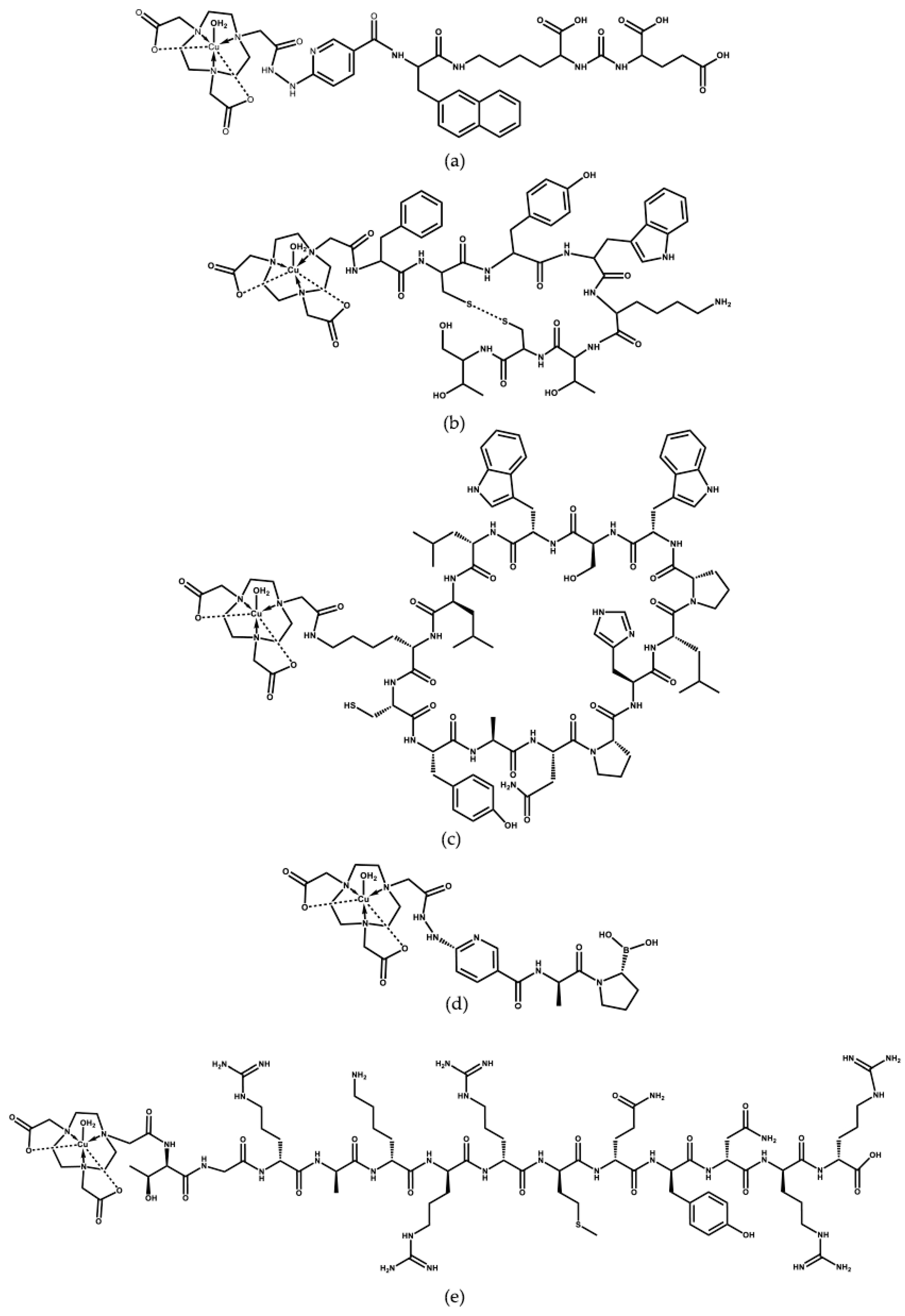
This research aimed to develop and to preclinically evaluate GMP-grade lyophilized formulations using gelatin for the one-step preparation of [64Cu]Cu-NOTA peptides, with high radiochemical purity and stability, targeting several receptors, including prostate-specific membrane antigen (PSMA; NOTA-iPSMA peptide), somatostatin receptor subtype 2 (SSTR2; NOTA-TOC peptide), programmed death ligand 1 (PD-L1; NOTA-iPD-L1 peptide), fibroblast activation protein (FAP; NOTA-iFAP peptide), and bacterial cell membranes (UBI 29–41 peptide) (Figure 1).

2. Results and Discussion

The use of 64Cu radiopharmaceuticals targeting various biomarkers in cancer diagnosis and combined therapies has significant clinical implications, particularly in enhancing diagnostic accuracy, personalizing treatment strategies, and improving patient outcomes. As shown below, 64Cu radiopharmaceuticals targeting PSMA, SSTR2, PD-L1, FAP, and bacterial membrane prepared from GMP-lyophilized gelatin-NOTA-peptide kits demonstrated high radiochemical purity, stability, and specificity, which is relevant for routine clinical use in PET nuclear medicine practice.

2.1. Kit Formulation and 64Cu Labeling

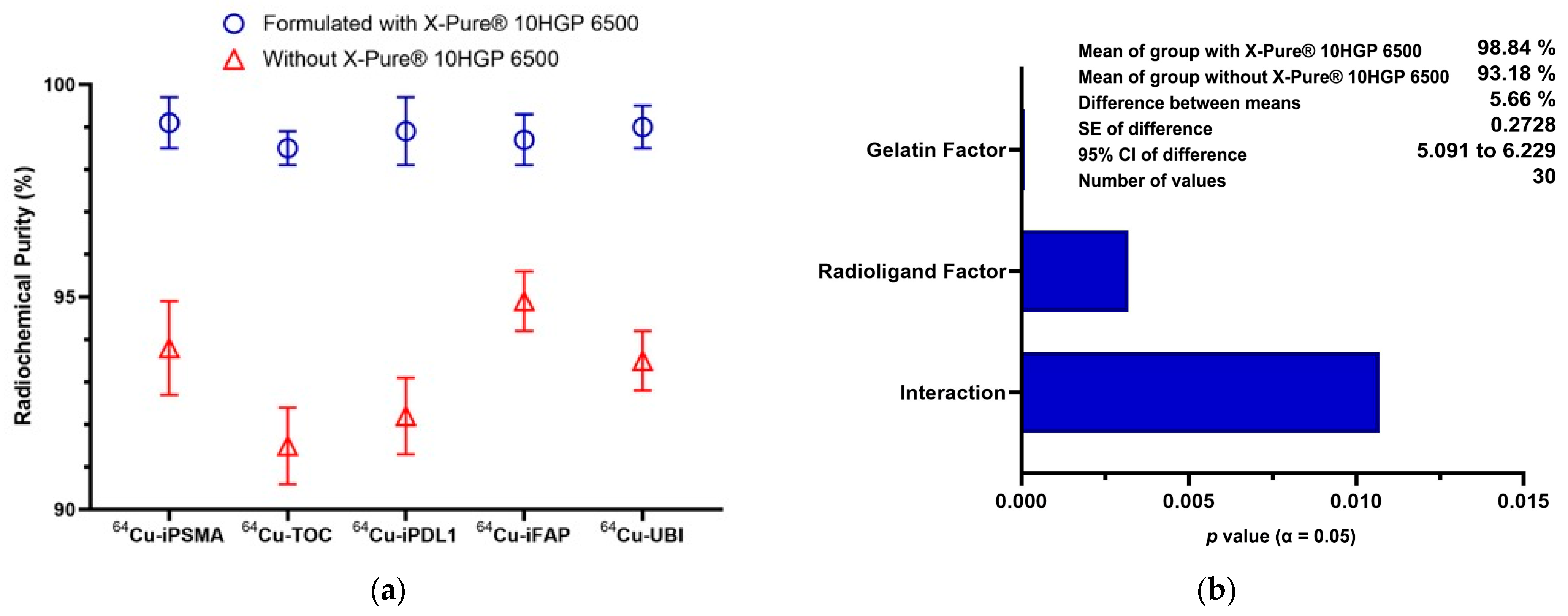
Figure 2 shows that the radiochemical purity of 64Cu radiopharmaceuticals was significantly higher (p < 0.0001) when X-PureR 10HGP 6500 gelatin was incorporated in the lyophilized formulations (mean value: 98.84%) compared to the formulation without gelatin (mean value: 93.18%) as a result of the hydrolyzed gelatin’s ability to chelate metal impurities [11,12]. While the radioligand type significantly influenced the final radiochemical purity, all 64Cu radiopharmaceuticals achieved purity levels exceeding 98%, indicating less than 2% free 64Cu. This characteristic is crucial because free 64Cu can lead to a non-specific distribution, reduced imaging accuracy, and potential toxicity due to its cytotoxic properties (β radiation) [18]. Free 64Cu may bind to unintended biological targets, such as serum albumin and accumulate in the liver, resulting in higher background signals and lower target-to-background ratios, reducing the spatial resolution and diagnostic accuracy of PET imaging [19].
The high radiochemical purities obtained also confirm the quality of the 64CuCl2 obtained from 18-MeV hospital cyclotrons in Mexico. Analysis of this 64CuCl2 revealed the chemical and radiochemical characteristics shown in Table 1. These results are consistent with the extensive studies on the production of 64Cu in hospital cyclotrons using enriched 64Ni (99%) as a solid target, which have demonstrated the ability to achieve high radionuclide and radiochemical purity, ensuring suitability for medical applications [3].

2.2. Kit Formulations Under GMP Conditions and 64Cu Labeling

After finalizing the formula, five freeze-dried kit formulations for the preparation of 64Cu radiopharmaceuticals were prepared under GMP conditions (Table A1). GMP provided a framework for maintaining consistent quality in radiopharmaceutical production. This practice included validated standard operating procedures, quality control measures, and traceability of production parameters. The GMP results ensured that even under these constraints, the products met stringent quality standards [20].
The radiopharmaceuticals 64Cu-iPSMA, 64Cu-TOC, 64Cu-iPD-L1, 64Cu-iFAP, and 64Cu-UBI exhibited the basic pharmaceutical quality characteristics required for clinical use when prepared from three distinct batches of their lyophilized formulations. These characteristics included colorless, clear, and sterile solutions; high radiochemical purity; and stability after 24 h and in human serum (Table 2). Since human serum contains physiological concentrations of Zn2+ (0.70–1.20 μg/mL), Fe2+ (0.60–1.75 μg/mL), Cu2+ (0.63–1.58 μg/mL), and Ni2+ (2 μg/L) [21], the stability observed after incubation at 37 °C for one hour with a radiochemical purity greater than 98% is evidence that Cu-NOTA-peptide complexes remain stable upon transchelation with transition metals present in blood.
All kits were prepared with the same molar amount of NOTA-peptide. Therefore, the molar activity was 925 ± 41 MBq/0.5 μmol immediately after 64Cu labeling. Each kit was designed as a multidose formulation, yielding 5 doses suitable for patient applications (185 MBq/100 nmol).
It is important to note that the radiochemical purity reported for other radiopharmaceuticals, such as 64Cu-DOTA-NT [22], 64Cu-PTSM [23], and 64Cu-NODAGA-RGD-BBN [24], is also 98–99%. However, all of them underwent purification using chromatographic cartridges after radiolabeling. In contrast, the radiochemical purity (>98%) obtained in all radiopharmaceuticals prepared in this study was achieved without any further purification process; they were all prepared by simple reconstitution of lyophilized formulations using a solution of 64CuCl2 in 1 M sodium acetate buffer at a pH of 5, and incubation at 95 °C for 10 min. In routine practice, this procedure for preparing 64Cu radiopharmaceuticals ensures the immediate availability of different PET radioligands, which can be labeled on the same day using different batches of 64CuCl2.

2.3. Preclinical Evaluation of 64Cu Radiopharmaceuticals

The in vitro preclinical evaluation was conducted using PD-L1-positive 4T1 breast cancer cells, SSTR2-positive AR42J pancreatic cancer cells, FAP-positive HT1080 human fibrosarcoma cells, and PSMA-positive LNCaP prostate cancer cells, in order to evaluate the uptake and specificity of the precursor ligands NOTA-iPD-L1, NOTA-TOC, NOTA-iFAP, and NOTA-iPSMA, respectively.
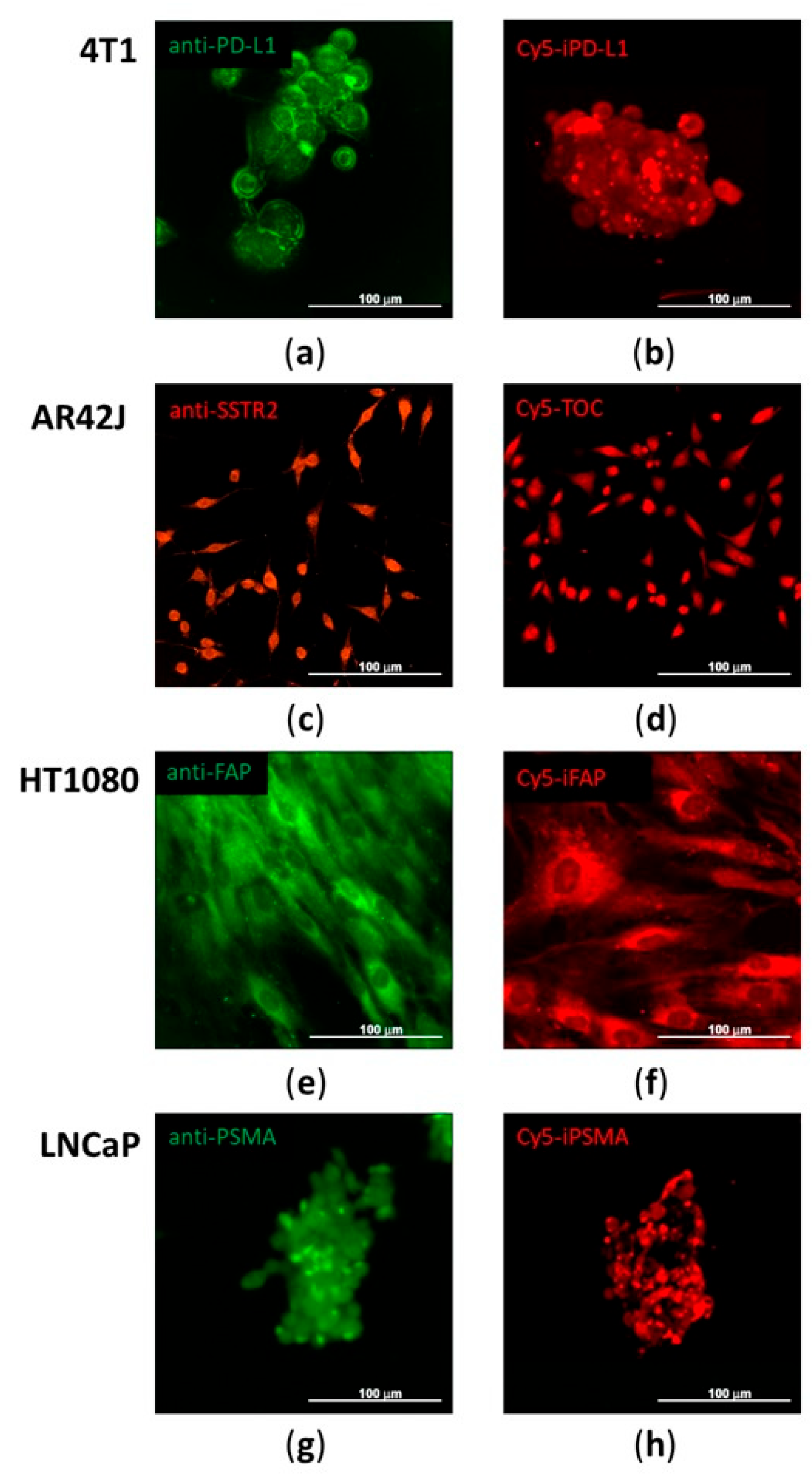
The confocal images in Figure 3 demonstrated that the molecular targets (PD-L1, SSTR2, FAP, and PSMA) were present in the cell lines expressing the respective receptors, as shown by immunodetection with a primary antibody and a fluorescently labeled secondary antibody. Furthermore, the same cell line bound the corresponding Cy5-labeled NOTA-peptide conjugate, indicating that the peptide specifically recognizes the same target identified by the antibody (Figure 3).
The positive expression of all receptors in the cells used in this research, which specifically bound Cy5-NOTA-peptides, is consistent with previous reports. For instance, Park et al. [25] established a 4T1 breast cancer model with stable PD-L1 overexpression to investigate the tumor’s response to anti-PD-L1 therapy. AR42J cells, which express SSTR2, have been widely used to study somatostatin receptor-mediated signaling and its role in cancer progression [26]. FAP overexpression in HT1080 cells has been shown to increase tumor growth and alter cell death mechanisms, including a switch from apoptosis to autophagy-dependent cell death [27]. LNCaP cells, which express PSMA, have been extensively used to study prostate cancer progression and therapeutic strategies [28].
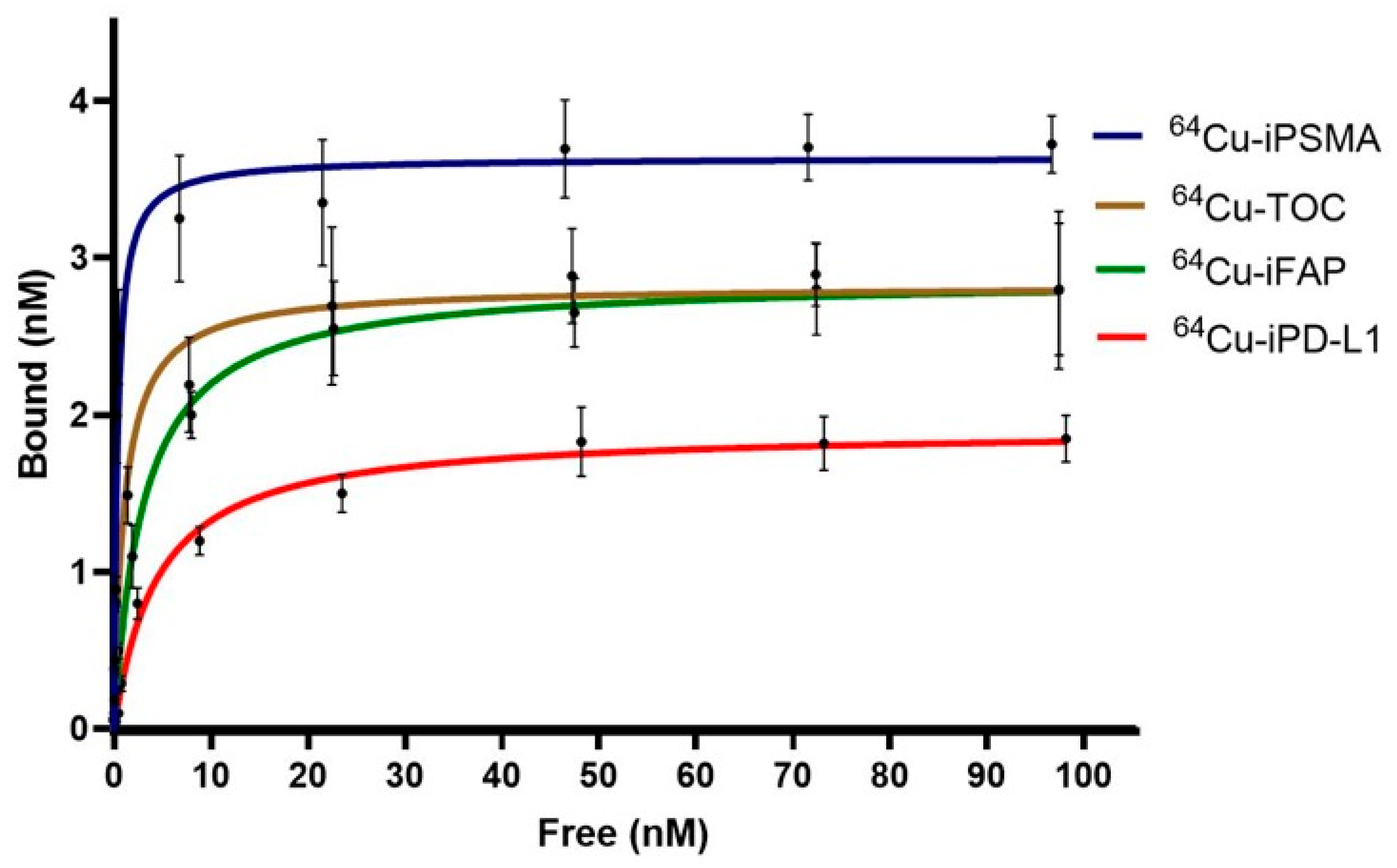
Saturation binding assays (Figure 4) demonstrated receptor-mediated, saturable binding of the kit-prepared radioligands in their corresponding target-positive cell models, supporting preservation of biological activity. Nonlinear regression analysis yielded nanomolar affinities and finite binding capacities (Table 3). 64Cu-iPSMA showed the highest affinity in LNCaP cells (Kd = 0.46 nM) with the highest Bmax (3.64 nM), followed by 64Cu-TOC in AR42J cells (Kd = 1.11 nM; Bmax = 2.83 nM). 64Cu-iFAP (HT1080) and 64Cu-iPD-L1 (4T1) also exhibited specific binding with Kd values of 3.06 nM and 4.39 nM, respectively, and comparable Bmax values (2.87 nM and 1.91 nM, respectively). Overall, the sigmoidal-to-plateau profiles and fitted Kd/Bmax parameters of all radioligands justified moving to in vivo work, as they confirmed high affinity (Kd in the nM range) and ensured proper target density or target availability (Bmax), both consistent with target-driven uptake.
A comparative assessment of the 64Cu-radiolabeled inhibitors highlights distinctions in binding affinity compared with previously published reports using other PET radionuclides. For PSMA-targeted agents, 64Cu-iPSMA exhibited the strongest affinity (Kd = 0.46 nM) compared to that reported for both 18F-PSMA-1007 (Kd ≈ 6.7 nM) and 68Ga-PSMA-11 (Kd ≈ 4.3 nM). However, all demonstrated high affinity within PSMA-expressing cell lines and robust Bmax values [29,30]. When evaluating PD-L1 inhibitors, 89Zr-DFO-KN035 (anti-PD-L1) stood out with high affinity (Kd = 1–15 nM) and elevated Bmax, while the peptide inhibitor 68Ga-NODA-CDV-Nb109 (Kd = 12.34 nM) and 64Cu-iPD-L1 (Kd = 4.39 nM) exhibited high-moderate affinity [31]. Importantly, 89Zr-labeled antibodies consistently achieved superior binding and receptor density compared to copper-64- or gallium-68-labeled small molecules, likely due to multivalent binding and enhanced tumor retention. However, in vivo pharmacokinetics have shown longer blood circulation and higher uptake in non-target organs compared with peptides. Interestingly, Begum et al. [32] reported an in silico study that systematically assessed how the half-lives of 68Ga-, 18F-, and 64Cu-labeled PSMA ligands with varying affinities affected tumor uptake in PET/CT imaging. Their results showed that the highest tumor uptake occurred at 1 h post-injection for 68Ga-PSMA, at 2 h for 18F-PSMA, and at 4–8 h for high-affinity 64Cu-PSMA. Notably, 64Cu-PSMA provided a 2.8–3.2-fold increase in tumor activity concentration compared to 68Ga-PSMA, and the optimal tumor-to-background ratio was achieved after 4 h using high-affinity 64Cu-PSMA [32].
As 64Cu-UBI is an antimicrobial peptide, it was used as a negative control for the in vitro and in vivo imaging evaluations of 64Cu-NOTA-peptides, which showed negligible uptake (<1%) in cancer cells. However, 64Cu-UBI in vitro uptake in a Staphylococcus aureus culture (107 CFU/mL) was 43.7 ± 5.7% (n = 6), in agreement with previous results [17].
As summarized in Table 3, biodistribution at 2 h post-injection showed predominant renal clearance for all radioligands, with the highest kidney uptake for 64Cu-iPD-L1 (29.36% ID/g) and 64Cu-TOC (28.57% ID/g), followed by 64Cu-iPSMA (21.72% ID/g) and 64Cu-iFAP (8.66% ID/g). Off-target liver activity was moderate for 64Cu-iPD-L1 (4.18% ID/g) and 64Cu-TOC (3.71% ID/g) and lower for 64Cu-iPSMA (2.83% ID/g) and 64Cu-iFAP (1.09% ID/g), while lung and spleen uptake remained low across tracers (≤0.53 and ≤0.94% ID/g, respectively). Intestinal activity was minimal for 64Cu-iPD-L1 and 64Cu-iPSMA (0.69 ± 0.34 and 0.19 ± 0.08% ID/g), increased for 64Cu-iFAP (0.23% ID/g), and was highest for 64Cu-TOC (2.49% ID/g). Tumor uptake ranged from 5.81% ID/g for 64Cu-iFAP to 8.17% ID/g for 64Cu-iPSMA (with 64Cu-iPD-L1 and 64Cu-TOC at 7.43 and 6.67% ID/g, respectively). In summary, tumor-to-lung ratios ranging from 14 to 290 at 2 h post-injection, with low liver uptake (2.95% ± 1.36% ID/g), support target-driven accumulation despite variable degrees of background uptake in clearance organs, as we previously reported for these ligands labeled with other radionuclides [13,14,15,16]. It is important to note that the design of dimeric 64Cu radiopharmaceuticals could further increase tumor uptake. For instance, 64Cu-iPSMA exhibited an LNCaP tumor uptake (8.17 ± 1.52% ID/g at 2 h) similar to that of 64Cu-Sar-PSMA (9 ± 1% ID/g at 1 h), which increased twice for the dimer 64Cu-Sar-bisPSMA (20% ID/g at 1 h) [33].
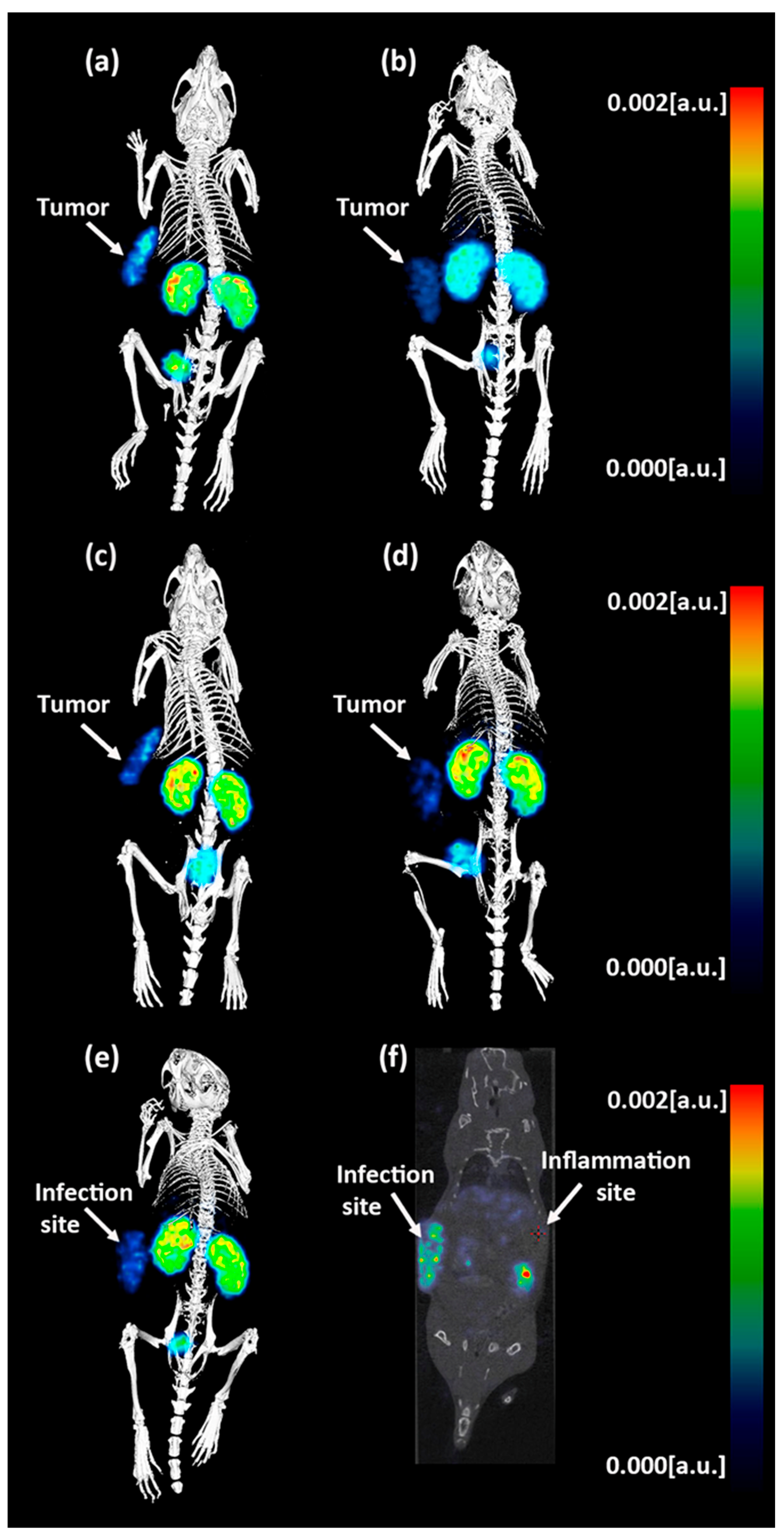
Representative micro-PET/CT images acquired 2 h post-injection (Figure 5) corroborated the in vitro specificity data and the ex vivo biodistribution profile, showing visually evident, target-driven uptake in each corresponding xenograft model. 64Cu-iPSMA produced a well-delineated LNCaP tumor signal, whereas 64Cu-iFAP showed clear localization in the HT1080 tumor, consistent with FAP expression in the tumor microenvironment. Similarly, 64Cu-TOC accumulated in the AR42J tumor, and 64Cu-iPD-L1 demonstrated uptake in the 4T1 tumor, supporting receptor-mediated retention at this imaging time point. In all cases, activity in clearance organs was also appreciable, in line with the predominant renal elimination observed for these peptide radioligands. As a negative control for tumor targeting, 64Cu-UBI did not highlight tumors. Instead, it localized to the S. aureus infection focus, and the fused CT images enabled anatomical confirmation and distinction between the infection site and the contralateral inflammation region (Figure 5).

3. Materials and Methods

3.1. Preparation of GMP-Lyophilized Gelatin-NOTA-Peptide Kits

NOTA-iPD-L1 (iPD-L1), NOTA-TOC (TOC), NOTA-iFAP (iFAP), NOTA-iPSMA (iPSMA), and NOTA-UBI 29–41 (UBI) were custom-synthesized by Yaxian Chemical (Shanghai, China) and chemically characterized by HPLC-MS (Waters, Milford, CT, USA), FTIR (ATR; PerkinElmer, Waltham, MA, USA), and reversed-phase HPLC (Shimadzu, Kyoto, Japan). Gelatin was X-Pure® 10HGP 6500, suitable for injectable applications (Rousselot BV, Belgium) (Table A2). Mannitol and ascorbic acid were supplied by Merck (Burlington, MA, USA).
In accordance with the GMP protocols, multidose lyophilized kits were produced in a sterile environment at the Radiopharmaceutical Production Plant, which has Good Manufacturing Practice certification issued by the Mexican regulatory agency COFEPRIS (certification No. 243300EL531063). iPD-L1, TOC, iFAP, iPSMA, or UBI (10 µmol) was dissolved in 10 mL of water suitable for injection, then vigorously stirred and heated to 70 °C. Separately, mannitol (0.5 g) and ascorbic acid (1.0 g) were each dissolved in another 10 mL of injectable-grade water. Once both the peptide and the mannitol/ascorbic acid mixtures were ready, they were blended, ensuring that the pH ranged between 2.5 and 3.5. The final blend was sterilized using a 0.22 µm Millipore filter. Afterward, 1 mL volumes were distributed into 20 depyrogenized ampoule vials. The vials were then lyophilized for 19 h, including freezing at −40 °C for 1 h, primary drying at −15 °C for 8 h, secondary drying at 0 °C for 4 h, and drying for 6 h each at 25 °C and 29 °C. Additional batches of all NOTA-peptides were prepared under the same conditions but with the addition of X-Pure® 10HGP 6500 gelatin (0.25 g).
In addition, a 1 M sodium acetate buffer solution at pH 5.0 was made in a volume of 30 mL, then passed through a 0.22 µm filter. Afterward, 1.5 mL volumes were dispensed into 20 ampoule vials under sterile conditions.
To validate the manufacturing process for each precursor, three sequential batches of 20 vials each were produced under uniform conditions to confirm consistency and reproducibility. All of these batches included X-Pure® 10HGP 6500 gelatin in the formulation. Quality assurance measures or process controls included checking the solution’s pH, assessing the dose volume by weighing samples (n = 3), testing filter integrity through a bubble point assay (Millipore, BP > 56 psi), and performing environmental surveillance for both non-viable and viable particulates in ISO-5 and ISO-6 zones, following the Mexican regulatory standard (NOM-241-SSA1-2025).

3.2. Quality Control and Stability

Quality control of lyophilized products involved checking parameters such as visual characteristics, color, pH, sterility, the presence of bacterial endotoxins, and radiochemical purity. The radiochemical purity was determined by reversed-phase HPLC using a 4.5 mm × 25 cm GIST C18 column with 5 μm particles (Shimadzu, Kyoto, Japan). The analysis was achieved over 30 min at a flow rate of 1 mL/min. The protocol employed a linear gradient, shifting phase A (aqueous solution containing 0.1% TFA) from 100% to 10%, while phase B (acetonitrile with 0.1% TFA) increased from 0% to 90%. The radiolabeled peptides, 64Cu-iPD-L1, 64Cu-TOC, 64Cu-iFAP, 64Cu-iPSMA, or 64Cu-UBI, prepared as described below, exhibited retention times of 13.0 ± 0.5, 12.6 ± 0.3, 10.8 ± 0.5, 11.2 ± 0.4, and 10.2 ± 0.1 min, respectively, whereas 64CuCl2 had a retention time of 3.5 ± 0.3 min. Stability assessments were conducted on each batch for 6 months following its production date. The stability of the radiolabeled compounds was assessed 24 h after production by reversed-phase HPLC. The stability of 64Cu-peptides in human serum was assessed by combining 100 µL (20 MBq) of the radiopeptide with 1 mL of certified human serum (Sigma-Aldrich, NIST-909c; St. Louis, MO, USA). Following a 1-h incubation period at 37 °C, 0.5 mL of acetonitrile was added to the mixture to precipitate serum proteins. The resulting solution was centrifuged for 10 min at 500 g. Subsequently, the radiochemical purity of the supernatant was determined by reverse-phase radio-HPLC, as previously outlined (n = 3).

3.3. Radiochemical Labeling

The radiochemical method began with the sterilization process of the ampoule vial containing the 64CuCl2 solution (925 ± 41 MBq/mL in 0.5 M HCl) (Cyclone Kiube IBA 100 μA, National Institute of Cancer, CDMX, Mexico; or Cyclone Kiube IBA 150 μA, Doctors Hospital Auna de Monterrey, NL, Mexico). The sterilization procedure has been previously validated, and the sterilized 64CuCl2 solution was assayed for radioactive concentration, specific activity, identity, radionuclide purity, chemical purity, radiochemical purity, sterility test, and bacterial endotoxin levels, using pharmacopoeia test methods (European Pharmacopoeia and United States Pharmacopeia) such as gamma-ray spectrometry with HPGe detector, ICP-MS, ionization chamber assay, thin layer chromatography, sterility test, and the LAL bacterial endotoxin test.
After sterilization, 1.5 mL of 1 M acetate buffer at pH 5.0 was added to the 64CuCl2 sterile solution. The entire mixture was collected with a sterile syringe and was subsequently used to reconstitute the lyophilized iPD-L1, TOC, iFAP, iPSMA, or UBI lyophilized kit. The reconstituted vial was heated to 95 °C for 10 min in a dry bath. Upon cooling to ambient temperature, the vial was vented, and injectable-grade water (Pisa, Mexico) was added to bring the total volume up to 10 mL using a sterile syringe. The dosing procedure was performed directly into delivery syringes (185 MBq/2 mL) with leaded glass protection or utilizing the Timo-2 dosing module from Comecer, Italy.
It is important to note that 64Cu-iPD-L1, 64Cu-TOC, 64Cu-iFAP, 64Cu-iPSMA, or 64Cu-UBI was radiolabeled in a shielded enclosure (Model Musa, Comecer, Italy) with a primary chamber, a waste section, and entry/exit compartments for materials. All sections were lined with lead blocks (98% purity, 2% Sb). The main compartment was fitted with an activity calibrator, controlled via specialized software and a touchscreen interface. Additional equipment included a UV lamp and a laminar airflow system with terminal HEPA filters (99.997% efficiency), programmed for vertical laminar flow at 0.3 m/s, ensuring ISO Class 5 cleanliness (Model Musa, Comecer, Italy). For incubation, a Cole–Parmer dry bath was positioned within the shielded cell.

3.4. Radiopharmaceutical Quality Control

To ensure the quality of the radiopharmaceuticals, samples were collected and analyzed for pH, sterility, bacterial endotoxin content, and radiochemical purity using reversed-phase HPLC with a gradient system, in accordance with the Mexican Pharmacopeia.

3.5. Cell Culture

Four mammalian cell lines representative of different cancer types were selected based on their expression of the molecular targets under study. The 4T1 (CRL-2539) breast carcinoma cells (PD-L1-positive), AR42J (CRL-1492) pancreatic cancer cells (positive for SSTR2), HT1080 (CCL-121) human fibrosarcoma cells (positive for FAP), and LNCaP (CRL-1740) human prostate adenocarcinoma cells (PSMA-positive) were cultured in RPMI 1640 medium (Roswell Park Memorial Institute Medium, Gibco Invitrogen Corp., Waltham, MA, USA) supplemented with 10% fetal bovine serum substitute (NuSeraTM, HIMEDIA, Germany) and a 1% antibiotic-antifungal solution (Merck, Burlington, MA, USA). The cultures were maintained in exponential growth under incubation conditions in a humidified atmosphere with 5% CO2 at 37 °C. For subculturing, 0.25% trypsin solution in PBS (Gibco Invitrogen Corp., USA) was used.

3.6. Saturation Binding Evaluation

Affinities of 64Cu-iPD-L1, 64Cu-TOC, 64Cu-iFAP, and 64Cu-iPSMA were assessed in the 4T1, AR42J, HT1080, and LNCaP cells, respectively. For the assessment of saturation binding, 96-well plates were seeded with 50,000 cells per well and incubated for 24 h at 37 °C in a CO2-enriched environment. Afterward, the plates were cooled for 30 min on ice, followed by a 2-h incubation at 4 °C with rising concentrations (ranging from 1 nM to 100 nM) of the 64Cu-peptide. Subsequently, cells were rinsed with PBS and lysed with 1 M NaOH. The resulting lysates were analyzed for radioactivity with a NaI(Tl) gamma counter (NML Inc., TX, USA). To measure non-specific binding, identical procedures were performed in the presence of the natCu-peptide at 1 μM. The values for dissociation constant (Kd) and maximum binding capacity (Bmax) were obtained by linear regression analysis of data (n = 3) from two separate experiments using GraphPad Prism software (version 10.6.1).

3.7. In Vitro Evaluation of 64Cu-UBI Uptake in Bacteria

For both in vitro and in vivo experiments, the microorganism Staphylococcus aureus ATCC 25923 was selected. After storage at −80 °C, the bacteria were cultured overnight at 37 °C with RPMI in a water bath with agitation. Following incubation, they were washed with 0.1 M phosphate buffer (pH 7.5) and then dispensed to obtain a bacterial suspension at approximately 107 CFU/mL.
A mixture was prepared by adding 0.1 mL of 64Cu-UBI and 0.1 mL of either bacterial (positive control) or 4T1, AR42J, HT1080, or LNCaP cell suspension (negative control) to 0.8 mL of incubation buffer (0.05 M PBS with 0.1% Tween 80 and 0.2% acetic acid, pH 5). The contents were vortexed gently and incubated at 37 °C. After a 1-h incubation with the radiolabeled peptide, samples were centrifuged at 1000 g for 5 min, the supernatant was discarded, and the pellets were resuspended in 1 mL incubation buffer before repeating the centrifugation. The supernatant was again removed, and the remaining pellet’s radioactivity was measured. The radioactivity bound to bacteria or tumor cells was reported as a percentage of the total 64Cu-UBI activity added.

3.8. Conjugation of Peptides with Cy5-NH2

Fluorescent labeling of the iPD-L1, TOC, iFAP, and iPSMA peptides was performed using the Cy5-NH2 fluorophore (Cy5 amine, MedChemExpress, Middlesex, NJ, USA). To carry out the reaction, solutions (1 mg/100 μL) of the EDC/NHS-activated NOTA-peptides and the Cy5-NH2 fluorophore (10 mM) were prepared in DMSO. To form the complex, both solutions were mixed in a 1:2 molar ratio with excess peptide and incubated at room temperature for 1 h. The binding of the fluorophore to the NOTA peptides was verified by vis-RP-HPLC using the gradient system described in Section 3.2, with the chromatogram obtained at 650 nm using a photodiode array detector (PDA).

3.9. Specific Uptake of the Peptides In Vitro

Cells from each cell line (1 × 10⁶ cells/well), adhered to 6-well plates, were incubated with Cy5-labeled peptides at a final concentration of 25 nM in culture medium for 1 h at 37 °C in the dark. To assess binding specificity, parallel incubations were performed in the presence of excess unlabeled peptide. After incubation, the medium was removed, and the cells were washed three times with PBS prior to kinetic uptake analysis using the Cytation 10 multimodal imaging system (Agilent Technologies, Santa Clara, CA, USA). Binding and internalization were visualized using a far-red excitation laser (640 nm) with 20× magnification at 1, 24, and 48 h.

3.10. Immunocytochemistry

Clean round coverslips were coated with poly-L-lysine (50 µg/mL) and incubated for 1 h at 37 °C. The solution was then removed by aspiration, and the coverslips were thoroughly washed with sterile water. The coverslips were placed in 24-well plates, and 2 × 10⁵ cells were seeded onto them, allowing for adhesion for 24 h before fixing the cultures with 4% paraformaldehyde (PFA) in PBS for 15 min at room temperature or 100% methanol. Cells permeabilized with 0.1% Triton X-100 were incubated for the immunodetection of the molecular targets (PD-L1, SSTR2, FAP, and PSMA) with the respective primary antibodies overnight at 4 °C (Abcam Limited, Cambridge, United Kingdom: anti-PD-L1 1:100, AB279292; anti-SSTR2 1:100, SC365502; anti-FAP 1:100, AB314456; anti-FOLH1 1:50, AB76104). After three washes with cold PBS, the corresponding secondary antibodies were added: Donkey anti-Mouse IgG (H&L) Alexa Fluor 594, A21203; Goat anti-Rabbit IgG (H&L) Alexa Fluor 488, A11008; Goat anti-Mouse IgG (H + L) Highly Cross-Adsorbed Alexa Fluor 488, A11029 (Thermo Fisher Scientific, Invitrogen, Waltham, MA, USA). Upon completion of the incubation with the secondary antibody, three washes with PBS were performed to remove unbound antibodies, and the preparations were mounted in Entellan (1.07961, Sigma-Aldrich, Merck KGaA, Darmstadt, Germany). For slide visualization, the Cytation 10 confocal microscopy multimodal system (Agilent 3.11 Technologies, Santa Clara, CA, USA) was used with 488 nm (green) and 594 nm (bright red) lasers at 20× magnification.

3.11. Molecular Imaging and Biodistribution

Given that the ligands used in this study have been shown to be specific for their respective receptors using other radionuclides [13,14,15,16,17], ex vivo biodistribution and tumor uptake were verified only at a single time point (2 h) (n = 3) in mice with induced tumors, immediately following micro-PET/CT imaging. In this way, the fundamental ethical principle for the humane use of animals in research (the 3Rs principle) was upheld.
To induce tumors, 8-week-old NOD/SCID mice (n = 12) received a subcutaneous injection of 1 × 10⁶ cells from 4T1 (n = 3), AR42J (n = 3), HT1080 (n = 3), or LNCaP (n = 3) cell suspensions into the left flank. Mice were housed in different identified and labeled plastic cages with a sawdust bedding, and their environment was controlled to maintain a temperature of 23–25 °C and a 12-h light–dark cycle. They were fed Purina Chow and provided with tap water ad libitum. Following the protocol approved by the ININ Internal Committee for Laboratory Animal Care Protocol No. CICUAL-01-25-02 and in accordance with the official standard (NOM-062-ZOO-1999), and once the tumor size averaged 0.3–0.4 cm in diameter, the mice were administered with 3.7 MBq/50 µL of 64Cu-iPD-L1, 64Cu-TOC, 64Cu-iFAP, or 64Cu-iPSMA via the tail vein. The mice, which had been previously anesthetized with 2% isoflurane, were scanned using a micro-PET/CT device (MiLabs VECTor5CT model, Utrecht, Netherlands) 2 h after administration of the radiopharmaceuticals (n = 3, 60 mm field of view). CT images were obtained using 600 micro-CT projections at 35 kV and 700 µA. Thereafter, the mice were euthanized (2% isoflurane), and tissues and organs, including the tumor, intestine, liver, spleen, lung, kidney, and heart, were extracted. Their radioactivity uptake was measured using a sodium iodide detector activated with thallium. The percentage of radioactivity in each tissue (% ID/g; mean ± SD) was determined relative to the initial injected dose, which was set to 100% of the administered activity. 64Cu-UBI is an antimicrobial peptide and was used as a negative control for the in vivo imaging evaluations of 64Cu-NOTA-peptides in tumors.

3.12. Inflammation and Infection Induction in Mice

64Cu-UBI was verified regarding its ability to target bacteria in vivo. To induce infection, 0.1 mL of the bacterial suspension (S. aureus; 107 CFU/mL) was subcutaneously injected into the left flank of 3 NOD/SCID mice. To induce inflammation, heat-killed bacteria (endotoxins, lipopolysaccharides) were used. Gram-negative bacteria (Klebsiella pneumoniae, sp.; 108 CFU/mL) in saline solution were heated at 100 °C for 2 h and aseptically dispensed into sterile vials in 1 mL aliquots. These were subjected to a few freezing and thawing cycles. Samples were checked for the absence of bacterial growth after culturing. Finally, 0.1 mL of such heat-killed bacterial suspension was injected into the right flank of the same 3 mice that had been injected for infection induction. After 24 h of inflammation and infection induction, 3.7 MBq/50 µL of 64Cu-UBI was administered intravenously in the tail vein of mice. After 2 h post-injection, the animals were anesthetized with 2% isoflurane and scanned using a micro-PET/CT device as described above.

4. Conclusions

GMP-grade lyophilized gelatin-NOTA-peptide kits enabled a simple, one-step preparation of 64Cu-labeled radiopharmaceuticals targeting PSMA, SSTR2, PD-L1, FAP, and bacterial membranes, yielding consistently high radiochemical purity (≥98%) without the need for post-labeling purification. The incorporation of hydrolyzed gelatin as a scavenger of metal impurities improved labeling performance and supported product stability in serum and over time while maintaining nanomolar receptor affinities in target-positive cancer cell lines. In vivo, the kit-prepared radioligands exhibited target-driven uptake in the corresponding tumor models and a biodistribution profile compatible with peptide-based pharmacokinetics, characterized by predominant renal clearance and limited non-target organ retention. These results support gelatin-NOTA-peptide kits as a practical GMP-compatible platform to expand the routine availability of 64Cu PET radiotracers, facilitating centralized production, flexible imaging schedules, and broader clinical translation of peptide-based molecular imaging probes.

Author Contributions

Conceptualization, G.F.-F. and M.L.-G.; funding acquisition, G.F.-F.; methodology, C.H.-G., N.L.-A., B.O.-G., C.S.-C., E.A.-V., N.J.-M., G.F.-F., M.L.-G. and L.M.-A.; formal analysis, C.H.-G., N.L.-A., B.O.-G., C.S.-C., E.A.-V., N.J.-M., G.F.-F., M.L.-G. and L.M.-A.; writing—original draft preparation, G.F.-F.; writing—review and editing, G.F.-F. and M.L.-G. All authors are responsible for all aspects of the work. All authors have read and agreed to the published version of the manuscript.

Funding

This research was funded by the Secretary of Science, Humanities, Technology, and Innovation (SECIHTI, Mexico), grant number MADTEC-2025-M-135. This study was also supported by the International Atomic Energy Agency (CRP-F22078; MEX6013).

Institutional Review Board Statement

All applicable institutional and international guidelines for the use and care of animals were followed. This research was approved by the Ethics Internal Committee of Use and Care of Laboratory Animals (CICUAL-ININ) of the National Institute of Nuclear Research, Approval No. CICUAL-01-25-02, 4 February 2025.

Informed Consent Statement

Not applicable.

Data Availability Statement

Data are contained within the article.

Acknowledgments

The authors thank the “Laboratorio Nacional de Investigaciones en Forense Nuclear” (LANAFONU) for the facilities provided for the ICP-MS analyses.

Conflicts of Interest

The authors declare no conflicts of interest.

Abbreviations

The following abbreviations are used in this manuscript:
Cy5Cyanine 5
FAPFibroblast activation protein
iFAPFibroblast activation protein inhibitor
GMPGood Manufacturing Practices
HPLCHigh-performance liquid chromatography
PD-L1Programmed death ligand 1
iPD-L1Programmed death ligand 1 inhibitor
SSTR2Somatostatin receptor subtype 2

Appendix A

Table A1. Controls of the production process and environmental monitoring during the preparation of lyophilized formulations as precursors of 64Cu radiopharmaceuticals.
ParametersSpecificationAverage of Three Production Batches
iPSMATOCiPDL1iFAPUBI
Formulation for freeze-drying
pH of the final mixture2.5–3.52.6 ± 0.22.5 ± 0.12.5 ± 0.12.5 ± 0.12.5 ± 0.1
Volume (determined by weight) (g)2.01.997–2.0062.003–2.0102.003–2.0101.998–2.0051.996–2.004
Filter integrity (bubble point test)>56 psi79.10 ± 0.3778.99 ± 0.4179.25 ± 0.3379.85 ± 0.1279.19 ± 0.22
1 M Sodium acetate buffer solution
pH of the buffer4.5–5.05.0 ± 0.05.0 ± 0.04.9 ± 0.14.9 ± 0.15.0 ± 0.0
Buffer volume (determined by weight) (g)1.4985–1.5015 (1.5 ± 0.1%)1.501–1.5091.501–1.5121.502–1.5111.502–1.5081.500–1.512
Filter integrity (bubble point test)>56 psi77.10 ± 0.7479.14 ± 0.2978.42 ± 0.6178.16 ± 0.4779.18 ± 0.36
Environmental
monitoring
ISO-5
Viable particles
(CFU)
Sedimentation ≤ 1 m300000
Contact ≤ 1 per plate00000
Air ≤ 5 per plate00000
Total particles/m30.5 µm ≤ 35201–2001–20
5.0 µm ≤ 2911–2000
ISO-6
Viable particles
(CFU)
Sedimentation ≤ 10 m30–10–1000
Contact ≤ 5 per plate0–11–21–20–20–1
Air ≤ 5 per plate1–32–31–21–31–2
Total particles/m30.5 µm ≤ 35,200651 ± 102702 ± 87515 ± 52589 ± 68593 ± 49
5.0 µm ≤ 29318 ± 417 ± 821 ± 620 ± 519 ± 8
Table A2. Physical, chemical, and microbial limits for X-Pure® 10HGP 6500: a non-gelling gelatin extracted from pig skin suitable for vaccine and injectable applications (Supplier: Rousselot BV, Belgium).
Standard ParametersSpecification CompliantTest Method
Endotoxin level≤10 EU/gLAL assay 1
Molecular weight≤6500 DaltonsRousselot
pH (1%, 55 °C)4.0–5.5EP, USP
Loss on drying≤7.0%EP, USP
Conductivity≤1000 μS.cm−1EP, USP
Residue limits
Iron≤30 ppmEP, USP
Chromium≤10 ppmEP, USP
Zinc≤30 ppmEP, USP
Arsenic≤0.8 ppmEP, USP
Heavy metals≤20 ppmEP, USP
Sulfites (SO2)≤20 ppmEP, USP, JP
Peroxides≤10 ppmEP, USP
Microbial limitsSpecificationsTest Method
Total aerobic microbial count-TAMC≤100 CFU/gEP, USP
Total yeasts and molds-TYMC≤10 CFU/gEP, USP
SalmonellaAbsence in 10 gEP, USP
E. coliAbsence in 1 gEP, USP
Pseudomonas aeruginosaAbsence in 1 gEP, USP
1 LAL assay (limulus amebocyte lysate), an FDA compliant method.

References

  1. Thieme, S.; Walther, M.; Pietzsch, H.J.; Henniger, J.; Preusche, S.; Mäding, P.; Steinbach, J. Module-assisted preparation of 64Cu with high specific activity. Appl. Radiat. Isot. 2012, 70, 602–608. [Google Scholar] [CrossRef]
  2. Hazari, P.P.; Singh, S.; Panchal, K.; Kumar, N.; Chaturvedi, S.; Singh, H. Production and Quality control of Cyclotron produced Copper-64: A Validation study for the use of 64CuCl2 as a radiochemical precursor for radiolabeling of ligands. J. Nucl. Med. 2025, 66, 252258. [Google Scholar]
  3. Jauregui-Osoro, M.; De Robertis, S.; Halsted, P.; Gould, S.M.; Yu, Z.; Paul, R.L.; Marsden, P.K.; Gee, A.D.; Fenwick, A.; Blower, P.J. Production of copper-64 using a hospital cyclotron: Targetry, purification and quality analysis. Nucl. Med. Commun. 2021, 42, 1024–1038. [Google Scholar] [CrossRef]
  4. Delpassand, E.S.; Ranganathan, D.; Wagh, N.; Shafie, A.; Gaber, A.; Abbasi, A.; Kjaer, A.; Tworowska, I.; Núñez, R. 64Cu-DOTATATE PET/CT for Imaging Patients with Known or Suspected Somatostatin Receptor-Positive Neuroendocrine Tumors: Results of the First U.S. Prospective, Reader-Masked Clinical Trial. J. Nucl. Med. 2020, 61, 890–896. [Google Scholar] [CrossRef]
  5. Braune, A.; Oehme, L.; Freudenberg, R.; Hofheinz, F.; van den Hoff, J.; Kotzerke, J.; Hoberück, S. Comparison of image quality and spatial resolution between 18F, 68Ga, and 64Cu phantom measurements using a digital Biograph Vision PET/CT. EJNMMI Phys. 2022, 9, 58. [Google Scholar] [CrossRef] [PubMed]
  6. Kubíček, V.; Böhmová, Z.; Ševčíková, R.; Vaněk, J.; Lubal, P.; Poláková, Z.; Michalicová, R.; Kotek, J.; Hermann, P. NOTA Complexes with Copper(II) and Divalent Metal Ions: Kinetic and Thermodynamic Studies. Inorg. Chem. 2018, 57, 3061–3072. [Google Scholar] [CrossRef] [PubMed]
  7. Kubinec, J.; Širůčková, V.; Havlíčková, J.; Kotek, J.; Kubíček, V.; Lubal, P.; Hermann, P. Complexes of NOTA-Monoamides with CuII Ions: Structural, Equilibrium, and Kinetic Study. Eur. J. Inorg. Chem. 2022, 19, e202200173. [Google Scholar] [CrossRef]
  8. Zhang, Y.; Hong, H.; Engle, J.W.; Bean, J.; Yang, Y.; Leigh, B.R.; Barnhart, T.E.; Cai, W. Positron emission tomography imaging of CD105 expression with a 64Cu-labeled monoclonal antibody: NOTA is superior to DOTA. PLoS ONE 2011, 6, e28005. [Google Scholar] [CrossRef] [PubMed]
  9. Liu, H.; Zhang, X.; Zhang, J.; Pan, Y.; Wen, H.; Xu, X.; Wu, S.; Wang, Y.; Zhang, C.; Ma, G.; et al. Comparison of 64Cu-DOTA-PSMA-3Q and 64Cu-NOTA-PSMA-3Q utilizing NOTA and DOTA as bifunctional chelators in prostate cancer: Preclinical assessment and preliminary clinical PET/CT imaging. Eur. J. Nucl. Med. Mol. Imaging 2025, 52, 2792–2803. [Google Scholar] [CrossRef]
  10. Shabsigh, M.; Solomon, L.A. Peptide PET Imaging: A Review of Recent Developments and a Look at the Future of Radiometal-Labeled Peptides in Medicine. Chem. Biomed. Imaging 2024, 2, 615–630. [Google Scholar] [CrossRef]
  11. Jiliding Marine Biotech Co., Ltd. Method for Removing Heavy Metal Ions by Using Gelatin. CN104946138A; Publication date 30 September 2015, Available online: https://patents.google.com/patent/CN104946138A/en (accessed on 1 February 2026).
  12. Mikhailov, O.V. Gelatin as It Is: History and Modernity. Int. J. Mol. Sci. 2023, 24, 3583. [Google Scholar] [CrossRef]
  13. Ferro-Flores, G.; Luna-Gutiérrez, M.; Ocampo-García, B.; Santos-Cuevas, C.; Azorín-Vega, E.; Jiménez-Mancilla, N.; Orocio-Rodríguez, E.; Davanzo, J.; García-Pérez, F.O. Clinical translation of a PSMA inhibitor for 99mTc-based SPECT. Nucl. Med. Biol. 2017, 48, 36–44. [Google Scholar] [CrossRef]
  14. Trujillo-Benítez, D.; Luna-Gutiérrez, M.; Ferro-Flores, G.; Ocampo-García, B.; Santos-Cuevas, C.; Bravo-Villegas, G.; Morales-Ávila, E.; Cruz-Nova, P.; Díaz-Nieto, L.; García-Quiroz, J.; et al. Design, Synthesis and Preclinical Assessment of 99mTc-iFAP for In Vivo Fibroblast Activation Protein (FAP) Imaging. Molecules 2022, 27, 264. [Google Scholar] [CrossRef]
  15. Ferro-Flores, G.; Ocampo-García, B.; Cruz-Nova, P.; Luna-Gutiérrez, M.; Bravo-Villegas, G.; Azorín-Vega, E.; Jiménez-Mancilla, N.; Michel-Sánchez, E.; García-Pérez, O.; Lara-Almazán, N.; et al. 99mTc-Labeled Cyclic Peptide Targeting PD-L1 as a Novel Nuclear Imaging Probe. Pharmaceutics 2023, 15, 2662. [Google Scholar] [CrossRef]
  16. Medina-García, V.; Ocampo-García, B.E.; Ferro-Flores, G.; Santos-Cuevas, C.L.; Aranda-Lara, L.; García-Becerra, R.; Ordaz-Rosado, D.; Melendez-Alafort, L. A freeze-dried kit formulation for the preparation of Lys(27)(99mTc-EDDA/HYNIC)-Exendin(9-39)/99mTc-EDDA/HYNIC-Tyr3-Octreotide to detect benign and malignant insulinomas. Nucl. Med. Biol. 2015, 42, 911–916. [Google Scholar] [CrossRef] [PubMed]
  17. Ferro-Flores, G.; Arteaga de Murphy, C.; Pedraza-López, M.; Meléndez-Alafort, L.; Zhang, Y.M.; Rusckowski, M.; Hnatowich, D.J. In vitro and in vivo assessment of 99mTc-UBI specificity for bacteria. Nucl. Med. Biol. 2003, 30, 597–603. [Google Scholar] [CrossRef]
  18. Paterson, B.M.; Donnelly, P.S. Macrocyclic bifunctional chelators and conjugation strategies for copper-64 radiopharmaceuticals. In Insights from Imaging in Bioinorganic Chemistry: Advances in Inorganic Chemistry; van Eldik, R., Hubbard, C.D., Eds.; Academic Press: San Diego, CA, USA, 2016; pp. 223–251. [Google Scholar] [CrossRef]
  19. Zhou, Y.; Li, J.; Xu, X.; Zhao, M.; Zhang, B.; Deng, S.; Wu, Y. 64Cu-based Radiopharmaceuticals in Molecular Imaging. Technol. Cancer Res. Treat. 2019, 18, 1533033819830758. [Google Scholar] [CrossRef] [PubMed]
  20. Faivre-Chauvet, A.; Bourdeau, C.; Bourgeois, M. Radiopharmaceutical good practices: Regulation between hospital and industry. Front. Nucl. Med. 2022, 2, 990330. [Google Scholar] [CrossRef]
  21. Bal, W.; Christodoulou, J.; Sadler, P.J.; Tucker, A. Multi-metal binding site of serum albumin. J. Inorg. Biochem. 1998, 70, 33–39. [Google Scholar] [CrossRef] [PubMed]
  22. Tudoroiu-Cornoiu, M.R.; Chilug, E.L.; Cocioabă, D.; Băruţă, S.; Şerban, R.; Ion, A.C.; Niculae, D. Assessment of chromatography separation parameters in the quality control of copper-64-labeled neurotensin-like peptides. J. Radioanal. Nucl. Chem. 2024, 333, 5531–5543. [Google Scholar] [CrossRef]
  23. Green, M.A.; Mathias, C.J. Synthesis and formulation of [64Cu]Cu-PTSM for PET perfusion imaging in small animal models. Appl. Radiat. Isot. 2022, 182, 110119. [Google Scholar] [CrossRef] [PubMed]
  24. Amraee, N.; Alirezapour, B.; Hosntalab, M.; Hadadi, A.; Yousefnia, H. Development of [64Cu]Cu-NODAGA-RGD-BBN as a Novel Radiotracer for Dual Integrin and GRPR-targeted Tumor PET Imaging. Curr. Radiopharm. 2025, 18, 37–44. [Google Scholar] [CrossRef]
  25. Park, A.Y.; Kim, J.H.; Lee, S.; Kim, H.S.; Kim, H.K.; Lee, H.B.; Han, W. Impact of PD-L1 upregulation on immune checkpoint inhibitor efficacy in triple-negative breast cancer using a 4T1 murine model. Int. J. Oncol. 2025, 67, 54. [Google Scholar] [CrossRef]
  26. Liu, A.M.; Wong, Y.H. Activation of nuclear factor {kappa}B by somatostatin type 2 receptor in pancreatic acinar AR42J cells involves G{alpha}14 and multiple signaling components: A mechanism requiring protein kinase, C.; calmodulin-dependent kinase, I.I.; ERK, and c-Src. J. Biol. Chem. 2005, 280, 34617–34625. [Google Scholar] [CrossRef] [PubMed]
  27. Baird, S.K.; Rigopoulos, A.; Cao, D.; Allan, L.; Renner, C.; Scott, F.E.; Scott, A.M. Integral membrane protease fibroblast activation protein sensitizes fibrosarcoma to chemotherapy and alters cell death mechanisms. Apoptosis 2015, 20, 1483–1498. [Google Scholar] [CrossRef]
  28. Kranzbühler, B.; Salemi, S.; Umbricht, C.A.; Deberle, L.M.; Müller, C.; Burger, I.A.; Hermanns, T.; Sulser, T.; Eberli, D. Concentration-dependent effects of dutasteride on prostate-specific membrane antigen (PSMA) expression and uptake of 177 Lu-PSMA-617 in LNCaP cells. Prostate 2019, 79, 1450–1456. [Google Scholar] [CrossRef]
  29. Cardinale, J.; Schäfer, M.; Benešová, M.; Bauder-Wüst, U.; Leotta, K.; Eder, M.; Neels, O.C.; Haberkorn, U.; Giesel, F.L.; Kopka, K. Preclinical Evaluation of 18F-PSMA-1007, a New Prostate-Specific Membrane Antigen Ligand for Prostate Cancer Imaging. J. Nucl. Med. 2017, 58, 425–431. [Google Scholar] [CrossRef]
  30. Butakova, N.S.; Uspenskaia, A.A.; Zyk, N.Y.; Petrov, S.A.; Popovicheva, K.A.; Lunev, A.S.; Petrosova, K.A.; Mitrofanov, I.A.; Ivashkovskaya, M.N.; Evteev, S.A.; et al. A series of conjugates based on prostate-specific membrane antigen ligands with the chelating agent DOTA: Synthesis, radiolabeling, and biological activity. Eur. J. Med. Chem. 2026, 303, 118433. [Google Scholar] [CrossRef] [PubMed]
  31. He, H.; Qi, X.; Fu, H.; Xu, J.; Zheng, Q.; Chen, L.; Zhang, Y.; Hua, H.; Xu, W.; Xu, Z.; et al. Imaging diagnosis and efficacy monitoring by [89Zr]Zr-DFO-KN035 immunoPET in patients with PD-L1-positive solid malignancies. Theranostics 2024, 14, 392–405. [Google Scholar] [CrossRef]
  32. Begum, N.J.; Glatting, G.; Eiber, M.; Beer, A.J.; Kletting, P. An in silico study on the effect of the radionuclide half-life on PET/CT imaging with PSMA-targeting radioligands. Nuklearmedizin 2021, 60, 33–37. [Google Scholar] [CrossRef] [PubMed]
  33. Zia, N.A.; Cullinane, C.; Van Zuylekom, J.K.; Waldeck, K.; McInnes, L.E.; Buncic, G.; Haskali, M.B.; Roselt, P.D.; Hicks, R.J.; Donnelly, P.S. A Bivalent Inhibitor of Prostate Specific Membrane Antigen Radiolabeled with Copper-64 with High Tumor Uptake and Retention. Angew. Chem. Int. Ed. Engl. 2019, 58, 14991–14994. [Google Scholar] [CrossRef] [PubMed]
Figure 1. Schematic chemical representation of the complexes (a) [64Cu]Cu-NOTA-iPSMA (64Cu-iPSMA), (b) [64Cu]Cu-NOTA-TOC (64Cu-TOC), (c) [64Cu]Cu-NOTA-iPD-L1 (64Cu-iPD-L1), (d) [64Cu]Cu-NOTA-iFAP (64Cu-iFAP), and (e) [64Cu]Cu-NOTA-UBI 29–41 (64Cu-UBI).
Inorganics 14 00146 g001
Figure 2. (a) Comparison of mean radiochemical purities of 64Cu radiopharmaceuticals obtained from lyophilized formulations containing X-PureR 10HGP 6500 gelatin (group means: 98.84%) and without gelatin (group means: 93.18%) (p < 0.0001). (b) Percentage of total variation explained by each factor. Gelatin contributed the largest proportion of variability (87.75%, p < 0.0001), followed by the type of radioligand and the interaction; both were statistically significant (the bars indicate the mean ± standard error) (two-way ANOVA).
Inorganics 14 00146 g002
Figure 3. Immunodetection of molecular targets was performed as follows: (a) PD-L1 was detected in 4T1 cells using anti-PD-L1 antibodies; (c) SSTR2 was identified in AR42J cells with anti-SSTR2 antibodies; (e) FAP was assessed in HT1080 cells utilizing anti-FAP antibodies; and (g) PSMA was evaluated in LNCaP cells with anti-PSMA antibodies. Alexa Fluor 488 (green) secondary antibodies were employed, except for SSTR2 detection, which used Alexa Fluor 594 (bright red) secondary antibodies. Specific binding of fluorescent peptide conjugates, (b) Cy5-iPD-L1, (d) Cy5-TOC, (f) Cy5-iFAP, and (h) Cy5-iPSMA, was visualized by fluorescence microscopy (scale bar = 100 µm) at 20× magnification, using the Cytation 10 confocal microscopy multimodal system (Agilent Technologies, Santa Clara, CA, USA).
Inorganics 14 00146 g003
Figure 4. Saturation binding curves of radioligands prepared from GMP-lyophilized gelatin-NOTA-peptide kits in target-positive cell lines: ( 64Cu-iPD-L1 in 4T1 cells (PD-L1), 64Cu-iPSMA in LNCaP cells (PSMA), 64Cu-TOC in AR42J cells (SSTR2), and 64Cu-iFAP in HT1080 cells (FAP). Cell-associated activity (specific binding) is shown as a function of radioligand concentration, and the data were fitted using a one-site binding model to estimate Kd and Bmax (Table 3).
Inorganics 14 00146 g004
Figure 5. Micro-PET/CT molecular imaging conducted 2 h post-administration demonstrated: (a) notable accumulation of 64Cu-iPSMA in the LNCaP tumor; (b) significant uptake of 64Cu-iFAP in the HT1080 tumor; (c) 64Cu-TOC localized in the AR42J tumor; (d) 64Cu-iPD-L1 detected in the 4T1 tumor; (e) 64Cu-UBI concentrated at the infection site (S. aureus); (f) CT imaging distinctly visualizes the infection site (left side) with 64Cu-UBI uptake and no accumulation of 64Cu-UBI in the inflammation site (right side).
Inorganics 14 00146 g005
Table 1. Analytical results of copper chloride, 64CuCl2, solutions (n = 3 batches) used for the labeling of peptide ligands formulated as lyophilized kits prepared under GMP conditions.
TestSpecificationTest Method (EP, USP)Result
Radioactive concentration/activity per vial925 ± 50 MBq/mLIonization chamber925 ± 41 MBq/mL
Specific activity≥3.7 GBq/μg CuGamma-ray spectrometry with HPGe detector/ICP-MS3.7 GBq/µg Cu
Identity of 64Cu511 keV and 1345 keVGamma-ray spectrometry with HPGe detectorComplies
Radionuclidic purity at EOB≥99%Gamma-ray spectrometry with HPGe detector99.96 ± 0.03
Chemical purityCo, Ni ≤ 0.1 µg/GBq
Pb ≤ 0.5 µg/GBq
Fe, Zn ≤ 1.0 µg/GBq
ICP-MS/
Ionization chamber
Ni 0.002 µg/GBq
Co < 0.001 µg/GBq
Pb < 0.002 µg/GBq
Zn 0.043 µg/GBq
Fe 0.012 µg/GBq
Radiochemical purity≥98%Thin-layer chromatography (TLC) using silica gel-coated plates as
stationary phase and methanol as mobile phase.
98.8 ± 0.6%
Sterility testSterileSterility testSterile
Bacterial endotoxin level<20 EU/mLLAL bacterial endotoxin testComplies
Table 2. Characteristics of 64Cu-iPSMA, 64Cu-TOC, 64Cu-iPD-L1, 64Cu-iFAP, and 64Cu-UBI radiopharmaceuticals (n = 3 batches for each one) obtained from lyophilized gelatin-NOTA-peptide kits prepared under GMP conditions.
ParametersSpecification
FEUM *
Average of Three Production Batches
64Cu-iPSMA64Cu-TOC64Cu-iPDL164Cu-iFAP64Cu-UBI
AppearanceLimpid solutionLimpid solutionLimpid solutionLimpid solutionLimpid solutionLimpid solution
ColorColorlessColorlessColorlessColorlessColorlessColorless
pH4.0–6.05.0 ± 0.05.0 ± 0.05.0 ± 0.05.0 ± 0.05.0 ± 0.0
SterilityNegativeNegativeNegativeNegativeNegativeNegative
Bacterial endotoxins<175 UE/V<0.625 UE/V<0.625 UE/V<0.625 UE/V<0.625 UE/V<0.625 UE/V
Radiochemical purity>97%99.1 ± 0.6%98.5 ± 0.4%98.9 ± 0.8%98.7 ± 0.6%99.0 ± 0.5%
Radiochemical purity at 24 h>97%98.8 ± 0.4%99.0 ± 0.6%98.5 ± 0.9%98.1 ± 0.5%98.7 ± 0.4%
Radiochemical purity after 1 h in human serum at 37 °C.>97%98.3 ± 0.7%98.7 ± 0.5%98.8 ± 0.3%98.3 ± 0.7%98.2 ± 0.6%
* Pharmacopoeia of the United Mexican States.
Table 3. Preclinical results (n = 3) of 64Cu-iPD-L1, 64Cu-iPSMA, 64Cu-TOC, and 64Cu-iFAP obtained from GMP-lyophilized gelatin-NOTA-peptide kits evaluated in 4T1, LNCaP, AR42J, and HT1080 cells and tumors (induced in NOD/SCID mice), respectively.
RadioligandKd (nM)
(95% CI)
Bmax (nM)
(95% CI)
Biodistribution (Tissue, % ID/g at 2 h)
LungKidneyIntestineLiverSpleenTumor
64Cu-iPDL14.39
(3.35 to 5.77)
1.91
(1.82 to 2.02)
0.53 ± 0.2129.36 ± 3.220.69 ± 0.344.18 ± 1.530.94 ± 0.277.43 ± 1.77
64Cu-iPSMA0.46
(0.38 to 0.57)
3.64
(3.44 to 3.83)
0.23 ± 0.1121.72 ± 2.310.19 ± 0.082.83 ± 1.190.63 ± 0.178.17 ± 1.52
64Cu-TOC1.11
(0.65 to 1.87)
2.83
(2.64 to 3.02)
0.24 ± 0.1628.57 ± 2.382.49 ± 0.643.71 ± 0.940.56 ± 0.136.67 ± 2.14
64Cu-iFAP3.06
(2.31 to 4.07)
2.87
(2.73 to 3.02)
0.02 ± 0.018.66 ± 1.450.23 ± 0.111.09 ± 0.210.14 ± 0.055.81 ± 1.27
Disclaimer/Publisher’s Note: The statements, opinions and data contained in all publications are solely those of the individual author(s) and contributor(s) and not of MDPI and/or the editor(s). MDPI and/or the editor(s) disclaim responsibility for any injury to people or property resulting from any ideas, methods, instructions or products referred to in the content.