Article

Engineering Perspectives on Medical Lasers: Gain-Medium Classification, Laser–Tissue Interaction, Dosimetry, Clinical Applications, and Future Intelligent Systems


1
Institute of Cited Scientists, Nicosia 2000, Cyprus
2
Faculty of Mechanical and Electrical Engineering, University of Petrosani, 332006 Petrosani, Romania
*
Author to whom correspondence should be addressed.
Photonics 2026, 13(6), 524; https://doi.org/10.3390/photonics13060524
Submission received: 30 March 2026 / Revised: 13 May 2026 / Accepted: 20 May 2026 / Published: 28 May 2026
(This article belongs to the Special Issue Laser Technology and Applications, 2nd Edition)

Abstract

Medical lasers are a heterogeneous class of interventional and therapeutic devices. They are differentiated based on their active gain medium, which includes solid-state, gaseous, dye, and semiconductor (diode) formulations. The present article undertakes a systematic evaluation and synthesis of the findings from a reproducible dataset. The present study yields novel scientific results, including a four-level classification of medical lasers that considers the chemical formula for each type of gain medium. In addition, a multisided systemic analysis of the engineering application of medical lasers in clinical practice is conducted, including an analysis of the main engineering challenges as a structured framework. Furthermore, a clustering of engineering applications for medical lasers in 2025 is performed, and a quantitative landscape of medical lasers by variables is presented. The following variables are analyzed: wavelength (nm), power (W)/irradiance (W/cm2), fluence (J/cm2), and exposure time/pulse duration. The objective is to create a year-by-year “trend analysis” for future engineering opportunities (2026–2030). The structure of the article is logical and roughly follows the IMRAD structure, and a thread of argumentation is demonstrated.

1. Introduction

The physical basis of laser technology emerged from Einstein’s theory of stimulated emission and was translated into practical devices through the maser–laser work of Townes, Basov, and Prokhorov [1]. Once engineering constraints were overcome, medical applications followed rapidly, especially in ophthalmology, dermatology, and surgery. Since their introduction into clinical practice in the 1960s, lasers have evolved from experimental devices into indispensable tools in modern medicine. Their value lies in the ability to deliver spatially controlled energy to tissue, enabling cutting, coagulation, ablation, fragmentation, or cellular stimulation depending on wavelength, pulse structure, and dose [2,3,4].
Early clinical experience established that lasers were not a single therapeutic category but a family of devices with different active media, beam characteristics, and tissue effects. This distinction remains central today: clinical outcomes depend less on the label ‘laser’ than on whether a specific platform is matched appropriately to the intended target and procedure.
The guiding framework for most interventional applications is selective photothermolysis, whereby wavelength is matched to a target chromophore such as water, hemoglobin, or melanin, and pulse duration is adjusted relative to the target’s thermal relaxation time to confine damage to the desired structure [3,4,5].
In high-power applications, lasers function as precise surgical instruments that can vaporize tissue, achieve hemostasis, and reduce mechanical trauma. These properties explain their widespread use in skin resurfacing, oncologic debulking, retinal photocoagulation, and lithotripsy, where precision and controlled collateral effects are critical [3,4,5].
At lower irradiances, lasers and other coherent or quasi-coherent light sources are also used for photobiomodulation (PBM). PBM aims to influence cellular metabolism and tissue repair rather than to cut or destroy tissue, but optimal dosimetry, mechanisms of action, and the reproducibility of clinical outcomes remain active areas of investigation [2,6,7,8].
Current medical use spans dermatology, dentistry, ophthalmology, urology, rehabilitation medicine, and selected oncologic and surgical applications. In each domain, however, efficacy must be balanced against device cost, operator skill, access limitations, safety requirements, and the need for procedure-specific protocols.
Because laser radiation can injure the eye and skin and may also create plume, fire, and electrical hazards, clinical use is regulated by formal safety frameworks. IEC 60825-1 [9] and the ANSI Z136 [10] family define classification, exposure limits, and operational safeguards for healthcare environments [6].
In practice, safe use of Class 4 medical lasers requires wavelength-specific eye protection, controlled-access areas, smoke evacuation, equipment checks, and trained personnel supervised by an appropriate laser safety structure [4,5,11,12]. Safety considerations are therefore inseparable from therapeutic decision-making.
Against this background, the present review synthesizes the clinical roles of major laser classes, the biophysical principles that support their use, the main translational barriers to broader adoption, and the directions most likely to shape the next phase of laser-enabled medicine.
Across specialties, contemporary practice has shifted from generic laser usage to indication-specific platform selection based on tissue optics, depth of pathology, and the desired biological effect.
In dermatology, fractional and ablative systems remain central for resurfacing, scar revision, and vascular or pigmented lesions. Recent comparative studies support the continued importance of balancing ablation depth, thermal coagulation, healing time, and patient tolerance when choosing between CO2, Er:YAG, pulsed dye, alexandrite, diode, and Nd:YAG systems [13,14,15,16,17,18].
PBM has moved from a largely empirical modality toward a more mechanistically informed field, with increasing attention to wavelength selection, energy density, and tissue penetration. Even so, evidence remains more heterogeneous than for established ablative applications, and standardization is still incomplete [2,19,20,21].
Ophthalmology offers some of the most protocol-driven uses of medical lasers, ranging from excimer-based corneal photoablation to subthreshold retinal therapies. Recent frameworks such as RELITE and retinal society guidelines reflect a broader move toward harmonized terminology and reporting [22,23,24,25,26,27,28].
In dentistry, erbium and diode platforms enable conservative hard- and soft-tissue procedures, while clinical adoption depends on a pragmatic balance among precision, operative time, hemostasis, postoperative comfort, and device cost [21,28,29].
In urology, laser lithotripsy and prostate procedures illustrate the advantage of fiber-delivered systems that combine endoscopic access with high local energy deposition. The choice between available platforms depends on fragmentation efficiency, retropulsion, fiber durability, and safety in confined operative fields [30,31,32,33].
Fiber lasers have become essential tools for minimally invasive surgery, urology, and dermatology. Unlike traditional lasers, they offer better beam quality, a compact design, efficient thermal management, and precise wavelength tunability. This makes them attractive for clinical applications where precision and reduced collateral damage are crucial. Recent advancements emphasize the importance of these technologies. A new strategy for tumor photothermal therapy based on the thermal accumulation dynamics of high-frequency, high-repetition-rate pulsed laser has demonstrated spatially confined ablation with reduced collateral damage [34]. At the same time, advances in erbium-doped lasers have expanded their diagnostic and therapeutic versatility [30,35], while thulium fiber laser technology has shown particular promise in urological lithotripsy and dermatological applications [36,37].
A notable recent development is the integration of sensing and feedback. Real-time optical discrimination between stone and soft tissue during lithotripsy, for example, suggests how intelligent laser systems may reduce non-target injury while improving procedural consistency [31].
Despite these advances, the wider deployment of medical lasers is constrained by persistent technical, clinical, economic, and regulatory barriers.
User safety and training remain recurrent concerns. Laser-associated adverse events are still commonly linked to workflow failures, incorrect parameter selection, or insufficient familiarity with device-specific risks [4,5,6,7,8,9,10,11,12].
Standardization is another limiting factor. While some fields now benefit from guidance on treatment settings and reporting, clinically meaningful variability in power, fluence, pulse structure, and endpoint definition remains substantial across specialties [22,24,25,27].
Economic barriers are also important. Acquisition costs, disposables, maintenance, and training requirements may restrict adoption to better-resourced institutions and can widen disparities in access to advanced care [27,29,30].
Regulatory complexity compounds these challenges, particularly for multifunctional platforms and AI-enabled systems. Accordingly, technical innovation must be accompanied by stronger evidence, clearer standards, and realistic implementation pathways.
Table 1 provides a concise chronological overview of the development of medical laser devices.
Although medical lasers are widely analyzed in relation to specific clinical specialties, existing studies separate clinical applications from the engineering characteristics of the laser source, quantitative dosimetry, beam delivery, and safety constraints. This separation limits the ability to understand why a given laser is appropriate for one tissue or indication but unsuitable for another. A photonics-engineering article is therefore needed to connect gain medium, wavelength, pulse structure, fluence, irradiance, penetration depth, tissue chromophores, delivery mechanisms, and safety protocols into a single interpretive framework. Such an approach allows medical laser technologies to be evaluated not only as clinical tools but also as engineered photonic systems whose therapeutic performance depends on precise matching between optical parameters and biological targets.
Therefore, this article aims to provide an engineering-oriented synthesis of medical laser technologies by (i) organizing major medical lasers according to gain medium, active species, and chemical composition; (ii) linking laser source characteristics to tissue interaction and clinical use; (iii) summarizing representative dosimetric parameters; (iv) identifying engineering, clinical, economic, and safety barriers; and (v) outlining future directions for intelligent, compact, and feedback-controlled laser systems.

2. Dataset and Methods

A data search was conducted by the formation of a dataset, the purpose of which was to reveal verified and reliable information about engineering and medical laser applications in medicine. The search was performed across datasets for reproducibility.
A comprehensive search strategy was employed to identify relevant studies, encompassing PubMed/MEDLINE systematic reviews from 2010 to 2025 and laser manufacturer specifications from prominent industry leaders such as Cynosure, Lumenis, Cutera, and BTL, along with clinical trial registries (e.g., ClinicalTrials.gov) and FDA summaries for laser devices (accessible via https://www.accessdata.fda.gov/scripts/cdrh/cfdocs/cfPMN/pmn.cfm (accessed on 27 April 2026)). The methodological workflow, meticulously designed to adhere to PRISMA guidelines, is elucidated in Figure 1.
During research, we were faced with the limitation of getting statistical data about medical laser applications in practical medicine. The existence of a centralized, public database that tracks the global number of surgeries performed with each specific laser type (classified by gain medium such as Nd:YAG, CO2, or diode) is highly improbable. The following reasons have been posited for this phenomenon:
Commercial Nature of Data: Detailed procedural data is considered to be valuable intellectual property, which is often held by private market research firms and sold at high costs.
Absence of a Central Registry: In contrast to certain pharmaceuticals or implanted devices, there is seldom a requirement to record every instance of a specific laser model in a national or international registry.
Terminology Mismatch: The query employs precise engineering and physics classifications, such as “Class” and “Gain Medium,” which are indicative of a technical and scientific approach. This approach complicates the identification of data that is formatted in the manner required.
In the absence of a globally recognized and unified classification system for lasers, there is a risk of confusion regarding the definition of specific laser types. To illustrate this point, we refer to the potential confusion when considering a fiber laser out of solid-state lasers. The methodology employed in this study entailed the implementation of a hierarchical classification system, with a particular focus on medical lasers that exhibit a chemical composition.

3. Results

3.1. Laser Classification and Medical Application

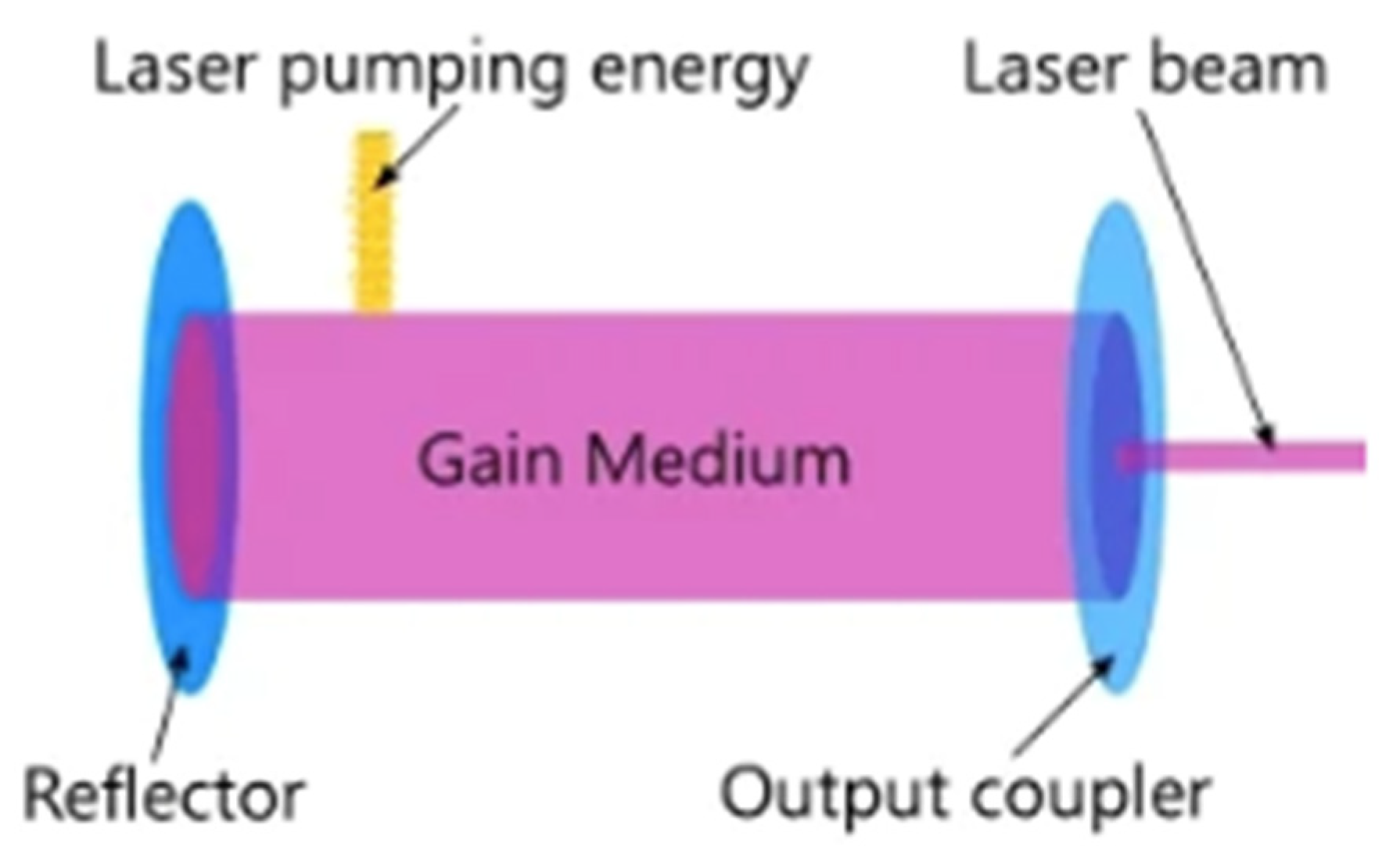
Depending on gain medium (the active substance that produces the laser beam), lasers have classically/historically been classified as solid-state, gas, dye (liquid), and semiconductor/diode. A simplified visualization of the gain medium is shown in Figure 2.
Creating a system which improves a fragmented classification is considered novel. Our approach of linking class → gain medium → specific type of lasers → chemical formula for each type of gain medium is distinct from systems that rely only on power output or wavelength. Our classification integrates physical state, host crystal, doping agent, and chemical structure into a single, clinically relevant hierarchy and enables systematic cross-type comparison and a different type of analysis.
Our four-level system (Table 2) applies hierarchical methods preserving information that flat classifications lose.
Single-level systems (“solid-state laser”) lose specificity about the host crystal.
Two-level systems (“YAG laser”) lose information about the active dopant.
Three-level systems (“Nd:YAG”) preserve the physical hierarchy and specific type of gain (dopant–host pair).
Our four-level system (Nd:Y3Al5O12) demonstrates which chemical elements constitute each type of gain.
Each level answers a distinct clinical question:
Level 1 (Class) → “What is the physical state of the gain medium?”
Level 2 (Gain Medium) → “What is the structural laser host?”
Level 3 (Type of Gain) → “What wavelength and tissue interaction does this produce?”
Level 4 (Chemical Formula) → “Which elements does each type of gain consist in?”
In Table 2, our chemical composition shows the difference between two solid-state lasers, Er:YAG and thulium fiber laser (TFL).
The Er:YAG laser is universally categorized as a solid-state crystal laser. This is due to its gain medium being a YAG crystal rod doped with erbium ions (Er:Y3Al5O12) rather than an optical fiber (Tm3+:SiO2). And we can see that their chemical formulas are different.
The Er:YAG laser can deliver energy through optical fibers. However, this is for beam delivery, not laser generation. A true fiber laser generates light inside a doped glass fiber. Er:YAG generates light inside a crystal rod.
It also belongs to the YAG crystal group, according to our classification, which is confirmed by sources such as the Library of Congress [35] and standard laser physics references.
This classification helps medical professionals transition from technical specifications (e.g., a part number) to clinical decisions (e.g., tissue interaction). Our framework is the first to integrate physical state, host crystal, doping agent, and chemical structure into a single, clinically relevant hierarchy. Our classification is ‘timely’ to adequately address the issues relevant to the target audience (e.g., engineering and medical practitioners) at a given time.
In addition, the proposed new classification system is a recognized form of scientific contribution across multiple fields: cheminformatics (chemical formula provided for each type of gain), scientometrics (novel typology and measurement framework), and hyper-spectral imaging (new hierarchical classification scheme). So, the new hierarchical framework solves a documented problem (fragmented, ambiguous existing classifications).
From Table 2, we can see that six rare-earth elements, ytterbium, yttrium, neodymium, thulium, holmium, and erbium, are needed for designing and functioning of six type of solid-state lasers: Yb:Fiber (ytterbium-doped fiber), neodymium-doped yttrium aluminum garnet (Nd:YAG), erbium-doped YAG (Er:YAG), holmium-doped YAG (Ho:YAG), thulium-doped YAG (Tm:YAG), and thulium fiber laser (TFL). Therefore, these types of lasers might be more expensive compared to others due to the limitation of their gain components being mined from the Earth. In addition, the amount of rare-earth elements on our planet is limited, which hinders the advancement of new solid-state laser manufacturing. But this is a topic for future research.

3.2. Engineering and Medical Application of Medical Lasers

The search results highlight the importance of matching a laser’s engineering parameters to the application. For example, a laser’s wavelength determines its primary absorber in tissue. A CO2 laser (10,600 nm) is strongly absorbed by water, making it a precise, superficial cutting tool, while a 980 nm diode laser targets hemoglobin for bloodless surgery. In tissue engineering, femtosecond lasers offer the nanoscale precision required for creating cell-instructive micropatterns in biomaterials.
Engineering applications for medical lasers can be clustered into four primary engineering domains based on their core function and interaction with tissue: surgical; therapeutic; imaging for diagnostics; tissue engineering and biomanufacturing.
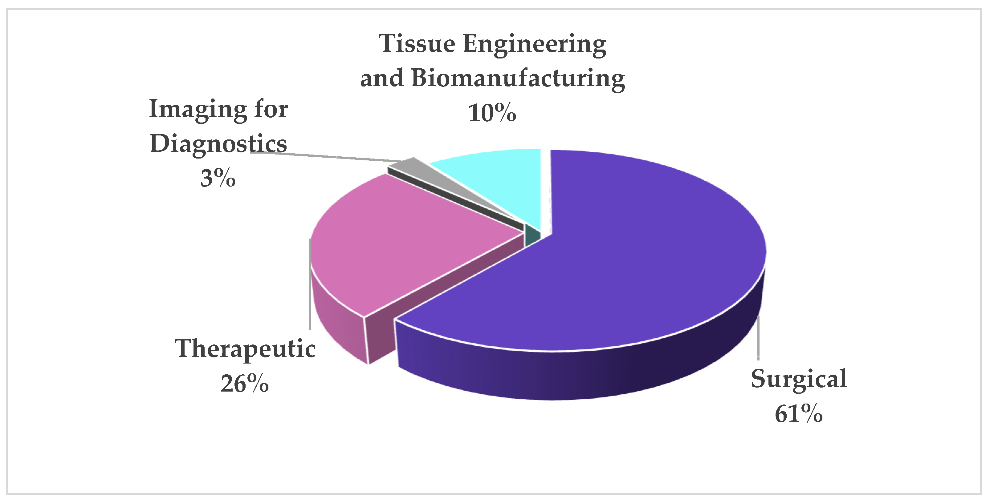
As a result of smart searching across the dataset, we have drawn a diagram representing the clustering of engineering applications for medical lasers for 2025 based on the market size breakdown for the four domains in 2025 (Figure 3).
Figure 3 demonstrates the clustering of engineering applications for medical lasers by application domain.
Surgical (~61%): This is the most solid data point. Market reports consistently show the growth of the surgical laser segment. This cluster includes lasers for cutting, vaporizing, and coagulating tissue in surgery.
Therapeutic (~26%): This is an inferred estimate. Market reports often group “Surgical and Therapeutic Uses” as one dominant segment. The therapeutic share is less than the surgical one. When isolating non-surgical therapeutics (like photodynamic therapy or pain relief) across all specialties, it likely falls into the 15–20% range.
Imaging for Diagnostics (~3%): This small share is consistent with our previous finding. While diagnostic lasers are distinct types and part of the “Diagnosis, Therapy, and Surgery” segment, their market is dwarfed by therapeutic and surgical applications.
Tissue Engineering and Biomanufacturing (~10%): This is an emerging field mentioned as a future growth area for advanced laser applications, not a current major market segment.
To summarize all laser types—solid, gas, semiconductor, and liquid— the authors created a comprehensive comparison table including their engineering and medical applications, and clinical rationale (Table 3).
As evidence basis, the authors confirm that Table 2 synthesizes a dataset from peer-reviewed sources referenced as [14,15,16,17,18,22,29,37,41].
Table 3 supports a thesis that evidence-based laser application requires integration of laser–tissue interaction mechanisms, validated clinical indications, and specialty-specific outcomes. By pairing each laser class with mechanistic rationale and referenced evidence, the table demonstrates that laser selection must be traceable to published data, elevating clinical decision-making from anecdotal practice to evidence-informed precision. The specific references cited [14,15,16,17,18,22,29,37,41] provide the evidentiary foundation for this structure.
In order to corroborate the genuine utilization of medical lasers within the medical field, the following figures are provided (see Table 4).
The available information in Table 4 does not provide a total count of surgical applications for all types of laser gain mediums, due to the reasons mentioned above in the Section 2. It is important to note that Table 4 data comes from scattered websites and articles rather than a central global registry, so it offers a patchwork view rather than a complete global picture.
For example, the global market report that provides the 700,000 figure focuses exclusively on thulium fiber laser (TFL) treatment devices. It states that these devices are now widely used in “urological surgical procedures,” and the market is segmented by application in stone management and BPH (Benign Prostatic Hyperplasia) treatment. Therefore, the 700,000 procedures represent urological interventions for these conditions. This data confirms the TFL’s rapid integration into surgical practice. According to this report’s public summary, the global TFL treatment operation volume in 2024 reached 700,000 procedures, with over 500,000 of these being for stone treatment. This report also provides a breakdown of where these procedures are concentrated and the growth trajectory:
Europe accounts for approximately 45% of the global market.
North America accounts for 25% of the market.
The number of procedures in China, while currently a fraction of the global total, is predicted to grow at an annual rate of nearly 50% from 2025 to 2030.
Industry analyses widely recognize TFL as a strong alternative to the holmium laser (the current gold standard) due to its technical advantages, such as a wavelength that is more efficiently absorbed by water and superior stone dusting capabilities.

3.3. Main Challenges, Roots, and Consequences in Medical Laser Application

Building on the laser classification (Table 2), and analysis of engineering and medical applications (Table 3 and Table 4), the current landscape is defined not only by incredible successes but also by significant, persistent challenges. These obstacles stem from the fundamental physics of light, the complexity of biology, and the stringent requirements of a regulated medical environment. These challenges ultimately affect system performance, patient safety, and clinical adoption. An analysis of the main engineering challenges in medical laser applications, their root causes, and their consequences is presented in Table 5. By explicitly linking each challenge to root causes, consequences, and evidence, the table provides a structured framework for understanding why laser adoption remains uneven despite proven efficacy, and guides strategies for overcoming these barriers to improve clinical implementation/application.
Table 5 directly supports a thesis that successful laser technology translation requires systematic identification and mitigation of technical, biological, economic, and safety barriers.
Table 5 synthesizes evidence from 17 peer-reviewed sources spanning laser physics [14,16,29,31,49,50], systematic reviews on clinical efficacy [14,15,16,17,18,20,21,50], photobiomodulation mechanisms [2,6,7,8], clinical outcome studies [17,18], economic and regulatory analyses [14,16,17,18,20,29,45,49,50], and established safety standards [9,10]. Each challenge category is grounded in documented limitations: the precision–damage trade-off [47,48], delivery system limitations [27,50], and biological variability [14,16,29]; the learning curve [17,27,50], photobiomodulation mechanisms [2,6,7,8], and postoperative complications [14,16,18]; high costs [28,45,49], regulatory hurdles [22,45], and maintenance requirements [27,49,50]; operational hazards are governed by international standards [9,10].
A more profound examination of the underlying factors and their ramifications is warranted. The most significant technical and physical challenges pertain to achieving equilibrium between precision and damage, and successfully targeting the intended area with minimal error. The latter is a particularly perilous process that renders lasers useful, namely, tissue ablation or coagulation via heat. The objective is to confine the effect to the intended target (e.g., a tumor or a corneal flap) while ensuring that adjacent healthy tissue is spared. Achieving perfection in the control of wavelength, power, pulse duration, and cooling is imperative. Although fiber optics have revolutionized endoscopy by enabling the delivery of laser energy to deeply buried or inaccessible areas (e.g., specific lobes of the brain or tumors behind major blood vessels), this remains a significant engineering challenge. The fiber must possess two essential qualities: firstly, it must be flexible; secondly, it must be durable. In addition, a clear working channel is often required, as it is a means of visualizing the target.
If the balance between precision and damage is off, it can lead to consequences like thermal necrosis of healthy cells, delayed healing, scarring, or even perforation of an organ. For cancer treatment, it is challenging to ensure that microscopic extensions of a tumor are destroyed, which is why laser therapy is often combined with other treatments.
Safety challenges are due to inherent operational hazards caused by the powerful sources of energy for medical lasers. The primary risks are to the eye (retinal burns from even diffuse reflection) and skin (burns). The high heat generated can also ignite surgical drapes, endotracheal tubes, or other materials in the oxygen-rich operating room environment, creating a serious fire risk. A consequence of this is the requirement for strict, non-negotiable safety protocols. This includes designated laser safety officers, specialized eyewear for everyone in the room, patient eye protection, fire safety measures (e.g., using laser-safe tubes), and controlled access to the treatment area. Failure to adhere to these protocols can result in catastrophic injury or death.
Meanwhile, treatment efficacy and safety remain incomplete without a quantitative analysis of light dose. The inclusion of quantitative dose parameters reveals several clinically important “dose–response” relationships that a pure discussion may obscure.
Fluence as a determinant of efficacy: For vascular lesion treatment with pulsed dye lasers, clinical efficacy requires fluences of 3–15 J/cm2. Below this range, photothermolysis is incomplete; above it, purpura and scarring risk increase nonlinearly.
Peak power distinguishes Q-switched and picosecond regimes: Although average power remains low (0.5–10 W), peak powers in the megawatt to gigawatt range are essential for the photoacoustic disruption of tattoo particles. This explains why continuous-wave or long-pulse lasers cannot achieve the same effect despite similar fluence values.
Exposure time and thermal confinement: For selective photothermolysis, exposure time must be less than the target’s thermal relaxation time (TRT). For example, a 50–100 μm blood vessel has a TRT of ~1–10 ms. Pulsed dye lasers (0.5–1.5 ms pulses) respect this; long-pulse Nd:YAG (10–100 ms) risks collateral thermal damage to surrounding tissue despite deeper penetration.
Irradiance as a safety constraint: In fiber-delivered endoscopic applications (e.g., Ho:YAG, 2100 nm), irradiance above 10,000 W/cm2 can damage the fiber tip, requiring careful power and spot-size management—a constraint not captured by wavelength or pulse duration alone.
The original synthesis prioritized parameters explicitly requested (wavelength, pulse duration, penetration depth, thermal confinement, beam delivery, and trade-offs) but did not systematically extract dose-related metrics (power, irradiance, fluence, or exposure time). To address this gap, the retained records were re-examined for quantitative dose information. Where present, values were extracted; where absent, the established biomedical laser literature was consulted to supplement representative clinical ranges. Table 4 presents the quantitative dose parameters for each laser class. Following the initial thematic synthesis, all retained records were secondarily screened for quantitative descriptors of laser dose, including power (W), irradiance (W/cm2), fluence (J/cm2), and exposure time (s). When a record contained a dose parameter for a given laser class, the reported value or range was recorded. For parameters not present in the retained records (e.g., typical fluence for diode lasers), representative values from standard biomedical laser references were added to ensure clinical completeness.
Table 6 highlights the core quantitative landscape of medical lasers that are not only clinically validated but also openly characterized. It serves as a reference for matching laser–tissue interactions—showing exactly which lasers can achieve pure ablation, selective photothermolysis, coagulation, or ultrafast cold cutting—based on their wavelength, fluence, and pulse duration.
Table 6 shows that the CO2 laser (10.6 µm) offers the broadest combined efficacy and safety across soft-tissue surgery. Its wavelength is universally absorbed by water, allowing predictable cutting and simultaneous hemostasis. A uniquely wide fluence range—from 1.1 J/cm2 for delicate resurfacing to over 1800 J/cm2 for rapid vaporization—enables micro-precision laryngeal work and en bloc tumor removal with a single device. Decades of clinical use have thoroughly defined its safety margins.

3.4. Future Engineering Opportunities for Medical Lasers

Creating a year-by-year “trend analysis” for future engineering opportunities (2026–2030) requires a shift in perspective. Instead of looking at the past, we must analyze the signals of emerging technologies, research breakthroughs, and market projections from recently published reports. The analysis below identifies the key innovation areas for each laser class, with data points that forecast their growth and transformation. The main trends are presented in Table 7. This table provides a data-driven forecast for the evolution of medical lasers.
The 2026–2030 trajectory for medical lasers is defined by three transformative shifts: AI-driven intelligence, ultrafast precision, and wavelength versatility. Diode lasers will dominate, expanding into multi-wavelength surgical platforms and enabling wearable phototherapy and UV-B diodes that may displace excimer systems. Thulium fiber lasers (TFLs) are poised to succeed Ho:YAG as the urological gold standard, growing at roughly 15% CAGR. Solid-state systems gain new clinical reach through tunable OPOs and yellow wavelengths, while CO2 lasers evolve into hybrid, AI-guided platforms. Dye lasers, though a shrinking standalone segment, find renewed purpose in nanomedicine and theranostics.
The modern perspective on laser-based medicine is mechanistic, seeking defined biological outcomes.
In daylight-PDT and metronomic PDT, pain is minimized and immune effects enhanced by prolonging low-level oxidative stress. Nanoparticle–photosensitizer conjugates improve tumor selectivity and allow imaging-guided treatment. Specific cell death programs are targeted (e.g., necroptosis and ferroptosis) to maximize immunogenicity. Biological monitoring of DAMP release, circulating tumor DNA, and T-cell repertoire serves as a predictive biomarker.
Photodynamic therapy initiates a chain of cell death, vascular disruption, and immunological activation, completing the laser-based treatment landscape. This mechanistic richness also indicates the way forward for next-generation, immunologically optimized light therapies.
Across all classes, closed-loop AI and robotics act as a universal value multiplier, enhancing precision and safety. The future belongs to compact, intelligent, and wavelength-agile systems that seamlessly merge diagnostics with therapy, advancing precision medicine from the operating room to the patient’s home.

4. Conclusions

As a result of the current study, novel scientific results were obtained: a new four-level classification of medical lasers considering the chemical formula for each type of gain medium; a multisided systemic analysis of engineering applications of medical lasers in clinical practice, including an analysis of the main engineering challenges as the structured framework; clustering by laser class of engineering applications for medical lasers for 2025; a designed quantitative landscape of medical lasers by variable (wavelength (nm), power (W)/irradiance (W/cm2), fluence (J/cm2), exposure time/pulse duration; and a year-by-year “trend analysis” for future engineering opportunities (2026–2030).
Our four-level classification of medical lasers
-
Resolves a demonstrated problem (fragmented existing systems);
-
Provides methodological innovation (hierarchical characterization applied to medical lasers).
Notwithstanding the advancements in technology, formidable obstacles continue to impede optimal utilization. The intrinsic biophysical interaction between light and tissue imposes a fundamental compromise between therapeutic accuracy and collateral thermal injury, thereby constraining the effective management of diffusely invasive neoplasms. Limitations in light delivery systems impede access to deeply situated or motile organs. Inter-individual biological heterogeneity contributes to clinical variability, necessitating advanced operator proficiency and leading to an extended training period. Furthermore, the underlying mechanisms of photobiomodulation are not yet fully elucidated, resulting in divergent clinical outcomes. Socioeconomic impediments, including substantial capital expenditure for equipment, rigorous regulatory frameworks, and requisite infrastructural support, exacerbate inequities in healthcare provision and decelerate the uptake of novel solutions. Additionally, potential safety risks necessitate the establishment of rigorous organizational guidelines.
Medical lasers will see three big changes by 2030: AI, ultrafast accuracy, and wavelength choice. Diode lasers will be the main technology, used in multi-colored surgical systems and in wearable phototherapy, and UV-B lasers that may replace excimer systems. Thulium fiber lasers (TFLs) may replace Ho:YAG as the urological gold standard, with a 15% CAGR. Solid-state systems will get better with tunable OPOs, while CO2 laser systems will become AI-guided hybrid systems. Dye lasers will have a new role in nanomedicine and theranostics. Closed-loop AI and robotics will improve accuracy and safety for all systems. The future belongs to compact, intelligent, and wavelength-agile hybrid systems. These will combine diagnostics and therapy in an operating room or at home.

Author Contributions

Conceptualization, T.A.; methodology, T.A. and S.R.; formal analysis, T.A.; resources, T.A. and S.R.; data curation, T.A. and S.R.; writing—original draft preparation, T.A.; writing—review and editing, T.A. and S.R.; visualization, S.R.; supervision, S.R.; project administration, T.A. All authors have read and agreed to the published version of the manuscript.

Funding

This research was funded by the Institute of Cited Scientists, project “Laser Applications by 2030”.

Data Availability Statement

This study has no additional data available.

Conflicts of Interest

The authors declare no conflicts of interest.

Abbreviations

The following abbreviations are used in this manuscript:
AMDAge-related Macular Degeneration
BPHBenign Prostatic Hyperplasia
BRVOBranch Retinal Vein Occlusion
CAGRCompound Annual Growth Rate
CSCCentral Serous Chorioretinopathy
DAMPDamage-Associated Molecular Pattern
DMEDiabetic Macular Edema
ErErbium (a rare-earth element)
Er, Cr:YSGGErbium, Chromium-doped Yttrium Scandium Gallium Garnet
FDAU.S. Food and Drug Administration
GaAsGallium Arsenide Laser
GaAlAsGallium Aluminum Arsenide Laser
He-Ne Helium–Neon (the gas mixture)
HIFUHigh-Intensity Focused Ultrasound
Ho:YAG (HoLEP)Holmium-doped Yttrium Aluminum Garnet Laser
InGaAsIndium Gallium Arsenide Laser
LASERLight Amplification by Stimulated Emission of Radiation
LASIKLaser-Assisted in Situ Keratomileusis
NdNeodymium (a rare-earth element)
OPOOptical Parametric Oscillator
PBMPhotobiomodulation
PDLPulsed Dye Laser
PDTPhotodynamic Therapy
PRKPhotorefractive Keratectomy
RFRadio Frequency
RELITERegenerative Retinal Laser and Light Therapies
TFLThulium Fiber Laser
YSGGYttrium Scandium Gallium Garnet
Yb:FiberYtterbium-doped Fiber Laser
YAGYttrium Aluminum Garnet, where Yttrium: A rare-earth metal; Aluminum: The metal; Garnet: A crystalline mineral structure (the same family as the gemstone)

References

  1. Nobel Foundation. The Nobel Prize in Physics 1964. 1964. Available online: https://www.nobelprize.org/prizes/physics/1964/summary/ (accessed on 22 March 2026).
  2. Hamblin, M.R. Photobiomodulation for the treatment of pain and inflammation: Mechanisms and clinical applications. Photobiomodul. Photomed. Laser Surg. 2023, 41, 449–465. [Google Scholar] [CrossRef]
  3. Niemz, M.H. Laser-Tissue Interactions: Fundamentals and Applications, 4th ed.; Springer: Cham, Switzerland, 2019. [Google Scholar] [CrossRef]
  4. Welch, A.J.; van Gemert, M.J.C. (Eds.) Optical-Thermal Response of Laser-Irradiated Tissue, 2nd ed.; Springer: Dordrecht, The Netherlands, 2011. [Google Scholar] [CrossRef]
  5. Anderson, R.R.; Parrish, J.A. Selective photothermolysis: Precise microsurgery by selective absorption of pulsed radiation. Science 1983, 220, 524–527. [Google Scholar] [CrossRef]
  6. Hamblin, M.R. Photobiomodulation for traumatic brain injury and stroke. J. Neurosci. Res. 2018, 96, 731–743, Erratum in J. Neurosci. Res. 2019, 97, 373. https://doi.org/10.1002/jnr.24376. [Google Scholar] [CrossRef] [PubMed]
  7. Chung, H.; Dai, T.; Sharma, S.K.; Huang, Y.Y.; Carroll, J.D.; Hamblin, M.R. The nuts and bolts of low-level laser (light) therapy. Ann. Biomed. Eng. 2012, 40, 516–533. [Google Scholar] [CrossRef]
  8. Hamblin, M.R. Mechanisms and applications of the anti-inflammatory effects of photobiomodulation. AIMS Biophys. 2017, 4, 337–361. [Google Scholar] [CrossRef]
  9. IEC 60825-1; Safety of Laser Products—Part 1: Equipment Classification and Requirements. IEC: Geneva, Switzerland, 2014. Available online: https://webstore.iec.ch/publication/3587 (accessed on 30 March 2026).
  10. ANSI Z136.3; American National Standard for Safe Use of Lasers in Health Care. Laser Institute of America: Orlando, FL, USA, 2018. Available online: https://www.lia.org/resources/laser-safety-standards/ansi-z1363-safe-use-lasers-health-care (accessed on 30 March 2026).
  11. Rhee, C.K.; Chung, P.S.; Jeong, P.S. Laser safety in laryngeal microsurgery using CO2 laser. Korean J. Bronchoesophagol. 1998, 4, 159–164. [Google Scholar]
  12. Woo, S.H. Safe use of medical lasers: A clinical review of the hazards, safety, and medical implications. J. Korean Soc. Laser Med. 2025, 14, 97–100. [Google Scholar] [CrossRef]
  13. Saki, N.; Heidari, M.; Kasaei, N.; Saki, S.; Farrokhi, N. Fractional CO2 laser alone versus combined with topical timolol or insulin for acne scar treatment: A randomized controlled clinical trial. Lasers Med. Sci. 2025, 40, 207. [Google Scholar] [CrossRef]
  14. van Dijk, E.H.C.; Fauser, S.; Breukink, M.B.; García-Ríos, P.; Pérez-Guzmán, N. Half-dose photodynamic therapy versus high-density subthreshold micropulse laser treatment in patients with chronic central serous chorioretinopathy. Ophthalmology 2018, 125, 1547–1555. [Google Scholar] [CrossRef]
  15. Clementi, A.; Cannarozzo, G.; Guarino, L.; Zappia, E.; Cassalia, F.; Alma, A.; Sannino, M.; Longo, C.; Nisticò, S.P. Sequential fractional CO2 and 1540/1570 nm lasers: A narrative review of preclinical and clinical evidence. J. Clin. Med. 2025, 14, 3867. [Google Scholar] [CrossRef] [PubMed]
  16. Pellegrini, M.; Bosisio, M.; Pulicari, F.; Todea, C.D.; Spadari, F. Laser therapy for vascular malformations of the oral cavity: A systematic review. Dent. J. 2025, 13, 416. [Google Scholar] [CrossRef]
  17. Rahimnia, A.; Ehsani, A.; Koohian Mohammadabadi, M.; Ehsani, A.; Nourmohammadpour, P.; Khosravi, D.; Hatami, P. Comparative efficacy and safety of traditional single-shot and two successive pass and diascopy method in Q-switched Nd:YAG laser-assisted tattoo removal: An evaluator-blinded trial. J. Photochem. Photobiol. B 2025, 272, 113283. [Google Scholar] [CrossRef]
  18. Lee, I.; Lee, S. Laser hair removal for Fitzpatrick skin types III–V: Efficacy and safety in Asian skin—A meta-analysis of 10 RCTs. J. Cosmet. Med. 2025, 9, 1–6. [Google Scholar] [CrossRef]
  19. von der Burchard, C.; Miura, Y.; Stanzel, B.; Chhablani, J.; Roider, J.; Framme, C.; Brinkmann, R.; Tode, J. Regenerative retinal laser and light therapies (RELITE): Proposal of a new nomenclature, categorization, and trial reporting standard. Lasers Surg. Med. 2024, 56, 693–708. [Google Scholar] [CrossRef]
  20. Tode, J. Macular laser picks up speed: The technology accelerating retinal therapies. Ophthalmol. Times Eur. 2025, 21, 6. Available online: https://europe.ophthalmologytimes.com/view/macular-laser-picks-up-speed-the-technology-accelerating-retinal-therapies (accessed on 27 March 2026).
  21. Chhablani, J.; SOLS (Subthreshold Laser Ophthalmic Society) writing committee. Subthreshold laser therapy guidelines for retinal diseases. Eye 2022, 36, 2234–2235. [Google Scholar] [CrossRef] [PubMed]
  22. Keunen, J.E.E.; Battaglia-Parodi, M.; Vujosevic, S.; Luttrull, J.K. International Retinal Laser Society guidelines for subthreshold laser treatment. Transl. Vis. Sci. Technol. 2020, 9, 15. [Google Scholar] [CrossRef]
  23. Haji Mohammadi, A.; Seirafianpour, F.; Khosravi, M.; Jafarzadeh, A.; Neshastesaz Kashi, H.; Baradaran, H.; Goodarzi, A. A systematic review of comparative clinical trials on the efficacy, safety, and patient satisfaction of ablative and non-ablative laser therapies for atrophic, hypertrophic, and keloid scars. Lasers Med. Sci. 2025, 40, 280. [Google Scholar] [CrossRef]
  24. Sorrentino, T.; Allegrini, D.; De Rosa, G.; Santoru, F.; Crepaldi, L.; Feo, A.; Zanellati, G.; Marconi, S.; Auricchio, F.; Romano, M.R. Settings and clinical applications of subthreshold micropulse laser therapy: A review. J. Clin. Med. 2024, 13, 5729. [Google Scholar] [CrossRef]
  25. Almutairi, M.N.; Alshehri, A.M.; Alhoumaily, A.Y.; Alnahdi, O.; Taha, M.A.; Gangadharan, S. Meta-analysis: Clinical outcomes of laser-assisted in situ keratomileusis (LASIK) and photorefractive keratectomy (PRK) in hyperopia. BMC Ophthalmol. 2025, 25, 140. [Google Scholar] [CrossRef] [PubMed]
  26. Sivaramakrishnan, G. Efficacy of Er:YAG, Er, Cr:YSGG, and Nd:YAG lasers for caries removal: A systematic review. J. Dent. 2025, 152, 106264. [Google Scholar] [CrossRef]
  27. Shang, X.; Xu, N.; Zong, M.; Yu, W.; Guo, L.; Gao, G.; Zhang, Z.; Zhang, H.; Su, L. ZrGeTe4 Nanoparticles as a Saturable Absorber for Mode-Locked Operations at 1 and 1.55 µm. Photonics 2026, 13, 305. [Google Scholar] [CrossRef]
  28. Korneva, N.; Budylin, G.; Tseregorodtseva, P.; Nikitin, D.; Andreeva, V.; Baytsaeva, O.; Rudomanova, P.; Musaeva, D.; Yakupov, F.; Baranov, A.; et al. Optical feedback for safe automatic laser lithotripsy: Tissue sensor implementation in a clinical system. Lasers Surg. Med. 2026, 58, 38–48. [Google Scholar] [CrossRef]
  29. Ashraf, Y.; Ali, S.; Zamzami, M.; Abedin, A. Next-generation Moses pulse modulation: An evaluation of whether this is hype or breakthrough. Cureus 2025, 17, e89298. [Google Scholar] [CrossRef]
  30. Fried, N.M.; Irby, P.B. Advances in laser technology and fibre-optic delivery systems in lithotripsy. Nat. Rev. Urol. 2018, 15, 563–573. [Google Scholar] [CrossRef] [PubMed]
  31. Chen, Z.; Meng, C.; Lin, H.; Fixler, D.; Zhang, H. Spatially confined tumor phototherapy enabled by GHz laser thermal accumulation dynamics. Adv. Photon. Nexus 2025, 4, 066009. [Google Scholar] [CrossRef]
  32. Guo, J.; Zhang, H.; Lin, W.; Xu, W. Optimization of Erbium-Doped Fiber to Improve Temperature Stability and Efficiency of ASE Sources. Photonics 2025, 12, 115. [Google Scholar] [CrossRef]
  33. Wang, Z.; Zhu, J.; Du, G.; Liu, J.; Song, Y. Noise-Like Rectangular Pulses with Asymmetric Spectral Stripes in a 1.7-μm Ultrafast Fiber Laser. J. Light. Technol. 2026, 44, 3218–3224. [Google Scholar] [CrossRef]
  34. Zhang, L.; Tang, P.; Zhu, J.; Zhou, Z.; Wu, H.; Wang, Z. Dynamic Behaviors of Pulsating Noise-like Pulses in an Ultrafast Fiber Laser. Photonics 2025, 12, 937. [Google Scholar] [CrossRef]
  35. Available online: https://id.loc.gov/authorities/subjects/sh2017004341.html (accessed on 8 May 2026).
  36. Wieneke, S.; Gerhard, C. Lasers in Medical Diagnosis and Therapy; Basics, Applications and Future Prospects; IOP Ebooks: Bristol, UK, 2018; ISBN 978-0-7503-1275-2. [Google Scholar]
  37. Medical Lasers Market Size, Share, Growth and Global Industry Analysis By Type & Application, Regional Insights and Forecast to 2026–2034. Available online: https://www.giiresearch.com/report/fbs1933281-medical-lasers-market-size-share-growth-global.html (accessed on 11 May 2026).
  38. Medical Laser Market Size & Share Analysis—Growth Trends & Forecasts 2026–2031. Available online: https://www.mordorintelligence.com/industry-reports/medical-laser-market (accessed on 2 May 2026).
  39. Global Medical Laser Market Assessment, By Product Type, By Application, By End-User, By Region, Opportunities and Forecast, 2017–2031F. Available online: https://www.marketsandata.com/industry-reports/medical-laser-market (accessed on 11 May 2026).
  40. Medical Lasers Market Report by Type (Surgical, Cosmetic, Dental, and Others), Product Type (Solid-State Laser Systems, Gas Laser Systems, Dye Lasers Systems, Diode Laser Systems), Application (Ophthalmology, Dermatology, Gynecology, Dentistry, Urology, Cardiovascular, and Others), End User (Hospitals, Specialty Clinics, Ambulatory Surgical Centers), and Region 2026–2034. Available online: https://www.imarcgroup.com/medical-lasers-market (accessed on 11 May 2026).
  41. Belokrylov, N.; Sotin, A.; Belokrylov, A.; Antipova, T. The Use Carbon Composite Material For Replacement of Postresection Bone Defects. JDS 2023, 5, 19–31. [Google Scholar] [CrossRef] [PubMed]
  42. Available online: https://pubmed.ncbi.nlm.nih.gov/37328009/ (accessed on 11 May 2026).
  43. Available online: http://oa.las.ac.cn/oainone/service/browseall/read1?ptype=JA&workid=JA202311106768129ZK (accessed on 11 May 2026).
  44. Available online: https://research-repository.uwa.edu.au/en/publications/12years-of-ablative-fractional-cosub2sub-laser-practice-logistics/ (accessed on 11 May 2026).
  45. Available online: https://dxpress.gelonghui.com/p/3329270 (accessed on 11 May 2026).
  46. Rice, P.; Somani, B.K. A systematic review of thulium fiber laser: Applications and advantages of laser technology in the field of urology. Res. Rep. Urol. 2021, 13, 519–527. [Google Scholar] [CrossRef] [PubMed]
  47. Jaffe, B.H. Laser surgery: Benefits, limitations, and future directions. Lasers Surg. Med. 2020, 52, 5–12. [Google Scholar] [CrossRef]
  48. Osman, M.A.; Kassab, A.N. Fractional Er:YAG laser versus fractional CO2 laser in the treatment of immature and mature scars: A comparative randomized study. Arch. Dermatol. Res. 2024, 316, 75. [Google Scholar] [CrossRef]
  49. Lonsdorf, A.S.; Keller, A.; Hartmann, J.; Enk, A.H.; Gholam, P. Ablative Fractional Laser-assisted Low-irradiance Photodynamic Therapy for Treatment of Actinic Keratoses in Organ Transplant Recipients. Acta Derm.-Venereol. 2022, 102, adv00694. [Google Scholar] [CrossRef] [PubMed]
  50. Meng, T.; He, L.; Chen, Y.; Zhang, Z. Efficacy of a single Nd:YAG laser dose in reducing pain after mandibular third molar extraction: A prospective randomized controlled clinical trial. Lasers Med. Sci. 2024, 39, 68. [Google Scholar] [CrossRef] [PubMed]
  51. Liu, Y.D.; Wang, Y.; Zeng, J.; Li, H.; Liu, H.L.; Qiu, H.X.; Gu, Y. Pulsed Dye Laser Versus Photodynamic Therapy for the Treatment of Port Wine Stain: A Systematic Review and Meta-analysis. Int. J. Dermatol. Venereol. 2025, 8, 225–237. [Google Scholar] [CrossRef]
  52. Menozzi-Smarrito, C.; Smarrito, S. Skin-toned tattoo ink: What should one expect in terms of ink darkening and removal when treated with a 755 nm pico-second laser? Aesthetic Med. 2024, 10, e2024004. [Google Scholar] [CrossRef]
  53. Ulvik, Ø.; Æsøy, M.S.; Juliebø-Jones, P.; Gjengstø, P.; Beisland, C. Thulium Fibre Laser versus Holmium:YAG for Ureteroscopic Lithotripsy: Outcomes from a Prospective Randomised Clinical Trial. Eur. Urol. 2022, 82, 73–79. [Google Scholar] [CrossRef]
  54. Saeed, W.M.; O’Brien, P.J.; Yoshino, J.; Restelli, A.R.; Traynham, A.J.; Fried, N.M. Comparison of quartz and sapphire optical chambers for infrared laser sealing of vascular tissues using a reciprocating, side-firing optical fiber: Simulations and experiments. Lasers Surg. Med. 2023, 55, 886–899. [Google Scholar] [CrossRef] [PubMed]
  55. Hamblin, M.R.; de Sousa, M.V. Photobiomodulation for radiation-induced fibrosis: Mechanisms and clinical evidence. Photobiomodul. Photomed. Laser Surg. 2023, 41, 97–107. [Google Scholar] [CrossRef]
  56. Binrayes, A. An Update on the Use of Lasers in Prosthodontics. Cureus 2024, 16, e57282. [Google Scholar] [CrossRef] [PubMed] [PubMed Central]
  57. U.S. Food and Drug Administration. DEN230045: AI-Assisted Laser Guidance System—De Novo Classification Request. FDA De Novo Database. 2024. Available online: https://www.accessdata.fda.gov/scripts/cdrh/cfdocs/cfpmn/denovo.cfm?id=DEN230045 (accessed on 11 May 2026).
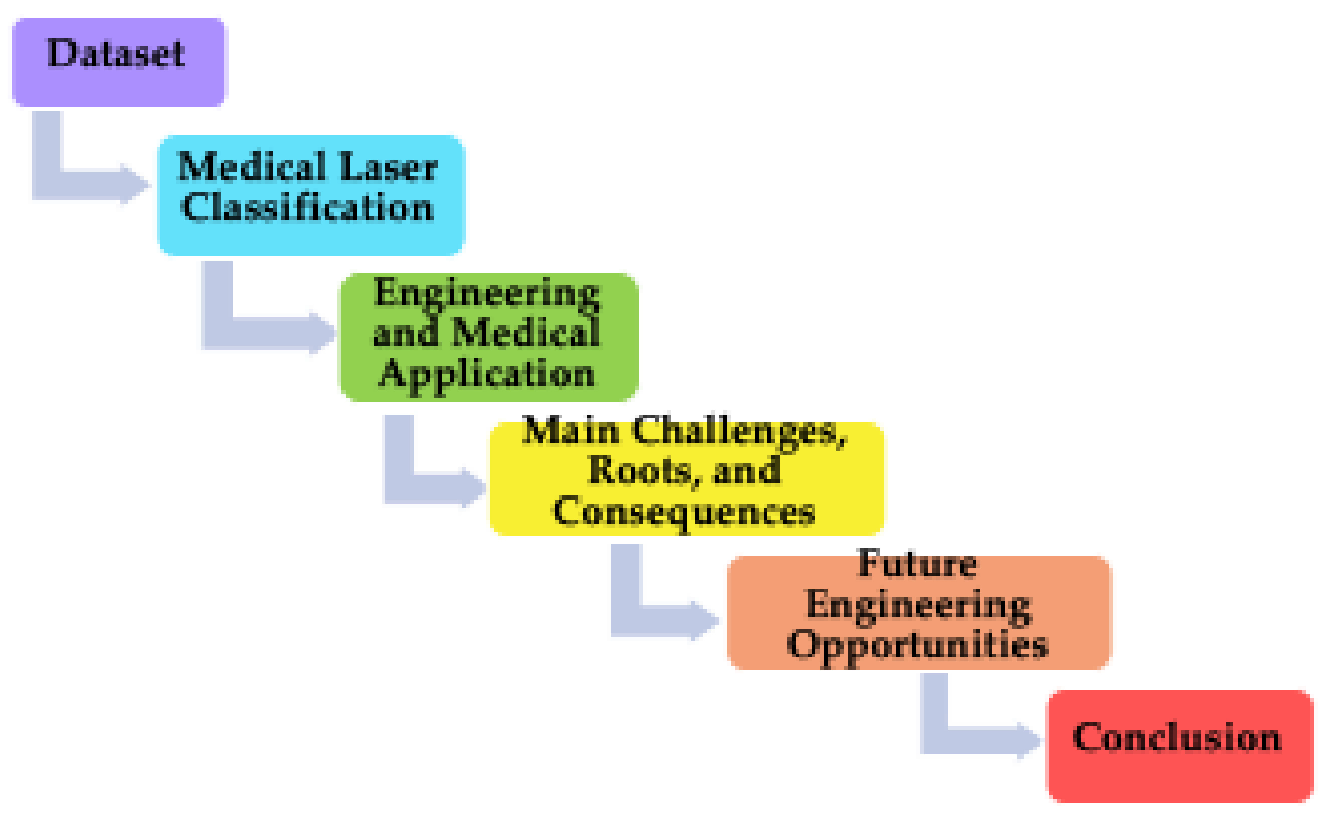
Figure 1. PRISMA workflow diagram. Source: authors’ own elaboration.
Photonics 13 00524 g001
Figure 2. Laser gain medium.
Photonics 13 00524 g002
Figure 3. Clustering of engineering application for medical lasers for 2025. Source: authors’ own elaboration based on [36,37,38,39,40].
Photonics 13 00524 g003
Table 1. Chronological overview of medical laser development.
PeriodDevelopment StageKey Characteristics and Milestones
1960sFundamental Research and ExplorationBirth of the first laser; initial studies on bio-effects; first applications in ophthalmology.
1970sWidespread Clinical ApplicationIntroduction of new lasers (CO2 and Nd: YAG); expansion into surgery, dermatology, and other fields.
1980s–1990sMaturation and
Specialization
Laser medicine becomes an independent discipline; development of specialized devices; revolutionary techniques like PRK, LASIK, and PDT emerge.
21st CenturyEra of Precision and
Intelligence
Focus on minimally invasive, precise treatment; rise of femtosecond lasers, theranostics, and nanoscale applications.
Table 2. Medical laser classification.
ClassGain MediumType of GainChemical Composition of
Type of Gain
Solid-State
Lasers
YAG Crystal Neodymium-doped Yttrium
Aluminum Garnet (Nd:YAG)
Nd:Y3Al5O12
Erbium-doped YAG (Er:YAG) Er:Y3Al5O12
Holmium-doped YAG (Ho:YAG) Ho:Y3Al5O12
Thulium-doped YAG (Tm:YAG) Tm:Y3Al5O12
Sapphire Crystal Titanium-doped Sapphire (Ti:Sapphire) Ti3+:Al2O3
Alexandrite Crystal Chromium-doped Chrysoberyl
(Alexandrite)
Cr3+:BeAl2O4
Ruby Crystal Chromium-doped Sapphire (Ruby) Cr3+:Al2O3
Silica Glass Thulium Fiber Laser (TFL) Tm3+:SiO2
Silica Glass Yb:Fiber (Ytterbium-doped Fiber) Yb3+:SiO2
YSGG Crystal Er, Cr:YSGG (Erbium, Chromium-doped Yttrium Scandium Gallium Garnet) Er3+, Cr3+:Y2.93Sc1.43Ga3.64O12
Gas LasersGas Mixture CO2CO2 + N2 + He + Xe + CO + D2
Argon-ion Ar+
HeNe (Helium–Neon) He + Ne
Excimer ArF + KrF + XeCl
Dye LasersOrganic Liquid Pulsed Dye Laser (PDL) C27H31N2O3Cl (Rhodamine 6G) or C40H42N2O8S (Sulforhodamine B)
Semiconductor (Diode) LasersSemiconductor Wafer GaAlAs (Gallium Aluminum Arsenide)
Diode Laser
Ga1−xAlxAs
Ternary Alloy of InAs and GaAs InGaAs (Indium Gallium Arsenide) InxGa1−xAs
Source: authors’ own elaboration.
Table 3. Engineering and medical applications by laser class.
Laser ClassEngineering ApplicationMedical
Application
Solid-StatePrecision tissue ablation: Er:YAG lasers (2.94 µm) are ideally suited to delicate soft-tissue surgery and dental treatments as their wavelength is highly absorbed by water, allowing for minimal thermal damage.
High-peak power applications: Q-switched solid-state lasers are used for high peak powers, essential for mechanical disruption, e.g., tattoo removal and lithotripsy (breaking up kidney stones).
General surgery and coagulation: 1.06 µm Nd:YAG lasers provide good penetration and coagulation.
Ophthalmic surgery: specific lasers are used for delicate eye surgeries. A 266 nm solid-state laser is being researched as an alternative to gas lasers for corneal reshaping.
Dermatology; ophthalmology;
esthetics; tissue ablation; lithotripsy
GasSoft tissue cutting: CO2 lasers cut skin and soft tissue while sealing blood vessels. They are used in dermatology and general surgery.
Bioelectronic device fabrication: CO2 lasers make laser-induced graphene (LIG) on polyimide films. This makes sensors for wearable healthcare devices.
Corneal reshaping: The Argon Fluoride (ArF) excimer laser is the standard tool for LASIK eye surgery. Its ultraviolet light is precise and does not cause thermal damage.
General surgery (cutting/coagulation); ophthalmology; dermatology; photocoagulation; lithotripsy
Dye Vascular lesion treatment: Although the search results do not provide specific information, dye lasers represent a well-established standard treatment for port wine stains and other vascular lesions, based on the precise tuning of the laser to the absorption peak of hemoglobin.
The following is a list of specialized diagnostics: The tunability of these materials renders them useful in scientific research applications, such as spectroscopy, which underlies numerous diagnostic technologies.
High-precision dermatology (vascular lesions; scar treatment); specialized surgical and diagnostic applications
Semiconductor
(Diode)
Hair and skin treatments: High-power diode lasers (typically 800–810 nm) are the standard for laser hair removal.
Minimally invasive surgery: Compact diode lasers are integrated into devices for procedures like endovenous laser ablation (treating varicose veins).
Photobiomodulation: Low-level diode lasers are used in physical medicine and rehabilitation for pain reduction, wound healing, and tissue regeneration.
Bioelectronic fabrication: Diode lasers are employed for precise tasks like sintering conductive nanoparticles and phase-modulating materials, which are critical engineering steps in creating advanced bioelectronic devices.
Diagnostic imaging; ophthalmology; aesthetics (hair removal); low-power therapeutic applications
Source: authors’ own compilation based on [14,15,16,17,18,22,29,37,41].
Table 4. Surgical applications of some types of medical lasers.
Type of
Laser Gain
Surgical
Application
Procedure VolumeStatistic PurposeStudy Period
Ho:YAGUrology6967Total Ho:YAG data
(national US database)
2000 to 2019
Nd:YAG Capsulotomy
(post-cataract)
312,103 National estimate for France 2017
CO2 Burn scar treatment 4005 Report for a single
Australian unit
2013–2024
TFL Urology 700,000 Global TFL Surgery
Estimate Report
2024
Source: authors’ own compilation based on [42,43,44,45].
Table 5. Engineering challenges, roots, and consequences in medical laser applications.
Challenge
Category
Challenge SubjectRoot CauseConsequence
Technical and PhysicalThe Balance
Between Precision and Damage
Fundamental physics of light-tissue interactionRisk of thermal damage to healthy tissue; limits treatment of diffuse or infiltrative tumors.
Reaching the TargetLimitations of current fiber optics and mechanics.Difficulty accessing deep, complex, or moving organs (e.g., brain or beating heart).
Inconsistent Tissue
Effects
Biological variability (tissue type, blood flow, and pigmentation).Unpredictable clinical outcomes; need for surgeon experience to adjust in real time.
System LevelThermal
Management
Low energy conversion efficiency (“wall-plug” efficiency), with a large portion of input energy converted to waste heat within the laser cavity and power electronicsThermal lensing and wavelength drift degrade beam quality; equipment requires bulky, noisy, and high-maintenance cooling systems (water and cryogenic) that limit portability and increase operational costs.
Beam Delivery
and Control
The challenge of efficiently and safely guiding laser light from the source to the target tissue, often through flexible optical fibers or articulated arms, while maintaining beam quality and alignment.Optical damage can cause catastrophic failure. Poor alignment leads to low treatment efficacy and potential collateral tissue damage. The exact cellular pathways, optimal dosages (“dose–response” relationships), and long-term effects are still under investigation.
System Size, Cost and MiniaturizationThe inherent complexity of high-precision optics, robust cooling, and specialized power supplies needed for medical-grade performance and safety.Limits accessibility, especially in smaller clinics and field settings. High acquisition and maintenance costs restrict market penetration and encourage use of less optimal technologies.
Application-
Specific
Precision and Selectivity in Surgery and TherapyThe complex, dynamic, and heterogeneous nature of biological tissues leads to variable light absorption and scattering, clouding the boundary between target and healthy tissue.Risk of unintended thermal damage, leading to scarring and nerve injury. Incomplete procedures may require retreatment.
Imaging Depth, Speed, and ResolutionThe fundamental trade-off between imaging depth and spatial resolution due to tissue optical properties; maintaining coherence and high-speed scanning in miniaturized probes.Limits ability to visualize deep structures with cellular-level detail. May miss early-stage pathologies, reducing diagnostic accuracy.
Cell Viability and Functionality in BiomanufacturingCells are vulnerable to thermal and mechanical stress from direct laser energy or the jetting process; precise 3D placement of multiple cell types is difficult.Functional tissue failure, limiting clinical application to simple structures.
Scalability and VascularizationSlow build speeds of high-res methods make large constructs impractical; lack of integrated vascular network leads to nutrient/waste exchange failure.Viability is limited to small, thin tissues, preventing production of complex organs. This is a critical bottleneck for regenerative medicine.
SafetyOperational HazardsHigh-power beams; electrical systems; potential for fire.Risk of eye injury, burns, and surgical fires for both patients and operating room staff.
Source: authors’ own compilation based on [14,16,17,18,22,29,46,47,48,49,50].
Table 6. Quantitative landscape of medical lasers.
Laser
Class
Type of GainWavelength, nmPower (W)/
Irradiance (W/cm2)
Fluence (J/cm2)Exposure Time/
Pulse Duration
Solid-State Nd:YAG 1064 10–40 W (CW)/
700–1500 W/cm2
60–190 (LP); 10–12 (QS tattoo);
70–110 (vascular)
15–60 ms (LP); 4–10 ns (QS)
Er:YAG 2940 1–25 W/
~1.5 J pulse energy
2–100 (ablation); 1 (thermal mode) 40–1000 µs; typical 230–350 µs
Ho:YAG 2100 30–100 W (max)/
0.5–4.0 J/pulse
150–927 (lithotripsy) 150–1500 µs; typical 350–600 µs
Er, Cr:YSGG 2780–2790 1.5–12 W/
up to 1800 mJ/pulse
2.5–28 100–600 µs; 200 ns (Q-switched)
Yb:Fiber 1030–1100 4–60 W/
3.8 × 10⁴ W/cm2 (CW)
0.28–5.38 <80–150 fs; 1.56 ps; 4 ns
Gas CO2 10,600 2–30 W (surgical)/480–780 W/cm2 (focused) 1.1–1840 CW or pulsed (µs–ms)
Excimer (ArF) 193 500 mW (average) 50–180 mJ/cm2 4–25 ns
DyeRhodamine 6G 570–610 (tunable) 50–100 J
(pumped pulse)/
0.5–2 W (average)
5.75–7.0 0.45 ms
Semiconductor InGaAs/GaAs 980 0.5–180 W 5–15 20–60 ms
Source: authors’ own compilation based on [48,49,50,51,52].
Table 7. Year-by-year engineering opportunities in medical laser applications.
Laser
Class
2026 2027 2028 2029 2030 (Outcome)
Solid-State Air-cooled DPSS, tunable OPOs enter clinics. TFL becomes standard in urology; 15–20% procedure time reduction. Yellow lasers (589 nm) approved for new retinal treatments. TFL enters mainstream dermatology and phlebology. Combination YAG/OPO platforms for multi-specialty. Ultrafast TFL for deep-tissue, non-invasive imaging. AI-driven wavelength selection standard. TFL combined with AI for autonomous stone dusting. OPO-based systems are a major segment. TFL equipment market grows at 15% CAGR.
Gas (CO2) Hybrid fractional CO2 + RF/HIFU devices. AI-guided beam shaping and dosimetry. Ultra-clear, all-skin-type protocols mature. Self-calibrating, autonomous resurfacing. Dominated by intelligent, connected systems.
Dye Nano-encapsulated ocular dyes for drug delivery. Biocompatible dye microlasers for internal diagnostics. First human trials of dye-based theranostics. Combined dye–nanoparticle platforms for oncology. Dye laser innovation shifts entirely to nanomedicine and theranostics.
Semiconductor (Diode) 4-wavelength platforms; UV-B CW diode prototype. UV-B diodes miniaturized into handheld medical devices. Multi-wavelength PBM devices with integrated diagnostics. Wavelength-on-demand modules for surgery. UV-B diodes disrupt excimer applications.
Source: authors’ own compilation based on 190 webpages, and [51,52,53,54,55,56,57].
Disclaimer/Publisher’s Note: The statements, opinions and data contained in all publications are solely those of the individual author(s) and contributor(s) and not of MDPI and/or the editor(s). MDPI and/or the editor(s) disclaim responsibility for any injury to people or property resulting from any ideas, methods, instructions or products referred to in the content.