Article

Photobiomodulation in Complex Female Infertility Profile: A Case Report with 12-Month Follow-Up and Review of Current Mechanism in Reproductive Photomedicine

1
Laser Medicine Centre, 134 Harley Street, London W1G 7JY, UK
2
Department of Restorative Dental Sciences, UCL Eastman Dental Institute, Medical Faculty, University College London, London WC1E 6DE, UK
3
Head and Neck Academic Centre, Integrated Research, Division of Surgery and Interventional Science, Medical Faculty, University College London, London W1W 7TY, UK
4
Department of Surgical Sciences and Integrated Diagnostics, University of Genoa, 16132 Genoa, Italy
5
Dental Faculty, Royal College of Surgeons Ireland (RCSI), 121-122 St Stephen’s Green, D02 H903 Dublin, Ireland
*
Author to whom correspondence should be addressed.
Photonics 2025, 12(10), 1021; https://doi.org/10.3390/photonics12101021
Submission received: 21 August 2025 / Revised: 5 October 2025 / Accepted: 14 October 2025 / Published: 16 October 2025
(This article belongs to the Special Issue Shining Light on Healing: Photobiomodulation Therapy)

Abstract

Female infertility from polycystic ovarian syndrome (PCOS) and endometriosis poses a challenge for both clinicians and women who are trying to conceive. The present clinical single case report aimed to evaluate the efficacy of multiple wavelengths of red and near-infrared (NIR) laser photobiomodulation (PBM) for increasing the potential of fertility in a woman with PCOS, endometriosis and low ovarian reserve. The observations helped to inform and establish the following: (1) any adverse effects; (2) the possibility of producing an effective PBM protocol; and (3) a healthy live birth. The case report concerns a female who failed to conceive naturally beyond five years and had experienced one unsuccessful IVF cycle. Methods: Case report of one female subject with infertility issues, which included failure to conceive naturally beyond five years. Previous conditions were recorded and then compared with outcomes from after the patient received a course of PBM treatments. PBM treatments were given at weekly and/or at two-week intervals over a 5-month period during the follicular stage of the menstrual cycle, using IR and NIR wavelengths between 600 and 1000 nm. Results: After five months a spontaneous conception was achieved. The case resulted in a full-term pregnancy and the birth of a healthy baby. Improvements in reproductive health outcomes in this case give reason to suggest that PBM helped to alleviate PCOS and endometriosis which could have been associated with a low ovarian reserve. Conclusions: The case report indicates that a multiwavelength of red and NIR-PBM laser therapy could have positively contributed to a healthy live birth in a female diagnosed with PCOS, endometriosis and a low ovarian reserve. Extensive studies with large data are warranted to validate our PBM dosimetry and treatment protocols to assess the potential impact of PBM for treating endometriosis and PCOS. Subsequently, to understand the genetic and phenotype biomarkers would be an important step further to standardise a range of PBM light dosimetry.

1. Introduction

1.1. Pathophysiology of Female Infertility

1.1.1. Endometriosis and Infertility

Endometriosis is a condition that affects women of reproductive age and it is distinguished by the development of endometrial-like tissue outside the uterine cavity. It is frequently accompanied by persistent pelvic discomfort and infertility [1]. Endometriosis is more often present and severe among infertile patients, with a risk of infertility estimated to be almost 20 times greater with endometriosis than without [2].
The pathophysiology of endometriosis is poorly known, with most gynaecologists of the belief that inflammation is a crucial source of irritation in endometriosis [3]. Altered ovulation and oocyte production is seen in endometriosis and associated with increased inflammatory cells in the peritoneal fluid and endometriomas [4].
A study in 2023 by Fan et al. found that decreased oocyte quality in patients with endometriosis is closely related to abnormal granulosa cells [5]. Granulosa cells, which aid meiosis and provide basic resources for oocyte development, can be affected by endometriosis to cause apoptosis, inflammation, oxidative stress, steroid synthesis obstacle and aberrant mitochondrial energy metabolism [5].
The peritoneal fluid of women with endometriosis has been found to contain higher amounts of macrophages, T-lymphocytes and beta lymphocytes, which are susceptible to apoptosis, again an indication of inflammation being present, and therefore an influential factor in the impact on fertility outcomes [6].
Angiogenesis is a key mediator of endometrial regeneration, facilitating appropriate nutrient and oxygen delivery, which is significantly important in the treatment of endometriosis and tissue healing [7].

1.1.2. Polycystic Ovarian Syndrome (PCOS) and Infertility

PCOS is a common endocrinological disorder in women, characterised by hormonal imbalances that disrupt the development and release of eggs, thereby increasing the risk of infertility [8]. It has a prevalence of approximately 5–15% [9] and is present in up to 83% of women with anovulatory infertility [10]. The pathophysiology of PCOS is hypothesised to involve a cyclical interaction between insulin resistance, hyperinsulinemia (elevated levels of insulin in the blood) and hyperandrogenism. This interplay, along with dysfunction of the pituitary gland, is believed to contribute to anovulation and subsequent infertility [11].
A population-based retrospective cohort study carried out in Western Australia [12] studied the potential implications of a PCOS diagnosis on a woman’s long-term health. The study concluded that PCOS had profound medical implications for the health of women with PCOS who were significantly more likely to experience adult-onset diabetes, obesity, hypertension, ischemic heart disease and arterial and venous disease. Furthermore, women diagnosed with PCOS had more hospitalisations for the treatment of various gynaecological conditions, and an extremely higher risk of endometriosis (26.4 vs. 4.4%) and endometrial glandular hyperplasia (1.8 vs. 0.1%) [12].
The role of chronic inflammation in PCOS is widely accepted. A review of 63 studies revealed higher circulating levels of the inflammatory-marker C-reactive protein (CRP) in women with PCOS, independent of obesity, and therefore indicative of chronic inflammation [13].
A relationship between PCOS and hypothyroidism has also been established, with an increase in ovarian volume and cystic changes in ovaries reported in primary hypothyroidism, and thyroid disorders being reportedly more common in women with PCOS [14].

1.1.3. Low Ovarian Reserve in the Context of Endometriosis and PCOS

Low ovarian reserve, also referred to as diminished ovarian reserve (DOR), is characterised by poor fertility outcomes, even when assisted reproductive techniques (ART) are employed [15].
By definition, DOR is similar to a poor ovarian responder (POR). The Bologna ESHRE consensus defines women as ‘poor ovarian responders’ when at least two of the three following characteristics apply: (i) advanced maternal age greater than 40; (ii) less than three oocytes with a conventional ART protocol; or (iii) an abnormal ovarian reserve test, i.e., antral follicular counts of less than 5–7 follicles or AMH less than 0.5–1.1 ng/mL [15].
Endometriosis, with its deeply rooted inflammatory characteristics, has emerged as a significant factor in negatively impacting ovarian reserve, and consequently the fertility potential of affected women [16]. A comprehensive review of studies on the relationship between endometriosis and diminished ovarian reserve highlighted a critical disruption in the follicular environment, whereby a surge in cytokines and ROS sets off a chain reaction that results in fibrosis of the ovarian cortex, adverse folliculogenesis and impaired gamete development [16].
Moreover, surgical interventions for endometriosis may further negatively impact ovarian tissue through effects such as vascular damage, removal of healthy ovarian tissue, post-operative adhesions and the risk of overstimulating primordial follicles [16]. A study of women diagnosed with superficial endometriosis, however, were not impacted with lower AMH levels than women without an endometriosis diagnosis [17].
A link between PCOS and low ovarian reserve is not considered, as PCOS is characterised by the presence of an elevated antral follicular count [18]. PCOS is thought to negatively impact fertility because of hormonal imbalances, which impair ovulation and cause chronic anovulation and hyperandrogenism [18]. However, a longitudinal study in 2018 showed a faster decline in anti-Müllerian hormone (AMH) and antral follicle count (AFC) in women with PCOS as they aged, compared to the control group, indicating a longer term impact and faster decline of the ovarian reserve [8].
The earlier mentioned population-based retrospective cohort study, which identified a link between PCOS and long-term health, also highlighted an infertility diagnosis which is considerably higher for women with PCOS than without PCOS (40.9 vs. 4.6%) [12]. Women with PCOS are not only more likely to have endometriosis (26.4 vs. 4.4%), but the combined diagnosis is likely to result in an increased likelihood of infertility [12].
PCOS and endometriosis are inflammatory conditions. Chronic inflammation in women with PCOS could be a considered a factor in ovarian ageing because of the detrimental effect that a sustained inflammatory response over a prolonged period can have on tissue and organs [19]. Several studies have shown the direct link between chronic inflammation and both IVF outcomes and the ovarian reserve, with the role of pro-inflammatory IL-1 identified as a contributing factor in diminishing the ovarian reserve [20].

1.2. Current Fertility Treatments

There are a number of known treatment options for a poor ovarian response, with IVF as the second line of treatment when conception has not been achieved after two years [21].
IVF outcomes may be supported with experimental procedures and supplementation support, which include (a) intra-uterine/intra-ovarian injection of platelet-rich plasma (PRP), (b) exosomes therapy, (c) ozone therapy, (d) dietary supplements, (e) acupuncture, (f) intravenous laser irradiation of blood (ILIB) and (g) photobiomodulation therapy (PBM) [22].
When endometriosis and/or PCOS impact poor ovarian response, the following approaches are also considered with arguable success.

1.2.1. Prescribed Medication

Metformin
The use of Metformin enhances insulin sensitivity at a cellular level, lowers insulin, reduces hyperinsulinemia and suppresses the excessive ovarian production of androgens, and therefore has the potential to improve symptoms and reproductive outcomes for women with PCOS [23].
Prescribed Metformin for women with polycystic ovary syndrome (PCOS) undergoing ovulation induction or in vitro fertilisation (IVF) cycles has been widely studied, because of Metformin’s ability to reduce hyperinsulinemia and suppress excessive ovarian production of androgens [24].
Clomiphene Citrate (CC)
CC is the first-choice treatment for PCOS-affected females to trigger ovulation [18]. It is an antioestrogen, which can raise FSH levels and counteract anovulation [25].
However, a recent systematic review and meta-analysis of random controlled trials carried out in 2024, which investigated the therapeutic management of women with a diminished ovarian reserve [26], revealed that the number of oocytes and metaphase II oocytes retrieved, clinical pregnancy rate, miscarriage rate and live birth rate were similar between all groups studied, which brings into question the effectiveness of Clomiphene Citrate to improve fertility outcomes for women with a low ovarian reserve.
Letrozole
Letrozole is a commonly prescribed medication for women with PCOS-related infertility [27]. It is a selective aromatase inhibitor which prevents the conversion of testosterone into estradiol and gives rise to increased FSH levels to prompt ovulation [18].
In the previously mentioned 2024 systematic review and meta-analysis of random controlled trials [26], analysis of the use of Letrozole to impact the number of oocytes and metaphase II oocytes retrieved, clinical pregnancy rate, miscarriage rate and live birth rate proved similar between all groups studied, and therefore Letrozole is questionable in its effectiveness to improve fertility outcomes for women with a low ovarian reserve.
Gonadotrophins
Gonadotrophins are considered a course of treatment for patients with a low ovarian reserve, and a study in 2015 showed a higher live birth rate compared with Clomiphene and Letrozole groups [27], yet when studied in the context of a systematic review assessing the fertility outcomes for patients with a low ovarian reserve, a lower number of eggs was retrieved and there was no difference in live birth or clinical pregnancy rates [26].

1.2.2. Surgery

Laparoscopic surgery, which involves removing or destroying endometriosis tissue and lesions, can reduce inflammation and restore pelvic anatomy.
A study published in 2015 supports the role of surgical intervention to enhance reproductive potential in women with advanced endometriosis [28]. The laparoscopic excision of severe endometriosis significantly improved fertility outcomes in the cohort, with a substantial proportion of women achieving pregnancy within a year post-surgery [28].
There are risks associated with laparoscopic surgery including potential damage to the ovarian reserve and premature ovarian failure in the post-operative stage [29]. Furthermore, adhesion formation after endometriosis surgery is a severe problem, affecting up to 90% of patients. This can cause secondary infertility and must therefore be mitigated with effective adhesion prophylaxis [30].

1.2.3. Anti-Inflammatory Diet

A key mechanism outlined in the literature relates to the adverse effects of inflammation on fertility, therefore dietary interventions, which act to reduce inflammation, may improve fertility outcomes [31]. Adherence to anti-inflammatory diets such as a Mediterranean diet, which encourages an uptake of monosaturated and n-3 polyunsaturated fatty acids, flavonoids and reduced intake of red and processed meat, improves fertility, assisted reproductive technology success and sperm quality in men [31].
In a review of clinical studies which assessed the impact of dietary modifications, most studies reported a positive effect on endometriosis [32].
Similarly, adherence to a Mediterranean diet, as outlined in a study by Barrea et al. 2019 [33], supports the therapeutic role of foods and nutrients of the Mediterranean dietary pattern in PCOS pathogenesis, which is likely to involve its inflammatory status, insulin resistance and hyperandrogenemia.

1.2.4. Dietary Supplements

CoQ10 can be a promising antioxidant for the treatment of poor ovarian response, [34] concluded that (CoQ10) supplementation can increase ovarian response to stimulation and improves oocyte and embryo quality in young, low-prognosis patients with diminished ovarian response, and is beneficial for clinical pregnancy and live birth.
The supplementation of omega-3 is potentially beneficial for female reproductive health. A systematic review conducted by Abodi et al. 2022 [35] aimed to summarise the evidence on the effect of omega-3 dietary intake on oocyte and embryo quality for a positive ART outcome. A recent systematic review and meta-analysis conducted by Trop-Steinberg et al. 2024 [36] reported that omega-3 intake significantly improves women’s pregnancy and fertilisation rates; however, the high heterogeneity of the studies in this review led to limitations in its interpretation.

1.2.5. Photobiomodulation Therapy

Photobiomodulation (PBM) is a form of light therapy which encompasses lasers and light-emitting diodes (LEDs), in the visible and non-visible ranges of the electromagnetic spectrum within the therapeutic optical window (660–1200 nm) [37].
This optical window is determined by the optical properties of human tissue; both the absorption and scattering of light are wavelength dependent. The principal tissue chromophore (haemoglobin) has high absorption bands at wavelengths shorter than 600 nm [38], and cytochrome C oxidase is regarded as the primary photo acceptor for the red-NIR range in mammalian cells [38]. Wavelengths between 780 nm and 950 nm are chosen for deep-seated tissues due to longer optical penetration distances through tissue [38].
The potential for PBM as an effective therapy for infertility when endometriosis and PCOS are present is considered for the following reasons.
(1) Red and near-infrared light absorbed in the tissue embraces selective activation of anti-inflammatory cytokine pathways and downregulation of pro-inflammatory mediators, resulting in a reduction in the inflammation [37].
(2) PBM enhances mitochondrial activities, resulting in ATP synthesis, ROS and nitric oxide (NO) modulation. Several studies show that PBM prompts healing and repair [39,40,41], reduces inflammation [37] and alleviates pain [42,43].
Furthermore, although the effects of PBM on reproductive medicine have not yet been fully explained [44], several in vitro and in vivo animal studies [45,46,47] showed the effectiveness of PBM for ovarian function.
In the case of female fertility, for women defined as being of advanced maternal age (maternal age definition, over 35 years [47]), there is evidence to suggest that PBM can improve live birth rates.
A prospective clinical study was conducted by Grinsted et al. 2022 [48] for women in the age group between 34 and 50 years old, which reported a 66% pregnancy rate with PBM treatment for 1 to 3 months in females who struggled with infertility. Another prospective clinical study was conducted by Oshiro et al. [49], which utilised PBM in women with an average age of 39 years old who severely struggled with infertility, and showed that an impressive 21.7% of women achieved successful fertilisation after PBM therapy.
However, to date there is no published data to explore how PBM can improve the fertility outcomes of women younger than 35, who fail to conceive as a result of specific reproductive health issues, such as endometriosis and PCOS which negatively impact ovarian reserve and ovarian response.
In general terms, PBM is considered to support reproductive health in the following ways: (1) Enhancing mitochondrial activity and ATP production, which collectively increase cellular proliferation and prompt healthy cells, which are essential for optimal reproductive function. (2) Increasing angiogenesis by enhancing the oxygenated blood flow by upregulating the vascular endothelial growth factor (VEGF). This would enhance the quality of eggs and increase the likelihood of successful fertilisation (OS reduction may also enhance the quality of eggs, increasing the likelihood of successful fertilisation). (3) Reducing inflammation and oxidative damage by upregulating the anti-inflammatory cytokines and creating a more favourable environment for conception. (4) Improving functionality in the scar and surrounding tissues, and creating better movement between layers of the skin, facscia and abdominal muscles. (5) Promoting tissue repair and energetic integrity. (6) Biogenesis and hormonal balance can major in fertility issues [22].
In light of the above, there is reason to suggest that PBM could help to improve a low ovarian reserve in the case of endometriosis and PCOS, by reducing inflammation associated with both conditions, by increasing angiogenesis and endothelial growth factor to improve ovarian tissue and follicular growth and, in the case of endometrial surgery, by promoting tissue repair and reducing scar tissue adhesions. We chose PBM as a safe and non-invasive therapy to reduce inflammation and potentially improve the reproductive environment for this patient with a complex infertility profile.
Drawing on the previous PBM clinical case study series related to fertility outcomes for women of an advanced maternal age (40–43 years) [22], this prospective case study aimed to evaluate the efficacy of multiwavelength red and near-infrared (NIR) laser PBM in enhancing fertility potential in a 27-year-old woman with PCOS, endometriosis and a low ovarian reserve. We elected to use the same PBM parameters, namely a dosimetry of 12,600 J delivered in total per treatment session, at key areas of the anatomy. The study objectives were as follows: (1) achieving natural conception, (2) delivering a live birth and (3) establishing a PBM protocol.

2. Materials and Methods

2.1. Study Design

A prospective observational case report of one 27-year-old female patient who presented as a poor ovarian responder and with infertility, probably a result of PCOS, endometriosis and a low/diminished ovarian reserve (DOR). The study was conducted by a single experienced laser operator in London, UK between 28/5/2023 and 5/9/2023.
As this study was an observational case study and not an RCT or a comparative study, we allocated two independent, experienced assessors for data collection and analysis to minimise the interobserver variability and bias. All the data were stored on an Excel spreadsheet.
The study was conducted in accordance with the Declaration of Helsinki. Informed written consent was obtained from all patients, and a full explanation of the treatment was provided, including a patient information leaflet. Additionally, informed written consent was obtained from the subject regarding publishing her clinical photos, if any, and our study in a scientific peer-reviewed journal. Figure 1 presents the flowchart outlining the methodology.

2.2. Subject Clinical Profile

The subject is a 27-year-old female presenting with primary infertility of six months or longer (six years in her case) and holding a diagnosis of low ovarian reserve [15]. She experiences a regular menstrual cycle and a documented failure to conceive despite previous attempts, including an unsuccessful IVF cycle which did not result in a live birth.
The subject has no history of systemic diseases. She was diagnosed with PCOS after elevated progesterone levels were observed in the first instance, and stage 1–2 endometriosis in 2022, which resulted in laparoscopic surgery to remove a small mass. Both conditions were considered contributory infertility factors. Her partner is healthy, with no abnormalities in sperm quality or quantity. Her body mass index was 28.8 kg/m2, and she has no history of sexually transmitted diseases.
Patient’s age (at conception and at birth), weight and height, body mass index (BMI) [50] and skin colour [51] were recorded and stored on an Excel spreadsheet by an independent healthcare professional who was not involved in the study.

2.3. Research Focused Questions and PICO

The research focused question was “Can multiwavelength PBM irradiation improve female fertility to support a live birth for a 27-year-old patient with a complex infertility profile?”
P: Female subject aged 27 years old diagnosed with infertility [52].
I: Multiwavelength PBM: PBM device using wavelengths of 660 nm, 800 nm, 905 nm and 970 nm.
C: Not applicable.
O: Uterine scans; fertility panel (blood tests).

2.4. Treatment Protocol and PBM Irradiation Points

The treatment protocol involved transdermal applications of PBM directly on the abdomen above the reproductive system and digestive tract, and other areas as detailed below:
  • Lymph nodes: a low-dose short protocol of PBM was applied to four lymph nodes to prepare the system for PBM:
    Groin: Bilateral inguinal lymph nodes;
    Clavicle: Bilateral thymus lymph nodes.
  • Inverted triangle in the lower abdominal area above the female reproductive system, primarily to target the ovaries and the uterus, approximately the size of a stretched open hand. To calculate dosimetry the irradiated area was assumed to be 10 cm2.
  • The area around the naval and upper digestive tract to aid with digestion and waste. PBM improves gut microbiome and digestive function, which has a positive effect on genital tract microbiome to aid fertility outcomes [53]. To calculate dosimetry the irradiated area was assumed to be 10 cm2.
  • Lower back, lumbar spine from L3 to the base of sacrum. PBM was applied to target the back of the uterus and lower abdominal area. Holes in the sacrum allow light wavelengths to reach the back of the uterus, and based on traditional Chinese medicine, the second holes on the sacrum are relative to the menstrual cycle. To calculate dosimetry the irradiated area was assumed to be 5 cm2.
  • Cervical spine from C1 to T1, to cover the nerves connecting to the thyroid gland and the Vagus nerve, to positively impact the parasympathetic nervous system. To calculate dosimetry the irradiated area was assumed to be 5 cm2.
  • The PBM dosimetry was determined based on previously published studies for PBM and female fertility [22,48,49,54]:
    12,600 J per PBM session in 20 min 45 s;
    Infrared wavelengths 800 nm, 900 nm and 970 nm for deep absorption into the tissue, and 660 nm to include superficial absorption in the tissue.

2.5. Assessment Tools

2.5.1. Method of Conception

The method of conception was identified as natural conception.

2.5.2. Uterine Scans

An external uterus ultrasound scan is a non-invasive tool and was employed to assess the progress of the pregnancy course by examining foetus heartbeat and growth [55].

2.5.3. Biochemical Markers

At 11 weeks of gestation, the subject underwent a fertility panel comprising blood tests to evaluate key biochemical markers relevant to early pregnancy screening listed below:
  • Pregnancy-associated plasma protein A (PAPP-A) is a biomarker included in the first-trimester combined screening, typically conducted between 11 and 14 weeks of gestation. Low levels of PAPP-A (≤0.4 MoM, multiples of the median) have been associated with an increased risk of adverse outcomes, including low birth weight, pre-term birth, preeclampsia (characterised by hypertension and proteinuria) and mid-trimester miscarriage [56].
  • Free beta human chorionic gonadotropin (β-hCG) is also measured during early screening to assess the risk of chromosomal abnormalities such as Down syndrome. Abnormal levels of free β-hCG, specifically values < 0.5 MoM or >2.0 MoM have been linked to various pregnancy complications, including late foetal loss, gestational hypertension, preeclampsia, intrauterine growth restriction (IUGR), pre-term delivery and intrauterine foetal demise (IUFD) [57].

2.6. Endpoints

2.6.1. Primary Outcome

The primary objective of applying PBM therapy was to evaluate the efficacy of PBM (multiwavelength) therapy for improving female fertility and achieving a healthy live birth. Drawing on previous clinical case studies and research in the field described in Section 1.2.5, the PBM treatment, which used a combination of red and infrared light wavelengths, could be considered a factor in successful conception and pregnancy.

2.6.2. Secondary Outcomes

  • We assessed and reported any adverse effects of PBM in the case of endometriosis, PCOS and low ovarian reserve.
  • We defined effective PBM and treatment protocols for future adjunct treatments for complex infertility profiles.

3. Case Description and Results

3.1. Cohort Demographic Characteristics

Table 1 shows the details of the subject’s age (at conception and at birth), body mass index (BMI), weight and height, as well as skin colour [51]. The subject had a full 9 months of gestation. The subject has type III skin colour based on the Fitzpatrick grading. This was considered when the PBM dosimetry and treatment protocol were formulated. Also, the subject’s BMI was taken into consideration.

3.2. Cohort Biochemical Markers

The subject’s blood results were taken at her second trimester (at 11-week gestation) to evaluate the level of PAPP-A and free β-hCG biomarkers. The values of those markers were within normal range, indicating no health complications were associated with the subject’s pregnancy: PAPP-A 0.58 (MoM), free β-hCG 0.63 (MoM).

3.3. Case Subject

27-year-old financially stable woman who failed to conceive for 6 years since 2017. The fertility journey was reported by the patient as follows:
  • 2018: Elevated progesterone levels indicated PCOS, and a clinical diagnosis.
  • 2019–2020: Clomid prescribed to support ovulation for a natural conception, without success.
  • 2021: Chemical pregnancy recorded in December (positive pregnancy test post-ovulation resulted in menstruation within the same cycle).
  • July 2022: Failed IVF cycle.
  • 2022: Surgery for the removal of a small mass/stage 1–2 endometriosis in December.
  • 2023: PBM therapy given pre-ovulation for 4 consecutive months June to September.
  • Metformin prescribed July-September.
  • From June to September the patient made dietary changes in line with the anti-inflammatory Mediterranean diet [33].
  • Patient progress assessed at each PBM treatment, with the patient reporting improved energy and a healthy weight loss. No side effects of the PBM treatment were observed.
  • September 2023: Natural conception achieved.

3.4. PBM Treatment Protocol Leading to a Natural Conception

A total of five PBM treatments were given during the first 14 days of each menstrual cycle leading up to ovulation, for four consecutive months between 28 May 2023 and 5 September 2023.
We delegated to give PBM treatments only during the follicular stage of the menstrual cycle (day 1–14) when an antral follicle develops, becomes dominant, and releases an egg at ovulation. Absorption of PBM red and infrared light wavelengths would aim to optimise absorption during the final stage of egg development. PBM could potentially increase the mitochondrial energy of the egg, reduce inflammation in the tissue and create more favourable conditions for ovulation and conception.
Once ovulation has taken place and conception potentially achieved, PBM is not given. We wait for either menstruation, which would indicate conception was not achieved, or a positive pregnancy test if menstruation fails to arrive at the anticipated time.
Table 2 shows the PBM dosimetry and treatment protocol regarded as potentially effective for patients with age-related unspecified fertility as described in a previous case series study [22]. It was logical to use a similar protocol for this patient. The same experienced laser operator carried out each treatment to ensure that the protocol was the same on each occasion. The PBM dosimetry was programmed into the laser equipment, again to ensure consistency of dosimetry applied at each session, and each irradiation area was calculated to be approximately 10 cm2.
No adverse side effects from the PBM treatment were reported by the patient.
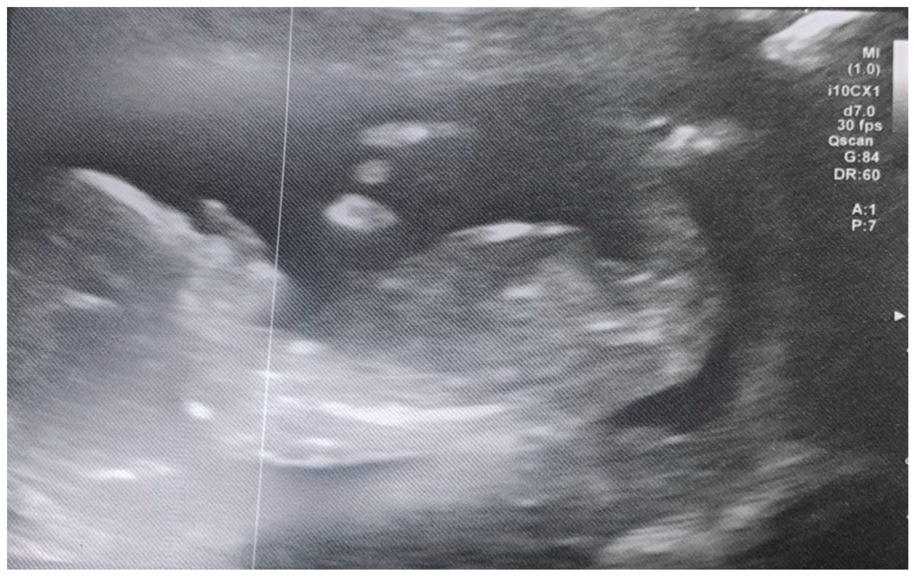
After the first fourth months of carrying out PBM sessions during the follicular stage of the menstrual cycle, a natural conception gave a positive pregnancy test, which led to normal 12-week scans (Figure 2). The scans and blood tests at the end of the first trimester (12 weeks) confirmed a healthy embryo with foetal biometry and foetal anatomy markers within range, and additional markers for aneuploidies also normal.
A healthy baby boy was born (from healthy patient) June 2024 after a full gestation period

4. Discussion

This case report showed the successful natural conception of a female patient with a complex infertility profile, including low ovarian reserve, PCOS and endometriosis, following a course of PBM, which ultimately resulted in the healthy birth of a child after a full-term pregnancy. Given the multifactorial reproductive challenges, the observed outcome suggests that combined red and NIR PBM may offer supportive therapeutic potential in reproductive medicine, particularly in cases considered resistant or suboptimal for conventional approaches. We interpreted our findings in the context of the current literature, as outlined below.

4.1. Role of PBM in Ovarian Health

PBM anti-inflammatory effects might address the chronic inflammation characteristic of PCOS and endometriosis, improving hormonal balance and ovarian function in our case study. This is supported by in vivo animal studies as follows:
An in vivo animal study conducted by He et al. 2024 [45] on naturally aged mice demonstrated that 650 nm PBM at fluence of 4 J/cm2 improved sex hormone levels, increased primordial and growing follicles, enhanced angiogenesis and reduced apoptosis. These effects were attributed to decreased oxidative stress, reduced chronic ovarian inflammation and improved mitochondrial function [45].
Another in vivo animal study conducted by Alves et al. 2019 [58] investigated PBM effects on ovarian activities in a PCOS-induced rat model. The results showed an increase in the number of the ovarian follicles and corpus luteum, decreased ovarian cysts and modulated reproductive and endocrine characteristics. These changes suggest the potential benefits of PBM for women with PCOS [58].

4.2. Role of PBM in Enhancing Endometrial Receptivity

Endometrial receptivity is a critical factor for successful embryo implantation and often serves as a limiting factor in ART outcomes, particularly in women with thin endometrium, chronic inflammation, or a history of implantation failure. In our case report, PBM showed potential to enhance endometrial receptivity, which is essential for achieving successful implantation. Hence, it is important to highlight the mechanisms by which PBM may improve endometrial receptivity, including:
  • Improved microcirculation: PBM has been shown to enhance local blood flow by stimulating NO release and vasodilation, leading to better oxygenation and nutrient delivery to the endometrial tissue [59].
  • Mitochondrial activation: PBM activates cytochrome c oxidase in the mitochondrial respiratory chain, increasing ATP production. This boost in cellular energy supports endometrial cell repair, angiogenesis and cellular proliferation [59].
  • Stimulation of growth factors and cytokines: PBM may promote the release of vascular endothelial growth factor (VEGF), epidermal growth factor (EGF) and fibroblast growth factors (FGFs), which are critical for endometrial growth, vascularization and receptivity [60].
  • Cytokine modulation: PBM appears to modulate the expression of pro-inflammatory and anti-inflammatory cytokines, creating an immune environment favourable for embryo implantation. This modulation is believed to be mediated through various mechanisms, including the activation of mitochondrial pathways and the regulation of ROS. These effects may contribute to improved endometrial receptivity and successful embryo implantation [61]. Hence, the evidence supports the role of PBM in modulating cytokine expression, which may contribute to creating an immune environment conducive to embryo implantation. However, further research is needed to fully understand the mechanisms involved and to establish standardised protocols for clinical application.

4.3. PBM Effects on Oocyte Quality and Maturation

An in vitro study on PCOS oocytes demonstrated that reduced ROS and increased glutathione levels indicate improved oocyte quality [62]. In our case study the use of PBM may have contributed to improved mitochondrial function and reducing ROS, and therefore potentially enhanced oocyte quality and follicular development.
Increased mitochondrial activity and DNA repair mechanisms are shown to stimulate the growth and maturation of immature oocytes. This stimulation can lead to improved quality and quantity of matured oocytes, potentially benefiting women with PCOS undergoing in vitro maturation [62].

4.4. Natural Conception as an Outcome

Natural conception in the presence of three independent infertility risk factors (low AMH, PCOS and endometriosis) is extremely rare without intervention. This case highlights the potential of PBM as a non-invasive, low-risk adjunct therapy capable of improving underlying conditions sufficiently to allow spontaneous conception.
While it is not possible to isolate PBM as the sole causative factor in this outcome, its timing and application correlate with an otherwise unexpected pregnancy. This supports the need for further studies, including controlled clinical trials, to better define PBM’s role, optimal parameters and patient selection criteria in reproductive medicine.

4.5. Study Limitations

This case report study, while valuable for highlighting novel PBM protocol for a rare case which led to natural conception and a long-term follow-up with a healthy birth, has several important limitations in terms of scientific rigour and clinical generalisability. Here are the key limitations to consider: (1) lack of generalisability: a single case reflects the outcome in one individual, which may not apply to the broader population; (2) no control group: without a comparison group, it is impossible to determine whether the outcome was due to the PBM, a placebo effect, spontaneous remission or other confounding variables; (3) no large data; (4) no statistical power.

5. Conclusions

As an observational case report it is not possible to draw a causal link between the PBM treatment and the outcome. However, PBM could be considered by clinicians as a compelling, non-invasive adjunctive therapy for women facing complex fertility challenges such as diminished ovarian reserve, polycystic ovary syndrome (PCOS) and endometriosis, for the reasons we have explored. By targeting mitochondrial function, enhancing ATP production and modulating oxidative stress and inflammation, PBM addresses key cellular pathways implicated in reproductive dysfunction. Emerging evidence supports the role of PBM in improving ovarian response and endometrial receptivity and suggests potential to enhance natural conception and ART outcomes. To fully integrate PBM into standard reproductive care, robust clinical trials are essential to establish optimal parameters, long-term safety, and efficacy.

Author Contributions

Conceptualisation, R.P.; methodology, R.P.; software, R.P.; validation, R.H. and R.P.; formal analysis, R.P.; investigation, R.P.; resources, R.H. and R.P.; data curation, R.H.; writing—original draft preparation, R.H. and R.P.; writing—review and editing, R.H. and R.P.; visualisation, R.P. and R.H.; supervision, R.H. and R.P.; project administration, R.P. All authors have read and agreed to the published version of the manuscript.

Funding

This research received no external funding.

Institutional Review Board Statement

Not applicable.

Informed Consent Statement

An informed written consent was obtained from all the subjects involved in the study. Also, a written informed consent was obtained for publishing an image of the subject in a peer-reviewed journal.

Data Availability Statement

All the data are provided in the text.

Conflicts of Interest

The authors declare no conflicts of interest.

References

  1. Chauhan, S.; More, A.; Chauhan, V.; Kathane, A. Endometriosis: A Review of Clinical Diagnosis, Treatment, and Pathogenesis. Cureus 2022, 14, e28864. [Google Scholar] [CrossRef]
  2. Strathy, J.H.; Molgaard, C.A.; Coulam, C.B.; Melton, L.J. Endometriosis and Infertility: A Laparoscopic Study of Endometriosis among Fertile and Infertile Women. Fertil. Steril. 1982, 38, 667–672. [Google Scholar] [CrossRef]
  3. Vercellini, P.; Viganò, P.; Somigliana, E.; Fedele, L. Endometriosis: Pathogenesis and Treatment. Nat. Rev. Endocrinol. 2014, 10, 261–275. [Google Scholar] [CrossRef]
  4. Macer, M.L.; Taylor, H.S. Endometriosis and Infertility. Obstet. Gynecol. Clin. N. Am. 2012, 39, 535–549. [Google Scholar] [CrossRef]
  5. Fan, W.; Yuan, Z.; Li, M.; Zhang, Y.; Nan, F. Decreased Oocyte Quality in Patients with Endometriosis Is Closely Related to Abnormal Granulosa Cells. Front. Endocrinol. 2023, 14, 1226687. [Google Scholar] [CrossRef]
  6. Prescott, J.; Farland, L.V.; Tobias, D.K.; Gaskins, A.J.; Spiegelman, D.; Chavarro, J.E.; Rich-Edwards, J.W.; Barbieri, R.L.; Missmer, S.A. A Prospective Cohort Study of Endometriosis and Subsequent Risk of Infertility. Hum. Reprod. 2016, 31, 1475–1482. [Google Scholar] [CrossRef] [PubMed]
  7. Gurtner, G.C.; Werner, S.; Barrandon, Y.; Longaker, M.T. Wound Repair and Regeneration. Nature 2008, 453, 314–321. [Google Scholar] [CrossRef] [PubMed]
  8. Ahmad, A.K.; Kao, C.-N.; Quinn, M.; Lenhart, N.; Rosen, M.; Cedars, M.I.; Huddleston, H. Differential Rate in Decline in Ovarian Reserve Markers in Women with Polycystic Ovary Syndrome Compared with Control Subjects: Results of a Longitudinal Study. Fertil. Steril. 2018, 109, 526–531. [Google Scholar] [CrossRef]
  9. Bozdag, G.; Mumusoglu, S.; Zengin, D.; Karabulut, E.; Yildiz, B.O. The Prevalence and Phenotypic Features of Polycystic Ovary Syndrome: A Systematic Review and Meta-Analysis. Hum. Reprod. 2016, 31, 2841–2855. [Google Scholar] [CrossRef] [PubMed]
  10. Kousta, E.; White, D.M.; Cela, E.; McCarthy, M.I.; Franks, S. The Prevalence of Polycystic Ovaries in Women with Infertility. Hum. Reprod. 1999, 14, 2720–2723. [Google Scholar] [CrossRef]
  11. Cunha, A.; Póvoa, A.M. Infertility Management in Women with Polycystic Ovary Syndrome: A Review. Porto Biomed. J. 2021, 6, e116. [Google Scholar] [CrossRef]
  12. Hart, R.; Doherty, D.A. The Potential Implications of a PCOS Diagnosis on a Woman’s Long-Term Health Using Data Linkage. J. Clin. Endocrinol. Metab. 2015, 100, 911–919. [Google Scholar] [CrossRef]
  13. Aboeldalyl, S.; James, C.; Seyam, E.; Ibrahim, E.M.; Shawki, H.E.-D.; Amer, S. The Role of Chronic Inflammation in Polycystic Ovarian Syndrome—A Systematic Review and Meta-Analysis. Int. J. Mol. Sci. 2021, 22, 2734. [Google Scholar] [CrossRef] [PubMed]
  14. Singla, R.; Gupta, Y.; Khemani, M.; Aggarwal, S. Thyroid Disorders and Polycystic Ovary Syndrome: An Emerging Relationship. Indian J. Endocr. Metab. 2015, 19, 25. [Google Scholar] [CrossRef]
  15. Cohen, J.; Chabbert-Buffet, N.; Darai, E. Diminished Ovarian Reserve, Premature Ovarian Failure, Poor Ovarian Responder—A Plea for Universal Definitions. J. Assist. Reprod. Genet. 2015, 32, 1709–1712. [Google Scholar] [CrossRef] [PubMed]
  16. Tan, Z.; Gong, X.; Wang, C.C.; Zhang, T.; Huang, J. Diminished Ovarian Reserve in Endometriosis: Insights from In Vitro, In Vivo, and Human Studies—A Systematic Review. Int. J. Mol. Sci. 2023, 24, 15967. [Google Scholar] [CrossRef] [PubMed]
  17. Lessans, N.; Gilan, A.; Dick, A.; Bibar, N.; Saar, T.D.; Porat, S.; Dior, U.P. Ovarian Reserve Markers of Women with Superficial Endometriosis. Int. J. Gynecol. Obstet. 2024, 165, 696–702. [Google Scholar] [CrossRef]
  18. Waghmare, S.V.; Shanoo, A. Polycystic Ovary Syndrome: A Literature Review With a Focus on Diagnosis, Pathophysiology, and Management. Cureus 2023, 15, e47408. [Google Scholar] [CrossRef]
  19. Zeng, Y.; Wang, C.; Yang, C.; Shan, X.; Meng, X.-Q.; Zhang, M. Unveiling the Role of Chronic Inflammation in Ovarian Aging: Insights into Mechanisms and Clinical Implications. Hum. Reprod. 2024, 39, 1599–1607. [Google Scholar] [CrossRef]
  20. Uri-Belapolsky, S.; Shaish, A.; Eliyahu, E.; Grossman, H.; Levi, M.; Chuderland, D.; Ninio-Many, L.; Hasky, N.; Shashar, D.; Almog, T.; et al. Interleukin-1 Deficiency Prolongs Ovarian Lifespan in Mice. Proc. Natl. Acad. Sci. USA 2014, 111, 12492–12497. [Google Scholar] [CrossRef]
  21. Treasure, T. Fertility: Assessment and Treatment for People with Fertility Problems; National Institute for Health and Clinical Excellence: Manchester, UK, 2013; ISBN 978-1-4731-0029-9. [Google Scholar]
  22. Phypers, R.; Berisha-Muharremi, V.; Hanna, R. The Efficacy of Multiwavelength Red and Near-Infrared Transdermal Photobiomodulation Light Therapy in Enhancing Female Fertility Outcomes and Improving Reproductive Health: A Prospective Case Series with 9-Month Follow-Up. J. Clin. Med. 2024, 13, 7101. [Google Scholar] [CrossRef] [PubMed]
  23. Morley, L.C.; Tang, T.M.H.; Balen, A.H. Metformin Therapy for the Management of Infertility in Women with Polycystic Ovary Syndrome: Scientific Impact Paper No. 13. BJOG 2017, 124, e306–e313. [Google Scholar] [CrossRef]
  24. Tso, L.O.; Costello, M.F.; Albuquerque, L.E.T.; Andriolo, R.B.; Macedo, C.R. Metformin Treatment before and during IVF or ICSI in Women with Polycystic Ovary Syndrome. Cochrane Database Syst. Rev. 2020, 2020, CD006105. [Google Scholar] [CrossRef]
  25. Romanski, P.A.; Bortoletto, P.; Malmsten, J.E.; Tan, K.S.; Spandorfer, S.D. Pregnancy Outcomes after Oral and Injectable Ovulation Induction in Women with Infertility with a Low Antimüllerian Hormone Level Compared with Those with a Normal Antimüllerian Hormone Level. Fertil. Steril. 2022, 118, 1048–1056. [Google Scholar] [CrossRef]
  26. Conforti, A.; Carbone, L.; Di Girolamo, R.; Iorio, G.G.; Guida, M.; Campitiello, M.R.; Ubaldi, F.M.; Rienzi, L.; Vaiarelli, A.; Cimadomo, D.; et al. Therapeutic Management in Women with a Diminished Ovarian Reserve: A Systematic Review and Meta-Analysis of Randomized Controlled Trials. Fertil. Steril. 2025, 123, 457–476. [Google Scholar] [CrossRef]
  27. Diamond, M.P.; Legro, R.S.; Coutifaris, C.; Alvero, R.; Robinson, R.D.; Casson, P.; Christman, G.M.; Ager, J.; Huang, H.; Hansen, K.R.; et al. Letrozole, Gonadotropin, or Clomiphene for Unexplained Infertility. N. Engl. J. Med. 2015, 373, 1230–1240. [Google Scholar] [CrossRef] [PubMed]
  28. Nesbitt-Hawes, E.M.; Campbell, N.; Maley, P.E.; Won, H.; Hooshmand, D.; Henry, A.; Ledger, W.; Abbott, J.A. The Surgical Treatment of Severe Endometriosis Positively Affects the Chance of Natural or Assisted Pregnancy Postoperatively. BioMed Res. Int. 2015, 2015, 438790. [Google Scholar] [CrossRef]
  29. La Marca, A.; Semprini, M.; Mastellari, E.; Donno, V.; Capuzzo, M.; Alboni, C.; Giulini, S. Fertility Preservation in Women with Endometriosis. Hum. Reprod. Open 2025, 2025, hoaf012. [Google Scholar] [CrossRef]
  30. Krämer, B.; Andress, J.; Neis, F.; Hoffmann, S.; Brucker, S.; Kommoss, S.; Höller, A. Adhesion Prevention after Endometriosis Surgery—Results of a Randomized, Controlled Clinical Trial with Second-Look Laparoscopy. Langenbeck’s Arch. Surg. 2021, 406, 2133–2143. [Google Scholar] [CrossRef]
  31. Alesi, S.; Villani, A.; Mantzioris, E.; Takele, W.W.; Cowan, S.; Moran, L.J.; Mousa, A. Anti-Inflammatory Diets in Fertility: An Evidence Review. Nutrients 2022, 14, 3914. [Google Scholar] [CrossRef] [PubMed]
  32. Nirgianakis, K.; Egger, K.; Kalaitzopoulos, D.R.; Lanz, S.; Bally, L.; Mueller, M.D. Effectiveness of Dietary Interventions in the Treatment of Endometriosis: A Systematic Review. Reprod. Sci. 2022, 29, 26–42. [Google Scholar] [CrossRef]
  33. Barrea, L.; Arnone, A.; Annunziata, G.; Muscogiuri, G.; Laudisio, D.; Salzano, C.; Pugliese, G.; Colao, A.; Savastano, S. Adherence to the Mediterranean Diet, Dietary Patterns and Body Composition in Women with Polycystic Ovary Syndrome (PCOS). Nutrients 2019, 11, 2278. [Google Scholar] [CrossRef]
  34. Ben-Meir, A.; Burstein, E.; Borrego-Alvarez, A.; Chong, J.; Wong, E.; Yavorska, T.; Naranian, T.; Chi, M.; Wang, Y.; Bentov, Y.; et al. Coenzyme Q10 Restores Oocyte Mitochondrial Function and Fertility during Reproductive Aging. Aging Cell 2015, 14, 887–895. [Google Scholar] [CrossRef]
  35. Abodi, M.; De Cosmi, V.; Parazzini, F.; Agostoni, C. Omega-3 Fatty Acids Dietary Intake for Oocyte Quality in Women Undergoing Assisted Reproductive Techniques: A Systematic Review. Eur. J. Obstet. Gynecol. Reprod. Biol. 2022, 275, 97–105. [Google Scholar] [CrossRef] [PubMed]
  36. Trop-Steinberg, S.; Gal, M.; Azar, Y.; Kilav-Levin, R.; Heifetz, E.M. Effect of Omega-3 Supplements or Diets on Fertility in Women: A Meta-Analysis. Heliyon 2024, 10, e29324. [Google Scholar] [CrossRef]
  37. Hamblin, M.R. Mechanisms and Applications of the Anti-Inflammatory Effects of Photobiomodulation. AIMS Biophys. 2017, 4, 337–361. [Google Scholar] [CrossRef]
  38. Hamblin, M.R.; Demidova, T.N. Mechanisms of Low Level Light Therapy. In Proceedings of the Mechanisms for Low-Light Therapy, San Jose, CA, USA, 21–26 January 2006; Hamblin, M.R., Waynant, R.W., Anders, J., Eds.; SPIE: Bellingham, WA, USA, 2006; Volumn 6140, p. 614001. [Google Scholar] [CrossRef]
  39. Hanna, R.; Miron, I.C.; Benedicenti, S. A Novel Therapeutic Approach of 980 Nm Photobiomodulation Delivered with Flattop Beam Profile in Management of Recurrent Aphthous Stomatitis in Paediatrics and Adolescents—A Case Series with 3-Month Follow-Up. J. Clin. Med. 2024, 13, 2007. [Google Scholar] [CrossRef] [PubMed]
  40. Iranpour, B.; Mohammadi, K.; Hodjat, M.; Hakimiha, N.; Sayar, F.; Kharazi Fard, M.J.; Sadatmansouri, S.; Hanna, R. An Evaluation of Photobiomodulation Effects on Human Gingival Fibroblast Cells under Hyperglycemic Condition: An in Vitro Study. Lasers Med. Sci. 2023, 39, 9. [Google Scholar] [CrossRef] [PubMed]
  41. Hanna, R.; Agas, D.; Benedicenti, S.; Ferrando, S.; Laus, F.; Cuteri, V.; Lacava, G.; Sabbieti, M.G.; Amaroli, A. A Comparative Study Between the Effectiveness of 980 Nm Photobiomodulation Delivered by Hand-Piece With Gaussian vs. Flat-Top Profiles on Osteoblasts Maturation. Front. Endocrinol. 2019, 10, 92. [Google Scholar] [CrossRef]
  42. Hanna, R.; Dalvi, S.; Bensadoun, R.J.; Raber-Durlacher, J.E.; Benedicenti, S. Role of Photobiomodulation Therapy in Neurological Primary Burning Mouth Syndrome. A Systematic Review and Meta-Analysis of Human Randomised Controlled Clinical Trials. Pharmaceutics 2021, 13, 1838. [Google Scholar] [CrossRef]
  43. Hanna, R.; Bensadoun, R.J.; Beken, S.V.; Burton, P.; Carroll, J.; Benedicenti, S. Outpatient Oral Neuropathic Pain Management with Photobiomodulation Therapy: A Prospective Analgesic Pharmacotherapy-Paralleled Feasibility Trial. Antioxidants 2022, 11, 533. [Google Scholar] [CrossRef]
  44. Oubiña, G.; Pascuali, N.; Scotti, L.; Di Pietro, M.; La Spina, F.A.; Buffone, M.G.; Higuera, J.; Abramovich, D.; Parborell, F. Low Level Laser Therapy (LLLT) Modulates Ovarian Function in Mature Female Mice. Prog. Biophys. Mol. Biol. 2019, 145, 10–18. [Google Scholar] [CrossRef] [PubMed]
  45. He, Y.; Ye, R.; Peng, Y.; Pei, Q.; Wu, L.; Wang, C.; Ni, W.; Li, M.; Zhang, Y.; Yao, M. Photobiomodulation Ameliorates Ovarian Aging by Alleviating Oxidative Stress and Inflammation Damage and Improving Mitochondrial Function. J. Photochem. Photobiol. B Biol. 2024, 260, 113024. [Google Scholar] [CrossRef] [PubMed]
  46. Oubiña, G.; Pascuali, N.; Scotti, L.; Bianchi, S.; May, M.; Martínez, J.E.; Marchese Ragona, C.; Higuera, J.; Abramovich, D.; Parborell, F. Local Application of Low Level Laser Therapy in Mice Ameliorates Ovarian Damage Induced by Cyclophosphamide. Mol. Cell. Endocrinol. 2021, 531, 111318. [Google Scholar] [CrossRef]
  47. Hochler, H.; Lipschuetz, M.; Suissa-Cohen, Y.; Weiss, A.; Sela, H.Y.; Yagel, S.; Rosenbloom, J.I.; Grisaru-Granovsky, S.; Rottenstreich, M. The Impact of Advanced Maternal Age on Pregnancy Outcomes: A Retrospective Multicenter Study. J. Clin. Med. 2023, 12, 5696. [Google Scholar] [CrossRef]
  48. Grinsted, A.; Grinsted Hillegass, M. Photobiomodulation for Infertility. Available online: https://ecronicon.com/ecgy/photobiomodulation-for-infertility (accessed on 1 July 2025).
  49. Ohshiro, T. Personal overview of the application of lllt in severely infertile japanese females. Laser Ther. 2012, 21, 97–103. [Google Scholar] [CrossRef]
  50. National Heart, Lung and Blood Institute. Available online: www.nhlbi.nih.gov/health/educational/lose_wt/BMI/bmicalc.htm (accessed on 1 July 2025).
  51. Gupta, V.; Sharma, V.K. Skin Typing: Fitzpatrick Grading and Others. Clin. Dermatol. 2019, 37, 430–436. [Google Scholar] [CrossRef]
  52. Vander Borght, M.; Wyns, C. Fertility and Infertility: Definition and Epidemiology. Clin. Biochem. 2018, 62, 2–10. [Google Scholar] [CrossRef]
  53. Jahani-Sherafat, S.; Taghavi, H.; Asri, N.; Rezaei Tavirani, M.; Razzaghi, Z.; Rostami-Nejad, M. The Effectiveness of Photobiomodulation Therapy in Modulation the Gut Microbiome Dysbiosis Related Diseases. Gastroenterol. Hepatol. Bed Bench 2023, 16, 386–393. [Google Scholar] [CrossRef]
  54. Fujii, S.; Ohshiro, T.; Ohshiro, T.; Sasaki, K.; Taniguchi, Y. Proximal priority treatment using the neck irradiator for adjunctive treatment of female infertility. Laser Ther. 2007, 16, 133–136. [Google Scholar] [CrossRef]
  55. Detti, L.; Francillon, L.; Christiansen, M.E.; Peregrin-Alvarez, I.; Goedecke, P.J.; Bursac, Z.; Roman, R.A. Early Pregnancy Ultrasound Measurements and Prediction of First Trimester Pregnancy Loss: A Logistic Model. Sci. Rep. 2020, 10, 1545. [Google Scholar] [CrossRef]
  56. Fruscalzo, A.; Cividino, A.; Rossetti, E.; Maurigh, A.; Londero, A.P.; Driul, L. First Trimester PAPP-A Serum Levels and Long-Term Metabolic Outcome of Mothers and Their Offspring. Sci. Rep. 2020, 10, 5131. [Google Scholar] [CrossRef]
  57. Younesi, S.; Eslamian, L.; Khalafi, N.; Taheri Amin, M.M.; Saadati, P.; Jamali, S.; Balvayeh, P.; Modarressi, M.-H.; Savad, S.; Amidi, S.; et al. Extreme βHCG Levels in First Trimester Screening Are Risk Factors for Adverse Maternal and Fetal Outcomes. Sci. Rep. 2023, 13, 1228. [Google Scholar] [CrossRef]
  58. Alves, E.D.; Bonfá, A.L.O.; Pigatto, G.R.; Anselmo-Franci, J.A.; Achcar, J.A.; Parizotto, N.A.; Montrezor, L.H. Photobiomodulation Can Improve Ovarian Activity in Polycystic Ovary Syndrome-Induced Rats. J. Photochem. Photobiol. B Biol. 2019, 194, 6–13. [Google Scholar] [CrossRef]
  59. Jafarabadi, M.; Farbod, Y.; Shariat, M. Low-Level Laser Therapy for Improvement of In Vitro Fertilization Outcomes in Patients with Recurrent Implantation Failure: A Randomized Clinical Trial. J. Lasers Med. Sci. 2024, 15, e15. [Google Scholar] [CrossRef]
  60. Lin, Y.; Qi, J.; Sun, Y. Platelet-Rich Plasma as a Potential New Strategy in the Endometrium Treatment in Assisted Reproductive Technology. Front. Endocrinol. 2021, 12, 707584. [Google Scholar] [CrossRef] [PubMed]
  61. Shamloo, S.; Defensor, E.; Ciari, P.; Ogawa, G.; Vidano, L.; Lin, J.S.; Fortkort, J.A.; Shamloo, M.; Barron, A.E. The Anti-Inflammatory Effects of Photobiomodulation Are Mediated by Cytokines: Evidence from a Mouse Model of Inflammation. Front. Neurosci. 2023, 17, 1150156. [Google Scholar] [CrossRef] [PubMed]
  62. Sahraeian, S.; Abbaszadeh, H.A.; Taheripanah, R.; Edalatmanesh, M.A. Extracellular Vesicle-Derived Cord Blood Plasma and Photobiomodulation Therapy Down-Regulated Caspase 3, LC3 and Beclin 1 Markers in the PCOS Oocyte: An In Vitro Study. J. Lasers Med. Sci. 2023, 14, e23. [Google Scholar] [CrossRef] [PubMed]
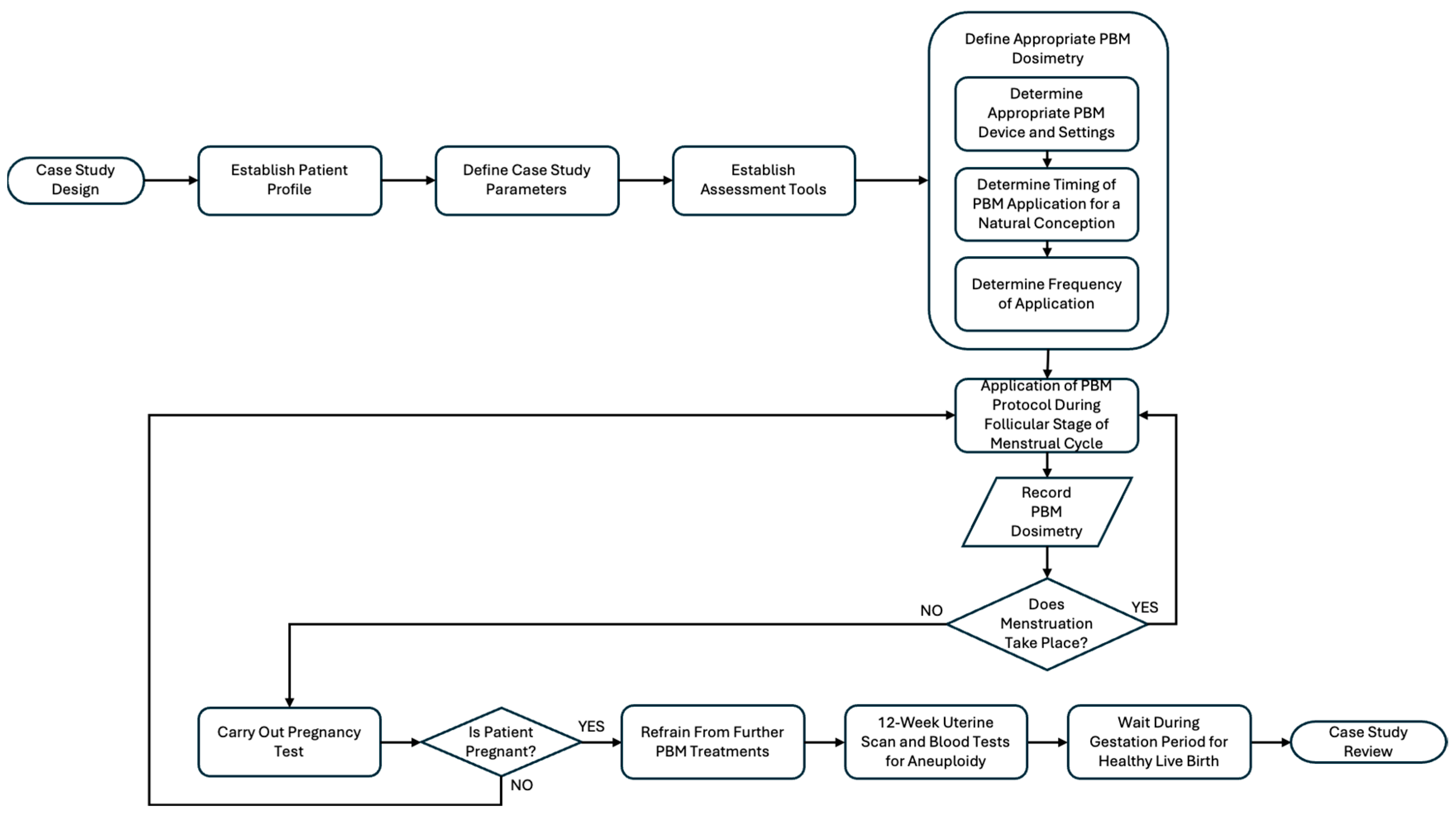
Figure 1. A flowchart showing the steps involved in the methodology.
Photonics 12 01021 g001
Figure 2. Shows 12-week gestation scan.
Photonics 12 01021 g002
Table 1. Presentation of the subject’s demographic characteristics.
Age at
Conception (Years)
Age at Birth (Years)Weight (Pre-Conception) (kg)Height
(cm)
BMISkin
Colour
Subject27287215828.8III
Table 2. Device specification, laser PBM dosimetry and treatment protocol [22].
ManufacturerK-Laser
Semiconductor materials (emitter type)GaAIAs
Probe design4 wavelengths probe
Device classificationType 4 Laser
Beam delivery systemFibre
Laser-aiming beamNone
Wavelength (nm)660, 800, 905, 970
Operating emission modeA combination of CW and SP
PolarisationLinear
Therapeutic power output for 800, 905, 970 (W)~15
Ratio of power output divided equally between 800, 905, 970 nm1:1:1
Therapeutic power output for 660 nm (mW)~120
Total fluence (J/cm2) per point (irradiation area of 10 cm2)3150
Total irradiation time over 10 cm25 mins and 15 s
Total number of irradiated points above the ovaries/uterus/abdomen 3
Total number of irradiated points at lower back/sacrum/cervical spine1
Total of fluence (J/cm2) per session12,600
Total irradiation time per session20 mins and 45 s
Time intervalRelatively every three weeks
Treatment frequency1 or 2 sessions per month
Total treatment sessions5 sessions
Treatment duration4 months
Scanning techniqueMoveable application
Light-skin tissue distance (cm)4.5
Disclaimer/Publisher’s Note: The statements, opinions and data contained in all publications are solely those of the individual author(s) and contributor(s) and not of MDPI and/or the editor(s). MDPI and/or the editor(s) disclaim responsibility for any injury to people or property resulting from any ideas, methods, instructions or products referred to in the content.