Article

CB1 and CB2 Receptor Expression in Type 1 Diabetic Neuropathic Rats Is Enhanced by Photobiomodulation Therapy

by
Danielle Paula Freitas Bataus Silva
1,
Natalia Vendrame
1,
Willians Fernando Vieira
1,2,3 and
Marucia Chacur
1,4,*
1
Laboratory of Functional Neuroanatomy of Pain, Department of Anatomy, Institute of Biomedical Sciences, University of São Paulo (USP), Av. Prof. Lineu Prestes, 2415, São Paulo 05508-900, Brazil
2
Laboratory of Neuroimmune Interface of Pain Research, Instituto e Centro de Pesquisas São Leopoldo Mandic, Faculdade São Leopoldo Mandic, Campinas 13045-755, Brazil
3
Department of Structural and Functional Biology, Institute of Biology, University of Campinas (UNICAMP), Campinas 13083-865, Brazil
4
Program in Neuroscience and Behavior, Institute of Psychology, University of São Paulo (USP), São Paulo 05508-900, Brazil
*
Author to whom correspondence should be addressed.
Photonics 2025, 12(11), 1060; https://doi.org/10.3390/photonics12111060
Submission received: 3 September 2025 / Revised: 16 October 2025 / Accepted: 23 October 2025 / Published: 27 October 2025
(This article belongs to the Special Issue Shining Light on Healing: Photobiomodulation Therapy)

Abstract

Background: The interaction between the endocannabinoid system (ECS), specifically the CB1 and CB2 cannabinoid receptors, and neuropathy has aroused great research interest due to the possible implications for treatment. Complications following type 1 diabetes, due to impaired glucose metabolism and chronic inflammation, may benefit from targeted therapeutic strategies involving the ECS. This study explores the effects of photobiomodulation therapy (PBMT) on streptozotocin (STZ)-induced diabetic peripheral neuropathy (DPN) in rats. The study assessed body mass, hyperglycemia, mechanical hyperalgesia, and the influence of PBMT on these conditions over four weeks. Results showed that while PBMT did not alter the metabolic aspects of type I diabetes, it significantly reduced mechanical hyperalgesia compared to untreated diabetic neuropathic rats. Notably, cannabinoid receptor antagonists for CB1 and CB2 elicited a transient reversal of this antihyperalgesic effect, indicating a potential role of these receptors in PBMT’s mechanism. However, CB2 modulation was not statistically significant, whereas changes in CB1 receptor expression were observed in the dorsal root ganglia, suggesting its involvement in PBMT’s effects. These findings highlight the importance of CB1 and CB2 receptors in DPN and suggest that PBMT may offer a therapeutic benefit by mitigating mechanical hyperalgesia. Further investigation into cannabinoid receptor dynamics in diabetes could help in new therapeutic strategies for managing diabetic complications.

1. Introduction

Diabetes mellitus, a chronic metabolic disease that affects nearly 10% of the global population, results from the body’s inability to produce (type 1 diabetes) or effectively use insulin (type 2 diabetes), leading to sustained hyperglycemia and peripheral nerve damage [1,2,3]. Among its complications, diabetic peripheral neuropathy (DPN) is notable for producing sensory abnormalities such as allodynia and mechanical hyperalgesia [4]. Although primarily affecting the peripheral nervous system (PNS), DPN also involves central alterations, including reduced spinal cord volume and cortical gray matter changes [5,6,7,8,9,10], which highlight the complex interplay between peripheral and central mechanisms in its pathophysiology. These changes in the central nervous system (CNS) underscore the complex interplay between peripheral and central mechanisms in the pathophysiology of DPN. This dual action highlights its potential as a comprehensive treatment strategy for managing DPN’s multifaceted pathology.
In the pursuit of methodologies to potentially delay, mitigate, or even manage DPN, investigations have unearthed studies demonstrating the potential efficacy of cannabinoids in mitigating nerve damage induced by conditions such as chronic constriction of the sciatic nerve [11], trigeminal neuralgia [12], chemotherapy-induced neuropathy [13] and streptozotocin (STZ)-induced DPN, among others [14]. Clinical evidence suggests that the endocannabinoid system (ECS), consisting of its receptors (CB1 and CB2), endogenous ligands, and enzymes responsible for synthesis and metabolism, may exert influence over several pathophysiological signaling pathways [15]. CB1 and CB2 receptor activation may exert effects similar to anxiolytic and antidepressant drugs, some of which are already in clinical use in specific circumstances and may have antiemetic, appetite-stimulating, and analgesic properties [16]. Remarkably, even at the peripheral level, the activation of CB1 receptors exhibits the capacity to alleviate both hyperalgesic and allodynic responses [14]. It is noteworthy that pharmacological interactions within the ECS can lead to potential functional modifications and may contribute adversely to the exacerbation of pathological pain levels, particularly in the context of DPN [17,18,19,20,21].
In recent years, photobiomodulation therapy (PBMT) has gained recognition as a non-pharmacological approach capable of modulating inflammation, oxidative stress, and nociceptive signaling [22,23,24,25,26,27,28,29,30]. PBMT utilizes non-ionizing light sources, including lasers and light-emitting diodes (LEDs), which differ in terms of coherence, penetration depth, and energy delivery [31,32]. While both modalities can stimulate photochemical reactions in mitochondria, laser devices provide higher beam collimation and energy precision, which is particularly advantageous for targeting deeper neural and muscular tissues relevant to neuropathic pain [31,33]. Preclinical and clinical studies have demonstrated that laser-based PBMT improves nerve regeneration, reduces neuroinflammation, and alleviates neuropathic pain in models of DPN and other nerve injuries, supporting its translational relevance [26,27,31,34,35,36].
Therefore, the present study aimed to investigate the antihyperalgesic effects of laser-based PBMT in a mouse model of STZ-induced DPN, focusing on its potential modulation of the ECS. Specifically, we sought to determine whether PBMT could attenuate mechanical hyperalgesia and influence ECS-related molecular markers in peripheral and central tissues.

2. Methods

2.1. Animals

We used a total of forty-five male Wistar rats (Rattus norvegicus), 8 weeks of age, weighing 200–240 g, purchased from the Central Animal Facility of the Institute of Biomedical Sciences (ICB) at the University of São Paulo (USP). All procedures were approved by the Animal Research Ethics Committee (CEUA) of the Institute of Biomedical Sciences, University of São Paulo (protocol number: 2017110820). Experiments were conducted in accordance with the guidelines for the ethical use of conscious animals in pain research established by the International Association for the Study of Pain (IASP) and in compliance with the ARRIVE guidelines [30]. Rats were kept at a room temperature of 22 ± 2 °C with a light/dark cycle (12:12 h). Food (pattern chow) and water were provided ad libitum. The rats were allowed to acclimatize to the testing rooms for at least 3 days before the onset of the study. They were randomly divided into five groups. N per group is shown in results and graph legends and the sample size was defined based on previous studies using comparable experimental models and molecular analyses, ensuring adequate statistical power while complying with the 3Rs (Reduction, Replacement, and Refinement) principle of animal research: 1—Naïve: rats were used as general control animals and did not receive any injection or treatment; 2—STZ: rats were subjected to five intraperitoneal (i.p.) injections (1 per day, for five consecutive days) of streptozotocin (25 mg/kg each one); 3—STZ + PBMT: rats received five i.p. injections of streptozotocin (25 mg/kg each one) and were subjected to photobiomodulation therapy (PBMT); 4—SCB: rats received five i.p. injections of sodium citrate buffer, SCB (STZ vehicle); 5—SCB + PBMT: rats received five i.p. injections of SCB and were submitted to PBMT.
Additionally, we used other 4 groups (5 animals per group) to measure the involvement of cannabinoid receptors CB1 and CB2 through pharmacological antagonists on the mechanical hyperalgesia of DPN rats and the PBMT antihyperalgesic response: 1—STZ + CB1: rats received five i.p. injections of STZ (25 mg/kg) and one lumbar intrathecal (i.t.) injection of the CB1 antagonist AM251 (30 μg/50 μL); 2—STZ + CB2: rats received five i.p. injections of STZ (25 mg/kg) and one lumbar i.t. injection of the CB2 antagonist AM630 (10 μg/50 μL); 3—STZ + PBMT + CB1: rats received five i.p. injections of STZ (25 mg/kg), were subjected to PBMT and received one lumbar i.t. injection of AM251 (30 μg/50 μL); 4—STZ + PBMT + CB2: rats received five i.p. injections of STZ (25 mg/kg), were subjected to PBMT, and received one lumbar i.t. injection of AM630 (10 μg/50 μL).

2.2. Streptozotocin (STZ)-Induced Type 1 Diabetes Mellitus

For the induction of type 1 diabetes, rats from STZ, STZ + CB1, STZ + CB2, STZ + PBMT, STZ + PBMT + CB1, and STZ + PBMT + CB2 groups received five i.p. injections of 25 mg/kg of STZ (Cayman Chemical®, Ann Arbor, MI, USA) diluted in 0.1 M sodium citrate buffer (SCB, pH 4.5), always applied at the same time each day, once daily, for five consecutive days. The SCB and SCB + PBMT groups received five i.p. injections of STZ vehicle (0.1 M SCB, pH 4.5) during the corresponding period. For the blood glucose assessment, rats from all groups had their blood samples collected periodically through caudal venous puncture with a 0.45 × 13 mm (26 G ½″) needle. Glycemia measurements were done with the aid of the Accu-Chek Sensor Comfort (Roche Diagnostics®, Basel, Switzerland) by using test strips impregnated with glucose oxidase. Diabetes was clinically considered when the rats reached a minimum of 250 mg/dL of blood glucose for two consecutive days [23,24,30,37,38,39,40].

2.3. Photobiomodulation Therapy (PBMT) by Low-Level Laser Irradiation

Rats from STZ + PBMT, SCB + PBMT, STZ + PBMT + CB1, and STZ + PBMT + CB2 groups were subjected to PBMT once a day, during 8 consecutive days (from the 21st to the 28th day after the start of STZ or vehicle injections). For the diabetic groups (STZ + PBMT, STZ + PBMT + CB1, and STZ + PBMT + CB2), only hyperalgesic rats were considered to receive the laser treatment. For that, rats were anesthetized with isoflurane (Cristália®, Itapira, Brazil) at a concentration of 3% for induction and 2% for maintenance. Irradiation was then applied through direct contact of the laser probe over carefully shaved areas located on the dorsal side, between the L4/L5 lumbar levels, bilaterally. A Laserpulse Diamond (IBRAMED® Ltda., Amparo, Brazil) laser emission device, 904 nm, Gallium Arsenide (GaAs), class IIIb, with scales graduated in Joules/cm2 and minutes/seconds, was used to perform the PBMT treatment. PBMT parameters are presented in Table 1 and were determined from studies previously carried out by our research group [7,23,24,37,41,42].

2.4. Intrathecal (i.t.) Injection of CB1 and CB2 Antagonists

On the 28th day, after finishing the last PBMT sessions, animals from the STZ + CB1, STZ + CB2, STZ + PBMT + CB1, and STZ + PBMT + CB2 groups received an i.t. injection (50 μL) of CB1 or CB2 antagonists while anesthetized by isoflurane inhalation. Injections were performed with a 29-G needle in the intervertebral region between L4 and L5, which was previously trichotomized [14]. The movement of the tail (flinch) indicated the needle’s proper position [43,44], then the following drugs were administered: N-(Piperidin-1-yl)-5-(4-iodophenyl)-1-(2,4-dichlorophenyl)-4-methyl-1H-pyrazole-3-carboxamide (AM251) (Cayman Chemical®, Ann Arbor, MI, USA), a CB1 receptor antagonist, or [6-Iodo-2-methyl-1-[2-(4-morpholinyl)ethyl]-1H-indol-3-yl]4-methoxyphenyl)methanone (AM630) (Cayman Chemical®, Ann Arbor, MI, USA), a CB2 receptor antagonist. AM251 and AM630 were initially dissolved in dimethyl sulfoxide (20%, Sigma-Aldrich, St. Louis, MO, USA) and then in saline (NaCl 0.9%) in the following concentrations: AM251: 30 µg/50 µL and AM630: 10 µg/50 µL [45].

2.5. Electronic Von Frey Test (Mechanical Hyperalgesia)

The development of hyperalgesia was determined by the mechanical withdrawal thresholds by applying the electronic von Frey test (Insight®, Ribeirão Preto, Brazil). It was performed using a universal 10 μL polypropylene pipette tip (T-300, Axygen®, Glendale, AZ, USA) adapted to a hand-held force transducer with crescent pressure in the plantar surface of the rat’s right and left hind paws. The measurement obtained from the paw surface area results in units of gram-force (g) when the paw was withdrawn. Rats were randomly placed into plastic cages with a metal mesh floor, followed by 30 min of acclimatization before the test. The test was applied to all experimental groups at 0, 7, 14, 21, 24, and 28 days after starting the STZ or SCB injections, always in the mid-morning period, by a blind examiner to the groups and treatment. Four different measurements were taken from each hind paw of each animal. Additionally, the Naïve, STZ, STZ + CB1, STZ + PBMT + CB1, STZ + CB2, and STZ + PBMT + CB2 groups were tested at 30 min, 1, 2, and 3 h after AM251 or AM630 i.t. injections.

2.6. Euthanasia, DRG and Thalamus Collection, and Homogenization

On the 28th day, at the end of all experimental procedures, rats were anesthetized under isoflurane (3%) (Cristália®, Itapira, Brazil) and humanely euthanized. After dissection, L4 and L5 DRG and thalamus from the Naïve, STZ, SCB, STZ + PBMT, and SCB + PBMT were collected, snap-frozen in liquid nitrogen, and stored at −80 °C for posterior homogenization. For the extraction of total protein from the DRG and thalamus, a lysis buffer containing 90 mM KCl, 10 mM HEPES, 3 mM MgCl2, 5 mM EDTA, 1% glycerol, 1 mM DTT, 0.04% SDS, 20 mM aprotinin, 20 mM pepstatin A, 20 mM leupeptin, 40 µM PMSF, and 100 mM orthovanadate was used.

2.7. Western Blotting (WB) Assay

Total protein concentration from L4 and L5 DRG and thalamic tissue lysates was determined using the Bradford method [46]. As mentioned above, tissue lysates for WB were obtained from the Naïve, STZ, SCB, STZ + PBMT, and SCB + PBMT groups. Thus, 45 μg of total protein were subjected to polyacrylamide gel electrophoresis (4% to 20%, gradient gel) and transferred to a nitrocellulose membrane (Bio-Rad®, Benicia, CA, USA), where it was stained with Ponceau solution. Subsequently, the membrane was incubated with primary antibodies anti-CB1 (Abcam, Cambridge, MA, USA; AB3558; 1:500; 40 kDa) and anti-CB2 (Abcam, Cambridge, MA, USA; AB45942, 1:5000; 60 kDa) under constant agitation at 4 °C overnight. After washing with basal solution (Tris-buffered saline with Tween® 20 detergent), the membrane was incubated with secondary anti-rabbit antibodies (1:5000) conjugated to peroxidase for 90 min at room temperature. The membrane was then washed again with the basal solution and exposed to a chemiluminescence detector (UviTec Gel Doc Systems, Rugby, UK). The bands corresponding to the target proteins (CB1 or CB2) were quantified by optical densitometry using the Image J software version 1.53t (NIH, Bethesda, MD, USA), with values expressed as percentages [38]. For internal control, GAPDH (Abcam, sc-32233; 1:5000; 37 kDa) was used. Two running beads were prepared with the internal control, washed with stripping solution, and subsequently used for both CB1 and CB2 expression analyses. All WB analyses were performed using biological replicates, with each replicate corresponding to an independent animal per experimental group (n = 5). Technical duplicates were performed only for the purpose of protein quantification.

2.8. Statistical Analyses

Results are presented as the mean ± standard error of the mean (SEM). Statistical analyses were generated using GraphPad Prism, version 9 (GraphPad Software Inc., San Diego, CA, USA). Statistical comparison of the groups was performed using a one-way analysis of variance (ANOVA); differences between means were tested by Bonferroni’s multiple comparison post hoc test. In all cases, p < 0.05 was considered statistically significant.

3. Results

3.1. Efficacy of Type 1 Diabetes Induction by STZ and the Effect of PBMT on Metabolic Parameters

The protocol for inducing type 1 diabetes using low doses of STZ was effective in establishing persistent hyperglycemia. Rats from the STZ, STZ + CB1, STZ + CB2, STZ + PBMT + CB1, and STZ + PBMT + CB2 groups reached blood glucose levels ≥ 250 mg/dL after receiving five consecutive daily injections of STZ (25 mg/kg per day). Most animals exceeded that blood glucose level threshold between the fourth and fifth days. Hyperglycemic animals exhibited classic signs of diabetes, including increased urine output (polyuria), excessive food intake (polyphagia), and excessive water intake (polydipsia). These rats also ceased gaining weight and experienced a slight weight reduction over the course of the experimental period. In contrast, systemic (i.p.) administration of STZ vehicle, SCB, had no impact on blood glucose levels or weight gain. Diabetic rats, whether treated with PBMT or not, exhibited comparable hyperglycemia levels on days 21, 24, and 28 of the experimental timeline. Similarly, body weight was influenced solely by the diabetic condition and remained unaffected by PBMT intervention.

3.2. Effect of CB1 and CB2 Antagonists on Mechanical Hyperalgesia After PBMT Treatment

PBMT was administered daily to diabetic hyperalgesic rats from day 21 to 28, resulting in a significant increase in mechanical withdrawal thresholds compared to the STZ group, particularly on day 28. To assess the involvement of cannabinoid receptors in the antihyperalgesic effect of PBMT, CB1 or CB2 antagonists (AM251 and AM630, respectively) were administered intrathecally following the last PBMT session (day 28). Nociceptive responses were measured at 30 min, 1 h, 2 h, and 3 h after antagonists’ injection. As shown in Figure 1 (Panels A and B), a transient reversal of the PBMT-induced antihyperalgesia was observed: a 28% reduction in threshold occurred 2 h after AM251 administration (F(4,40) = 7.704, p < 0.0001), and a 24% reduction was noted 1 h after AM630 administration (F(4,40) = 7.876, p < 0.0001). No significant effects of CB1 or CB2 antagonists were observed 3 h after injection, indicating that the antihyperalgesic effect of PBMT was restored by this time.

3.3. Effect of PBMT on CB1 and CB2 Receptors Expression in the DRG (L4 and L5)

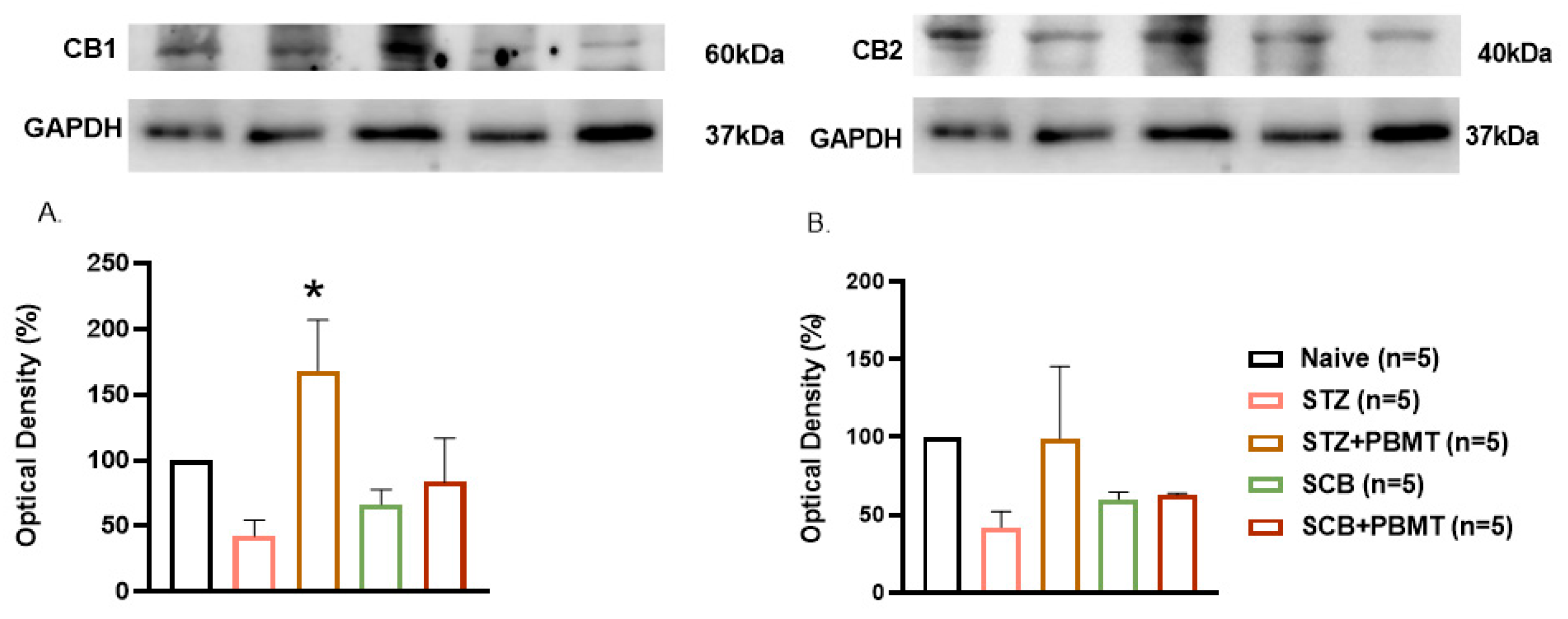
To investigate whether PBMT could modulate cannabinoid receptor expression in the PNS, we evaluated CB1 and CB2 protein levels in the DRG (L4 and L5) of DPN rats submitted (or not) to the PBMT treatment. WB analysis revealed a significant increase in CB1 protein expression in the STZ + PBMT group compared to the STZ group [F(4,17) = 3.828, p = 0.021] and all other groups, as depicted in Figure 2 (Panel A). This result suggests the possible involvement of CB1 receptors in the peripheral mechanisms underlying the antihyperalgesic effects of PBMT. In contrast, no statistically significant differences were observed in CB2 expression among groups (F(4,14) = 1.405, p = 0.263) (Figure 2, Panel B).

3.4. Effect of PBMT on CB1 and CB2 Receptor Expression in the Thalamus

To assess whether the effects of PBMT extended beyond peripheral structures, we also analyzed CB1 and CB2 receptor expression in the thalamus, a central region involved in nociceptive processing. However, no significant differences were found in CB1 (F(4,17) = 2.054, p = 0.13) or CB2 (F(4,9) = 0.456, p = 0.69) expression among the experimental groups, as shown in Figure 3 (Panels A and B). These findings suggest that the modulatory effects of PBMT observed in this model of DPN are likely restricted to peripheral mechanisms, particularly at the DRG level, without inducing measurable supraspinal changes in cannabinoid receptor expression under the tested conditions.

4. Discussion

Our findings suggest that the modulatory effects of PBMT in the DPN model involve partial engagement of the ECS, yet remain primarily restricted to peripheral pathways rather than central mechanisms. The absence of detectable changes in CB1 and CB2 receptor expression within the thalamus suggests that, under the tested parameters, PBMT does not significantly influence supraspinal components of the ECS. This supports the notion that its antihyperalgesic and neuroprotective effects are likely mediated by local or spinal-level mechanisms rather than central modulation.
The interaction between the ECS and diabetes has been extensively investigated, with particular focus on the cannabinoid receptors CB1 and CB2. Diabetes mellitus, especially type 1, is characterized by impaired glucose homeostasis, insulin deficiency, and a chronic pro-inflammatory state [1]. Given that the ECS is deeply involved in the regulation of energy balance, glucose metabolism, and immune modulation, alterations in CB1 and CB2 receptor signaling have been implicated in the pathophysiology of diabetes and its complications. Therefore, understanding how diabetes affects ECS components, and conversely, how ECS modulation may influence metabolic and inflammatory pathways, offers valuable insights for developing novel therapeutic approaches targeting cannabinoid receptors [18].
As mentioned elsewhere, PBMT involves the use of low-level laser or LED devices to deliver non-thermal photons to biological tissues, thereby triggering photochemical and photophysical reactions that modulate cellular metabolism and function [31,32]. In the present study, a low-level laser was selected as the light source due to its well-established precision in dose delivery and tissue penetration. PBMT has been extensively investigated for its therapeutic potential across a wide range of medical conditions, including wound healing, inflammation control, pain modulation, tissue regeneration [47,48], and neuropsychiatric conditions [24,49,50,51,52]. The beneficial effects are primarily attributed to enhanced mitochondrial activity, increased ATP production, and the regulation of oxidative stress and inflammatory mediators, which together contribute to improved cellular homeostasis and recovery [53]. Here, we suggest that these processes can alter the activity or expression of cannabinoid receptors, contributing to PBMT’s analgesic and anti-inflammatory effects, especially in neuropathic pain conditions such as DPN.
In this experimental study, type 1 diabetes was induced in rats through multiple low doses of STZ, resulting in sustained hyperglycemia (plasma glucose ≥ 250 mg/dL). Notably, hyperglycemia remained stable throughout the four-week experimental period, and diabetic rats exhibited classic symptoms of diabetes, including increased urination, excessive water and food intake, and impaired weight gain. PBMT application did not significantly affect the body mass of diabetic animals or change the symptoms of uncontrolled diabetes. A progressive reduction in the nociceptive threshold was observed beginning on the 7th day after the initiation of STZ injections, with a persistent decrease maintained throughout the study. This mechanical hyperalgesia represented reductions of approximately 24%, 38%, and 42% at 7, 14, and 21 days, respectively, compared to Naïve controls. Following PBMT treatment, an improvement in hyperalgesia was detected, with the significant attenuation of pain sensitivity relative to the untreated DPN group. However, this improvement was not evident immediately after the first PBMT session (day 21), becoming significant only on the 24th and 28th days, suggesting a cumulative and time-dependent therapeutic effect. These findings are in agreement with previous data obtained by our research group [24,37].
The administration of selective CB1 and CB2 antagonists produced intriguing results. Within 2 h of CB1 antagonist administration, a significant 28% reversal of the PBMT-induced antihyperalgesia was observed. Similarly, the administration of the CB2 antagonist resulted in a 24% reversal within just 1 h. However, by 3 h post-administration, both antagonists had lost their effect, indicating that the reversal was transient. Taken together, these findings demonstrate that CB1 and CB2 receptor blockade transiently counteracted the antihyperalgesic effects of PBMT. Additionally, WB analyses revealed distinct alterations in CB1 receptor expression within the DRG at the lumbar level (L4-L5), suggesting the potential involvement of this receptor subtype in the mechanisms underlying PBMT’s therapeutic effects in DPN. In contrast, no significant differences were observed in the expression of these cannabinoid receptors within the thalamus. This suggests that, in our model, applying PBMT to the lumbar region did not induce supraspinal alterations in receptor expression. Interestingly, this finding differs from that reported by [41], in which PBMT administered to a distinct anatomical region produced supraspinal effects, including modulation of opioid receptor expression and attenuation of glial cell activation. Such discrepancies highlight the importance of the irradiation site in shaping both peripheral and central therapeutic outcomes, suggesting that PBMT may engage region-specific mechanisms in the modulation of pain and inflammation. Beyond the regulation of CB1 and CB2 receptors, the therapeutic actions of PBMT also encompass the modulation of pro- and anti-inflammatory cytokines [37], such as IL-6 and IL-10, as well as the participation of ATP-sensitive K+ channels and p38 mitogen-activated protein kinases [28]. As previously postulated, CB1 and CB2 receptors are key components of the ECS; CB1 receptors are predominantly expressed in the CNS and peripheral tissues, whereas CB2 receptors are mainly found in immune cells and peripheral tissues. Both receptors have been implicated in several aspects of diabetes, including neuropathic pain and low-grade chronic inflammation. Therefore, the capacity of PBMT to modulate the ECS, alongside its effects on inflammatory mediators and ion channel activity, underscores its translational potential as a non-invasive and multifactorial therapeutic strategy for DPN and other metabolic-inflammatory disorders.
Our results raise important questions regarding the potential differential roles of CB1 and CB2 receptors in the context of diabetes and neuropathic pain. While the expression levels of these receptors may remain relatively stable, it is worth exploring how their functions or downstream signaling pathways may be influenced by the diabetic state. Furthermore, understanding the nuances of cannabinoid receptor regulation in diabetes can provide valuable insights into potential therapeutic avenues for managing DPN.
Importantly, the mechanisms identified in this study suggest potential translational relevance for PBMT in human DPN. The modulation of ECS components and the reduction in hyperalgesia observed here support the therapeutic potential of laser-based PBMT as an adjunct, non-pharmacological approach for pain management in patients with DPN. Future studies should focus on bridging the gap between preclinical and clinical applications by 1) optimizing PBMT parameters (wavelength, dose, and treatment frequency) for human tissue penetration; 2) evaluating sex-related and disease-stage differences in response to therapy; 3) integrating neuroimaging and biochemical biomarkers to assess ECS modulation in humans; and 4) conducting controlled clinical trials to confirm efficacy, safety, and long-term outcomes. Together, these directions could pave the way for translating the observed preclinical benefits into effective therapeutic strategies for human DPN.

5. Conclusions

The assessment of CB1 and CB2 receptor expression and stability in type I diabetic rat models treated with PBMT is crucial, as it may reveal how PBMT modulates the ECS in the context of diabetes-related complications. Continued research in this field is essential to deepen our understanding of PBMT’s underlying mechanisms and its potential to alleviate hyperalgesia while providing neuroprotective benefits in DPN. Furthermore, by elucidating the molecular pathways involved in PBMT-induced analgesia, this study lays the groundwork for translating these findings into clinical applications. Optimizing PBMT parameters and incorporating ECS-related biomarkers in clinical trials may facilitate the development of personalized, non-invasive therapeutic approaches for patients with DPN.

Author Contributions

All authors contributed to the study conception and design. Material preparation, data collection and analysis were performed by D.P.F.B.S. and N.V. The first draft of the manuscript was written by D.P.F.B.S., W.F.V. and M.C. and all authors commented on previous versions of the manuscript. All authors have read and agreed to the published version of the manuscript.

Funding

This study was funded by the Sao Paulo Research Foundation (FAPESP) processes numbers 2019/21158-8, 2021/02897-4, 2024/05615-8 and 2022/08212-6. Danielle P.F.B. Silva has received grant number 2024/05615-8, Willians F. Vieira has received grant number 2019/21158-8 and Marucia Chacur grants 2021/02897-4 and 2022/08212-6.

Institutional Review Board Statement

All procedures were approved by the Animal Research Ethics Committee (CEUA) of Institute of Biomedical Science, University of Sao Paulo-ICB/USP (protocol number: 2017110820).

Informed Consent Statement

A statement confirming that all authors have reviewed and agreed to the publication of images should appear in the manuscript.

Data Availability Statement

The materials used in this study, as well as the raw data, are available from the corresponding author upon reasonable request.

Conflicts of Interest

The authors have no relevant financial or non-financial interests to disclose.

References

  1. Magliano, D.J.; Boyko, E.J.; Committee IDFD Atlas. Idf Diabetes Atlas; International Diabetes Federation: Brussels, Belgium, 2021. [Google Scholar]
  2. American Diabetes Association. Standards of Medical Care in Diabetes-2020 Abridged for Primary Care Providers. Clin. Diabetes 2020, 38, 10–38. [Google Scholar] [CrossRef]
  3. Pham, V.M.; Thakor, N. Insulin enhances neurite extension and myelination of diabetic neuropathy neurons. Korean J. Pain 2022, 35, 160–172. [Google Scholar] [CrossRef] [PubMed]
  4. Harris, K.; Boland, C.; Meade, L.; Battise, D. Adjunctive therapy for glucose control in patients with type 1 diabetes. Diabetes, Metabolic Syndrome and Obesity. 2018, 11, 159–173. [Google Scholar] [CrossRef] [PubMed]
  5. Eaton, S.E.; Harris, N.D.; Rajbhandari, S.M.; Greenwood, P.; Wilkinson, I.D.; Ward, J.D.; Griffiths, P.D.; Tesfaye, S. Spinal-cord involvement in diabetic peripheral neuropathy. Lancet 2001, 358, 35–36. [Google Scholar] [CrossRef]
  6. Selvarajah, D.; Wilkinson, I.D.; Maxwell, M.; Davies, J.; Sankar, A.; Boland, E.; Gandhi, R.; Tracey, I.; Tesfaye, S. Magnetic resonance neuroimaging study of brain structural differences in diabetic peripheral neuropathy. Diabetes Care 2014, 37, 1681–1688. [Google Scholar] [CrossRef]
  7. Correia Rocha, I.R.; Chacur, M. Modulatory effects of photobiomodulation in the anterior cingulate cortex of diabetic rats. Photochem. Photobiol. Sci. 2021, 20, 781–790. [Google Scholar] [CrossRef]
  8. Liu, S.; Li, D.; Yu, T.; Zhu, J.; Semyachkina-Glushkovskaya, O.; Zhu, D. Transcranial photobiomodulation improves insulin therapy in diabetic microglial reactivity and the brain drainage system. Commun. Biol. 2023, 6, 1239. [Google Scholar] [CrossRef]
  9. de la Torre, J.C. Treating cognitive impairment with transcranial low level laser therapy. J. Photochem. Photobiology B: Biology. 2017, 168, 149–155. [Google Scholar] [CrossRef] [PubMed]
  10. Tesfaye, S.; Boulton, A.J.; Dyck, P.J.; Freeman, R.; Horowitz, M.; Kempler, P.; Lauria, G.; Malik, R.A.; Spallone, V.; Vinik, A.; et al. Diabetic neuropathies: Update on definitions, diagnostic criteria, estimation of severity, and treatments. Diabetes Care 2010, 33, 2285–2293. [Google Scholar] [CrossRef]
  11. Wen, J.; Sackett, S.; Tanaka, M.; Zhang, Y. Therapeutic Effects of Combined Treatment with the AEA Hydrolysis Inhibitor PF04457845 and the Substrate Selective COX-2 Inhibitor LM4131 in the Mouse Model of Neuropathic Pain. Cells 2023, 12, 1275. [Google Scholar] [CrossRef]
  12. Liang, Y.C.; Huang, C.C.; Hsu, K.S. Therapeutic potential of cannabinoids in trigeminal neuralgia. Curr. Drug Targets CNS Neurol. Disord. 2004, 3, 507–514. [Google Scholar] [CrossRef] [PubMed]
  13. Starobova, H.; Vetter, I. Pathophysiology of Chemotherapy-Induced Peripheral Neuropathy. Front. Mol. Neurosci. 2017, 10, 174. [Google Scholar] [CrossRef] [PubMed]
  14. Binda, K.H.; Chacur, M.; Martins, D.O. Exercise Improves Orofacial Pain and Modifies Neuropeptide Expression in a Rat Model of Parkinson’s Disease. Neurotox. Res. 2023, 41, 459–470. [Google Scholar] [CrossRef]
  15. Zou, S.; Kumar, U. Cannabinoid Receptors and the Endocannabinoid System: Signaling and Function in the Central Nervous System. Int. J. Mol. Sci. 2018, 19, 833. [Google Scholar] [CrossRef]
  16. Saito, V.M.; Wotjak, C.T.; Moreira, F.A. Pharmacological exploitation of the endocannabinoid system: New perspectives for the treatment of depression and anxiety disorders? Braz. J. Psychiatry 2010, 32 (Suppl. S1), 57–514. [Google Scholar] [CrossRef]
  17. Schreiber, A.K.; Neufeld, M.; Jesus, C.H.; Cunha, J.M. Peripheral antinociceptive effect of anandamide and drugs that affect the endocannabinoid system on the formalin test in normal and streptozotocin-diabetic rats. Neuropharmacology 2012, 63, 1286–1297. [Google Scholar] [CrossRef]
  18. Gruden, G.; Barutta, F.; Kunos, G.; Pacher, P. Role of the endocannabinoid system in diabetes and diabetic complications. Br. J. Pharmacol. 2016, 173, 1116–1127. [Google Scholar] [CrossRef]
  19. Yu, L.L.; Zhou, S.J.; Wang, X.Y.; Liu, J.F.; Xue, Y.X.; Jiang, W.; Lu, L. Effects of cannabinoid CB1 receptor antagonist rimonabant on acquisition and reinstatement of psychostimulant reward memory in mice. Behav. Brain Res. 2011, 217, 111–116. [Google Scholar] [CrossRef]
  20. Toth, C.C.; Jedrzejewski, N.M.; Ellis, C.L.; Frey, W.H. Cannabinoid-mediated modulation of neuropathic pain and microglial accumulation in a model of murine type I diabetic peripheral neuropathic pain. Mol. Pain 2010, 6, 16. [Google Scholar] [CrossRef]
  21. Yagihashi, S.; Mizukami, H.; Sugimoto, K. Mechanism of diabetic neuropathy: Where are we now and where to go? J. Diabetes Investig. 2011, 2, 18–32. [Google Scholar] [CrossRef] [PubMed]
  22. de Oliveira, M.E.; Da Silva, J.T.; Brioschi, M.L.; Chacur, M. Effects of photobiomodulation therapy on neuropathic pain in rats: Evaluation of nociceptive mediators and infrared thermography. Lasers Med. Sci. 2021, 36, 1461–1467. [Google Scholar] [CrossRef]
  23. Rocha, I.R.C.; Ciena, A.P.; Rosa, A.S.; Martins, D.O.; Chacur, M. Photobiostimulation reverses allodynia and peripheral nerve damage in streptozotocin-induced type 1 diabetes. Lasers Med. Sci. 2017, 32, 495–501. [Google Scholar] [CrossRef] [PubMed]
  24. Vieira, W.F.; de Magalhães, S.F.; Farias, F.H.; de Thomaz, A.A.; Parada, C.A. Raman spectroscopy of dorsal root ganglia from streptozotocin-induced diabetic neuropathic rats submitted to photobiomodulation therapy. J. Biophotonics 2019, 12, e201900135. [Google Scholar] [CrossRef] [PubMed]
  25. Mazuqueli Pereira, E.S.B.; Basting, R.T.; Abdalla, H.B.; Garcez, A.S.; Napimoga, M.H.; Clemente-Napimoga, J.T. Photobiomodulation inhibits inflammation in the temporomandibular joint of rats. J. Photochem. Photobiology B Biology 2021, 222, 112281. [Google Scholar] [CrossRef]
  26. Yamada, E.F.; Bobinski, F.; Martins, D.F.; Palandi, J.; Folmer, V.; da Silva, M.D. Photobiomodulation therapy in knee osteoarthritis reduces oxidative stress and inflammatory cytokines in rats. J. Biophotonics 2020, 13, e201900204. [Google Scholar] [CrossRef]
  27. Tomazoni, S.S.; Frigo, L.; Dos Reis Ferreira, T.C.; Casalechi, H.L.; Teixeira, S.; de Almeida, P.; Muscara, M.N.; Marcos, R.L.; Serra, A.J.; de Carvalho, P.T.C.; et al. Effects of photobiomodulation therapy and topical non-steroidal anti-inflammatory drug on skeletal muscle injury induced by contusion in rats-part 2: Biochemical aspects. Lasers Med. Sci. 2017, 32, 1879–1887. [Google Scholar] [CrossRef] [PubMed]
  28. Neves, L.M.S.; Gonçalves, E.C.D.; Cavalli, J.; Vieira, G.; Laurindo, L.R.; Simões, R.R.; Coelho, I.S.; Santos, A.R.S.; Marcolino, A.M.; Cola, M.; et al. Photobiomodulation Therapy Improves Acute Inflammatory Response in Mice: The Role of Cannabinoid Receptors/ATP-Sensitive K+ Channel/p38-MAPK Signalling Pathway. Mol. Neurobiol. 2018, 55, 5580–5593. [Google Scholar] [CrossRef]
  29. Moheghi, A.; Noori Mougehi, S.M.H.; Amini, A.; Mostafavinia, A.; Rezaei, F.; Bagheri Tadi, F.; Chien, S.; Bayat, M. Anti-inflammatory, Antioxidant, and Wound-Healing Effects of Photobiomodulation on Type-2 Diabetic Rats. J. Lasers Med. Sci. 2023, 14, e45. [Google Scholar] [CrossRef]
  30. Zhang, Y.; Su, J.; Ma, K.; Li, H.; Fu, X.; Zhang, C. Photobiomodulation promotes hair regeneration in injured skin by enhancing migration and exosome secretion of dermal papilla cells. Wound Repair Regen. 2022, 30, 245–257. [Google Scholar] [CrossRef]
  31. Heiskanen, V.; Hamblin, M.R. Photobiomodulation: Lasers vs. light emitting diodes? Photochem. Photobiol. Sci. 2018, 17, 1003–1017. [Google Scholar] [CrossRef]
  32. Zhang, W.W.; Wang, X.Y.; Chu, Y.X.; Wang, Y.Q. Light-emitting diode phototherapy: Pain relief and underlying mechanisms. Lasers Med. Sci. 2022, 37, 2343–2352. [Google Scholar] [CrossRef]
  33. Scarcella, G.; Tardugno, R.; Crupi, P.; Muraglia, M.; Clodoveo, M.L.; Corbo, F. Fluorescent Light Energy (FLE) Generated through Red LED Light and a Natural Photoconverter Gel as a New, Non-Invasive Approach for Facial Age Control: A Pilot Study. Cosmetics 2023, 10, 74. [Google Scholar] [CrossRef]
  34. Bettleyon, J.; Kaminski, T.W. Does Low-Level Laser Therapy Decrease Muscle-Damaging Mediators After Performance in Soccer Athletes Versus Sham Laser Treatment? A Critically Appraised Topic. J. Sport Rehabil. 2020, 29, 1210–1213. [Google Scholar] [CrossRef]
  35. Vieira, W.F.; Kenzo-Kagawa, B.; Britto, M.H.M.; Ceragioli, H.J.; Sakane, K.K.; Baranauskas, V.; da Cruz-Höfling, M.A. Vibrational spectroscopy of muscular tissue intoxicated by snake venom and exposed to photobiomodulation therapy. Lasers Med. Sci. 2018, 33, 503–512. [Google Scholar] [CrossRef] [PubMed]
  36. Vieira, W.F.; Kenzo-Kagawa, B.; Alvares, L.E.; Cogo, J.C.; Baranauskas, V.; da Cruz-Höfling, M.A. Exploring the ability of low-level laser irradiation to reduce myonecrosis and increase Myogenin transcription after Bothrops jararacussu envenomation. Photochem. Photobiol. Sci. 2021, 20, 571–583. [Google Scholar] [CrossRef]
  37. Vieira, W.F.; Malange, K.F.; de Magalhães, S.F.; Lemes, J.B.P.; Dos Santos, G.G.; Nishijima, C.M.; de Oliveira, A.L.R.; da Cruz-Höfling, M.A.; Tambeli, C.H.; Parada, C.A. Anti-hyperalgesic effects of photobiomodulation therapy (904 nm) on streptozotocin-induced diabetic neuropathy imply MAPK pathway and calcium dynamics modulation. Sci. Rep. 2022, 12, 16730. [Google Scholar] [CrossRef] [PubMed]
  38. Athie, M.C.P.; Vieira, A.S.; Teixeira, J.M.; Dos Santos, G.G.; Dias, E.V.; Tambeli, C.H.; Sartori, C.R.; Parada, C.A. Transcriptome analysis of dorsal root ganglia’s diabetic neuropathy reveals mechanisms involved in pain and regeneration. Life Sci. 2018, 205, 54–62. [Google Scholar] [CrossRef]
  39. Teixeira, J.M.; Dos Santos, G.G.; Neves, A.F.; Athie, M.C.P.; Bonet, I.J.M.; Nishijima, C.M.; Farias, F.H.; Figueiredo, J.G.; Hernandez-Olmos, V.; Alshaibani, S.; et al. Diabetes-induced Neuropathic Mechanical Hyperalgesia Depends on P2X4 Receptor Activation in Dorsal Root Ganglia. Neuroscience 2019, 398, 158–170. [Google Scholar] [CrossRef]
  40. Vieira, W.F.; Malange, K.F.; de Magalhães, S.F.; Dos Santos, G.G.; de Oliveira, A.L.R.; da Cruz-Höfling, M.A.; Parada, C.A. Gait analysis correlates mechanical hyperalgesia in a model of streptozotocin-induced diabetic neuropathy: A CatWalk dynamic motor function study. Neurosci. Lett. 2020, 736, 135253. [Google Scholar] [CrossRef]
  41. Rocha, I.R.C.; Perez-Reyes, E.; Chacur, M. Effect of photobiomodulation on mitochondrial dynamics in peripheral nervous system in streptozotocin-induced type 1 diabetes in rats. Photochem. Photobiol. Sci. 2021, 20, 293–301. [Google Scholar] [CrossRef]
  42. Ferreira, N.L.; Rocha, I.R.C.; Chacur, M. Unraveling the RAGE-NF-κB pathway: Implications for modulating inflammation in diabetic neuropathy through photobiomodulation therapy. Lasers Med. Sci. 2024, 39, 222. [Google Scholar] [CrossRef]
  43. Mestre, C.; Pélissier, T.; Fialip, J.; Wilcox, G.; Eschalier, A. A method to perform direct transcutaneous intrathecal injection in rats. J. Pharmacol. Toxicol. Methods 1994, 32, 197–200. [Google Scholar] [CrossRef]
  44. Rosa, A.S.; Freitas, M.F.; Rocha, I.R.; Chacur, M. Gabapentin decreases microglial cells and reverses bilateral hyperalgesia and allodynia in rats with chronic myositis. Eur. J. Pharmacol. 2017, 799, 111–117. [Google Scholar] [CrossRef]
  45. Binda, K.H.; Lillethorup, T.P.; Real, C.C.; Bærentzen, S.L.; Nielsen, M.N.; Orlowski, D.; Brooks, D.J.; Chacur, M.; Landau, A.M. Exercise protects synaptic density in a rat model of Parkinson’s disease. Exp. Neurol. 2021, 342, 113741. [Google Scholar] [CrossRef]
  46. Bradford, M.M. A rapid and sensitive method for the quantitation of microgram quantities of protein utilizing the principle of protein-dye binding. Anal. Biochem. 1976, 72, 248–254. [Google Scholar] [CrossRef]
  47. Tomazoni, S.S.; Machado, C.; De Marchi, T.; Casalechi, H.L.; Bjordal, J.M.; de Carvalho, P.T.C.; Leal-Junior, E.C.P. Infrared Low-Level Laser Therapy (Photobiomodulation Therapy) before Intense Progressive Running Test of High-Level Soccer Players: Effects on Functional, Muscle Damage, Inflammatory, and Oxidative Stress Markers-A Randomized Controlled Trial. Oxidative Med. Cell. Longev. 2019, 2019, 6239058. [Google Scholar] [CrossRef] [PubMed]
  48. Ramezani, F.; Neshasteh-Riz, A.; Ghadaksaz, A.; Fazeli, S.M.; Janzadeh, A.; Hamblin, M.R. Mechanistic aspects of photobiomodulation therapy in the nervous system. Lasers Med. Sci. 2022, 37, 11–18. [Google Scholar] [CrossRef]
  49. Vieira, W.F.; Gersten, M.; Caldieraro, M.A.K.; Cassano, P. Photobiomodulation for Major Depressive Disorder: Linking Transcranial Infrared Light, Biophotons and Oxidative Stress. Harv. Rev. Psychiatry 2023, 31, 124–141. [Google Scholar] [CrossRef] [PubMed]
  50. Coelho, D.R.A.; Salvi, J.D.; Vieira, W.F.; Cassano, P. Inflammation in obsessive-compulsive disorder: A literature review and hypothesis-based potential of transcranial photobiomodulation. J. Neurosci. Res. 2024, 102, e25317. [Google Scholar] [CrossRef]
  51. Coelho, D.R.A.; Renet, C.; López-Rodríguez, S.; Cassano, P.; Vieira, W.F. Transcranial photobiomodulation for neurodevelopmental disorders: A narrative review. Photochem. Photobiol. Sci. 2024, 23, 1609–1623. [Google Scholar] [CrossRef]
  52. Vieira, W.F. Beyond Digital Therapy: Augmenting Internet-Based Cognitive Behavioral Therapy With Brain Stimulation. Biol. Psychiatry Glob. Open Sci. 2025, 5, 100514. [Google Scholar] [CrossRef] [PubMed]
  53. de Freitas, L.F.; Hamblin, M.R. Proposed Mechanisms of Photobiomodulation or Low-Level Light Therapy. IEEE J. Sel. Top. Quantum Electron. 2016, 22, 348–364. [Google Scholar] [CrossRef]
Figure 1. Effect of CB1 and CB2 receptor antagonists on mechanical withdrawal thresholds after PBMT treatment in STZ-induced DPN rats. PBMT was administered daily from day 21 to day 28 (shaded areas). Intrathecal (i.t.) injection of AM251 (30 µg/50 µL) (CB1 antagonist) or AM630 (10 µg/50 µL) (CB2 antagonist) was performed on day 28 (black arrows), after the last PBMT session. Nociceptive thresholds were measured 30 min, 1, 2, and 3 h after antagonists’ injection. (Panel A) STZ + PBMT + CB1 group showed a transient reduction in the antihyperalgesic effect induced by PBMT (28% reversal of the withdrawal threshold) 2 h after AM251 administration. (Panel B) STZ + PBMT + CB2 group also exhibited a transient reduction in PBMT-induced antihyperalgesia (24% reversal of threshold) 1 h after AM630 administration. Both antagonists completely lost their effect 3 h after injection. Data are expressed as mean ± standard error of the mean (SEM). Statistical comparisons were performed using one-way ANOVA followed by the Bonferroni post hoc test. Symbol (*) means that all STZ-injected groups are statistically different (p < 0.05) from the Naïve group; symbol (#) means that the STZ + PBMT + CB1 group is statistically different (p < 0.05) from STZ and STZ + CB1 groups (Panel A); also, the symbol (#) means that the STZ + PBMT + CB2 group is statistically different (p < 0.05) from STZ and STZ + CB2 groups (Panel B).
Photonics 12 01060 g001
Figure 2. Analysis of the effects of PBMT on CB1 and CB2 receptor protein expression in the L4–L5 DRG of DPN rats. CB1 and CB2 expression levels in the L4-L5 DRG were quantified by WB in the Naïve (general control), STZ (DPN rats), STZ + PBMT (DPN + PBMT), SCB (STZ vehicle), and SCB + PBMT (vehicle + PBMT) groups; representative protein bands are shown in this order. Expression levels of the CB1 (60 kDa) and CB2 (40 kDa) receptors were normalized to those of GAPDH (37 kDa, endogenous control) and expressed as percentages relative to the Naïve group (100%). (Panel A) Expression levels of CB1: The STZ + PBMT group showed significantly higher CB1 expression compared to the STZ group. (Panel B) Expression levels of CB2: No statistically significant differences in CB2 expression were observed between the experimental groups. Data are expressed as mean ± standard error of the mean (SEM). Statistical comparisons were performed using one-way ANOVA followed by the Bonferroni post hoc test. Symbol (*) means that the STZ + PBMT group is statistically different (p < 0.05) from the STZ group (particularly for CB1 expression).
Photonics 12 01060 g002
Figure 3. Analysis of the effects of PBMT on CB1 and CB2 receptor protein expression in the thalamus of DPN rats. CB1 and CB2 expression levels in the thalamus were quantified by WB in the Naïve (general control), STZ (DPN rats), STZ + PBMT (DPN + PBMT), SCB (STZ vehicle), and SCB + PBMT (vehicle + PBMT) groups; representative protein bands are shown in this order. Expression levels of the CB1 (60 kDa) and CB2 (40 kDa) receptors were normalized to those of GAPDH (37 kDa, endogenous control) and expressed as percentages relative to the Naïve group (100%). (Panel A) Expression levels of CB1: No statistically significant differences in CB1 expression were detected in the thalamus among the experimental groups. (Panel B) Expression levels of CB2: Similarly to CB1, no statistically significant differences in CB2 expression were detected in the thalamus among the experimental groups. Data are expressed as mean ± standard error of the mean (SEM). Statistical comparisons were performed using one-way ANOVA followed by the Bonferroni post hoc test.
Photonics 12 01060 g003
Table 1. Laser parameters used for PBMT.
LaserλOperation ModeFrequencyTime
GaAs904 nmPulsed9500 Hz18″ per point
Total EnergyNº of pointsNº of sessionsTreated areaContact
0.81 J2/animal/day8 sessions; 1/day1.17 cm2Direct
GaAs = Gallium-arsenide; λ = wavelength; Hz = Hertz; (″) = seconds; J = Joule; nm = nanometers; cm2 = square centimeters.
Disclaimer/Publisher’s Note: The statements, opinions and data contained in all publications are solely those of the individual author(s) and contributor(s) and not of MDPI and/or the editor(s). MDPI and/or the editor(s) disclaim responsibility for any injury to people or property resulting from any ideas, methods, instructions or products referred to in the content.