Review

Separation of Serum and Plasma Proteins for In-Depth Proteomic Analysis

School of Pharmacy, Cedarville University, Cedarville, OH 45314, USA
*
Author to whom correspondence should be addressed.
Separations 2022, 9(4), 89; https://doi.org/10.3390/separations9040089
Submission received: 2 March 2022 / Revised: 21 March 2022 / Accepted: 29 March 2022 / Published: 1 April 2022
(This article belongs to the Special Issue Separations and Analysis of Proteins in Biological Samples)

Abstract

:
There are probably no biological samples that did more to spur interest in proteomics than serum and plasma. The belief was that comparing the proteomes of these samples obtained from healthy and disease-affected individuals would lead to biomarkers that could be used to diagnose conditions such as cancer. While the continuing development of mass spectrometers with greater sensitivity and resolution has been invaluable, the invention of strategic strategies to separate circulatory proteins has been just as critical. Novel and creative separation techniques were required because serum and plasma probably have the greatest dynamic range of protein concentration of any biological sample. The concentrations of circulating proteins can range over twelve orders of magnitude, making it a challenge to identify low-abundance proteins where the bulk of the useful biomarkers are believed to exist. The major goals of this article are to (i) provide an historical perspective on the rapid development of serum and plasma proteomics; (ii) describe various separation techniques that have made obtaining an in-depth view of the proteome of these biological samples possible; and (iii) describe applications where serum and plasma proteomics have been employed to discover potential biomarkers for pathological conditions.

1. Introduction

Testing for the levels of specific molecules within blood samples have played a major role in diagnosing and treating various health conditions for decades [1,2]. These blood tests begin as soon as a baby is born through the various newborn screening tests that are used to screen for metabolic (e.g., phenylketonuria, maple syrup urine disease, etc.), hormonal (e.g., hypothyroidism and adrenal hyperplasia), and genetic (e.g., sickle cell anemia, cystic fibrosis, etc.) disorders [3]. Blood-based tests continue throughout our lives in the form of clinical tests such as complete cell counts and metabolic panels [4,5]. These routine tests are used to detect abnormal function of a patient’s liver, kidneys, immune system, blood chemistry, etc. Since no cell in the body is more than four cell units removed from the circulatory system, blood is perhaps the most important window through which a physician can readily view a patient’s health status. Acting as a transportation system, the circulatory system not only contains classic blood proteins, such as albumin, immunoglobulins, and hemoglobin, but also a variety of proteins that are shed from both the exterior and interior of cells.
Owing to its intimacy with the body, considerable effort has gone into characterizing the molecular content of serum and plasma. Over the past couple of decades, the continued development of higher-resolution chromatography and mass spectrometry (MS) technologies have enabled serum and plasma proteomes (i.e., the entire protein complement of a biological sample) to be characterized at an increasingly deeper level. The progress has been outstanding. In the early part of this century, the ability to identify a few hundred proteins in a single serum or plasma sample was considered state-of-the-art [6,7]. Fast forward to today, where exploratory studies can identify thousands of plasma proteins in tens of samples [8,9]. While the development of more powerful mass spectrometers has received most of the accolades, none of the achievements in proteomics would be possible without advances in separation technologies, particularly chromatography. In this article, we describe some of the advances that have enabled the proteomes of both serum and plasma to be explored.

2. Plasma vs. Serum

The obvious question in deciding whether to use plasma or serum is “Which one is the best to use for proteomics?” Answering this question requires understanding the differences between the two samples (Table 1). The main difference between plasma and serum can be summarized by the word “clotting”. In preparing plasma, blood is drawn via venipuncture in the presence of an anticoagulant such as ethylenediamine tetraacetic acid (EDTA), heparin, or sodium citrate [10]. After centrifugation to remove cells such as red (RBCs) and white blood cells (WBCs), and the platelets, the straw-colored plasma sample remains. Serum is also acquired via venipuncture but without anticoagulants being present. The serum is allowed to sit for approximately 30 min during which time the blood clots. Centrifugation removes the clot and blood cells leaving behind serum [10].
Both plasma and serum contain a high protein concentration (i.e., 60–80 mg/mL), with albumin and immunoglobulin G (IgG) making up approximately 90% of this total [11]. The primary protein difference between plasma and serum is the lower amount of fibrinogen found in serum. Another protein that has been shown to be lower in serum than plasma is the coagulation factor, platelet factor 4. While the levels of other coagulation factors, such as factors IX, X, XI, and VII/VIIa, would be expected to be absent in serum, studies show trace amounts of both in serum and plasma. Studies have shown that during the clotting process, platelets, RBCs, and WBCs secrete specific proteins into the serum. For example, platelet-secreted vascular endothelial growth factor levels were shown to be 230 ± 63 and 38 ± 8 pg/mL in serum and plasma obtained from normal individuals, respectively [12]. Other studies have also shown differences in the composition of serum and plasma; however, little consideration beyond availability is made when selecting which sample type to use in a proteomics study, even though the Human Proteome Project recommended that plasma prepared using EDTA be used for all proteomic studies [13].
While serum remains the preferred sample for clinical chemistry tests, both serum and plasma have been used extensively (and almost equally) for proteomic analysis. There were 10,461 and 10,075 entries that included serum/proteomics and plasma/proteomics, respectively, in their search terms within Pubmed as of 2 December 2021. Almost 1800 of these articles included both serum and plasma. Regardless of which specimen is used, both are highly complex samples that require high-resolution separation techniques to enable their proteomes to be characterized.

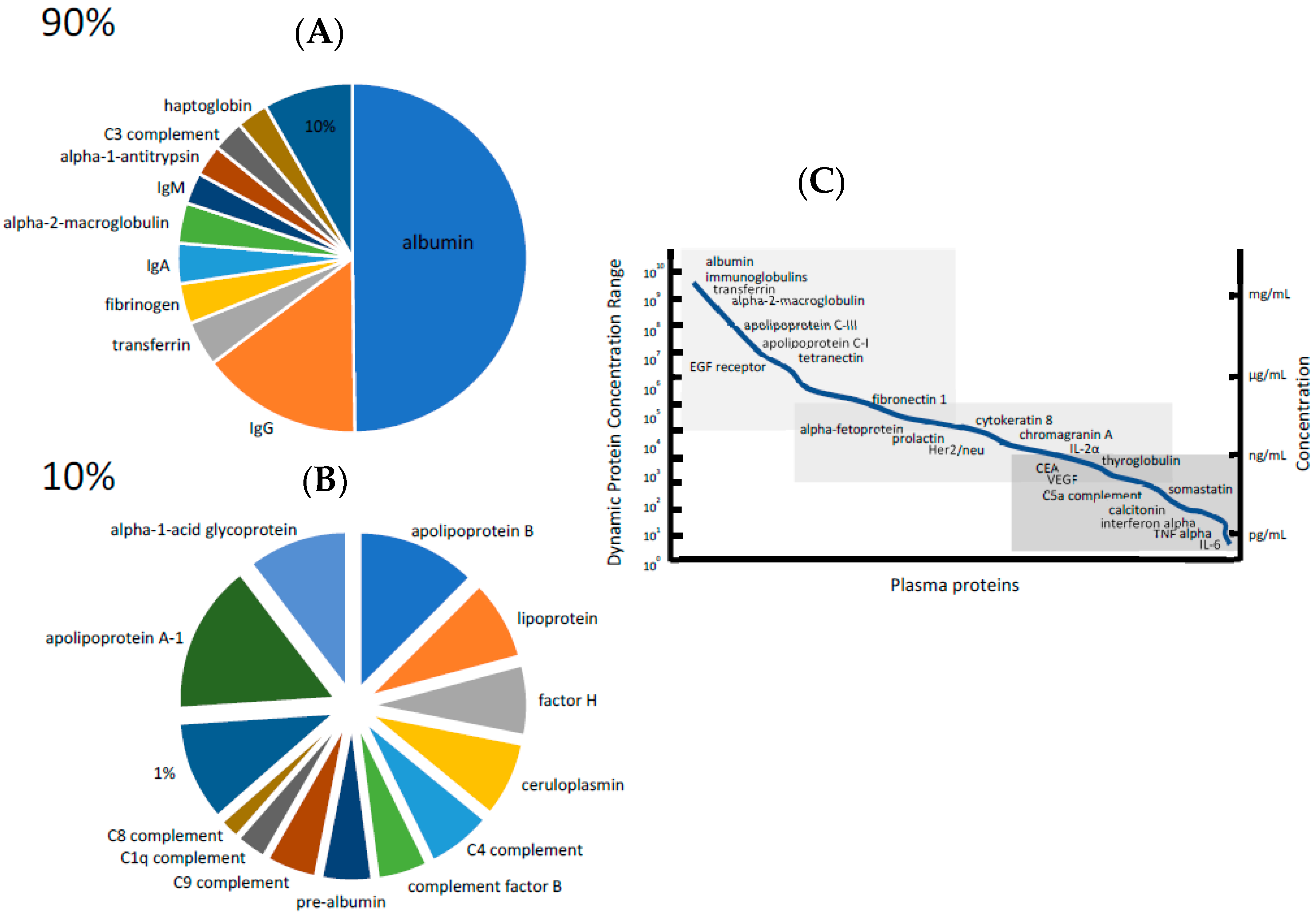
3. The Dynamic Range of Protein Concentration Problem

While every proteomic study requires the separation of a complex mixture of proteins, serum and plasma have two unique features that set them apart. These features are an extremely large dynamic range of protein concentration and having most of their protein concentration made up by only a small number of proteins (Figure 1). For example, 10 proteins make up about 90% of the entire protein concentration of serum and only 12 proteins make up about 90% of the remaining 10% [11]. For most proteomic studies that focus on identifying diagnostic, therapeutic, or prognostic biomarkers, the most interesting proteins are within the low-abundance region, which makes up only 1% of the entire protein content [14,15,16]. The concentrations of protein within serum and plasma range over approximately 12 orders of magnitude from albumin at the high end to proteins such as cytokines at the low end. While it is often asked, “What is the limit of detection (LOD) and limit of quantitation for the proteomic analysis of serum and plasma?”, this question cannot be accurately answered because the LOD and LOQ pertains to a specific analyte, which would be a peptide in the case of serum/plasma proteomics. The LOD and LOQ is specific to each peptide and needs to be experimentally determined for each using internal standards and calibration curves. While studies can claim that proteins present at concentrations in the nanogram per liter range can be detected, these claims simply rely on targeted studies in which specific serum/plasma LODs and LOQs have been established for these specific proteins.

3.1. High-Abundance Protein Depletion

Analyzing serum and plasma proteomes is essentially the reverse of the old idiom “can’t see the forest for the trees”. While this saying refers to being unable to see the entire situation because you are too involved in the details, proper analysis of blood’s proteome requires looking past the forest of high-abundance proteins to see the details represented by the low-abundance ones. Therefore, to identify potentially critical biomarkers requires removing the higher-abundance proteins prior to protein identification. Since albumin makes up more than 50% of the protein amount of a typical serum or plasma sample, many of the earliest efforts were focused on removing this protein prior to further proteomic analysis. Many of these efforts used resins to which dye-ligands, such as Cibacron Blue or anti-albumin antibodies, were coupled [17,18,19]. While dye-ligand columns have a large loading capacity, they lack the specificity of antibody-based depletion columns [20]. Albumin-depletion methods generally remove more than 96% of the protein, while other high-abundance proteins remain within the samples, particularly immunoglobulin G (IgG).
A major step forward in the removal of high-abundance proteins were the development of Multiple Affinity Removal System (MARS) columns [21]. The original MARS columns were designed to remove albumin only; however, they progressed to a stage in which 14 highly abundant proteins could be removed using a single column [22,23]. These devices were produced specifically for both human and mouse samples and are available in the column or spin cartridge formats. Studies have generally shown that the MARS columns eliminate about 85% of the total protein content of serum or plasma through the depletion of high-abundance proteins. This depletion expands the dynamic range of the downstream assay by enabling a greater loading capacity of lower-abundance proteins.
One criticism of the use of affinity resins to remove highly abundant proteins was that they would result in the depletion of the low-abundance, low molecular weight proteins via protein–protein interactions. To investigate this possibility, six highly abundant proteins (albumin, IgG, IgA, IgM, transferrin, and apolipoprotein) were extracted from serum using affinity resins and identified using MS [24]. A total of 210 proteins were identified, ranging from a high of 63 bound to albumin to 19 bound to IgM. Twelve of these proteins (e.g., prostate specific antigen, dihydropteridine reductase, meningioma-expressed antigen, etc.) were being used as clinical biomarkers at the time of the study. Almost 140 of these proteins had not been identified in a global study of serum in which approximately 1500 proteins were characterized [25]. Not all the proteins that were identified as being bound to these six highly abundant proteins are present in low concentrations in serum. Many low- and high-affinity interactions occur between proteins in complex samples, especially involving albumin that is not only highly abundant but is well-known as a sticky protein to which many other species bind [26]. This study does not recommend eliminating the depletion steps, but rather suggests there are various other methods to enrich for proteins in serum and plasma that may be of biological interest. Indeed, several subsequent studies used albumin as a bait to enrich for biologically important low-abundance proteins [27,28].
A study utilizing albumin as a bait to isolate low molecular weight proteins analyzed pooled sera from 40 women who were at high-risk of developing ovarian cancer but did not have the condition, 30 women with stage I ovarian cancer, and 40 women with stage III ovarian cancer [29]. Albumin was isolated using a solid-phase affinity capture column. The samples were bound to the column and the column was washed using non-denaturing conditions to capture the proteins that bound to albumin. After eluting the bound proteins using 70% acetonitrile containing 0.2% formic acid, they were fractionated using one-dimensional PAGE. Visualized protein bands were cut from the gel and digested into tryptic peptides. The peptides were identified using reversed-phase chromatography (5-μm C18 silica-bonded beads packed in a 10-cm long, 75-μm i.d. column) coupled directly online with MS. The overall analysis resulted in the identification of over 1200 unique proteins, including over 700 that had not previously been identified in serum. Of specific interest was the identification of BRCA2, a low-abundance protein associated with ovarian cancer susceptibility that is not predicted to be present in circulation, owing to its native size (i.e., 390 kDa) and localization to the cell’s nucleus [30]. Its identification using the albumin-baiting technique suggests that fragments of this protein may be released from the cell and are available for identification using LC-MS. Another interesting protein identified in this study was the known ovarian cancer biomarker CA125 [31]. This protein was identified in serum samples taken from women with stage III ovarian cancer, but not those at high risk or with stage I ovarian cancer. Overall, this study demonstrated that using high-abundance proteins in serum/plasma could be an effective separation technique to increase the chance of identifying disease-specific biomarkers.

3.2. Low-Abundance Protein Enrichment

There are two ways to increase the availability to detect low-abundance proteins in serum and plasma: remove the high-abundance proteins (as described above) or increase the concentration of low-abundance proteins. Increasing their relative concentration is the strategy behind the combinatorial peptide ligand library (CPLL) method for identifying low-abundance proteins in complex proteome samples [32]. In CPLL, a combinatorial library, automated solid-phase synthesis is used to couple ligands to resin beads. Theoretically, each bead has millions of copies of a unique ligand and individual beads have different ligands. The ligands provide surfaces to which specific proteins can then bind. If the library is sufficiently diverse, it is possible that every protein in a complex proteome will bind to a high-affinity ligand and be captured. If the CPLL library is exposed to serum, highly abundant proteins will quickly saturate their ligands, resulting in most of their concentration being unbound and lost in the column eluate. Conversely, most copies of a low-abundance protein will be bound as there is an insufficient amount of it to completely saturate their ligand. This results in an enrichment of low-abundance proteins relative to high-abundance proteins based on the principle of saturation overloading. After the proteins have bound to their corresponding ligands, the beads are washed to reduce the low-affinity interactions and the remaining absorbed proteins are eluted using denaturing conditions. The major disadvantage of the CPLL technique is that it tends to normalize the concentration of all proteins within a complex mixture, resulting in a loss of comparative quantitative information. CPLL technology has been made available to general laboratories through the ProteoMiner Protein Enrichment Kits that are manufactured by BioRad [33]. The application of CPLL has been very useful in the detection of trace food allergens in various extracts, particularly for the detection of IgE-binding proteins. CPLL has been used to detect allergens in milk whey [34], eggs [35], maize [36], as well as fungal allergens in the blood of patients with aspergillosis [37]. A list of some commercially available devices for either depleting high-abundance proteins or enriching low-abundance proteins in serum and plasma is provided in Table 2.

4. Electrophoresis for the Fractionation of Serum and Plasma Proteins

Although it has been largely replaced by chromatography, electrophoresis was the foremost and major separation technique early in the development of serum and plasma proteomics. Indeed, the field of proteomics can trace its history back to the electrophoretic separation of E. coli proteins. In his seminal paper, Pat O’Farrell demonstrated the ability to resolve 1000 protein components extracted from E. coli using two-dimensional polyacrylamide gel electrophoresis (2D-PAGE), which separates proteins based on their isoelectric point (pI) in one dimension followed by molecular weight in the other [38]. Even the term proteomics can be traced back to a study in which Mycoplasma genitalium proteins were fractionated by 2D-PAGE and identified using matrix-assisted laser desorption–ionization MS [39]. Fifty years ago, a 2D-PAGE gel was able to resolve just over 110 protein spots: most of them originating from highly abundant proteins such as albumin, IgG, transferrin, and haptoglobulin [40]. Although this technology provided little proteomic coverage compared to today’s methods, it was used in a biomarker discovery study comparing sera from controls and patients affected with leukemia, myeloma, and Hodgkin’s diseases. They found several variants of albumin in the disease-affected samples that were not seen in the controls.
Recognizing the utility of 2D-PAGE, the father and son pioneers in the field of serum and plasma proteomics, Norman and N. Leigh Anderson, were able to resolve 300 distinct proteins and their isoforms. While this number almost tripled the number of proteins that could be observed, it was still limited to mostly high-abundance proteins [41]. It was clear that greater resolution was going to be required to observe low-abundance proteins in blood.

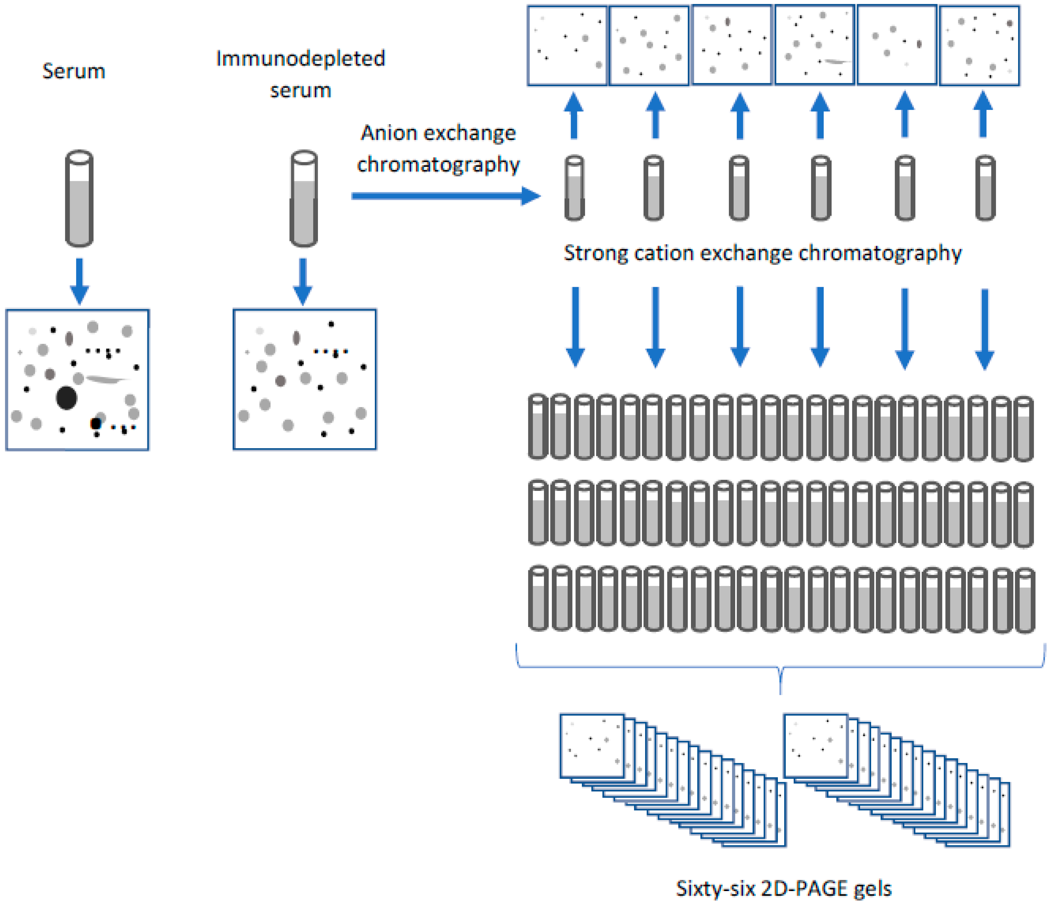
4.1. Two-Dimensional Gels Using Isoelectric Immobilized pH Gradients

Two different approaches were taken to achieve this goal. One approach increased the resolution of 2D-PAGE by using immobilized pH gradient (IPG) gels that essentially fractionated proteins within a specific pI range prior to loading them onto the gel, where they would then be separated based on molecular weight [42]. By using a series of overlapping narrow range (i.e., 1 pH unit) or ultra-narrow (i.e., 0.4 pH units) IPG strips, only those proteins that possess pI’s in a specific range (e.g., 4.5–5.5) will be resolved. By using several IPG strips of different ranges, a greater number of proteins will be resolved by increasing the resolution of the first separation dimension. The proteins within each of these strips are subsequently loaded onto individual gels and the proteins separated based on molecular weight.
Using this approach, a group separated out approximately 40 mg (i.e., 80 μL) of serum proteins (from which the six highest abundance proteins had been depleted) using seven overlapping narrow range IPG strips and were able to observe approximately 2–3000 proteins [43]. Of these, the pH ranges 4.5–5.5 and 5.5–6.7 accounted for almost 1150 of the identified proteins. While most of the identified proteins were relatively highly abundant acute-phase proteins, others that had concentrations less than 10 μg/mL in healthy individuals (e.g., ficolin) was also identified.
In a biomarker discovery study, tissue and plasma samples obtained from patients with colorectal cancer (CRC) were analyzed using a narrow-range IPG strip encompassing a pH range of 5.5–6.7 [44]. Comparison of proteins extracted from normal and matched tumor tissues showed that Nm23-H1 (a metastasis-related protein), S100A8, and S100A9 were all elevated in tumor tissue. The investigators then analyzed plasma samples taken from a larger number of patients. They showed that S100A8 and S100A9 were significantly increased in plasma samples of CRC patients compared to healthy individuals. This study demonstrated an excellent combination of IPG strips of tumor tissue discovery with further validation in plasma samples, a more readily obtainable clinical sample.

4.2. Combining Two-Dimensional Gels with Chromatography

Continuing his quest to further serum and plasma proteomics, N. Leigh Anderson combined a series of fractionation methods, including high-abundance protein depletion, chromatography, and 2D-PAGE (Figure 2), to resolve and identify as many proteins as possible in serum [45]. Eight high-abundance proteins (i.e., albumin, haptoglobulin, transferrin, transthyretin, α-1-antitrypsin, α-1-acid glycoprotein, hemopexin, and β-2-macroglobulin) were immuno-depleted from a single serum sample, which was then separated into six fractions using anion-exchange (AEX) chromatography. Each of these six fractions were separated into 11 fractions using strong cation exchange (SCX) chromatography, creating a total of 66 new aliquots. An aliquot of each fraction was taken and separated using 2D-PAGE, resulting in 74 gels (including the original non-depleted, unfractionated serum sample). The resulting gels showed approximately 20,000 individual spots, of which about 3700 were unique. Approximately 1800 of these unique spots were identified and found to originate from 325 unique proteins. One encouraging fact of this study was that 41% of the identified proteins were classified as either intracellular or cell-surface proteins, showing that serum contained species that may reflect the cellular condition of the individual.

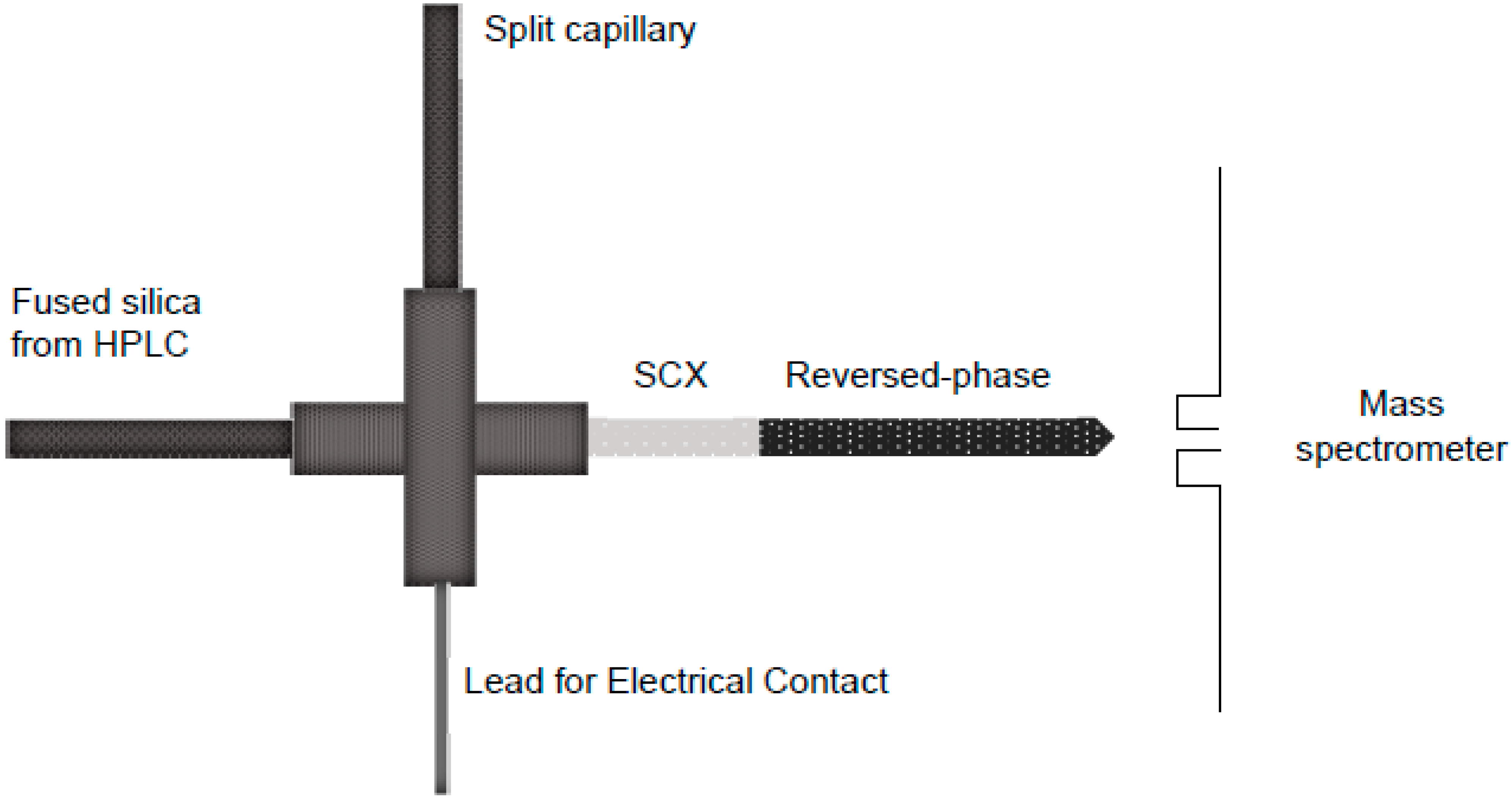
5. Chromatography for the Fractionation of Serum and Plasma Proteins

In the early days, 2D-PAGE was the major separation technique used in serum and plasma proteomics. Unfortunately, 2D-PAGE is quite labor-intensive, lacks sensitivity and dynamic range for protein detection, and has limited reproducibility. A simplistic Pubmed search using various combination of the terms “plasma”, “serum”, “proteome”, and “2D-PAGE” shows that the use of 2D-PAGE grew steadily between the years 2000 and 2012, and then started to decrease. This trend does not mean that it is an obsolete technique; 2D-PAGE has been critical in deciphering the protein content of blood. Indeed, with the incorporation of fluorescent tags to enable direct quantitative comparisons of proteins within a single gel (i.e., 2D difference in gel electrophoresis; DIGE), 2D-PAGE continues to produce important results [46,47,48,49]. In the year 2001, however, the combination of two well-known separation techniques would revolutionize how complex proteomes, such as serum and plasm, were analyzed.

Multidimensional Protein Identification Technology

Reversed-phased and SCX chromatography have been utilized for the separation of molecules since the 1970s [50,51]. In 2001, John Yates III’s lab prepared a single 15 cm-long chromatography column that was packed with 10 cm of C18 reversed-phase particles followed by 5 cm of SCX resin (Figure 3) [46]. This chromatography system was coupled with MS and termed MudPIT (multidimensional protein identification technology) [52]. The first published application of MudPIT, also commonly known as “shotgun” proteomics, was the analysis of the Saccharomyces cerevisiae (yeast) proteome. In the use of MudPIT, the proteins are proteolytically digested into peptides using trypsin or Lys-C. The proteins are digested for several reasons, related to both chromatography and MS. While it is possible to separate intact proteins using SCX and reversed-phase chromatography, many will not be soluble under the elution conditions used in these techniques. Using detergents to increase the intact protein solubility is challenging because many detergents interfere with MS detection. Related to MS, the sensitivity of the mass spectrometer is much greater for peptides than for proteins. The sequence of a peptide obtained from a tryptic or Lys-C digest will result in at least two potentially ionizable amino acid residues within each. These residues enable the necessary ionization to occur, which is required for MS analysis. Mass spectrometers are also most efficient at obtaining sequence information from peptides that are between 5 and 20 amino acids in length rather than from intact proteins that are not only much larger but are complicated by the possibility of different isoforms. Finally, software programs such as Sequest and Mascot are designed to readily calculate the sequence information from the tandem MS spectra of peptides.
The separation strategy of MudPIT involves loading the peptides onto the SCX column followed by cycles of eluting a fraction of these peptides onto the reversed-phase column, which are then eluted off this column into the mass spectrometer for analysis. In the first application of MudPIT, four elution buffers were used: buffer A (5% acetonitrile (ACN)/0.02% heptafluorobutyric acid (HFBA)), buffer B (80% ACN/0.02% HFBA), buffer C (250 mM ammonium acetate/5% ACN/0.02% HFBA), and buffer D (500 mM ammonium acetate/5% ACN/0.02% HFBA). Buffers A and B were to elute peptides from the reversed-phase column, while C and D performed the same function for the SCX column. The sample was loaded onto the column over 80 min by first applying a 70 min gradient 0 to 80% buffer B, which was then held for 10 min. The next 12 steps involved eluting fractions of peptides from the SCX column onto the reversed-phase column and then into the mass spectrometer. Each of these steps were 110 min in length. These steps consisted of 5 min of 100% buffer A, followed by 2 min of x% buffer C, 3 min of 100% buffer A, 10 min gradient from 0 to 10% buffer B, and finally a 90 min gradient from 10–45% buffer B. The step involving buffer C was ramped up in each of these 12 cycles from 10% to 100%, with the purpose of eluting new groups of peptides from the SCX column onto the reversed-phase column, where they could be eluted by the increasing buffer B concentration within each cycle. In the final step, the SCX column was washed using 100% buffer D.
So how effective was MudPIT at characterizing the yeast proteome? In this first application, 5540 peptides originating from 1484 proteins were identified from approximately 1 mg of total protein. These numbers are extraordinary when compared to the fact that the greatest number of yeast proteins identified by 2D-PAGE coupled to MS at this point was only 279 [53]. Quite importantly, the MudPIT method was able to identify low-abundance proteins (e.g., transcription factors and kinases) and proteins from a variety of subcellular locations (e.g., cytosolic and membrane).
While these numbers were incredibly impressive, the yeast proteome is significantly different than that of serum or plasma. As mentioned previously, both serum and plasma have an enormous dynamic range of protein concentrations compared to other proteomes. One of the first labs to apply this new MudPIT strategy to serum was that of Richard D. Smith, a pioneer in applying high-performance MS to proteomics. In the first step of the analysis, this group passed 0.5 mL of a standard serum sample over Protein A and G immobilized to beads to selectively remove IgGs [54]. In this study, albumin was not depleted since it is known to transport a variety of compounds in the blood and removing it would also result in the concomitant removal al many low-abundance proteins such as cytokines, peptide hormones, and lipoproteins. The immunoglobulin-depleted serum was digested with trypsin. While a MudPIT fractionation strategy (i.e., SCX and reversed-phase chromatography) was used to separate the serum peptides, unlike the original study in which the two chromatograph techniques were performed within a single column, this study collected fractions off a separate SCX column (i.e., off-line) and then separated each of these using a reversed-phase column coupled directly online with the mass spectrometer.
In the SCX chromatography step, IgG-depleted serum peptides were dissolved in 2 mL of 75% 10 mM ammonium formate and 25% acetonitrile, which was adjusted to pH 3.0 using formic acid (mobile phase A). The peptides were separated on a polysulfoethyl A column containing 5 μm particles with pores of 300 Å. After further equilibration with mobile phase A for 5 min, the peptides were eluted using a linear gradient from 0–100% mobile phase B (75% 200 mm ammonium formate, 25% acetonitrile, pH 8.0) over 30 min. The final peptides were then eluted from the column by maintaining the gradient at 100% mobile phase B for 25 min. The separation was conducted at a flow rate of 4 mL/minute. A total of 120 fractions were collected over the 60-min separation. These fractions were lyophilized and dissolved in 50 μL of H2O containing 0.1% formic acid (mobile phase A). Each peptide sample (8 μL) was sequentially injected onto a 60 cm reversed-phase capillary column (150 μm inner diameter and 360 μm outer diameter) packed with 5 μm Jupiter C18 particles. Peptides were loaded using mobile phase A and eluted using a gradient of 5–70% mobile phase B (acetonitrile containing 0.1% formic acid) over 80 min. The reversed-phase column was interfaced with an ion trap mass spectrometer. The mass spectrometer was operated in a data-dependent mode in which the three most intense peptide signals observed in an initial MS scan were sequentially isolated and subjected to tandem MS for identification.
The analysis resulted in the identification of 490 serum proteins. The concentrations of proteins identified ranged from albumin (~30–50 mg/mL) to prostate-specific antigen (~1 pg/mL). While common serum proteins, such as complement and coagulation factors, blood transporters, etc., were readily observed in the analysis, so were many intracellular (e.g., transcription factors) and membrane proteins (e.g., integrins), confirming the hypothesis that the circulatory systems contain intracellular proteins that are shed from tissues. Because of the peculiar dynamic range of protein concentrations within serum, the number of proteins identified in serum was only about one-third of that identified in yeast; however, it was substantially greater than the number of proteins previously identified in the massive chromatography/2D-PAGE study of N. Leigh Anderson [45].

6. Other Separation Techniques for Characterizing the Serum/Plasma Proteome

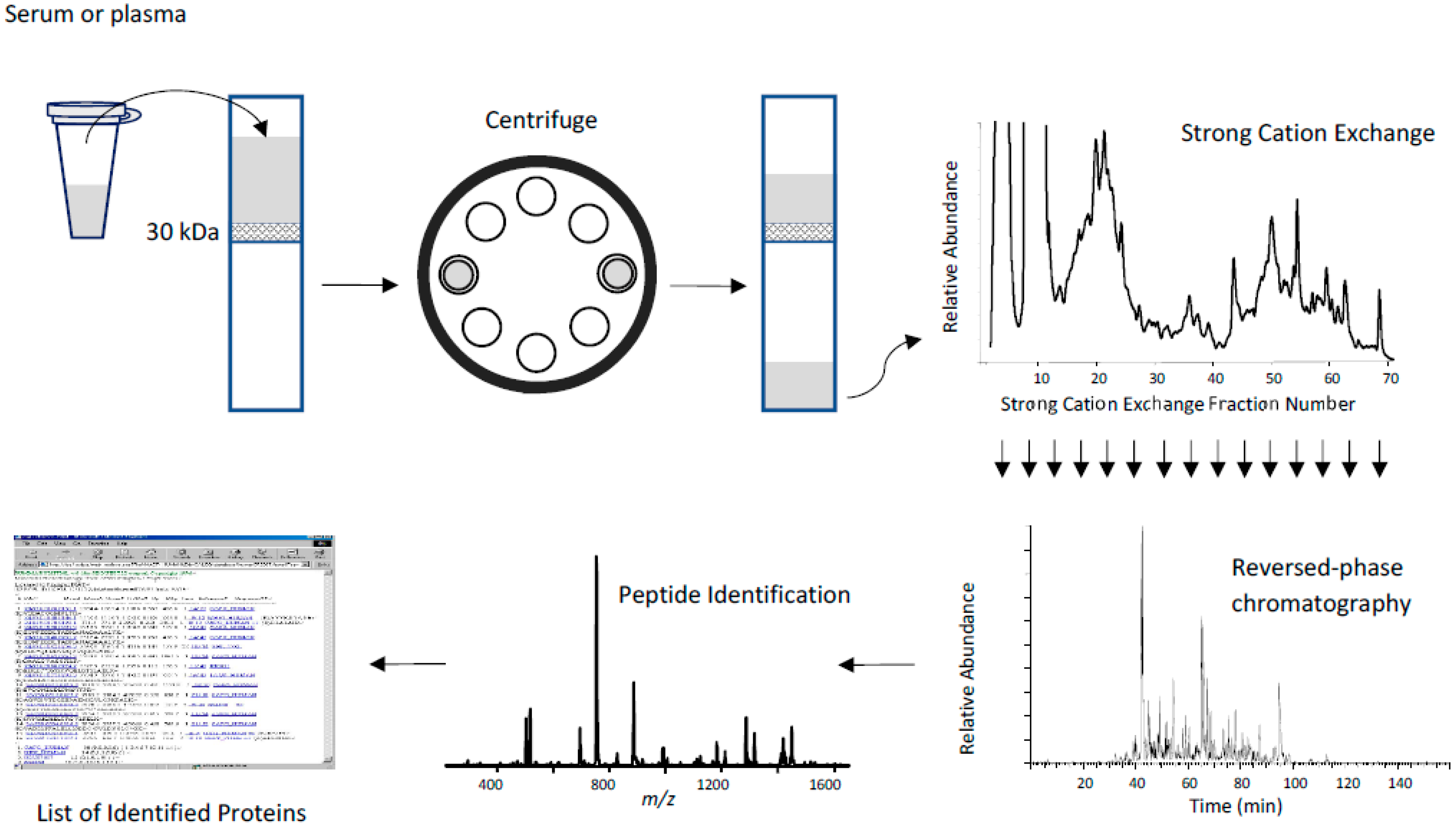
Low Molecular Weight Fractionation

In 2003, our group utilized a 30 kDa cutoff membrane to remove highly abundant proteins and recover the low molecular weight (LMW) proteome of serum [6]. The hypothesis was that by eliminating proteins with molecular weights greater than 30 kDa, high-abundance proteins such as albumin, immunoglobulins, lipoproteins, and transferrin would be greatly reduced in abundance. Enriching for the LMW proteome would enable greater coverage of biologically significant proteins such as cytokines, chemokines, peptide hormones, and proteolytic fragments of larger proteins. After the LMW fraction of serum was extracted using a 30 kDa molecular weight cutoff (MWCO) centrifugal filter, the sample was digested with trypsin, separated off-line using SCX into 20 fractions that were analyzed using reversed-phase chromatography coupled online with MS (Figure 4). Since previous studies have shown that many proteins bind to albumin, acetonitrile was added to a final concentration of 20% v/v to the serum sample prior to LMW filtration. While this addition did not affect the depletion of albumin and other HMW proteins, it did increase the amount of LMW proteins that were extracted.
A total of 341 unique proteins were identified in the LMW fraction of human serum. The efficacy of the LMW ultrafiltration method is demonstrated by the complete lack of any albumin peptides being detected in the analyzed samples. The proteins identified include common circulatory proteins, coagulation and complement factors, transport and binding proteins, cytokines, growth factors, and hormones. Other proteins, such as transcription factors, nuclear proteins, channels, and receptors, were also identified, corroborating the hypothesis that cell contents may be released into the bloodstream during necrosis, apoptosis, and hemolysis.

7. Applications

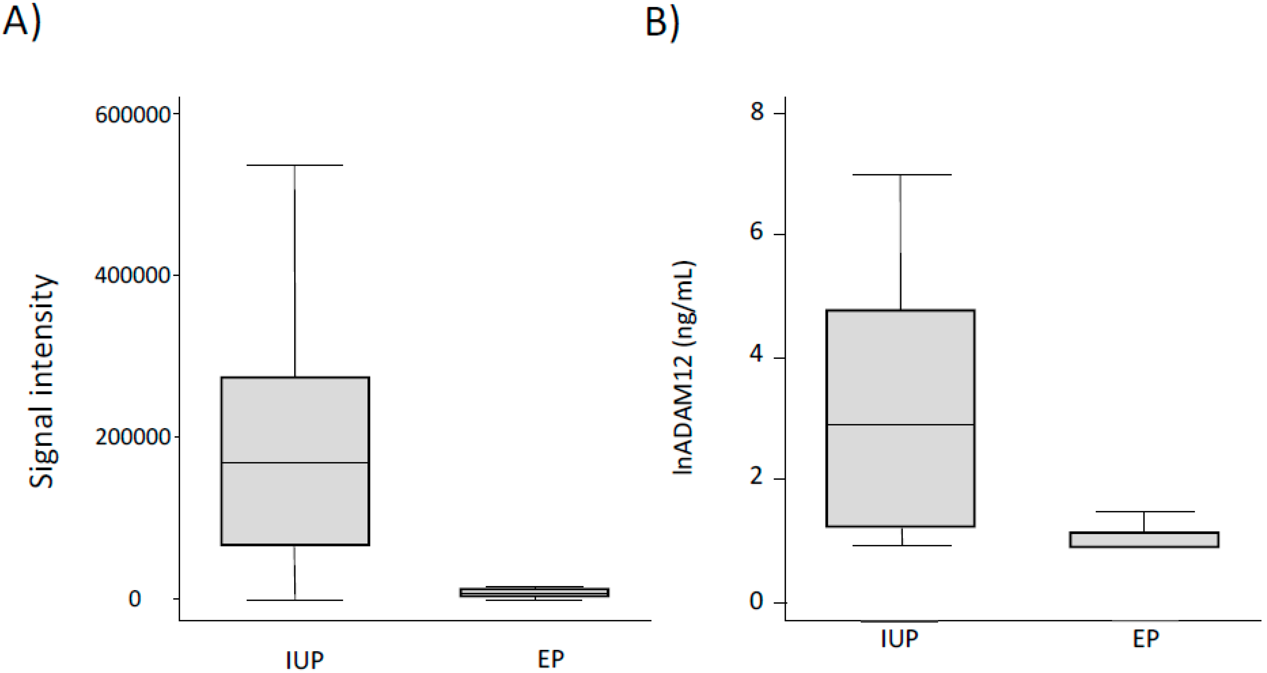
7.1. Biomarkers of Ectopic Pregnancy

Ectopic pregnancy (EP), the second leading cause of maternal death in the first trimester of pregnancy, is a clinical condition in which an embryo implants outside of the uterus, typically within a fallopian tube [55]. As the fetus grows, the mother’s life is threatened by the potential for tubal rupture and internal hemorrhage [56]. Ectopic pregnancy is diagnosed using a combination of serial β-human chorionic gonadotrophin measurements and transvaginal ultrasound [57]. Unfortunately, approximately 50% of EPs are misdiagnosed, particularly at an early stage [58], in part due to the fact that almost one-third of all cases are asymptomatic and 9% exhibit no symptoms prior to tubal rupture [59]. As with many conditions, there exists an urgent need to identify biomarkers that can improve the early diagnosis of EP. To try to meet this need, the group of David Speicher used a three-stage fractionation strategy to compare the proteomes of serum samples obtained from women with EP and those with normal intrauterine pregnancy (IUP) [60]. Nine samples from each cohort were collected and matched based on gestational age, β-hCG levels, and diagnosis. The samples were pooled to make three samples from women with EP and three with IUP.
The first step of fractionation was the depletion of 20 abundant proteins from 100 μL of serum using a ProteoPrep20 Immunodepletion Column (Sigma Aldrich). This immunodepletion step removes approximately 97% of the protein content of serum or plasma. The sensitivity of modern proteomics technology is demonstrated when one considers that only 100 μL of each sample was used. After each sample was immunodepleted, they were separated using 1D-PAGE. Each of the six gel lanes (corresponding to each pooled serum sample) was cut into 21 uniform 1 mm slices. After destaining, the proteins in these gel slices were digested into tryptic peptides, which were extracted from the gel. The peptides were identified using a BEH nanocapillary column (75 μm i.d. × 25 cm; Waters) packed with 1.7 μm C18 particles. Peptides were eluted using a 77 min-long gradient of increasing acetonitrile containing 1% formic acid. The flowrate was 200 nL/min. A total of 252 LC/MS runs were performed, as each gel piece was analyzed in duplicate.
The MS data from the EP and IUP samples were compared using a label-free approach in which the intensity values of the peptides are compared. Using this strategy, twelve candidate biomarkers were identified. Of these, 5 were selected for multiple-reaction monitoring (MRM)-MS in which the signal intensity of the peptides from these specific proteins are specifically monitored. These five proteins included ADAM12 (disintegrin and metalloproteinase domain-containing protein 12), CGA (glycoprotein hormones, alpha polypeptide), CGB (glycoprotein hormones, alpha polypeptide), CSH1 (chorionic somatomammotropin hormone 1), and PAEP (progestogen-associated endometrial protein). Instead of using pooled samples, individual samples from EP and IUP cases were analyzed by MRM-MS. The MRM-MS results for ADAM12 showed a particularly good diagnosis of EP (Figure 5A), with a large decrease in the signal intensity for peptides originating from this protein. Specifically, the EP diagnostic sensitivity and specificity of ADAM12 was 78% and 100%, respectively. ADAM12 is known to be expressed in a secreted form in the placenta that may initiate myogenesis (i.e., the formation of skeletal muscle) during first trimester of pregnancy [61]. Given its localization and role in cell-fusion in other tissues, it is biologically plausible that low ADAM12 levels can play a role in abnormal embryonic implantation, as seen in EP.
While the discovery and pre-validation of ADAM12 as a potential biomarker for EP using MRM-MS is exciting, this juncture is where current proteomics technology that relies heavily on chromatography and MS needs to pass the analysis to higher-throughput techniques. While separation-based proteomics technology has revolutionized the discovery of potential biomarkers, immunoassay-based methods are still critical as they possess the necessary throughput required for validation studies. In a follow-up study, the ADAM12 levels were quantitated in serum from 99 and 100 women with EP and IUP, respectively, with an immunoassay that used dissociation enhanced lanthanide fluorescence for greater sensitivity [62,63]. A statistically significant (p < 0.0001) decrease in the ADAM12 concentrations were observed in serum taken from the EP compared to the IUP cohort (i.e., 11.7 ng/mL vs. 115.4 ng/mL). The MRM-MS and immunoassay studies gave similar results (Figure 5B) and discriminatory power as assessed by the receiver operating characteristics (AUC = 0.81 and 0.82). A most intriguing result was that only 16% of the serum samples from IUP patients had a level of ADAM12 below the detection limit of the assay, while the same for EP patients was almost 70%. Overall, this study demonstrated the utility of both a discovery proteomics approach that utilized three sequential separation strategies (i.e., immunodepletion, 1D-PAGE, and reversed-phase chromatography) to identify potential biomarkers and a higher throughput immunoassay for validation.

7.2. Identification of a Novel Proatherogenic Peptide Hormone

As described above, extraction of the LMW serum proteome has been shown to be a simple and effective method to remove highly abundant proteins, while potentially containing biologically relevant biomarkers. Since the original study [6], many groups have tweaked the method to improve the extraction efficiency and increase the number of identified proteins [64,65,66]. One such study used an organic solvent to precipitate high molecular weight proteins from a 50 μL sample of plasma [64]. The peptides within the supernatant were separated using a nano-capillary reversed-phase column packed with C18 particles (particle diameter 3 µm, 75 μm i.d. × 125 mm). The peptides were separated at a flow rate of 300 nL/min using a gradient composed of solvent A (0.1% FA) and solvent B (0.1% FA and 90% ACN) (0–20 min, 5–32% B; 20–26 min, 32–55% B; 26–27 min, 55–95% B; 27–30 min, 95% B). Peptides eluting from the column were identified using a high-performance quadrupole Orbitrap mass spectrometer coupled directly online with the chromatography column.
The method resulted in the high confidence identification of almost 8000 unique peptide sequences. Instead of simply determining the levels of these peptides in serum, this group did something rather interesting. The group analyzed the list of identified peptides to determine which of these may possess bioactivity. Bioinformatic analysis and solubility testing resulted in 129 synthetic peptides being produced. The activity of these synthetic peptides was tested in human macrophages and vascular cells in vitro. One peptide in particular corresponding to residues 22–51 of human glucose-dependent insulinotropic polypeptide (GIP_HUMAN[22-51]) was found to activate NF-κB signaling in these cells. Since GIP is known to be involved in production of atherosclerotic plaques, the effect of GIP_HUMAN[22-51] was tested by infusing ApoE−/− mice with the synthetic peptides.
Atherosclerotic lesion development was accelerated in GIP_HUMAN[22-51]-infused ApoE−/− mice. Atheromatous plaque formation and macrophage accumulation induced by GIP_HUMAN[22-51] was inhibited in mice treated with an anti-GIP_HUMAN[22-51] antibody. Serum samples taken from ApoE−/− mice infused for 4 weeks of with or without GIP_HUMAN[22-51] or anti-GIP_HUMAN[22-51] IgG that were applied to protein antibody arrays detected 111 mouse cytokines and chemokines. Serum samples from mice infused with GIP_HUMAN[22-51] contained higher levels of several proinflammatory and proatherosclerotic proteins, including angiopoietin-2, serum amyloid P (SAP), CXC chemokine ligand 16 (CXCL16), proprotein convertase subtilisin kexin type 9 (PCSK9), fetuin A, and MMP-3. Serum from mice treated with anti-GIP_HUMAN[22-51] IgG showed lower levels of intercellular adhesion molecule-1 (ICAM-1), vascular cell adhesion molecule-1 (VCAM-1), fetuin A, PCSK9, CXCL16, C-reactive protein, and SAP. Taken together, these results suggest that GIP_HUMAN[22-51] induces various proinflammatory and proatherosclerotic proteins.
While both the in vitro and in vivo results demonstrate its proatherogenic activity, this activity is physiologically irrelevant to humans if the peptide is not expressed within human tissues. Analysis of a human tissue array using an anti-GIP_HUMAN[22-51] polyclonal antibody detected the high expression of this peptide within cerebellum, liver, stomach, kidney, heart, small intestine, and colon, and lower expression in the cerebral cortex, tonsils, lymph nodes, and testes. While the identification of this novel proatherogenic peptide was significant, the greater result of this study is the demonstration of how the combination of a serum fractionation method, MS analysis, bioinformatics, and systematic validation can lead to the identification of endogenous bioactive peptides and biomarkers for diagnosis, prognosis, and treatment of diseases.

8. Current Challenges in Serum and Plasma Proteomics

The goal of a vast majority of serum and plasma proteome studies is to identify a clinical biomarker that can be used for disease diagnosis or therapeutic monitoring. This requires that the study aims be precisely defined with respect to the desired application and target patient population. If the aim is to identify a biomarker for disease screening, the disease incidence and prevalence needs to be carefully considered as the cost of false positive and false negative results drive the required sensitivity and specificity of any putative biomarker. Designing a study aimed at discovering a therapeutic biomarker needs to carefully consider the specific therapy that is used, the response rates to this therapy, and survival analysis when given this therapy.
Considering that most of the biomarkers sought in serum/plasma proteomic studies originate from specific cell populations and undergo significant dilution prior to acquisition, their abundance difference in two cohorts of patients would be expected to be small. This expectation means that any variability in pre-analytical sample handling and processing must be minimal. Overcoming these variability issues requires careful planning of the number of samples that must be analyzed to obtain the desired power, limit the number of false discoveries, and control the technical and biological variation. Studies have shown that the ability to detect a 1.2-fold difference in a specific plasma protein with 80% power would require the comparison of 50 cases and 50 controls [67,68]. While these numbers can be a useful estimate, the number of samples required will differ based on both the condition being studied and the analytical process. Fortunately, with the increasing amounts of information regarding analytical and biological variability observed in serum/plasma proteomes along with fold-changes observed for specific proteins, software tools have been developed that enable sample size estimates to be calculated [69,70].
The pre-analytical stages, such as sample collection and storage time and temperature, need to be very consistent. A recent study evaluated the intra- and inter-laboratory reproducibility in the proteome analysis of both serum and cerebrospinal fluid (CSF) [71]. They took eight pooled serum and eight CSF samples and processed them using an identical procedure. Aliquots were analyzed in triplicate within a single lab as well as in two different labs using the same reversed-phase chromatography separation and mass spectrometer conditions. To assess the sensitivity, linearity, and dynamic range of the LC-MS method, serial dilutions of 18 stable isotope-labeled peptides were added to the serum and CSF samples. The lower limit of quantification (LLOQ) for these peptides ranged from 0.1 to 0.5 fmol, with excellent linearity (r2 > 0.978) for both samples. While intra-laboratory reproducibility for peptide identification was excellent for both serum and CSF (medium coefficient of variation (CV) of 12.5% and 17.3%, respectively), the inter-laboratory reproducibility was significantly higher (CVs of 23.8% for serum and 32.0% for CSF). Probably the most encouraging result from this study related to the intra- and inter-laboratory results comparing peptide quantitation. The Pearson correlation value was 0.98 for the replicate samples analyzed both intra- and inter-laboratory when the quantitative peptide results for the peptides were compared. These results show that under highly controlled conditions, quantitative proteomic analysis of serum samples can provide reliable quantitative results.
While the resolution of separation technologies used for proteomic analysis have greatly increased, their sheer complexity and degeneracy makes it currently impossible to fully resolve all the peptides within serum/plasma samples. The faster duty cycles and data-independent acquisition methods available with modern mass spectrometers has lessened the need for complete resolution; however, minimizing the number of peptides that enter the mass spectrometer per unit time increases the proteome coverage, making it currently impossible to fully resolve these samples. As a single chromatography is insufficient, proteomic scientists have relied on combining multiple separation techniques to minimize the number of peptides that are concurrently being analyzed [72,73]. Unfortunately, adding more separation steps increases the experimental variability and time. Each separation step can increase the number of aliquots that need to be analyzed per sample by an order of magnitude. For example, while immunodepleting a single serum sample produces one aliquot, once this sample is fractionated using SCX (for example), it may result in 10–100 aliquots that require MS analysis. Considering that some calculations suggest that 100 samples are necessary to detect the types of quantitative differences expected for biomarkers with reasonable confidence, simply combining two chromatography methods could require the analysis of 1000–10,000 aliquots. It is very challenging to maintain low inter-laboratory variability over this large number of samples. While pooling samples to reduce the time and resources required to complete a serum/plasma study is tempting, this method should be avoided as it assumes that specific samples within a cohort have similar biological values [74].
The time, equipment, and personnel commitment required for acquiring serum/plasma proteomic data is just the tip of the iceberg. Once the raw MS data have been acquired, it must be processed to be turned into peptide identifications. This step has been historically done using software programs such as Sequest [75] and Mascot [76]. Spectral library searching is becoming an increasingly popular tool in proteomics as it allows MS spectra to be queried against libraries of experimental reference spectra of previously identified peptides [77]. This method has been applied to peptides, phosphopeptides, glycopeptides, as well as cross-linked peptides [78,79,80,81]. Studies have shown that some spectral library searching tools can increase the number of peptides identified by approximately 30% compared to conventional search engines that compare experiment spectra to theoretical fragment ions of putative peptides [82]. While spectral matching has been used for the analysis of metabolites for years, it is now currently becoming more popular in proteomics with the development of search tools such as SpectraST [83], COSS [84], Pepitome [85], and Calibr [86]. Not only is spectral library searching faster, it is also more accurate and results in lower false discovery rates than using theoretical fragment ion algorithms.

9. Conclusions

Biomarkers have been used by physicians, epidemiologists, and scientists for centuries. There are four classes of biomarkers: molecular (e.g., DNA, protein, and metabolite levels, etc.), histologic (e.g., cell shape), radiographic (e.g., tumor size), and physiologic (e.g., blood pressure). Unlocking the blood proteome to enable the identification of clinically validated biomarkers would have a huge impact on public health. One of the primary attributes of blood is its accessibility. Having been used in clinical chemistry for decades, blood samples are routinely taken at doctor visits and analyzed for a variety of molecular and histological markers. Imagine the impact that being able to routinely test blood samples for various cancers and neurological disorders would have on public health. Early diagnosis provides the opportunity for more accurate treatment decisions, resulting in higher quality of life and overall prognosis [86,87,88]. Not only would blood-based diagnostic biomarkers impact public health, but biomarkers for monitoring treatment would benefit drug development. Considering the resources focused on their discovery, it is astonishing that most biomarkers were discovered over two decades ago and only a small fraction (out of the more than 14,000) of human diseases have companion tests [89]. Why, then, has the current era of serum and plasma proteomics not produced more validated biomarkers? The answer is simple: serum and plasma proteomes are very difficult to analyze and compare between cohorts because they are extremely complicated. The proteomes of samples taken from cells and other biofluids (e.g., urine, cerebrospinal fluid, etc.) simply do not possess the large dynamic range of protein concentration and sheer number of proteins as found in serum and plasma.
While the development of more sensitive and higher-resolution mass spectrometers has had a tremendous impact on the ability to characterize complex proteomes, the importance of separations technology cannot be underestimated. Although very-fast mass spectrometers can only identify a single (or very small number) peptide per cycle. They are analogous to hitting tennis balls. If 100 tennis balls are served sequentially, the receiver can successfully hit each one. If all 100 are served simultaneously, the receiver is only able to hit those within the area of the racket. Separating proteins with the highest resolution possible is critical for maximizing the number of proteins that can be characterized in a serum or plasma proteome. While a shotgun separation strategy (i.e., SCX followed by reversed-phase chromatography) is standard for most proteome analyses, serum and plasma has been subjected to many different combination strategies. Immunodepleting highly abundant blood proteins is critical for identifying low-abundance proteins, which are the most likely source of clinically useful biomarkers.
The progress made in characterizing serum and plasma proteomes has been remarkable. In the span of 20 years, scientists went from being able to identify about 300 proteins to more than 1000 [90,91], with one report even identifying more than 5000 proteins in plasma [92]. Inventive separation technologies have been a key to this progress and will continue to play a major role if we hope to discover the much-needed biomarkers that can impact human health.
Chromatography and separation methods will continue to improve enabling a greater coverage of serum and plasma proteomes. Future developments will include continued decreases in stationary-phase particle sizes, which will lead to improvements in peak capacity. Improving peak capacity allows shorter columns to be used and faster separations. Using decreasing particle sizes results in higher column back pressures; however, instrument innovations that provide robust operation at higher pressures are expected to continue. Outside of chromatography, peptide separation using ion mobility–mass spectrometry (IM-MS) will continue to increase in use. IM-MS adds an extra separation dimension between chromatographic separation and MS detection [93]. As peptides enter the mass spectrometer from the chromatography column, they are separated based on their drift time (or ion mobility), which is related to the size, shape, and charge state. The advantage of IM-MS is that it increases the overall analytical resolution without increasing the analysis time. This MS method is versatile in that it can be coupled with both chromatography and electrophoretic techniques. In the future, IM-MS will play an increasingly bigger role in proteomic analysis.
The importance of being able to accurately characterize the proteomes of serum, plasma, and other clinical samples cannot be understated as medicine has only begun to mine the treasure of biomarkers present within these samples. The discovery of biomarkers that could diagnose various cancers at early stages would save millions of lives every year. Identification of therapeutic biomarkers could assist in ensuring that patients are provided the most effective treatment as soon as possible. While the discovery of novel biomarkers has been slower than anticipated, serum and plasma proteomics has come a long way since the early days, when being able to identify 100 proteins in these complex samples was considered state-of-the-art. While the development of high-performance mass spectrometers deserved much of the credit for these advances, the importance of the continued improvements seen in electrophoretic and chromatographic separations should not be ignored. Separations will continue to be an integral part in the march towards gaining a greater understanding of the serum and plasma proteomes.

Author Contributions

Conceptualizaiton, J.P. and T.D.V. research, J.P. and T.D.V.; writing—original draft preparation, J.P. and T.D.V.; writing—review and editing, J.P. and T.D.V. All authors have read and agreed to the published version of the manuscript.

Funding

The research received no external funding.

Acknowledgments

The authors would like to thank Cedarville University for their support.

Conflicts of Interest

The authors declare no conflict of interest, financial or otherwise.

References

  1. Shorthouse, J.; Wilson, S. Use of blood components in clinical practice. Nurs. Stand. 2019, 34, 76. [Google Scholar] [CrossRef] [PubMed]
  2. Blood Test. Available online: https://www.hopkinsmedicine.org/health/treatment-tests-and-therapies/blood-test (accessed on 4 January 2022).
  3. Chace, H.D.; Kalas, T.A.; Naylor, E.W. Use of tandem mass spectrometry for multianalyte screening of dried blood specimens from newborns. Clin. Chem. 2003, 49, 1797. [Google Scholar] [CrossRef] [PubMed] [Green Version]
  4. Basic Metabolic Panel (BMP). Available online: https://medlineplus.gov/lab-tests/basic-metabolic-panel-bmp/ (accessed on 5 January 2022).
  5. Complete Blood Count (CBC). Available online: https://www.mayoclinic.org/tests-procedures/complete-blood-count/about/pac-20384919 (accessed on 5 January 2022).
  6. Tirumalai, R.S.; Chan, K.C.; Prieto, D.A.; Issaq, H.J.; Conrads, T.P.; Veenstra, T.D. Characterization of the low molecular weight serum proteome. Mol. Cell. Proteom. 2003, 2, 1096. [Google Scholar] [CrossRef] [PubMed] [Green Version]
  7. Anderson, N.L.; Polanski, M.; Pieper, R.; Gatlin, T.; Tirumalai, R.S.; Conrads, T.P.; Veenstra, T.D.; Adkins, J.N.; Pounds, J.G.; Fagan, R.; et al. The human plasma proteome: A nonredundant list developed by combination of four separate sources. Mol. Cell. Proteom. 2004, 3, 311–326. [Google Scholar] [CrossRef] [PubMed] [Green Version]
  8. Geyer, P.E.; Voytik, E.; Treit, P.V.; Doll, S.; Kleinhempel, A.; Niu, L.; Müller, J.B.; Buchholtz, M.L.; Bader, J.M.; Teupser, D.; et al. Plasma proteome profiling to detect and avoid sample-related biases in biomarker studies. EMBO Mol. Med. 2019, 11, e10427. [Google Scholar] [CrossRef] [PubMed]
  9. Almeida, N.; Rodriguez, J.; Pla Parada, I.; Perez-Riverol, Y.; Woldmar, N.; Kim, Y.; Oskolas, H.; Betancourt, L.; Valdés, J.G.; Sahlin, K.B.; et al. Mapping the melanoma plasma proteome (MPP) using single-shot proteomics interfaced with the WiMT database. Cancers 2021, 13, 6224. [Google Scholar] [CrossRef]
  10. Lygirou, V.; Makridakis, M.; Vlahou, A. Biological sample collection for clinical proteomics: Existing SOPs. Methods Mol. Biol. 2015, 1243, 3–27. [Google Scholar]
  11. Anderson, N.L.; Anderson, N.G. The human plasma proteome: History, character, and diagnostic prospects. Mol. Cell. Proteom. 2002, 1, 845–867. [Google Scholar] [CrossRef] [Green Version]
  12. Maloney, J.P.; Silliman, C.C.; Ambruso, D.R.; Wang, J.; Tuder, R.M.; Voelkel, N.F. In vitro release of vascular endothelial growth factor during platelet aggregation. Am. J. Physiol. 1998, 275, H105401061. [Google Scholar] [CrossRef]
  13. Omenn, G.S. Plasma proteomics, the human proteome project, and cancer-associated alternative splice variant proteins. Biochim. Biophys. Acta 2014, 1844, 866–873. [Google Scholar] [CrossRef] [Green Version]
  14. Das, L.; Murthy, V.; Varma, A.K. Comprehensive analysis of low molecular weight serum proteome enrichment for mass spectrometric studies. ACS Omega 2020, 5, 28877–28888. [Google Scholar] [CrossRef] [PubMed]
  15. Nie, S.; Shi, T.; Fillmore, T.L.; Schepmoes, A.A.; Brewer, H.; Gao, Y.; Song, E.; Wang, H.; Rodland, K.D.; Qian, W.J.; et al. Deep-dive targeted quantification for ultrasensitive analysis of proteins in nondepleted human blood plasma/serum and tissues. Anal. Chem. 2017, 89, 9139–9146. [Google Scholar] [CrossRef] [PubMed] [Green Version]
  16. Fredolini, C.; Pathak, K.V.; Paris, L.; Chapple, K.M.; Tsantilas, K.A.; Rosenow, M.; Tegeler, T.J.; Garcia-Mansfield, K.; Tamburro, D.; Zhou, W.; et al. Shotgun proteomics coupled to nanoparticle-based biomarker enrichment reveals a novel panel of extracellular matrix proteins as candidate serum protein biomarkers for early-stage breast cancer detection. Breast Cancer Res. 2020, 22, 135–150. [Google Scholar] [CrossRef] [PubMed]
  17. Echan, L.A.; Tang, H.Y.; Ali-Khan, N.; Lee, K.; Speicher, D.W. Depletion of multiple high-abundance proteins improves protein profiling capacities of human serum and plasma. Proteomics 2005, 5, 3292–3303. [Google Scholar] [CrossRef] [PubMed]
  18. Hinerfeld, D.; Innamorati, D.; Pirro, J.; Tam, S.W. Serum/plasma depletion with chicken immunoglobulin Y antibodies for proteomic analysis from multiple Mammalian species. J. Biomol. Tech. 2004, 15, 184–190. [Google Scholar]
  19. Chromy, B.A.; Gonzales, A.D.; Perkins, J.; Choi, M.W.; Corzett, M.H.; Chang, B.C.; Corzett, C.H.; McCutchen-Maloney, S.L. Proteomic analysis of human serum by two-dimensional differential gel electrophoresis after depletion of high-abundant proteins. J. Proteome Res. 2004, 3, 1120–1127. [Google Scholar] [CrossRef]
  20. Björhall, K.; Miliotis, T.; Davidsson, P. Comparison of different depletion strategies for improved resolution in proteomic analysis of human serum samples. Proteomics 2005, 5, 307–317. [Google Scholar] [CrossRef]
  21. Multiple Affinity Removal System. Available online: https://www.agilent.com/en/product/agilent-multiple-affinity-removal-spin-columns-cartridges (accessed on 27 February 2022).
  22. Dardé, V.M.; Barderas, M.G.; Vivanco, F. Depletion of high-abundance proteins in plasma by immunoaffinity subtraction for two-dimensional difference gel electrophoresis analysis. Methods Mol. Biol. 2007, 357, 351–364. [Google Scholar]
  23. Johansen, E.; Schilling, B.; Lerch, M.; Niles, R.K.; Liu, H.; Li, B.; Allen, S.; Hall, S.C.; Witkowska, H.E.; Regnier, F.E.; et al. A lectin HPLC method to enrich selectively-glycosylated peptides from complex biological samples. J. Vis. Exp. 2009, 1, 1398–1401. [Google Scholar] [CrossRef] [Green Version]
  24. Zhou, M.; Lucas, D.A.; Chan, K.C.; Issaq, H.J.; Petricoin, E.F., 3rd; Liotta, L.A.; Veenstra, T.D.; Conrads, T.P. An investigation into the human serum “interactome”. Electrophoresis 2004, 25, 1289–1298. [Google Scholar] [CrossRef]
  25. Chan, K.C.; Lucas, D.A.; Hise, D.; Schaefer, C.F.; Xiao, Z.; Janini, G.M.; Buetow, K.H.; Issaq, H.J.; Veenstra, T.D.; Conrads, T.P. Analysis of the human serum proteome. Clin. Proteom. 2004, 1, 101–225. [Google Scholar] [CrossRef] [Green Version]
  26. Finn, T.E.; Nunez, A.C.; Sunde, M.; Easterbrook-Smith, S.B. Serum albumin prevents protein aggregation and amyloid formation and retains chaperone-like activity in the presence of physiological ligands. J. Biol. Chem. 2012, 287, 21530–21540. [Google Scholar] [CrossRef] [PubMed] [Green Version]
  27. Petricoin, E.F.; Belluco, C.; Araujo, R.P.; Liotta, L.A. The blood peptidome: A higher dimension of information content for cancer biomarker discovery. Nat. Rev. Cancer 2006, 6, 961–967. [Google Scholar] [CrossRef] [PubMed]
  28. Mehta, A.I.; Ross, S.; Lowenthal, M.S.; Fusaro, V.; Fishman, D.A.; Petricoin, E.F., 3rd; Liotta, L.A. Biomarker amplification by serum carrier protein binding. Dis. Markers 2003, 19, 1–10. [Google Scholar] [CrossRef] [Green Version]
  29. Lowenthal, M.S.; Mehta, A.I.; Frogale, K.; Bandle, R.W.; Araujo, R.P.; Hood, B.L.; Veenstra, T.D.; Conrads, T.P.; Goldsmith, P.; Fishman, D.; et al. Analysis of albumin-associated peptides and proteins from ovarian cancer patients. Clin. Chem. 2005, 51, 1933–1945. [Google Scholar] [CrossRef] [Green Version]
  30. Jonkers, J.; Meuwissen, R.; van der Gulden, H.; Petersen, H.; van der Valk, M.; Berns, A. Synergistic tumor suppressor activity of BRCA2 and p53 in a conditional mouse model for breast cancer. Nat. Genet. 2001, 29, 418–425. [Google Scholar] [CrossRef]
  31. Moss, E.L.; Hollingworth, J.; Reynolds, T.M. The role of CS125 in clinical practice. J. Clin. Pathol. 2005, 58, 308–312. [Google Scholar] [CrossRef] [Green Version]
  32. Boschetti, E.; Zilberstein, G.; Righetti, P.G. Combinatorial peptides: A library that continuously probes low-abundance proteins. Electrophoresis 2022, 43, 355–369. [Google Scholar] [CrossRef]
  33. ProteoMiner Protein Enrichment Kits. Available online: https://www.bio-rad.com/en-us/product/proteominer-protein-enrichment-kits?ID=1dd94f06-7658-4ab4-b844-e29a1342a214 (accessed on 21 January 2022).
  34. D’Amato, A.; Bachi, A.; Fasoli, E.; Boschetti, E.; Peltre, G.; Sénéchal, H.; Righetti, P.G. In-depth exploration of cow’s whey proteome via combinatorial peptide ligand libraries. J. Proteome Res. 2009, 8, 3925–3936. [Google Scholar] [CrossRef]
  35. Martos, G.; López-Fandiño, R.; Molina, E. Immunoreactivity of hen egg allergens: Influence on in vitro gastrointestinal digestion of the presence of other egg white proteins and of egg yolk. Food Chem. 2013, 136, 775–781. [Google Scholar] [CrossRef]
  36. Boschetti, E.; Bindschedler, L.V.; Tang, C.; Fasoli, E.; Righetti, P.G. Combinatorial peptide ligand libraries and plant proteomics: A winning strategy at a price. J. Chromatogr. A. 2009, 1216, 1215–1222. [Google Scholar] [CrossRef] [PubMed]
  37. Fekkar, A.; Pionneau, C.; Brossas, J.Y.; Marinach-Patrice, C.; Snounou, G.; Brock, M.; Mazier, D. DIGE enables the detection of a putative serum biomarker of fungal origin in a mouse model of invasive aspergillosis. J. Proteome 2012, 75, 2536–2549. [Google Scholar] [CrossRef] [PubMed]
  38. O’Farrell, P.H. High resolution two-dimensional electrophoresis of proteins. J. Biol. Chem. 1975, 250, 4007–4021. [Google Scholar] [CrossRef]
  39. Wasinger, V.C.; Cordwell, S.J.; Cerpa-Poljak, A.; Yan, J.X.; Gooley, A.A.; Wilkins, M.R.; Duncan, M.W.; Harris, R.; Williams, K.L.; Humphery-Smith, I. Progress with gene-product mapping of the Mollicutes: Mycoplasma genitalium. Electrophoresis 1995, 16, 1090–1094. [Google Scholar] [CrossRef] [PubMed]
  40. Wright, G.L., Jr. High resolution two-dimensional polyacrylamide electrophoresis of human serum proteins. Am. J. Clin. Pathol. 1972, 57, 173–185. [Google Scholar] [CrossRef] [PubMed] [Green Version]
  41. Anderson, L.; Anderson, N.G. High resolution two-dimensional electrophoresis of human plasma proteins. Proc. Natl. Acad. Sci. USA 1977, 74, 5421–5425. [Google Scholar] [CrossRef] [PubMed] [Green Version]
  42. Görg, A.; Drews, O.; Lück, C.; Weiland, F.; Weiss, W. 2-DE with IPGs. Electrophoresis 2009, 30, S122–S132. [Google Scholar] [CrossRef] [PubMed]
  43. Vasudev, N.S.; Ferguson, R.E.; Cairns, D.A.; Stanley, A.J.; Selby, P.J.; Banks, R.E. Serum biomarker discovery in renal cancer using 2-DE and prefractionation by immunodepletion and isoelectric focusing; increasing coverage or more of the same? Proteomics 2008, 8, 5074–5085. [Google Scholar] [CrossRef]
  44. Kim, H.J.; Kang, H.J.; Lee, H.; Lee, S.T.; Yu, M.H.; Kim, H.; Lee, C. Identification of S100A8 and S100A9 as serological markers for colorectal cancer. J. Proteome Res. 2009, 8, 1368–1379. [Google Scholar] [CrossRef]
  45. Pieper, R.; Gatlin, C.L.; Makusky, A.J.; Russo, P.S.; Schatz, C.R.; Miller, S.S.; Su, Q.; McGrath, A.M.; Estock, M.A.; Parmar, P.P.; et al. The human serum proteome: Display of nearly 3700 chromatographically separated protein spots on two-dimensional electrophoresis gels and identification of 325 distinct proteins. Proteomics 2003, 3, 1345–1364. [Google Scholar] [CrossRef]
  46. Issaq, H.J.; Veenstra, T.D. The role of electrophoresis in disease biomarker discovery. Electrophoresis 2007, 28, 1980–1988. [Google Scholar] [CrossRef] [PubMed]
  47. Arentz, G.; Weiland, F.; Oehler, M.K.; Hoffmann, P. State of the art of 2D DIGE. Proteom. Clin. Appl. 2015, 9, 277–288. [Google Scholar] [CrossRef] [PubMed]
  48. Lazensky, R.; Silva-Sanchez, C.; Kroll, K.J.; Chow, M.; Chen, S.; Tripp, K.; Walsh, M.T.; Denslow, N.D. Investigating an increase in Florida manatee mortalities using a proteomic approach. Sci. Rep. 2021, 11, 4282–4295. [Google Scholar] [CrossRef] [PubMed]
  49. Zhang, X.; Yu, W.; Cao, X.; Wang, Y.; Zhu, C.; Guan, J. Identification of serum biomarkers in patients with Alzheimer’s disease by 2D-DIGE proteomics. Gerontology 2022, 12, 1–13. [Google Scholar] [CrossRef]
  50. Molnár, I.; Horváth, C. Reverse-phase chromatography of polar biological substances: Separation of catechol compounds by high-performance liquid chromatography. Clin. Chem. 1976, 22, 1497–1502. [Google Scholar] [CrossRef]
  51. Atack, C.V.; Magnusson, T. Individual elution of noradrenaline (together with adrenaline), dopamine, 5-hydroxytryptamine and histamine form a single, strong cation exchange column, by means of mineral acid-organic solvent mixtures. J. Pharm. Pharmacol. 1970, 22, 625–627. [Google Scholar] [CrossRef]
  52. Wolters, D.A.; Washburn, M.P.; Yates, J.R., 3rd. An automated multidimensional protein identification technology for shotgun proteomics. Anal. Chem. 2001, 73, 5683–5690. [Google Scholar] [CrossRef]
  53. Perrot, M.; Sagliocco, F.; Mini, T.; Monribot, C.; Schneider, U.; Shevchenko, A.; Mann, M.; Jenö, P.; Boucherie, H. Two-dimensional gel protein database of Saccharomyces cerevisiae (update 1999). Electrophoresis 1999, 20, 2280–2298. [Google Scholar] [CrossRef]
  54. Adkins, J.N.; Varnum, S.M.; Auberry, K.J.; Moore, R.J.; Angell, N.H.; Smith, R.D.; Springer, D.L.; Pounds, J.G. Toward a human blood serum proteome: Analysis by multidimensional separation coupled with mass spectrometry. Mol. Cell. Proteom. 2002, 1, 947–955. [Google Scholar] [CrossRef] [Green Version]
  55. Barnhart, K.T. Clinical practice. Ectopic pregnancy. N. Engl. J. Med. 2009, 361, 379–387. [Google Scholar] [CrossRef]
  56. Seeber, B.E.; Barnhart, K.T. Suspected ectopic pregnancy. Obstet. Gynecol. 2006, 107, 399–413. [Google Scholar] [CrossRef] [PubMed]
  57. Kirk, E.; Bottomley, C.; Bourne, T. Diagnosing ectopic pregnancy and current concepts in the management of pregnancy of unknown location. Hum. Reprod. Update 2014, 20, 250–261. [Google Scholar] [CrossRef] [PubMed]
  58. Refaat, B.; Bahathiq, A.O. The performances of serum activins and follistatin in the diagnosis of ectopic pregnancy: A prospective case-control study. Clin. Chim. Acta. 2020, 500, 69–74. [Google Scholar] [CrossRef] [PubMed]
  59. Tay, J.I.; Moore, J.; Walker, J.J. Ectopic pregnancy. BMJ 2000, 320, 916–919. [Google Scholar] [CrossRef] [PubMed]
  60. Beer, L.A.; Tang, H.Y.; Sriswasdi, S.; Barnhart, K.T.; Speicher, D.W. Systematic discovery of ectopic pregnancy serum biomarkers using 3-D protein profiling coupled with label-free quantitation. J. Proteome Res. 2011, 10, 1126–1138. [Google Scholar] [CrossRef] [PubMed] [Green Version]
  61. Gilpin, B.J.; Loechel, F.; Mattei, M.G.; Engvall, E.; Albrechtsen, R.; Wewer, U.M. A novel, secreted form of human ADAM 12 (meltrin alpha) provokes myogenesis in vivo. J. Biol. Chem. 1998, 273, 157–166. [Google Scholar] [CrossRef] [PubMed] [Green Version]
  62. Rausch, M.E.; Beer, L.; Sammel, M.D.; Takacs, P.; Chung, K.; Shaunik, A.; Speicher, D.; Barnhart, K.T. A disintegrin and metalloprotease protein-12 as a novel marker for the diagnosis of ectopic pregnancy. Fertil. Steril. 2011, 95, 1373–1378. [Google Scholar] [CrossRef]
  63. Parnham, A.J.; Tarbit, I.F. “Delfia” and “Amerlite”: Two sensitive nonisotopic immunoassay systems for assay of thyrotropin compared. Clin. Chem. 1987, 33, 1421–1424. [Google Scholar] [CrossRef]
  64. Masaki, T.; Kodera, Y.; Terasaki, M.; Fujimoto, K.; Hirano, T.; Shichiri, M. GIP_HUMAN[22-51] is a new proatherogenic peptide identified by native plasma peptidomics. Sci. Rep. 2021, 11, 14470–14483. [Google Scholar] [CrossRef]
  65. Zhou, C.X.; Xie, S.C.; Li, M.Y.; Huang, C.Q.; Zhou, H.Y.; Cong, H.; Zhu, X.Q.; Cong, W. Analysis of the serum peptidome associated with Toxoplasma gondii infection. J. Proteom. 2020, 222, 103805–103813. [Google Scholar] [CrossRef]
  66. Padoan, A. The impact of pre-analytical conditions on human serum peptidome profiling. Proteom. Clin. Appl. 2018, 12, e1700183–e1700184. [Google Scholar] [CrossRef] [PubMed]
  67. Bhawal, R.; Oberg, A.L.; Zhang, S.; Kohli, M. Challenges and opportunities in clinical applications of blood-based proteomics in cancer. Cancers 2020, 12, 2428. [Google Scholar] [CrossRef] [PubMed]
  68. Chang, C.Y.; Picotti, P.; Huttenhain, R.; Heinzelmann-Schwarz, V.; Jovanovic, M.; Aebersold, R.; Vitek, O. Protein significance analysis in selected reaction monitoring (SRM) measurements. Mol. Cell Proteom. 2012, 11, M111.014662. [Google Scholar] [CrossRef] [PubMed] [Green Version]
  69. Skates, S.J.; Gillette, M.A.; LaBaer, J.; Carr, S.A.; Anderson, L.; Liebler, D.C.; Ransohoff, D.; Rifai, N.; Kondratovich, M.; Tezak, Z.; et al. Statistical design for biospecimen cohort size in proteomics-based biomarker discovery and verification studies. J. Proteome Res. 2013, 12, 5383–5394. [Google Scholar] [CrossRef]
  70. Muntel, J.; Kirkpatrick, J.; Bruderer, R.; Huang, T.; Vitek, O.; Ori, A.; Reiter, L. Comparison of protein quantification in a complex background by dia and tmt workflows with fixed instrument time. J. Proteome Res. 2019, 18, 1340–1351. [Google Scholar] [CrossRef]
  71. Cho, K.C.; Oh, S.; Wang, Y.; Rosenthal, L.S.; Na, C.H.; Zhang, H. Evaluation of the sensitivity and reproducibility of targeted proteomic analysis using data-independent acquisition for serum and cerebrospinal fluid proteins. J. Proteome Res. 2021, 20, 4284–4291. [Google Scholar] [CrossRef]
  72. Wang, H.; Dey, K.K.; Chen, P.C.; Li, Y.; Niu, M.; Cho, J.H.; Wang, X.; Bai, B.; Jiao, Y.; Chepyala, S.R.; et al. Integrated analysis of ultra-deep proteomes in cortex, cerebrospinal fluid and serum reveals a mitochondrial signature in Alzheimer’s disease. Mol. Neurodegener. 2020, 15, 43–62. [Google Scholar] [CrossRef]
  73. Goldman, A.R.; Beer, L.A.; Tang, H.Y.; Hembach, P.; Zayas-Bazan, D.; Speicher, D.W. Proteome analysis using gel-LC-MS/MS. Curr. Protoc. Protein Sci. 2019, 96, e93. [Google Scholar] [CrossRef]
  74. Borras, E.; Canto, E.; Choi, M.; Maria Villar, L.; Alvarez-Cermeno, J.C.; Chiva, C.; Montalban, X.; Vitek, O.; Comabella, M.; Sabido, E. Protein-based classifier to predict conversion from clinically isolated syndrome to multiple sclerosis. Mol. Cell Proteom. 2016, 15, 318–328. [Google Scholar] [CrossRef] [Green Version]
  75. Yates, J.R.; Eng, J.K.; Clauser, K.R.; Burlingame, A.L. Search of sequence databases with ininterpreted high-energy collision-induced dissociation spectra of peptides. J. Am. Soc. Mass Spectrom. 1996, 7, 1089–1098. [Google Scholar] [CrossRef] [Green Version]
  76. Chalkley, R.J.; Baker, P.R.; Huang, L.; Hansen, K.C.; Allen, N.P.; Rexach, M.; Burlingame, A.L. Comprehensive analysis of a multidimensional liquid chromatography mass spectrometry dataset acquired on a quadrupole selecting, quadrupole collision cell, time-of-flight mass spectrometer: New developments in Protein Prospector allow for reliable and comprehensive automatic analysis of large datasets. Mol. Cell. Proteom. 2005, 4, 1194–1204. [Google Scholar]
  77. Willems, P.; Fels, U.; Staes, A.; Gevaert, K.; Van Damme, P. Use of hybrid data-dependent and -independent acquisition spectral libraries empowers dual-proteome profiling. J. Proteome Res. 2021, 20, 1165–1177. [Google Scholar] [CrossRef] [PubMed]
  78. Yang, Y.; Horvatovich, P.; Qiao, L. Fragment mass spectrum prediction facilitates site localization of phosphorylation. J. Proteome Res. 2021, 20, 634–644. [Google Scholar] [CrossRef] [PubMed]
  79. Shu, Q.; Li, M.; Shu, L.; An, Z.; Wang, J.; Lv, H.; Yang, M.; Cai, T.; Hu, T.; Fu, Y.; et al. Large-scale identification of N-linked intact glycopeptides in human serum using HILIC enrichment and spectral library search. Mol. Cell. Proteom. 2020, 19, 672–689. [Google Scholar] [CrossRef] [PubMed] [Green Version]
  80. Schweppe, D.K.; Chavez, J.D.; Navare, A.T.; Wu, X.; Ruiz, B.; Eng, J.K.; Lam, H.; Bruce, J.E. Spectral library searching to identify cross-linked peptides. J. Proteome Res. 2016, 15, 1725–1731. [Google Scholar] [CrossRef] [PubMed] [Green Version]
  81. Yang, Y.; Lin, L.; Qiao, L. Deep learning approaches for data-independent acquisition proteomics. Expert Rev. Proteom. 2021, 18, 1031–1043. [Google Scholar] [CrossRef]
  82. Wang, J.H.; Choong, W.K.; Chen, C.T.; Sung, T.Y. Calibr improves spectral library search for spectrum-centric analysis of data independent acquisition proteomics. Sci. Rep. 2022, 12, 2045–2057. [Google Scholar] [CrossRef]
  83. Lam, H.; Deutsch, E.W.; Eddes, J.S.; Eng, J.K.; King, N.; Stein, S.E.; Aebersold, R. Development and validation of a spectral library searching method for peptide identification from MS/MS. Proteomics 2007, 7, 655–667. [Google Scholar] [CrossRef]
  84. Shiferaw, G.A.; Vandermarliere, E.; Hulstaert, N.; Gabriels, R.; Martens, L.; Volders, P.J. COSS: A fast and user-friendly tool for spectral library searching. J. Proteome Res. 2020, 19, 2786–2793. [Google Scholar] [CrossRef]
  85. Dasari, S.; Chambers, M.C.; Martinez, M.A.; Carpenter, K.L.; Ham, A.J.; Vega-Montoto, L.J.; Tabb, D.L. Pepitome: Evaluating improved spectral library search for identification complementarity and quality assessment. J. Proteome Res. 2012, 11, 1686–1695. [Google Scholar] [CrossRef]
  86. Biskup, E.; Wils, R.S.; Hogdall, C.; Hogdall, E. Prospects of improving early ovarian cancer diagnosis using cervical cell swabs. Anticancer Res. 2022, 42, 1–12. [Google Scholar] [CrossRef] [PubMed]
  87. Kulczyńska-Przybik, A.; Dulewicz, M.; Słowik, A.; Borawska, R.; Kułakowska, A.; Kochanowicz, J.; Mroczko, B. The clinical significance of cerebrospinal fluid reticulon 4 (RTN4) levels in the differential diagnosis of neurodegenerative diseases. J. Clin. Med. 2021, 10, 5281. [Google Scholar] [CrossRef] [PubMed]
  88. Duca, R.B.; Massillo, C.; Dalton, G.N.; Farré, P.L.; Graña, K.D.; Gardner, K.; De Siervi, A. MiR-19b-3p and miR-101-3p as potential biomarkers for prostate cancer diagnosis and prognosis. Am. J. Cancer Res. 2021, 11, 2802–2820. [Google Scholar] [PubMed]
  89. Anderson, N.L. The clinical plasma proteome: A survey of clinical assays for proteins in plasma and serum. Clin. Chem. 2010, 56, 177–185. [Google Scholar] [CrossRef]
  90. Cao, Z.; Tang, H.Y.; Wang, H.; Liu, Q.; Speicher, D.W. Systematic comparison of fractionation methods for in-depth analysis of plasma proteomes. J. Proteome Res. 2012, 11, 3090–3100. [Google Scholar] [CrossRef] [Green Version]
  91. Addona, T.A.; Shi, X.; Keshishian, H.; Mani, D.R.; Burgess, M.; Gillette, M.A.; Clauser, K.R.; Shen, D.; Lewis, G.D.; Farrell, L.A.; et al. A pipeline that integrates the discovery and verification of plasma protein biomarkers reveals candidate markers for cardiovascular disease. Nat. Biotechnol. 2011, 29, 635–643. [Google Scholar] [CrossRef] [Green Version]
  92. Keshishian, H.; Burgess, M.W.; Gillette, M.A.; Mertins, P.; Clauser, K.R.; Mani, D.R.; Kuhn, E.W.; Farrell, L.A.; Gerszten, R.E.; Carr, S.A. Multiplexed, quantitative workflow for sensitive biomarker discovery in plasma yields novel candidates for early myocardial injury. Mol. Cell. Proteom. 2015, 14, 2375–2393. [Google Scholar] [CrossRef] [Green Version]
  93. Ewing, M.A.; Glover, M.S.; Clemmer, D.E. Hybrid ion mobility and mass spectrometry as a separation tool. J. Chromatogr. A 2016, 1439, 3–25. [Google Scholar] [CrossRef]
Figure 1. (A) Approximately 90% of the protein content of these samples is made up of only 22 proteins, with albumin making up between almost half of the total amount. (B) Separation strategies to characterize serum and plasma focus on methods to enable proteins within the lower 10% concentration level to be identified. (C) The dynamic range of protein concentration of serum and plasma is very broad, covering approximately 12 orders of magnitude. Adapted from [7].
Separations 09 00089 g001
Figure 2. Serum proteome analysis using a combined immunodepletion, liquid chromatographic, and two-dimensional polyacrylamide gel electrophoresis (2D-PAGE) strategy. In the first stage of this strategy, high-abundance proteins were immunodepleted from a serum sample that was then fractionated into six aliquots using anion exchange chromatography. These six aliquots were subsequently fractionated into a total of 66 aliquots using strong cation exchange chromatography. Each aliquot throughout the process was fractionated using 2D-PAGE. Visualized spots within the gel were identified using mass spectrometry. Adapted from [36].
Separations 09 00089 g002
Figure 3. Multidimensional protein identification technology (MudPIT) for the separation of complex proteome samples. MudPIT consists of a biphasic column packed with strong cation exchange (SCX) and reversed-phase resins. Peptides derived via a tryptic digest of a proteome sample are loaded onto the SCX resin and eluted using a step gradient of increasing salt concentrations onto the reversed-phase column. The peptides are eluted from the reversed-phase column using a linear gradient directly into the mass spectrometer for identification. Repeated cycles of this process using increasing salt concentrations enables the peptides to be separated providing the mass spectrometer more opportunity to identify a greater number of peptides. Adapted from [43].
Separations 09 00089 g003
Figure 4. Analysis of a low molecular weight (LMW) serum proteome. In this analysis, serum (or plasma) is centrifuged through a 30 kilodalton (kDa) low molecular weight membrane. The flow-through is digested into tryptic peptides that are fractionated using strong cation exchange (SCX) chromatography. Fractions collected off the SCX column are separated using reversed-phase chromatography. Peptides coming off this column elute directly into the mass spectrometer, where they are analyzed to generate a list of identified proteins.
Separations 09 00089 g004
Figure 5. Statistical analysis of the intensity of ADAM12 peptides observed in a quantitative proteomics analyses using (A) multiple reaction monitoring mass spectrometry (MRM-MS) and (B) immunoassay comparing serum samples obtained from woman with ectopic pregnancies (EP) and normal intrauterine pregnancies (IUP). The MRM-MS data were compiled from the analysis of 9 EP and 9 IUP serum samples, while the immunoassay data was compiled using 99 EP and 100 IUP samples. Adapted from [51].
Separations 09 00089 g005
Table 1. Comparison of plasma and serum.
PlasmaSerum
Straw-coloredClear, yellowish fluid
Composed of serum and clotting factorsBlood without clotting factors
Acquired by centrifuging blood to which an anticoagulant has been addedAcquire by centrifuging blood that has been allowed to clot for ~30 min
Easier and faster to prepareTakes longer and is more difficult to prepare
Long shelf-life (i.e., up to ten years)Shorter shelf life (i.e., a few months)
Table 2. List of commercially available products for high-abundance protein depletion or low-abundance protein enrichment of serum and plasma samples.
ProductManufacturerCapture AgentCaptured Proteins
Albumin
Depletion Kit
ThermoFisher ScientificAntibodyAlbumin
High-Select HSA/Immunoglobulin Depletion Spin ColumnsThermoFisher ScientificAntibodiesAlbumin, IgG, IgM, IgE, IgD, and IgA
High-Select Top14 Abundant Protein Depletion Spin ColumnsThermoFisher ScientificAntibodiesAlbumin, IgG, IgA, IgM, IgD, IgE, Alpha-1-Acid glycoprotein, Alpha-1-Antitrypsin, Alpha-2-Macroglobulin, Apolipoproteins A-I, Fibrinogen, Haptoglobin, Transferrin
Albumin/IgG Removal KitThermoFisher ScientificCibacron Blue Dye and Protein AAlbumin and IgG
Multiple Affinity Removal Column Human 14AgilentAntibodiesalbumin, IgG, antitrypsin, IgA, transferrin, haptoglobin, fibrinogen, alpha-2-macroglobulin, alpha1-acid glycoprotein, IgM, apolipoprotein AI, apolipoprotein AII, complement C3 and transthyretin
ProteoExtract Albumin/IgG Removal KitMillipore Sigmaalbumin-specific affinity resin and protein AAlbumin and IgG
ProteoMiner Protein Enrichment BioRadCombinatorial library of hexapeptidesAll proteins
Publisher’s Note: MDPI stays neutral with regard to jurisdictional claims in published maps and institutional affiliations.

