Abstract
Objectives: To develop a robust liquid chromatography-tandem mass spectrometry (LC–MS/MS) method to simultaneously measure seven human plasma androgens, namely testosterone (T), dihydrotestosterone (DHT), androstenedione (A4), dehydroepiandrosterone sulfate (DHEAS), dehydroepiandrosterone (DHEA), 11-ketotestosterone (11-KetoT), and 11β-hydroxytestosterone (11β-OHT). Design and Methods: Plasma was extracted via a solid phase extraction method, and the analytical performance of the assay was validated according to the Clinical & Laboratory Standards Institute guidelines. Overall, 73 apparently healthy volunteers were recruited to evaluate the distribution of these seven androgens; their levels in 25 females with acne and 33 obese females were also evaluated. Results: The developed method exhibited a good precision, with the total coefficient variations (CV) and the intra-assay CVs being within 10%. Furthermore, the recoveries of T, DHT, A4, DHEA, DHEAS, 11-KetoT, and 11β-OHT were 90.3–105.8, 88.7–98.1, 92.4–102.5, 90.5–106.7, 87.6–99.9, 93.3–105.3, and 90.2–104.4%, respectively, and no significant matrix effect was observed after internal standard correction (<20%). Moreover, the limits of quantification were 0.01, 0.01, 0.01, 0.10, 5.00, 0.02, and 0.02 ng/mL for T, DHT, A4, DHEA, DHEAS, 11-KetoT, and 11β-OHT, respectively, which are adequate for their accurate measurement in human plasma samples. It was also determined that patients diagnosed with acne had significantly higher levels of DHT, A4, and DHEAS, while those suffering from obesity had significantly higher levels of T and A4 but lower levels of DHT. Conclusions: A robust LC-MS/MS method for the simultaneous determination of seven androgens in plasma samples was successfully established and validated, which plays important roles in clinical application.
1. Introduction
Androgen hormones are critical for the development and maintenance of male characteristics, and they are also related to many metabolic disorders in females, including hypogonadism, polycystic ovary syndrome (PCOS), and tumors of the ovary and breast [1,2,3]. Moreover, associations between androgen hormones and other diseases such as obesity [4], acne [5], diabetes [6], Alzheimer’s disease [7], and even mortality in men [8] have also been found. In addition to testosterone (T)–the primary androgen hormone–many other androgen hormones play important roles in the diagnosis and treatment of various diseases, with example androgens including dihydrotestosterone (DHT), androstenedione (A4), dehydroepiandrosterone sulfate (DHEAS), and dehydroepiandrosterone (DHEA) [9,10]. Moreover, emerging evidence has proposed a set of 11-oxygenated 19-carbon (11oxC19) adrenal-derived steroids, including 11-ketotestosterone (11-KetoT) and 11β-hydroxytestosterone (11β-OHT), to be clinically important androgens that play vital roles in many endocrine disorders, such as 21-hydroxylase deficiency [11]. Thus, the accurate measurement of androgen hormones from biological samples is of great importance.
Currently, the measurement of androgen hormones in clinical applications generally depends on immunoassays. Although such assays allow a large sample throughput, interference can occur due to the presence of heterophilic or autoantibodies, and the high-dose hook effects can affect the accurate measurement of sandwich assays [12]. Following intense development, the liquid chromatography-tandem mass spectrometry (LC–MS/MS) technique has become the method of choice for steroid analyses in a growing number of routine clinical laboratories. More specifically, LC–MS/MS has been employed to analyze the concentrations of androgen hormones in females, children, and males undergoing androgen suppression therapies [10,13], wherein up to 20 androgen hormones can be simultaneously measured [14]. However, previous reports [9,14] failed to investigate the simultaneous measurement of these seven androgens, especially for 11β-OHT and 11-KetoT. Even though 11-KetoT has a greater bioactivity comparable to that of T [15,16,17], its application in many endocrine disorders, such as acne and obesity, remains unknown due to the lack of available measurement methods and related studies.
To address these issues, we established and validated a robust LC–MS/MS method for the simultaneous measurement of seven plasma androgens–namely T, DHT, A4, DHESA, DHEAS, 11-KetoT, and 11β-OHT. In addition, we evaluated their distributions in healthy volunteers, and explore their clinical roles in acne and obesity.
2. Materials and Methods
2.1. Chemicals and Reagents
The standard materials of T, DHT, A4, DHEA, and DHEAS were purchased from Sigma-Aldrich (St. Louis, MO, USA), while 11-KetoT and 11β-OHT were purchased from Steraloids Inc. (Newport, RI, USA). The internal standards (IS) of T (T-2,3,4-13C3) and DHT (5α-DHT-16,16,17-d3) were purchased from Sigma-Aldrich, while those of A4 (A4-3,17-dione-13C3), DHEA (DHEA-2,2,3,4,4,6-d6), and DHEAS (DHEAS-2,2,3,4,4,6-d6 sulfate sodium salt) were purchased from Isosciences (Ambler, PA, USA). Hormone-free plasma was purchased from Seracare (Milford, MA, USA). High-performance liquid chromatography-grade methanol and acetonitrile were purchased from Thermo Fisher Scientific (Fair Lawn, NJ, USA). Ammonium fluoride (NH4F) was obtained from Sinopharm Chemical Reagent Co. (Beijing, China), and the deionized water was obtained from A.S. Watson (Hong Kong, China).
2.2. Instrumentation and Analytical Conditions
Controlled using MassLynx 4.2 software (Waters. Milford, MA, USA), a Waters Acquity I-Class UPLC system coupled with a Waters TQ-S triple quadrupole MS/MS system was used for the determination of these seven androgens in human plasma. A Waters Acquity UPLC BEH C8 column (100 mm × 2.1 mm, 1.7 μm) was chosen for the chromatographic separation. Mobile phase A consisted of a 0.5 mM NH4F solution prepared in ddH2O, while mobile phase B was pure methanol. Gradient elution was performed at a flow rate of 0.3 mL/min according to the following procedure: 0.0–0.5 min, 40% B; 0.5–4.0 min, 40–55% B; 4.0–6.5 min, 55–75% B; 6.5–7.2 min, 75–90% B; and 7.2–8.0 min, 90–40% B. The column temperature was maintained at 50 °C, and the sample compartment temperature was 15 °C. Electrospray Ionization mode was chosen, all seven analytes were detected in the positive electrospray ion mode, and the optimized MS/MS settings can be found in Table 1. The final ion source settings were as follows: desolvation gas flow rate, 1100 L/h; cone gas flow rate, 150 L/h; nebulizer, 7.0 bar; capillary voltage, 2.0 kV; cone voltage, 50 V; and desolvation temperature, 550 °C.

Table 1.
The optimized MS/MS parameters and retention time of seven androgens.
2.3. Preparation of the Stock Solutions, Calibration Standards, and Quality Control Samples
The T, DHT, A4, DHEA, and DHEAS standard solutions were prepared at concentrations of 1 mg/mL, and were further diluted with methanol/ddH2O (1:1, v/v) prior to storage at −80 °C. The 11-KetoT and 11β-OHT standard solutions were dissolved in methanol to give final concentrations of 2 mg/mL, and were then further diluted with methanol/ddH2O (1:1, v/v) prior to storage at −80 °C. A mixed standard stock solution was prepared, and this solution contained 2 μg/mL T, 1 μg/mL DHT, 2 μg/mL A4, 5 μg/mL DHEA, 500 μg/mL DHEAS, 2 μg/mL 11-KetoT, and 2 μg/mL 11β-OHT. The IS solutions of T, DHT, and A4 were prepared at a concentration of 100 μg/mL, while those of DHEAS and DHEA were prepared at concentrations of 1 and 5 mg/mL, respectively. Since the IS materials of 11-KetoT and 11β-OHT were unavailable, T-2,3,4-13C3 was used for their correction. A mixed IS stock solution was prepared, and this solution contained 0.5 μg/mL T, 2 μg/mL DHT, 0.5 μg/mL A4, 4 μg/mL DHEA, and 20 μg/mL DHEAS. Three levels of quality controls were prepared by spiking hormone-free plasma with T, DHT, A4, DHEA, DHEAS, 11-KetoT, and 11β-OHT at the desired concentrations. The calibration and control solutions were stored at −20 °C prior to analysis.
2.4. Sample Preparation
A 200 μL plasma/quality control/calibration sample (10-fold diluted with hormone-free plasma) was mixed with the IS solution (20 μL) at room temperature (22–24 °C) for 2 min. Proteins were precipitated by the addition of methanol (300 μL), and then ddH2O (400 μL) was added. After subjecting the resulting mixture to centrifugation for 10 min at 13,000 rpm, an aliquot (700 μL) of the clear supernatant was transferred to a 96-well PRiME HLB solid-phase extraction (SPE) plate (Waters, Milford, MA, USA), which had been pre-washed using methanol (200 μL) and ddH2O (200 μL). The sample was allowed to flow through the SPE plate under a Positive Pressure-96 (Waters, Milford, MA, USA), and then acetonitrile/ddH2O (200 μL, 1:9, v/v), and hexane (200 μL) were added successively to wash the SPE plate. Subsequently, the bound substances were eluted using acetonitrile/ddH2O (40 μL, 9:1, v/v), then mixed with ddH2O (40 μL) in a 700 μL 96-well collection plate. After mixing and centrifugation, an aliquot (10 μL) of the sample was injected into the LC–MS/MS for determination of the target compounds.
2.5. Method Validation
The method was validated in terms of its limit of detection (LOD), limit of quantification (LOQ), linearity, sensitivity, precision, and recovery according to the CLSI C62-A and EP-15 A3 guidelines. The carryover and matrix effect were also assessed for these seven androgens. A set of 13 non-zero calibration standards were prepared for each analyte to obtain their corresponding calibration curves, which were generated by the plotting peak area ratio (Y) of the analyte and the IS against the concentration (X ng/mL) of each analyte. The LOD was defined as the lowest concentration with a signal-to-noise ratio >3 and a coefficient of variation (CV) < 20%. The LOQ was defined as the lowest concentration with a signal-to-noise >10 and a CV < 20%. The method precision was evaluated by means of five replicated measurements at the three levels of quality controls for all analytes within 1 d (intra-assay precision) and over a period of 5 d (inter-assay precision). To evaluate the recovery and accuracy, known concentrations of seven androgens at low, medium, and high levels were added to hormone-free plasma. The matrix effect was evaluated by spiking the post-extracted plasma samples from six random individuals with two appropriate concentrations of the analytes, and the absolute responses (peak area) of the standards and the relative responses (ratio of the analyte peak area/IS area) in the solvent were compared with those of the extracted plasma samples to determine whether the isotope-labeled IS can compensate for any potential matrix effect. To estimate the carryover, a blank sample was injected after the calibration solution with the highest concentration. All the concentration levels used for the evaluation of the method validation were shown in the below results.
2.6. Clinical Application
A total of 73 apparently healthy volunteers were recruited to evaluate the distribution of these seven androgens. All these Han ethnic adults had lived in Beijing city for at least 1 year without the presence of acute or chronic diseases requiring medical intervention or associated drugs taking. Female participants were not pregnant, breastfeeding, or within 1 year of delivery. Exclusion criteria included: (1) blood donation in the last 3 months, (2) systolic pressure > 140 mmHg or diastolic pressure > 90 mmHg, (3) body mass index > 28 kg/m2, (4) known carrier of hepatitis B virus, hepatitis C virus, or human immunodeficiency virus, (5) has been working regular night shifts for almost a month and has had disturbed sleep patterns for almost a week. Between the hours of 07:00 and 09:00 a.m., fasting EDTA anticoagulant samples were collected from the volunteers in the recumbent position for at least 2 h. The samples were then subjected to centrifugation at 3000 rpm for 10 min and subjected to the established analytical method described above. In addition, the leftover plasma samples of 25 females diagnosed with acne (26 ± 6 years), and 33 females suffering from obesity (25 ± 7 years), as registered in the LIS system of the Peking Union Medical College Hospital (PUMCH), were collected and the seven target androgens were analyzed using the newly established method.
This study was reviewed and approved by the Ethics Committee of PUMCH of the Chinese Academy of Medical Sciences (S-T483), and all volunteers provided written informed consent prior to participation in the study.
2.7. Statistical Analysis
SPSS version 22.0 (IBM, Chicago, IL, USA) and GraphPad Prism 7.0 (GraphPad Software, San Diego, CA, USA) were used for the purpose of statistical analysis. Analytes with a normal distribution were presented as mean ± standard deviation, and those with non-normal distributions were presented as a median (25th and 75th percentiles). The Mann-Whitney U test or the Kruskal-Wallis test was used for comparison among several groups. To evaluate the correlation among different analytes, Pearson’s correlation analysis was used for the analytes with a normal distribution, whereas Spearman’s correlation analysis was used for analytes with a non-normal distribution. Two-sided p values < 0.05 were considered statistically significant.
3. Results
3.1. Representative Chromatography
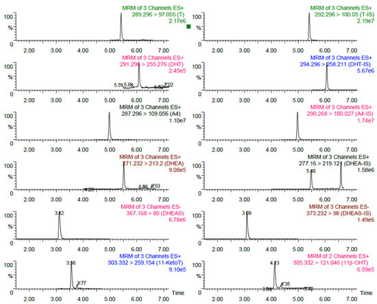
As shown in Figure 1, the total analysis time for this method was 8 min, and during this time the chromatography peaks of all seven androgens were symmetrical and well-separated, except for T and DHEA, which could be separated by the difference of MRM transitions.

Figure 1.
Representative LC-MS/MS chromatograms for the seven androgens in a random plasma sample. T, Testosterone; DHT, Dihydrotestosterone; A4, Androstenedione; DHEA, Dehydroepiandrosterone; DHEAS, Dehydroepiandrosterone sulfate; 11-KetoT, 11-ketotestosterone; 11β-OHT, 11β-hydroxytestosterone.
3.2. Method Validation
3.2.1. Linearity
The calibration curves were linear over concentration ranges of 0.01–20 ng/mL for T and A4, 0.01–10 ng/mL for DHT, 0.1–50 ng/mL for DHEA, 5–5000 ng/mL for DHEAS, and 0.02–20 ng/mL for 11-KetoT and 11β-OHT; these concentrations encompassed the concentration ranges of the corresponding compounds in human plasma. The linearity was monitored for five consecutive days, and the average coefficients of linear regression (r2) were >0.998 for all seven androgens (see Supplementary Figure S1).
3.2.2. LOD and LOQ
The LODs for T, DHT, A4, DHEA, DHEAS, 11-KetoT, and 11β-OHT were determined to be 0.005, 0.010, 0.010, 0.100, 5.000, 0.020, and 0.020 ng/mL, respectively, while the LOQs were determined to be 0.01, 0.01, 0.01, 0.10, 5.00, 0.02, and 0.02 ng/mL, respectively. These results indicated that the developed method allowed the accurate determination of all seven androgens in human plasma samples.
3.2.3. Accuracy and Precision
As shown in Table 2, the recoveries for T, DHT, A4, DHEA, DHEAS, 11-KetoT, and 11β-OHT were 90.3–105.8, 88.7–98.1, 92.4–102.5, 90.5–106.7, 87.6–99.9, 93.3–105.3, and 90.2–104.4%, respectively, and the bias compared with that of the added theoretical concentration was within 15% for each analyte, thereby confirming an excellent method accuracy. To assess the precision, mixed plasma samples at three different concentrations were successively determined within 5 days. As listed in Table 3, the reproducibility values at the three concentrations were good, with intra-assay CVs of 2.7–3.3, 2.5–5.4, 2.0–4.9, 3.1–5.6, 3.5–4.2, 2.4–5.9, and 2.5–6.4% for T, DHT, A4, DHEA, DHEAS, 11-KetoT, and 11β-OHT, respectively, whereas the total CVs were 3.7–4.1, 4.5–7.7, 2.7–5.1, 5.6–6.4, 4.0–6.4, 3.2–7.3, and 3.0–6.9%.

Table 2.
Analytical recovery assays for the seven androgens.

Table 3.
Method precision results for the seven androgens.
3.2.4. Matrix Effect
The absolute responses obtained for these seven androgens indicated that both ion suppression and enhanced matrix effect existed without IS correction. However, it was found that the observed matrix effect could be compensated via IS correction, as outlined in Table 4.

Table 4.
The matrix effect of seven androgens.
3.2.5. Carryover
In this study, the carryover for all seven androgens can be ignored since there was no visible peak for the blank sample after injecting the standard solution containing the highest concentrations of T (20 ng/mL), DHT (10 ng/mL), A4 (20 ng/mL), DHEA (50 ng/mL), DHEAS (5000 ng/mL), 11-KetoT (20 ng/mL), and 11β-OHT (20 ng/mL).
3.3. Clinical Application
A total of 73 apparently healthy volunteers (aged 42 ± 13 years) including 29 males (aged 43 ± 14 years) and 44 females (aged 42 ± 13 years) were recruited to preliminarily evaluate the distribution of these seven androgens. More specifically, the distributions of T, DHT, A4, DHEA, DHEAS, 11-KetoT, and 11β-OHT were 4.64 (3.51, 5.36), 0.24 (0.19, 0.30), 0.75 (0.65, 0.86), 3.39 (2.07, 4.52), 1406.83 (1153.22, 2268.97), 0.45 (0.31, 0.71), and 0.25 (0.16, 0.34) ng/mL in males; and 0.17 (0.13, 0.24), 0.04 (0.02, 0.07), 0.61 (0.36, 0.90), 2.55 (1.66, 3.73), 930.90 (648.86, 1414.27), 0.33 (0.27, 0.49), and 0.15 (0.12, 0.20) ng/mL in females. Furthermore, significant correlations were observed between T and DHT (r = 0.754), DHT and DHEA (r = −0.443), DHEA and DHEAS (r = 0.557), A4 and DHEA (r = 0.400), and 11-KetoT and 11β-OHT (r = 0.878) in males; significant correlations were also found between T and DHT (r = 0.781), T and DHEA (r = 0.641), T and DHEAS (r = 0.606), DHT and DHEA (r = 0.655), DHT and DHEAS (r = 0.663), DHEA and DHEAS (r = 0.840), and 11-KetoT and 11β-OHT (r = 0.822) in females.
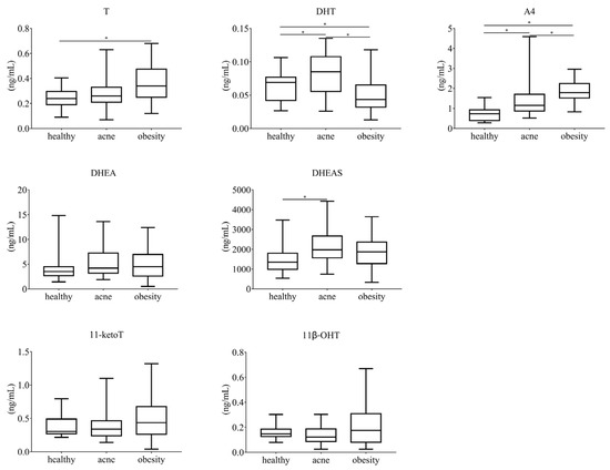
Furthermore, the results of 25 females diagnosed with acne (26 ± 6 years) and 33 females suffering from obesity (25 ± 7 years) were analyzed, and 19 individuals ranging in age from 20 to 40 years (29 ± 7 years) were selected from the 44 apparently healthy female volunteers as a control group. As outlined in Figure 2, the T, DHT, A4, DHEA, DHEAS, 11-KetoT, and 11β-OHT levels in the acne group were 0.26 (0.21, 0.33), 0.09 (0.06, 0.11), 1.15 (0.87, 1.71), 4.20 (3.20, 7.25), 1972.00 (1564.50, 2667.38), 0.34 (0.24, 0.46), and 0.12 (0.09, 0.19) ng/mL, respectively, while the corresponding levels in the obesity group were 0.34 (0.25, 0.48), 0.04 (0.03, 0.06), 1.79 (1.45, 2.20), 4.50 (2.60, 6.59), 1865.10 (1193.00, 2358.87), 0.43 (0.25, 0.67), and 0.18 (0.08, 0.31) ng/mL, respectively. Thus, the levels of DHT, A4, and DHEAS were significantly higher in the acne group than in the control group, while the levels of T and A4 were significantly higher, and that of DHT was significantly lower, in the obesity group than in the control group.

Figure 2.
Distribution of these seven androgens in females diagnosed with acne or obesity. T, Testosterone; DHT, Dihydrotestosterone; A4, Androstenedione; DHEA, Dehydroepiandrosterone; DHEAS, Dehydroepiandrosterone sulfate; 11-KetoT, 11-ketotestosterone; 11β-OHT, 11β-hydroxytestosterone. * means p values < 0.05.
4. Discussion
In previous studies [14,18,19,20,21,22,23], the simultaneous determination of a maximum of only four androgens (i.e., T, DHT, A4, DHEA, or DHEAS) was achieved using LC–MS/MS methods, and the additional simultaneous determination of 11-KetoT and 11β-OHT were not effectively cooperated with these more common androgens [24,25]. However, recent evidence has shown that 11oxC19 steroids, such as 11-KetoT and 11β-OHT, are clinically important androgens, and their trajectories throughout adulthood differ from other androgens [25]. As a downstream derivative of T, 11-KetoT was previously reported to exhibit a maximal bioactivity comparable to that of T [15,16,17], and the ratio of 11-KetoT/T was considered as an excellent clinical indicator for a 21-hydroxylase deficiency, particularly in post-pubertal males. Moreover, 11-KetoT can also be derived from 11β-OHT under catalysis by the 11β-hydroxysteroid dehydrogenase type 2 enzyme. Our results therefore indicate that we successfully established and verified a robust LC–MS/MS method for the simultaneous measurement of seven androgens, including 11-KetoT and 11β-OHT, in human plasma for the first time. This method is robust, sensitive, and simple for clinical application.
Based on the distributions obtained for 73 apparently healthy volunteers, all seven androgens were found to be present in significantly lower quantities in females than in males, which is reasonable [19]. The levels of some androgens were like those of one previous report [22], but significantly lower than those described in another report [19]. These differences were likely related to the different cohorts, and so it is apparent that population-specific reference intervals (RIs) must be established. Since the levels of androgens are affected not only by sex, but also by age, menstrual cycle, and other factors, larger cohorts with different characteristics should be recruited to accurately establish these RIs.
Since the excess of androgens and the resulting sebum production are involved in the development of obesity and acne [26], we evaluated the distribution of the seven target androgens in patients suffering from obesity or acne. We found that patients diagnosed with acne had significantly higher levels of DHT, A4, and DHEAS, but no significant change in T was observed, which is consistent with previous studies [27,28]. Indeed, it was previously reported that the levels of DHEA and A4, but not T, were most frequently elevated in females with acne [28]. Moreover, we found that the individuals diagnosed with obesity had significantly higher levels of T and A4, but lower levels of DHT. In this study, no significant differences were found for the 11-KetoT and 11β-OHT levels between the acne/obesity groups and the healthy controls. However, a previous study found that the levels of 11-KetoT in males and 11β-OHT in females were directly correlated with their body mass index [25]. This inconsistency may be related to the small sample size employed in the current study. Further studies are also required to decipher the roles of 11-KetoT and 11β-OHT in aging individuals of both sexes. It should also be noted here that in contrast to other diseases, such as PCOS, obesity or acne can be accurately diagnosed in an initial medical consultation, and so we only reported the distribution of these seven androgens in acne, obesity, and healthy volunteer groups. However, the roles of these androgens in other important endocrine diseases such as congenital adrenal hyperplasia and PCOS should also be investigated in the future.
5. Conclusions
In summary, we successfully established and verified a robust LC–MS/MS method to simultaneously determinate seven androgens in human plasma, including T, DHT, A4, DHEA, DHEAS, 11-KetoT, and 11β-OHT, which may be beneficial to the diagnosis and monitoring of related endocrine disorders.
Supplementary Materials
The following supporting information can be downloaded at: https://www.mdpi.com/article/10.3390/separations9110377/s1, Figure S1. The linear calibration plots for the seven androgens. (A) Testosterone; (B) Dihydrotestosterone; (C) Androstenedione; (D) Dehydroepiandrosterone; (E) Dehydroepiandrosterone sulfate; (F) 11-ketotestosterone; (G) 11β-hydroxytestosterone.
Author Contributions
Conceptualization, L.Q. and S.Y.; methodology, Y.Y., J.Y. and Q.L.; software, Y.Z.; validation, S.X. and W.L.; data curation, Y.Z.; writing—original draft preparation, S.Y. and Y.Z.; writing—review and editing, X.M. and D.W.; supervision, L.Q.; project administration, L.Q.; funding acquisition, L.Q. All authors have read and agreed to the published version of the manuscript.
Funding
This work was funded by research grants from National Key Research and Development Program of China (No. 2021YFC2009300/2021YFC2009302), National High Level Hospital Clinical Research Funding (No. 2022-PUMCH-A-138), and National High Level Hospital Clinical Research Funding (No. 2022-PUMCH-B-073).
Institutional Review Board Statement
The study was conducted in accordance with the Declaration of Helsinki, and approved by the Ethics Committee of PUMCH of the Chinese Academy of Medical Sciences (S-T483) on 22 5 2018.
Informed Consent Statement
Written Informed consent was obtained from all subjects involved in the study.
Data Availability Statement
Not applicable.
Conflicts of Interest
The authors declare no conflict of interest.
References
- Wu, F.C.; Tajar, A.; Beynon, J.M.; Pye, S.R.; Silman, A.J.; Finn, J.D.; O’Neill, T.W.; Bartfai, G.; Casanueva, F.F.; Forti, G.; et al. Identification of late-onset hypogonadism in middle-aged and elderly men. N. Engl. J. Med. 2010, 363, 123–135. [Google Scholar] [CrossRef] [PubMed]
- Legro, R.S.; Arslanian, S.A.; Ehrmann, D.A.; Hoeger, K.M.; Murad, M.H.; Pasquali, R.; Welt, C.K. Diagnosis and treatment of polycystic ovary syndrome: An Endocrine Society clinical practice guideline. J. Clin. Endocrinol. Metab. 2013, 98, 4565–4592. [Google Scholar] [CrossRef] [PubMed]
- Walsh, P.C. American Society of Clinical Oncology recommendations for the initial hormonal management of androgen-sensitive metastatic, recurrent, or progressive prostate cancer. J. Urol. 2005, 173, 1966. [Google Scholar] [CrossRef]
- Kelly, D.M.; Jones, T.H. Testosterone and obesity. Obes. Rev. 2015, 16, 581–606. [Google Scholar] [CrossRef]
- Bienenfeld, A.; Azarchi, S.; Sicco, K.L.; Marchbein, S.; Shapiro, J.; Nagler, A.R. Androgens in women: Androgen-mediated skin disease and patient evaluation. J. Am. Acad. Dermatol. 2019, 80, 1497–1506. [Google Scholar] [CrossRef]
- Wittert, G.; Bracken, K.; Robledo, K.P.; Grossmann, M.; Yeap, B.B.; Handelsman, D.J.; Stuckey, B.; Conway, A.; Inder, W.; McLachlan, R.; et al. Testosterone treatment to prevent or revert type 2 diabetes in men enrolled in a lifestyle programme (T4DM): A randomised, double-blind, placebo-controlled, 2-year, phase 3b trial. Lancet Diabetes Endocrinol. 2021, 9, 32–45. [Google Scholar] [CrossRef]
- Lv, W.; Du, N.; Liu, Y.; Fan, X.; Wang, Y.; Jia, X.; Hou, X.; Wang, B. Low Testosterone Level and Risk of Alzheimer’s Disease in the Elderly Men: A Systematic Review and Meta-Analysis. Mol. Neurobiol. 2016, 53, 2679–2684. [Google Scholar] [CrossRef]
- Kyriazis, J.; Tzanakis, I.; Stylianou, K.; Katsipi, I.; Moisiadis, D.; Papadaki, A.; Mavroeidi, V.; Kagia, S.; Karkavitsas, N.; Daphnis, E. Low serum testosterone, arterial stiffness and mortality in male haemodialysis patients. Nephrol. Dial. Transplant. 2011, 26, 2971–2977. [Google Scholar] [CrossRef]
- Cao, Z.; Lu, Y.; Cong, Y.; Liu, Y.; Li, Y.; Wang, H.; Zhang, Q.; Huang, W.; Liu, J.; Dong, Y.; et al. Simultaneous quantitation of four androgens and 17-hydroxyprogesterone in polycystic ovarian syndrome patients by LC-MS/MS. J. Clin. Lab. Anal. 2020, 34, e23539. [Google Scholar] [CrossRef]
- Janse, F.; Eijkemans, M.J.; Goverde, A.J.; Lentjes, E.G.; Hoek, A.; Lambalk, C.B.; Hickey, T.E.; Fauser, B.C.; Norman, R.J. Assessment of androgen concentration in women: Liquid chromatography-tandem mass spectrometry and extraction RIA show comparable results. Eur. J. Endocrinol. 2011, 165, 925–933. [Google Scholar] [CrossRef]
- Turcu, A.F.; Auchus, R.J. Clinical significance of 11-oxygenated androgens. Curr. Opin. Endocrinol. Diabetes Obes. 2017, 24, 252–259. [Google Scholar] [CrossRef] [PubMed]
- Bloem, L.M.; Storbeck, K.H.; Swart, P.; Toit, T.d.; Schloms, L.; Swart, A.C. Advances in the analytical methodologies: Profiling steroids in familiar pathways-challenging dogmas. J. Steroid. Biochem. Mol. Biol. 2015, 153, 80–92. [Google Scholar] [CrossRef] [PubMed]
- Zhou, Y.; Wang, S. A robust LC-MS/MS assay with online cleanup for measurement of serum testosterone. J. Sep. Sci. 2019, 42, 2561–2568. [Google Scholar] [CrossRef] [PubMed]
- Wang, Z.; Wang, H.; Peng, Y.; Chen, F.; Zhao, L.; Li, X.; Qin, J.; Li, Q.; Wang, B.; Pan, B.; et al. A liquid chromatography-tandem mass spectrometry (LC-MS/MS)-based assay to profile 20 plasma steroids in endocrine disorders. Clin. Chem. Lab. Med. 2020, 58, 1477–1487. [Google Scholar] [CrossRef]
- Rege, J.; Turcu, A.F.; Kasa-Vubu, J.Z.; Lerario, A.M.; Auchus, G.C.; Auchus, R.J.; Smith, J.M.; White, P.C.; Rainey, W.E. 11-Ketotestosterone Is the Dominant Circulating Bioactive Androgen During Normal and Premature Adrenarche. J. Clin. Endocrinol. Metab. 2018, 103, 4589–4598. [Google Scholar] [CrossRef]
- Imamichi, Y.; Yuhki, K.I.; Orisaka, M.; Kitano, T.; Mukai, K.; Ushikubi, F.; Taniguchi, T.; Umezawa, A.; Miyamoto, K.; Yazawa, T. 11-Ketotestosterone Is a Major Androgen Produced in Human Gonads. J. Clin. Endocrinol. Metab. 2016, 101, 3582–3591. [Google Scholar] [CrossRef]
- Barnard, L.; Schiffer, L.; du-Toit, R.L.; Tamblyn, J.A.; Chen, S.; Africander, D.; Arlt, W.; Foster, P.A.; Storbeck, K.H. 11-Oxygenated Estrogens Are a Novel Class of Human Estrogens but Do not Contribute to the Circulating Estrogen Pool. Endocrinology 2021, 162, bqaa231. [Google Scholar] [CrossRef]
- Häkkinen, M.R.; Heinosalo, T.; Saarinen, N.; Linnanen, T.; Voutilainen, R.; Lakka, T.; Jääskeläinen, J.; Poutanen, M.; Auriola, S. Analysis by LC-MS/MS of endogenous steroids from human serum, plasma, endometrium and endometriotic tissue. J. Pharm. Biomed. Anal. 2018, 152, 165–172. [Google Scholar] [CrossRef]
- Eisenhofer, G.; Peitzsch, M.; Kaden, D.; Langton, K.; Pamporaki, C.; Masjkur, J.; Tsatsaronis, G.; Mangelis, A.; Williams, T.A.; Reincke, M.; et al. Reference intervals for plasma concentrations of adrenal steroids measured by LC-MS/MS: Impact of gender, age, oral contraceptives, body mass index and blood pressure status. Clin. Chim. Acta 2017, 470, 115–124. [Google Scholar] [CrossRef]
- Desai, R.; Harwood, D.T.; Handelsman, D.J. Simultaneous measurement of 18 steroids in human and mouse serum by liquid chromatography-mass spectrometry without derivatization to profile the classical and alternate pathways of androgen synthesis and metabolism. Clin. Mass. Spectrom. 2019, 11, 42–51. [Google Scholar] [CrossRef]
- Büttler, R.M.; Martens, F.; Kushnir, M.M.; Ackermans, M.T.; Blankenstein, M.A.; Heijboer, A.C. Simultaneous measurement of testosterone, androstenedione and dehydroepiandrosterone (DHEA) in serum and plasma using Isotope-Dilution 2-Dimension Ultra High Performance Liquid-Chromatography Tandem Mass Spectrometry (ID-LC-MS/MS). Clin. Chim. Acta 2015, 438, 157–159. [Google Scholar] [CrossRef] [PubMed]
- Kushnir, M.M.; Blamires, T.; Rockwood, A.L.; Roberts, W.L.; Yue, B.; Erdogan, E.; Bunker, A.M.; Meikle, A.W. Liquid chromatography-tandem mass spectrometry assay for androstenedione, dehydroepiandrosterone, and testosterone with pediatric and adult reference intervals. Clin. Chem. 2010, 56, 1138–1147. [Google Scholar] [CrossRef]
- Gaudl, A.; Kratzsch, J.; Bae, Y.J.; Kiess, W.; Thiery, J.; Ceglarek, U. Liquid chromatography quadrupole linear ion trap mass spectrometry for quantitative steroid hormone analysis in plasma, urine, saliva and hair. J. Chromatogr. A 2016, 1464, 64–71. [Google Scholar] [CrossRef] [PubMed]
- Häkkinen, M.R.; Murtola, T.; Voutilainen, R.; Poutanen, M.; Linnanen, T.; Koskivuori, J.; Lakka, T.; Jääskeläinen, J.; Auriola, S. Simultaneous analysis by LC-MS/MS of 22 ketosteroids with hydroxylamine derivatization and underivatized estradiol from human plasma, serum and prostate tissue. J. Pharm. Biomed. Anal. 2019, 164, 642–652. [Google Scholar] [CrossRef] [PubMed]
- Davio, A.; Woolcock, H.; Nanba, A.T.; Rege, J.; O’Day, P.; Ren, J.; Zhao, L.; Ebina, H.; Auchus, R.; Rainey, W.E.; et al. Sex Differences in 11-Oxygenated Androgen Patterns Across Adulthood. J. Clin. Endocrinol. Metab. 2020, 105, e2921–e2929. [Google Scholar] [CrossRef]
- Koseki, J.; Matsumoto, T.; Matsubara, Y.; Tsuchiya, K.; Mizuhara, Y.; Sekiguchi, K.; Nishimura, H.; Watanabe, J.; Kaneko, A.; Hattori, T.; et al. Inhibition of Rat 5α-Reductase Activity and Testosterone-Induced Sebum Synthesis in Hamster Sebocytes by an Extract of Quercus acutissima Cortex. Evid.-Based Complement. Alternat. Med. 2015, 2015, 853846. [Google Scholar] [CrossRef]
- Sansone, G.; Reisner, R.M. Differential rates of conversion of testosterone to dihydrotestosterone in acne and in normal human skin—A possible pathogenic factor in acne. J. Investig. Dermatol. 1971, 56, 366–372. [Google Scholar] [CrossRef]
- Da Cunha, M.G.; Fonseca, F.L.; Machado, C.D. Androgenic hormone profile of adult women with acne. Dermatology 2013, 226, 167–171. [Google Scholar] [CrossRef]
Publisher’s Note: MDPI stays neutral with regard to jurisdictional claims in published maps and institutional affiliations. |
© 2022 by the authors. Licensee MDPI, Basel, Switzerland. This article is an open access article distributed under the terms and conditions of the Creative Commons Attribution (CC BY) license (https://creativecommons.org/licenses/by/4.0/).