Article

Liquid Chromatography Fingerprint Analysis of Released Compounds in Plasma Samples of Stroke Patients after Thrombolytic Treatment

by
Mar Castellanos
1,2,
Dolores Fernández-Couto
1,2,
Andrés Da Silva-Candal
1,2,
Maria J. Feal-Painceiras
1,2,
Manuel Rodríguez-Yáñez
3,
Carme Gubern-Mérida
4 and
Juan M. Sanchez
2,5,*
1
Department of Neurology, A Coruña University Hospital, 15006 A Coruña, Spain
2
Biomedical Research Institute A Coruña (INIBIC), 15006 A Coruña, Spain
3
Department of Neurology, Complejo Hospitalario Universitario de Santiago de Compostela, 15706 Santiago de Compostela, Spain
4
Department of Neurology, Girona Biomedical Research Institute (IdIBGi), 17190 Salt, Spain
5
Department of Chemistry, University of Girona, 17006 Girona, Spain
*
Author to whom correspondence should be addressed.
Separations 2023, 10(1), 34; https://doi.org/10.3390/separations10010034
Submission received: 30 November 2022 / Revised: 28 December 2022 / Accepted: 3 January 2023 / Published: 5 January 2023
(This article belongs to the Special Issue Chromatographic Analysis of Biological Samples)

Abstract

:
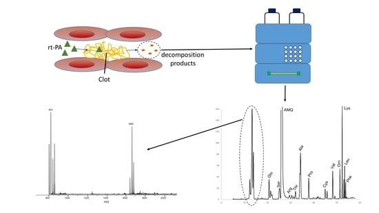
Plasma samples obtained from stroke patients treated with recombinant tissue-type plasminogen activator (rt-PA) and not treated with rt-PA were evaluated with different HPLC methodologies to obtain information about the possible release of small molecules as a result of the thrombolytic treatment. Plasma samples, without derivatization and derivatized with 6-aminoquinolyl-N-hydroxysuccinimidyl carbamate (AQC), were evaluated with a HPLC gradient method, which consisted of a mobile phase of 10 mM ammonium acetate buffered solution (pH = 5.3) and acetonitrile. Three different detection methods were applied: UV, fluorescence, and ESI-MS. The results obtained showed that a group of new highly hydrophilic compounds appeared in most samples analyzed from treated patients, just after the administration of rt-PA. These compounds appeared shortly after the administration of the drug and were detected during the first 24 h after treatment, disappearing from plasma after this time. These new compounds were not detected either in controls or in non-treated stroke patients, which suggests that they were released into the plasma as a consequence of the thrombolytic effect of the drug. Our results suggest that these new compounds might be free glycans. The use of AQC as a derivatizing reagent has demonstrated that the new compounds detected cannot contain primary or secondary amine groups in their structure. The molecular mass determined by ESI-MS (821 Da) suggests that if these compounds are free glycans they might be a high-mannose type.

Graphical Abstract

1. Introduction

Human tissue plasminogen activator (t-PA) is a glycosylated serine protease with three N-linked glycosylation sites (Asn117, Asn184, and Asn448) [1]. Type I t-PA is glycosylated at the three positions, whereas type II lacks a carbohydrate at position 184 [1]. As other glycoproteins, t-PA can be manufactured using biotechnology techniques as recombinant tissue-type plasminogen activator (rt-PA). Thrombolysis with intravenous rt-PA, given within the first 4.5 h of onset, is currently the most commonly administered fibrinolytic medical therapy for patients with acute ischemic stroke [2,3].
Recombinant biological drugs are produced from nonhuman cells but must have a human-like glycosylation pattern [4]. This is important because protein–carbohydrate interactions play a participatory role in many processes affecting disease progression [5]. In the case of both rt-PA and native t-PA, the structure of the Asn-linked oligosaccharides carries primarily high-mannose and hybrid type glycans at Asn117, while more complex type N-glycans are most abundant at Asn184 and Asn448 [1,6,7,8,9]. In a recent study, it was found that these glycans may also contain mannose 6-phosphate at the Asn448 in both recombinant and native t-PA [8]. Glycosylation is one of the most complex and common protein posttranslational modifications, linking carbohydrates to proteins, that exhibits significant roles in protein functions [10]. Characterization of a protein’s glycans after they are released from glycoproteins is very common, and different methodologies, based on high-performance liquid chromatography (HPLC), mass spectrometry (MS), and LC-MS, have been developed to facilitate a glycoscience study [11]. Usually, the analysis of glycans involves preliminary cleavage of these compounds at the protein glycosylation site using chemical/chemo-enzymatic methodologies, followed by subsequent derivatization with fluorescent labels for fluorescence detection, due to the lack of an exploitable chromophore in natural glycans and the anomeric mutual rotation at the reducing end [11,12,13]. Probably, the most common derivatization methodology applied before HPLC analysis for free-reducing glycans is reductive amination with fluorescent amines, being 2-aminopyridine (2-AP), 2-aminobenzamide (2-AB), 2-aminobenzoic acid, or anthranilic acid (2-AA), the most common fluorescent amines used. However, reductive amination also breaks the reducing end ring structure, which affects the glycan structural integrity. Permethylation is also a common derivatization method, where glycan hydrogens are replaced with methyl groups to increase hydrophobicity of the glycan and allowing efficient separation on a reversed phase C18 column [14]. However, both derivatization methodologies can be interfered by detergents and salts during protein denaturation, requiring extra purification steps before derivatization [15]. For these reason, other derivatization reagents, such as 6-aminoquinolyl-N-hydroxysuccinimideyl carbamate (AQC), have been proposed. AQC derivatization is a different derivatization mechanism as this reagent binds to free amino groups, as the alpha-amino group of the N-terminal amino acid (N-acetylglucosamine monomeric unit). This derivatization reagent presents the advantage that is not interfered by salts or detergents [16].
Each type of derivatization induces different properties and modifies glycan retention on different chromatographic columns. Glycans can be separated using reversed phase, normal phase, and weak anion exchange columns and also with HILIC columns, except for the permethylated glycans [10]. Reversed phase chromatography is usually applied for derivatized glycans [17], but, in the case of native glycans, they cannot be efficiently retained on these types of columns, especially when the molecule is small and contains a high number of hydrophilic monomeric units [18]. Zhuo et al. described unexpected peak splitting for Man8 and Man9 due to weak interaction between the glycan and stationary phase [10].
The primary function of rt-PA in the treatment of ischemic stroke patients includes catalyzing the proteolytic conversion of plasminogen into plasmin, the primary enzyme involved in degrading insoluble fibrin to soluble by-products [19,20]. However, the use of rt-PA can also be associated with secondary effects that it can exacerbate brain edema and hemorrhage through a plasmin-mediated extrinsic pathway [21]. Different studies have demonstrated that rt-PA treatment changes the plasma degradomic profile in acute ischemic stroke patients, which usually differs from patients not receiving rt-PA [22,23]. Such reactivity suggests that during the fibrin assembly process fibrin(ogen) undergoes conformational changes, resulting in the exposure of its multiple interaction sites and modulation of various activities. The exposure of rt-PA and plasminogen-binding sites on fibrin represents a clear example demonstrating the importance of such conformational changes in the regulation of fibrinolysis [24]. Plasmin activity in vivo is restricted mainly to places of fibrin deposition, allowing effective dissolution of fibrin without proteolytic damage of other proteins, which occurs through interactions between newly exposed binding sites on fibrin and complementary sites of t-PA and plasminogen, resulting in the activation of plasminogen by t-PA and the localization of fibrinolysis [24].
The potentiation of fibrinogenolysis in the presence of fibrin is mediated by soluble fibrin degradation products (FDP), which are responsible for the ongoing fibrinogenolysis. FDP stimulates fibrinogenolysis by binding t-PA and plasminogen. It has been found that rt-PA-induced degradation of cross-linked fibrin can potentiate rt-PA-mediated fibrinogenolysis by providing a surface for t-PA and plasminogen binding, thereby promoting plasmin generation [25].
Taking into account these considerations, it is thought that some type of small soluble molecules might be released into blood due to the thrombolytic treatment. Some studies have confirmed that glycoproteins in cytosol may release significant amounts of soluble free glycans [26,27], which seems to be mainly derived from high mannose-type glycans. However, little study has been undertaken regarding their presence in human serum samples [28]. Therefore, the objective of this study is to assess whether liquid chromatography techniques might be useful in detecting whether small soluble molecules are formed in plasma samples obtained from ischemic stroke patients as a result of rt-PA treatment.

2. Materials and Methods

2.1. Subjects

A total of 108 patients with acute ischemic stroke within the first 4.5 h of evolution were included in the study. Of these, 88 patients (mean age 71 ± 13 years, 51% men) received rt-PA (between 22 and 290 min after beginning of stroke symptomatology; mean = 167 min, SD = 55 min), and 20 (77 ± 14 years, 60% men) did not receive the treatment due to different exclusion criteria for the administration of the drug. Forty healthy subjects were also included in the study as controls.
Clinical and neurological examination, as well as blood and coagulation tests were carried out at arrival at the emergency department. For the purposes of this study, neurological deficit was evaluated and quantified by certified neurologists using the National Institutes of Health Stroke Scale (NIHSS) score at admission (rt-PA-treated patients: mean = 12.9 ± 6.0, median = 13.0, interquartile range = 7.5–18.5; non-treated patients: mean = 10.6 ± 7.1, median = 12.0, interquartile range = 4.0–17.0) and at 24 h of evolution. In order to determine the possible association between the appearance of new compounds in plasma and the thrombolytic effect of rt-PA, early neurological improvement, which was defined as a decrease of ≥4 points on the NIHSS score at 24 h compared with the baseline assessment [29], was considered as a surrogate marker of this thrombolytic effect. All subjects, or their relatives when subjects were unable to sign for themselves, gave their informed consent to participate in the study. The study was conducted in accordance with the Declaration of Helsinki, and the protocol was approved by the Ethics Committee.

2.2. Blood Sample Collection

Once obtained at arrival at the emergency department, blood samples were collected in EDTA BD Vacutainer tubes. Immediately after drawing, blood samples were processed to obtain plasma, aliquoted, and preserved at −80 °C until analysis.
For patients receiving thrombolytic treatment, blood samples were collected at admission (pre-rt-PA: time 0), and at different times post-rt-PA administration: 2 h (time 1), 6 h (time 2), 24 h (time 3), and 72 h (time 4) after rt-PA. For patients who did not receive thrombolytic treatment, blood samples were collected at admission (time 0), and 2 h (time 1) and 6 h (time 2) after admission. In treated patients, the rt-PA dose applied was 0.9 mg rt-PA/kg body weight (not to exceed 90 mg total treatment dose), infused over 60 min.

2.3. Chromatographic Analysis

As the main objective of this study was to assess whether new small soluble molecules were detected in blood after rt-PA treatment, plasma samples were first ultrafiltrated (UF) through 3 kDa centrifugal filters (Amicon Ultra-0.5 Centrifugal Filter Unit, Merck, Cork, Ireland), which limits the size of molecules that can be detected with the proposed methodology. This avoids the interference of proteins and other macromolecules in the chromatographic analyses. The purification of plasma samples is a significant issue in this type of matrix. When proteins are eliminated from the sample some of the less hydrophilic compounds may be bonded to proteins and be eliminated together with this fraction. UF was chosen in our case as previous studies have demonstrated that UF results in lower losses by adsorption on proteins that those due to co-precipitation with proteins using conventional protein precipitation methodologies using organic solvents or organic acids [30,31,32,33]. In the conditions applied, free amino acids present in plasma can also be analyzed with the proposed methodology when samples are derivatized with 6-aminoquinolyl-N-hydroxysuccinimidyl carbamate (AQC) [30].
As other studies have shown that glycoproteins in cytosol may release significant amounts of soluble glycans [26,27], a preliminary hypothesis was that free glycans could be released into circulating blood during the thrombolytic process. Taking into account that N-glycosylation (attachment of N-acetylglucosamine, GlcNAc, to the nitrogen atom of an Asn side chain by a β-1N linkage) is common in glycoproteins, it could be expected that the linked conjugates contain a GlcNAc2 mannose (Man) core [34]. Therefore, pre-column derivatization of samples with AQC (AccQ·Tag derivatization kit, Waters Corp., Milford, MA, USA) was applied as this reagent enhances the sensitivity in the analysis of this type of N-glycans [35] and yields highly stable N-glycan derivatives, through urea linkage, within 5 min reaction under ambient conditions [16,35,36].
HPLC analysis of both derivatized and non-derivatized plasma samples was achieved using a Gemini-NX C18 column (250 mm × 4.6 mm, 5 μm) (Phenomenex, Torrance, CA, USA) thermostated at 40 °C. Two HPLC instruments were used: (i) a 1260 Infinity II LC System (Agilent Technologies, Santa Clara, CA, USA) with UV (detection at 254 nm) and fluorescence (FL, excitation at 250 nm and emission at 395 nm) detectors, and (ii) a 1200 LC System (Agilent Technologies, Santa Clara, CA, USA) coupled to an Esquire 6000 Ion Trap Mass Spectrometer (Bruker, Billerica, MA, USA).
A gradient elution with a binary mobile phase system was used to separate the compounds at a constant flow rate of 1.0 mL·min−1. Mobile phase A consisted of a buffer solution containing 10 mM ammonium acetate (pH = 5.3 ± 0.1, adjusted with TFA), while solution B consisted of acetonitrile/water (60:40, v/v). All solutions were filtered through 0.45 μm filters (Whatman, Buckinghanshire, UK) and degassed before use. In order to have the maximum separation power and information about the possible presence of hydrophilic compounds, the gradient applied started with a 100% aqueous solution and increased slowly to 6% acetonitrile over 25 min. For this reason, the gradient applied started at 100% A and was brought down as follows: 98% at 0.5 min, 93% at 15 min, 90% at 25 min, 74% at 45 min, 67% at 48 min, and 0% at 63 min where it was maintained for 7 min. A reconditioning time of 15 min with 100% solution A was needed between samples in order to obtain reproducible results with the proposed HPLC methodology.

3. Results

After analyzing derivatized plasma samples with HPLC-UV-FL, no qualitative differences were observed between samples obtained pre-rt-PA administration in the group of patients treated with this drug and those obtained from controls and non-treated patients. In all these samples, only the presence of derivatized amino acids was detected (group of peaks detected at retention times between 16 and 60 min in Figure 1a,b). However, at the first sampling point post-rt-PA (2 h after administration), a new group of peaks was observed at the beginning of the chromatograms in 70 (80%) out of the 88 patients who received thrombolysis. These new peaks were not detected using fluorescence detection (Figure 1b). A second HPLC analysis of the same plasma samples without preliminary derivatization confirmed that these peaks did not correspond to derivatized compounds, as the same new group of peaks was also found with the non-derivatized solutions using the UV detector (Figure 1c).
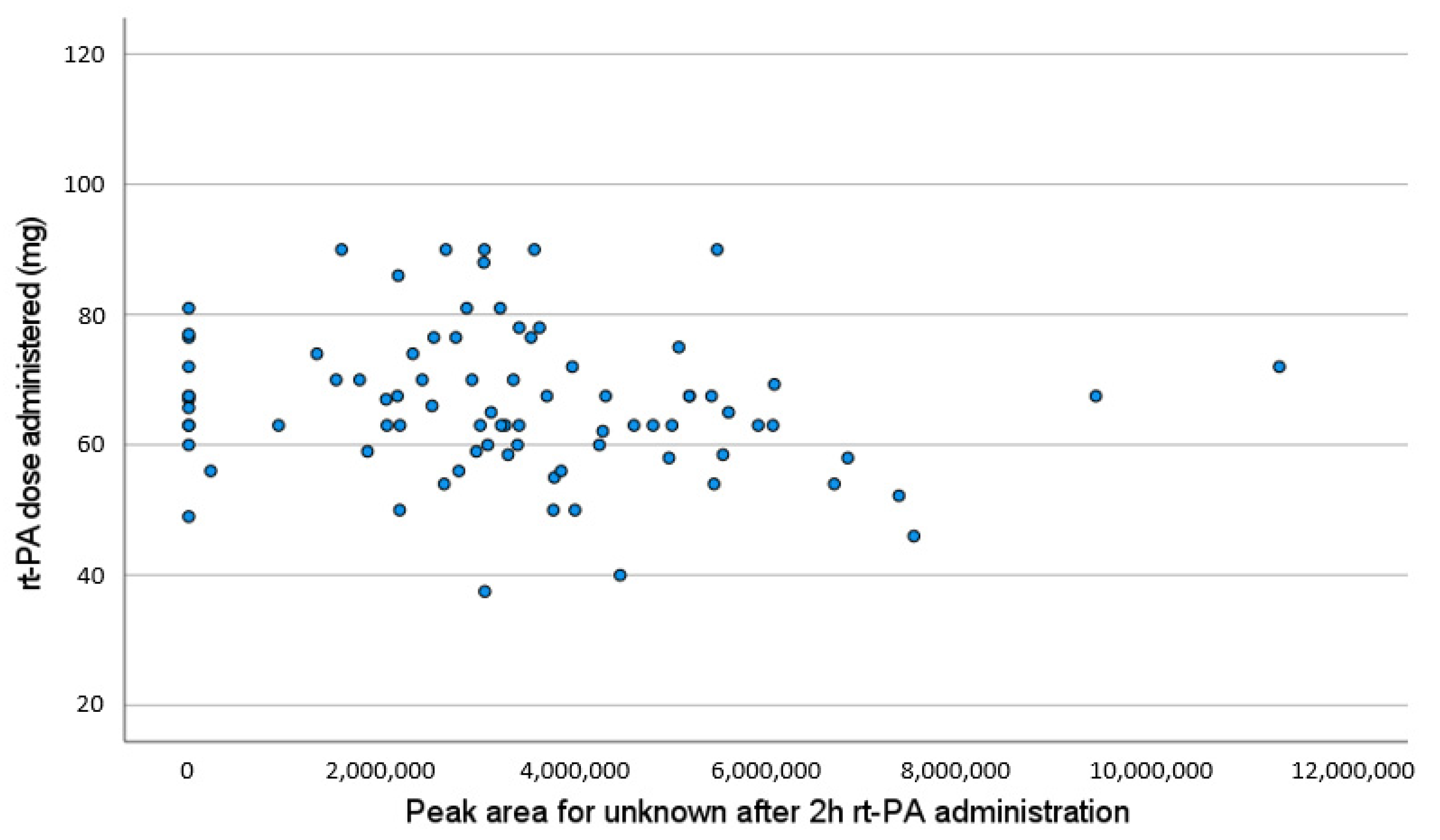
In the group of patients in whom we observed the new peaks just after rt-PA administration, the presence of these compounds was detected for a maximum of 24 h after treatment, and none were detected at time 4 (72 h after rt-PA). As can be seen from the time evolution profiles showed in Figure 2, the new compounds appeared shortly after rt-PA administration. In some cases, the maximum level was reached at the first sampling point taken after the treatment (2 h after), but in others, the maximum appeared after 6 h. Thereafter, there was always a decrease in the amounts detected, and they were never found after >24 h of treatment. No significant correlation was observed between the amount of the new compounds and the dose of rt-PA administered (median dose of rt-PA administered = 63.0 mg, interquartile range = [58.0,71.5]) (Figure 3, r = −0.178, p = 0.103).
In the group of patients that did not receive thrombolytic treatment, these new peaks were never detected at any of the different sampling points evaluated. In both this group of patients and the control group, only peaks corresponding to derivatized amino acids were observed.
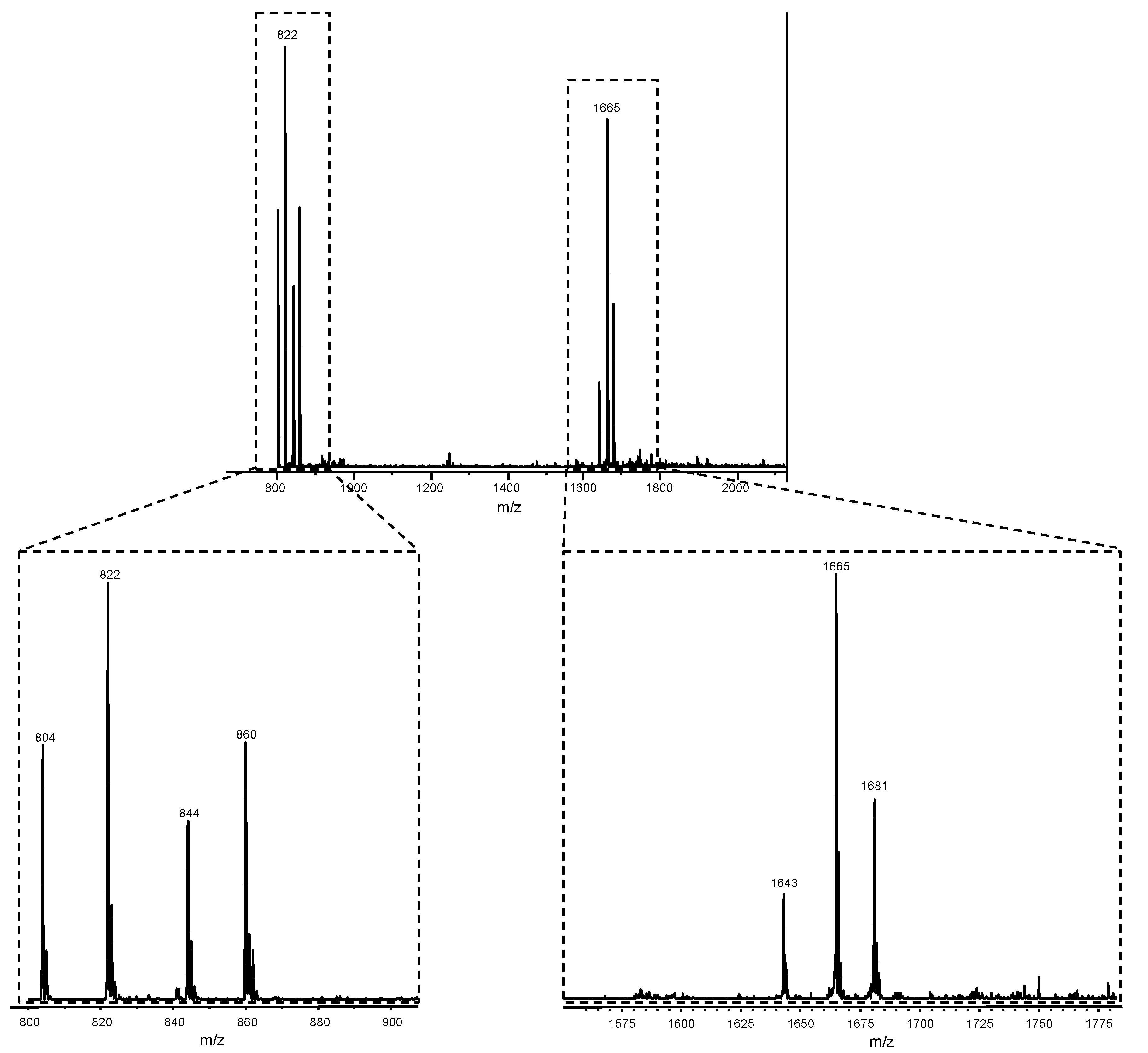
In order to obtain more information about the possible structure of the newly detected compounds, derivatized plasma samples were also analyzed by HPLC with electrospray ionization-mass spectrometry (ESI-MS) detection. The MS results confirmed that the peaks detected between 16 and 60 min were the result of derivatized amino acids with the AQC reagent. The positive ion mode analysis of the new peaks detected after rt-PA administration showed the same mass spectra for all these peaks (Figure 4), with a main peak at m/z = 822, which could be associated to the protonated molecular ion [M+H]+. Other peaks were found at m/z = 844 ([M+Na]+), 860 ([M+K]+), and 804 (possible loss of a water molecule from the protonated molecular ion). A second group of peaks was observed at higher masses, which could be attributed to a dimmer formed during ionization: m/z = 1643 ([2M+H]+), 1665 ([2M+Na]+), and 1681 ([2M+K]+). The peak observed at m/z = 804 can be associated to a loss of a water molecule from the protonated molecular ion. The presence of this peak suggests that these compounds may be a type of carbohydrate since protonated underivatized carbohydrates can easily be decomposed through a dehydration reaction in ESI-MS [37]. Moreover, analyses by MS/MS confirmed that the fragmentation of the ion at m/z = 822 yields an ion at m/z = 804, due to the loss of a water molecule. The ESI-MS in negative mode only presented a peak at m/z = 934, which may be attributed to [M+TFA]-, with a molecular mass of 821 Da. These results suggest that all new compounds have the same molecular ion with 821 Da, and the detection of different peaks may be due to the presence of different isomers of this compound.
A second set of experiments was performed using a more concentrated buffer solution as mobile phase A (50 mM ammonium acetate). With this mobile phase, the same MS spectra was obtained in positive mode, plus a new peak at m/z = 839, which may be associated to [M+NH4]+ for the 821 Da molecular ion.
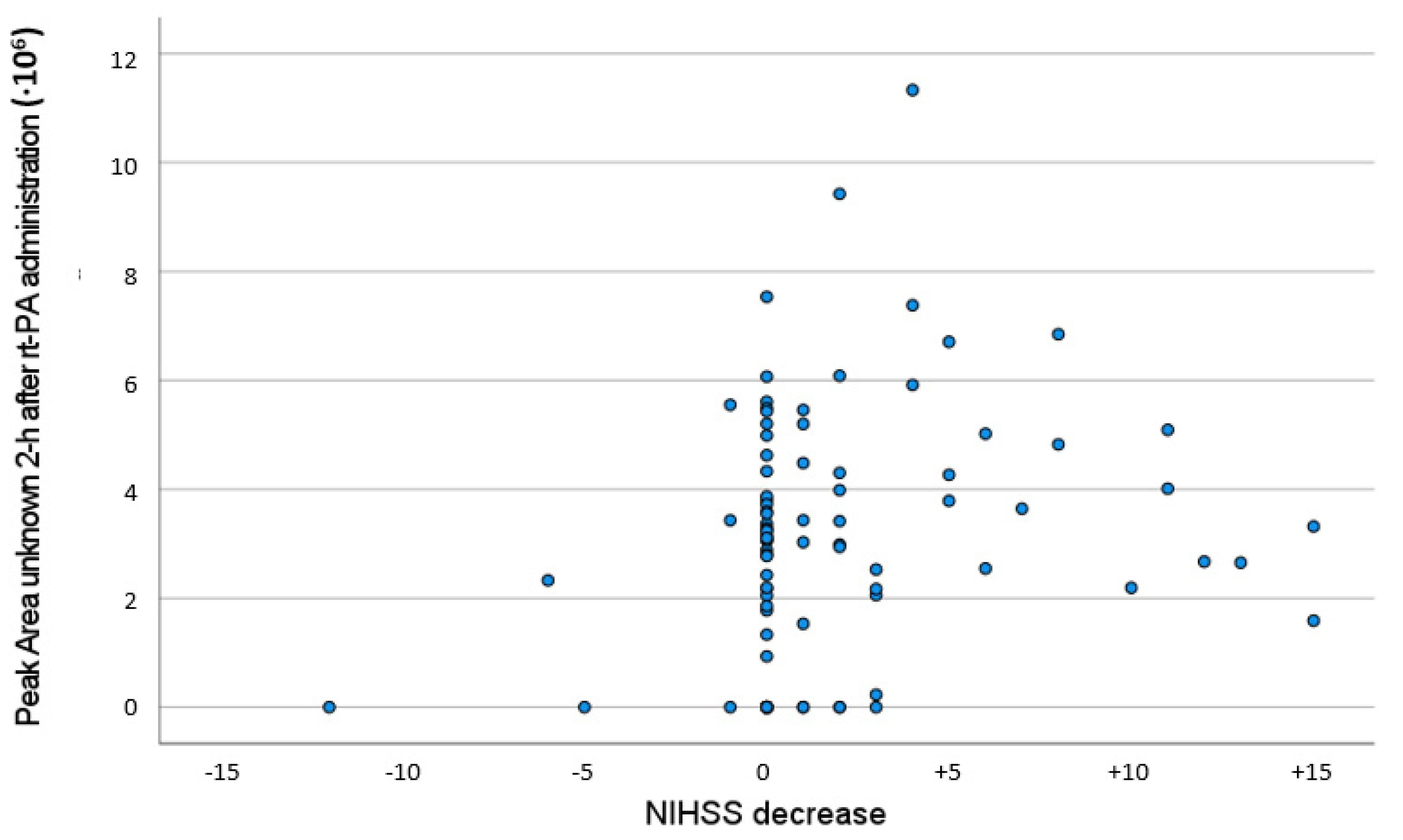
A significant correlation was found between the concentration of new compounds and the variation between NIHSS at 24 h and baseline (Figure 5, r = 0.216, p = 0.041). With regards to the association between the presence of new compounds in plasma and the neurological evolution of patients, 49 (70%) rt-PA-treated patients had early neurological improvement, whereas 21 (30%) did not (p = 0.026).

4. Discussion

New compounds detected in plasma samples after rt-PA administration could be associated to some of the components present in the recombinant drug other than the active ingredient (i.e., arginine, phosphoric acid, and polysorbate 80). However, different facts suggest that this in fact is not the case and rather that they might be the result of the interaction of the rt-PA with the clot. First, MS results are clearly not related to any of these compounds. In this respect, all samples from patients treated with rt-PA showed there was a significant increase in plasma arginine levels after rt-PA administration. This amino acid is necessary to help to solubilize rt-PA (3.5 g Arg/100 mg rt-PA), and so, high levels of this amino acid are administered to the patients receiving this treatment (up to 3.15 g of Arg) [38,39]. Second, the new peaks detected appeared in 70 of the treated patients (80%) but were not detected in the other 18 (20%), which suggests that these compounds are not present in the drug itself and supports the hypothesis that they may be the result of some type of interaction of the drug with the clot. Moreover, no significant correlation was obtained between the amount of the new compounds (determined from the peak area obtained) and the dose of rt-PA (Figure 3, r = −0.178, p = 0.103).
The new compounds found after rt-PA administration in the HPLC analyses using a reversed-phase (RP) stationary phase appeared at the front of the chromatograms, before the most polar amino acid peak, when the elution gradient contained >93% of mobile phase A (i.e., <4.2% acetonitrile in the gradient mixture). This suggests that these new compounds are highly hydrophilic, which agrees with the hypothesis that some glycans could be released during the interaction of rt-PA with the fibrin clot. Supporting this, Lareau et al. [40] analyzed glycans with RP-HPLC and found that non-derivatized free glycans elute with the initial aqueous solvent plug in typical RP-HPLC separations. Moreover, they also found that these compounds are not well separated with this chromatographic separation, a fact that was also observed in the present study (Figure 1a).
In our study, the use of two detectors, UV and FL, confirmed that the new peaks found did not correspond to derivatized compounds with AQC, as they were only observed with UV detection (Figure 1). These results suggest that the new compounds detected do not contain any primary or secondary amine in their structure. Therefore, they cannot contain N-acetylhexosamine (HexNAc), N-glycolylneuraminic acid (Neu5Gc), N-acetylneuramic acid (Neu5Ac), or sialic acid groups as all these molecules have amino groups and would have been detected in their derivatized form with AQC with the FL detector. As a consequence, in case that they are glycans, these compounds must be some type of terminal mannose residues. However, non-derivatized mannose residues have low intrinsic UV absorbance due to the lack of chromophore groups.
The study of plasma samples by MS poses additional challenges due to the inherent preference of alkali metal-coordinated compounds in endogenous biological matrices, especially in the case of non-derivatized glycans [40]. Therefore, although compounds are predominately ionized as protonated ions ([M+H]+), alkali metal-coordinated ions ([M+Na]+ or [M+K]+) can also be found in ESI-MS. This could explain the presence of peaks with m/z = 844 ([M+Na]+) and 860 ([M+K]+) in the HPLC-ESI-MS analysis, with a molecular mass of 821 Da. It should be noted that the peak corresponding to the ion [M+NH4]+ (m/z = 839) only appeared when the concentration of ammonium acetate was increased to 50 mM. Another result that suggests that the new compounds are isomers of some polysaccharides is the presence of a peak with a loss of a water molecule from the protonated molecular ion, which is common in the analysis of underivatized carbohydrates by ESI-MS [37].
Unfortunately, it has not been possible to associate the found peaks to any specific molecule. Despite this, some information can be extracted from the results obtained. First, the new molecules found are highly hydrophilic. Second, the molecular mass determined with the ESI-MS results (821 Da) suggests that if these compounds are free glycans, they must be high-mannose type, probably residues containing five hexoses, which always show limited interaction with an RP chromatographic column and are eluted at low percentages of acetonitrile [18,41]. In another study, Pabst et al. [42] analyzed fibrin(ogen) N-glycans from various species, including humans, and found that all fibrins contained diantennary N-glycans. Moreover, on analyzing different isomers of non-derivatized glycans using single ion monitoring MS, they detected α-Gal containing glycans with a total of five hexose residues at a mass/charge ratio of 822. Another study analyzing mixtures of maltose standards also found that Man5 residues appeared at m/z values between 800–850 [40].
Different studies have evaluated mannosidase activity in human serum when investigating the clearance of different glycoforms of a therapeutic antibody [43,44,45]. Chen et al. [43] concluded that the change in clearance rate of high mannose glycoforms was a result of glycan cleavage. Other studies also found that high mannose glycans were cleared faster than other glycans [44,45]. Yakolev et al. [24] found that although plasmin exhibits proteolytic activity toward many plasma proteins, its activity in vivo is restricted mainly to places of fibrin deposition, allowing effective dissolution of fibrin without proteolytic damage of other proteins. It was reported that this natural adaptation occurs through a number of orchestrated interactions between newly exposed binding sites on fibrin and complementary sites on t-PA and plasminogen, resulting in activation of plasminogen by t-PA and localization of fibrinolysis.
Some studies have analyzed glycan groups that are linked to glycosylated proteins in serum samples, through cleavage of oligosaccharides from glycosylated proteins, finding significant differences in the glycan patterns when comparing data from healthy subjects and patients [46,47,48,49,50,51].

5. Possible Clinical Implications

The lack of correlation between the detection of these new compounds and the dose of rt-PA administered, together with the fact that patients who did not receive thrombolytic treatment never presented these compounds, suggests that the formation of these compounds is not the result of an rt-PA dose-effect but the result of the rt-PA thrombolytic effect. Our study demonstrates a significant association between the presence of new compounds and the frequency of early neurological improvement, which may be related to thrombus dissolution and the corresponding clinical effects as a results of the rt-PA thrombolytic effect.

6. Conclusions

The results obtained in the present study show, for the first time, that new hydrophilic compounds are released into plasma after rt-PA administration to stroke patients. Despite not having been possible to associate the peaks found to specific molecules, the evolution of patients in whom these new compounds have been found allows us to hypothesize that they are released into plasma as a consequence of the thrombolytic effect of rt-PA. The molecular mass determined by ESI-MS (821 Da) suggests that if these compounds are free glycans, they might be a high-mannose type. However, more studies are needed to characterize these compounds and the mechanisms involved in their appearance in the plasma of rt-PA-treated stroke patients.

Author Contributions

Conceptualization, J.M.S. and M.C.; Methodology, J.M.S. and M.C.; Samples Collection, C.G.-M., D.F.-C., M.R.-Y. and M.J.F.-P.; Neurological Evaluation, M.C., M.R.-Y., D.F.-C. and M.J.F.-P.; Laboratory Analyses, J.M.S., C.G.-M. and A.D.S.-C.; Resources, M.C.; Data Interpretation, J.M.S. and M.C.; Writing–Original Draft Preparation, J.M.S. and M.C.; Writing–Review and editing, J.M.S., D.F.-C., M.J.F.-P., C.G.-M. and M.C.; Funding Acquisition, J.M.S. and M.C. All authors have read and agreed to the published version of the manuscript.

Funding

This work was supported by grants from the Instituto de Salud Carlos III (PI17/02123 and RD21/0006/0017). A. Da Silva-Candal was the recipient of a Sara Borrell grant (CD20/00054).

Institutional Review Board Statement

The study was conducted in accordance with the Declaration of Helsinki, and approved by the Ethics Committee of Comité Autonómico de Ética de la Investigación de Galicia (protocol code 2017/368, 11 July 2017).

Informed Consent Statement

Informed consent was obtained from all subjects involved in the study.

Data Availability Statement

Data from this study are available on request to the principal investigator of the funding supporting studies, Mar Castellanos.

Conflicts of Interest

The authors declare no conflict of interest.

References

  1. Chevilley, A.; Lesept, F.; Lenoir, S.; Ali, C.; Parcq, J.; Vivien, D. Impacts of tissue-type plasminogen activator (tPA) on neuronal survival. Front. Cell Neurosci. 2015, 9, 415. [Google Scholar] [CrossRef] [PubMed] [Green Version]
  2. Hacke, W.; Kaste, M.; Bluhmi, E.; Brozman, M.; Dávalos, A.; Guidetti, D.; Larrue, V.; Lees, K.R.; Medeghri, A.; Machnig, T.; et al. ECASS Investigators. Thrombolysis with alteplase 3 to 4.5 hours after acute ischemic stroke. N Engl. J. Med. 2008, 359, 1317–1329. [Google Scholar] [CrossRef] [PubMed] [Green Version]
  3. Powers, W.J.; Rabinstein, A.A.; Ackerson, T.; Adeoye, O.M.; Bambakidis, N.C.; Becker, K.; Biller, J.; Brown, M.; Demaerschalk, B.M.; Hoh, B.; et al. Guidelines for the early management of patients with acute ischemic stroke: 2019 update to the 2018 guidelines for the early management of acute ischemic stroke: A guideline for healthcare professionals from the American Heart Association/American Stroke Association. Stroke 2019, 50, e344–e418. [Google Scholar] [PubMed]
  4. Leymarie, N.; Zaia, J. Effective use of mass spectrometry for glycan and glycopeptide structural analysis. Anal. Chem. 2012, 84, 3040–3048. [Google Scholar] [CrossRef] [Green Version]
  5. Wood, C.L.; Maxon, M.; Desaire, H. Software for automated interpretation of mass spectrometry data from glycans and glycopeptides. Analyst 2013, 138, 2793–2803. [Google Scholar]
  6. Itoh, S.; Kawasaki, N.; Ohta, M.; Hyuga, M.; Hyuga, S.; Hayakawa, T. Simultaneous microanalysis of N-linked oligosaccharides in a glycoprotein using microbore graphitized carbon column liquid chromatography-mass spectrometry. J. Chromatogr. A 2002, 968, 89–100. [Google Scholar] [CrossRef]
  7. Papac, D.I.; Briggs, J.B.; Chin, E.T.; Jones, A.J.S. A high-throughput microscale method to release N-linked oligosaccharides from glycoproteins from matrix-assisted laser desorption/ionization time-of-flight mass spectrometric analysis. Glycobiology 1998, 8, 445–454. [Google Scholar] [CrossRef] [Green Version]
  8. Miller, J.J.; Bohnsack, R.N.; Olson, L.J.; Ishihara, M.; Aoki, K.; Tiemeyer, M.; Dahms, N.M. Tissue plasminogen activator is a ligand of cation-independent mannose 6-phosphate receptor and consists of glycoforms that contain mannose 6-phospate. Sci. Rep. 2021, 11, 8213. [Google Scholar] [CrossRef]
  9. Spellman, M.W.; Basa, L.J.; Leonard, C.K.; Chakel, J.A.; O’Connor, J.V.; Wilson, S.; van Halbeek, H. Carbohydrate structures of human tissue plasminogen activator expressed in Chinese hamster ovary cells. J. Biol. Chem. 1989, 264, 14100–14111. [Google Scholar] [CrossRef]
  10. Zhuo, S.; Veillon, L.; Dong, X.; Huan, Y.; Mechref, Y. Direct comparison of derivatization strategies for LC-MS/MS analysis of N-glycans. Analyst 2017, 142, 4446–4455. [Google Scholar] [CrossRef]
  11. Zhang, Q.; Li, Z.; Song, X. Preparation of complex glycans from natural sources for functional study. Front. Chem. 2020, 8, 508. [Google Scholar] [CrossRef] [PubMed]
  12. Yang, S.; Zhang, H. Glycomic analysis of glycans released from glycoproteins using chemical immobilization and mass spectrometry. Curr. Protoc. Chem. Biol. 2015, 6, 191–208. [Google Scholar] [CrossRef] [PubMed] [Green Version]
  13. Wilkinson, H.; Saldova, R. Current methods for the characterization of O-glycans. J. Proteom. Res. 2020, 19, 3890–3905. [Google Scholar] [CrossRef] [PubMed]
  14. Zhou, S.; Wooding, K.; Mechref, Y. Analysis of permethylated glycan by liquid chromatography (LC) and mass spectrometry (MS). Methods Mol. Bio. 2017, 1503, 83–96. [Google Scholar]
  15. Parker, N.H.; Lawson, M.A.; Jardine, D.R.; Redmond, J.W. A general approach to desalting oligosaccharides released from glycoproteins. Glycoconj. J. 1998, 15, 737–747. [Google Scholar]
  16. Wu, Y.; Sha, Q.; Du, J.; Wang, C.; Zang, L.; Liu, B.F.; Lin, Y.; Liu, X. Determination of N-glycans by high performance liquid chromatography using 6-aminoquinolyl-N-hydroxysuccinimidyl carbamate as the glycosylamine labeling reagent. J. Chromatogr. A 2018, 1535, 114–122. [Google Scholar] [CrossRef]
  17. Vreeker, G.C.M.; Wuhrer, M. Reversed-phase separation methods for glycan analysis. Anal. Bioanal. Chem. 2017, 409, 359–378. [Google Scholar] [CrossRef] [Green Version]
  18. Kolarich, D.; Jensen, P.H.; Altmann, F.; Packer, N.H. Determination of site-specific glycan heterogeneity on glycoproteins. Nat. Protoc. 2012, 7, 1285–1289. [Google Scholar] [CrossRef]
  19. Gurman, P.; Miranda, O.R.; Nathan, A.; Washington, C.; Rosen, Y.; Elman, N.M. Recombinant tissue plasminogen activators (rtPA): A review. Clin. Pharmacol. Ther. 2015, 97, 274–285. [Google Scholar] [CrossRef]
  20. Jilani, T.N.; Siddiqui, A.H. Tissue plasminogen activator. In StatPearls; StatPearls Publishing: Treasure Island, FL, USA, 2022. Available online: https://www.ncbi.nlm.nih.gov/books/NBK507917/ (accessed on 28 November 2022).
  21. Zhao, X.J.; Larkin, T.M.; Lauver, M.A.; Ahmad, S.; Ducruet, A.F. Tissue plasminogen activator mediates deleterious complement cascade activation in stroke. PLoS ONE 2017, 12, e0180822. [Google Scholar] [CrossRef] [Green Version]
  22. Ning, M.M.; Sarracino, D.A.; Buonano, F.S.; Krastins, B.; Chou, S.; McMullin, D.; Wang, X.; Lopez, M.; Lo, E.H. Proteomic protease substrate profiling of tPA treatment in acute ischemic stroke patients: A step toward individualizing thrombolytic therapy at the bedside. Transl. Stroke Res. 2010, 1, 268–275. [Google Scholar] [CrossRef] [PubMed] [Green Version]
  23. Dagonnier, M.; Cooke, I.R.; Faou, P.; Sidon, T.K.; Dewey, H.M.; Donnan, G.A.; Howells, D.W. Discovery and longitudinal evaluation of candidate biomarkers for ischaemic stroke by mass spectrometry-based proteomics. Biomark. Insights 2017, 12, 1177271917749216. [Google Scholar] [CrossRef] [PubMed] [Green Version]
  24. Yakolev, S.; Makogonenko, E.; Kurochkina, N.; Nieuwwnhuizen, W.; Ingham, K.; Medved, L. Conversion of fibrinogen to fibrin: Mechanism of exposure of tPA- and plasminogen-binding sites. Biochemistry 2000, 39, 15730–15741. [Google Scholar] [CrossRef] [PubMed]
  25. Weitz, J.I.; Leslie, B.; Ginsberg, J. Soluble fibrin degradation products potentiate tissue plasminogen activator-induced fibrinogen proteolysis. J. Clin. Investig. 1991, 87, 1082–1090. [Google Scholar] [CrossRef] [Green Version]
  26. Kmiécik, D.; Herman, V.; Stroop, C.J.M.; Michalski, J.C.; Mir, A.M.; Labiau, O.; Verbert, A.; Cacan, R. Catabolism of glycan moieties of lipid intermediates leads to a single Man5GLcNAc oligosaccharide isomer: A study with permeabilized CHO cells. Glycobiology 1995, 5, 483–494. [Google Scholar] [CrossRef]
  27. Yanagida, K.; Natsuka, S.; Hase, S. Structural diversity of cytosolic free oligosaccharides in the human hepatoma cell line, HepG2. Glycobiology 2006, 16, 294–304. [Google Scholar] [CrossRef] [Green Version]
  28. Iwatsuka, K.; Watanabe, S.; Kinoshita, M.; Mamisue, K.; Yamada, D.; Hayakawa, T.; Suzuki, T.; Kakehi, K. Free glycans derived from glycoproteins present in human sera. J. Chromatogr. B Analyt. Technol. Biomed. Life Sci. 2013, 928, 16–21. [Google Scholar] [CrossRef]
  29. National Institute of Neurological Disorders and Stroke rt-PA Stroke Study Group. Tissue plasminogen activator for acute ischemic stroke. N. Engl. J. Med. 1995, 333, 1581–1587. [Google Scholar] [CrossRef]
  30. Castellanos, M.; van Eendeburg, C.; Gubern, C.; Sanchez, J.M. Ethyl-bridged hybrid column as an efficient alternative for HPLC analysis of plasma amino acids by pre-column derivatization with 6-aminoquinolyl-N-hydroxysuccinimidyl carbamate. J. Chromatogr. B Analyt. Technol. Biomed. Life Sci 2016, 1029, 137–144. [Google Scholar] [CrossRef]
  31. Liu, H.; Worthen, H.G. Measurement of free amino acid level in ultrafiltrates of blood plasma by high-performance liquid chromatography with automated pre-column derivatization. J. Chromatogr. B 1992, 579, 215–224. [Google Scholar] [CrossRef]
  32. Ralston, P.B.; Strein, T.G. A study of deproteinization methods for subsequent serum analysis with capillary electrophoresis. Microchem. J. 1997, 55, 270–283. [Google Scholar] [CrossRef]
  33. Jaworska, M.; Stanczyk, M.; Wilk, M.; Klaczkow, G.; Anuszewska, E.; Barzal, J.; Rzepecki, P. New approach for amino acid profiling in human plasma by selective fluorescence derivatization. Amino Acids 2012, 43, 1653–1661. [Google Scholar] [CrossRef] [PubMed]
  34. Reily, C.; Stewart, T.J.; Renfrow, M.B.; Novak, J. Glycosylation in health and disease. Nat. Rev. Nephrol. 2019, 15, 346–355. [Google Scholar] [CrossRef] [PubMed]
  35. Laubert, M.A.; Yu, Y.Q.; Brousmiche, D.W.; Hua, Z.; Koza, S.M.; Magnelli, P.; Guthrie, E.; Taron, C.H.; Fountain, K.J. Rapid preparation of released N-glycans for HILIC analysis using a labelling reagent that facilitates sensitive fluorescence and ESI-MS detection. Anal. Chem. 2015, 87, 5401–5409. [Google Scholar] [CrossRef] [PubMed]
  36. Ullmer, R.; Plemati, A.; Rizzi, A. Derivatization by 6-aminoquinolyl-N-hydroxysuccinimidyl carbamate for enhancing the ionization yield of small peptides and glycopeptides in matrix-assisted laser desorption/ionization and electrospray ionization mass spectrometry. Rapid. Commun. Mass. Spectrom. 2006, 20, 1469–1479. [Google Scholar] [CrossRef]
  37. Chen, J.L.; Lee, C.; Lu, I.C.; Chien, C.L.; Lee, Y.T.; Hu, W.P.; Ni, C.K. Theoretical investigation of low detection sensitivity for underivatized carbohydrates in ESI and MALDI. J. Mass. Spectrom. 2016, 51, 1180–1186. [Google Scholar] [CrossRef]
  38. Hovest, A.S.; Horne, M.K. The effect of arginine on coagulation and fibrinolysis in vitro. Fibrinolysis Proteol. 1999, 13, 31–34. [Google Scholar] [CrossRef]
  39. Arakawa, T.; Tsumoto, K.; Kita, Y.; Chang, B.; Ejima, D. Biotechnology applications of amino acids in protein purification and formulations. Amino Acids 2007, 33, 587–605. [Google Scholar] [CrossRef]
  40. Lareau, N.M.; May, J.C.; McLean, J.A. Non-derivatized glycan analysis by reverse phase liquid chromatography and ion mobility-mass spectrometry. Analyst 2015, 140, 3335–3388. [Google Scholar] [CrossRef] [Green Version]
  41. Kawasaki, N.; Ohta, M.; Hyuga, S.; Hashimoto, O.; Hayakawa, T. Analysis of carbohydrate heterogeneity in a glycoprotein using liquid chromatography/mass spectrometry and liquid chromatography with tandem mass spectrometry. Anal. Biochem. 1999, 269, 297–303. [Google Scholar] [CrossRef]
  42. Pabst, M.; Bondili, J.S.; Stadlmann, J.; Mach, L.; Altmann, F. Mass + retention time = structure: A strategy for the analysis of N-glycans by carbon LC-ESI-MS and its application to fibrin N-glycans. Anal. Chem. 2007, 79, 5051–5057. [Google Scholar] [CrossRef] [PubMed]
  43. Chen, X.; Liu, Y.D.; Flynn, G.C. The effect of Fc glycan forms on human IgG2 antibody clearance in humans. Glycobiology 2009, 19, 240–249. [Google Scholar] [CrossRef] [PubMed]
  44. Goetze, A.M.; Liu, Y.D.; Zhang, Z.; Shah, B.; Lee, E.; Bondarendi, P.V.; Flynn, G.C. High-mannose glycans on the Fc region of therapeutic IgG antibodies increase serum clearance in humans. Glycobiology 2011, 21, 949–959. [Google Scholar] [CrossRef] [PubMed]
  45. Yu, M.; Brown, D.; Reed, C.; Chung, S.; Lutman, J.; Stefanich, E.; Wong, A.; Stephan, J.P.; Bayer, R. Production, characterization and pharmacokinetic properties of antibodies with N-linked Mannose-5 glycans. MAbs 2012, 4, 475–487. [Google Scholar] [CrossRef] [PubMed] [Green Version]
  46. An, H.J.; Miyamoto, S.; Lancaster, K.S.; Kirmiz, C.; Li, B.; Lam, K.S.; Leiserowitx, G.S.; Lebrilla, C.B. Profiling of glycans in serum for the discovery of potential biomarkers for ovarian cancer. J. Proteome Res. 2006, 5, 1626–1635. [Google Scholar] [CrossRef]
  47. Kirmiz, B.C.; Li, H.J.; An, B.H.; Clowers, H.K.; Chew, K.S.; Lam, A.; Ferrige, R.; Alecio, A.D.; Borowsky, S.; Sulaimon, C.B.; et al. Miyamoto. A serum glycomics approach to breast cancer biomarkers. Mol. Cell Proteom. 2007, 6, 43–55. [Google Scholar] [CrossRef] [Green Version]
  48. Kyselova, Z.; Mechref, Y.; Kang, P.; Goetz, J.A.; Dobrolecki, L.E.; Sledge, G.W.; Schnaper, L.; Hickey, R.J.; Malkas, L.H.; Novotny, M.V. Breast cancer diagnosis and prognosis through quantitative measurements of serum glycan profiles. Clin. Chem. 2008, 54, 1166–1175. [Google Scholar] [CrossRef] [Green Version]
  49. Isailovic, D.; Kurulugama, R.T.; Plasencia, M.D.; Stokes, S.T.; Kyselova, Z.; Godman, R.; Mechref, Y.; Novotny, M.V.; Clemmer, D.E. Profiling of human serum glycans associated with liver cancer and cirrhosis by IMS-MS. J. Proteome Res. 2008, 7, 1109–1117. [Google Scholar] [CrossRef] [Green Version]
  50. Bereman, M.S.; Williams, T.I.; Muddlman, D.C. Development of a nanoLC LTQ Orbitrap mass spectrometric method for profiling glycans derived from plasma from healthy, benign tumor control, and epithelial ovarian cancer patients. Anal. Chem. 2009, 81, 1130–1136. [Google Scholar] [CrossRef] [Green Version]
  51. Liu, Y.; Wang, C.; Wang, R.; Wu, Y.; Zhang, L.; Liu, B.F.; Cheng, K.; Liu, X. Isomer-specific profiling of N-glycans derived from human serum for potential biomarker discovery in pancreatic cancer. J. Proteom. 2018, 181, 160–169. [Google Scholar] [CrossRef]
Figure 1. HPLC-UV-FL chromatograms obtained in the analysis of a treated patient at time 1 (2 h after rt-PA): (a) Derivatized plasma sample with UV detection at 254 nm, (b) derivatized plasma sample with fluorescence detection, (c) non-derivatized plasma sample with UV detection, and (d) non-derivatized plasma sample with fluorescence detection. The dashed circle marks the new compounds detected. Individual standard solutions of each amino acid were used for the determination of the retention times of these compounds with UV and FL detection. The assignation was confirmed with the ESI-MS results obtained applying the HPLC-MS method for derivatized samples.
Separations 10 00034 g001
Figure 2. Time evolution profiles of the amount of the new compounds detected after the rt-PA administration (determined from the peak area obtained with the UV detector and normalized to the highest peak area measured for each patient). Quality control measurements were daily performed to assure the accuracy and reproducibility for UV and FL measurements. Each colored line corresponds to the results obtained for the same treated patient.
Separations 10 00034 g002
Figure 3. Correlation plot between the amount of new compounds detected and the administered dose of rt-PA (r = −0.178, p = 0.103).
Separations 10 00034 g003
Figure 4. ESI positive-ion mass spectrum of the new compounds detected; those with retention times between 12 and 15 min are shown in Figure 1.
Separations 10 00034 g004
Figure 5. Correlation plot between the variation of NIHSS score in treated patients after 24 h of rt-PA administration and the amount of new compounds detected at time 1 (2 h after rt-PA) (r = 0.216, p = 0.041).
Separations 10 00034 g005
Disclaimer/Publisher’s Note: The statements, opinions and data contained in all publications are solely those of the individual author(s) and contributor(s) and not of MDPI and/or the editor(s). MDPI and/or the editor(s) disclaim responsibility for any injury to people or property resulting from any ideas, methods, instructions or products referred to in the content.