Article

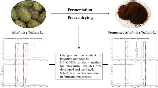
Development and Validation of an Analytical Method for Deacetylasperulosidic Acid, Asperulosidic Acid, Scopolin, Asperuloside and Scopoletin in Fermented Morinda citrifolia L. (Noni)

1
Department of Food Biotechnology and Environmental Science, Kangwon National University, Chuncheon 24341, Korea
2
NSTBIO Co., Ltd., Incheon 21984, Korea
3
Atomy R&D Center, Gongju 32511, Korea
*
Authors to whom correspondence should be addressed.
These authors contributed equally to this work.
Separations 2021, 8(6), 80; https://doi.org/10.3390/separations8060080
Submission received: 30 April 2021 / Revised: 2 June 2021 / Accepted: 3 June 2021 / Published: 5 June 2021
(This article belongs to the Section Analysis of Food and Beverages)

Abstract

:
Fermentation is a technology that enhances biologically active ingredients, improves the absorption rate and induces the generation of new functional ingredients by the catalytic action of enzyme systems possessed by microorganisms. In this study, changes in the content of five kinds of bioactive compounds (deacetylasperulosidic acid, asperulosidic acid, scopolin, asperuloside and scopoletin) of Morinda citrifolia L. were confirmed by fermentation, and a high-performance liquid chromatography-photodiode array (HPLC-PDA) analysis method for measuring analytes was developed and validated. HPLC method for the determination of five bioactive compounds in Morinda citrifolia L. extracts (MCE) was validated in terms of sensitivity, linearity, selectivity, limit of detection (LOD) and quantification (LOQ), precision and accuracy. The coefficient of determination of the calibration curve for bioactive compounds (1.56–100 μg/mL) showed linearity (R2 ≥ 0.9999). LOD and LOQ were in the range 0.04–0.97 and 0.13–2.95 μg/mL, respectively. The range of intra- and intraday accuracies values (recovery) were 97.5–121.9% and 98.8–118.1%, respectively, and precision value (RSDs) of the bioactive compounds were <4%. In addition, changes in the content of five bioactive compounds in MCE by fermentation were confirmed. These results indicate that the developed fermentation and analysis method could be applied in the development of potential functional food ingredients.

Graphical Abstract

1. Introduction

Morinda citrifolia L. is found in Polynesia, India and China and has been used as a traditional folk medicinal plant for about 2000 years ago. Polynesians have used not only the flesh, but also roots, stems, bark, leaves and flowers to treat wounds [1]. M. citrifolia fruit also shows astringent or bitter taste when ripe and a strong rancid smell similar to butyric acid. In addition, the functional ingredients including minerals, vitamins and phenolic compounds have been studied [2]. Additionally, the efficacy of M. citrifolia on antioxidant, anti-inflammatory, anticancer, antibacterial, diabetes and cardiovascular disease has been reported [3,4,5]. Due to the variety of adaptations and uses of plant structures for various therapeutic purposes, M. citrifolia has attracted the attention of researchers in food and pharmaceutical industries, and the possibility as a functional food material has been raised [2,6].
M. citrifolia has already been reported to contain more than 160 phytochemicals such as alkaloids, phenolic and organic acids [7]. Among the alkaloids already identified, proxeronine, a precursor of xeronine, was discovered by Heinicke [8]. The proxeronine is converted to the xeronine in the human intestine by proxeroninase [9]. Xeronine produced in the body is an essential alkaloid for cell regulation. It regulates the structure and function of proteins to regenerate damaged cells, activates all functions in the body, and plays an important role in helping nutrient absorption functions [4]. Iridoids are phytochemicals produced by the plants self-defense mechanism [10]. Iridoids have many biological functions in human such as ant-inflammatory, immunomodulatory, hepatoprotective, neuroprotective, cardioprotective, anticancer, antioxidant, antimicrobic and hypoglycemic [11]. They are found mainly in the leaves and young stems, but very rarely in fruits [12]. However, M. citrifolia has been reported to contain a large amount of iridoid compounds, among which deacetylasperulosidic acid, asperulosidic acid and asperuloside are major iridoid in M. citrifolia [13,14]. Coumarins play an important role in regulation of plants growth and metabolites [15]. These are also a wide range of biological function such as anti-inflammatory, antioxidant, antibacterial and anticancer [16,17,18]. Among these, scopoletin is one of the representative coumarin derivative in M. citrifolia [19].
Functional foods including designer foods, therapeutic foods, medifoods, medicinal foods, therapeutic foods, foodceuticals and superfoods are foods manufactured using bioactive compounds [20]. Functional foods are aimed at reducing the risk of disease occurrence or maintaining and promoting health through activation of biological functions [21]. Fermentation is a process in which microorganisms decompose organic matter using enzymes, and involves converting large molecules into small molecules or including molecular oxidation/reduction mechanisms mediated by selected microorganisms [22]. The purpose of fermentation is to increase the content of active ingredients, improve the absorption rate and induce the generation of new functional ingredients by inducing structural changes of natural physiologically active substances through biological methods [23]. Additionally, it is being used to develop foods with enhanced physiological activity as a method of increasing the functionality of natural materials [24]. In this study, fermentation was applied to enhance the bioactive compounds and was a suggested possibility for function foods ingredients.
In order to develop functional foods, it is necessary to scientifically prove the functionality and stability of the bioactive compounds they possess. It is also required to standardize the bioactive compounds present in the food [25]. Standardization is a technology that can produce consistent quality by minimizing fluctuations in functional ingredients contained in natural substances [26]. Standardization requires technology and information management used throughout the manufacturing process from the production of raw materials to the final product development. In general, constant quality is confirmed by using changes in the marker compound [27]. Therefore, it is necessary to use a recognized method or a precise analytical method to measure the content of the marker compound, and the scientific validity and reliability of the analysis method should be verified in order to establish a reference standard.
In this study, in order to standardize fermented M. citrifolia L. extracts (MCE) as a health functional food ingredient, we developed and validated a high-performance liquid chromatography-photodiode array (HPLC-PDA) method. PDA detector is a detector that uses the principle that the target analyte absorbs light in the UV or visible range, and the analyte requires a chromophore for detection [28]. Additionally, the PDA detector uses a photodiode imaging sensor to monitor the entire UV-vis spectrum of the material passing through the flow cell. Coumarin is a crystalline soluble in ether, chloroform, ethanol and water. Reverse-phase (RP) HPLC is most commonly used for the identification and quantification of coumarin [29]. The physical properties of most natural coumarins are that they are fluorescent in UV light [30]. Sproul et al. [31] reported that the diode array detection HPLC method is suitable for the analysis of coumarin in various foods. Iridoid compounds with COOH and COOR substituents in C4 have strong absorption between 230 and 240 nm due to the chromophore α·β-unsaturated acid, ester and lactone structure in the molecule [32]. Therefore, the maximum absorption wavelength of five bioactive compounds in MCE with chromophores was investigated, and a wavelength range for simultaneous analysis of them was selected. Additionally, the changes in their content during the fermentation process was measured.

2. Materials and Methods

2.1. Chemicals and Standards

Acetonitrile (ACN; ≥99%) was acquired from J.T. Baker (Philipsburg, NJ, USA) and formic acid (≥98%; Junsei, Tokyo, Japan) was diluted with distilled water to prepare a mobile phase with 0.1%. The chemical standards deacetylasperulosidic acid (CAS. 14259-55-3; ≥98%), asperulosidic acid (CAS. 25368-11-0; ≥98%), scopolin (CAS. 531-44-2; ≥98%), asperuloside (CAS. 14259-45-1; ≥98%) and scopoletin (CAS. 92-61-5; ≥98%) were obtained from ChemFaces (Wuhan, Hubei, China). All standards were accurately weighed with an appropriate weight, and a stock solution was prepared by dissolving in distilled water.

2.2. Sample Preparation

M. citrifolia L. freshly harvested in French Polynesia was provide through NST Bio Noni Farm Co., Ltd. (Incheon, Korea). M. citrifolia L. was fermented with Lactobacillus brevis (NST707) using the fermentation system as follows: M. citrifolia L. washed with lukewarm water was cut into pieces. The samples (100 g) were fermented with 500 mL of water and 2% (8 × 10¹¹ CFU/g) Lactobacillus brevis (NST707) at 30 °C for 72 h. At the end of the fermentation period, the ferment was filtered to eliminate debris and fruit particles. Then the ferment was freeze-dried to obtain the fermented MCE. Additionally, non-fermented MCE was obtained by proceeding under the same method with fermented MCE except for the treatment of Lactobacillus brevis (NST707). The non-fermented MCE and fermented MCE (25 mg) were mixed with 25 mL of distilled water. The mixture was sonicated for 10 min and filtered using 0.22 μm PVDF filter (Millex-HV, Millipore, Bedford, MA, USA).

2.3. HPLC Instrument Conditions

The chromatography was separated on a Waters 2695 separation module HPLC system (Waters Co., Milford, MA, USA) coupled with the Waters 996 photodiode array detector, and equipped with Shiseido Capcell Pak C18 UG120 column (Shiseido, 4.6 mm 250 mm, 5.0 μm, Tokyo, Japan). The temperature of the column was maintained at 30 °C, and the pump was connected to two mobile phases (A: 0.1% formic acid in distilled water; v/v and B: acetonitrile) with a gradient elution as follows: 0–5 min, maintained at 100% A; 5–30 min, linear from 100 to 65% A; 30–35 min, linear from 60 to 100%. The mobile phase was filtered with 0.45 μm membrane filter (Whatman, Amersham, UK) and degassed under vacuum. The injection volume and flow rate were set at 10 μL and 1.0 mL/min, respectively. The photodiode array detector was monitored for all standards at 254 nm for quantitative analysis. HPLC system operation and processing were performed using Waters Empower software.

2.4. Method Validation

International Conference on Harmonization (ICH) suggested guidance and recommendations on how to consider the validation for analytical procedure in 2005 [33]. According to ICH guidelines, the HPLC method for the determination of five bioactive compounds (deacetylasperulosidic acid, asperulosidic acid, scopolin, asperuloside and scopoletin) in MCE was validated in terms of sensitivity, linearity, selectivity, limit of detection (LOD) and quantification (LOQ), precision and accuracy. The calibration curve was estimated by diluting the stock solution with distilled water in seven concentration increments ranging from 1.56 to 100 μg/mL for all standards. In the process of the analysis method development and verification, the calibration curve evaluation shows sensitivity and linearity. The LOD and LOQ of bioactive compounds were calculated using the slope and standard deviation of a calibration curve and determined by signal-to-noise ratios of 3.3 and 10, respectively as following formula: LOD = 3.3(δ/S); LOQ = 10(δ/S), with δ being the standard deviation at 6.25 μg/mL and S the slope of the calibration curve. Additionally, matrix-matched calibration curves were prepared by spiking standards into non-fermented and fermented MCE to confirm the interference with the matrix peak. For selectivity evaluation of HPLC method, chromatograms were confirmed to ensure that the ingredients in non-fermented and fermented MCE did not interfere with the analysis of the target analyte. The precision and accuracy were determined in two matrices at three different concentration of standards (12.5, 25 and 50 μg/mL). Furthermore, the results were assessed by intraday (three times repeated in a day) and interday (three times repeated in three different days).

3. Results

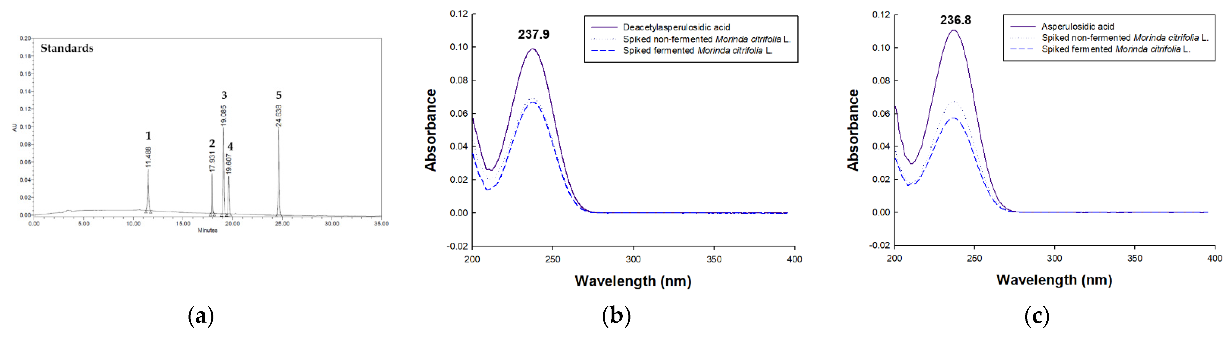
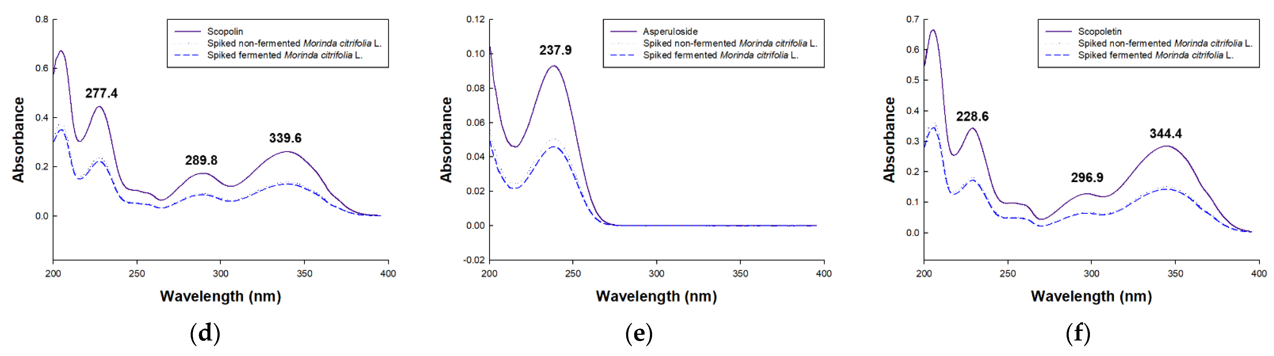
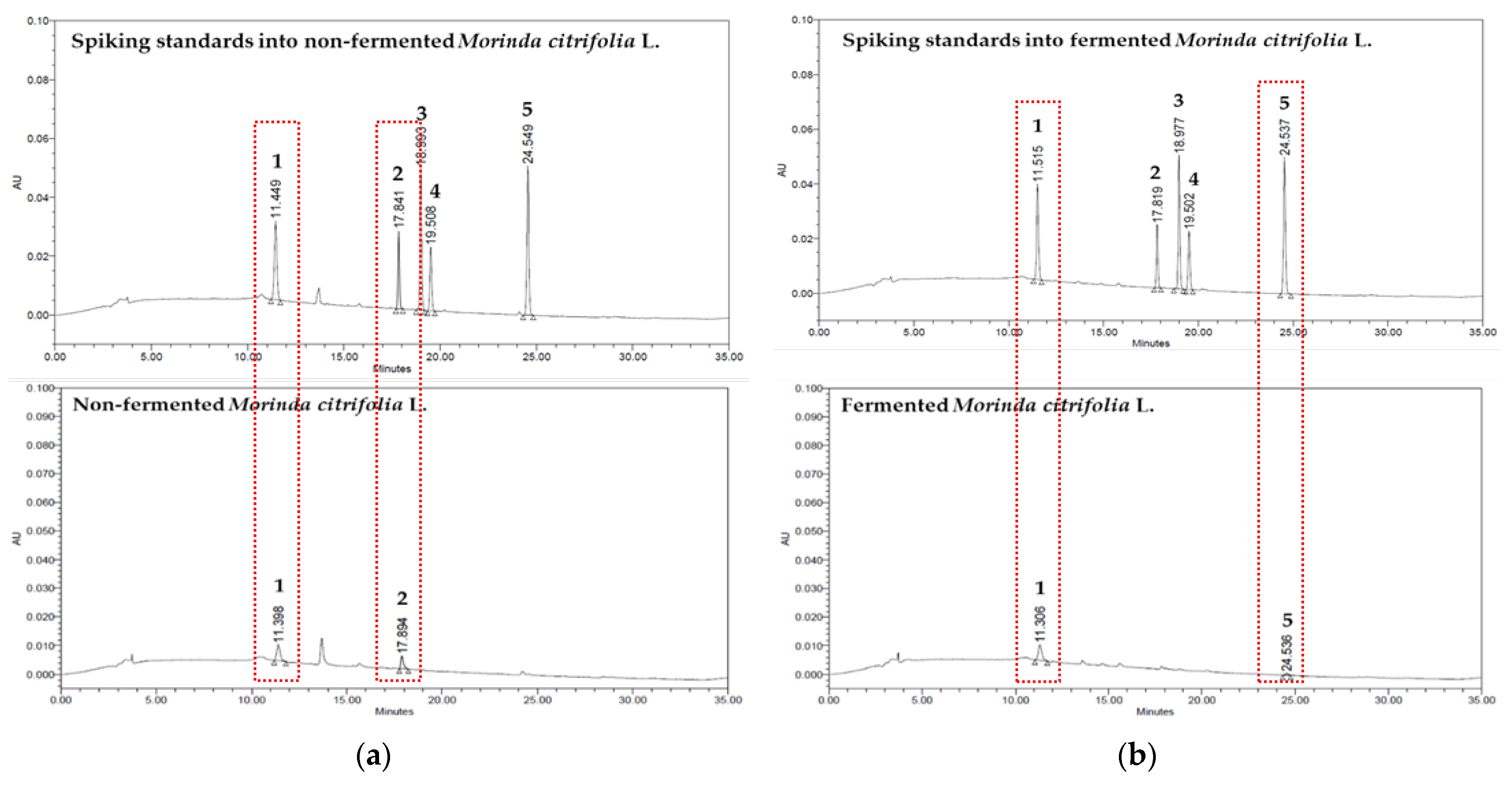
3.1. Method Development

Suitable simultaneous determination of the bioactive compounds of MCE including deacetylasperulosidic acid, asperulosidic acid, scopolin, asperuloside and scopoletin were performed by the HPLC-PDA method. They were efficiently set to the separation conditions using a Shiseido C18 column (4.6 mm × 250 mm, 5.0 μm) maintained at 30 °C and a mobile phase system consisted of formic acid and distilled water gradient conditions. Using the established assay, bioactive compounds were separated within 35 min (Figure 1a). The measurement wavelength range was set from 200 to 400 nm, and five standards were detected up to 256 nm wavelengths, of which 254 nm was used in consideration of the baseline and the degree of separation with Rs value ≥ 1.60. Method specificity was determined by comparing PDA spectrums and retention time of five standards as follows (Figure 1e–f): deacetylasperulosidic acid (λmax = 238 nm; RT = 11.5 min), asperulosidic acid (λmax = 237 nm; RT = 17.9 min), scopolin (λmax = 277, 290 and 340 nm; RT = 19.1 min), asperuloside (λmax = 238 nm; RT = 19.6 min) and scopoletin (λmax = 229, 297 nm and 344; RT = 24.6 min).

3.2. Method Validation

3.2.1. Selectivity, Linearity, LOD and LOQ

Selectivity of the HPLC method refers to the degree to which a particular component can be determined without interference from other components in the food matrix of a complex mixture [34]. As shown Figure 2, the chromatograms of bioactive compounds spiked in the non-fermented and fermented MCE were efficiently separated with no interference and coelution peaks using the Capcell Pak C18 column. The linearity of the established HPLC-PDA method was determined in the 1.56–100 μg/mL for bioactive compounds. The coefficient of determination (R2) of the bioactive compounds were in the range of 0.9999–1.0000. As shown Table 1, the LOD and LOQ values of the bioactive compounds ranged from 0.04 to 0.97 and 0.13 to 2.95 μg/mL, respectively.

3.2.2. Precision and Accuracy

The recovery results for the bioactive compounds were performed using developed analytical method by spiking non-fermented and fermented MCE with 12.5, 25 and 50 μg/mL of bioactive compounds (Table 2). The average recoveries in non-fermented MCE were 103.0–117.7% for deacetylasperulosidic acid, 112.9–118.1% for asperulosidic acid, 97.5–101.5% for scopolin, 99.7–103.1% for asperuloside and 99.3–102.2% for scopoletin. In fermented MCE, the average recoveries were 105.2–121.9% for deacetylasperulosidic acid, 100.2–102.6% for asperulosidic acid, 97.5–99.1% for scopolin, 99.5–100.6% for asperuloside and 100.2–100.9% for scopoletin.
The precision of bioactive compounds analysis was determined using the intraday and interday RSDs. The respective intraday and interday precisions ranges for all standards are as follows: In non-fermented MCE, from 1.53 to 3.41% and from 0.21 to 3.95% for deacetylasperulosidic acid; from 0.82 to 3.58% and from 0.83 to 3.26% for asperulosidic acid; from 0.15 to 3.31% and from 1.35 to 3.44% for scopolin; from 0.27 to 3.60% and from 1.74 to 3.60% for asperuloside; from 0.03 to 2.09% and from 1.06 to 2.03% for scopoletin; in fermented MCE, from 1.54 to 2.13% and from 1.10 to 2.06% for deacetylasperulosidic acid; from 0.54 to 2.38% and from 1.57 to 2.89% for asperulosidic acid; from 0.15 to 0.66% and from 1.90 to 3.44% for scopolin; from 0.27 to 0.97% and from 1.74 to 1.80% for asperuloside; from 0.23 to 0.74% and from 1.06 to 1.42% for scopoletin.

3.3. Quantitative Analysis of Bioactive Compound in Non-Fermented and Fermented Morinda citrifolia L. Extracts

The bioactive compounds were analyzed in the non-fermented and fermented MCE using the developed and validated HPLC-PDA analytical method. Each analyte peak was identified by comparing the retention time and PDA spectrum of the reference standard. Using the calibration curves of each standard, the amounts of the bioactive compounds in the three batches of the non-fermented and fermented MCE were calculated. The HPLC-PDA based method showed that non-fermented and fermented MCE contained 15.71 ± 0.74 mg/mL of deacetylasperulosidic acid and 15.16 ± 0.38 mg/mL of asperulosidic acid, and 18.52 ± 0.71 mg/mL of deacetylasperulosidic acid and 1.01 ± 0.07 mg/mL of scopoletin, respectively (Figure 2 and Table 3). During fermentation, the contents of deacetylasperulosidic acid and scopoletin increased, while the contents of asperulosidic acid decreased.

4. Discussion

M. citrifolia L. has been used as a traditional medicine and it contains a wide range of bioactive compounds with proven biological effects [35]. However, M. citrifolia L. fruit has a bitter taste and distinctive rancid taste, making it unsuitable for fresh consumption [3]. According to a recent study, the fermentation process becomes partly free from the undesirable smell [36,37]. However, for the development of functional foods, raw material standardization experiments through setting and analysis of marker compound are required [25]. The marker compound is a chemical determined for the purpose of quality control among the identified components contained in the raw material during the fermentation [38]. In this study, the marker compounds of M. citrifolia L. were selected according to ingredients that exist specifically or have a differential content variation depending on the raw material or manufacturing method (specificity) and exhibit functionality depending on the content and presence in the extract (functionality and representative). In addition, the stability of the indicator components and an easily accessible analyzer such as HPLC were considered. The analyzed deacetylasperulosidic acid, asperulosidic acid and asperuloside are representative iridoid of M. citrifolia L., and their various biological effects have been reported [39,40]. In addition, scopoletin and its glucoside scopolin were analyzed among coumarins, which are known to have efficacy in improving immune responses, and aglycon formed by hydrolysis by fermentation was analyzed to confirm its potential as an indicator component [41,42].
In this study, a method for the quantification of five bioactive compounds of MCE, was developed and validated. The analytical method was verified according to the procedure such as accuracy, precision, specificity, linearity, LOD and LOQ presented in the ICH guidelines. As a result, the validated HPLC-PDA analysis method is suitable for the determination of five bioactive compounds in non-fermented and fermented MCE. In addition, changes in their contents through fermentation were also determined. Fermentation induced changes in the content of bioactive compounds in fermented MCE. The HPLC-PDA based method showed that non-fermented and fermented MCE contained 15.71 ± 0.74 mg/mL of deacetylasperulosidic acid, 15.16 ± 0.38 mg/mL of asperulosidic acid, 18.52 ± 0.71 mg/mL of deacetylasperulosidic acid and 1.01 ± 0.07 mg/mL of scopoletin, respectively. Deacetylasperulosidic acid, whose content was increased through fermentation, is a major active phytochemical constituent of M. citrifolia L. and it has been reported to be effective on antioxidant [43] and immune response regulation [5]. In addition, the efficacy of scopoletin on antioxidant [44], anti-inflammatory [45], antiaging [46], antitumoral [47] and antihypertension [48] has been reported. In addition, it was reported that the biological effect of M. citrifolia L. was improved through fermentation [35,49]. However, as a result of the analysis of this study, the contents of deacetylasperulosidic acid and scopoletin slightly increased, but asperulosidic acid was not detected in MCE. Kim et al. [50] reported that fermentation improved immune balance and increased the content of deacetylasperulosidic acid and asperulosidic acid in M. citrifolia L. The results of this study showed a decrease in asperulosidic acid, which was thought to be a difference in fermentation strains and fermentation conditions. Additionally, Lee et al. [51] reported an increase in the scopoletin content of M. citrifolia L. by fermentation. The changes of the five bioactive compounds analyzed in this study were not consistent with bioactive effects. This is expected to be a change in bioactive compounds excluding the five selected species. Thus, future studies should analyze the compounds that play a major role in enhancing bioactive effects such as an immune response. However, an increase in the content of deacetylasperulosidic acid and scopoletin indicated the possibility of use as a marker compound for this fermentation process of M. citrifolia L. Standardization of fermentation processes and physiological and pathological role of fermented M. citrifolia L. will also be studied using these as marker compounds.

Author Contributions

Conceptualization, G.-S.S. and O.-H.L.; methodology, S.-I.C. and S.-J.L.; software, H.-Y.K. and Y.-H.J.; validation, I.-J.L., Y.-H.J. and X.H.; formal analysis, Y.-D.K. and X.M.; investigation, X.H. and S.-J.L.; resources, G.-S.S. and O.-H.L.; data curation, I.-J.L.; writing—original draft preparation, S.-I.C. and H.-Y.K.; writing—review and editing Y.-D.K. and O.-H.L.; visualization, O.-H.L.; supervision, G.-S.S.; project administration, O.-H.L.; funding acquisition, G.-S.S. All authors have read and agreed to the published version of the manuscript.

Funding

This work was supported by the Technology development Program (S2840161) funded by the Ministry of SMEs and Startups (MSS, Korea).

Institutional Review Board Statement

Not applicable.

Informed Consent Statement

Not applicable.

Data Availability Statement

The data presented in this study are available upon request.

Conflicts of Interest

The authors declare no conflict of interest relevant to this study.

References

  1. Wang, M.Y.; West, B.J.; Jensen, C.J.; Nowicki, D.; Su, C.; Palu, A.K.; Anderson, G. Morinda citrifolia (noni): A literature review and recent advances in noni research. Acta Pharmacol. Sin. 2002, 23, 1127–1141. [Google Scholar] [PubMed]
  2. Chan-Blanco, Y.; Vaillant, F.; Mercedes Perez, A.; Reynes, M.; Brillouet, J.M.; Brat, P. The noni fruit (Morinda citrifolia L.): A review of agricultural research, nutritional and therapeutic properties. J. Food Compos. Anal. 2006, 19, 645–654. [Google Scholar] [CrossRef]
  3. Motshakeri, M.; Ghazali, H.M. Nutritional, phytochemical and commercial quality of noni fruit: A multi-beneficial gift from nature. Trends Food Sci. Technol. 2015, 45, 118–129. [Google Scholar] [CrossRef]
  4. García-Vilas, J.A.; Quesada, A.R.; Medina, M.A. Damnacanthal, a noni anthraquinone, inhibits c-Met and is a potent antitumor compound against HepG2 human hepatocellular carcinoma cells. Sci. Rep. 2015, 5, 8021. [Google Scholar] [CrossRef] [Green Version]
  5. Murata, K.; Abe, Y.; Futamura-Masuda, M.; Uwaya, A.; Isami, F.; Deng, S.; Matsuda, H. Effect of Morinda citrifolia fruit extract and its iridoid glycosides on blood fluidity. J. Nat. Med. 2014, 68, 498–504. [Google Scholar] [CrossRef]
  6. Almeida, É.S.; de Oliveira, D.; Hotza, D. Properties and applications of Morinda citrifolia (noni): A review. Compr. Rev. Food Sci. Food Saf. 2019, 18, 883–909. [Google Scholar] [CrossRef] [Green Version]
  7. Wang, M.Y.; Su, C. Cancer preventive effect of Morinda citrifolia (Noni). Ann. N. Y. Acad. Sci. 2001, 952, 161–168. [Google Scholar] [CrossRef]
  8. Heinicke, R.M. The pharmacologically active ingredient of noni. Pac. Trop. Bot. Gard. Bull. 1985, 15, 10–14. [Google Scholar]
  9. Sanni, D.M.; Fatoki, T.H.; Kolawole, A.O.; Akinmoladun, A.C. Xeronine structure and function: Computational comparative mastery of its mystery. In Silico Pharmacol. 2017, 5, 1–7. [Google Scholar] [CrossRef] [Green Version]
  10. Israili, Z.H.; Lyoussi, B. Ethnopharmacology of the plants of genus Ajuga. Pak. J. Pharm. Sci. 2009, 22, 425–462. [Google Scholar]
  11. Tundis, R.; Loizzo, M.R.; Menichini, F.; Statti, G.A.; Menichini, F. Biological and pharmacological activities of iridoids: Recent developments. Mini Rev. Med. Chem. 2008, 8, 399–420. [Google Scholar] [CrossRef]
  12. Kucharska, A.Z.; Szumny, A.; Sokół-Łętowska, A.; Piórecki, N.; Klymenko, S.V. Iridoids and anthocyanins in cornelian cherry (Cornus mas L.) cultivars. J. Food Compost. Anal. 2015, 40, 95–102. [Google Scholar] [CrossRef]
  13. Deng, S.; West, B.J.; Palu, A.K.; Jensen, C.J. Determination and comparative analysis of major iridoids in different parts and cultivation sources of Morinda citrifolia. Phytochem. Anal. 2011, 22, 26–30. [Google Scholar] [CrossRef] [PubMed]
  14. Abou Assi, R.; Darwis, Y.; Abdulbaqi, I.M.; Vuanghao, L.; Laghari, M.H. Morinda citrifolia (Noni): A comprehensive review on its industrial uses, pharmacological activities, and clinical trials. Arab. J. Chem. 2017, 10, 691–707. [Google Scholar] [CrossRef]
  15. Robe, K.; Izquierdo, E.; Vignols, F.; Rouached, H.; Dubos, C. The Coumarins: Secondary metabolites playing a primary role in plant nutrition and health. Trends Plant Sci. 2020, 26, 248–259. [Google Scholar] [CrossRef]
  16. Bansal, Y.; Sethi, P.; Bansal, G. Coumarin: A potential nucleus for anti-inflammatory molecules. Med. Chem. Res. 2013, 22, 3049–3060. [Google Scholar] [CrossRef]
  17. Zhu, J.J.; Jiang, J.G. Pharmacological and nutritional effects of natural coumarins and their structure activity relationships. Mol. Nutr. Food Res. 2018, 62, 1701073. [Google Scholar] [CrossRef] [PubMed]
  18. Wu, L.; Wang, X.; Xu, W.; Farzaneh, F.; Xu, R. The structure and pharmacological functions of coumarins and their derivatives. Curr. Med. Chem. 2009, 16, 4236–4260. [Google Scholar] [CrossRef]
  19. Ikeda, R.; Wada, M.; Nishigaki, T.; Nakashima, K. Quantification of coumarin derivatives in Noni (Morinda citrifolia) and their contribution of quenching effect on reactive oxygen species. Food Chem. 2009, 113, 1169–1172. [Google Scholar] [CrossRef]
  20. Shah, N.P. Functional cultures and health benefits. Int. Dairy J. 2007, 17, 1262–1277. [Google Scholar] [CrossRef]
  21. Hasler, C.M. Functional foods: Benefits, concerns and challenges—A position paper from the American Council on Science and Health. J. Nutr. 2002, 132, 3772–3781. [Google Scholar] [CrossRef] [PubMed] [Green Version]
  22. Yadav, H.; Jain, S.; Rastamanesh, R.; Bomba, A.; Catanzaro, R.; Marotta, F. Fermentation technology in the development of functional foods for human health: Where we should head. Ferment. Technol. 2011, 1, 1–2. [Google Scholar] [CrossRef] [Green Version]
  23. Rein, M.J.; Renouf, M.; Cruz-Hernandez, C.; Actis-Goretta, L.; Thakkar, S.K.; da Silva Pinto, M. Bioavailability of bioactive food compounds: A challenging journey to bioefficacy. Br. J. Clin. Pharmacol. 2013, 75, 588–602. [Google Scholar] [CrossRef] [Green Version]
  24. Joana Gil-Chávez, G.; Villa, J.A.; Fernando Ayala-Zavala, J.; Basilio Heredia, J.; Sepulveda, D.; Yahia, E.M.; González-Aguilar, G.A. Technologies for extraction and production of bioactive compounds to be used as nutraceuticals and food ingredients: An overview. Compr. Rev. Food Sci. Food Saf. 2013, 12, 5–23. [Google Scholar] [CrossRef]
  25. Cho, B.Y.; Park, M.R.; Lee, J.H.; Ra, M.J.; Han, K.C.; Kang, I.J.; Lee, O.H. Standardized Cirsium setidens Nakai ethanolic extract suppresses adipogenesis and regulates lipid metabolisms in 3T3-L1 adipocytes and C57BL/6J mice fed high-fat diets. J. Med. Food 2017, 20, 763–776. [Google Scholar] [CrossRef]
  26. Yadav, N.P.; Dixit, V.K. Recent approaches in herbal drug standardization. Int. J. Integr. Biol. 2008, 2, 195–203. [Google Scholar]
  27. Ong, E.S. Extraction methods and chemical standardization of botanicals and herbal preparations. J. Chromatogr. B. 2004, 812, 23–33. [Google Scholar] [CrossRef]
  28. Swartz, M. HPLC detectors: A brief review. J. Liq. Chrom. Relat. Technol. 2010, 33, 1130–1150. [Google Scholar] [CrossRef]
  29. Scotter, M.J.; Roberts, D.P.; Rees, G.O. Development and single-laboratory validation of an HPLC method for the determination of coumarin in foodstuffs using internal standardization and solid-phase extraction cleanup. Anal. Methods 2011, 3, 414–419. [Google Scholar] [CrossRef]
  30. Nahata, A.; Dixit, V.K. Spectrofluorimetric estimation of scopoletin in Evolvulus alsinoides Linn. and Convulvulus pluricaulis choisy. Indian J. Pharm. Sci. 2008, 70, 834–837. [Google Scholar]
  31. Raters, M.; Matissek, R. Analysis of coumarin in various foods using liquid chromatography with tandem mass spectrometric detection. Eur. Food Res. Technol. 2008, 227, 637–642. [Google Scholar] [CrossRef]
  32. Liu, H.W. Extraction and isolation of compounds from herbal medicines. In Traditional Herbal Medicine Research Methods: Identification, Analysis, Bioassay, and Pharmaceutical and Clinical Studies; Liu, W.J.H., Ed.; John Wiley & Sons: New York, NY, USA, 2011; pp. 81–138. [Google Scholar]
  33. International Conference on Harmonization (ICH). International Conference on Harmonization of Technical Requirements for Registration of Pharmaceuticals for Human Use. ICH Harmonised Tripartite Guidline. Validation of Analytical Procedures: Text and Methodology. Available online: https://www.ich.org/fileadmin/Public_Web_Site/ICH_Products/Guidelines/Quality/Q2_R1/Step4/Q2_R1_Guideline.pdf (accessed on 11 March 2019).
  34. Marın, A.; Garcıa, E.; Garcıa, A.; Barbas, C. Validation of a HPLC quantification of acetaminophen, phenylephrine and chlorpheniramine in pharmaceutical formulations: Capsules and sachets. J. Pharm. Biomed. Anal. 2002, 29, 701–714. [Google Scholar] [CrossRef]
  35. Zhang, L.; Liu, S.; Chen, Y.; Guo, J.; Li, C.; Zhang, J. Metatranscriptomic approach reveals the functional and enzyme dynamics of core microbes during noni fruit fermentation. Food Res. Int. 2021, 141, 109999. [Google Scholar] [CrossRef] [PubMed]
  36. Wang, Z.; Dou, R.; Yang, R.; Cai, K.; Li, C.; Li, W. Changes in Phenols, Polysaccharides and Volatile Profiles of Noni (Morinda citrifolia L.) Juice during Fermentation. Molecules 2021, 26, 2604. [Google Scholar] [CrossRef] [PubMed]
  37. Zhang, K.; Meng, J.; Li, X.; Tang, X.; Ma, S.; Lv, Y.; Yang, S. Noni (Morinda citrifolia L.) wine prevents the oxidative stress and obesity in mice induced by high-fat diet. J. Food Biochem. 2020, 44, e13460. [Google Scholar] [CrossRef]
  38. Liang, Y.Z.; Xie, P.; Chan, K. Quality control of herbal medicines. J. Chromatogr. B 2004, 812, 53–70. [Google Scholar] [CrossRef]
  39. Kamiya, K.; Tabnaka, Y.; Endgang, H.; Umar, M.; Satake, T. New anthraquinone and iridoid from the fruits of Morinda citrifolia. Chem. Pharm. Bull. 2005, 53, 1597–1599. [Google Scholar] [CrossRef] [PubMed] [Green Version]
  40. Levand, O.; Larson, H.O. Some chemical constituents of Morinda citrifolia. Planta Med. 1979, 36, 186–187. [Google Scholar] [CrossRef]
  41. Manuele, M.G.; Ferraro, G.; Arcos, M.L.B.; López, P.; Cremaschi, G.; Anesini, C. Comparative immunomodulatory effect of scopoletin on tumoral and normal lymphocytes. Life Sci. 2006, 79, 2043–2048. [Google Scholar] [CrossRef]
  42. Pawlus, A.D.; Su, B.N.; Keller, W.J.; Kinghorn, A.D. An anthraquinone with potent quinone reductase-inducing activity and other constituents of the fruits of Morinda citrifolia (noni). J. Nat. Prod. 2005, 68, 1720–1722. [Google Scholar] [CrossRef]
  43. Ma, D.L.; Chen, M.; Su, C.X.; West, B.J. In vivo antioxidant activity of deacetylasperulosidic acid in noni. J. Anal. Methods Chem. 2013, 2013, 804504. [Google Scholar] [CrossRef] [PubMed] [Green Version]
  44. Zhang, X.; Xu, M.; Zhang, J.; Wu, L.; Liu, J.; Si, J. Identification and evaluation of antioxidant components in the flowers of five Chimonanthus species. Ind. Crops Prod. 2017, 102, 164–172. [Google Scholar] [CrossRef]
  45. Leema, G.; Tamizhselvi, R. Protective effect of scopoletin against cerulein-induced acute pancreatitis and associated lung injury in mice. Pancreas 2018, 47, 557–585. [Google Scholar] [CrossRef] [PubMed]
  46. Jamuna, S.; Karthika, K.; Paulsamy, S.; Thenmozhi, K.; Kathiravan, S.; Venkatesh, R. Confertin and scopoletin from leaf and root extracts of Hypochaeris radicata have anti-inflammatory and antioxidant activities. Ind. Crops Prod. 2015, 70, 221–230. [Google Scholar] [CrossRef]
  47. Pinto, D.; Silva, A.M.S. Anticancer natural coumarins as lead compounds for the discovery of new drugs. Curr. Top. Med. Chem. 2017, 17, 3190–3198. [Google Scholar] [CrossRef]
  48. Panda, S.; Kar, A. Evaluation of the antithyroid, antioxidative and antihyperglycemic activity of scopoletin from Aegle marmelos leaves in hyperthyroid rats. Phytother. Res. 2006, 20, 1103–1105. [Google Scholar] [CrossRef]
  49. Guo, M.; Mao, B.; Sadiq, F.A.; Hao, Y.; Cui, S.; Yi, M.; Hong, Q.; Lee, Y.H.; Zhao, J. Effects of noni fruit and fermented noni juice against acute alcohol induced liver injury in mice. J. Funct. Foods 2020, 70, 103995. [Google Scholar] [CrossRef]
  50. Kim, S.H.; Seong, G.S.; Choung, S.Y. Fermented Morinda citrifolia (Noni) alleviates DNCB-induced atopic dermatitis in NC/Nga mice through modulating immune balance and skin barrier function. Nutrients 2020, 12, 249. [Google Scholar] [CrossRef] [PubMed] [Green Version]
  51. Lee, H.H.; Yin, L.J.; Tai, H.M.; Jiang, S.T. Facilitating the release of bionutrients from Morinda citrifolia (noni) by cellulase hydrolysis and lactic acid bacteria fermentation and their effects on α-amylase and α-glucosidase activities. J. Mar. Sci. Technol. 2016, 24, 648–655. [Google Scholar]
Figure 1. HPLC chromatographic of bioactive compounds marked with 1–5 represent deacetylasperulosidic acid, asperulosidic acid, scopolin, asperuloside and scopoletin (a). PDA spectrums of bioactive compounds in STD spiked non-fermented and fermented Morinda citrifolia L. extracts (bf).
Separations 08 00080 g001aSeparations 08 00080 g001b
Figure 2. HPLC chromatographic of bioactive compounds marked with 1–5 represent deacetylasperulosidic acid, asperulosidic acid, scopolin, asperuloside and scopoletin spiked non-fermented (a) and fermented (b) Morinda citrifolia L. extracts.
Separations 08 00080 g002
Table 1. Coefficient of determination of the calibration curves, LOD and LOQ of bioactive compounds analysis for non-fermented and fermented Morinda citrifolia L. extracts.
AnalytesRange
(μg/mL)
SlopeInterceptCoefficient of
Determination (R2)
LOD 1
(μg/mL)
LOQ 2
(μg/mL)
Deacetylasperulosidic acid1.56–1004081.36360.711.00000.762.30
Asperulosidic acid1.56–1002716.59736.720.99990.260.77
Scopolin1.56–1006052.101032.581.00000.441.34
Asperuloside1.56–1002797.46−167.511.00000.972.95
Scopoletin1.56–1007441.30619.611.00000.040.13
1 LOD is the limit of detection. 2 LOQ is the limit of quantification.
Table 2. Recoveries of bioactive compounds (three different concentrations) in non-fermented and fermented Morinda citrifolia L. extracts.
MatrixAnalytesConcentration
(μg/mL)
Mean ± SD
(μg/mL)
RSD 1 (%)Recovery (%)
Non-fermented
Morinda citrifolia L.
Deacetylasperulosidic acidIntraday12.512.87 ± 0.201.53103.0
2527.15 ± 0.722.66110.5
5060.95 ± 2.083.41117.7
Interday12.513.30 ± 0.030.21106.4
2527.62 ± 0.511.85110.5
5058.83 ± 2.323.95117.7
Asperulosidic acidIntraday12.514.27 ± 0.130.93114.1
2528.23 ± 0.260.93112.9
5058.75 ± 2.103.58117.5
Interday12.514.15 ± 0.120.83113.2
2528.49 ± 0.541.91114.0
5059.05 ± 1.923.26118.1
ScopolinIntraday12.512.18 ± 0.050.4097.5
2524.43 ± 0.050.2097.7
5050.70 ± 1.683.31101.4
Interday12.512.39 ± 0.171.3599.1
2524.69 ± 0.512.0898.8
5050.73 ± 1.753.44101.5
AsperulosideIntraday12.512.46 ± 0.110.8799.7
2525.06 ± 0.230.91100.2
5051.73 ± 1.963.60103.1
Interday12.512.46 ± 0.352.8399.7
2525.13 ± 0.612.42100.5
5051.57 ± 1.863.60103.1
ScopoletinIntraday12.512.42 ± 0.070.5799.4
2524.95 ± 0.010.0399.8
5051.11 ± 1.072.09102.2
Interday12.512.42 ± 0.181.4299.3
2525.06 ± 0.331.31100.2
5050.95 ± 1.032.03101.9
Fermented
Morinda citrifolia L.
Deacetylasperulosidic acidIntraday12.513.15 ± 0.282.13105.2
2527.82 ± 0.802.87108.6
5057.80 ± 0.891.54121.9
Interday12.513.37 ± 0.151.10107.0
2528.13 ± 0.512.06112.5
5057.59 ± 1.051.82115.2
Asperulosidic acidIntraday12.512.71 ± 0.302.38101.7
2525.22 ± 0.210.82100.9
5051.10 ± 0.280.54102.2
Interday12.512.52 ± 0.332.60100.2
2525.44 ± 0.732.89101.7
5051.30 ± 0.811.57102.6
ScopolinIntraday12.512.19 ± 0.080.6697.5
2524.45 ± 0.040.1597.8
5049.33 ± 0.300.6198.7
Interday12.512.40 ± 0.241.9099.1
2524.69 ± 0.512.0898.8
5050.73 ± 1.753.4499.0
AsperulosideIntraday12.512.47 ± 0.030.2799.8
2524.98 ± 0.240.9799.9
5049.77 ± 0.380.7699.5
Interday12.512.56 ± 0.221.78100.5
2525.14 ± 0.451.80100.6
5049.86 ± 0.871.7499.7
ScopoletinIntraday12.512.57 ± 0.090.74100.6
2525.12 ± 0.060.23100.5
5050.47 ± 0.290.58100.9
Interday12.512.55 ± 0.181.42100.4
2525.06 ± 0.331.31100.2
5050.40 ± 0.541.06100.8
1 RSD is the relative standard deviation.
Table 3. HPLC-PDA determination of bioactive compounds in non-fermented and fermented Morinda citrifolia L. extracts.
SampleAnalytesMean ± SD (mg/g)RSD (%)
Non-fermented Morinda citrifolia L.Deacetylasperulosidic acid15.71 ± 0.744.71
Asperulosidic acid15.16 ± 0.382.51
ScopolinN.D. 1-
AsperulosideN.D.-
scopoletinN.D.-
Fermented Morinda citrifolia L.Deacetylasperulosidic acid18.52 ± 0.713.83
Asperulosidic acidN.D.-
ScopolinN.D.-
AsperulosideN.D.-
scopoletin1.01 ± 0.077.24
1 ND is the not detected.
Publisher’s Note: MDPI stays neutral with regard to jurisdictional claims in published maps and institutional affiliations.

