Review

A Review on the Extraction, Purification, and Biological Activities of Polysaccharides from Elaeagnus angustifolia Fruits


1
Institute of Advanced Carbon Conversion Technology, Huaqiao University, Xiamen 361021, China
2
Fujian Provincial Key Laboratory of Biomass Low-Carbon Conversion, Huaqiao University, Xiamen 361021, China
3
College of Chemical Engineering, Huaqiao University, Xiamen 361021, China
*
Author to whom correspondence should be addressed.
Separations 2026, 13(5), 139; https://doi.org/10.3390/separations13050139
Submission received: 13 March 2026 / Revised: 1 April 2026 / Accepted: 16 April 2026 / Published: 1 May 2026

Abstract

Elaeagnus angustifolia L., belonging to the family Elaeagnaceae and genus Elaeagnus, which is a medicinal and edible homologous material with significant economic and ecological value. Its polysaccharides are one of its key active components, exhibiting bioactivities, including antioxidant, immunomodulatory, antitumor, anti-fatigue, and hypolipidemic effects. This paper reviews the research progress on the extraction, purification, structural features, and bioactivities of E. angustifolia polysaccharides, aiming to provide a theoretical basis and reference for their high-value development and utilization.

1. Introduction

Elaeagnus angustifolia L., belonging to the family Elaeagnaceae and genus Elaeagnus, is a deciduous tree or small tree, which is widely distributed across Asia and Europe. In China, it is mainly concentrated in the “Three North” regions (Northeast, North, and Northwest China), including Liaoning, Hebei, Gansu, Inner Mongolia, and Xinjiang. It is a typical sandy–soil plant known for its tolerance to drought, saline-alkali conditions, and cold temperatures [1,2,3,4]. E. angustifolia possesses a highly developed root system, with a taproot that can penetrate 6–7 m deep and lateral roots that can extend horizontally up to 10–14 m. It plays a vital role in windbreak, sand fixation, and soil improvement, while also holding significant nutritional, medicinal, and economic value [5]. This species exhibits a strong attraction to the Asian long-horned beetle (Anoplophora glabripennis), which attracts adults to feed, gnaw oviposition grooves, and lay eggs. Concurrently, E. angustifolia exudes gum at the gnawinng sites, encapsulating the eggs and any hatchable larvae, ultimately leading to their mortality [6]. These characteristics make it an excellent tree species integrating ecological protection with resource utilization. The fruits, flowers, leaves, and stems of E. angustifolia can all be utilized [7]. The fruit is nutritionally rich, containing crude polysaccharides, proteins, various amino acids, and trace elements. Historically, it has served as a “woody grain” for people in sandy regions and has also been used medicinally. It is traditionally believed to nourish the liver and kidneys, invigorate the spleen and regulate menstruation, and ralieve diarrhea and inflammation. In folk medicine practices in regions such as Xinjiang and Iran, it is commonly used to alleviate gastrointestinal discomfort [8]. The flowers can be used to extract essential oils and serve as a high-quality nectar source. The gum exuded from the stems can be used as a substitute for gum arabic and is widely applied in the food industry [7]. Modern pharmacological studies have demonstrated that E. angustifolia possesses various therapeutic activities, including anti-inflammatory [9], antibacterial [10], antioxidant [11], analgesic [12], and anticancer effects [8].
Polysaccharides are defined as natural high-molecular-weight compounds formed by the linking of monosaccharides through glycosidic bonds [13]. They represent one of the important bioactive components in Elaeagnus angustifolia L., primarily found in its fruits and flowers [14]. Due to advantages such as low toxicity, biodegradability, and good biocompatibility, natural plant polysaccharides show great promise for applications in fields including food [15], cosmetics [16], medicine [17], and animal feed [18]. Their diverse physiological activities, including immunomodulatory [19], antioxidant [20], and antitumor effects [21], have made them a research focus in the field of natural products worldwide. This paper reviews the research progress on the extraction, purification, structural characteristics, and physiological activities of E. angustifolia polysaccharides, with the aim of providing a reference for further in-depth investigation and industrial development of these compounds.

2. Extraction, Isolation, and Purification of Polysaccharides from Elaeagnus angustifolia Fruits

Figure 1 illustrates the extraction and purification process of Elaeagnus angustifolia polysaccharides, which primarily includes raw material pretreatment, polysaccharide extraction, crude polysaccharide impurity removal, and homogeneous polysaccharide purification. E. angustifolia raw materials typically require drying to completely remove moisture, thereby preventing microbial growth and polysaccharide degradation during subsequent extraction. After drying, the material is crushed and sieved to obtain uniform E. angustifolia powder. To remove lipids, low-molecular-weight sugars, and other impurities present in the powder—and thus improve the extraction yield of E. angustifolia polysaccharides—reflux treatment with organic solvents such as 95% ethanol or petroleum ether is necessary [22,23,24].
Hot water extraction (HWE) is commonly employed for extracting E. angustifolia polysaccharides. This method operates under mild conditions, disrupting the plant cell wall structure to facilitate the sufficient dissolution of polysaccharides located within the cells, without altering their molecular structure and thus preserving their native characteristics effectively [25,26]. Li [22] extracted E. angustifolia polysaccharides using HWE, achieving a yield of 4.68% at 61 °C for 118.4 min with a solid-to-liquid ratio of 1:26 (g/mL). Chen et al. [27] employed the same method at 90 °C for 5 h with a solid-to-liquid ratio of 1:35 (g/mL), obtaining a yield of 5.39%. The phenol-sulfuric acid method [28] and anthrone–sulfuric acid method [29] are frequently used to determine the polysaccharide content in extracts, from which the yield is subsequently calculated. Response surface methodology (RSM), as an efficient process optimization tool, has been applied to optimize the extraction process of E. angustifolia polysaccharides [30,31]. This approach typically uses the polysaccharide extraction rate as the response value, identifies key factors influencing extraction efficiency and their suitable ranges through single-factor experiments, and then optimizes the critical process conditions using response surface design to maximize the yield [31]. However, traditional HWE has significant drawbacks, including lengthy extraction time, low efficiency, and high energy consumption, making it unfavorable for large-scale industrial production [26,32]. To overcome these limitations, novel auxiliary technologies have been introduced for extracting E. angustifolia polysaccharides. Ultrasound-assisted extraction (UAE) utilizes the energy generated by cavitation effects during ultrasound propagation in liquid to disrupt plant cell walls, increasing the contact between intracellular polysaccharides and the extraction solvent, thereby enhancing extraction efficiency [33]. Du et al. [23] employed UAE to extract polysaccharides from E. angustifolia, using Box–Behnken design (BBD) combined with RSM to optimize the process. Under the optimized conditions (extraction time: 43 min; ultrasonic power: 282 W; temperature: 79 °C; solid-to-liquid ratio: 30:1 (g/mL); three extraction cycles), the yield of E. angustifolia polysaccharides reached 9.82 ± 0.38%. Microwave-assisted extraction (MAE) relies on rapid polarization and rotation of water molecules induced by microwaves, generating heat through intermolecular friction. This results in internal heating, causing cell membrane expansion and rupture, thereby releasing intracellular polysaccharides [34]. Li et al. [35] compared the extraction efficiencies of HWE, UAE, and MAE for E. angustifolia polysaccharides. The results showed that the optimal extraction yields for the three methods were 5.1% (HWE), 7.5% (UAE), and 8.9% (MAE), in descending order: MAE > UAE > HWE. It is evident that UAE/MAE can significantly enhance the yield of E. angustifolia polysaccharides while shortening extraction time. Additionally, Li et al. [36] combined UAE and MAE, employing ultrasonic/microwave-assisted extraction (UAME) for E. angustifolia polysaccharides. This method integrates the cavitation effect of ultrasonic vibration with the high-energy effect of microwaves, generating uniform internal energy within the sample matrix by reducing binding forces and promoting rapid dissolution of target substances. They compared the yields with HWE, UAE, and MAE under specific conditions and obtained the following results: UAME (solid-to-liquid ratio 1:23 g/mL, ultrasonic power 252 W, microwave power 417 W, time 11 min) yielded 7.85%; UAE (ultrasonic power 252 W, time 11 min) yielded 5.74%; MAE (microwave power 417 W, time 11 min) yielded 6.86%; and HWE (solid-to-liquid ratio 1:25 g/mL, two extractions) yielded 4.91%. Although ultrasound- and microwave-assisted extraction can effectively increase polysaccharide yield and shorten extraction time, they carry the potential to cleave glycosidic bonds in E. angustifolia polysaccharide molecules, potentially disrupting the polysaccharide structure and altering its biological activity [37,38].
Besides physical methods, biological approaches are increasingly used for plant polysaccharide extraction, including enzyme-assisted extraction (EAE) and microbial fermentation. EAE utilizes the high specificity of enzymes to degrade specific components of the plant cell wall, commonly employing enzymes such as cellulase, pectinase, and papain [39,40]. Lian et al. [41] employed composite enzyme-assisted ultrasound extraction for E. angustifolia polysaccharides. Under the optimized conditions (1.5% cellulase, 1.5% pectinase; enzymatic hydrolysis: 40 min at 55 °C; ultrasound: 25 min at 400 W and 60 °C; solid-to-liquid ratio: 1:30), the extraction rate of E. angustifolia polysaccharides reached 12.35%. However, the high cost of enzyme preparations and difficulties in enzyme recovery limit their industrial application [42]. It is worth mentioning that microorganisms are also gradually being applied in plant polysaccharide extraction. Sakr et al. [43] treated Asparagus sprengeri through fermentation with Lactobacillus plantarum DMS 20174. Under the conditions of 5.0 g asparagus, 84 h cultivation, and 2.0% inoculum size, the polysaccharide yield was 10.34 g/L. Compared to unfermented polysaccharides, the molecular weight of the fermented polysaccharides decreased (from 1770 Da to 1229 Da), and their physiological activities—including antibacterial, antioxidant, anticoagulant, fibrinolytic, lipase inhibitory, and anticancer effects—were enhanced. During metabolism, microorganisms may biochemically modify polysaccharides, altering their structure and biological activity. However, fermentation broths obtained through microbial fermentation often contain numerous metabolites, which complicate subsequent purification [44].
The crude polysaccharide extract obtained as described above requires deproteinization, decolorization, alcohol precipitation, and dialysis to yield crude E. angustifolia polysaccharides [23,24,45]. Currently, common methods for deproteinizing E. angustifolia polysaccharides include the Sevage method and the trichloroacetic acid (TCA) method. The Sevage method is simple to operate, employs mild conditions, and causes minimal damage to the polysaccharide structure. Its principle involves shaking a chloroform-n-butanol mixture with the polysaccharide solution, causing protein impurities to denature and precipitate, after which they are extracted into the organic solvent phase. However, this method has drawbacks, including incomplete deproteinization and the need for repeated operations [46,47]. The TCA method exhibits higher deproteinization efficiency, as TCA denatures and precipitates protein impurities, which are then removed by centrifugation. However, high concentrations of TCA may cause slight damage to the polysaccharide structure, potentially affecting its biological activity [48]. Pigment impurities are also a significant factor affecting the purification and subsequent study of E. angustifolia polysaccharides. They not only lead to uneven coloration of the polysaccharide product but also interfere with experimental results such as structural analysis and molecular weight determination [49]. Therefore, decolorization of the E. angustifolia polysaccharide extract is necessary. Currently, activated carbon adsorption is commonly used for decolorizing E. angustifolia polysaccharides. This method is simple to operate, cost-effective, and offers fast decolorization, effectively adsorbing both fat-soluble and water-soluble pigments present in the extract [50]. Li et al. [51] compared the decolorization effects of three decolorizing agents: granular activated carbon, powdered activated carbon, and polyamide. The results indicated that granular activated carbon exhibited superior decolorization compared to the other two agents. The optimal conditions were decolorization at 55 °C for 75 min, with three cycles. Under these conditions, the decolorization rate reached 45.52%, with a polysaccharide retention rate of 95.55%. The polysaccharide solution, after deproteinization and decolorization, often still contains small-molecule impurities (such as monosaccharides, amino acids, and inorganic salts). These impurities can affect the purity of E. angustifolia polysaccharides and subsequent studies, so dialysis using permeable membranes is typically required for removal. Dialysis relies on the selective permeability of semi-permeable membranes: small-molecule impurities diffuse through the membrane pores into the dialysate, while larger polysaccharide molecules are retained. This achieves separation of polysaccharides from small-molecule impurities, further enhancing the purity of E. angustifolia polysaccharides [46,47].
To obtain higher-purity homogeneous polysaccharide fractions, column chromatography is necessary after crude polysaccharide purification. Commonly employed techniques include gel filtration and ion-exchange chromatography (IEC). Gel filtration chromatography separates polysaccharides based on differences in their molecular size. During elution, polysaccharides with larger molecular weights elute first, followed by smaller ones, enabling the separation of E. angustifolia polysaccharide components with different molecular weights. DEAE-52 cellulose column is frequently used for ion-exchange chromatography of E. angustifolia polysaccharides [52,53]. IEC separates and purifies polysaccharide molecules based on differences in their charge. It effectively removes charged impurities from the polysaccharide solution while simultaneously separating polysaccharide components with different charge properties. For gel filtration, Sephadex G-75 or G-100 is often employed in studies of E. angustifolia polysaccharides [54]. In practice, gel filtration and ion-exchange chromatography are often used in combination, and high-performance gel permeation chromatography (HPGPC) is ultimately employed to assess the homogeneity of the purified polysaccharides. Huojiaaihemaiti et al. [53] used DEAE-52 to purify crude E. angustifolia polysaccharides obtained via HWE, yielding three fractions: EAP-N, EAP-H-a1, and EAP-H-a2. Further purification using Sephadex G-75 revealed that EAP-H-a1 and EAP-H-a2 were homogeneous polysaccharides, which were then subjected to further analysis.
In summary, the research on the separation and purification of E. angustifolia polysaccharides can be outlined as follows: Fresh E. angustifolia materials are cleaned, then oven-dried to constant weight. Subsequently, they are pulverized using a mill and passed through a sieve of a specific mesh size (commonly 40–80 mesh) to obtain uniform E. angustifolia powder. This powder is then subjected to reflux extraction or soaking pretreatment with 95% ethanol or petroleum ether. After filtration and drying, pretreated E. angustifolia powder is obtained. Following this, based on experimental requirements and process optimization goals, suitable extraction techniques such as HWE, UAE, or MAE are employed to extract polysaccharides from the pretreated material. The extract is filtered, and the supernatant is collected as the crude polysaccharide extract. This crude extract then undergoes a series of purification steps: first, deproteinization using methods like the Sevage method or TCA method to remove protein impurities; then, decolorization using activated carbon adsorption to eliminate pigments that could interfere with subsequent studies; followed by dialysis to remove small-molecule impurities such as monosaccharides and inorganic salts. After dialysis, excess anhydrous ethanol is added to the polysaccharide solution for alcohol precipitation to fully precipitate the polysaccharides, which are then collected by centrifugation. The precipitate is dissolved and freeze-dried to obtain crude E. angustifolia polysaccharide powder. To obtain high-purity homogeneous polysaccharide fractions, the crude polysaccharide powder requires further purification, typically by DEAE-52 ion-exchange chromatography followed by Sephadex G-100 gel filtration chromatography. The resulting fractions are tested for homogeneity using HPGPC. Once confirmed as homogeneous polysaccharide fractions, they are used for subsequent structural analysis, biological activity studies, and application development.

3. Structural Characterization of Polysaccharides from Elaeagnus angustifolia Fruits

The biological activities of polysaccharides are often influenced by their structure. Structural studies of polysaccharides primarily focus on primary and higher-order structures [55]. Primary structure represents the fundamental architecture, primarily comprising molecular weight, monosaccharide composition, glycosidic linkage patterns, and the types and positions of substituent groups. Higher-order structures comprise secondary, tertiary, and quaternary structures, which involve spatial conformation and aggregation states [56,57,58]. Current research on the structural characterization of Elaeagnus angustifolia polysaccharides primarily focuses on the primary structure level, with an emphasis on fundamental parameters such as molecular weight, monosaccharide composition, and infrared spectroscopy analysis of crude or homogeneous fractions. The structural information related to E. angustifolia polysaccharides is presented in Table 1, and the following section provides a review of this information.

3.1. Molecular Weight

The molecular weight and distribution uniformity are often closely correlated with the pharmacological activities of polysaccharides. E. angustifolia polysaccharides with different molecular weight ranges exhibit significant differences in bioactivities such as antioxidant and immunomodulatory effects. Specifically, fractions with moderate molecular weights typically demonstrate more pronounced biological activities [60].
Currently, HPGPC is a common method for molecular weight analysis of E. angustifolia polysaccharides. Crude polysaccharides are characterized by high molecular weights and broad distributions. For instance, the crude polysaccharide EMP, extracted by Chen using HWE, displayed a molecular weight range of 42.4–0.9 kDa and a polydispersity index (PDI) between 1.236 and 1.505, indicating a wide molecular weight distribution [27]. Furthermore, homogeneous polysaccharides after meticulous purification exhibit significantly lower molecular weights compared to crude extracts. Notably, ultrasound-assisted extraction can reduce the molecular weight of E. angustifolia polysaccharides. For example, Du et al. [23] UAE to prepare crude E. angustifolia polysaccharides, which, after purification on DEAE-52 cellulose and Sephadex G-100 columns, yielded homogeneous fractions EAP-1a and EAP-1b with molecular weights of 8.70 kDa and 4.39 kDa, respectively. Haibaier et al. isolated two acidic polysaccharides, EAP-H-a1 and EAP-H-a2,which were determined to be 405.796 kDa and 439.853 kDa, respectively [53]. Polysaccharides with lower molecular weights typically exhibit smaller particle sizes and a higher abundance of functional groups, which enable them to penetrate cells more effectively and often results in greater bioactivity [61]. However, no studies to date have reported such characteristics for Elaeagnus angustifolia polysaccharides. This aspect was highlighted in the study by Ju et al., in which polysaccharides extracted from Ficus hirta Vahl using different methods (HWE, UAE, EAE, FAE) were found to have varying molecular weights, which in turn affected their antioxidant and immunomodulatory activities [62].

3.2. Monosaccharide Compositions

The monosaccharide compositions of crude polysaccharides or homogeneous fractions obtained from E. angustifolia vary depending on the extraction and purification methods employed. The predominant constituent monosaccharides include glucose, galactose, arabinose, rhamnose, xylose, fructose, and mannose. Some fractions also contain fucose, ribose, galacturonic acid, and glucuronic acid. As shown in Table 1, Wang et al. [45] reported that EAP-2, purified from an HWE extract, was composed of L-rhamnose, D-xylose, L-arabinose, D-fructose, D-mannose, D-glucose, and D-galactose. Chen’s analysis of EMP identified mannose (Man), rhamnose (Rha), glucuronic acid (Glc-UA), galacturonic acid (Gal-UA), glucose (Glc), galactose (Gal), and arabinose (Ara) [27]. Liu et al. [54] purified a homogeneous polysaccharide, EAP-1a, the composition of which was determined to be rhamnose, arabinose, xylose, mannose, glucose, and galactose in a molar ratio of 0.029:0.74:0.51:0.031:1.0:5.19. Du et al. [23] fractionated the UAE-derived polysaccharide EAP into two homogeneous fractions, EAP-1a and EAP-1b. The monosaccharide composition of EAP was Man, Rha, Glc, Gal, Xyl, and Ara; that of EAP-1a was Man, Rha, Glc, Gal, and Xyl; and that of EAP-1b was Man, Rha, Glc, and Gal. Huojiaaihemaiti et al. [53] characterized two acidic polysaccharides, EAP-H-a1 and EAP-H-a2. Both contained the same monosaccharides, namely Rha, Ara, Xyl, Glc, and Gal, but in different molar ratios. For EAP-H-a1, the ratio was 13.7:20.5:23.3:8.8:33.4, while for EAP-H-a2, it was 24.8:19.7:8.2:8.4:38.6. Li et al. [36] obtained EAP-UMₜ, whose monosaccharide composition consisted of Man, Rib, Rha, GlcA, GalA, Glc, Gal, Xyl, Ara, and Fuc, with a molar ratio of 0.685:0.113:0.510:0.370:0.744:1.862:2.175:0.195:1.366:0.180.

3.3. Morphological Featuress

The bioactivity of polysaccharides is not only dependent on their molecular weight and monosaccharide composition but is also intimately linked to their morphological features and chain conformation. The microstructure of polysaccharides directly influences physicochemical properties such as solubility and emulsifying capacity, thereby indirectly influencing their pharmacological activities [63,64]. Common techniques for investigating structural features, chain architecture, and microstructure include scanning electron microscopy (SEM) and atomic force microscopy (AFM), which enable direct visualization of morphology, porosity, and aggregation states. Chen [27] utilized SEM to examine the microstructure of the crude polysaccharide EMP. Observations revealed that at 500× and 1000× magnification, the EMP sample exhibited large aggregates alongside some dispersed material. The particles appeared as irregular spheres with numerous pores between them. At higher magnifications (5000× and 10,000×), a layered stacking of densely packed, smooth-surfaced spheres was observed, with a finer texture and significantly reduced inter-particle gaps. Liu et al. [52] employed AFM to visualize the structure of the purified fraction EAP-1a. The images revealed sheet-like structures, suggesting that EAP-1a molecules might exist as long chains capable of intertwining to form rod-like or spherical aggregates. Huojiaaihemaiti et al. [53] used SEM to compare the microstructures of the two acidic polysaccharides EAP-H-a1 and EAP-H-a2. EAP-H-a1 displayed a fibrous structure with spherical microparticles attached to its surface. In contrast, EAP-H-a2 presented a ribbon-like morphology interspersed with micropores, while also exhibiting fibrous surface characteristics. Li et al. [36] prepared E. angustifolia polysaccharides using different methods: EAP-UM (UAME), EAP-U (UAE), EAP-M (MAE), and EAP-H (HWE) and observed their microstructures via SEM. EAP-UM (UAME) showed a partially honeycombed surface with more pronounced collapse and folding compared to samples from other methods. EAP-U (UAE) displayed fragmented tissue organization, a wrinkled surface, irregular geometric shapes, and a coarse texture. EAP-M (MAE) exhibited large voids, an uneven surface with prominent protrusions, and an aggregated distribution. EAP-H (HWE) had a rough surface with irregular folds but no significant structural disruption. These findings indicate that different extraction methods can lead to diverse microstructural morphologies and appearances of E. angustifolia polysaccharides.

3.4. Other Structural Features

Beyond the molecular weight, monosaccharide composition, and morphological characteristics discussed above, other crucial structural information includes glycosidic linkage patterns, the types and positions of substituent groups, and anomeric configurations [65]. In polysaccharide structural elucidation, methylation analysis and nuclear magnetic resonance (NMR) spectroscopy (e.g., 1H-NMR, 13C-NMR) are key techniques for determining linkage patterns and anomeric configurations. These methods provide essential information on the linkage sites and configurations of individual monosaccharide residues, thereby providing a crucial foundation for structure-activity relationship studies [66,67]. However, research on such detailed structural features of E. angustifolia polysaccharides remains limited. Current studies primarily employ Fourier-transform infrared (FT-IR) spectroscopy for preliminary characterization of basic structural features. For instance, Wang et al. [45] employed FT-IR spectroscopy to analyze EAP-2, indicating the presence of α-glycosidic linkages. Huojiaaihemaiti et al. [53] used FT-IR to suggest that EAP-H-a1 and EAP-H-a2 are acidic polysaccharides, potentially containing pyranose rings and α/β-glycosidic linkages. The Congo red test is employed to detect the presence of triple-helical structures in polysaccharide polymers. Polysaccharides possessing a triple helix can form complexes with Congo red under weakly alkaline conditions, resulting in a bathochromic shift (redshift) in the maximum absorption wavelength (λmax) compared to Congo red alone. As alkalinity increases, strong alkali can disrupt the triple-helical conformation by breaking interchain hydrogen bonds, leading to dissociation into single chains. These single chains cannot form complexes with Congo red, causing a rapid decrease in λmax [68,69]. Chen [27] applied the Congo red test and found that EMP possesses a triple-helical structure. Conversely, Huojiaaihemaiti et al. [53] reported that EAP-H-a1 and EAP-H-a2 do not exhibit a triple-helical structure based on the Congo red assay. The β-(1→3)-D-glucan backbone is a common feature of many polysaccharides that adopt a triple-helical conformation. However, the stable formation of such a conformation typically requires specific side-chain substitution patterns, such as β-(1→6)-linked monosaccharide branches [70]. To date, no studies have reported the glycosidic linkage patterns, side-chain structures, or detailed structural information of Elaeagnus angustifolia polysaccharides, despite the critical importance of such information for understanding their structure–activity relationships. Therefore, future studies are recommended to employ methylation analysis combined with gas chromatography–mass spectrometry (GC–MS), nuclear magnetic resonance (NMR) spectroscopy, and two-dimensional NMR (2D NMR) spectroscopy to elucidate the higher-order structure of Elaeagnus angustifolia polysaccharides.

4. Pharmacological Activities of Elaeagnus angustifolia Fruits Polysaccharides

Studies have shown that Elaeagnus angustifolia polysaccharides exhibit various pharmacological activities, including antioxidant, immunomodulatory, antitumor, anti-fatigue, hypoglycemic, and hypolipidemic effects. In-depth investigation of their pharmacological effects is crucial for advancing the high-value utilization of E. angustifolia resources. The biological activities of E. angustifolia polysaccharides are reviewed below.

4.1. Antioxidant Properties

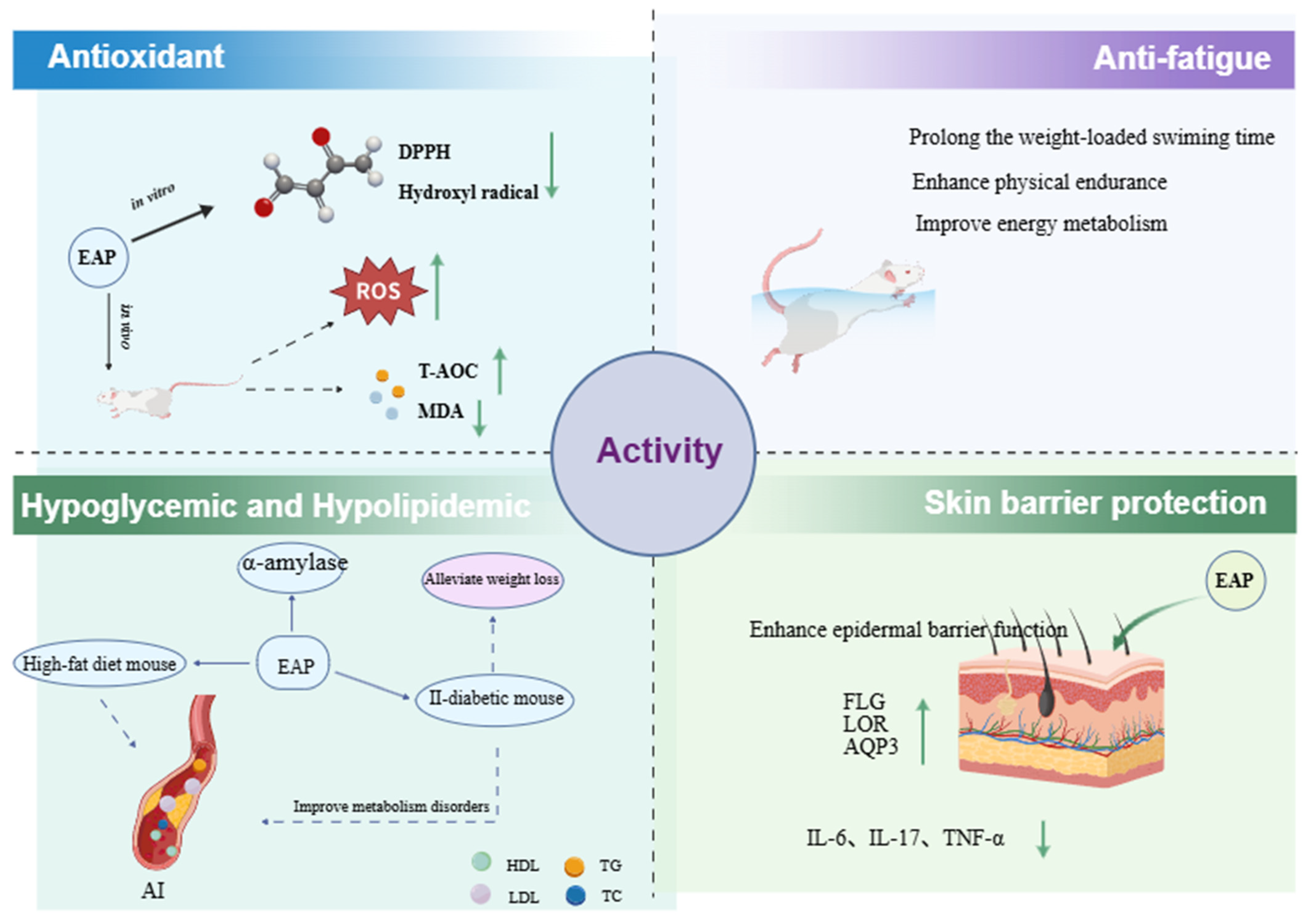
In recent years, antioxidants derived from natural products, particularly polysaccharides, have garnered increasing attention [71]. The evaluation of antioxidant activity typically includes both in vitro and in vivo experiments. As shown in Figure 2, numerous studies have demonstrated that E. angustifolia polysaccharides exhibit significant in vitro antioxidant activity in a dose-dependent manner. Ding et al. [72] found that E. angustifolia polysaccharides effectively scavenged DPPH and hydroxyl radicals in a clear dose-dependent manner. The IC50 values for DPPH and hydroxyl radical scavenging were 380.58 mg/L and 21.11 g/L, respectively, compared to 319.97 mg/L and 14.13 g/L for the positive control, Vitamin C. Purified fractions obtained from E. angustifolia polysaccharides also displayed significant antioxidant activity. Huojiaaihemaiti et al. [53] isolated two acidic polysaccharides, EAP-H-a1 and EAP-H-a2, from E. angustifolia, and found that both exhibited significant antioxidant capacity within the concentration range of 0.2–1 mg/mL in a dose-dependent manner. Furthermore, the extraction method influences the antioxidant activity of E. angustifolia polysaccharides. Compared to HWE, polysaccharides obtained via UAME showed stronger antioxidant activity, with IC50 values of 0.079 mg/mL and 0.083 mg/mL, respectively [36]. In vivo studies have also confirmed the antioxidant activity of E. angustifolia polysaccharides. Experimental results indicated that intervention with E. angustifolia polysaccharides significantly increased serum total antioxidant capacity (T-AOC) and superoxide dismutase (SOD) activity, while reducing malondialdehyde (MDA) content in high-fat model rats, demonstrating certain in vivo antioxidant capacity [73].

4.2. Hypoglycemic and Hypolipidemic Activity

Elaeagnus angustifolia polysaccharides exhibit inhibitory activity against α-amylase, which is influenced by the extraction method. Li et al. [36] reported that the IC50 values of four E. angustifolia polysaccharides (EAP-H, EAP-M, EAP-U, EAP-UM) against α-amylase were 3.236 mg/mL, 3.154 mg/mL, 3.257 mg/mL, and 2.826 mg/mL, respectively, with EAP-UM obtained by UAME exhibiting the strongest inhibitory effect. Further in vivo experiments demonstrated that E. angustifolia polysaccharides significantly reduced blood glucose levels in a type 2 diabetic mouse model. By the fourth week of administration, its hypoglycemic effect was comparable to that of the acarbose positive control group. Moreover, E. angustifolia polysaccharides effectively alleviated diabetes-induced weight loss, improved lipid metabolism disorders, significantly reduced serum triglyceride and total cholesterol levels, and increased high-density lipoprotein levels [74].
Regarding hypolipidemic effects, E. angustifolia polysaccharides significantly reduce serum total cholesterol, triglyceride, and low-density lipoprotein levels, lower the atherosclerosis index, and increase high-density lipoprotein levels in high-fat diet mouse models. Additionally, they effectively mitigate high-fat diet-induced weight gain and decrease liver and kidney coefficients, indicating a notable organ-protective effect [52,73].

4.3. Antitumor and Immunomodulatory Activity

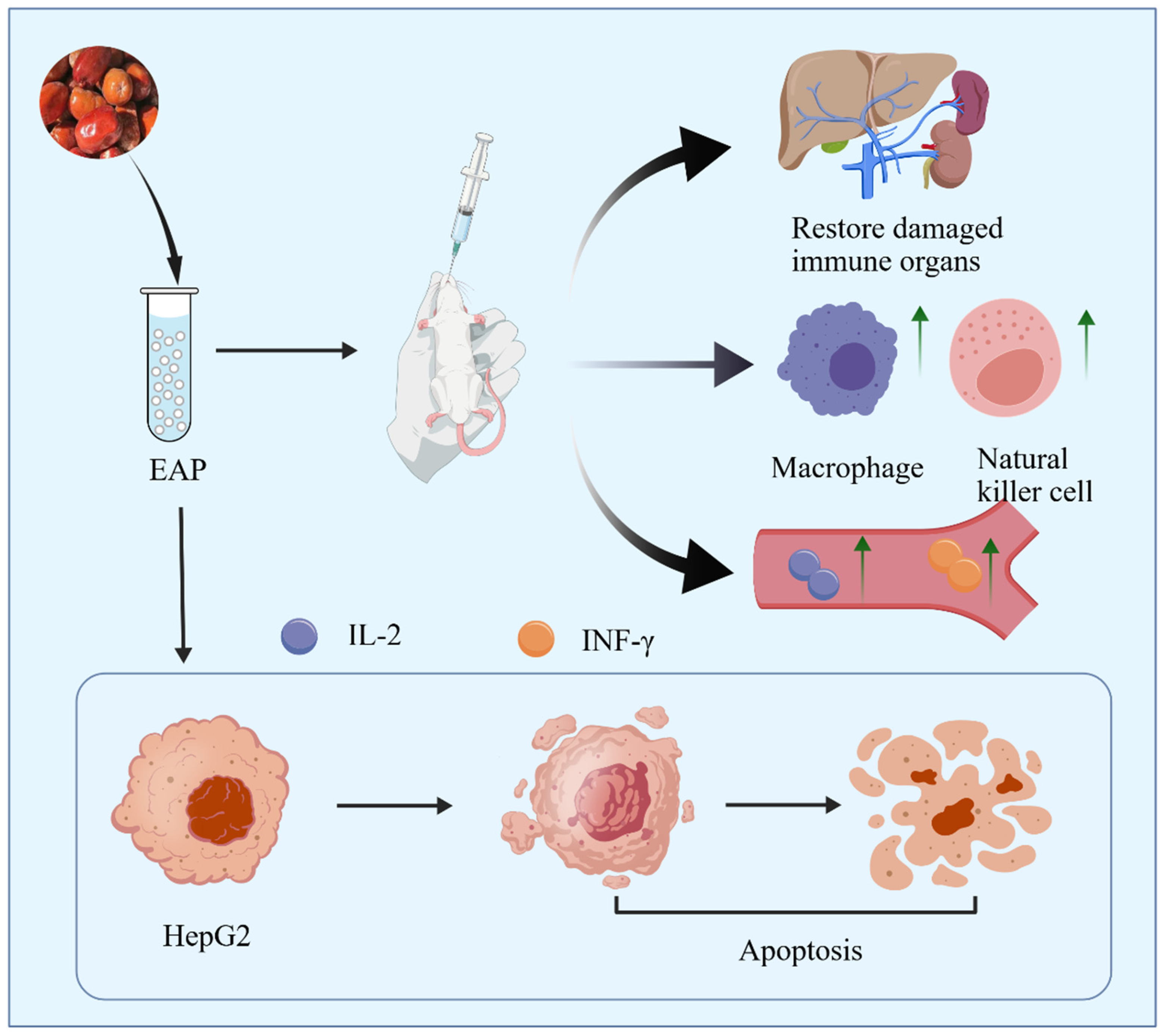
Compared with traditional chemotherapeutic drugs, polysaccharides typically exhibit very low toxicity and good biocompatibility. Their antitumor effects are exerted via direct mechanisms such as inducing tumor cell apoptosis and arresting the cell cycle, or indirectly through immunomodulatory pathways, including activating macrophages and NK cells, promoting the secretion of cytokines (e.g., IL-2, IFN-γ), and modulating the tumor microenvironment. Furthermore, polysaccharides can enhance the efficacy of chemotherapeutic agents while reducing their toxic side effects [75,76]. The antitumor and immunomodulatory effects of Elaeagnus angustifolia polysaccharides are illustrated in Figure 3. Research by Liu et al. [52] showed that E. angustifolia polysaccharides (EAP) inhibited the proliferation of human hepatocellular carcinoma HepG2 cells. After treatment with 50–400 μg/mL EAP for 24 h and 48 h, EAP suppressed HepG2 cell proliferation in a concentration- and time-dependent manner, with inhibition rates increasing with higher concentrations and longer treatment durations. HE staining and Hoechst 33342 staining combined with laser confocal microscopy revealed that the number of tumor cells gradually decreased with prolonged treatment time, exhibiting typical apoptotic morphological changes. Additionally, EAP showed no significant cytotoxicity towards normal human hepatocytes L-02, with inhibition rates below 10%, indicating minimal impact on the growth and proliferation of normal liver cells and suggesting a degree of selectivity.
In terms of immunomodulation, E. angustifolia polysaccharides can restore impaired immune function by enhancing macrophage phagocytosis, increasing NK cell activity, and upregulating cytokine levels (e.g., IL-2, IFN-γ). Wang et al. [77] found that EAP increased the spleen and thymus indices, enhanced mononuclear macrophage phagocytic function, elevated serum hemolysin levels, and promoted lymphocyte proliferation in cyclophosphamide-induced immunosuppressed mice. Wang et al. [78] further confirmed that EAP could increase the spleen index, promote lymphocyte transformation, and enhance the function of antibody-producing cells and the erythrocyte hemolysis value (half value) in immunosuppressed mice, indicating its enhancing effects on both humoral and cellular immunity. Zhao et al. [79] showed that EAP could increase IL-2 and antibody levels in mice infected with respiratory syncytial virus, with the neutralizing antibody titer (1:218) in the 200 mg/kg dose group being significantly higher than that in the virus control group (1:103). Sun et al. [54] discovered that the EAP-1a fraction dose-dependently increased the spleen index, enhanced NK cell activity, stimulated peritoneal macrophage phagocytic function, and upregulated IL-2, IFN-γ, and antibody levels in immunosuppressed mice. Du et al. [23] also reported that the crude E. angustifolia polysaccharide EAP and its fractions EAP-1a and EAP-1b could dose-dependently stimulate NO release from RAW264.7 macrophages and promote cell growth without significant cytotoxicity. Studies by Huojiaaihemaiti et al. [53] similarly demonstrated that fractions such as EAP-H-C, EAP-H-a1, and EAP-H-a2 could activate macrophage function and enhance NO release from RAW264.7 cells in a dose-dependent manner.

4.4. Other Bioactivities

In addition to antioxidant, hypoglycemic, hypolipidemic, antitumor, and immunomodulatory effects, EAP also show potential applications in radiation protection, anti-fatigue, and skin barrier protection. Zhao et al. [80] demonstrated that EAP could significantly increase white blood cell and lymphocyte counts, elevate the spleen index, effectively alleviate radiation-induced immunosuppression, and enhance immune function in radiation-injured mice. Concurrently, EAP enhanced the activities of superoxide dismutase (SOD) and glutathione peroxidase (GSH-Px), improved antioxidant capacity, and reduced oxidative stress damage, thereby exerting protective effects against radiation-induced multi-system dysfunction. Ding et al. [81] found that EAP significantly prolonged the weight-loaded swimming time, increased liver glycogen stores, enhanced physical endurance, and improved energy metabolism in mice. Furthermore, EAP reduced serum urea nitrogen levels and decreased blood lactate accumulation, thereby regulating metabolite accumulation and alleviating post-exercise metabolic burden.
Wang et al. [82] showed that EAP significantly reduced nitric oxide (NO) levels in LPS-induced HaCaT cells and dose-dependently inhibited the release of pro-inflammatory cytokines such as IL-6, IL-1β, and TNF-α. In an acetone/ether-induced dry skin mouse model, EAP effectively enhanced epidermal barrier function, reduced inflammatory cell infiltration and epidermal hyperplasia, and accelerated barrier repair. It also upregulated the expression of filaggrin (FLG), loricrin (LOR), and aquaporin 3 (AQP3) at both the protein and gene levels, alleviated skin dryness and structural damage, and significantly reduced the expression levels of pro-inflammatory factors such as IL-6, IL-17, and TNF-α. In summary, E. angustifolia polysaccharides can protect and repair the dry skin barrier by inhibiting inflammatory responses and upregulating the expression of structure-related proteins.

5. Conclusions and Perspectives

In recent years, significant progress has been made in the isolation, purification, structural characterization, and bioactivity studies of Elaeagnus angustifolia polysaccharides. Currently, the development of E. angustifolia resources is primarily focused on areas such as food processing and ecological protection, while systematic research on its active components, particularly polysaccharides, remains relatively underdeveloped. Numerous scientific questions still need to be addressed, including the optimization of efficient extraction and purification processes, structural modification of polysaccharides, detailed structural elucidation, in-depth explanation of physiological activity mechanisms, and industrial application. Given the unique ecological value and rich resource advantages of E. angustifolia, combined with the broad application prospects of natural polysaccharides, conducting in-depth research on E. angustifolia polysaccharides will not only provide theoretical basis and technical support for their further development and application but also vigorously promote the high-value utilization of E. angustifolia resources.
Currently, several key issues remain to be addressed in research on Elaeagnus angustifolia polysaccharides. First, the fine structural features of these polysaccharides remain largely unexplored. In-depth information regarding the glycosidic linkage patterns and spatial conformations of homogeneous polysaccharide fractions is still insufficient, despite the well-established correlation between polysaccharide structure and bioactivity. For instance, polysaccharides featuring a linear β-(1→3)-D-glucose backbone with a high proportion of short β-(1→6)-D-glucose branches typically exhibit higher anticancer activity, whereas those with a β-(1→6)-D-glucose backbone tend to show lower anticancer activity [83]. This gap hinders the investigation of the structure–activity relationships of Elaeagnus angustifolia polysaccharides. Techniques such as methylation analysis [84], high-resolution mass spectrometry (MALDI-TOF/ESI-MS) [85], and two-dimensional nuclear magnetic resonance (2D NMR) spectroscopy [86] offer promising approaches to address this gap. Second, polysaccharide derivatization often enhances bioactivity; however, research on the derivatization of Elaeagnus angustifolia polysaccharides remains largely unexplored. Derivatization strategies such as phosphorylation, selenylation, and bioconjugation could be applied to modify these polysaccharides [87,88]. Third, pharmacological studies on Elaeagnus angustifolia polysaccharides have largely been confined to phenotypic observations at the cellular or animal model level, while elucidation of the underlying molecular mechanisms—including key signaling pathways, target recognition, and molecular interactions—remains insufficient. Techniques such as Western blotting, qPCR, transcriptomics, and metabolomics will be essential for elucidating the metabolic pathways and signaling networks regulated by these polysaccharides in vivo [89,90].
In recent years, as awareness of green and natural food additives has increased, natural products have gained growing attention for such applications. Elaeagnus angustifolia polysaccharides can be utilized as food or feed additives in the food and feed industries [91]. In addition, owing to their favorable film-forming properties and biocompatibility, they can be employed in the development of capsules, microspheres, and nanoparticles for drug release and targeted delivery [92]. However, their mechanical strength, thermal stability, and water resistance currently limit their broader application in high-end industrial sectors. Targeted modification to alter their physicochemical properties represents a key strategy for overcoming these challenges [93].
In summary, Elaeagnus angustifolia polysaccharide is a natural bioactive compound with antioxidant, antitumor, immunomodulatory, and hypoglycemic activities, while numerous research questions remain to be addressed.

Author Contributions

Writing—original draft, X.F., supervision, W.W., writing—review and editing, W.W. and X.F. All authors have read and agreed to the published version of the manuscript.

Funding

This work was supported by National Key R&D Program of China (2024YFD1300804-1), Scientific Research Funds of Huaqiao University.

Data Availability Statement

Not applicable.

Conflicts of Interest

The authors confirm that this article content has no conflicts of interest.

Abbreviations

The following abbreviations are used in this manuscript:
HWEHot water extraction
RSMResponse surface methodology
UAEUltrasound-assisted extraction
BBDBox–Behnken design
MAEMicrowave-assisted extraction
UAMEUltrasonic/microwave-assisted extraction
EAEEnzyme-assisted extraction
TCATrichloroacetic acid
IECIon-exchange chromatography
HPGPCHigh-performance gel permeation chromatography
EAPElaeagnus angustifolia polysaccharides
SEMScanning electron microscopy
AFMAtomic force microscopy
NMRNuclear magnetic resonance
FT-IRFourier-transform infrared

References

  1. Farzaei, M.H.; Bahramsoltani, R.; Abbasabadi, Z.; Rahimi, R. A comprehensive review on phytochemical and pharmacological aspects of Elaeagnus angustifolia L. J. Pharm. Pharmacol. 2015, 67, 1467–1480. [Google Scholar] [CrossRef]
  2. Wang, Z.L.; Zhao, X.; Liu, L.D.; Bai, X.F.; Zhu, J.J. A comparative study on the adaptation of Populus euphratica and Elaeagnus angustifolia to desert environments. J. Desert Res. 2015, 35, 160–166. (In Chinese) [Google Scholar]
  3. Wu, H.W.; Yang, X.Y.; Wang, J.P.; Liu, Z.X.; Zhu, J.F.; Zhang, H.X. A review on the improvement of salt-affected soil nutrients by Elaeagnus angustifolia L. Chin. J. Ecol. 2019, 38, 3527–3534. (In Chinese) [Google Scholar]
  4. Azmat, M.A.; Shahzad, A.; Shaban, M.; Khan, A.A.; Shakoor, S. Exploring the Multifaceted Potential of Elaeagnus angustifolia L.: A Comprehensive Review of Its Nutritional, Pharmacological, and Environmental Significance. Food Sci. Nutr. 2025, 13, e70486. [Google Scholar] [CrossRef]
  5. Katz, G.L.; Shafroth, P.B. Biology, ecology and management of Elaeagnus angustifolia L. (Russian olive) in western North America. Wetlands 2003, 23, 763–777. [Google Scholar] [CrossRef]
  6. Hao, K.Y.; Zhou, Q.; Li, C.C.; Qi, R.H.; Tian, Y.; Pei, J.H.; Ren, L.L.; Luo, Y.Q. Effects of irrigation amount on gum exudation of Elaeagnus angustifolia and its capacity to attract and kill Anoplophora glabripennis in arid regions. Chin. J. Appl. Entomol. 2025, 62, 53–64. (In Chinese) [Google Scholar]
  7. An, X.; Yang, R.; Chen, C.; Liu, W. Research progress on the chemical constituents and pharmacological effects of Elaeagnus angustifolia flower. Xinjiang J. Tradit. Chin. Med. 2023, 41, 105–110. (In Chinese) [Google Scholar]
  8. Zakaria, Z.Z.; Ahen, Y.M.A.; Ahmed, N.H.; Sokary, S.; Abu-hijleh, H.; Bawadi, H.; Al-Asmakh, M. Elaeagnus Angustifolia: Unveiling Anti-Cancer Potential from Ancient Remedies to Modern Therapeutics. Cancer Manag. Res. 2026, 18, 1–13. [Google Scholar] [CrossRef]
  9. Mahboubi, M. Elaeagnus angustifolia and its therapeutic applications in osteoarthritis. Ind. Crops Prod. 2018, 121, 36–45. [Google Scholar] [CrossRef]
  10. Zor, M.; Saglamtas, R.; Demirci, A.; Ceyran, E.; Fettahoglu, K.; Demir, Y. Chemical Composition, Antimicrobial, and Enzyme Inhibitory Properties of Elaeagnus angustifolia L. Chem. Biodivers. 2025, 22, e202402802. [Google Scholar] [CrossRef]
  11. Cansev, A.; Sahan, Y.; Celik, G.; Taskesen, S.; Ozbey, H. Chemical Properties and Antioxidant Capacity of Elaeagnus angustifolia L. Fruits. Asian J. Chem. 2011, 23, 2661–2665. [Google Scholar]
  12. Ahmadiani, A.; Hosseiny, J.; Semnanian, S.; Javan, M.; Saeedi, F.; Kamalinejad, M.; Saremi, S. Antinociceptive and anti-inflammatory effects of Elaeagnus angustifolia fruit extract. J. Ethnopharmacol. 2000, 72, 287–292. [Google Scholar] [CrossRef] [PubMed]
  13. Ullah, S.; Khalil, A.A.; Shaukat, F.; Song, Y.D. Sources, Extraction and Biomedical Properties of Polysaccharides. Foods 2019, 8, 304. [Google Scholar] [CrossRef]
  14. Li, Y.Z.; Jasar, A.; Wei, W.; Wan, J.Y.; Hadiya, A.; Eksan, P.; Zumrat, A. Research Progress on the Structural Characteristic and Biological Activity of Eleagnus angustifolia L. Polysaccharides. Spec. Wild Econ. Anim. Plant Res. 2026, 48, 179–183+201. (In Chinese) [Google Scholar]
  15. Emre, U.T.; Sirin, S.; Dolanbay, S.N.; Aslim, B. Harnessing polysaccharides for sustainable food packaging. Polym. Bull. 2025, 82, 2779–2825. [Google Scholar] [CrossRef]
  16. Lu, S.Y.; Zhou, T.; Shabbir, I.; Choi, J.; Kim, Y.H.; Park, M.; Aweya, J.J.; Tan, K.; Zhong, S.Y.; Cheong, K.L. Marine algal polysaccharides: Multifunctional bioactive ingredients for cosmetic formulations. Carbohydr. Polym. 2025, 353, 123276. [Google Scholar] [CrossRef]
  17. Luo, Y.H.; Han, P.; Tu, L.X.; Cheng, M. Research progress on extraction technology of traditional Chinese medicine polysaccharides and their application in drug delivery systems. J. Guangdong Pharm. Univ. 2025, 82, 180–190. [Google Scholar]
  18. Jin, W.X.; Zhang, Z.Q.; Guo, F.F.; Huang, S.C. Research Progress on the Application of Rehmannia glutinosa Polysaccharides in Livestock and Poultry Production. J. Shanxi Agric. Sci. 2025, 53, 39–46. (In Chinese) [Google Scholar]
  19. Murphy, E.J.; Fehrenbach, G.W.; Abidin, I.Z.; Buckley, C.; Montgomery, T.; Pogue, R.; Murray, P.; Major, I.; Rezoagli, E. Polysaccharides-Naturally Occurring Immune Modulators. Polymers 2023, 15, 2373. [Google Scholar] [CrossRef]
  20. Wei, P.P.; Deng, M.Y.; Ding, Y.Z.; Xia, D.; Yan, Z.J.; Chen, T. Antioxidant activity of polysaccharides from natural sources. Acta Pol. Pharm. 2023, 80, 705–716. [Google Scholar]
  21. Guo, R.; Chen, M.; Ding, Y.Y.; Yang, P.Y.; Wang, M.J.; Zhang, H.H.; He, Y.Q.; Ma, H.L. Polysaccharides as Potential Anti-Tumor Biomacromolecules-A Review. Front. Nutr. 2022, 9, 838179. [Google Scholar] [CrossRef]
  22. Li, X.H. Study on Extraction, Isolation and Purification of Polysaccharides from Xinjiang Elaeagnus angustifolia. Master’s Thesis, Shihezi University, Shihezi, China, 2011. (In Chinese) [Google Scholar]
  23. Chen, Q.Q.; Yang, J.F.; Liu, H. Purification and Antioxidant Activity of Polysaccharide from Elaeagnus angustifolia in Xinjiang. Food Res. Dev. 2016, 37, 37–40. (In Chinese) [Google Scholar]
  24. Du, H.T.; Chen, J.C.; Tian, S.; Gu, H.L.; Li, N.; Sun, Y.; Ru, J.J.; Wang, J.R. Extraction optimization, preliminary characterization and immunological activities in vitro of polysaccharides from Elaeagnus angustifolia L. pulp. Carbohydr. Polym. 2016, 151, 348–357. [Google Scholar] [CrossRef]
  25. Nai, J.J.; Zhang, C.; Shao, H.L.; Li, B.Q.; Li, H.; Gao, L.; Dai, M.M.; Zhu, L.Q.; Sheng, H.G. Extraction, structure, pharmacological activities and drug carrier applications of Angelica sinensis polysaccharide. Int. J. Biol. Macromol. 2021, 183, 2337–2353. [Google Scholar] [CrossRef]
  26. Cheng, Y.; Gan, J.; Yan, B.W.; Wang, P.; Wu, H.; Huang, C.X. Polysaccharides from Russula: A review on extraction, purification, and bioactivities. Front. Nutr. 2024, 11, 1406817. [Google Scholar] [CrossRef]
  27. Chen, L. Study on Preparation, Structural Characteristics and Emulsifying Properties of Polysaccharides from Large-Fruited Elaeagnus angustifolia. Master’s Thesis, Tarim University, Alar, China, 2024. (In Chinese) [Google Scholar]
  28. Fu, T.T.; Sun, J.C.; Han, S.C.; Zhang, Y.X.; Guo, J.P.; Luan, T.L.; Wu, H.J.; Xu, X.Y. Experimental Principle Verification and Application of Phenol-sulfuric Acid Method for Determination of Total Polysaccharide Content. Guangzhou Chem. Ind. 2025, 53, 128–131+138. (In Chinese) [Google Scholar]
  29. Lun, Q.J.; Luo, P.Y. Comparison of Phenol-sulfuric Acid Method and Anthrone Sulfate Method for Determining the Content of Ganoderma lucidum Polysaccharides in Ganoderma lucidum Spore. China Food Saf. 2024, 14, 89–91+95. (In Chinese) [Google Scholar]
  30. Ferreira, S.L.C.; Bruns, R.E.; Ferreira, H.S.; Matos, G.D.; David, J.M.; Brandao, G.C.; da Silva, E.G.P.; Portugal, L.A.; Reis, P.S.; Souza, A.S.; et al. Box-Behnken design: An alternative for the optimization of analytical methods. Anal. Chim. Acta 2007, 597, 179–186. [Google Scholar] [CrossRef]
  31. Yang, Y.; Li, M.; Sun, J.W.; Qin, S.H.; Diao, T.T.; Bai, J.W.; Li, Y.H. Microwave-assisted aqueous two-phase extraction of polysaccharides from Hippophae rhamnoide L.: Modeling, characterization and hypoglycemic activity. Int. J. Biol. Macromol. 2023, 254, 127626. [Google Scholar] [CrossRef]
  32. Hang, H.; Chen, R.Z.; Wang, C.B.; Sun, Y.R.; Du, D.S. A review of the extraction processes and biological characteristics of Chrysanthemum polysaccharides. Int. J. Biol. Macromol. 2024, 285, 138224. [Google Scholar] [CrossRef]
  33. Teng, H.; He, Z.G.; Hong, C.Z.; Xie, S.Z.; Zha, X.Q. Extraction, purification, structural characterization and pharmacological activities of polysaccharides from sea buckthorn (Hippophae rhamnoides L.): A review. J. Ethnopharmacol. 2024, 324, 117809. [Google Scholar] [CrossRef]
  34. Zhang, S.; Chen, L.; Shang, N.; Wu, K.F.; Liao, W. Recent Advances in the Structure, Extraction, and Biological Activity of Sargassum fusiforme Polysaccharides. Mar. Drugs 2025, 23, 98. [Google Scholar] [CrossRef]
  35. Li, X.H.; Tong, J.M. Optimization of extraction technology of polysaccharides from Xinjiang Elaeagnus angustifolia. Farm Prod. Process. 2011, 5, 73–76. [Google Scholar]
  36. Li, J.F.; Chen, Y.; Yan, Q.; Li, C.Y.; Yao, Y.H.; Zhao, J. Ultrasonic/microwave-assisted extraction and properties of polysaccharides from Elaeagnus angustifolia. Prep. Biochem. Biotechnol. 2025, 55, 776–786. [Google Scholar] [CrossRef]
  37. Chen, L.H.; Cui, C.F.; Wang, Z.H.; Che, F.H.; Chen, Z.X.; Feng, S.B. Structural Characterization and Antioxidant Activity of β-Glucans from Highland Barley Obtained with Ultrasonic-Microwave-Assisted Extraction. Molecules 2024, 29, 684. [Google Scholar] [CrossRef]
  38. Xiong, W.Y.; Kumar, G.; Zhang, B.; Dhital, S. Sonication-mediated modulation of macronutrient structure and digestibility in chickpea. Ultrason. Sonochem. 2024, 106, 106904. [Google Scholar] [CrossRef]
  39. You, Q.H.; Yin, X.L.; Zhao, Y.P. Enzyme assisted extraction of polysaccharides from the fruit of Cornus officinalis. Carbohydr. Polym. 2013, 98, 607–610. [Google Scholar] [CrossRef]
  40. Lin, B.B.; Wang, S.S.; Zhou, A.Q.; Hu, Q.R.; Huang, G.L. Ultrasound-assisted enzyme extraction and properties of Shatian pomelo peel polysaccharide. Ultrason. Sonochem. 2023, 98, 607–610. [Google Scholar] [CrossRef]
  41. Lian, Y.J.; Li, Y.J.; Liu, H.; Ma, Y.M.; Chen, H.Y. Study on Extraction Process of Polysaccharides from Elaeagnus angustifolia L. by Enzymatic-assisted Ultrasonic Methods. Chin. J. Inf. Tradit. Chin. Med. 2009, 16, 47–48+59. (In Chinese) [Google Scholar]
  42. Bhati, N.; Shreya; Sharma, A.K. Cost-effective cellulase production, improvement strategies, and future challenges. J. Food Process Eng. 2021, 44, e13623. [Google Scholar] [CrossRef]
  43. Sakr, E.A.E. Structural characterization and health benefits of a novel fructan produced by fermentation of an Asparagus sprengeri extract by Lactobacillus plantarum DMS 20174. Process Biochem. 2022, 118, 370–380. [Google Scholar] [CrossRef]
  44. Drenker, C.; El Mazouar, D.; Seib, T.; Weisshaupt, S.; Stephan, D.; Kunz, S.; Reineke, A.; Rondot, Y.; Linkies, A.; Koch, E. Biocontrol of plant pathogens by Lysobacter enzymogenes isolate LEC: Efficacy of shake flask broth, fermentation broth and dried preparations. Biocontrol Sci. Technol. 2025, 35, 1–20. [Google Scholar] [CrossRef]
  45. Wang, Q.N.; Lu, X.L.; Peng, Y.S.; Wu, J.L. Extraction, Purification and Preliminary Analysis of Eleagnus angustifolius L. Polysaccharide-2. Food Sci. 2007, 28, 43–46. (In Chinese) [Google Scholar]
  46. Chen, Y.J.; Cheng, J.Y.; Zhu, L.W.; Li, F. Study on Different Methods of Deproteinization of Polygonatum Sibiricum Polysaccharide and Its Antioxidant Activity. Guangdong Chem. Ind. 2023, 50, 9–11. (In Chinese) [Google Scholar]
  47. Yang, W.J.; Huang, G.L. Preparation and analysis of polysaccharide from Solanum tuberdsm. Ultrason. Sonochem. 2023, 98, 106520. [Google Scholar] [CrossRef]
  48. Huang, G.L.; Chen, F.; Yang, W.J.; Huang, H.L. Preparation, deproteinization and comparison of bioactive polysaccharides. Trends Food Sci. Technol. 2021, 109, 564–568. [Google Scholar] [CrossRef]
  49. Hu, J.X.; Cao, Y.; Di, D.L.; Wu, Y.X.; Zhao, L.C.; Pei, D.; Liu, J.F. Plant-Derived Polysaccharide Ultrafast Purification Reinvented: A Green Aqueous Two-Phase System with Deep Eutectic Solvents for Simultaneous Decolorization and Deproteinization. Anal. Chem. 2026, 98, 2244–2256. [Google Scholar] [CrossRef]
  50. Shao, L.J.; Sun, Y.; Liang, J.; Li, M.Q.; Li, X.L. Decolorization affects the structural characteristics and antioxidant activity of polysaccharides from Thesium chinense Turcz: Comparison of activated carbon and hydrogen peroxide decolorization. Int. J. Biol. Macromol. 2020, 155, 1084–1091. [Google Scholar] [CrossRef] [PubMed]
  51. Li, Y.Y.; Li, X.; Lian, Y.J.; Li, B.Q. Optimization for conditions on Elaeagnus polysaccharides decoloring material. Food Sci. Technol. 2012, 37, 210–213. (In Chinese) [Google Scholar]
  52. Liu, X.Q. Purification, Structural Characterization and Bioactivities of Polysaccharides from Elaeagnus angustifolia. Master’s Thesis, Tianjin University of Science and Technology, Tianjin, China, 2015. [Google Scholar]
  53. Huojiaaihemaiti, H.; Mutaillifu, P.; Omer, A.; Nuerxiati, R.; Duan, X.M.; Xin, X.L.; Yili, A. Isolation, Structural Characterization, and Biological Activity of the Two Acidic Polysaccharides from the Fruits of the Elaeagnus angustifolia Linnaeus. Molecules 2022, 27, 6415. [Google Scholar] [CrossRef] [PubMed]
  54. Sun, N.X.; Liu, H.P.; Liu, X.H.; Zhang, Y.; Liu, X.Q.; Wang, S.; Xu, X.X.; Tian, W.T. Immunological activities of polysaccharide extracted from Elaeagnus angustifolia L. Cyta-J. Food 2018, 16, 995–1002. [Google Scholar] [CrossRef]
  55. Guo, J.; Jia, G.J.; Tao, L.; Wang, R.X. Research Progress on Structure and Biological Function of Polysaccharides. J. Food Saf. Guide 2021, 33, 137–139. (In Chinese) [Google Scholar]
  56. Liu, H.Z.; Huang, H.Y.; Yang, L.N.; Wang, S.N.; Li, J.; Zhu, L.J.; Zhu, D.S.; He, Y.T. Research progress on structural analysis of polysaccharides. J. Bohai Univ. (Nat. Sci. Ed.) 2018, 28, 97–106. [Google Scholar]
  57. Chen, S.K.; Wang, X.; Guo, Y.Q.; Song, X.X.; Yin, J.Y.; Nie, S.P. Exploring the partial degradation of polysaccharides: Structure, mechanism, bioactivities, and perspectives. Compr. Rev. Food Sci. Food Saf. 2023, 22, 4831–4870. [Google Scholar] [CrossRef] [PubMed]
  58. Liu, H.; Xu, J.X.; Xu, X.Y.; Yuan, Z.H.; Song, H.; Yang, L.N.; Zhu, D.S. Structure/function relationships of bean polysaccharides: A review. Crit. Rev. Food Sci. Nutr. 2023, 63, 330–344. [Google Scholar]
  59. Chen, K. Research on Extraction, Purification and Antioxidant Activity of Polysaccharides from Elaeagnus angustifolia. Master’s Thesis, Northwest A&F University, Xianyang, China, 2015. [Google Scholar]
  60. Lee, Q.C.; Xue, Z.X.; Luo, Y.J.; Lin, Y.P.; Lai, M.Y.; Xu, H.Y.; Liu, B.; Zheng, M.F.; Lv, F.; Zeng, F. Low molecular weight polysaccharide of Tremella fuciformis exhibits stronger antioxidant and immunomodulatory activities than high molecular weight polysaccharide. Int. J. Biol. Macromol. 2024, 281, 136097. [Google Scholar] [CrossRef]
  61. Hui, H.P.; Mao, H.J.; Wei, J.; Xue, X.D.; Cheng, M. Optimization of microwave-assisted extraction for Herba Patriniae polysaccharide and its impact on physicochemical properties, antioxidant and hypoglycemic capacities: Primary structure-activity relationship. Int. J. Biol. Macromol. 2025, 319, 145396. [Google Scholar] [CrossRef]
  62. Ju, T.; Yin, Y.S.; Gao, H.Q.; Wang, J.Y.; Ye, G.J.; Li, J.F.; Zhang, Y.C.; Miao, F.J.; Lu, W.H. Structural characteristics, antioxidant and immunological activities of polysaccharides from Ficus hirta Vahl based on different extraction methods. Ind. Crops Prod. 2026, 241, 122741. [Google Scholar] [CrossRef]
  63. Chen, N.; Jiang, T.Y.; Xu, J.X.; Xi, W.J.; Shang, E.; Xiao, P.; Duan, J.A. The relationship between polysaccharide structure and its antioxidant activity needs to be systematically elucidated. Int. J. Biol. Macromol. 2024, 270, 132391. [Google Scholar] [CrossRef]
  64. Shi, L.T.; He, Q.; Li, J.; Liu, Y.L.; Cao, Y.L.; Liu, Y.Q.; Sun, C.D.; Pan, Y.J.; Li, X.; Zhao, X.Y. Polysaccharides in fruits: Biological activities, structures, and structure-activity relationships and influencing factors-A review. Food Chem. 2024, 451, 139408. [Google Scholar] [CrossRef]
  65. Bu, Y.K.; Yin, B.F.; Qiu, Z.C.; Li, L.Y.; Zhang, B.; Zheng, Z.J.; Li, M.M. Structural characterization and antioxidant activities of polysaccharides extracted from Polygonati rhizoma pomace. Food Chem.-X 2024, 23, 101778. [Google Scholar] [CrossRef]
  66. Yao, H.Y.Y.; Wang, J.Q.; Yin, J.Y.; Nie, S.P.; Xie, M.Y. A review of NMR analysis in polysaccharide structure and conformation: Progress, challenge and perspective. Food Res. Int. 2021, 143, 110290. [Google Scholar] [CrossRef] [PubMed]
  67. Liu, Y.Q.; Gao, L.N.; Yu, Z.L. Revealing the Complexity of Polysaccharides: Advances in NMR Spectroscopy for Structural Elucidation and Functional Characterization. Appl. Sci. 2025, 15, 5246. [Google Scholar] [CrossRef]
  68. Chen, K.W.; Zhang, Q.Q.; Yang, S.Z.; Zhang, S.Y.; Chen, G.J. Comparative Study on the Impact of Different Extraction Technologies on Structural Characteristics, Physicochemical Properties, and Biological Activities of Polysaccharides from Seedless Chestnut Rose (Rosa sterilis) Fruit. Foods 2024, 13, 772. [Google Scholar] [CrossRef] [PubMed]
  69. Li, Y.Y.; Li, X.Q.; Yuan, Q.; Zhao, J.L.; Wu, W.; Gui, Y.B.; Wang, H.L.; Wang, L.J.; Luo, Y.Y.; Zhou, G.; et al. Polysaccharides from Balanophora harlandii Hook: Isolation, characterization, and anti-inflammation activities. J. Pharm. Biomed. Anal. 2024, 246, 116252. [Google Scholar] [CrossRef] [PubMed]
  70. Guo, X.Y.; Kang, J.; Xu, Z.Y.; Guo, Q.B.; Zhang, L.F.; Ning, H.F.; Cui, S.W. Triple-helix polysaccharides: Formation mechanisms and analytical methods. Carbohydr. Polym. 2021, 262, 117962. [Google Scholar] [CrossRef]
  71. Fernandes, P.R.; Coimbra, M.A. The antioxidant activity of polysaccharides: A structure-function relationship overview. Carbohydr. Polym. 2023, 314, 120965. [Google Scholar] [CrossRef]
  72. Ding, Y.S.; Ma, R.L.; Niu, Q.; Feng, G.L.; Xu, S.Z.; Lyu, F.L.; Tang, M.E. Experimental study on antioxidant activity of polysaccharides from Elaeagnus angustifolia. Agric. Reclam. Med. 2009, 31, 511–513. [Google Scholar]
  73. Bai, Y.L. Study on Lipid-Lowering, Weight-Reducing and Antioxidant Effects of Elaeagnus angustifolia Polysaccharides in Rats. Master’s Thesis, Jilin University, Changchun, China, 2007. (In Chinese) [Google Scholar]
  74. Wang, Y.; Zou, H.M.; Sun, C.; Guo, T.; Feng, Z.P. Hypoglycemic potential of Elaeagnus angustifolia fruit polysaccharide on streptozotocin-induced type 2 diabetic mice. Bangladesh J. Pharmacol. 2018, 13, 297–301. [Google Scholar] [CrossRef]
  75. Xiong, H.T.; Han, X.P.; Cai, L.; Zheng, H.G. Natural polysaccharides exert anti-tumor effects as dendritic cell immune enhancers. Front. Oncol. 2023, 13, 1274048. [Google Scholar] [CrossRef]
  76. Sun, X.Q.; Wei, Z.X.; Su, Y.W.; Fang, R.; Fan, Y.Z.; Zeng, D.J.; Ding, Q.K.; Miao, Y.Q.; Liu, J.H.; Sun, Q. Structural characteristics and anti-tumor activities of a novel polysaccharide from Klebsiella sp. Carbohydr. Polym. 2025, 356, 123368. [Google Scholar] [CrossRef]
  77. Chen, H.Y.; Yang, Y.Y.; Lian, Y.J.; Li, B.Q. Study on effects of polysaccharides from three characteristic Xinjiang medicinal plants on immune function in mice. Lishizhen Med. Mater. Medica Res. 2011, 22, 2226–2227. (In Chinese) [Google Scholar]
  78. Wang, Z.; Ma, R.L.; Niu, Q.; Xu, S.Z.; Feng, G.L.; Tang, M.E.; Ding, Y.S. Effect of polysaccharides from Elaeagnus angustifolia L. on immune function in mice. Mod. Prev. Med. 2013, 40, 222–224. (In Chinese) [Google Scholar]
  79. Zhao, H.J.; Li, J.; Zhang, G.Y.; Qu, J.L.; Tang, H.B.; Tian, L.L.; Wang, B.G. Effect of Elaeagnus angustifolia polysaccharides on humoral immune function in mice infected with respiratory syncytial virus. Chin. J. Gerontol. 2013, 33, 4180–4181. (In Chinese) [Google Scholar]
  80. Zhao, H.J.; Zhang, G.Y.; Tang, H.B.; Tian, L.L.; Wang, B.G. The assessment of assisting irradiation hazard protection function of Elaeagnus angustifolia polysaccharide on mice. Chin. Prev. Med. 2012, 13, 107–108. (In Chinese) [Google Scholar]
  81. Ding, Y.S.; Wang, Z.; Ma, R.L.; Feng, G.L.; Xu, S.Z.; Lu, F.L.; Tang, M.E.; Zheng, R.H. Anti-fatigue Effect and Mechanism of Polysaccharides from Eleagnus angustifolius L. Food Sci. 2010, 31, 255–257. (In Chinese) [Google Scholar]
  82. Wang, B.Y.; Tai, M.L.; Zhang, K.; Chen, H.X.; Gan, X.S.; Che, B.; Abudukelimu, N.; Wang, G.P.; Xin, X.L.; Lin, L.; et al. Elaeagnus L gum polysaccharides alleviate the impairment of barrier function in the dry skin model mice. J. Cosmet. Dermatol. 2021, 20, 647–656. [Google Scholar] [CrossRef]
  83. Li, N.Y.; Wang, C.F.; Georgiev, M.I.; Bajpai, V.K.; Tundis, P.; Simal-Gandara, J.; Lu, X.M.; Xiao, J.B.; Tang, X.Z.; Qiao, X.G. Advances in dietary polysaccharides as anticancer agents: Structure-activity relationship. Trends Food Sci. Technol. 2021, 111, 360–377. [Google Scholar] [CrossRef]
  84. Pan, Z.C.; Li, A.Z.; Zeng, N.K.; Yang, X.Q.; Xie, H.J.; Chen, J.; Zhao, K.; Wang, Y. Structural characteristics and hypoglycemic activity of a polysaccharide from an edible bolete Phlebopus portentosus. Int. J. Biol. Macromol. 2025, 308, 142587. [Google Scholar] [CrossRef] [PubMed]
  85. Pandeirada, C.O.; Hageman, J.A.; Janssen, H.G.; Westphal, Y.; Schols, H.A. Identification of plant polysaccharides by MALDI-TOF MS fingerprinting after periodate oxidation and thermal hydrolysis. Carbohydr. Polym. 2022, 292, 119685. [Google Scholar] [CrossRef]
  86. Zdorovenko, E.L.; Sigida, E.N.; Dmitrenok, A.S.; Konnova, S.A.; Fedonenko, Y.P. Structure of O-polysaccharide from the Azospirillum himalayense ptl-3T lipopolysaccharide. Carbohydr. Res. 2025, 552, 109471. [Google Scholar] [CrossRef]
  87. Cheng, L.Z.; Wang, Y.F.; He, X.X.; Wei, X.L. Preparation, structural characterization and bioactivities of Se-containing polysaccharide: A review. Int. J. Biol. Macromol. 2018, 120, 82–92. [Google Scholar] [CrossRef] [PubMed]
  88. Gu, H.F.; Liang, L.; Zhu, X.P.; Jiang, X.H.; Du, M.; Wang, Z.C. Optimization of enzymatic extraction, characterization and bioactivities of Se-polysaccharides from Se-enriched Lentinus edodes. Food Biosci. 2022, 51, 102346. [Google Scholar] [CrossRef]
  89. Tao, S.C.; Wang, H.; Ji, Q.F.; Yang, Y.S.; Wei, G.; Li, R.M.; Zhou, B.J. Integration of Metabolomics and Transcriptomics to Reveal the Antitumor Mechanism of Dendrobium officinale Polysaccharide-Based Nanocarriers in Enhancing Photodynamic Immunotherapy in Colorectal Cancer. Pharmaceutics 2025, 17, 97. [Google Scholar] [CrossRef] [PubMed]
  90. Zhang, W.S.; Cui, N.; Su, F.Z.; Liu, M.; Li, B.; Sun, Y.P.; Zeng, Y.N.; Yang, B.Y.; Kuang, H.X.; Wang, Q.H. Effects of Rehmanniae Radix Praeparata polysaccharides on LPS-induced immune activation in mice based on gut microbiota, metabolomics and transcriptomics. Int. J. Biol. Macromol. 2025, 311, 143981. [Google Scholar] [CrossRef]
  91. Luo, M.C.; Zhang, X.Y.; Wu, J.; Zhao, J.M. Modifications of polysaccharide-based biomaterials under structure-property relationship for biomedical applications. Carbohydr. Polym. 2021, 266, 118097. [Google Scholar] [CrossRef]
  92. Chen, X.X.; Shen, M.Y.; Yu, Q.; Chen, Y.; Xie, J.H. Recent advance in chemistry modified methods of natural polysaccharides and their applications. Trends Food Sci. Technol. 2023, 144, 104317. [Google Scholar] [CrossRef]
  93. Wang, Y.; Zou, Y.; Gunarathne, R.; Lu, J.; Sun, P.L. Sulfated seaweed polysaccharides and edible mushroom polysaccharide-based biomaterials for food applications. Int. J. Biol. Macromol. 2026, 344, 150530. [Google Scholar] [CrossRef]
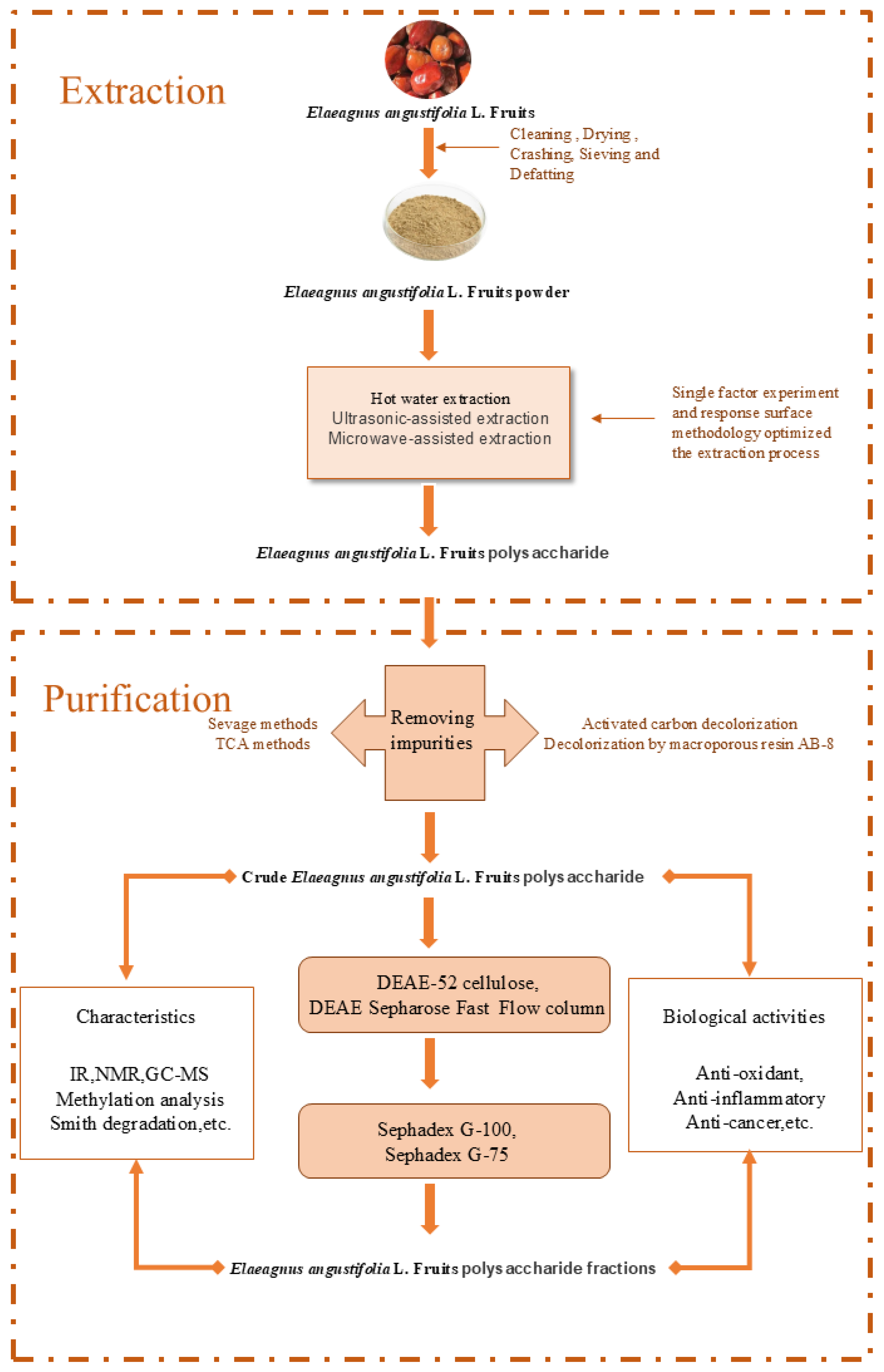
Figure 1. Overview of Extraction, purification, structural characterization, and Bioactivity Study of Elaeagnus angustifolia polysaccharides.
Separations 13 00139 g001
Figure 2. Schematic diagram of the antioxidant, hypoglycemic, and other pharmacological activities of Elaeagnus angustifolia polysaccharides.
Separations 13 00139 g002
Figure 3. Schematic diagram of the antitumor and immunomodulatory effects of Elaeagnus angustifolia polysaccharides.
Separations 13 00139 g003
Table 1. Selected polysaccharides from Elaeagnus angustifolia.
Extraction, Separation, and PurificationYield (%)Compound NameMW
(Da)
Monosaccharide
Composition
Main Structural
Information
BioactivityReference
HWE (solid–liquid ratio 1:10 (g/mL), 3 h, 2 times); TCA + Sevage method; activated carbon; DEAE-Cellulose 0.74 ± 0.01EAP-2NAL-Rha, D-Xyl, L-Ara, D-Fru, D-Man, D-Glc, D-Galα-glycosidic bondNA[45]
HWE (solid–liquid ratio 1:26, temperature 61 °C, time 118.4 min); Sevage method; DEAE-52 + Sephadex G-1004.68 ± 0.64EAP1;
EAP2;
EAP3;
EAP4
4.88 × 10⁴;
3.26 × 10⁴;
1.92 × 10⁴;
1.19 × 10³
Fru, Glc (1:10.681); Glc; Fru:Glc(1:1.545); GlcAll contain α-type glycosidic bondsAntioxidant[59]
HWE (solid–liquid ratio 1:35, temperature 90 °C, time 5 h); AB-8 resin (resin amount 25 g/100 mL, temperature 40 °C, time 3 h); papain method (pH = 6.0, enzyme amount 2.5%, temperature 50 °C, time 1.5 h)5.39 ± 0.63EMP4.24~0.80 × 10³Man, Rha, Glc-UA, Gal-UA, Glc, Gal, AraPossible presence of pyranose ring groups; possible presence of α- or β-glycosidic bonds; possible presence of triple helix structure; negatively charged and relatively stable in solvent systems; clear granular structure, smooth micro-surface, plump morphology, tightly arranged, irregular spherical macromolecular substancesEmulsifying activity[27]
HWE (time 2 h, temperature 80 °C, solid–liquid ratio 1:35, 2 extractions); DEAE-52 + Sephadex G-1005.9EAP-1a6.14 × 103Rha, Ara, Xyl, Man, Glc, and Gal(0.029:0.74:0.51:0.031:1.0:5.19)Has α-pyranose configuration, may contain longer side chains and more branches, possible helical structure in aqueous solutionAntioxidant; Antitumor; Hypolipidemic[54]
UAE (43 min, 282 W, 79 °C, 30:1, 3 times); DEAE-52 + Sephadex G-1009.82 ± 0.38EAP; EAP-1a; EAP-1b8.70 × 103; 4.39 × 10³ (EAP-1a; EAP-1b)EAP: Man (8.5%), Rha (22.2%), Glc (27.8%), Gal (25.1%), Xyl (2.4%), Ara (4.0%); EAP-1a: Man (6.3%), Rha (37.2%), Glc (35.7%), Gal (18.9%), Xyl (1.9%); EAP-1b: Man (8.2%), Rha (15.2%), Glc (50.1%), Gal (26.5%)β-glycosidic bondsImmunomodulatory[25]
HWE (90 °C, 1:30 g/mL, 2 h); DEAE-52 + Sephadex G-75NAEAP-H-a1; EAP-H-a27.05 × 105; 4.39 × 10⁵Rha, Ara, Xyl, Glc, Gal (13.7:20.5:23.3:8.8:33.4; 24.8:19.7:8.2:8.4:38.6)Acidic polysaccharides; possible presence of pyranose ring structure; possible presence of α/β configuration; no triple helix structure; have irregular fibrous, filamentous surfaces; both have crystalline and amorphous structuresAntioxidant; Immunomodulatory[53]
HWE (80 °C); DEAE-52; Sephadex G-100NAPEA-1;
PEA-2
9.11 × 10³; 5.02 × 10³PEA-1: Rha, Xyl, Man, Glc, Gal; PEA-2: Rha, Man, Glc, GalNAAntioxidant[37]
UAME (1:23 g/mL, ultrasound 252 W, microwave 417 W, 11 min); UAE (252 W, 11 min); MAE (417 W, 11 min); HWE (1:25 g/mL, 2 times)7.85; 5.74; 6.86; 4.91EAP-UM; EAP-U; EAP-M; EAP-HNAEAP-UM: Man, Rib, Rha, GlcA, GalA, Glc, Gal, Xyl, Ara, Fuc(0.685:0.113:0.510:0.370:0.744:1.862:2.175:0.195:1.366:0.180); EAP-H: Man, Rib, Rha, GlcA, GalA, Glc, Gal, Xyl, Ara, Fuc (0.285:0.186:0.271:0.114:0.668:4.934:1.939:0.112:2.051:0.131)NAAntioxidant[36]
Note: HWE, hot water extraction; UAE, ultrasound-assisted extraction; UAME, ultrasonic/microwave-assisted extraction; DEAE-52: Diethylaminoethyl Cellulose 52; MW, molecular weight; Glc, glucose; Man, mannose; Gal, galactose; Rha, rhamnose; Ara, arabinose; Xyl, xylose; Fru, fructose; Fuc, fucose; Rib, ribose; GalA, galacturonic acid; GlcA, glucuronic acid; NA, Not Available.
Disclaimer/Publisher’s Note: The statements, opinions and data contained in all publications are solely those of the individual author(s) and contributor(s) and not of MDPI and/or the editor(s). MDPI and/or the editor(s) disclaim responsibility for any injury to people or property resulting from any ideas, methods, instructions or products referred to in the content.