Abstract
The global demand for oils and lipids, particularly those derived from vegetable sources with high polyunsaturated fatty acid content, has posed significant challenges for the food industry. This trend is largely driven by growing consumer awareness of health and nutrition. To meet this demand, it is essential to not only identify richer sources of lipids but also develop efficient, sustainable, and environmentally friendly methods for their extraction, isolation, and characterization. In this context, the present work provides a comprehensive review of current perspectives on the extraction, isolation, and identification of lipids and fatty acids, comparing conventional and green methodologies for food applications. Ideally, analytical and processing methodologies for obtaining food-grade materials should prioritize low energy consumption, minimal or no use of hazardous substances, and the generation of non-polluting residues, thereby safeguarding both human health and the environment. In recent years, green extraction techniques have emerged as promising alternatives to conventional methods, offering partial or complete replacements, such as ultrasound-assisted extraction, microwave-assisted extraction, supercritical and subcritical fluid extraction, and others. However, significant advancements are still required to fully address these concerns. Techniques such as chromatography and spectrometry play pivotal roles in the isolation and identification process, especially gas chromatography coupled with mass spectrometry or with flame ionization detectors; while separating individual fatty acids based on their chain length and degree of unsaturation, reversed-phase high-performance liquid chromatography (HPLC) is quite a helpful approach. Furthermore, the isolation and structural elucidation of fatty acids are critical steps in ensuring the nutritional quality and commercial viability of lipid products.
1. Introduction
Vegetable oils have been a relevant source of lipids to food and non-food industries, which have made their demand increase over the past years. This demand is related not only to the growth of the world’s population but also to the increase in their diverse applications. The global production of vegetable oils has increased by more than 200% from 2001 to 2020 [1]. The increasing demand for edible lipids has led to an expansion of agricultural practices in tropical countries, such as Brazil in South America and Malaysia and Indonesia in Southeast Asia, especially with respect to soybeans and palm trees [1]. Despite soybean and palm trees, there are up to 40 plants that are commercially available as sources of edible oils and lipids, such as coconuts, sunflowers, linseeds, rapeseeds, olives, and others [2,3]. These plants are specialized in storing lipids and oils in fruits and seeds. Even though the species and the inner quality of each lipid source are important, the main challenge in obtaining oils and fatty acids is related to the extraction process [2,4]. The extraction method should consider different aspects related to the oil source and to its final application, such as for the food or non-food industries, since this step will significantly influence the oil yield and quality.
The most common oil extraction techniques are now called conventional extractions, which are mainly formed by mechanical and solvent-based extractions. Many concerns have been raised on the use of these conventional techniques, especially because of the unpredictable yield, more time and energy needed, and higher impact on the environment due to the hazardous solvents used [5]. In this context, the so-called unconventional or green extraction methods have been raised as alternative techniques for separating oils and lipids. These techniques explore the use of non-hazardous solvents, such as aqueous media or green solvents, e.g., ionic liquids, terpenes, alcohols, and deep eutectic solvents, as well as the use of non-polluting equipment, i.e., microwave, ultra-high pressure, supercritical and subcritical fluids, ultrasound, and others [1]. The advantages of using greener extraction methods include obtaining products with higher quality under efficient time processing, reduced energy consumption, the use of eco-friendly technology, a fair cost-effective balance, and the potential use of co-products for other purposes, which agrees with a circular economy perspective.
As the chemical structures of fatty acids are very similar, with low molecular weight and very nonpolar substances, their identification methodologies should present high resolutions upon separation and include a spectrometric tool, such as mass spectrometry, and standards [6], or both. To differentiate between two carbons, with one exhibiting unsaturation and the other comprising a double bond, chemists apply gas chromatography coupled with mass spectrometry techniques [7]. The retention index system concept originally proposed by Kováts has been used over the past 60 years for qualitative and quantitative analyses, such as correlations, calculations, or estimations of other physicochemical data, e.g., carbon number, molar mass, refraction, stationary-phase “polarity”, and others [8,9]. The proper application and comprehension of retention indices are crucial for deriving accurate conclusions regarding chemical identities, mixture compositions, and the significance of results that rely on precise identification. Numerous inaccurate or doubtful identifications reported in scholarly publications from incorrect applications or the misinterpretation of retention indices can undermine the fundamental basis of these investigations [8]. In this context, fatty acid standards are no longer needed since gas chromatography retention times are correlated with retention indices, such as Kovats indices or van der Dool–Kratz indices [8]. Both correlate the retention time of fatty acids with that of linear hydrocarbons in a homologous series. As published, these indices were ubiquitously used to identify nonpolar substances from several different and complex matrices [8]. Also, the development of computational tools, such as large and well-organized data libraries with several hundreds of thousands of spectra, enabled trustworthy automatic comparisons of recently obtained spectra using data libraries [10]. Once isolated, mainly by chromatography techniques such as preparative liquid column chromatography, fatty acids could have their chemical structures successfully described in detail using nuclear magnetic resonance [11]. Hydrogen and the isotope of carbon thirteen are the common nuclei used in fatty acid analysis [11].
In this context, the main objective of this work was to address a comprehensive review of current perspectives on the extraction, isolation, and identification of fatty acids, comparing conventional and green methodologies for food applications. For this end, a careful examination of scientific journals and institutional records, with a specific focus on green chemistry, the extraction of fatty acids, the extraction of vegetable oils, and fatty acid identification and characterization, was undertaken. The primary sources utilized for this review comprised scientific articles sourced from esteemed databases, including Web of Science; Elsevier’s Science Direct; Wiley Online Library; Springer Nature; Taylor & Francis; BMC; Hindawi; Scielo; ACS—American Chemical Society; and Google Scholar, among others. For the survey, a comprehensive set of descriptors in both English and Portuguese was employed, encompassing “green chemistry”; “extraction”; “microwave assisted-extraction”; “ultrasound assisted-extraction”; “fatty acids”; “vegetable oils”, “fatty acids identification”, “chromatography”, “gas-chromatography coupled with mass spectrometry detector”, “gas-chromatography coupled with flame ionization detector”, and “high-performance liquid chromatography”. Following the initial search for scientific articles, a subsequent search was conducted by combining the terms using the Boolean operator “AND”. The search for scientific articles in the mentioned databases had no time or field restrictions.
2. Fat Extraction Methods
2.1. Conventional Extraction Methods
Extraction is a separation technique used on target molecules for later identification and proper application to diverse ends. Many chemical and physical processes can be investigated using extraction, which will vary with the inner characteristics of the molecule of interest, the material source, and its final use, such as for analytical, processing, or industrial purposes. Mainly, pressing using a hydraulic press and solvent-based methods are known as traditional or conventional techniques for extracting biological compounds. This is no different for lipids and oil extraction. Among the various unit operations in the processing of oilseed oils, extraction stands out as one of the most crucial steps, as it defines both the quality and quantity of the oil obtained. In this sense, the initial oil content in the seeds is a determining factor in the choice of processing and extraction methods used [2]. The steps prior to the extraction are also important and can influence the quality of the extracted lipids. Depending on the material source, some preparation stages are welcome, such as cleaning, drying, cracking, dehulling, flaking, cooking, cooling, and others [1]. Mainly, three conventional methods stand out in the industrial extraction of vegetable oils: mechanical extraction by pressing using a hydraulic press, mechanical pressing using a continuous press (expeller), and extraction with organic solvents [12].
2.1.1. Mechanical Extraction Using a Hydraulic and Mechanical Press
Since the first century AD, people have used screw presses and oil expellers, techniques developed by the Greeks in the Mediterranean, to press olives. These basic methods continue to be widely used by vegetable oil extraction companies around the world regardless of the size of the company [13]. This is one of the oldest methods for extracting oil, involving mechanical hydraulic or screw presses that are driven by a motor. It is a method widely used in the food industry, where there is liquid–solid separation using compressive forces [2]. Essentially, the seeds are placed between barriers, which are compressed against each other to force the oil out of its matrix.
When studying the extraction of Jatropha oil (Jatropha curcas L. Kernel), Subroto et al. [14] identified that hydraulic presses are the best option, considering the robustness of the method for application in rural areas, with lower initial and operational costs, and they can be operated by people with little training. The use of a hydraulic mechanical press is efficient; however, the oil yield is low [2]. For small-scale production, continuous presses are available, which enable the extraction of oil from vegetable raw materials with low oil contents. Pressing is a viable method that can be used in low-moisture oleaginous raw materials, such as seeds and almonds, for which their oil contents range from 20 to 60% [15]. The advantages of the screw press over the hydraulic press include superior performance and the ability to be adjusted to the process [2]. In this type of press, the raw material is inserted into worm-type screws, which compress and move the material forward. At the end of this process, two products are obtained: the press cake (solid part resulting from pressing) and the crude oil or fat (which may contain solid particles resulting from pressing) [4]. The press cake can also be used for solvent extraction [2].
Screw presses are currently designed for continuous extraction processes. Although they comprise the method with the lowest investment cost and relatively short extraction time, which is more advantageous than solvent extraction, they have some disadvantages, such as high heat generation caused by the deterioration of machinery parts; an increase in operating costs; and a reduction in oil quality caused by friction between screw parts, generating more heat [13]. A temperature control parameter in the process is essential because the greater the volume of raw materials with the same screw speed, the greater the friction inside the press. However, for lower feed rates with the same screw speed, the residence time increases. As a result, the material remains in contact with the internal surfaces of the press for longer, raising the oil’s temperature [15]. On the other hand, studies have shown that there is a linear correlation between the pressing force and the duration of oil yield extractions [2].
2.1.2. Conventional Solvent or Solid–Liquid Extraction
Solvent-based extractions are techniques based on the solvent’s capacity to dissolve and carry the lipid fraction from the plant matrix. For this end, the solvent must have the ability to solubilize the fats and oils using a batch or a continuous procedure [2]. Generally, the solvents used in solid–liquid extractions have a high affinity with the lipidic fraction, and consequently, this technique has high efficiency compared to mechanical/hydraulic pressing. Solvent extraction can reduce the oil content to less than 1%, while mechanical/hydraulic pressing reduces the oil fraction to 5–10% and can have lower operating costs per unit [1]. A schematic layout of the mechanical and continuous pressing techniques is shown in Figure 1.
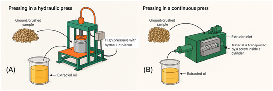
Figure 1.
Schematic layout of mechanical (A) and continuous (B) pressing techniques.
Generally, in solid–liquid extractions, oil solubilization by the solvent occurs in two ways: by direct contact with damaged plant cells or by diffusion through the semipermeable intact cell walls [4]. This process allows the transfer of soluble components to the solvent; then, the lipids can be recovered by solvent evaporation. The recovered lipid fraction is commonly known as crude oil and needs to undergo refining processes for food or feed utilization since it can contain contaminants in its raw form [4]. The main solvent-based method used in lipid separation is known as Soxhlet extraction. This method has been a standard technique and a primary reference used to measure the oil content of food matrices in the last century.
Originally, the Soxhlet method was used to determine fat content in milk and was named after its inventor. Von Soxhlet developed a new extraction system in 1879, which is called the Soxhlet apparatus. In this system, the sample is placed in a thimble holder that is filled with condensed solvents from a distillation flask, which carries the dissolved substances into the bulk liquid. The solvent is heated and distilled once again, purifying the sample over and over. This operation is often repeated until exhaustion [16]. Soxhlet extraction is an easy method to apply, requiring little training, and it has higher oil yield recovery compared to other techniques. However, some disadvantages are its longer sample preparation time and the higher quantity of solvent extractant needed, as well as the necessity of special disposal methods, which often impact the environment [16]. Essentially, the Soxhlet method is limited by solvent use, the oil sample application (to food or feed or cosmetic applications, etc.), the mass transfer resistances, and its low automation level, making its use on the industrial scale more constrained. Other drawbacks with using this method are that thermally sensitive components can easily deteriorate due to the heating process, which can result in low extraction yields [17]. In Figure 2, the schematic layout of solid–liquid extractions is shown using the Soxhlet apparatus.
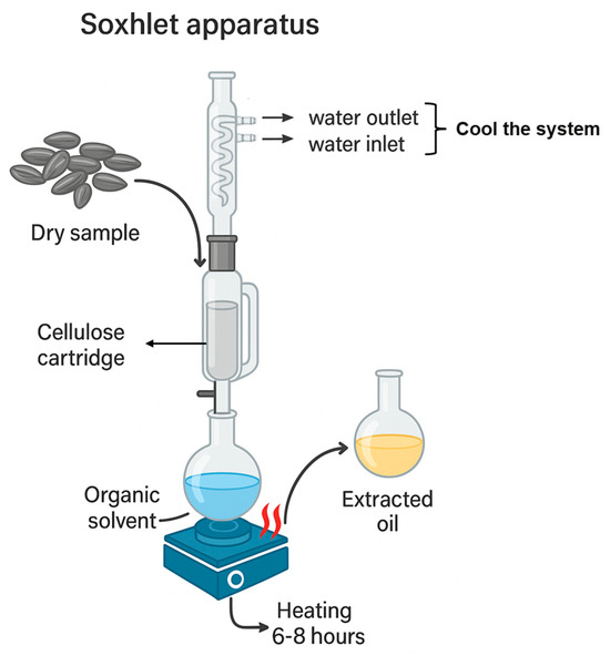
Figure 2.
Schematic layout of solid–liquid extraction using the Soxhlet apparatus.
Generally, the solvents used in lipid extraction have high oil solubility, high selectivity for lipidic substances, low viscosities and surface tension, low boiling points, and others. However, not all solvent reagents only display advantages. Some of those that are currently used have high toxicity and negative environmental impacts, and they are banned or discouraged in some countries, such as hexane, carbon disulfide, benzene, and trichloroethylene [1]. Despite this discommendation, hexane is the most used solvent for oilseed extraction due to its low boiling point (64–70 °C), low viscosity, and high oil recovery, in addition to low production costs [12,16]. The disadvantage of using hexane is related to its high flammability and its hazardousness to the environment and human health. To overcome this, some alternative solvents, such as isopropanol, ethanol, hydrocarbons, and even water, have been used in solid–liquid extractions [17]. Hexane is also obtained from the distillation of oil, and some very toxic benzenoids, such as benzene, toluene, and xylenes (BTXs), could be present in low amounts. In several countries, soy oil, which is an extract with hexane, presents regulatory issues related to the amount of harmful residual BTXs.
2.2. Green Extraction Methods
Extraction is an important tool for the food, cosmetic, and pharmaceutical industries. Conventional methods of extraction have been frequently used for diverse ends; therefore, they have been linked to environmental and health concerns, as previously presented. In this context, green extraction techniques emerge as more sustainable alternatives to these conventional methods, since they generally have less environmental impacts due to the use of low-toxicity environmentally friendly solvents and their lower energy needs, and they produce less waste by converting them into by-products and others, making the process safer [18]. Green extraction methods have also been named as unconventional extraction methods. The most frequently studied unconventional techniques are supercritical fluid extraction (SFE), microwave-assisted extraction (MAE), ultrasound-assisted extraction (UAE), pressurized liquid extraction (PLE), pulsed electric fields (PEFs), and enzymatic-assisted extraction (EAE). These methods are mostly used to obtain bioactive compounds and lipids from diverse sources, especially from plants and their processed residues (biomass) [18,19].
SFE is among the advanced techniques that use substances in their near-supercritical state as solvents, i.e., substances acting at temperatures and pressures above their critical point, in which the gas and liquid phases become indistinguishable [18,19]. Supercritical CO2 has been widely used as an eco-friendly technique because it has intermediate properties that lie between a gas and a liquid, which allows it to penetrate solid materials and thus facilitate the dissolution of some substances [18,19]. CO2 is a good working solvent for this technique since it becomes supercritical above 31.1 °C and 73.8 Bar of pressure, and it has low toxicity and requires low costs, is recyclable and non-flammable, and is widely available [18]. Recent works have shown that supercritical CO2 fluid extraction is a good method for obtaining high-quality vegetable oils from various sources without leaving harmful residues for health and the environment [18,20,21]. SFE has been applied to extract different types of vegetable oils, such as sunflower oil [22]; soybean oil [23]; babassu seed oil (Orbignya phalerata) [24]; uxi (Endopleura uchi) oil [25]; sapucaia (Lecythis pisonis Camb.); nut oil [26]; Brazil nut oil (Bertolletia excelsa H.B.K) [27]; tucuma fruit oil (Astrocaryum aculeatum and Astrocaryum vulgare) [28]; and Terminalia catappa fruit oil [29]. Recently, a study from Scognamiglio et al. [30] demonstrated that SFE-CO2 coupled with fractional separation can isolate cuticular waxes from diverse vegetable matrices, such as basil leaves, cannabis inflorescence (Cannabis sativa L.), chamomile flower heads, clove buds, ginger rhizomes, lavender inflorescence, marjoram leaf (Origanum majorana L.), rosemary leaf, tangerine peels, and tobacco leaves. These cuticular waxes were most abundantly composed of paraffinic compounds with high molecular weights, such as C27, C29, and C31, making the studied cuticular waxes interesting sources of high-added-value substances in diverse medical and pharmaceutical applications [30]. In Figure 3, the schematic layout of the carbon dioxide supercritical fluid extraction process is shown.
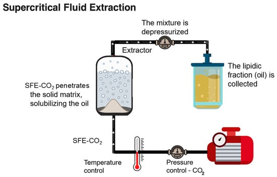
Figure 3.
Schematic layout of the carbon dioxide supercritical fluid extraction process.
Subcritical fluid extraction or subcritical water extraction (SWE) is a novel technology that uses subcritical water in a short 30 min extraction time, aiming to obtain less-polar substances at temperatures ranging from 100 to 374 °C under high pressure. These conditions reduce water viscosity and density, allowing higher and deeper penetration into the sample matrix [19,20]. As water has universal solvent abilities for both polar and nonpolar phytochemicals, SWE has been used to dissolve diverse phytochemicals, such as steviol glycosides and antioxidants (both polar and nonpolar), quercetin from onion waste, fatty acids from Camellia oleifera and Castanea sativa seeds, and polyphenols from Vitis vinifera [19].
Another emerging green extraction method is ultrasound-assisted extraction (UAE) (Figure 4). UAE involves the application of ultrasonic waves, which can reach 10 Hz to 20 KHz. It is a non-invasive technique that generates ultrasonic cavitation, which creates microbubbles that collapse, generating heat and localized pressures. The temperature and pressure rise causes the rupture of plant cells, releasing the target compounds [19]. The advantage of this technique is that the samples do not have direct contact with wave transducers. UAE also produces better quality extracted compounds, and it requires fewer solvents and less energy; it also reduces extraction times when compared to conventional methods [18,19,20]. Different solvents can be used in UAE, e.g., acidified water solutions, alcohols, ethanol, acetone, water, their mixtures, and others. UAE has been used to extract oil from diverse plant and animal species, such as soybean germs and cultivated marine microalgae, as is carried out in the work of Cravotto et al. [1]; pumpkin seed oil (Cucurbita pepo), as carried out by Hernández-Santos et al. [31]; and passion fruit seed oil (Passiflora edulis f. flavicarpa), as carried out by Oliveira, Barros, & Gimenes [32].
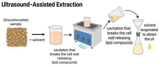
Figure 4.
Schematic layout of ultrasound-assisted extraction.
Microwave-assisted extraction (MAE) is a green extraction method based on the use of microwave energy, and it is used to extract soluble compounds in some solvents. The method relies on the application of microwave radiation to selectively heat solvents and samples, resulting in fast and efficient extraction media (Figure 5) [18,33]. Originally, MAE was employed pre-treat samples as a digestion step, but soon, it was largely accepted for extracting natural substances, especially when involving thermolabile compounds, since heating occurs in a uniform and controlled manner, preserving the integrity of the sample [33]. MAE can be carried out on two types of systems: single-mode systems, in which microwaves are only applied to samples, ensuring improved extraction; and multi-mode systems, where the waves are applied to the space, guaranteeing the uniform dispersion of energy [18,20]. Through this technique, humidity within the samples considerably affects extraction since it evaporates during the process. Modifications in the existing pressure further favor cell rupture and the solvent action on samples, which significantly reduce solvent consumption and energy use [20]. The extractions of curcuma oil (Curcuma longa L.) with ethanol, Sapindus mukorossi seed oil with hexane, volatile oils from soy sauce with cyclohexane, polyphenols from eggplant peel (Solanum melongena L.), and polyphenols and proteins from pineapple peel are among the uses of MAE [19].
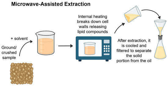
Figure 5.
Schematic layout of microwave-assisted extraction.
Enzyme-assisted extraction (EAE) is a trending technology that uses different enzymes to extract target compounds through complex polysaccharide hydrolysis, e.g., lignin, cellulose, and cell wall rupture [20]. EAE is a sustainable technique that combines enzyme efficiency with environmental preservation; hence, it is in accordance with green chemistry principles. In this method, specific enzymes, such as cellulase, pectinase, and amylase, are used during extraction to promote the breakdown of cellular walls and hydrolyze complex polysaccharides; then, the cell wall rupture occurs, allowing the solvent’s permeation to extract the target compounds [19]. There are two types of EAE methods: The first is enzyme-assisted aqueous extraction (EAAE), which is applied for extracting oils from diverse seeds, and the second is enzyme-assisted cold pressing (EACP), which hydrolyzes the cellular wall of plant seeds [19]. However, it is possible to combine EAE with other extraction methods, such as MAE or UAE. Moreover, this technique not only preserves the quality of extracts but also reduces volatile compound emissions, allowing for the reuse of plant waste [20,34]. A disadvantage of this technique is that the enzymes are very expensive, and their use on an industrial scale is limited [34]. EAAE was used to obtain pumpkin seed oil, which is characterized by the composition of fatty acids, antioxidant activities, and pharmacological potential applications, by Prommaban et al. [35]. This highlights its effectiveness in preserving bioactive components, and it can also greatly enhance the antioxidant properties of extracted oils. Some extraction methods used for obtaining lipids and fatty acids are presented in Table 1.

Table 1.
Some extraction methods used for obtaining lipids and fatty acids.
In this context, the application of green extraction methods for obtaining plant compounds offers several advantages, such as reducing environmental impact, reducing or even eliminating the use of toxic solvents, and, consequently, reducing pollution. It is worth saying that green extraction guarantees safer and cleaner processes, which results in final products that are healthier for human consumption, whether in the cosmetic, food, or pharmaceutical industries [19]. Therefore, the products also provide a high degree of purity and quality since bioactive compounds are found in high concentrations [19,20]. The incorporation of innovative and ecological methods not only meets environmental and health requirements but also promotes the efficiency and quality of final products. Continued research and development in this area is crucial for the transition to a green economy and the sustainable future of industries. In Figure 6, the main characteristics of the use of conventional and green extraction methods are summarized.
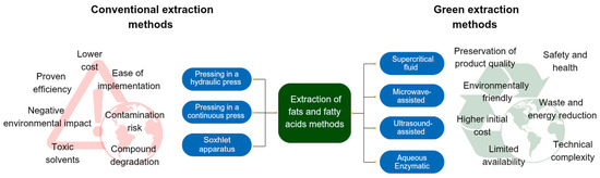
Figure 6.
Main characteristics of using conventional and green extraction methods.
3. Identification of Fatty Acids
3.1. Chemical Structure
Fatty acids (FAs) are carboxylic acids in the terminal position of an aliphatic chain (R-COOH). They are formed when fats (triglycerides) are broken down, and they can remain free or be attached to another biomolecule [48]. FAs can be found saturated (when the carbons have single (sigma-σ) bonds) or unsaturated (when they have one or more double bonds (Pi-π)); they can also have different chain lengths (Figure 7). In general, FAs exhibit low water solubility, and their soluble nature rapidly diminishes as the carbon chain lengthens [49]. Fatty acids from foods give the human body the energy it needs to function [50]. Free fatty acids can occur in biological systems, and they are most frequently found to be bonded in forms like cholesterol and phospholipids [51]. Based on the length of the chain, they can be categorized into four groups: short-chain fatty acids (< = C6), medium-chain fatty acids (C6–C12), long-chain fatty acids (C13–C21), and very-long-chain fatty acids (C22) [52]. Furthermore, different places within the aliphatic chain of fatty acids may include varying quantities of double bonds, giving rise to enormous families of isomeric fatty acids, such as geometric isomers and structural isomers [53].
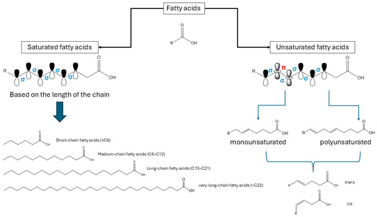
Figure 7.
Chemical classification and the chain types of the chemical structure of fatty acids.
Solid fat, or saturated fatty acids, occurs more frequently in animal sources, such as whole milk, butter, cheese, lard, bacon, meat fat, and the skins of chickens and fish. However, some vegetable sources can also present solid fat, such as coconut oil and other oily tropical fruits. The frequent intake of foods with high contents of saturated fat and the so-called “junk food” is unhealthy because they raise blood cholesterol levels and increase the risk of cardiovascular diseases, like embolism and atherosclerosis [50]. Lipids from plant sources are usually liquid oils because they contain a higher content of unsaturated fatty acids [49,50]. Trans fatty acids are found in processed foods like hydrogenated vegetable fat, which is used to extend their shelf life and enhance the flavor and consistency of food. Because trans fatty acids cause a rise in blood cholesterol levels, their excess can be detrimental to the body’s homeostasis [51]. Plant-based fats contain cis fatty acids, which the body can readily absorb. They are easily oxidized and converted into saturated fat because they are less stable than trans fat [51,52,53].
Fatty acid is referred to as a monounsaturated fatty acid (MUFA) if there is only one double bond in the chain; polyunsaturated fatty acid (PUFA) presents two or more double bonds [50,53]. Vegetable oils from sunflower, corn, soybeans, cottonseeds, chestnuts and almonds, and some fish species are mainly composed of PUFAs, while peanut oil, canola oil, and olive oil have higher contents of MUFAs [54]. The polyunsaturated fatty acids that are referred to as essential fatty acids are not produced by the body’s cells and must instead be obtained through diet. Omega-3 (linolenic acid) and omega-6 (linoleic acid) are two of the three categories of important fatty acids [50]. Alpha-linolenic acid (ALA), docosahexaenoic acid (DHA), and eicosapentaenoic acid (EPA) are the three primary forms of omega-3 fatty acids [50,53]. Seaweed and fish, including salmon, tuna, and sardines, include EPA and DHA, which are vital components of cell membranes, particularly those in the brain [50,55]. They are essential for proper brain function and have anti-inflammatory properties, especially for the cardiovascular system. Lower blood levels of triglycerides and total cholesterol are associated with the frequent intake of food composed of both EPA and DHA [50,55]. Alpha-linolenic acid is the most prevalent member of this category and is mostly found in flaxseeds and chia seeds, which can also be used to extract oil [50,53]. Significant amounts of ALAs are also present in walnuts and rapeseed oil (commonly known as canola oil). They are required for the upkeep of cell membranes, the enhancement of brain activity, and the transmission of nerve impulses [50,55].
On the other hand, vegetable oils like sunflower, corn, soybean, and cotton provide omega-6 fatty acids [50,56]. Linoleic acid (LA), which is linked to lowering total cholesterol and shielding the body against hypertension and excessive blood sugar levels, is the primary representative of this group. After consumption, linoleic acid can be converted into other members of the fatty acid group, including arachidonic acid (ARA) [50,55]. Meat and egg yolks are two foods that directly provide arachidonic acid, which can be obtained through diet. The inflammatory response of the body is significantly influenced by omega-6 content. For instance, arachidonic acid is a precursor to molecules that are involved in inflammation, a process that is vital to the body’s ability to fight off infections and heal wounds [57].
Since the human body can produce them, omega-9 fatty acids, a subtype of monounsaturated fat, are not regarded as necessary, in contrast to omega-3 and omega-6 [50]. On the contrary, eating foods that are high in omega-9 can have several health advantages. The primary member of this category is oleic acid, which also has anti-inflammatory properties and slows down the aging process of cells. It also aids in lowering blood cholesterol levels [57]. Nuts (almonds, walnuts, and chestnuts), avocados, olive and rapeseed oils, and olives can all be included in a diet to include oleic acid. Even if omega-9s are advantageous, it is crucial to consume all fatty acids in moderation. A diet rich in healthy fats, including omega-3, omega-6, and omega-9, can provide a wide range of health benefits: from improving heart function to reducing inflammation [50,56]. In Figure 8, some types of omega fatty acids and their respective structures and common natural origins are presented.

Figure 8.
Types of omega fatty acids and their respective structures and common natural origins.
Analyzing the fatty acid profile is essential for accurately determining the nutritional and therapeutic properties of these lipids. Several nutritional quality indices have been proposed based on the proportion of fatty acids among the oil and fat sources, e.g., monounsaturated/saturated fatty acid ratio (MUFA/SFA), polyunsaturated/saturated fatty acid (PUFA/SFA) ratio, omega-3/omega-6 ratio, atherogenicity index (AI), thrombogenic index (TI), hypocholesterolemic/hypercholesterolemic ratio (h/H), health-promoting index (HPI), calculated oxidizability value (COX), and others [58,59]. These parameters serve as valuable tools for assessing and comparing the quality and nutritional merits, as well as the evaluation of dietary impacts on the cardiovascular health of various lipidic matrices and their fatty acid composition, which can help reduce the risk of coronary and other metabolism-related diseases [58]. In this context, the quality of fatty acids in lipidic sources produced artificially in industrial or related operations may affect consumers’ biology. For this reason, it is critical that we have quick and accurate techniques for determining the structures of fatty acids.
3.2. Identification Techniques
In investigations on human nutrition and metabolism, a variety of chromatographic techniques for separation and quantification, particularly in vegetable oils and fats, are crucial [60]. Moreover, the examination of fatty acids plays a crucial role in regulating technical products; conducting medical diagnostic research; and assessing the quality, source, and freshness of food [61]. Due to the complex structure of the samples, it is imperative to employ selective and sensitive procedures [62]. The use of such techniques in combination is best considered in relation to specific methods. For example, it is not always possible to extract all the information from a sample during a single gas chromatography (GC) experiment, despite the strength of some of these techniques. If several isomeric chemicals are present, the GC column’s resolution might not be adequate, or there might be uncertainties about how to interpret a mass spectrum alone [63]. Coupling several techniques together is presented as a solution.
Lipid researchers have access to several potent hyphenated techniques for determining the structure of fatty acids [64]. Methods involving gas chromatography coupled with mass spectrometry or flame ionization detectors (GC-MS or GC-FID) and high-performance liquid chromatography (HPLC) techniques have been employed to analyze both saturated and unsaturated fatty acids (Figure 9) [65]. GC linked to Fourier transform infrared spectroscopy (GC-FTIR) and silver-ion high-performance liquid chromatography (Ag-HPLC) are also available, and these are useful techniques [66].

Figure 9.
Combined chromatographic techniques for fatty acid analysis.
Each technique has a specific advantage: reversed-phase and silver-ion HPLC can be used for the isolation of pure fatty acids or at least simpler fractions. Gas chromatography–mass spectrometry facilitates the determination of structure, and any doubt can be removed by using this technique in conjunction with derivatization procedures such as deuteration [67]. The elution order determined via silver-ion chromatography can give a good indication of the geometry of double bonds, but GC-FTIR will provide confirmation [68]. Although high-resolution nuclear magnetic resonance (NMR) spectroscopy (especially 1H and 13C NMR) is an extremely powerful tool, it appears to require relatively large amounts of pure single components in comparison to other methods [67,68].
Any new sample will probably be analyzed using GC on a capillary column coated with a polar or moderately polar column as the initial approach [67]. Silver-ion chromatography is a helpful characterization tool, and basic degradative processes like hydrogenation followed by GC analysis can yield insightful supplementary data [68]. The isolation of the target fatty acids, chemical degradation, mass spectrometry, and/or additional spectroscopic evidence will be necessary for a definitive identification.
3.2.1. Gas Chromatographic (GC) Analysis
An appropriate column with high resolution is necessary for the analysis of fatty acid isomeric mixtures. GC separation on a fused silica capillary column covered with a suitable stationary phase is the first step in the analysis of every new sample [67,68]. Many columns have been demonstrated to be effective for separating fatty acids with different chain lengths, degrees of saturation, double-bond locations, and cis or trans isomers [65,66]. For further information or to achieve better separation in particular situations, such as with trans fatty acids, a column with a higher polarity may be maintained [65].
High-polarity columns, such as the HP-88 column (88%-cyanopropyl aryl-polysiloxane), DB-FFAP column (nitroterephthalic acid-modified polyethylene glycol), and SLB-IL series columns (ionic liquids), are commonly used for fatty acid analysis [69]. Previous studies have indicated that ionic liquid (IL) columns provide better selectivity and can separate geometric and positional fatty acid isomers [70]. One of the Carbowax TM types is typically advised as a standard. The application of long polar capillary columns (50–100 m) for gas–liquid chromatography has improved the resolution of many positional and geometric fatty acid isomers [71].
In a chromatogram, the association between the retention elution time of peaks on a GC trace and their identity is quickly apparent to fatty acid analysts [69]. It is important to remember that the conditions under which the retention times and separations are achieved depend on several variables, including the carrier gas flow rate, column age, and, most importantly, column temperature [72]. For identification purposes, the analyst should ideally obtain suitable primary standards with defined compositions and use natural mixes for which trustworthy compositional data are available as secondary standards. GC retention times should only be used as a guide for fatty acid identification and must be supplemented with further chromatographic and spectroscopic information [73] or other methods, with the most typical being mass spectrometry.
The retention times of free fatty acids (FFAs) or fatty acid methyl esters (FAMEs) could be compared with their standards, for example, analyzing co-elution, or with the retention times of linear hydrocarbons in a homologous series to obtain retention indices, such as Kovats indices or van der Dool–Kratz indices [8,9]. Correlating the retention time of fatty acids with those of linear hydrocarbons, retention indices are fully available in the literature, enabling the comparison in any similar equipment using the same chromatographic conditions. The automatic comparison between spectra from FFA or PUFA with databases such as NIST and Wiley also contributes to positive identifications [8,9].
In the GC analysis of fatty acids, methyl esters are frequently used, and the choice of the optimal derivative was not given much concern until recently [74]. For fatty acid analysis by GC-MS, derivatization is typically required, particularly for fatty acids with carbon numbers greater than 10. FAMEs, which are subsequently identified by GC-MS, are frequently formed via the derivatization of fatty acids [73]. In general, FFA and esterified fatty acids can be treated with acid derivatization techniques.
Hydrochloric acid (HCl), acetyl chloride (CH3COCl), sulfuric acid (H2SO4), and boron trifluoride (BF3) are some of the most often utilized acid derivatization reagents. Because of its ease of operation, HCl derivatization is one of the most widely used fatty acid analysis techniques [74]. In HCl derivatization, dried lipid extracts are combined with methanolic HCl, and the mixture is heated for a predetermined amount of time [75]. However, adding an additional solvent prior to the derivatization stage could be required because some lipids are soluble in methanolic HCl. Following derivatization, the samples are neutralized, and an organic solvent is used to extract the FAMEs in preparation for additional GC analyses [65]. Methyl esters can be prepared using a variety of easy techniques, and there is a wealth of published information regarding their GC retention characteristics [76]. Because they have the lowest molecular weight and are the easiest ester derivatives to make, they elute from GC columns at a lower temperature than other derivatives. Furthermore, because of their low polarity, they can be separated using GC, thin-layer chromatography (TLC), and HPLC techniques. Sometimes methyl esters are very volatile, which makes it challenging to analyze lipids that contain short-chain fatty acids [77].
Fatty acid analysis using, for example, GC-MS-based analytical procedures typically involves three steps: (1) fatty acid extraction from the sample matrix, (2) fatty acid derivatization, and (3) GC-MS analysis. Following hydrolysis, the most intense ions in mass spectra (M+ − 15) are identified as fatty acids. A capillary GC–MS technique employing electron-capture negative-ion technology was created to quantify the total FAs in C8–C26. Compared to GC-FID, GC-MS could provide more structural information [78]. Moreover, GC-MS has well-established databases for FA identification, with higher efficiency and better selectivity compared to GC-FID. As a result, GC-MS is the most frequently used method for fatty acid isolation and identification. There are numerous well-established extraction techniques that, in principle, can be used with a variety of sample types; nevertheless, method tuning is necessary to acquire the best results for target analytes.
3.2.2. Isolation of Fatty Acids for Structural Analysis by HPLC
Obtaining fatty acid as a pure single component makes identification much easier to accomplish and allows for the application of chemically degradative and spectroscopic techniques. The ability to convert a complex mixture to simpler fractions, however, is often adequate to ensure that there are no overlapping components that could diminish the usefulness of coupled chromatography–spectroscopy techniques [67,68]. Numerous zones with overlapping peaks are revealed when fatty acids are separated based on saturation levels using HPLC columns [77]. A mixture of C12–C18 saturated fatty acids and the positional or geometrical isomers of unsaturated fatty acids with 18 carbon atoms can be separated by employing acetonitrile and water as the mobile phases in a binary gradient process. Natural samples can benefit from improved resolution efficiency, which makes the binary gradient algorithm more optimal [76,77]. The kind of separation that takes place depends on the total number of carbon atoms, double bonds, and geometric isomers of unsaturated fatty acids. The HPLC method is appropriate for routinely determining certain saturated and unsaturated fatty acids using C18 columns, which facilitate the quick and easy synthesis of free fatty acid extracts [73].
Preparative high-performance liquid chromatography is utilized to separate the distinct classes of phospholipids, while reverse-phase HPLC (RP-HPLC) is employed to isolate molecular species on the C18-phase column [79]. Lower analytical temperatures, which lower the chance of double-bond isomerization, and the ability to separate fractions for additional analysis are the main benefits of HPLC over GC [80]. Fatty acids are transformed into a derivative with a chromophore before being used in HPLC analysis with ultraviolet (UV) detection because they have a relatively low molar absorption at 220 nm [70]. HPLC-MS methods for fatty acid analysis showed some disadvantages, such as greater solvent consumption and lower selectivity [77]. To separate individual fatty acids based on their chain length and the degree of unsaturation, reversed-phase HPLC is quite a helpful approach. The development of derivatizing reagents that enable the sensitive detection of fatty acids by UV or fluorescence spectroscopy has received significant attention in recent years [76]. Reversed-phase HPLC may be advantageous for analyses in certain unusual cases, and this may include applications involving fatty acids that are sensitive to the high temperatures needed for gas-phase separation [81].
Reversed-phase HPLC (C18) is most useful as a micro-preparative technique for isolating individual fatty acids or simpler fractions from complex mixtures for further analysis [81]. The process of partitioning a solute between liquid stationary and mobile phases is known as reversed-phase HPLC, and the name “reversed-phase” suggests that the mobile phase is more polar than the stationary phase. Most of the most-utilized stationary phase is made up of octadecylsilyl (“C18” or “ODS”) groups that are covalently bonded to a silanol surface. Mobile phases have nearly always been based on combinations of methanol and water or acetonitrile and water [81]. The different fatty acids are easily confused due to the dual nature of the separation process; thus, it is important to take great care to verify that the components separated by reversed-phase HPLC are correctly recognized [82]. The order in which the various components elute follows logically when one understands the nature of the separation process, but it may be confusing to someone unfamiliar with the method. Saturated fatty acids with the same chain lengths elute much later than unsaturated fatty acids, with each double bond shortening the retention period by roughly two methylene groups. Individual positional and configurational isomers can be separated with much more precision if the HPLC separation is carefully tuned using isocratic elution, moderate gradients, and the appropriate fatty acid derivative [82].
Methyl esters of fatty acids with conjugated double-bond systems can be detected with great sensitivity by UV spectroscopy, following separation by reversed-phase HPLC. The detection and quantification of conjugated double-bond systems benefit greatly from the application of ultraviolet spectroscopy [76]. The technique is commonly used for the examination of hydroperoxides, but it also plays a significant role when paired with HPLC for the characterization of other naturally occurring conjugated fatty acids [71]. Conjugated dienes may be detected and quantified more easily due to the development of UV spectroscopy. Conjugated dienes have a distinctive absorbance at 234 nm, but this can interfere with analysis because it is typically seen as a shoulder above the wide band at 200 nm relative to the end absorption of lipids [78]. A second derivative that separated a distinct peak from a shoulder was generated by taking the differential of the first derivative spectrum. The smaller bands with minima instead of maxima were obtained, indicating improved resolutions. Natural samples showed two bands: the trans–trans band at 233 nm and the cis–trans-conjugated diene band at 242 nm. Since differentiation had no effect on the Beer–Lambert law, quantification was also enhanced [76].
3.2.3. Silver-Ion Chromatography
The ability of silver ions to reversibly form polar complexes with unsaturated centers in organic compounds, such as lipids, is used in silver-ion chromatography to achieve separations [83]. This makes it possible to separate molecules based on the quantity, kind, geometrical arrangement, and location of double bonds or other unsaturated functional moieties. Thin-layer chromatography (TLC), which makes use of layers of silica gel coated with silver nitrate, was employed in a large portion of earlier studies [84]. When silver-ion chromatography is used in conjunction with other separation methods, it becomes significant. Thus, elements that could overlap if a single technique is used are resolved by combined techniques. Fatty acid methyl ester fractions produced by silver-ion chromatography may need more extensive GC or GC-MS analyses than the original material might allow. Because silver-ion chromatographic profiles are typically simple to understand, they are ideal for concentrating on small components or for the early simplification of complex combinations [83].
For silver-ion HPLC, two basic column types have been utilized: those based on silver ions linked to cation exchange media and those comprising silica gel impregnated with silver nitrate. However, these systems are problematic due to the solvent’s corrosive properties and are no longer in use. Comparably, most of the historical significance of silica gel/silver nitrate columns comes from the fact that the elution of silver ions in the mobile phase reduces the column’s lifetime and contaminates some fractions during preparative separations [85]. These kinds of silver-ion HPLC columns have been used for two main applications in the separation of simple fatty acid derivatives: group separations of components with different levels of unsaturation and the separation of individual positional and configurational isomers [83]. With the former, binary gradient elution techniques are used to obtain high-resolution fatty acid derivatives relative to fractions. A gradient of dichloromethane-1,2-dichloroethane (1:1, v/v) to dichloromethane-1,2-dichloroethane-methanol-acetonitrile is used to improve resolutions after an initial gradient of methanol to methanol–acetonitrile (9:1, v/v) [60]. Sample collections are made possible by inserting a stream splitter into an evaporative light-scattering detector. This process has been widely used to simplify a variety of intricate natural fatty acid combinations, including their methyl esters, in order to proceed to GC-MS structural investigations [71]. Some isolation and identification methods for identifying fatty acids from diverse materials are presented in Table 2.
Table 2.
Some isolation and identification methods for identifying fatty acids from diverse materials.
4. Conclusions and Perspectives
The extraction of fats and fatty acids is a dynamic and crucial field for several industries. Traditionally, conventional techniques such as Soxhlet extraction, maceration, and cold pressing have dominated the sector due to their effectiveness in extracting high amounts of lipids. However, these methods often resort to the use of chemical solvents that can be harmful to both the environment and human health. Growing concerns about the impacts of these conventional techniques have driven the adoption of green extraction methods. Some techniques, such as extraction with supercritical fluids (especially using CO2) and extraction assisted by ultrasound and microwaves, have been explored as they present themselves as a superior alternative. These methods not only mitigate the environmental and health risks associated with traditional solvents but can also offer advantages such as lower energy consumption, reduced waste, and the preservation of the nutritional and sensory qualities of extracts. However, it is expected that innovation will go hand in hand with more sustainable techniques, meeting environmental regulations and consumer desires for cleaner products. Therefore, the improvement of green extraction methods such that they can operate on larger scales is necessary for the industry to see them as main alternatives for extracting fats and fatty acids. The proper separation and quantification of fatty acids in vegetable oils and fats are crucial to well-balanced nutrition and health safety. As food samples have a complex structure of macro- and micronutrients, it is imperative to employ selective and sensitive procedures for the isolation and quantification of their components, such as fatty acids, using different chromatography techniques; this will allow their chemical structures to be successfully described in detail using nuclear magnetic resonance. More sensitive methods like GC×GC-MS (two-dimensional gas chromatography) and LC-MS (liquid chromatography with mass spectrometry) have been used more frequently for the analysis of fatty acids in biological samples because of their high sensitivity when combined with new technologies that are in line with analytical strategies based on gas chromatography coupled with mass spectrometry (GC-MS). This situation enables the identification of chemicals at low concentrations and enhances the separation processes of isomeric molecules. These well-adjusted automatic extraction and derivatization methods improve the reliability of results, cut down manual errors, and shorten the time needed to prepare samples. Furthermore, these methods expedite the discovery of novel lipid biomarkers that are crucial for identifying lipid profiles linked to conditions, including diabetes, cancer, and cardiovascular disorders. Since developments in the substitution of more ecologically friendly solvents, like supercritical CO2, for harmful ones during the extraction stages have become crucial steps in the development of novel techniques, the use of sustainable solvents is also a reality. In the future, it is expected that green extraction technologies will gradually surpass conventional methods as standard approaches to recover and analyze lipids. Further advances in the design of equipment and the increased use of green solvents or aqueous media (e.g., ionic liquids, terpenes, alcohols, deep eutectic solvents) and tuning parameters associated with optimized processes will play an important role in improving the yield of extractions and the quality of products. Additionally, improvements in analytical techniques—particularly chromatographic methods such as GC-MS, GC-FID, HPLC, and silver-ion chromatography—will continue to support the precise identification and structural elucidation of fatty acids. Continued investment in research and innovation is crucial to ensure that extraction and characterization methods remain aligned with sustainability principles and regulatory demands, ultimately supporting a safer and more responsible lipid supply chain.
Author Contributions
Conceptualization, B.E.T.-C. and V.F.V.-J.; methodology, B.E.T.-C. and V.F.V.-J.; investigation, Y.L.-P., E.M.O.d.S., D.S.d.R., I.G.C.B.-S. and K.S.L.M. data curation, Y.L.-P., E.M.O.d.S., D.S.d.R., I.G.C.B.-S. and K.S.L.M.; writing—original draft preparation, Y.L.-P., E.M.O.d.S., D.S.d.R., I.G.C.B.-S., K.S.L.M., B.E.T.-C. and V.F.V.-J.; writing—review and editing, B.E.T.-C. and V.F.V.-J.; supervision, B.E.T.-C. and V.F.V.-J.; project administration, B.E.T.-C. All authors have read and agreed to the published version of the manuscript.
Funding
This study was partially financed (with scholarships) by the Coordenação de Aperfeiçoamento de Pessoal de Nível Superior—Brasil (CAPES)—Finance Code 001 and by FAPERJ—Carlos Chagas Filho Foundation for Research Support of the State of Rio de Janeiro (SEI-RJ Process: E-26/204.549/2024).
Conflicts of Interest
The authors declare no conflicts of interest.
Abbreviations
The following abbreviations are used in this manuscript:
| HPLC | High-performance liquid chromatography; |
| AD | Anno Domini from Gregorian calendar; |
| BTXs | Benzene, toluene, and xylenes; |
| SFE | Supercritical fluid extraction; |
| SFE-CO2 | Carbon dioxide supercritical fluid extraction; |
| MAE | Microwave-assisted extraction; |
| UAE | Ultrasound-assisted extraction; |
| PLE | Pressurized liquid extraction; |
| PEF | Pulsed electric fields; |
| EAE | Enzyme-assisted extraction; |
| SWE | Subcritical water extraction; |
| EACP | Enzyme-assisted cold pressing; |
| EAAE | Enzyme-assisted aqueous extraction; |
| FAs | Fatty acids; |
| MUFAs | Monounsaturated fatty acids; |
| PUFAs | Polyunsaturated fatty acids; |
| SFAs | Saturated fatty acids; |
| ALA | Alpha-linolenic acid; |
| DHA | Docosahexaenoic acid; |
| EPA | Eicosapentaenoic acid; |
| LA | Linoleic acid; |
| ARA | Arachidonic acid; |
| AI | Atherogenicity index; |
| TI | Thrombogenic index; |
| h/H | Hypocholesterolemic/hypercholesterolemic ratio; |
| HPI | Health-promoting index; |
| COX | Calculated oxidizability value; |
| GC | Gas chromatography; |
| GC-MS | Gas chromatography coupled with mass spectrometry detector; |
| GC-FID | Gas chromatography coupled with flame ionization detector; |
| GC-FTIR | Gas chromatography coupled with Fourier transform infrared spectroscopy; |
| Ag-HPLC | Silver-ion high-performance liquid chromatography; |
| NMR | Nuclear magnetic resonance; |
| HP-88 | High-polarity column (88%-cyanopropyl aryl-polysiloxane); |
| DB-FFAP | Nitroterephthalic acid-modified polyethylene glycol column; |
| IL | Ionic liquid; |
| FFAs | Free fat acids; |
| FAMEs | Fatty acid methyl esters; |
| NIST | National Institute of Standards and Technology; |
| TLC | Thin-layer chromatography. |
References
- Cravotto, C.; Claux, O.; Bartier, M.; Fabiano-Tixier, A.-S.; Tabasso, S. Leading Edge Technologies and Perspectives in Industrial Oilseed Extraction. Molecules 2023, 28, 5973. [Google Scholar] [CrossRef]
- Nde, D.; Foncha, A. Optimization Methods for the Extraction of Vegetable Oils: A Review. Processes 2020, 8, 209. [Google Scholar] [CrossRef]
- Sharma, M.; Gupta, S.K.; Mondal, A.K. Production and Trade of Major World Oil Crops. In Technological Innovations in Major World Oil Crops; Springer: New York, NY, USA, 2012; Volume 1, pp. 1–15. [Google Scholar] [CrossRef]
- Ramalho, H.F.; Suarez, P.A.Z. The Chemistry of Oils and Fats and Their Extraction and Refining Processes. Rev. Virtual Química 2013, 5, 2–15. [Google Scholar] [CrossRef]
- Mwaurah, P.W.; Kumar, S.; Kumar, N.; Attkan, A.K.; Panghal, A.; Singh, V.K.; Garg, M.K. Novel Oil Extraction Technologies: Process Conditions, Quality Parameters, and Optimization. Compr. Rev. Food Sci. Food Saf. 2020, 19, 3–20. [Google Scholar] [CrossRef]
- van Den Dool, H.; Kratz, P.D. A Generalization of the Retention Index System Including Linear Temperature Programmed Gas—Liquid Partition Chromatography. J. Chromatogr. A 1963, 11, 463–471. [Google Scholar] [CrossRef] [PubMed]
- Olfert, M.; Knappe, C.; Sievers-Engler, A.; Masberg, B.; Lämmerhofer, M. Determination of Double Bond Positions in Unsaturated Fatty Acids by Pre-Column Derivatization with Dimethyl and Dipyridyl Disulfide Followed by LC-SWATH-MS Analysis. Anal. Bioanal. Chem. 2025, 417, 2753–2766. [Google Scholar] [CrossRef] [PubMed]
- Bizzo, H.R.; Brilhante, N.S.; Nolvachai, Y.; Marriott, P.J. Use and Abuse of Retention Indices in Gas Chromatography. J. Chromatogr. A 2023, 1708, 464376. [Google Scholar] [CrossRef] [PubMed]
- Babushok, V.I. Chromatographic Retention Indices in Identification of Chemical Compounds. TrAC Trends Anal. Chem. 2015, 69, 98–104. [Google Scholar] [CrossRef]
- Lieng, B.Y.; Quaile, A.T.; Domingo-Almenara, X.; Röst, H.L.; Montenegro-Burke, J.R. Computational Expansion of High-Resolution-MSn Spectral Libraries. Anal. Chem. 2023, 95, 17284–17291. [Google Scholar] [CrossRef]
- Pati, S.; Nie, B.; Arnold, R.D.; Cummings, B.S. Extraction, Chromatographic and Mass Spectrometric Methods for Lipid Analysis. Biomed. Chromatogr. 2016, 30, 695–709. [Google Scholar] [CrossRef]
- Rani, H.; Sharma, S.; Bala, M. Technologies for Extraction of Oil from Oilseeds and Other Plant Sources in Retrospect and Prospects: A Review. J. Food Process. Eng. 2021, 44, e13851. [Google Scholar] [CrossRef]
- Ofori-Boateng, C.; Keat Teong, L.; JitKang, L. Comparative Exergy Analyses of Jatropha curcas Oil Extraction Methods: Solvent and Mechanical Extraction Processes. Energy Convers. Manag. 2012, 55, 164–171. [Google Scholar] [CrossRef]
- Subroto, E.; Manurung, R.; Heeres, H.J.; Broekhuis, A.A. Optimization of Mechanical Oil Extraction from Jatropha curcas L. Kernel Using Response Surface Method. Ind. Crops Prod. 2015, 63, 294–302. [Google Scholar] [CrossRef]
- Wilhelm, A.E.; Antoniassi, R.; Faria-Machado, A.F.; Bizzo, H.R.; Reis, S.L.R.; Cenci, S.A. Different Feed Rates of Expeller Press on the Extraction Efficiency and Quality of Passion Fruit Seed Oil. Ciência Rural. 2014, 44, 1312–1318. [Google Scholar] [CrossRef]
- Luque de Castro, M.D.; Priego-Capote, F. Soxhlet Extraction: Past and Present Panacea. J. Chromatogr. A 2010, 1217, 2383–2389. [Google Scholar] [CrossRef]
- Danlami, J.M.; Arsad, A.; Ahmad Zaini, M.A.; Sulaiman, H. A Comparative Study of Various Oil Extraction Techniques from Plants. Rev. Chem. Eng. 2014, 30, 605–626. [Google Scholar] [CrossRef]
- Martins, R.; Barbosa, A.; Advinha, B.; Sales, H.; Pontes, R.; Nunes, J. Green Extraction Techniques of Bioactive Compounds: A State-of-the-Art Review. Processes 2023, 11, 2255. [Google Scholar] [CrossRef]
- Usman, M.; Nakagawa, M.; Cheng, S. Emerging Trends in Green Extraction Techniques for Bioactive Natural Products. Processes 2023, 11, 3444. [Google Scholar] [CrossRef]
- Masoodi, L.; Gull, A.; Masoodi, F.A.; Gani, A.; Nissar, J.; Ahad, T.; Nayik, G.A.; Mukarram, S.A.; Kovács, B.; Prokisch, J.; et al. An Overview on Traditional vs. Green Technology of Extraction Methods for Producing High Quality Walnut Oil. Agronomy 2022, 12, 2258. [Google Scholar] [CrossRef]
- Vági, E.; Balázs, M.; Komoczi, A.; Mihalovits, M.; Székely, E. Fractionation of Phytocannabinoids from Industrial Hemp Residues with High-Pressure Technologies. J. Supercrit. Fluids 2020, 164, 104898. [Google Scholar] [CrossRef]
- Rai, A.; Mohanty, B.; Bhargava, R. Supercritical Extraction of Sunflower Oil: A Central Composite Design for Extraction Variables. Food Chem. 2016, 192, 647–659. [Google Scholar] [CrossRef] [PubMed]
- Jokic, S.; Svilovic, S.; Vidovic, S. Modelling the Supercritical CO2 Extraction Kinetics of Soybean Oil. Croat. J. Food Sci. Technol. 2015, 7, 52–57. [Google Scholar] [CrossRef]
- de Oliveira, N.A.; Mazzali, M.R.; Fukumasu, H.; Gonçalves, C.B.; Oliveira, A.L.d. Composition and Physical Properties of Babassu Seed (Orbignya phalerata) Oil Obtained by Supercritical CO2 Extraction. J. Supercrit. Fluids 2019, 150, 21–29. [Google Scholar] [CrossRef]
- Pinto, R.H.H.; Menezes, E.G.O.; Freitas, L.C.; Andrade, E.H.d.A.; Ribeiro-Costa, R.M.; Silva Júnior, J.O.C.; Carvalho Junior, R.N. Supercritical CO2 Extraction of Uxi (Endopleura uchi) Oil: Global Yield Isotherms, Fatty Acid Profile, Functional Quality and Thermal Stability. J. Supercrit. Fluids 2020, 165, 104932. [Google Scholar] [CrossRef]
- Santos, O.V.d.; Carvalho, R.N.; Costa, C.E.F.d.; Lannes, S.C.d.S. Chemical, Chromatographic-Functional, Thermogravimetric-Differential and Spectroscopic Parameters of the Sapucaia Oil Obtained by Different Extraction Methods. Ind. Crops Prod. 2019, 132, 487–496. [Google Scholar] [CrossRef]
- Santos, O.V.; Corrêa, N.C.F.; Carvalho, R.N.; Costa, C.E.F.; Lannes, S.C.S. Yield, Nutritional Quality, and Thermal-Oxidative Stability of Brazil Nut Oil (Bertolletia excelsa H.B.K) Obtained by Supercritical Extraction. J. Food Eng. 2013, 117, 499–504. [Google Scholar] [CrossRef]
- Costa, B.E.T.; Santos, O.V.d.; Corrêa, N.C.F.; França, L.F.d. Comparative Study on the Quality of Oil Extracted from Two Tucumã Varieties Using Supercritical Carbon Dioxide. Food Sci. Technol. 2016, 36, 322–328. [Google Scholar] [CrossRef]
- Santos, O.V.; Lorenzo, N.D.; Souza, A.L.G.; Costa, C.E.F.; Conceição, L.R.V.; Lannes, S.C.d.S.; Teixeira-Costa, B.E. CO2 Supercritical Fluid Extraction of Pulp and Nut Oils from Terminalia catappa Fruits: Thermogravimetric Behavior, Spectroscopic and Fatty Acid Profiles. Food Res. Int. 2021, 139, 109814. [Google Scholar] [CrossRef]
- Scognamiglio, M.; Baldino, L.; Reverchon, E. Fractional Separation and Characterization of Cuticular Waxes Extracted from Vegetable Matter Using Supercritical CO2. Separations 2022, 9, 80. [Google Scholar] [CrossRef]
- Hernández-Santos, B.; Rodríguez-Miranda, J.; Herman-Lara, E.; Torruco-Uco, J.G.; Carmona-García, R.; Juárez-Barrientos, J.M.; Chávez-Zamudio, R.; Martínez-Sánchez, C.E. Effect of Oil Extraction Assisted by Ultrasound on the Physicochemical Properties and Fatty Acid Profile of Pumpkin Seed Oil (Cucurbita pepo). Ultrason. Sonochem. 2016, 31, 429–436. [Google Scholar] [CrossRef]
- de Oliveira, R.C.; de Barros, S.T.D.; Gimenes, M.L. The Extraction of Passion Fruit Oil with Green Solvents. J. Food Eng. 2013, 117, 458–463. [Google Scholar] [CrossRef]
- Destandau, E.; Michel, T.; Elfakir, C. CHAPTER 4. Microwave-assisted Extraction. In RSC Green Chemistry; Mauricio, A.R., Juliana, M.P., Eds.; Royal Society of Chemistry: London, UK, 2013; pp. 113–156. [Google Scholar] [CrossRef]
- Kumar, S.P.J.; Prasad, S.R.; Banerjee, R.; Agarwal, D.K.; Kulkarni, K.S.; Ramesh, K.V. Green Solvents and Technologies for Oil Extraction from Oilseeds. Chem. Cent. J. 2017, 11, 9. [Google Scholar] [CrossRef]
- Prommaban, A.; Kuanchoom, R.; Seepuan, N.; Chaiyana, W. Evaluation of Fatty Acid Compositions, Antioxidant, and Pharmacological Activities of Pumpkin (Cucurbita moschata) Seed Oil from Aqueous Enzymatic Extraction. Plants 2021, 10, 1582. [Google Scholar] [CrossRef]
- Thilakarathna, R.C.N.; Siow, L.F.; Tang, T.-K.; Chan, E.-S.; Lee, Y.-Y. Physicochemical and Antioxidative Properties of Ultrasound-Assisted Extraction of Mahua (Madhuca longifolia) Seed Oil in Comparison with Conventional Soxhlet and Mechanical Extractions. Ultrason. Sonochem 2023, 92, 106280. [Google Scholar] [CrossRef]
- Thomsen, K.; Raak, N.; Gregersen, S.B.; Månsson, L.; Miquel Becker, E. Enzyme-Assisted Extraction of Rapeseed Oil with Minimum Water Addition: A Proof-of-Concept Study. Int. J. Food Sci. Technol. 2024, 5, 3013–3019. [Google Scholar] [CrossRef]
- Sabarish, C.S.; Sebastian, J.; Muraleedharan, C. Extraction of Oil from Rubber Seed through Hydraulic Press and Kinetic Study of Acid Esterification of Rubber Seed Oil. Procedia Technol. 2016, 25, 1006–1013. [Google Scholar] [CrossRef]
- Polmann, G.; Teixeira, G.L.; Santos, P.H.; Rivera, G.Á.; Ibañez, E.; Cifuentes, A.; Ferreira, S.R.S.; Block, J.M. Chemical Characterization of Gurguéia Nut (Dipteryx lacunifera Ducke) and Press Cake Oil Obtained by Hydraulic Pressing and Supercritical Extraction. Biomass Conv. Bioref. 2024, 14, 19065–19080. [Google Scholar] [CrossRef]
- Dar, I.H.; Junaid, P.M.; Ahmad, S.; Shams, R.; Dash, K.K.; Shaikh, A.M.; Béla, K. Optimization of Ultrasound-Assisted Extraction of Nigella Sativa Seed Oil for Enhancement of Yield and Antioxidant Activity. Discov. Appl. Sci. 2024, 6, 104. [Google Scholar] [CrossRef]
- Senrayan, J.; Venkatachalam, S. Optimization of ultrasound-assisted solvent extraction (UASE) based on oil yield, antioxidant activity and evaluation of fatty acid composition and thermal stability of Coriandrum sativum L. seed oil. Food Sci. Biotechnol. 2019, 28, 377–386. [Google Scholar] [CrossRef]
- da Rosa, A.C.S.; Stevanato, N.; Iwassa, I.; dos Santos Garcia, V.A.; da Silva, C. Obtaining Oil from Macauba Kernels by Ultrasound-Assisted Extraction Using Ethyl Acetate as the Solvent. Braz. J. Food Technol. 2019, 22, e2018195. [Google Scholar] [CrossRef]
- Santos, O.V.; Lemos, Y.S.; da Conceição, L.R.V.; Teixeira-Costa, B.E. Lipids from the Purple and White Açaí (Euterpe Oleracea Mart) Varieties: Nutritional, Functional, and Physicochemical Properties. Front. Nutr. 2024, 11, 1385877. [Google Scholar] [CrossRef] [PubMed]
- De Oliveira Palheta, H.C.; Lima dos Santos, M.P.; Vasconcelos dos Santos, O.; Miranda Mendes, P.; Macedo Rogero, M.; Vieira da Conceicão, L.R.; Carrera Silva-Junior, J.O.; Teixeira-Costa, B.E.; De Souza Figueira, M. Functionality of Lipid Extracts Obtained by Green Extraction from Red Pupunha (Bactris Gasipaes Kunt). Sci. Plena 2025, 21. [Google Scholar] [CrossRef]
- Yeasmin, M.S.; Uddin, M.J.; Dey, S.S.; Barmon, J.; Ema, N.T.; Rana, G.M.M.; Rahman, M.M.; Begum, M.; Ferdousi, L.; Ahmed, S.; et al. Optimization of Green Microwave-Assisted Extraction of Essential Oil from Lemon (Citrus limon) Leaves: Bioactive, Antioxidant and Antimicrobial Potential. Curr. Res. Green Sustain. Chem. 2024, 8, 100413. [Google Scholar] [CrossRef]
- Fernández-Marín, R.; Fernandes, S.C.M.; Andrés, M.A.; Labidi, J. Microwave-Assisted Extraction of Curcuma longa l. Oil: Optimization, Chemical Structure and Composition, Antioxidant Activity and Comparison with Conventional Soxhlet Extraction. Molecules 2021, 26, 1516. [Google Scholar] [CrossRef] [PubMed]
- Buranachokpaisan, K.; Muangrat, R.; Chalermchat, Y. Supercritical CO2 Extraction of Residual Oil from Pressed Sesame Seed Cake: Optimization and Its Physicochemical Properties. J. Food Process Preserv. 2021, 45, e15722. [Google Scholar] [CrossRef]
- Janßen, H.; Steinbüchel, A. Fatty Acid Synthesis in Escherichia Coli and Its Applications towards the Production of Fatty Acid Based Biofuels. Biotechnol. Biofuels 2014, 7, 7. [Google Scholar] [CrossRef]
- Mu, H. The Digestion of Dietary Triacylglycerols. Prog. Lipid Res. 2004, 43, 105–133. [Google Scholar] [CrossRef]
- Saini, R.K.; Keum, Y.-S. Omega-3 and Omega-6 Polyunsaturated Fatty Acids: Dietary Sources, Metabolism, and Significance—A Review. Life Sci. 2018, 203, 255–267. [Google Scholar] [CrossRef]
- Micha, R.; Mozaffarian, D. Saturated Fat and Cardiometabolic Risk Factors, Coronary Heart Disease, Stroke, and Diabetes: A Fresh Look at the Evidence. Lipids 2010, 45, 893–905. [Google Scholar] [CrossRef]
- De Carvalho, C.; Caramujo, M. The Various Roles of Fatty Acids. Molecules 2018, 23, 2583. [Google Scholar] [CrossRef]
- Wang, Z.; Yang, T.; Brenna, J.T.; Wang, D.H. Fatty Acid Isomerism: Analysis and Selected Biological Functions. Food Funct. 2024, 15, 1071–1088. [Google Scholar] [CrossRef]
- Orsavova, J.; Misurcova, L.; Ambrozova, J.; Vicha, R.; Mlcek, J. Fatty Acids Composition of Vegetable Oils and Its Contribution to Dietary Energy Intake and Dependence of Cardiovascular Mortality on Dietary Intake of Fatty Acids. Int. J. Mol. Sci. 2015, 16, 12871–12890. [Google Scholar] [CrossRef]
- Smolińska, K.; Szopa, A.; Sobczyński, J.; Serefko, A.; Dobrowolski, P. Nutritional Quality Implications: Exploring the Impact of a Fatty Acid-Rich Diet on Central Nervous System Development. Nutrients 2024, 16, 1093. [Google Scholar] [CrossRef]
- Botella-Martínez, C.; Pérez-Álvarez, J.Á.; Sayas-Barberá, E.; Navarro Rodríguez de Vera, C.; Fernández-López, J.; Viuda-Martos, M. Healthier Oils: A New Scope in the Development of Functional Meat and Dairy Products: A Review. Biomolecules 2023, 13, 778. [Google Scholar] [CrossRef]
- Tapiero, H.; Nguyen Ba, G.; Couvreur, P.; Tew, K.D. Polyunsaturated Fatty Acids (PUFA) and Eicosanoids in Human Health and Pathologies. Biomed. Pharmacother. 2002, 56, 215–222. [Google Scholar] [CrossRef]
- Santos, O.V.d.; Dias, P.C.S.; Soares, S.D.; da Conceição, L.R.V.; Teixeira-Costa, B.E. Artisanal Oil Obtained from Insects’ Larvae (Speciomerus ruficornis): Fatty Acids Composition, Physicochemical, Nutritional and Antioxidant Properties for Application in Food. Eur. Food Res. Technol. 2021, 247, 1803–1813. [Google Scholar] [CrossRef]
- Dongho Dongmo, F.F.; Fogang Mba, A.R.; Njike Ngamga, F.H.; Djeukeu Asongni, W.; Zokou, R.; Simo Noutsa, B.; Ngo Hagbe, D.; Tchuenbou-Magaia, F.L.; Ebelle Etame, R.M. An Overview of Fatty Acids-Based Nutritional Quality Indices of Fish Oils from Cameroon: Impact of Fish Pre-Treatment and Preservation Methods. J. Food Compos. Anal. 2024, 131, 106250. [Google Scholar] [CrossRef]
- Srivastava, S.; Pandey, V.K.; Singh, K.; Dar, A.H.; Dash, K.K.; Shams, R.; Mukarram Shaikh, A.; Kovács, B. Advances in Detection Technology for Authentication of Vegetable Oils: A Comprehensive Review. Heliyon 2024, 10, e34759. [Google Scholar] [CrossRef]
- Ferreira, R.; Lourenço, S.; Lopes, A.; Andrade, C.; Câmara, J.S.; Castilho, P.; Perestrelo, R. Evaluation of Fatty Acids Profile as a Useful Tool towards Valorization of By-Products of Agri-Food Industry. Foods 2021, 10, 2867. [Google Scholar] [CrossRef]
- Pianosi, F.; Beven, K.; Freer, J.; Hall, J.W.; Rougier, J.; Stephenson, D.B.; Wagener, T. Sensitivity Analysis of Environmental Models: A Systematic Review with Practical Workflow. Environ. Model. Softw. 2016, 79, 214–232. [Google Scholar] [CrossRef]
- Sparkman, O.D.; Penton, Z.E.; Kitson, F.G. Gas Chromatography. In Gas Chromatography and Mass Spectrometry: A Practical Guide; Kitson, F.G., Larsen, B.S., McEwen, C.N., Eds.; Elsevier: Amsterdam, The Netherlands, 2011; pp. 15–83. [Google Scholar] [CrossRef]
- Quehenberger, O.; Armando, A.M.; Dennis, E.A. High Sensitivity Quantitative Lipidomics Analysis of Fatty Acids in Biological Samples by Gas Chromatography–Mass Spectrometry. Biochim. Biophys. Acta (BBA) Mol. Cell Biol. Lipids 2011, 1811, 648–656. [Google Scholar] [CrossRef]
- Chiu, H.-H.; Kuo, C.-H. Gas Chromatography-Mass Spectrometry-Based Analytical Strategies for Fatty Acid Analysis in Biological Samples. J. Food Drug Anal. 2020, 28, 60–73. [Google Scholar] [CrossRef]
- Salerno, T.M.G.; Donato, P.; Frison, G.; Zamengo, L.; Mondello, L. Gas Chromatography—Fourier Transform Infrared Spectroscopy for Unambiguous Determination of Illicit Drugs: A Proof of Concept. Front. Chem. 2020, 8, 624. [Google Scholar] [CrossRef]
- Rohde, J.K.; Fuh, M.M.; Evangelakos, I.; Pauly, M.J.; Schaltenberg, N.; Siracusa, F.; Gagliani, N.; Tödter, K.; Heeren, J.; Worthmann, A. A Gas Chromatography Mass Spectrometry-Based Method for the Quantification of Short Chain Fatty Acids. Metabolites 2022, 12, 170. [Google Scholar] [CrossRef]
- Holčapek, M.; Lísa, M. Silver-Ion Liquid Chromatography–Mass Spectrometry. In Handbook of Advanced Chromatography/Mass Spectrometry Techniques; Elsevier: Amsterdam, The Netherlands, 2017; pp. 115–140. [Google Scholar] [CrossRef]
- Chardigny, J.; Sébédio, J.; Grandgirard, A.; Martine, L.; Berdeaux, O.; Vatèle, J. Identification of Novel Trans Isomers of 20:5n−3 in Liver Lipids of Rats Fed a Heated Oil. Lipids 1996, 31, 165–168. [Google Scholar] [CrossRef] [PubMed]
- Harvey, D.J. Identification of Long-Chain Fatty Acids and Alcohols from Human Cerumen by the Use of Picolinyl and Nicotinate Esters. Biol. Mass. Spectrom. 1989, 18, 719–723. [Google Scholar] [CrossRef]
- Christie, W.W. Structural Analysis of Fatty Acids. In Advances in Lipid Methodology; Elsevier: Amsterdam, The Netherlands, 2012; pp. 119–169. [Google Scholar] [CrossRef]
- Christie, W.W. Gas Chromatography-mass Spectrometry Methods for Structural Analysis of Fatty Acids. Lipids 1998, 33, 343–353. [Google Scholar] [CrossRef]
- Rohman, A.; Irnawati; Windarsih, A.; Riswanto, F.D.O.; Indrayanto, G.; Fadzillah, N.A.; Riyanto, S.; Bakar, N.K.A. Application of Chromatographic and Spectroscopic-Based Methods for Analysis of Omega-3 (ω-3 FAs) and Omega-6 (ω-6 FAs) Fatty Acids in Marine Natural Products. Molecules 2023, 28, 5524. [Google Scholar] [CrossRef]
- Ichihara, K.; Fukubayashi, Y. Preparation of Fatty Acid Methyl Esters for Gas-Liquid Chromatography. J. Lipid Res. 2010, 51, 635–640. [Google Scholar] [CrossRef]
- Aboelazayem, O.; Gadalla, M.; Saha, B. Derivatisation-Free Characterisation and Supercritical Conversion of Free Fatty Acids into Biodiesel from High Acid Value Waste Cooking Oil. Renew. Energy 2019, 143, 77–90. [Google Scholar] [CrossRef]
- Carvalho, M.S.; Mendonça, M.A.; Pinho, D.M.M.; Resck, I.S.; Suarez, P.A.Z. Chromatographic Analyses of Fatty Acid Methyl Esters by HPLC-UV and GC-FID. J. Braz. Chem. Soc. 2012, 23, 763–769. [Google Scholar] [CrossRef]
- Gerhardtova, I.; Jankech, T.; Majerova, P.; Piestansky, J.; Olesova, D.; Kovac, A.; Jampilek, J. Recent Analytical Methodologies in Lipid Analysis. Int. J. Mol. Sci. 2024, 25, 2249. [Google Scholar] [CrossRef]
- Banni, S.; Carta, G.; Contini, M.S.; Angioni, E.; Deiana, M.; Dessì, M.A.; Melis, M.P.; Corongiu, F.P. Characterization of Conjugated Diene Fatty Acids in Milk, Dairy Products, and Lamb Tissues. J. Nutr. Biochem. 1996, 7, 150–155. [Google Scholar] [CrossRef]
- Guevara-Zambrano, J.M.; Michels, D.; Verkempinck, S.H.E.; Infantes-Garcia, M.R.; Hendrickx, M.E.; Van Loey, A.M.; Grauwet, T. HPLC-CAD Method to Quantify Lipolysis Products from Plant-Based Oils Rich in Unsaturated Fatty Acids. J. Food Compos. Anal. 2023, 121, 105400. [Google Scholar] [CrossRef]
- Teutenberg, T. Potential of High Temperature Liquid Chromatography for the Improvement of Separation Efficiency—A Review. Anal. Chim. Acta 2009, 643, 1–12. [Google Scholar] [CrossRef]
- Poole, C.F.; Lenca, N. Reversed-Phase Liquid Chromatography. In Liquid Chromatography; Elsevier: Amsterdam, The Netherlands, 2017; Volume 1, pp. 91–123. [Google Scholar] [CrossRef]
- Horká, P.; Vrkoslav, V.; Kindl, J.; Schwarzová-Pecková, K.; Cvačka, J. Structural Characterization of Unusual Fatty Acid Methyl Esters with Double and Triple Bonds Using HPLC/APCI-MS2 with Acetonitrile In-Source Derivatization. Molecules 2021, 26, 6468. [Google Scholar] [CrossRef]
- Dobson, G.; Christie, W.W.; Nikolova-Damyanova, B. Silver Ion Chromatography of Lipids and Fatty Acids. J. Chromatogr. B Biomed. Sci. Appl. 1995, 671, 197–222. [Google Scholar] [CrossRef]
- Tsujikawa, K.; Okada, Y.; Segawa, H.; Yamamuro, T.; Kuwayama, K.; Kanamori, T.; Iwata, Y.T. Thin-Layer Chromatography on Silver Nitrate-Impregnated Silica Gel for Analysis of Homemade Tetrahydrocannabinol Mixtures. Forensic Toxicol. 2022, 40, 125–131. [Google Scholar] [CrossRef]
- Al-Anber, M.A.; Al Ja’afreh, M.; Al-Momani, I.F.; Hijazi, A.K.; Sobola, D.; Sagadevan, S.; Al Bayaydah, S. Loading of Silver (I) Ion in L-Cysteine-Functionalized Silica Gel Material for Aquatic Purification. Gels 2023, 9, 865. [Google Scholar] [CrossRef]
- Czauderna, M.; Kowalczyk, J. Separation of Some Mono-, Di- and Tri-Unsaturated Fatty Acids Containing 18 Carbon Atoms by High-Performance Liquid Chromatography and Photodiode Array Detection. J. Chromatogr. B Biomed. Sci. Appl. 2001, 76, 165–178. [Google Scholar] [CrossRef]
- Liu, P.; Kang, S.; Huang, Y.; Song, T.; Wu, Z.; Lu, Z.; Deng, R. Ultrasonic-Assisted Extraction, Fatty Acids Identification of the Seeds Oil and Isolation of Chemical Constituent from Oil Residue of Belamcanda Chinensis. Ultrason. Sonochem 2022, 90, 106200. [Google Scholar] [CrossRef]
- Liu, P.; Xu, Y.; Gao, X.; Zhu, X.; Du, M.; Wang, Y.; Deng, R.; Gao, J. Optimization of Ultrasonic-Assisted Extraction of Oil from the Seed Kernels and Isolation of Monoterpene Glycosides from the Oil Residue of Paeonia Lactiflora Pall. Ind. Crops Prod. 2017, 107, 260–270. [Google Scholar] [CrossRef]
- Sprynskyy, M.; Monedeiro, F.; Monedeiro-Milanowski, M.; Nowak, Z.; Krakowska-Sieprawska, A.; Pomastowski, P.; Gadzała-Kopciuch, R.; Buszewski, B. Isolation of Omega-3 Polyunsaturated Fatty Acids (Eicosapentaenoic Acid - EPA and Docosahexaenoic Acid - DHA) from Diatom Biomass Using Different Extraction Methods. Algal Res. 2022, 62, 102615. [Google Scholar] [CrossRef]
- Dhayanithy, G.; Mukherjee, S.; Subban, K.; Radhakrishnan, S.; Chelliah, J. Unsaturated Fatty Acid, Nonacosenoic Acid Isolated from an Endophyte Chaetomium Nigricolor Inhabiting the Stem of Catharanthus Roseus and Its Bioactivity. Fungal Biol. 2024, 12, 1876–1884. [Google Scholar] [CrossRef]
- Manjoo, R.; Deepa, S.; Yadav, A.K.; Singh, N.K. Isolation and Characterization of Fusarium Verticillioides NKF1 for Unsaturated Fatty Acid Production. Curr. Microbiol. 2017, 74, 1301–1305. [Google Scholar] [CrossRef] [PubMed]
- Ntumba, J.K.; Collard, L.; Taba, K.M.; Robiette, R. Isolation of a Series of Fatty Acid Components of Ongokea Gore Seed (Isano) Oil and Their Detailed Structural Analysis. Lipids 2015, 50, 313–322. [Google Scholar] [CrossRef]
- Schlotterbeck, J.; Kolb, A.; Lämmerhofer, M. Free Fatty Acid Profiling in Marine Algae Extract by LC-MS/MS and Isolation as Well as Quantification of the Ω-3 Fatty Acid Hexadeca-4,7,10,13-tetraenoic Acid. J. Sep. Sci. 2018, 41, 4286–4295. [Google Scholar] [CrossRef]
- Singla, R.K.; Ali, M.; Kamal, M.A.; Dubey, A.K. Isolation and Characterization of Nuciferoic Acid, a Novel Keto Fatty Acid with Hyaluronidase Inhibitory Activity from Cocos Nucifera Linn. Endocarp. Curr. Top. Med. Chem. 2019, 18, 2367–2378. [Google Scholar] [CrossRef]
- Wakamatsu, K.; Masaki, T.; Itoh, F.; Kondo, K.; Sudo, K. Isolation of Fatty Acid Amide as an Angiogenic Principle from Bovine Mesentery. Biochem. Biophys. Res. Commun. 1990, 168, 423–429. [Google Scholar] [CrossRef]
- Cantrell, C.L.; Case, B.P.; Mena, E.E.; Kniffin, T.M.; Duke, S.O.; Wedge, D.E. Isolation and Identification of Antifungal Fatty Acids from the Basidiomycete Gomphus Floccosus. J. Agric. Food Chem. 2008, 56, 5062–5068. [Google Scholar] [CrossRef]
- Khan, M.N.A.; Cho, J.Y.; Lee, M.C.; Kang, J.Y.; Nam, G.P.; Fujii, H.; Hong, Y.K. Isolation of Two Anti-Inflammatory and One pro-Inflammatory Polyunsaturated Fatty Acids from the Brown Seaweed Undaria Pinnatifida. J. Agric. Food Chem. 2007, 55, 6984–6988. [Google Scholar] [CrossRef] [PubMed]
- James, D.W.; Dooner, H.K. Isolation of EMS-Induced Mutants in Arabidopsis Altered in Seed Fatty Acid Composition. Theor. Appl. Genet. 1990, 80, 241–245. [Google Scholar] [CrossRef] [PubMed]
- Abe, M.; Ito, Y.; Suzuki, A.; Onoue, S.; Noguchi, H.; Yamada, S. Isolation and Pharmacological Characterization of Fatty Acids from Saw Palmetto Extract. Anal. Sci. 2009, 25, 553–557. [Google Scholar] [CrossRef] [PubMed]
- Chi, X.; Yang, Q.; Pan, L.; Chen, M.; He, Y.; Yang, Z.; Yu, S. Isolation and Characterization of Fatty Acid Desaturase Genes from Peanut (Arachis hypogaea L.). Plant Cell Rep. 2011, 30, 1393–1404. [Google Scholar] [CrossRef]
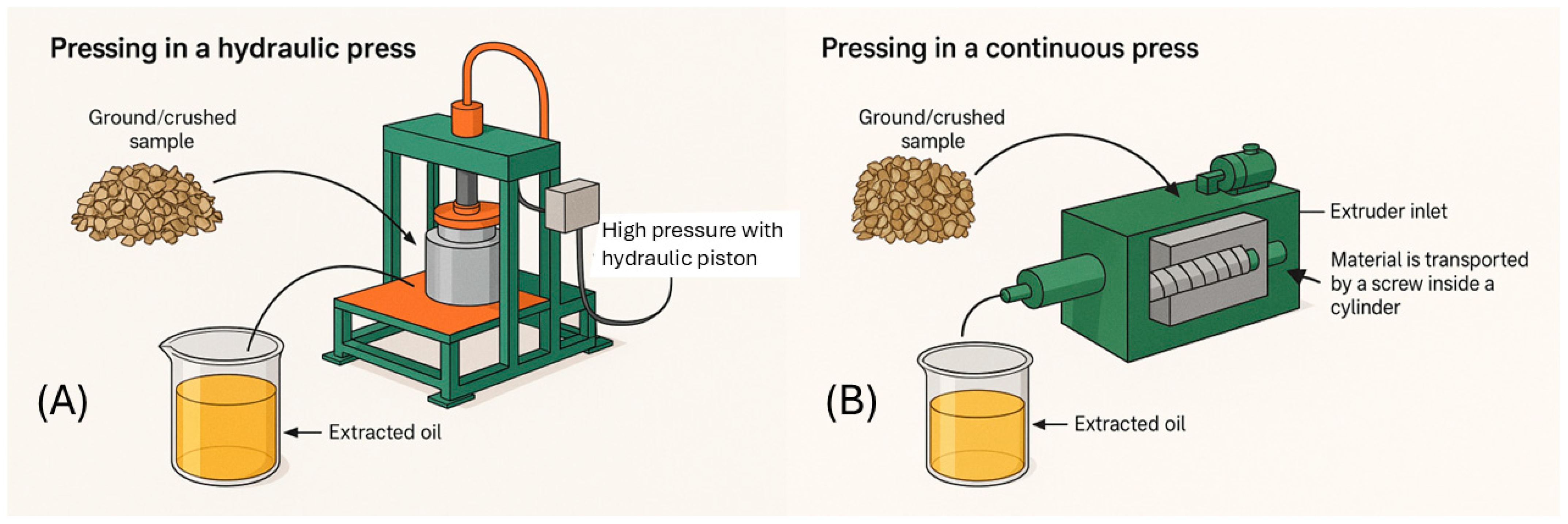
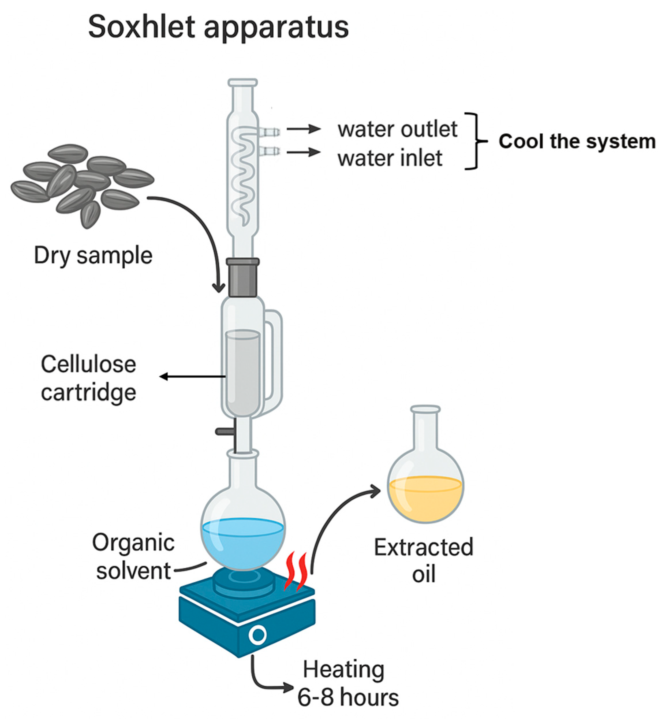
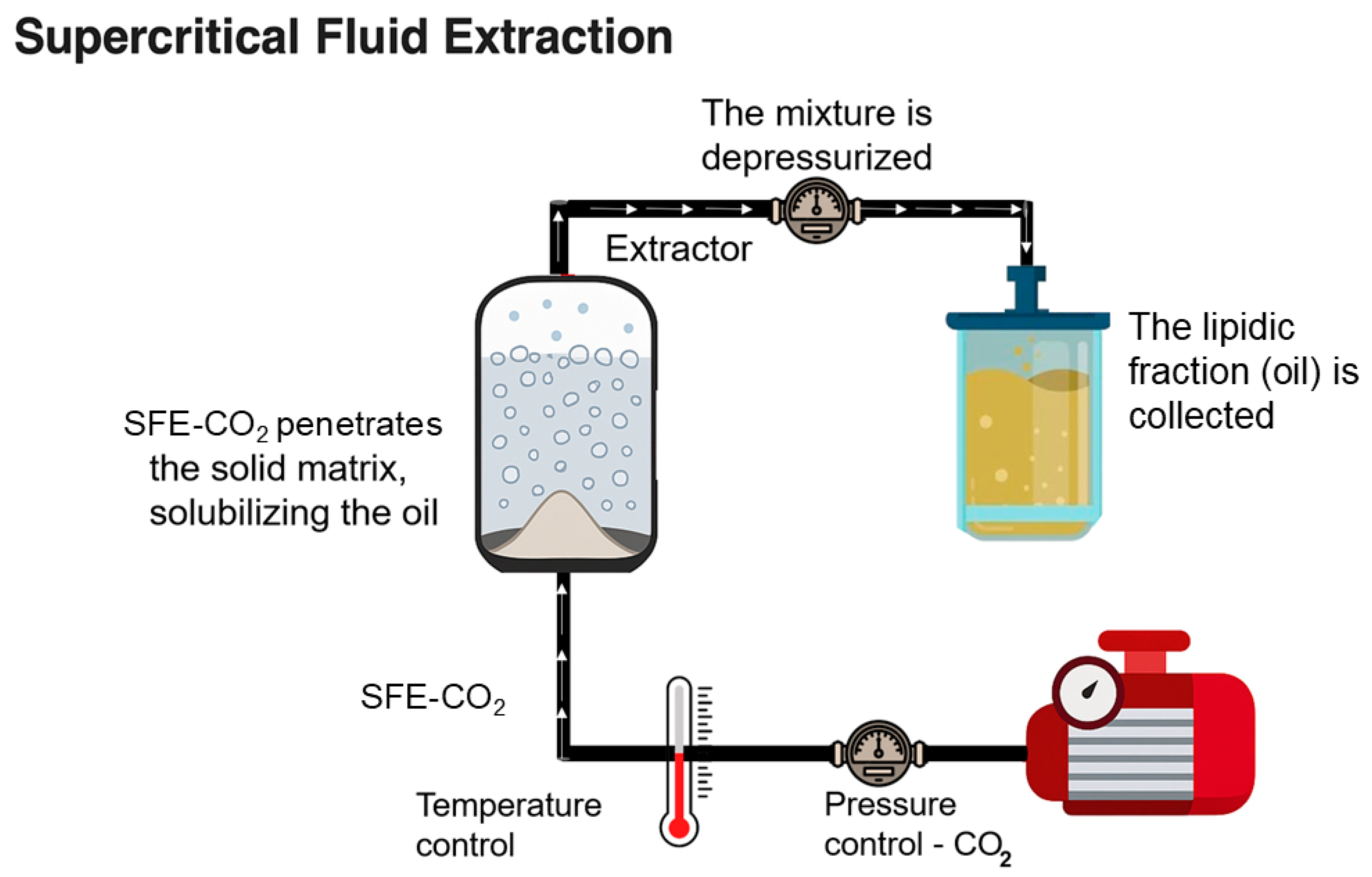
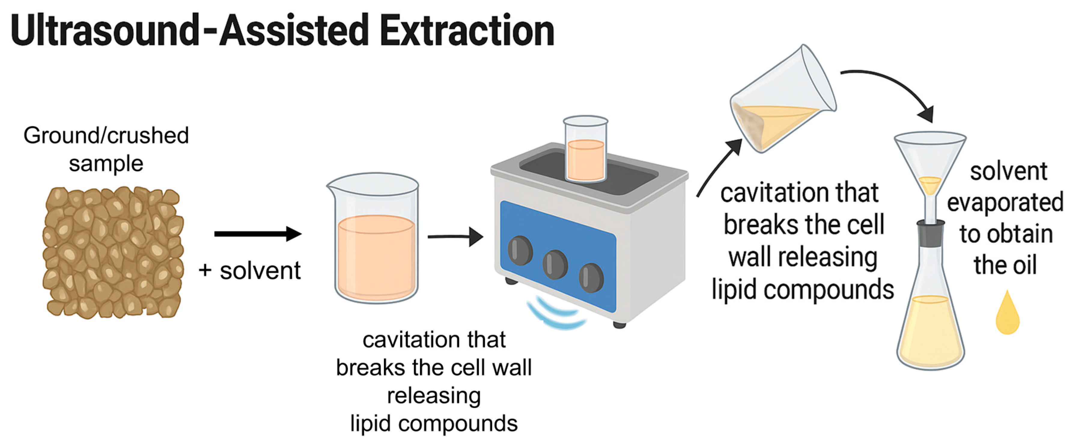

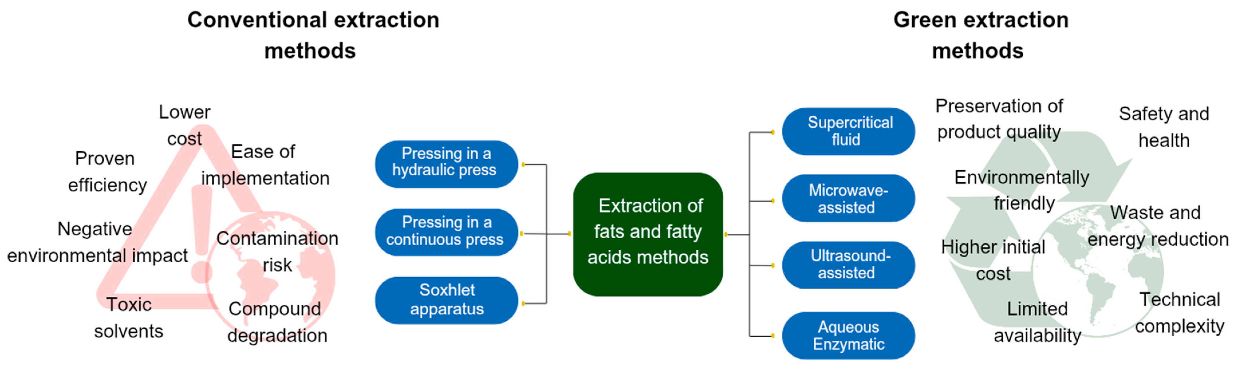
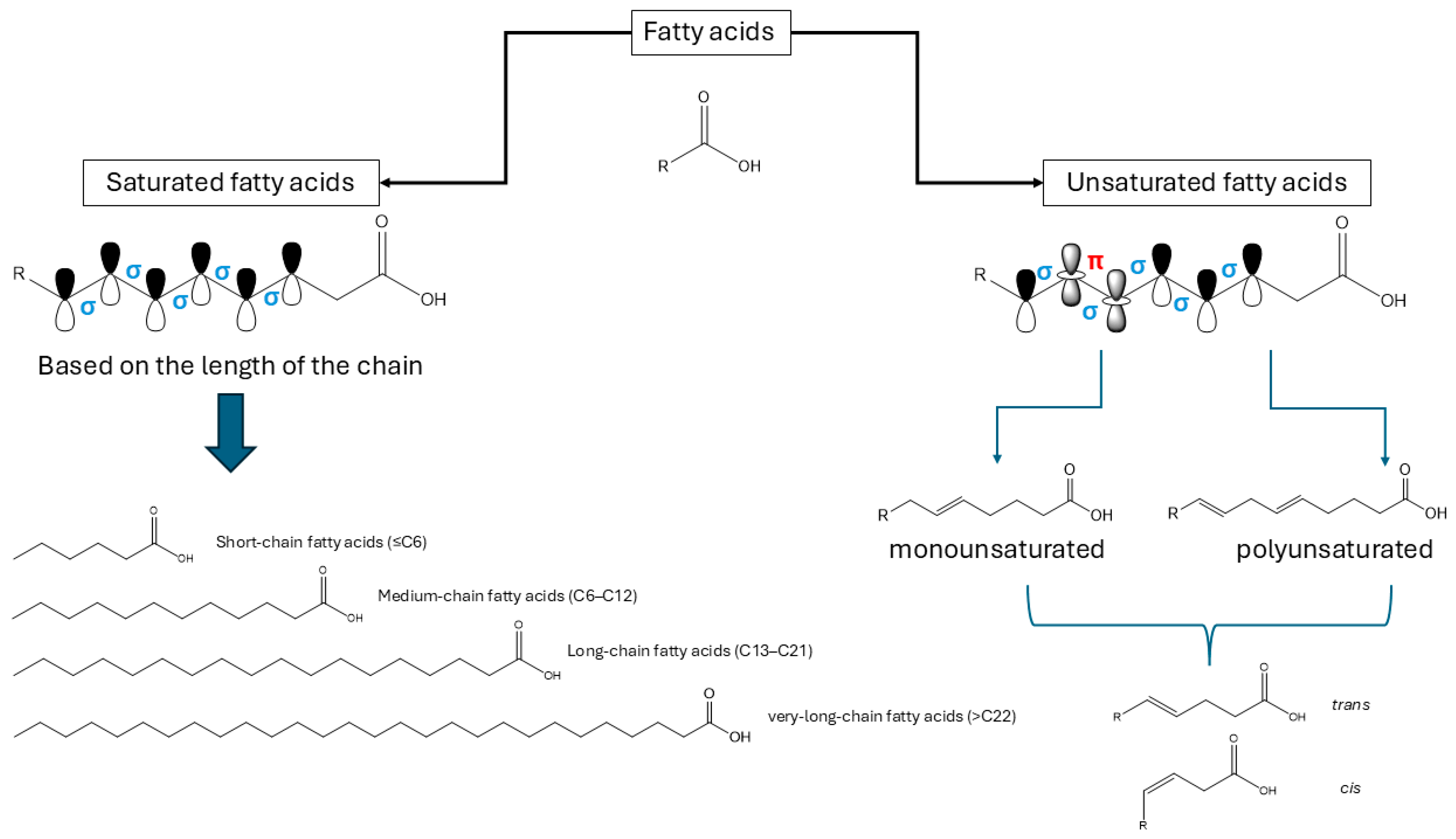
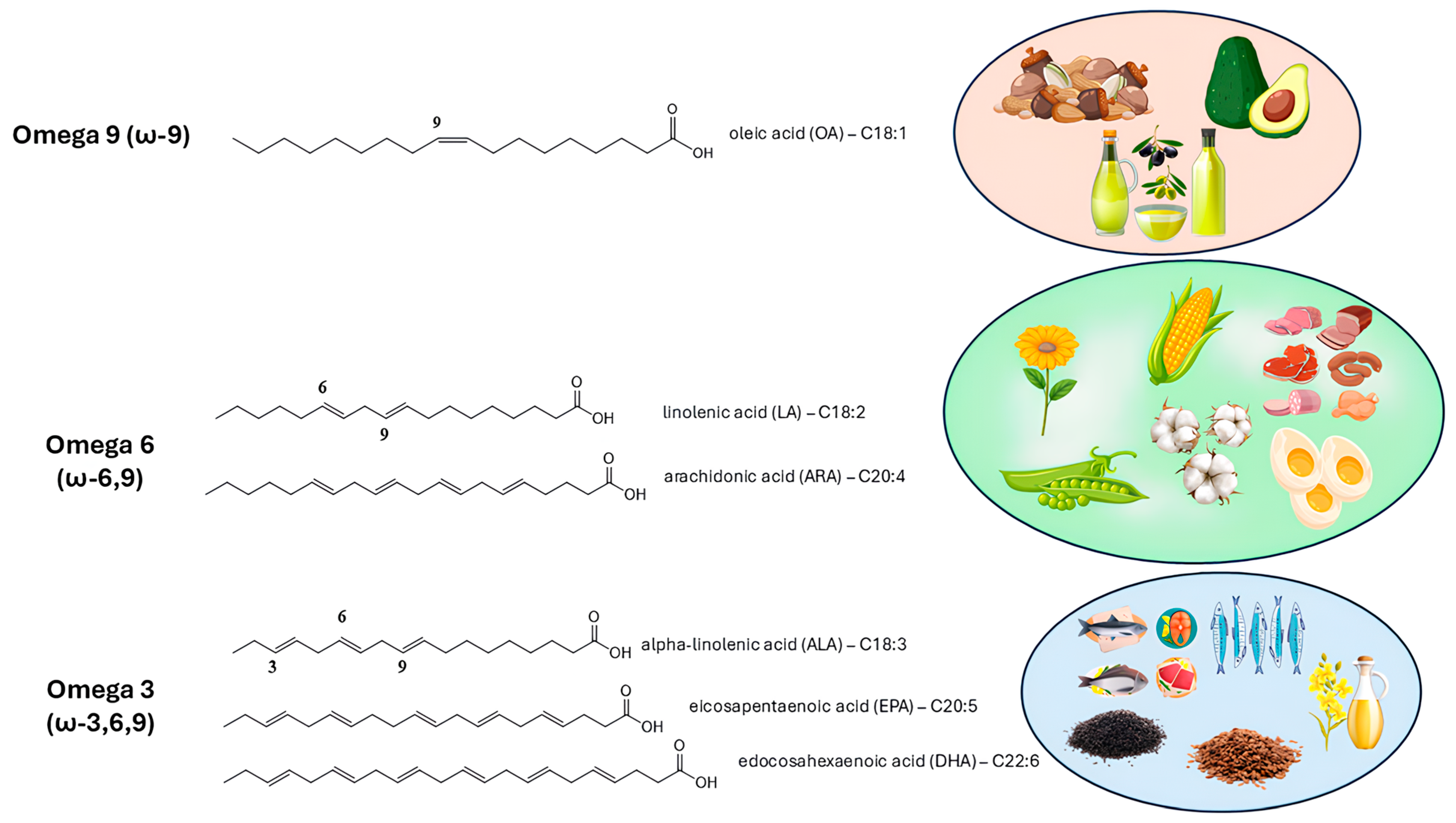
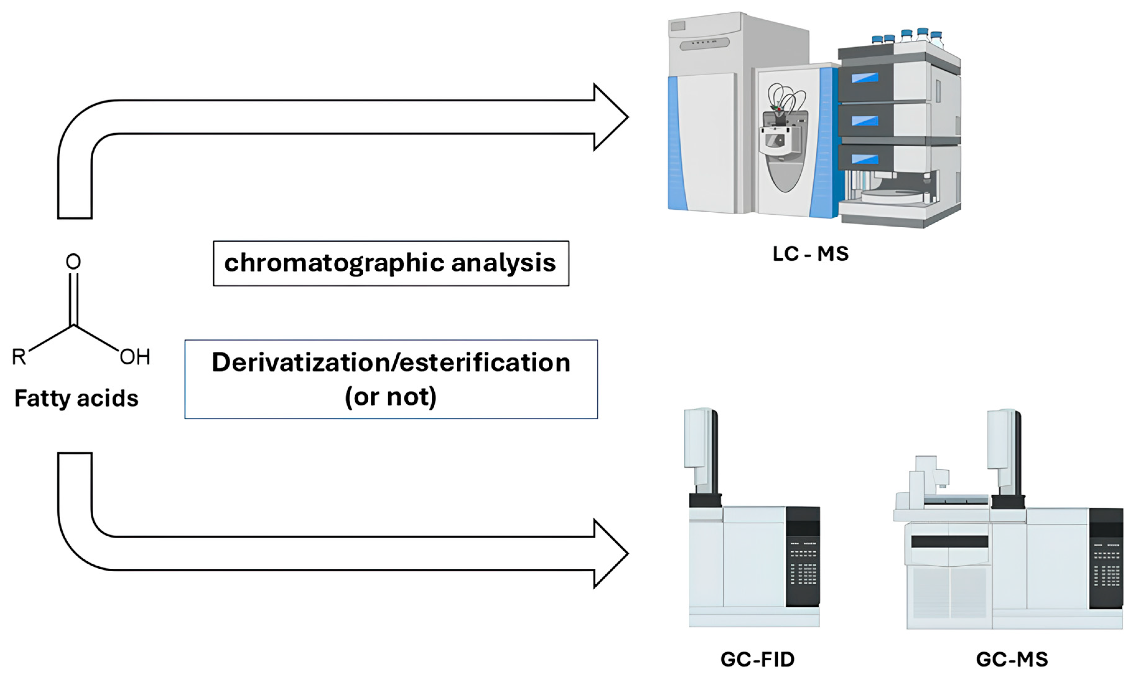
Disclaimer/Publisher’s Note: The statements, opinions and data contained in all publications are solely those of the individual author(s) and contributor(s) and not of MDPI and/or the editor(s). MDPI and/or the editor(s) disclaim responsibility for any injury to people or property resulting from any ideas, methods, instructions or products referred to in the content. |
© 2025 by the authors. Licensee MDPI, Basel, Switzerland. This article is an open access article distributed under the terms and conditions of the Creative Commons Attribution (CC BY) license (https://creativecommons.org/licenses/by/4.0/).