Article

Essential Oils as Nature’s Dual Powerhouses for Agroindustry and Medicine: Volatile Composition and Bioactivities—Antioxidant, Antimicrobial, and Cytotoxic

by
Javier Rocha-Pimienta
1,*,
Javier Espino
2,
Sara Martillanes
1 and
Jonathan Delgado-Adámez
3
1
High-Technology Incubator in Bioeconomy and Circular Economy, Center for Scientific and Technological Research of Extremadura, Polígono Industrial El Prado, C. Pamplona, s/n, 06800 Mérida, Spain
2
Neuroimmunophysiology and Chrononutrition Research Group, Department of Physiology, Faculty of Science, University of Extremadura, 06006 Badajoz, Spain
3
Technological Agri-Food Institute of Extremadura, Center for Scientific and Technological Research of Extremadura, 06007 Badajoz, Spain
*
Author to whom correspondence should be addressed.
Separations 2025, 12(6), 145; https://doi.org/10.3390/separations12060145
Submission received: 16 April 2025 / Revised: 21 May 2025 / Accepted: 27 May 2025 / Published: 1 June 2025
(This article belongs to the Section Analysis of Natural Products and Pharmaceuticals)

Abstract

:
Essential oils (EOs), which are complex mixtures of plant-derived volatile compounds, have been utilized for centuries in the medical, food, and pharmaceutical industries because of their diverse biological properties. In recent years, there has been growing interest in elucidating the bioactivities of essential oils and their underlying mechanisms of action. This study aimed to investigate the antioxidant, antimicrobial, and cytotoxic characteristics of Laurus nobilis, Eucalyptus camaldulensis, Rosmarinus officinalis, and Mentha suaveolens oils and relate them to their volatile compound content. The volatile compounds of the essential oils were characterized and quantified by gas chromatography, the antioxidant activity was quantified using the ABTS assay, the antibacterial activity was quantified using broth microdilution and agar diffusion techniques, and the MTT assay was used to establish the cytotoxic potential. This study revealed a significant antioxidant capacity, which correlated with the proportion of terpenes known for their antioxidant properties. The antioxidant potency was ranked in descending order: R. officinalis, M. suaveolens, E. camaldulensis, and L. nobilis. Antimicrobial testing demonstrated that all the examined essential oils were effective against the evaluated microbial species, including both Gram-positive (Listeria innocua) and Gram-negative (Escherichia coli) bacteria. Additionally, all the tested essential oils triggered cell death in the human epithelioid cervical carcinoma (HeLa) cell line. Collectively, this article highlights the promising therapeutic and alimentary potential of essential oils and underscores the need for further research to fully harness their benefits in industrial settings.

1. Introduction

The problem of waste management generated by the industry is leading to the development of policies that advocate the reuse of waste, focusing on obtaining bioactive compounds. Food- and plant-derived bioactive substances are a major focus for human health owing to their low toxicity, limited cost, and wide availability.
Natural compounds in the field of agri-foods have advantages over synthetic compounds. These natural compounds are often perceived to be safer and more environmentally friendly. They can also provide additional health benefits, such as antioxidant properties or nutritional value, beyond their primary function in food preservation or enhancement. Moreover, the use of natural compounds aligns with the growing trend towards clean label products and sustainable food production practices. Synthetic preservatives added to foods, such as antimicrobials and antioxidants, have no potential adverse effects and are classified as generally safe. However, there have been concerns regarding the safety of some chemicals, such as the possibility of allergies and formation of compounds with possible carcinogenicity.
Essential oils (EOs) are complex mixtures of volatile compounds, mostly hydrocarbons and oxygenated compounds synthesized by plants, and have various functions, including defense against microorganisms and animals. Essential oils are recognized as products extracted through steam distillation or pressing the outer layers of certain fruits. These substances are commonly referred to as essences, although this term is more inclusive and encompasses various compounds obtained through different extraction techniques [1].
Their applications in medicine and food are based on their anticancer, antiviral, antibacterial, and antioxidant properties [2]. In the second case, their use is focused on increasing the shelf life of foods [3] because there is currently a social trend of rejecting chemical additives used in the agri-food industry, owing to their possible adverse effects on health. EOs are generally derived from one or more plant parts, such as flowers, leaves, stems, bark, wood, roots, seeds, fruits, rhizomes, gums, or oleoresin exudations.
Several factors influence the chemical composition of plant essential oils, such as the species, part of the plant, time of harvest, geographical origin, and method of extraction, and consequently, their bioactive properties. It is widely recognized that plants of the same species, when grown in diverse locations, can exhibit significant variations in their chemical composition, consequently leading to differences in their biological activities. Essential oils are commonly found in various types of vegetation, and certain plants contain particularly high concentrations of these substances. Monoterpenoids, the main volatile components of essential oils, have historically been used because of their potent properties. Members of the genera Mentha, Rosmarinus, Laurus, and Eucalyptus produce some of the most commonly used essential oils. Several species have been studied, which show differences in oil composition depending on various intrinsic and extrinsic factors [4,5]. These plants are widespread in the Mediterranean basin and are important because of the wide variety of applications in which they are used. Their functionalities are mainly due to monoterpenoids, such as 1,8-cineole, found in their essential oils at high concentrations, which are responsible for their antimicrobial, antioxidant, cytotoxic, and anti-inflammatory activities [6].
The accumulation of aromatic and medicinal plants that fail to meet industrial quality standards presents a significant waste management challenge. These plants, cultivated for their essential oils, active compounds, or medicinal properties, may be rejected due to various factors, such as insufficient concentration of desired compounds, contamination, or improper handling during cultivation or processing. Consequently, large quantities of plant material that could potentially have value are discarded. This waste not only represents a loss of resources and economic potential but also poses environmental concerns if not properly managed. Addressing this issue requires the development of innovative approaches to utilize or repurpose these plants, potentially in new products, composting, or as feedstock for other industrial processes, thereby minimizing waste and maximizing resource efficiency in the aromatic and medicinal plant industries.
This study aimed to deepen our knowledge of the essential oils obtained from waste from the aromatic and medicinal plant industry in order to determine their functionalities and biological properties, and thus to establish a future application for key industries in the sector, such as food, medicine, pharmaceuticals, and cosmetics. The main objective was to characterize the aromatic and functional profiles of essential oils from R. officinalis, E. camaldulensis, M. suaveolens, and L. nobilis. Their antioxidant, antimicrobial, and antiproliferative activities were characterized to evaluate their potential future applications in different fields.

2. Materials and Methods

2.1. Preparation of Raw Material and Essential Oils Extraction

Leaves of laurel (L. nobilis), eucalyptus (E. camaldulensis), rosemary (R. officinalis), and apple mint (M. suaveolens) were collected from a local company (Badajoz, Spain). The samples were collected in the morning; leaves from different parts of the plants were taken, immediately transported to the laboratory in ventilated storage trays, and vacuum-packaged in plastic bags to avoid compositional changes. Before the essential oil extraction treatment, fresh leaves were washed with distilled water and ground in a domestic knife mill to obtain particles (0.5–3.0 mm). The material was stored at room temperature under vacuum until further use.
The EOs were obtained by distillation according to the European Pharmacopoeia. In this process, water produces steam when heated, carrying more volatile compounds in the plant material. The steam was then cooled, and the resulting distillate was collected. The EOs floated on the hydrosol (distilled water) and were separated. The EOs were kept away from light at refrigeration (4 °C) in amber-colored glass bottles.

2.2. Analysis of Volatile Compounds by Solid Phase Microextraction—Gas Chromatography–Mass Spectrometry (SPME-GC-MS)

2.2.1. HS-SPME

An amount of 10 µL from each sample was placed in a 20-mL vial with a PTFE/silicon septum. Then, the vial was placed in a CombiPAL autosampler (CTC Analytics, Zwingen, Switzerland) fitted with a GC–MS. The Combi-Pal program, in the SPME mode, was set as follows: the vial was heated at 37 °C for 5 min with agitation at 250 rpm (preextraction period); a 10 mm SPME fiber coated with a 50/30 µm layer of divinylbenzene-carboxen-polydimethylsiloxane (DVD-CAR-PDMS) was then inserted into the headspace, where extraction occurred for 30 min with continued heating and agitation; the fiber was subsequently desorbed in the GC injector at 270 °C for 10 min in the splitless mode.

2.2.2. GC–MS Analysis

The GC–MS analysis of the samples was carried out in an Agilent Technologies 6850 gas chromatograph equipped with a 5975 VL selective mass detector and a fused silica capillary column (HP-5, 5% phenyl methyl siloxane) (Agilent Technologies, Santa Clara, CA, USA, 30 m × 0.25 mm id and 0.25 µm film thickness) [5]. The carrier gas was helium (5.6 grade) at a flow rate of 1 mL/min and 7.95 psi. The column temperature program was held at 35 °C for 10 min, raised to 250 °C at 7 °C/min and held for 5 min (total running time: 45 min). The MS parameters were a mass range of 40–300 m/z, a 5.27 scan/s sampling rate, an ionization energy of 70 eV, 230 °C for the MS source, and 150 °C for the MS Quad.
The compounds were identified by comparison of their mass spectra and linear retention indexes (LRI) with those of standards (Sigma–Aldrich, St. Louis, MO, USA), or with mass spectra included in the NIST library by using the Agilent MSD Chemstation E.02.01.1177 software. Only the compounds found in at least four out of the five replicate packages per group, at least in one group, were taken into account.

2.3. Antibacterial Activity

2.3.1. Broth Microdilution

Broth microdilution was used to determine the antimicrobial activities of the essential oils [5]. The microorganisms used, Escherichia coli (CECT 45), and Listeria innocua (CECT 910), were obtained from the Spanish Type Culture Collection (CECT) of Valencia University, and were grown in Mueller-Hinton broth (Oxoid Ltd., Madrid, Spain) for 24 h to obtain actively growing cultures. The suspensions were diluted to a standard turbidity of 0.5 McFarland. A 96-well microtiter plate was used, and 160 μL of Mueller–Hinton broth, 20 μL of diluted bacteria, and 20 μL aliquots of the oil solutions were added. A positive control (containing inoculum but no EOs) and negative control (containing oils but no inoculum) were included in each microplate. The contents of the wells were mixed, and the microplates were incubated at 37 °C for 24 h under microaerophilic conditions. The absorbance was determined using a microplate reader (Infinite M200; Tecan Austria GmbH, Groedig, Austria) at 450 nm. Antibacterial activity was calculated using the following formula:
% Inhibition = Δ Abs reference Δ Abs assay Δ Abs reference × 100
ΔAbsreference: increase in absorbance of the positive control sample.
ΔAbsassay: increase in absorbance of oil-containing samples.

2.3.2. Agar Diffusion

The technique is based on the method originally described by Kirby-Bauer [7] and is based on the relationship between the concentration of the test substance required to inhibit bacterial growth and the growth inhibition halo it generates. For this purpose, the bacteria were homogeneously seeded on an agar plate, and filter paper discs (6 mm diameter) were placed equidistantly on the plate. The test substance (10 μL) was added to the discs and incubated at the appropriate temperature for 24 h. Finally, to read the results, the diameters of the growth inhibition halos around the discs are measured. Ampicillin (10 μg), an antibiotic of known spectrum, was added to one of the discs on each agar plate as a control.

2.4. Antioxidant Activity

Antioxidant activity was determined according to the method proposed by Turoli et al. [8] with slight modifications. This method is based on a reaction that occurs when the colorless compound ABTS ([2,2′-azinobis (3-ethylbenzothiazoline-6-sulphonic acid)]) is in the presence of potassium persulfate (K2S2O8), thus constituting an equilibrium with its ABTS* radical (blue-green chromophore). Antioxidant capacity was determined by measuring the decrease in absorbance of the ABTS* radical, as this reaction is reversed if an antioxidant substance is present in the medium. This radical was generated by its reaction with persulphate in the presence of phosphate buffer (pH 7.5, 50 mM). The decrease in absorbance was measured at 750 nm wavelength using a microplate reader (Infinite M200; Tecan Austria GmbH, Groedig, Austria). To quantify antioxidant activity, the external standard method was performed using Trolox (6-hydroxy-2,5,7,8-tetramethylchromane-2-carboxylic acid) as a synthetic antioxidant, and the results were expressed as mmol equivalents of Trolox.

2.5. Antiproliferative Activity

Antiproliferative activity was tested in the HeLa cervical cancer cell line using the CellTiter 96® AQueous One Solution Assay (Promega, Madrid, Spain), which is based on the reduction of the tetrazolium compound MTS. Cells were seeded in 96-well plates at a density of 2 × 10⁴ cells/well. After treating the cultures for 24 h, assays were performed by adding 10 µL of CellTiter 96® AQueous One Solution reagent directly to the culture wells, incubating for 2 h at 37 °C, and recording the absorbance on a microplate reader (Infinite M200; Tecan Austria GmbH, Groedig, Austria) at an assay wavelength of 490 nm and reference wavelength of 650 nm to subtract the background. Cell viability was calculated as a percentage of the control value (untreated samples).

2.6. Statistical Analysis

Statistical analysis was performed using Graph Prism 9.1 software (GraphPad Software, Inc., San Diego, CA, USA). These were studied statistically by applying t-tests for the comparison of means between samples and one-way analysis of variance (ANOVA). For the latter, Tukey’s test for the comparison of means (p < 0.05) was applied in case of significant differences. Statistical significance was set at p < 0.05.

3. Results and Discussion

3.1. Aromatic Profiles of the Essential Oils

Gas chromatography coupled with mass spectrometry (GC–MS) is considered the reference technique for the qualitative and quantitative analysis of volatile compounds in essential oils because of its high resolution, sensitivity, and structural identification capabilities. Unlike methods such as thin-layer chromatography (TLC) or Fourier-transform infrared spectroscopy (FTIR), which provide limited information on compound identity, GC–MS allows for the efficient separation of complex mixtures, followed by the identification of each analyte through its mass spectrum, using spectral libraries such as NIST and Wiley. Furthermore, compared with the flame ionization detector (GC–FID), which offers good quantification but lacks structural identification capabilities, GC–MS provides the added advantage of unambiguous identification based on molecular fragmentation patterns. Its applicability to complex matrices and compatibility with preconcentration methods, such as solid-phase microextraction (SPME), further strengthen its suitability. For these reasons, both the European Pharmacopoeia and ISO Technical Committee on Essential Oils (ISO TC 54) recommend the use of GC–MS as the standard technique for the characterization of these natural products.
The characteristics of the volatile compounds in the oils are listed in Table 1.
The determination percentages were 96.71%, 91.07%, 93.60%, and 94.13% for R. officinalis, E. camaldulensis, M. suaveolens, and L. nobilis, respectively. The main compounds vary depending on the plant, and there are similarities between them. The most abundant molecule was eucalyptol, also called 1,8-cineole, in rosemary (20.77%), eucalyptus (27.91%), and laurel (33.36%), an oxygenated monoterpene with an ether function. This compound has been recognized for a long list of properties, among which its antimicrobial, antifungal, anti-inflammatory, antioxidant, and anticancer activity stands out [6]. In apple mint, the majority of the compounds were piperitone oxide, accounting for more than half of the total content of volatile compounds in this oil (55.73%). It is an oxide formed from a ketone monoterpene with antimicrobial, antioxidant, insecticidal, analgesic, anti-inflammatory, and cytotoxic properties [9]. Considering those that were found in lower concentrations, in the case of rosemary, the compounds that followed eucalyptol were terpenes such as camphor (14.80%), α-pinene (13.18%), and L-bornyl acetate (10.66%)—a cyclic ketone and two monoterpenes, respectively, to which antimicrobial, antimutagenic and anticancer, anti-inflammatory, antioxidant, neuroprotector and gastroprotective properties are attributed, and which are widely used in cosmetics, perfumery, and as food additives [10,11,12]. R. officinalis is one of the plant species considered a natural source of camphor, along with others from the laurel and basil family. The presence of α-pinene and L-bornyl acetate in this essential oil has also been widely described, and it is shared mainly with some species of conifers [13,14].
In the case of eucalyptus, the following compounds were identified: L-aloaromadendrene (13.49%) and (+)-ledene (9.69%). The first, also known as aromadendrene, is a sesquiterpene with potent antimicrobial properties and is widely used against antibiotic-resistant bacteria [15]. This aromatic profile is mainly present in this genus of plants, in which eucalyptol is the most abundant [16,17].
In apple mint, high concentrations of β-cubebene (15.83%) and 4-terpineol (8.69%) were also found. Cubebene is a tricyclic sesquiterpene with neuroprotective and anti-inflammatory properties [18]. Terpineol is an alcohol monoterpene with potent anticancer properties both in vitro and in vivo [19] as well as antimicrobial properties against bacteria that cause eye and skin infections in humans [20]. Apple mint essential oil has three possible aromatic profiles: the first is rich in pulegone and the second in piperitenone oxide, and the third contains similar amounts of piperitenone oxide and piperitone oxide, depending on the species and growing conditions [9].
For laurel, the next most important compounds were α-terpineol acetate (24.32%) and linalool (17.29%).
The volatile composition of essential oils depends not only on the plant genus but also on the species itself, and even on the environmental conditions in which they grow. This is evident when comparing our results with those of previous studies carried out by other researchers, where the proportions of different compounds and the presence or absence of some of them vary directly. These premises are very important for establishing the future applicability of oils, requiring a study prior to extraction to determine the optimum conditions for obtaining specific aromatic profiles.

3.2. Evaluation of the Antioxidant Activity of the EOs

Essential oils contain a wide range of bioactive compounds, including outstanding antioxidant activity. Figure 1 shows the in vitro antioxidant potential of rosemary, eucalyptus, apple mint, and laurel oils.
According to the results obtained, the essential oils with the highest antioxidant activity were rosemary (361.1 equiv·mg Trolox·mL−1) and apple mint (337.8 equiv·mg Trolox·mL−1), with no significant differences between them, followed by eucalyptus (324.2 equiv·mg Trolox·mL−1) and, finally, laurel (257.8 equiv·mg Trolox·mL−1). In view of the results obtained previously, which showed the profile of aromatic compounds in each oil, the high antioxidant potential exhibited by these oils was justified.
High antioxidant potential was detected in rosemary essential oils with an aromatic profile very similar to ours, in which 1,8-cineole, α-pinene, and camphor predominated, establishing an IC50 of 22.61 μg/mL using the ABTS method [21]. The importance of these compounds has been demonstrated in oils rich in them; Chraibi et al. [22] established an IC50 value of 2.77 mg/mL in rosemary essential oils rich in 1,8-cineole (33.88%). As with the aromatic profile, an important factor to consider is the harvesting and development stage of the plant. Beretta et al. [4] compared the radical scavenging capacity of essential oils at the flowering, post-flowering, and vegetative stages. The best activity was observed at the flowering stage, with IC50 values of 36.78 μg/mL, followed by the post-flowering and vegetative stages (79.69 and 111.94 μg/mL, respectively).
Studies have also determined the IC50 value for eucalyptus and its antioxidant activity to be 94.7 μg/mL [23]. In contrast, Noumi et al. [24] described the importance of granulometry and plant material size for oil extraction. They analyzed the activity of oils dominated by α-pinene, 1,8-cineole, and α-terpineol, and determined that the percentage of radical elimination by the ABTS method ranged from 18 to 89%, depending on the amount of the raw material used to obtain the oil.
Finally, for apple mint and bay laurel essential oils, which also have significant antioxidant properties, with the latter being the least potent of those tested in this study, their radical scavenging capacity is well known. Recent studies characterize the essential oils of laurel leaves for their possible use in cosmetic applications, obtaining a profile of volatiles like ours (predominance of compounds such as 1,8-cineole, α-terpineol acetate and linalool) and determining that their antioxidant potential by the ABTS radical method has IC50 values of 124.01 μg/mL [25]. Similar results were obtained by Guedri et al. [26] for laurel oils collected from different locations (Tunisia, France, and Austria). The volatile composition varied significantly depending on the place of origin, but all three shared a 1,8-cineole content. Antioxidant activity also varied, with IC50 values of 44.8, 76.4, and 81.4 μg/mL, respectively. This study concludes that 1,8-cineole is one of the components responsible for the antioxidant potential of laurel oils, as its increase led to higher activity in the essential oil of Tunisian origin.
On the other hand, Bouyahya et al. [27] compared the volatile composition, antioxidant, and antimicrobial activity of M. suaveolens oil. The apple mint essential oil in this study contains a similar percentage of piperitone oxide (56.28 vs. 55.73%) and other minority components, which exhibited an antioxidant activity with IC50 values of 82.73 μg/mL by the ABTS method. Al-Mijalli et al. [28] also support our results with their study, in which they determined the antioxidant, antibacterial, and antidiabetic capacity of apple mint and sage in relation to their volatile composition. They found that apple mint oil has an IC50 for its antioxidant activity of 75.19 μg/mL, which is more active than that of sage (129.74 μg/mL). They attributed this potential to the high piperazine and piperitone oxide content.
These results highlight the wide variety of factors that affect the different compositions of oils and the contents of certain compounds, leading to significant differences in their biological properties.

3.3. Evaluation of the Antimicrobial Activity of the EOs

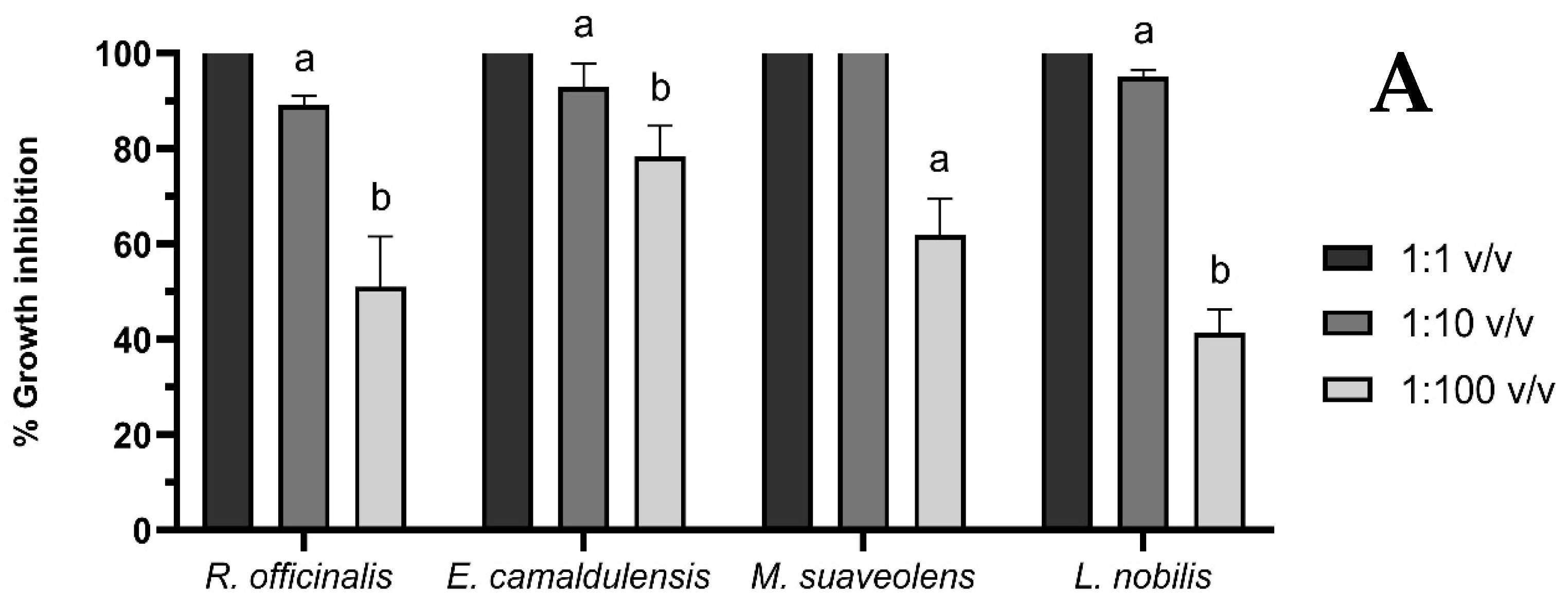
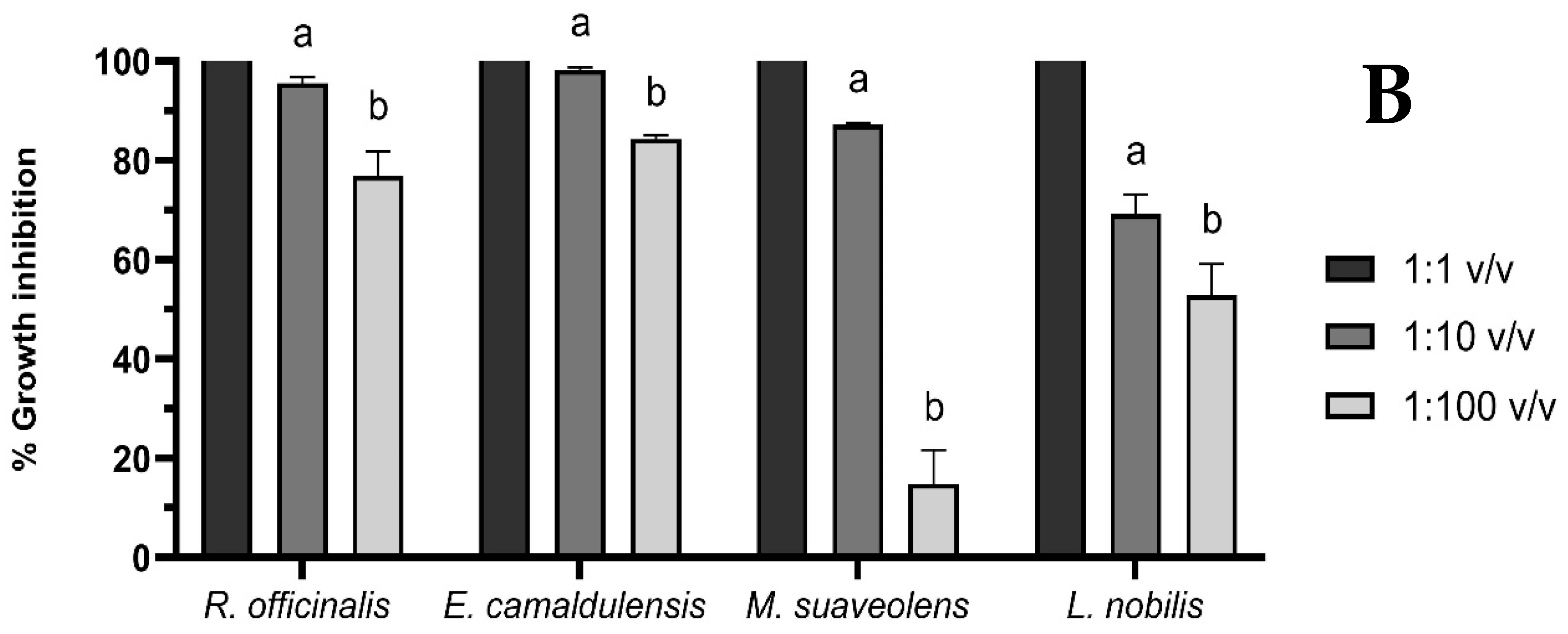
In vitro antibacterial capacity was determined against two model microorganisms, E. coli and L. innocua (Figure 2).
From the results obtained, we can affirm that all the oils tested showed bactericidal activity when applied directly without dilution to the two bacteria (100 μL/mL), since growth inhibition was complete in all cases. Similarly, all of them showed dose-dependent activity, as the three concentrations tested, as they were diluted, significantly decreased their activity against the two microorganisms. For E. coli, the 1:10 dilutions of rosemary, eucalyptus, and laurel were very close to 100% inhibition of bacterial growth (89.12%, 92.93%, and 95.04%, respectively), indicating that their MICs were close to this application dose, standing at 11.3, 10.8 and 10.3 µL/mL, respectively. However, in the case of apple mint, the total growth inhibition is exceeded; therefore, at this dose, it also exhibits bactericidal activity, with an MIC of 1.7 µL/mL. Against L. innocua, rosemary and eucalyptus are also those which, in their 1:10 dilutions, come close to the MIC (95.93% and 98.19% inhibition, respectively), being 10.4 and 10.2 µL/mL, respectively. In contrast, apple mint, unlike against E. coli, did not exhibit total growth inhibition at this concentration (87.19%), with an MIC of 11.5 µL/mL. The most significant decrease was observed with bay laurel, which reduced the efficacy by 69.19%, showing the highest MIC of all oils (14.6 µL/mL).
Although, as mentioned above, the oils applied directly to the bacterial cultures showed that they had bactericidal activity, the bactericidal activity was determined using the agar diffusion method with discs (Table 2).
The reference antibiotic ampicillin exhibited the expected bactericidal activity, showing statistically significant differences for all oils. The oils that created the largest inhibition halos against E. coli cultures were laurel and rosemary, at 19.50 and 15.80 mm in diameter. Apple mint had the lowest activity (12.50 mm), with significant differences compared to laurel, while eucalyptus had the lowest activity (8.75 mm). In contrast, rosemary, eucalyptus, and laurel had the highest activity against L. innocua, with no differences between them, whereas apple mint had the lowest bactericidal capacity.
Considering the research mentioned in the previous section where we analyzed the antioxidant capacity, Bajalan et al. [29] quantified the antibacterial capacity against S. agalactiae, S. aureus, K. pneumoniae, and E. coli, showing that the highest bactericidal capacity was against the latter. The inhibition halos ranged from 18.51 to 13.97 mm in diameter, as there are substances in the oils that cause a specific increase in the antibacterial capacity of Gram-negative bacteria [30].
Chraibi et al. [22] also determined the antimicrobial potential of rosemary essential oil alone and in combination with carvacrol against bacteria (S. aureus, M. luteus, B. cereus, B. subtilis, E. coli, S. enterica, and P. aeruginosa) and yeasts (C. albicans and C. tropicalis). They found strong antimicrobial potency against all Gram-positive bacteria, with MIC values of 7–31 μL/mL. For Gram-negative bacteria, the tested oil was most effective against E. coli and S. enterica, with MICs of 62 μL/mL and 125 μL/mL, respectively. The combination with carvacrol enhanced the activity of the oil (synergistic effect) and significantly decreased the MICs of all tested microorganisms.
Hassine et al. [23] also characterized the antimicrobial capacity of oils and aqueous extracts of eucalyptus oils against bacteria, molds, and yeasts. Their results are in agreement with ours, but exhibit higher activity in the case of L. innocua, since the MIC values presented are 0.78 mg/mL. In contrast, their oil showed no activity against Gram-negative bacteria, including E. coli. These results, compared with those presented by other researchers and by us, show that the differences in the volatile composition of the oils make some of them ineffective against certain bacteria, thus causing oils of the same species obtained under different conditions to have completely different capacities.
Bay laurel has also been characterized for its efficacy against bacterial growth. An example of this again is the study by Guedri et al. [26], who, apart from determining the volatile profile and antioxidant activity of oils from Tunisia, France, and Austria, also established its effectiveness against B. subtilis, S. aureus, L. monocytogenes, E. coli, K. pneumonaea, and S. enterica, and against three molds and two yeasts. The oil showed activity against S. aureus, which was greater than that of the control antibiotic (ampicillin). Activity against Listeria was moderate (17–20 mm inhibition halo), and the oil was insensitive to, or had very little activity against, E. coli at the concentrations tested (0.5–50 μL/mL). In its activity against other bacteria, the oil follows the same trend as described above, that is, it is more effective against Gram-positive bacteria. It also showed significant activity against molds and yeast.
Other investigations have also calculated the MIC and bactericidal capacity of the essential oil of M. suaveolens against Gram-positive and -negative bacteria (E. coli, S. aureus, L. monocytogenes, P. mirabilis, P. aeruginosa, and B. subtilis). MIC values are 0.5 mg/mL for Listeria and >1 mg/mL for E. coli. In the case of inhibition halos, the same occurs as with bacteriostatic activity; the values are 13 mm for E. coli and 24 mm for Listeria, which is five times higher than ours; therefore, against E. coli, it is our most potent oil [27]. In this study, the activity of the oil did not exceed that of chloramphenicol against any of the tested microorganisms.
As with antioxidant and antidiabetic activity, Al-Mijalli et al. [28] characterized the antimicrobial capacity of peppermint essential oils against a large number of microorganisms such as S. aureus, S. epidermidis, E. faecalis, L. monocytogenes, B. cereus, E. coli, S. typhimurium, K. pneumoniae, P. mirabilis, and P. aeruginosa, determining both its MIC and the presence of bactericidal activity. As for the MIC calculation, their results are higher than ours since they obtained amounts of 1.56 and 0.78 mg/mL for E. coli and Listeria, respectively. In our case, the application of a 1:10 dilution (8.5 mg/mL oil) did not show bactericidal activity. In general, they established, as in most studies, that oil is more effective against Gram-positive bacteria than Gram-negative bacteria. Finally, their results showed better bactericidal activity than ours. The inhibition halos were 29.6 mm for L. innocua and 21.7 mm for E. coli. This shows that their oils have activity even higher than that of chloramphenicol (a 27.5 mm halo against Listeria). All other microorganisms also showed significant bactericidal activity (inhibition halos of 9.2–31.6 mm), which demonstrates the broad spectrum of action of this essential oil.
These results, including ours, highlight the great potential of essential oils when applied as antimicrobials, as well as the importance of their composition. However, when they are used in industries such as the food industry, it must be taken into account that volatile compounds sometimes provide a very strong odor and flavor, which can modify the organoleptic characteristics of the products and make them undesirable for the consumer. Therefore, it is necessary to investigate the form and conditions of applications for food preservation.

3.4. Evaluation of the Cytotoxic Activity of the EOs

Finally, the cytotoxic capacity of essential oils has been widely studied because they include in their composition molecules that act at different levels to kill cells that develop different types of cancer. Figure 3 shows the cytotoxic activity of rosemary, eucalyptus, apple mint, and laurel essential oils against the HeLa cell line at different concentrations.
Oils diluted 1:100, 1:500, and 1:1000 were used because when more concentrated oils were added to the cell cultures, they showed 0% cell viability. Regardless of the dilution applied, the most effective treatments, with no statistically significant differences between doses, were eucalyptus and apple mint. This decreased cell viability by 5.95–17% and 10.34–14.12%, respectively. In terms of the dose-dependent effect, rosemary did not show significant differences in the two most concentrated doses (1:100 and 1:500) but did show significant differences in the dose diluted to 1:1000 (3.49, 9.63, and 78.67% viability, respectively). Finally, bay laurel had a clear dose-dependent effect, as the cell viability decreased significantly as the dose was diluted.
The IC50 values of rosemary, eucalyptus, peppermint, and laurel against HeLa cells were 0.125%, 0.031%, 0.006%, and 0.167%, respectively.
Essential oils, owing to their content of compounds with a multitude of functionalities, have also been attributed with cytotoxic properties against tumor cell lines. This has been a source of research for the development of drugs to treat different types of cancer, and there is the prospect that essential oils can be used as therapeutic agents and confer benefits to human health. Focusing specifically on rosemary oil, research such as that of Santos et al. [31] extols the role and cytotoxic efficacy of rosemary oil, as well as turmeric and ginger oils, on the HeLa cell line. They obtained an oil rich mainly in 1,8-cineole, α-pinene and camphor, and through the MTT assay, determined that it possessed an IC50 of 909.6 μg/mL. This application dose is higher than ours and is therefore less effective. This can be explained by the fact that the oils analyzed in this study contained almost twice as much 1,8-cineole as ours, and this compound has low cytotoxicity against tumor cell lines. This was described by Wang et al. [32], who related antibacterial and cytotoxic activities to the major compounds in rosemary oil: 1,8-cineole, α-pinene, and β-pinene. They established that both α-pinene and β-pinene are more important in providing antimicrobial and antiproliferative functionality than 1,8-cineole. This was observed by analyzing the compounds together in the oil and separately, showing significant differences, and concluding that, apart from some compounds being more important than others, the antibacterial and anticancer activities of rosemary essential oil are the cooperative results of its components. Finally, a recent study showed that inclusion of rosemary essential oil in nanoemulsions (50% Tween, 15% Span 80, and 35% essential oil) enhanced the properties and functionalities of the oil in terms of antioxidant, antimicrobial, and anticancer activities [33]. In the first case, its capacity became 10 times greater than that of Trolox, MICs against bacteria such as P. aeruginosa, K. pneumoniae and methicillin-resistant S. aureus decreased significantly, and cytotoxic activity against human hepatocellular carcinoma cell lines (Hep3B) and cervical carcinoma cells (HeLa) decreased IC50 values with respect to directly applied oil.
On the other hand, eucalyptus essential oil has aroused great interest due to its cytotoxic properties against a wide range of cell lines, establishing itself as one of the future alternatives in natural therapies against cancer [34]. These are mainly associated with the monoterpene group, such as 1,8-cineole, γ-terpinene, terpinen-4-ol, and α-terpineol, among others [35]. In recent years, research such as that of Pauzer et al. [36] has studied in detail the essential oils of E. cinerea by determining their volatile content, antioxidant and antimicrobial activity, and cytotoxicity. Against the same cell line as ours, E. cinerea oil possessed little activity, but it exhibited activity against the Calu-3 line, showing a significant reduction in viability, with an IC50 of 689.79 μg/mL. They observed that this was due to an increase in the percentage of cells in the sub-G0 and S phases, with an analogous reduction in the percentage of cells in the G0/G1 and G2/M phases, also providing DNA fragmentation of 29.73%.
Regarding the cytotoxic properties of mint, many studies have demonstrated the effect of compounds present in this species in inhibiting cell proliferation in numerous tumor lines by acting on mitochondrial dysfunction, induction of apoptosis, and autophagy processes [37]. In our study, this essential oil had the highest cytotoxic capacity against HeLa cells. Spagnoletti et al. [38] characterized, in terms of volatile composition, the antifungal, antioxidant, and cytotoxic activities of peppermint, thyme, oregano, and rosemary essential oils. With a majority composition of piperitone oxide (86.5%), the peppermint oil analyzed in this study exhibited potent antitumor activity against HaCaT and A549 cell lines, with IC50 of 35.7 and 75.6 μg/mL, respectively, and was the most potent of the four analyzed. Stringaro et al. [39] studied the cytotoxicity of M. suaveolens essential oils against the human breast adenocarcinoma cell line SKBR3. Analysis of their composition revealed a high percentage of piperitenone oxide (90%), and morphological and ultrastructural studies using electron microscopy revealed that the cells were damaged at the plasma membrane with many superficial alterations.
Finally, similarly to most essential oils, bay laurel oil was characterized and defined by its cytotoxic potential. In our study, it showed the lowest activity, as its IC50 was the highest. Oils with volatile profiles very similar to ours, composed of 1,8-cineole (27.41%), linalool (19.37%), α-terpineol acetate (14.65%), eugenol (7.73%), α-terpineol, (2.84%), terpinen-4-ol (2.09%), elemicin (2.08%), and sabinene (1.80%), exhibited potent cytotoxic properties against the human liver cancer cell line (Hep-G2), with an IC50 of 1.83 μg/mL. By analyzing the molecular coupling of the oil components against caspase-3 (an apoptotic enzyme), the ability of laurel compounds to interact with this enzyme at different sites and initiate its activity has been observed [40].

4. Conclusions

The results demonstrated that the chemical composition of the EOs of L. nobilis, E. camaldulensis, R. officinalis, and M. suaveolens are rich in volatile compounds. EOs from these plant species exhibit strong antioxidant and antimicrobial activities. Moreover, the results clearly showed that these EOs possessed cytotoxic activity in vitro, as they exerted cytotoxic actions against the tested cell line (HeLa). However, further studies are required to elucidate these cytotoxic mechanisms. From these results, it is evident that the amounts of certain compounds, or the presence or absence of others, are also crucial for determining whether an essential oil has bioactive capacities. Considering this, the optimization of growing conditions, plant selection, extraction conditions, and other factors that may influence the aromatic profile should be the first and most important step when applying a particular oil. Overall, these findings suggest that the tested EOs are a potential source of pharmaceuticals.

Author Contributions

Conceptualization, J.D.-A.; Formal analysis, J.R.-P., J.E. and S.M.; Investigation, J.R.-P., J.E. and S.M.; Methodology, J.D.-A.; Resources, J.D.-A.; Writing—original draft, J.R.-P.; Writing—review & editing, J.E. and S.M. All authors have read and agreed to the published version of the manuscript.

Funding

The authors thank the Regional Government of Extremadura and FEDER funds for the economic aid they received (GR18192 (AGA002)). J. Rocha-Pimienta thanks to Regional Government of Extremadura and the European Social Found (ESF) for the predoctoral formation contract (PD18075).

Data Availability Statement

Data is contained within the article.

Conflicts of Interest

The authors declare no conflicts of interest.

References

  1. Bruneton, J. Factores de variabilidad de los aceites esenciales. In Aceites Esenciales; Acribia S.A.: Zaragoza, Spain, 2001. [Google Scholar]
  2. Adorjan, B.; Buchbauer, G. Biological Properties of Essential Oils: An Updated Review. Flavour Fragr. J. 2010, 25, 407–426. [Google Scholar] [CrossRef]
  3. Holley, R.A.; Patel, D. Improvement in Shelf-Life and Safety of Perishable Foods by Plant Essential Oils and Smoke Antimicrobials. Food Microbiol. 2005, 22, 273–292. [Google Scholar] [CrossRef]
  4. Beretta, G.; Artali, R.; Facino, R.M.; Gelmini, F. An Analytical and Theoretical Approach for the Profiling of the Antioxidant Activity of Essential Oils: The Case of Rosmarinus officinalis L. J. Pharm. Biomed. Anal. 2011, 55, 1255–1264. [Google Scholar] [CrossRef]
  5. Delgado-Adámez, J.; Garrido, M.; Bote, M.E.; Fuentes-Pérez, M.C.; Espino, J.; Martín-Vertedor, D. Chemical Composition and Bioactivity of Essential Oils from Flower and Fruit of Thymbra capitata and Thymus Species. J. Food Sci. Technol. 2017, 54, 1857–1865. [Google Scholar] [CrossRef] [PubMed]
  6. Salehi, B.; Sharifi-Rad, J.; Quispe, C.; Llaique, H.; Villalobos, M.; Smeriglio, A.; Trombetta, D.; Ezzat, S.M.; Salem, M.A.; Zayed, A.; et al. Insights into Eucalyptus Genus Chemical Constituents, Biological Activities and Health-Promoting Effects. Trends Food Sci. Technol. 2019, 91, 609–624. [Google Scholar] [CrossRef]
  7. Hudzicki, J. Kirby-Bauer Disk Diffusion Susceptibility Test Protocol. Am. Soc. Microbiol. 2009, 15, 1–23. [Google Scholar]
  8. Turoli, D.; Testolin, G.; Zanini, R.; Bellù, R. Determination of Oxidative Status in Breast and Formula Milk. Acta Paediatr. 2004, 93, 1569–1574. [Google Scholar] [CrossRef] [PubMed]
  9. Božovic, M.; Pirolli, A.; Ragno, R. Mentha suaveolens Ehrh. (Lamiaceae) Essential Oil and Its Main Constituent Piperitenone Oxide: Biological Activities and Chemistry. Molecules 2015, 20, 8605–8633. [Google Scholar] [CrossRef]
  10. Malabadi, R.B.; Kolkar, K.P.; Meti, N.T.; Chalannavar, R.K. Camphor Tree, Cinnamomum camphora (L.); Ethnobotany and Pharmacological Updates. Biomedicine 2021, 41, 181–184. [Google Scholar] [CrossRef]
  11. Allenspach, M.; Steuer, C. α-Pinene: A Never-Ending Story. Phytochemistry 2021, 190, 112857. [Google Scholar] [CrossRef]
  12. Zhao, Z.J.; Sun, Y.L.; Ruan, X.F. Bornyl Acetate: A Promising Agent in Phytomedicine for Inflammation and Immune Modulation. Phytomedicine 2023, 114, 154781. [Google Scholar] [CrossRef] [PubMed]
  13. Jafari-Sales, A.; Pashazadeh, M. Study of Chemical Composition and Antimicrobial Properties of Rosemary (Rosmarinus officinalis) Essential Oil on Staphylococcus aureus and Escherichia coli in Vitro. Int. J. Life Sci. Biotechnol. 2020, 3, 62–69. [Google Scholar] [CrossRef]
  14. Hcini, K.; Sotomayor, J.A.; Jordan, M.J.; Bouzid, S. Chemical Composition of the Essential Oil of Rosemary (Rosmarinus officinalis L.) of Tunisian Origin. Asian J. Chem. 2013, 25, 2601–2603. [Google Scholar] [CrossRef]
  15. Mulyaningsih, S.; Sporer, F.; Zimmermann, S.; Reichling, J.; Wink, M. Synergistic Properties of the Terpenoids Aromadendrene and 1,8-Cineole from the Essential Oil of Eucalyptus globulus against Antibiotic-Susceptible and Antibiotic-Resistant Pathogens. Phytomedicine 2010, 17, 1061–1066. [Google Scholar] [CrossRef]
  16. Taher Maghsoodlou, M.; Kazemipoor, N.; Valizadeh, J.; Falak, M.; Seifi, N.; Rahneshan, N. Essential Oil Composition of Eucalyptus microtheca and Eucalyptus viminalis. Avicenna J. Phytomed. 2015, 5, 540–552. [Google Scholar]
  17. Pereira, S.I.; Freire, C.S.R.; Neto, C.P.; Silvestre, A.J.D.; Silva, A.M.S. Chemical Composition of the Essential Oil Distilled from the Fruits of Eucalyptus globulus Grown in Portugal. Flavour. Fragr. J. 2005, 20, 407–409. [Google Scholar] [CrossRef]
  18. Park, S.Y.; Jung, W.J.; Kang, J.S.; Kim, C.M.; Park, G.; Choi, Y.W. Neuroprotective Effects of α-Iso-Cubebene against Glutamate-Induced Damage in the HT22 Hippocampal Neuronal Cell Line. Int. J. Mol. Med. 2015, 35, 525–532. [Google Scholar] [CrossRef]
  19. Liu, S.; Zhao, Y.; Cui, H.F.; Cao, C.Y.; Zhang, Y.B. 4-Terpineol Exhibits Potent in Vitro and in Vivo Anticancer Effects in Hep-G2 Hepatocellular Carcinoma Cells by Suppressing Cell Migration and Inducing Apoptosis and Sub-G1 Cell Cycle Arrest. J. Buon 2016, 21, 1195–1202. [Google Scholar]
  20. Su, C.W.; Tighe, S.; Sheha, H.; Cheng, A.M.S.; Tseng, S.C.G. Safety and Efficacy of 4-Terpineol against Microorganisms Associated with Blepharitis and Common Ocular Diseases. BMJ Open Ophthalmol. 2018, 3, e000094. [Google Scholar] [CrossRef]
  21. Leporini, M.; Bonesi, M.; Loizzo, M.R.; Passalacqua, N.G.; Tundis, R. The Essential Oil of Salvia rosmarinus Spenn. from Italy as a Source of Health-Promoting Compounds: Chemical Profile and Antioxidant and Cholinesterase Inhibitory Activity. Plants 2020, 9, 798. [Google Scholar] [CrossRef]
  22. Chraibi, M.; Farah, A.; Elamin, O.; Iraqui, H.; Fikri-Benbrahim, K. Characterization, Antioxidant, Antimycobacterial, Antimicrobial Effcts of Moroccan Rosemary Essential Oil, and Its Synergistic Antimicrobial Potential with Carvacrol. J. Adv. Pharm. Technol. Res. 2020, 11, 25–29. [Google Scholar] [CrossRef] [PubMed]
  23. Ben Hassine, D.; Abderrabba, M.; Yvon, Y.; Lebrihi, A.; Mathieu, F.; Couderc, F.; Bouajila, J. Chemical Composition and in Vitro Evaluation of the Antioxidant and Antimicrobial Activities of Eucalyptus gillii Essential Oil and Extracts. Molecules 2012, 17, 9540–9558. [Google Scholar] [CrossRef] [PubMed]
  24. Noumi, V.D.; Nguimbou, R.M.; Tsague, M.V.; Deli, M.; Rup-Jacques, S.; Amadou, D.; Baudelaire, E.N.; Sokeng, S.; Njintang, N.Y. Phytochemical Profile and In Vitro Antioxidant Properties of Essential Oils from Powder Fractions of Eucalyptus camaldulensis Leaves. Am. J. Plant Sci. 2021, 12, 329–346. [Google Scholar] [CrossRef]
  25. Kivrak, Ş.; Göktürk, T.; Kivrak, İ. Assessment of Volatile Oil Composition, Phenolics and Antioxidant Activity of Bay (Laurus nobilis) Leaf and Usage in Cosmetic Applications. Int. J. Second. Metab. 2017, 4, 148–161. [Google Scholar] [CrossRef]
  26. Guedri, M.M.; Romdhane, M.; Lebrihi, A.; Mathieu, F.; Bouajila, J. Chemical Composition and Antimicrobial and Antioxidant Activities of Tunisian, France and Austrian Laurus nobilis (Lauraceae) Essential Oils. Not. Bot. Horti Agrobot. Cluj Napoca 2020, 48, 1929–1940. [Google Scholar] [CrossRef]
  27. Bouyahya, A.; Belmehdi, O.; Abrini, J.; Dakka, N.; Bakri, Y. Chemical Composition of Mentha suaveolens and Pinus halepensis Essential Oils and Their Antibacterial and Antioxidant Activities. Asian Pac. J. Trop. Med. 2019, 12, 117–122. [Google Scholar] [CrossRef]
  28. Al-Mijalli, S.H.; Assaggaf, H.; Qasem, A.; El-Shemi, A.G.; Abdallah, E.M.; Mrabti, H.N.; Bouyahya, A. Antioxidant, Antidiabetic, and Antibacterial Potentials and Chemical Composition of Salvia officinalis and Mentha suaveolens Grown Wild in Morocco. Adv. Pharmacol. Pharm. Sci. 2022, 2022, 2844880. [Google Scholar] [CrossRef]
  29. Bajalan, I.; Rouzbahani, R.; Pirbalouti, A.G.; Maggi, F. Antioxidant and Antibacterial Activities of the Essential Oils Obtained from Seven Iranian Populations of Rosmarinus officinalis. Ind. Crop. Prod. 2017, 107, 305–311. [Google Scholar] [CrossRef]
  30. Zaouali, Y.; Bouzaine, T.; Boussaid, M. Essential Oils Composition in Two Rosmarinus officinalis L. Varieties and Incidence for Antimicrobial and Antioxidant Activities. Food Chem. Toxicol. 2010, 48, 3144–3152. [Google Scholar] [CrossRef]
  31. Santos, P.A.S.R.; Avanço, G.B.; Nerilo, S.B.; Marcelino, R.I.A.; Janeiro, V.; Valadares, M.C.; Machinski, M. Assessment of Cytotoxic Activity of Rosemary (Rosmarinus officinalis L.), Turmeric (Curcuma longa L.), and Ginger (Zingiber officinale R.) Essential Oils in Cervical Cancer Cells (HeLa). Sci. World J. 2016, 2016, 9273078. [Google Scholar] [CrossRef]
  32. Wang, W.; Li, N.; Luo, M.; Zu, Y.; Efferth, T. Antibacterial Activity and Anticancer Activity of Rosmarinus officinalis L. Essential Oil Compared to That of Its Main Components. Molecules 2012, 17, 2704–2713. [Google Scholar] [CrossRef] [PubMed]
  33. Eid, A.M.; Jaradat, N.; Issa, L.; Abu-Hasan, A.; Salah, N.; Dalal, M.; Mousa, A.; Zarour, A. Evaluation of Anticancer, Antimicrobial, and Antioxidant Activities of Rosemary (Rosmarinus officinalis) Essential Oil and Its Nanoemulgel. Eur. J. Integr. Med. 2022, 55, 102175. [Google Scholar] [CrossRef]
  34. Abiri, R.; Atabaki, N.; Sanusi, R.; Malik, S.; Abiri, R.; Safa, P.; Shukor, N.A.A.; Abdul-Hamid, H. New Insights into the Biological Properties of Eucalyptus-Derived Essential Oil: A Promising Green Anti-Cancer Drug. Food Rev. Int. 2022, 38, 598–633. [Google Scholar] [CrossRef]
  35. Sobral, M.V.; Xavier, A.L.; Lima, T.C.; De Sousa, D.P. Antitumor Activity of Monoterpenes Found in Essential Oils. Sci. World J. 2014, 2014, 953451. [Google Scholar] [CrossRef]
  36. Pauzer, M.S.; Borsato, T.D.O.; de Almeida, V.P.; Raman, V.; Justus, B.; Pereira, C.B.; Flores, T.B.; Maia, B.H.L.N.S.; Meneghetti, E.K.; Kanunfre, C.C.; et al. Eucalyptus cinerea: Microscopic Profile, Chemical Composition of Essential Oil and Its Antioxidant, Microbiological and Cytotoxic Activities. Braz. Arch. Biol. Technol. 2021, 64, e21200772. [Google Scholar] [CrossRef]
  37. Brahmi, F.; Hadj-Ahmed, S.; Zarrouk, A.; Bezine, M.; Nury, T.; Madani, K.; Chibane, M.; Vejux, A.; Andreoletti, P.; Boulekbache-Makhlouf, L.; et al. Evidence of Biological Activity of Mentha Species Extracts on Apoptotic and Autophagic Targets on Murine RAW264.7 and Human U937 Monocytic Cells. Pharm. Biol. 2017, 55, 286–293. [Google Scholar] [CrossRef]
  38. Spagnoletti, A.; Guerrini, A.; Tacchini, M.; Vinciguerra, V.; Leone, C.; Maresca, I.; Simonetti, G.; Sacchetti, G.; Angiolella, L. Chemical Composition and Bio-Efficacy of Essential Oils from Italian Aromatic Plants: Mentha suaveolens, Coridothymus capitatus, Origanum hirtum and Rosmarinus officinalis. Nat. Prod. Commun. 2016, 11, 1517–1520. [Google Scholar] [CrossRef] [PubMed]
  39. Stringaro, A.; Colone, M.; Angiolella, L. Antioxidant, Antifungal, Antibiofilm, and Cytotoxic Activities of Mentha spp. Essential Oils. Medicines 2018, 5, 112. [Google Scholar] [CrossRef]
  40. Nagah, N.; Mostafa, I.; Dora, G.; El-Sayed, Z.; Ateya, A.M. Essential Oil Composition, Cytotoxicity against Hepatocellular Carcinoma, and Macro and Micro-Morphological Fingerprint of Laurus nobilis Cultivated in Egypt. Egypt. J. Bot. 2021, 61, 521–540. [Google Scholar] [CrossRef]
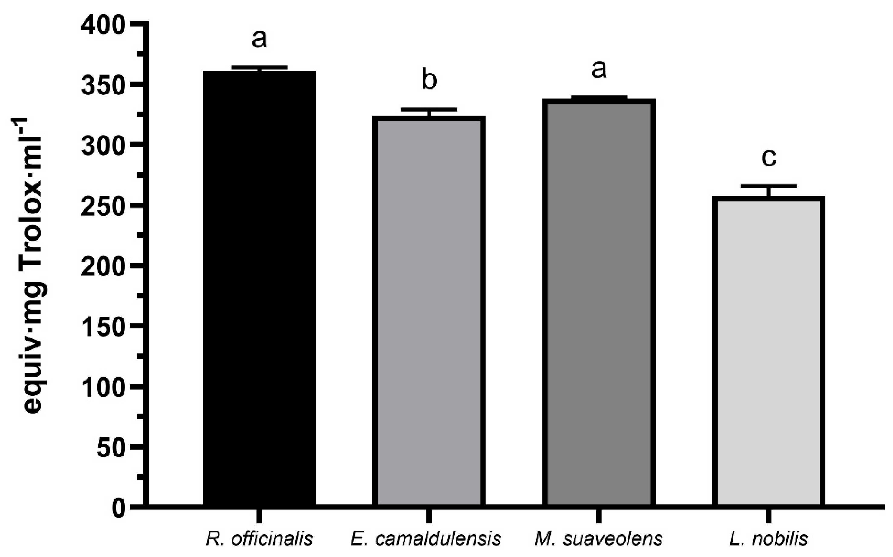
Figure 1. Antioxidant activities of R. officinalis, E. camaldulensis, M. suaveolens, and L. nobilis essential oils. The results are expressed as mean ± standard deviation in milligram equivalents of Trolox per milliliter of oil. a–c Different letters indicate statistically significant differences (p < 0.05) in antioxidant activity between the essential oils.
Separations 12 00145 g001
Figure 2. Antibacterial activity of R. officinalis, E. camaldulensis, M. suaveolens, and L. nobilis essential oils against E. coli (A) and L. innocua (B). They were administered at three doses (1:1, 1:10, and 1:100 v/v). The results are expressed as the mean ± standard deviation of cell growth inhibition. a,b Different letters in the same group indicate statistically significant differences (p < 0.05) in the % inhibition of bacterial growth.
Separations 12 00145 g002aSeparations 12 00145 g002b
Figure 3. Antiproliferative activity of R. officinalis, E. camaldulensis, M. suaveolens, and L. nobilis essential oils against HeLa cells. They were applied at three doses (1:100, 1:500, and 1:1000 v/v). The results are expressed as the mean ± standard deviation of cell survival. a–c Different letters in the same oil indicate statistically significant differences (p < 0.05) in cell survival among the different doses applied. * Statistically significant differences (p < 0.05) in cell survival compared to the control (100% viability).
Separations 12 00145 g003
Table 1. Chemical composition of volatiles in S. rosmarinus, E. camaldulensis, M. suaveolens, and L. nobilis essential oils. The compounds listed showed a minimum concentration of 1% in at least one essential oil. Values are presented as percentages of normalized peak areas without correction factors.
CompoundsS. rosmarinusE. camaldulensisM. suaveolensL. nobilis
Eucalyptol20.7727.91-33.36
Camphor14.80---
α-Pinene13.185.29-1.06
L-bornyl acetate10.66---
cis-Verbenone5.06---
β-Pinene4.52---
Borneol4.23---
Camphene4.09---
L-β-pinene3.25--1.23
γ-Terpinene2.74---
α-Terpinolen2.66---
p-Menth-1-en-8-ol2.377.53-2.93
4-Terpineol1.752.498.693.65
Linalol1.55--17.29
3-Pinanone1.37---
α-Fellandrene1.30---
p-Mentha-1,4(8)-diene-1.22---
m-cymene1.19---
L-alloaromadendrene-13.49--
(+)-Ledene-9.691.30-
B-Caryophyllene oxide-8.58--
α-Gurjunene-7.47--
D-Limonene-3.87--
1-Methyl-1-(4-methylphenyl) ethyl phenylcarbamate -2.74--
Epiglobulol-2.01--
Piperitone oxide--55.73-
β-Cubebene--15.83-
L-Caryophyllene--6.73-
4-Terpinenyl acetate--5.32-
α-Terpineol acetate---24.32
Eugenol---6.13
Sabinene---2.88
Pseudolimonen---1.28
Table 2. Susceptibility of Escherichia coli and Listeria innocua to different edible essential oils. Ampicillin was loaded at a concentration of 20 μg/mL. Oils were loaded 10 μL in each disc. Values are presented as mean ± standard deviation of the inhibition zones (mm diameter).
Essential OilE. coliL. innocua
Ampicillin29.00 ± 1.40 a34.50 ± 2.10 a
R. officinalis15.80 ± 1.83 cd17.50 ± 1.41 b
E. camaldulensis8.75 ± 0.35 b15.50 ± 0.70 b
M. suaveolens12.50 ± 1.41 bd9.12 ± 0.88 c
L. nobilis19.50 ± 2.12 c14.25 ± 0.35 b
a–d Different letters in the same column indicate statistically significant differences (p < 0.05).
Disclaimer/Publisher’s Note: The statements, opinions and data contained in all publications are solely those of the individual author(s) and contributor(s) and not of MDPI and/or the editor(s). MDPI and/or the editor(s) disclaim responsibility for any injury to people or property resulting from any ideas, methods, instructions or products referred to in the content.