Article

Determining the Hydrophobicity Index of Protected Amino Acids and Common Protecting Groups

1
Department of Chemical Engineering, Imperial College London, London SW7 AZ, UK
2
Eli Lilly and Company, Indianapolis, IN 46285, USA
*
Author to whom correspondence should be addressed.
These authors contributed equally to this work.
Separations 2023, 10(8), 456; https://doi.org/10.3390/separations10080456
Submission received: 31 July 2023 / Revised: 18 August 2023 / Accepted: 18 August 2023 / Published: 20 August 2023
(This article belongs to the Special Issue Advances in Chromatographic Analysis of Bioactive Compounds)

Abstract

:
Peptides are in great demand in the pharmaceutical arena and a majority of these peptides contain 20 or more amino acids. They are infrequently synthesised using the fragment condensation approach. A key limitation in adopting this approach more commonly is that protected peptide fragments with high purity are often required prior to the final condensation steps. It is hypothesized that understanding the hydrophobic nature of the protected amino acids will assist with designing optimal fragment purification processes when needed. Whilst a myriad of hydrophobicity indices are reported in the literature for unprotected amino acids, the literature lacks any data regarding the protected amino acids which form the key precursor for the fragment condensation task. In this current study, hydrophobicity indices for protected amino acids with common α-amino and sidechain protecting groups were experimentally determined. Different positions for each amino acid within the peptide chain were considered, namely at the C-terminal and N-terminal as well as internal positions. These data give deep insights on the hydrophobicity of each amino acid with respect to its position in the peptide chain. The data acquired in this research facilitated the prediction of the retention time of protected peptide fragments with an uncertainty of less than ±1.5%.

Graphical Abstract

1. Introduction

Macromolecular chains usually consisting of 10 to 50 amino acids which are linked together by amide bonds are known as peptides [1]. The number, type and order of the amino acids within each peptide differentiate them from each other, and they are generally classified as medium-sized molecules [2]. A human body consists of more than 7000 peptides that occur naturally, and these peptides play a vital role by helping in the physiobiological process within the living organism [1]. Peptides functions include but are not limited to antibodies, transducing signals, helping in cellular growth and reproduction, and in the communication between cells [1,3]. Due to the technological advancements witnessed in the synthetic and biotherapeutic formulation strategies, drug delivery and purification techniques of peptides, they are now of great interest for the pharmaceutical industry [1,4,5]. Peptides have high target affinity and specificity, and can degrade easily or stay in the targeted location for a long time depending on the intended use. All these positive attributes make peptides one of the most important drugs in the market today, with a total of 26 approvals between 2016 and 2022 [6]. Research projects are now being undertaken to further study and exploit peptides in a range of applications, as they can exhibit excellent performance not only in the field of medical therapeutics, but also in tissue engineering, cosmetics, biosensing, and antibacterial agents, among other fields [7,8].
In 2006, there were more than 40 FDA-approved peptides on the market and 400 in clinical trials. Currently, there are 120 FDA-approved peptides drugs on the market, 500 in preclinical trials and 140 in clinical trials. These data clearly show that the demand for peptides on the market is increasing and growing fast [8,9,10,11,12]. The peptide market is expected to grow from USD 29 Bn to USD 51 Bn between 2020 and 2026 [8].
Some of the peptides on the market are synthesised using solution phase synthesis, especially if they are short peptides and easy to synthesise [1,2,13,14]. However, the Nobel laureate Merrifield revolutionised the field of peptide chemistry by introducing solid-phase peptide synthesis (SPPS) methodology [15]. SPPS is the method of choice for synthesising peptides both in research and industrial settings. This methodology helped in synthesising longer and more complicated peptides following the stepwise approach [15]. However, the efficiency starts to diminish with peptides of more than 30 amino acids, and sometimes 20 amino acids depending on the type of amino acids [16,17]. The fragment condensation method proved to be a better alternative to the stepwise approach for these longer peptides, as the desired sequence is divided into multiple fragments which are synthesised using the SPPS stepwise approach, and later these fragments are merged into the final product. T20 is one of these drugs which is currently on the market and is synthesised using this approach [18]. A blood–brain barrier peptide called TD2.2 was also successfully synthesised following the fragment condensation approach [19]. Protected peptide fragments are also important for synthesising other peptide families, such as cyclic peptides [20].
It is vital to maintain the purity standard of peptides, as it is set by good manufacturing practice (GMP), and it is also important to study their applications further. HPLC is the most popular technique for this purpose due to its ability to separate the structurally related impurities from the target peptide [8,21,22]. Significant improvements in the chromatographic techniques have been witnessed for both analytical and manufacturing scales of purification [22,23].
Among the physicochemical properties of peptides, hydrophobicity is the major property that governs the chromatographic separation of peptides [22,23,24]. Various studies have established the hydrophobic indices of unprotected amino acids [22], which allow the predicting of the retention times of peptides with a known amino acid sequence, hence saving experimental time, costs and laboratory efforts [22]. However, there are no data in the literature with respect to the hydrophobicity of protected amino acids which in turn could be used in predicting the retention time of protected peptide fragments, the main precursors in the fragment condensation methodology.
In this work, we aim to establish a hydrophobicity index for the protected amino acids with common α-amino and sidechain protecting groups. This approach will be helpful in predicting the separation and purification processes of protected peptide fragments, which are key precursors in the fragment condensation reactions. Hence, to fully understand their hydrophobicity, 20 natural amino acids were considered in a model peptide, as well as various positions within the peptide sequence, including acid and peptide amide, and acetylated forms were investigated using reversed-phase liquid chromatography (RPLC).

2. Materials and Methods

2.1. Materials

PuroSynth CTC (1.0 mmol/g) and Merck Sieber amide (0.75 mmol/g) resins with manufacturer’s specifications were used. Other solvents and reagents that were used in the process include diethyl ether, N,N-dimethylformamide (DMF), trifluoroacetic acid (TFA), triisopropylsilane (TIS), dichloromethane (CH2Cl2), chloroform, N,N-diisopropylethylamine (DIEA), piperdine, acetonitrile (CH3CN), N,N′-diisopropylcarbodiimide (DIC) and methanol. These reagents, amino acids, and organic solvents were used without further purification unless otherwise stated. Analytical HPLC was performed on a Shimadzu LC20 system using Lab solution software for data processing with a Symmetry Luna C18 (3.6 μm, 4.6 × 150 mm) column. Another HPLC and columns were used to verify the model: a Shimadzu LC40 system using Lab solution software for data processing using Symmetry Luna C8 (5 μm, 4.6 × 150 mm). The mobile phase flow rate was 1.0 mL/min and UV detection was at 220 nm. Mobile phase A was 0.1% TFA in H2O, and mobile phase B was 0.1% TFA in CH3CN.

2.2. Peptide Synthesis

2.2.1. Incorporation Procedure

CTC resin
The first amino acids were incorporated onto CTC resin using dry CH2Cl2. CTC resin was swelled in CH2Cl2 for 10−20 min. The Fmoc-amino acids (2 equiv) were dissolved in a minimum amount of the CH2Cl2 (0.5 mL/100 mg resin) and sonicated for 10 min. DIEA (4 equiv) was then added to the solution, which in turn was added to the previously swelled resin and allowed to react for 1 h under mechanical shaking. After this, MeOH (80 μL/100 mg of resin) was added to endcap any unreacted chloride of the CTC resin. Finally, the resin was washed twice with CH2Cl2 and vacuum dried.
Sieber amide resin
The first amino acids were incorporated onto Rink amide resin using dry DMF. Rink Amide resin was swelled in DMF for 10−20 min. Fmoc was removed using 20% piperidine/DMF and the mixture was allowed to shake for 2 and 7 min. The Fmoc-amino acids (3 equiv) and OxymaPure (3 equiv) were dissolved in a minimum amount of the DMF (0.5 mL/100 mg resin) and sonicated for 10 min. DIC (3 equiv) was then added to the solution, which in turn was added to the previously swelled resin and allowed to react for 1 h under mechanical shaking. Finally, the resin was washed twice with CH2Cl2 and dried over vacuum.

2.2.2. Peptide Assembly

Peptides were synthesised following the standard methodology performed in our laboratory (3 equiv. of Fmoc-AA-OH, 3 equiv. of OxymaPure, 3 equiv. of DIC) in DMF and then shaken for 1 h. Fmoc was then removed (see under Rink amide resin section).

2.3. Cleavage Protocols

2.3.1. Protected Fragments

Peptide resin was placed in a syringe fitted with porous polyethylene filter. It was then swelled with CH2Cl2 for 10 min. The solvent was then filtered off, the cleavage solution (2 mL, 2% TFA (v/v) in CH2Cl2) was added per 100 mg of the peptide resin, and the syringe was closed with a cap and shaken for 30 min at room temperature. Finally, the filtrate was collected over CH3CN (4.0 mL), and the cleaved resin was washed 4 times with CH3CN (1 mL each time). The filtrates were again collected in the same tube. An aliquot (5 μL) was then injected into the HPLC system to record the retention time.

2.3.2. Unprotected Fragments

CTC resin
The final synthesised peptide was cleaved from the resin using TFA/TIS/H2O (95:2.5:2.5) (1 mL/100 mg) under mechanical shaking for 60 min. Chilled diethyl ether was then added (5 times the cleavage solution volume), and the solution was kept in an ice bath for 30 min. The solution was then centrifuged for 5 min at 5000 rpm, and the supernatant was decanted. A new amount of the ether (5 times the cleavage solution volume) was added to repeat this step. Any remaining ether was dried under N2. Finally, the precipitate was dissolved in CH3CN-H2O (1:1). A small amount of the solution was injected into the HPLC system to record the retention time.
Sieber amide resin
As the peptide amides were soluble in the ether, another workup procedure was followed. First, TFA was removed from the cleavage solution under vacuum. Next, 10% (v/v) of aqueous acetic acid was added to the dried peptidyl resin to dissolve the peptide. Then, chloroform (1 mL/100 mg) was added to the mixture and then shaken vigorously. The mixture was then allowed to settle, and the lower layer (chloroform and the released protecting groups) was discarded. This step was performed twice or thrice. Finally, the peptide in the 10% (v/v) solution was diluted in CH3CN-H2O (1:1). A small amount of the solution was injected into the HPLC system to record the retention time.

3. Results and Discussions

Six different peptide sequences were considered, in which the target amino acid was placed at various positions to investigate its influence on the peptide’s hydrophobicity (see Figure 1). The previous position has been shown to influence the hydrophobicity of amino acids [24,25].
Amino acids were located at the C-terminal, N-terminal and internal positions. To be able to determine the hydrophobicity of a particular protected amino acid, a short peptide with one amino acid that had a functional side chain was considered. Here, the pentapeptide Leu-enkephalin, which is an endogenous opioid peptide neurotransmitter, was considered [26]. This peptide has five amino acids, and one of them has a functional side chain which requires protection prior to any synthesis work. This amino acid will be interchanged with the 20 natural amino acids in this study. However, the sequences have been designed in a way that the target amino acid is placed next to a Gly residue to ensure unrestricted rotation around the amide bond, and hence the hydrophobicity data will be more accurate and representative for each amino acid [27]. To understand and reflect the hydrophobic nature of each amino acid, the target amino acid was placed at different positions, as follows: (i) N-terminal of the sequence, (ii) internal position, and (iii) C-terminal. These three sequences were considered for the acid peptide, with an additional three sequences of the same peptides but with an amidated C-terminal. Hence, the first three sequences were synthesised using 2-chlorotrityl (2-CTC) resin. The second three sequences were synthesised using Sieber amide resin. Both resins are highly acid-labile resins, where protected peptides can be cleaved from the resin with a mild acidolysis reaction [28,29].

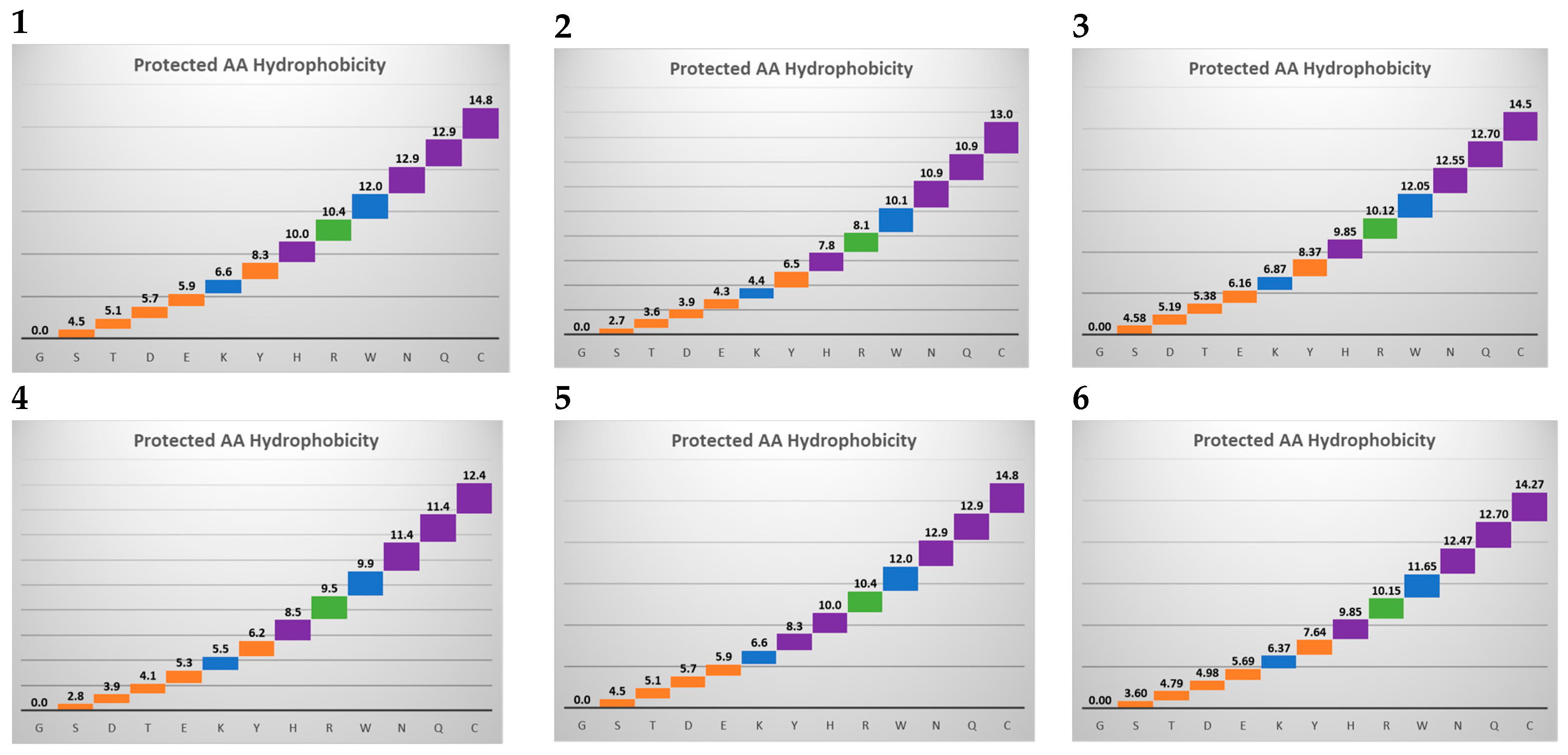
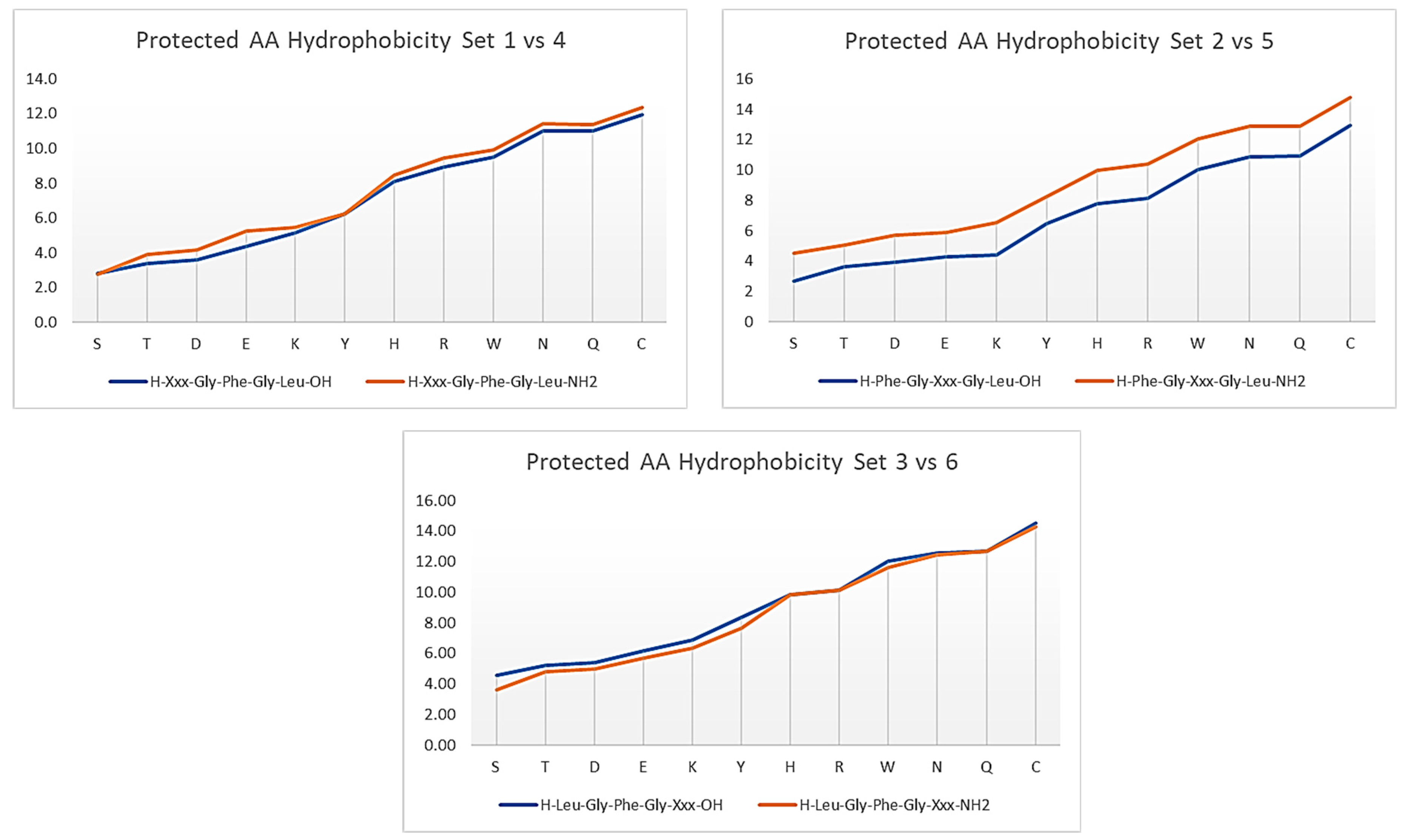
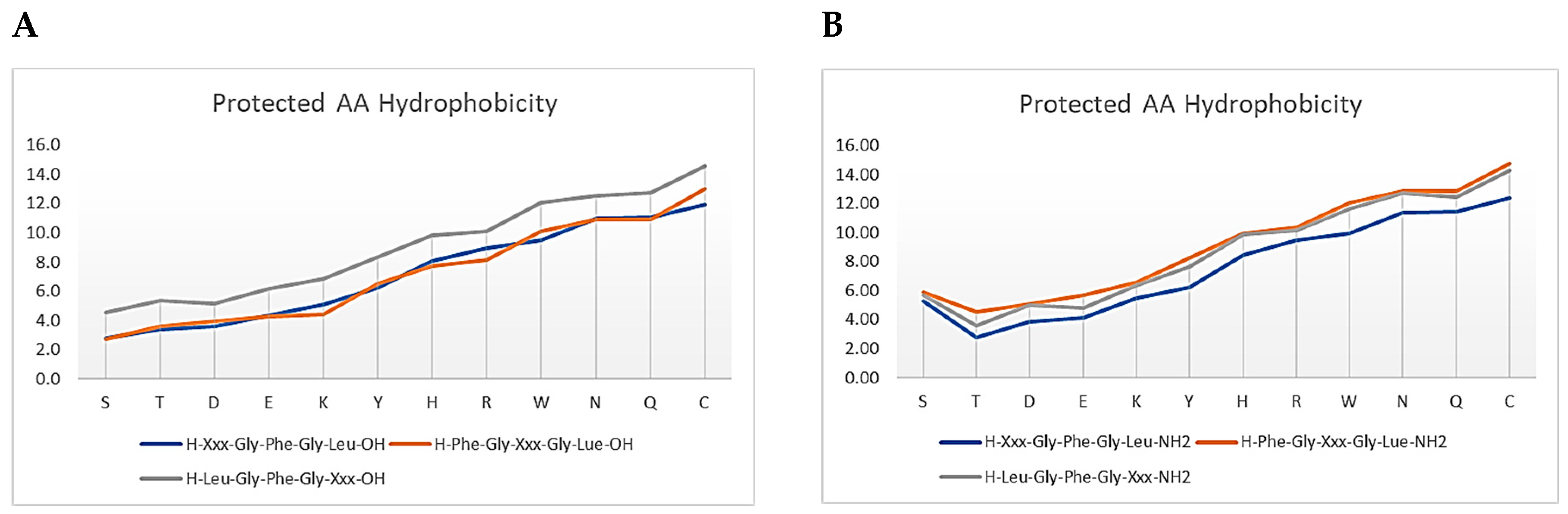
3.1. Amino Acids Hydrophobicity

Twelve out of the twenty natural amino acids have a functional sidechain, and hence appropriate protection is deemed necessary to avoid complications during the synthesis process. The following are the common sidechain protecting groups for the Fmoc/tBu peptide synthesis strategy: Trityl (Trt), tert-butyloxycarbonyl (Boc), 2,2,4,6,7-pentamethyldihydrobenzofuran-5-sulfonyl (Pbf). Several amino acids share the same sidechain protecting group, in which His, Asn, Gln, and Cys have the same Trt group to protect their functional sidechain, whereas Asp, Glu, Ser, Thr, and Tyr have tBu, and Trp and Lys have Boc. Pbf is the sidechain protecting Arg only. Hence, it would be interesting to establish and compare the hydrophobicity index of these protecting groups on different amino acids.
Given that Gly has only H on its sidechain, the Gly-peptide analogue was considered as a reference to estimate the hydrophobicity index of the protected amino acids. Hydrophobicity indices were determined for all sequences by subtracting the retention time of the peptide with the amino acid of interest from the retention time of the Gly-peptide of the same sequence (Figure 2) (Supplementary Table S1) (Supplementary Table S2) (Figure S1).
In all of the investigated peptide sequences, and regardless of the amino acid’s position in the sequence, Cys(Trt) was the most hydrophobic protected amino acid and Ser(tBu) was the least. The position of the amino acid does play a significant role in its hydrophobicity. In the acid peptides, amino acids showed a more hydrophobic character when placed at the C-terminal than when at the N-terminal, and the least values when they were in the internal positions (Figure 3).
However, the overall hydrophobicity pattern of all the protected amino acids showed an excellent correlation. The hydrophobicity index in the first three acid peptide sequences resulted in the following r2correlation values:
Sequence 1 versus sequence 2 r2 = 0.98;
Sequence 1 versus sequence 3, r2 = 0.99;
Sequence 2 versus sequence 3, r2 = 0.99;
Sequence 4 versus sequence 5, r2 = 0.98;
Sequence 4 versus sequence 6, r2 = 0.99;
Sequence 5 versus sequence 6, r2 = 0.99 (Figure S2).
The situation was a bit different in the peptide amide sequences. Even though the protected amino acids showed a higher hydrophobic character when at the C-terminal, they also showed a higher hydrophobic character when in the internal positions, and in some cases higher than when at the C-terminal. This observation is an unsurprising fact, and it is ascribed to the amidated C-terminal (CONH2), which is hydrophobic in its nature, whereas in acid peptides there is the hydrophilic OH group instead. This phenomenon caused the protected amino acids at the N-terminal to show the lowest hydrophobic character in the peptide amide sequences (Figure 3).
Protected amino acids were also compared when present in acid peptides versus peptide amides, with the same position as shown below in Figure 4.
Unsurprisingly, in all cases the peptide amide showed higher hydrophobicity than the acid form. The difference was more pronounced when the amino acid was placed in the internal positions. Excellent correlations were obtained as follows: 1 versus 4, r2 = 0.99; 2 versus 5, r2 = 0.99; and 3 versus 6, r2 = 0.98 (Figure S3).
However, though this study focuses on protected amino acids, we also investigated the unprotected ones in order to allow the retention time prediction reported at the end of this study (Supplementary Table S2) (see Figure S1). Trp was shown to be the most hydrophobic unprotected amino acid, whereas Lys was the least. Very good correlations were also obtained in the overall hydrophobicity pattern, in which:
Sequence 1 versus sequence 2, r2 = 0.92;
Sequence 1 versus sequence 3, r2 = 0.92;
Sequence 2 versus sequence 3, r2 = 0.96;
Sequence 4 versus sequence 5, r2 = 0.93;
Sequence 4 versus sequence 6, r2 = 0.91;
Sequence 5 versus sequence 6, r2 = 0.97 (see Figure S2).
When comparing the same amino acid, in the same position but in the acid and peptide amide, a very good correlation was also obtained, as follows:
Sequence 1 versus sequence 4, r2 = 0.93;
Sequence 2 versus sequence 5, r2 = 0.93;
Sequence 3 versus sequence 6, r2 = 0.92 (see Figure S3).

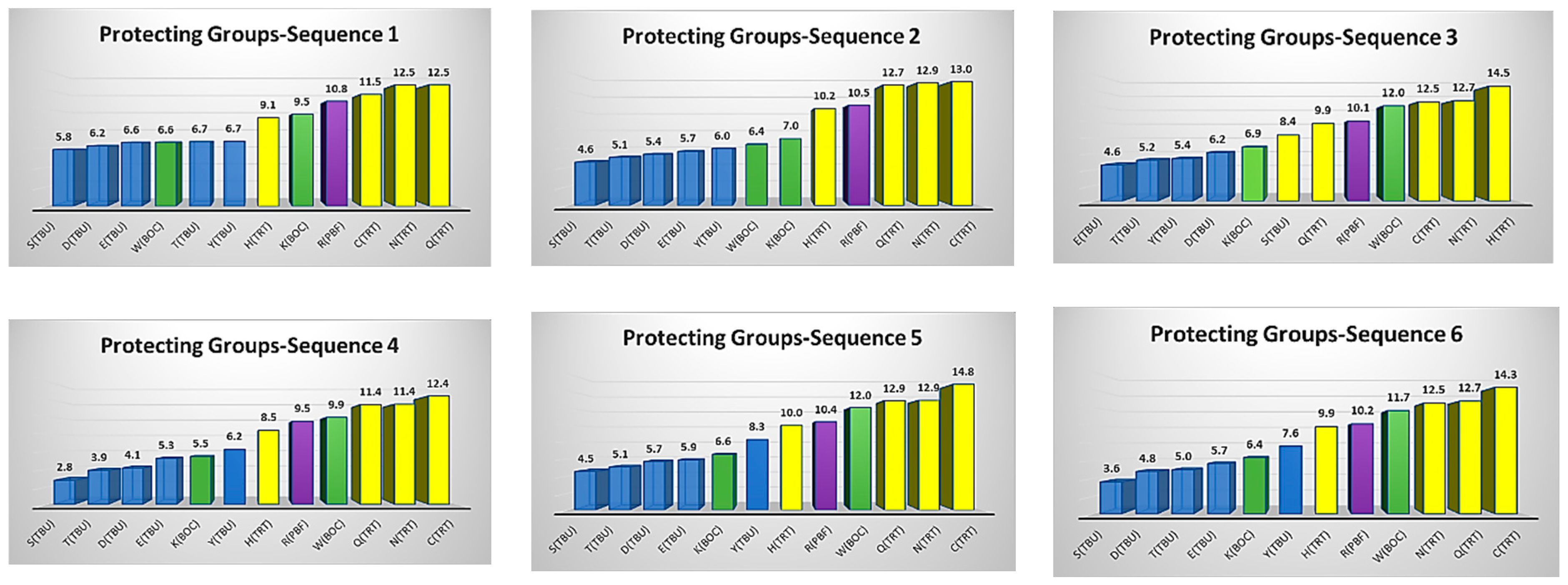
3.2. Protecting Groups’ Hydrophobicity

Additional processes were also considered to establish the hydrophobicity index for the protecting groups themselves. The hydrophobicity indices of the twelve protected amino acids with respect to the Gly-peptide of the same sequence are shown below in Figure 5.
Hydrophobicity indices were determined for the protecting groups themselves by subtracting the retention time of the protected peptide from the unprotected analogue of the same peptide. This applies to any protecting group, either at the α-amino or the sidechain of the amino acid of interest. Investigating the hydrophobicity indices of the sidechain protecting groups that can protect different amino acids showed that a large protecting group would have the same hydrophobicity index regardless of the amino acid type (Figure S4). For example, the Trt group showed a hydrophobicity index that ranged from about 10 to 14 min, with four amino acids (His(Trt), Asn(Trt), Glu(Trt) and Cys(Trt)). It is worth highlighting that Trt had the lowest hydrophobicity index of 10 min with the least hydrophobic amino acid, which was His. Other than the fact that His is hydrophilic, its ring within its structure probably experienced less interaction with the stationary phase and hence had a lower hydrophobicity value. Nevertheless, the Trt group was the most hydrophobic protecting group among all the protecting groups incorporated in this study. The next protecting group in terms of the hydrophobicity was the Pbf group, which protects the side chain of Arg only, then Boc group, and the last one was the tBu group. Unlike large protecting groups, Boc and tBu are small protecting groups and hence they are affected by the type of amino acid they are protecting. Their hydrophobicity ranged from 6 to 12 min and 4 to 8 min, respectively. With such small protecting groups, the influence of the amino acid and its interaction with the chromatographic components, and mainly the stationary phase, will be more pronounced than in the case of the large protecting group. All the protecting groups showed a uniform hydrophobicity pattern regardless of their position within the sequence, whether the peptide was an acid or a peptide amide one.
The protection of α-amino functionality is often carried out using Boc and/or Fmoc protecting groups. However, Fmoc is more convenient and an easier methodology that does not require special laboratory infrastructure, unlike the case for the Boc strategy [1]. Therapeutic peptides are often presented in the acetylated form to enhance their stability. Therefore, the hydrophobicity index was also determined for Fmoc amino acids, both protected and unprotected, Ac amino acids (protected and unprotected), the Fmoc protecting group and the acetyl group to understand their hydrophobicity, and such data were also helpful for the prediction task (Supplementary Tables S3 and S4) (Figure S4). For the Fmoc group, only sequence 1 was selected, while for the acetyl group sequence 4 was selected. The selection was made based on how peptide fragments are presented during the fragment condensation synthetic approach (Fmoc-fragment-OH). In the case of acetyl, as explained above, therapeutic peptides often exist as (Ac-peptide-NH2); hence, sequence 4 was considered. There was a difference in the hydrophobicity indices of Fmoc amino acids and the Fmoc protecting group itself, where the values ranged from 9 to 27 min. The difference was even higher in the case of Ac amino acids and the acetyl group itself (0.2 to 17 min), which was again attributed to the small size of the acetyl group with respect to that of Fmoc.

3.3. Retention Time Prediction

Using the extensive dataset generated, the prediction of retention times for various peptide fragments was undertaken as described below. The following peptides were employed in the study: Fmoc-APPPS(tBu)-OH, Fmoc-GPS(tBu)S(tBu)G-OH and Fmoc-E(tBu)FIE(tBu)-OH. The predicted retention (τ) time was calculated by summing up the hydrophobicity index of the amino acids in the peptide chains, as per Equation (1):
τ = R c
where R c represents the sum of the retention coefficients for all amino acid residues and the termini in the given peptide sequence.
The predictive capability in our work was satisfactory regarding the uncertainty <3.5% (Table 1).
A Symmetry Luna C18 (3.6 μm, 4.6 × 150 mm) column was used, with 1.0 mL/min and UV detection was at 220 nm. The mobile phase A was 0.1% TFA in H2O, and the mobile phase B was 0.1% TFA in CH3CN.
It should be noted that having the Gly residue at the C-terminal will not shield the subsequent amino acid from the effect of that location. Thus, in the second peptide (Fmoc-GPS(tBu)S(tBu)G-OH), the hydrophobicity index of S(tBu) amino acid should be taken from sequence 1 as if S(tBu) is at the C-terminal position with a hydrophobicity index of 4.6 min rather than sequence 2 that represents the internal positions, provided that its hydrophobicity index in the internal position is lower (2.6 min) and influences the prediction capability with a difference of 1.7 min.
To further validate our approach, we also predicted the retention times for peptide fragments by changing the gradient profile using the same HPLC and column and repeated the procedure using another HPLC system and column. Simple correction factors must be adopted to account for these changes. Therefore, in the case of changing the gradient profile, the ratio between both gradient profiles has to be incorporated as per Equation (2), where 16.8 is the gradient profile for the gradient used to estimate the hydrophobicity index in this study (15–70% in 30 min).
τ = R c   ×   current   gradient   profile / 16.8
In the case of using the new HPLC system, a standard peptide should be used as a reference, and it is recommended to consider a standard that shares common amino acids and/or protecting groups with the peptides of interest. Next, the difference between the predicted and actual retention times of the standard peptide should be subtracted as per Equation (3).
τ = R c t s
where R c represents the sum of the retention coefficients for all amino acid residues and the termini in the given peptide sequence, the current gradient profile represents the amount of organic modifier needed to elute the peptide, and ts represents the difference between the predicted and actual retention time of the standard peptide.
The first and second peptides (Fmoc-APPPS(tBu)-OH and Fmoc-GPS(tBu)S(tBu)G-OH) were selected to validate our approach. Our model was able to predict the retention time when changing the gradient profile. For example, using a gradient profile of 5–95% in 15 min instead of the original one, which was 15–70% in 30 min, and employing Equation (2) resulted in a good prediction accuracy. A small difference of only −0.4 min and +1.0 min was observed for the first and the second peptides, respectively. Furthermore, using another HPLC and column while keeping the same gradient profile (15–70% in 30 min) exhibited an excellent prediction capability for our model using Equation (3). While both columns had the same length and diameter, they were different in their alkyl chain length and particle size: 3.6 μm versus 5 μm and C18 versus C8, respectively. After employing Equation (3) and considering H-S(tBu)GFGL-OH as a standard peptide for making the t s correction, the final prediction capability was as follows: −1.9 min and −1.2 min for the first and the second peptides, respectively.
This study illustrates the excellent versatility of our first order model to predict the RPLC elution behaviour of a protected peptides with a known composition. However, it should be highlighted that there are other factors not incorporated into this model that can alter the elution behaviour, including the structural conformations, peptide length and neighbouring group effect, among others [24]. Thus, ultimately, our model needs to be coupled with an a priori computational model which can consider all of these additional factors affecting the elution process and incorporate them into a more advanced prediction model. Nevertheless, this first order model had offered excellent predictive performance for the peptides investigated in the current study.

4. Conclusions

For the first time, the hydrophobicity indices for protected amino acids with common α-amino and sidechain protecting groups were determined for a large set of synthesised peptides. The data in this work will help in predicting the retention time of protected peptide fragments that are the main precursors in the current peptide synthesis field. These data gave deep insights into the hydrophobicity of each amino acid with respect to its position in the peptide chain. The data acquired in this research facilitated the prediction of the RPLC retention time of protected peptide fragments with an uncertainty of <±1.5%. Therefore, being able to predict the retention time of these protected peptide fragments will definitely save time, funds and effort during their manufacture and purification. Based on previous work reported for unprotected amino acids in the literature [22,24], we plan for this model to be enhanced by coupling it with a molecular computational tool. This enhancement will allow for more complex hydrophobic phenomena to be understood, including any potential observed deviations from the current model. We will endeavour to exploit the available models, including machine learning and computational tools, to establish a more accurate prediction model.

Supplementary Materials

Supplementary information contains the hydrophobicity indices for unprotected amino acids and for protecting groups. The following supporting information can be downloaded at: https://www.mdpi.com/article/10.3390/separations10080456/s1, Table S1: Hydrophobicity index of protected amino acids. Table S2: Hydrophobicity index for unprotected amino acids. Table S3: Hydrophobicity index for Fmoc, Fmoc-amino acid. Table S4: Hydrophobicity index for Acetyl, Ac-amino acid. Figure S1: Hydrophobicity index for the unprotected amino acids in different positions of the peptide chains. The figure number represents the sequence of the peptide as per Figure 1. Figure S2: Correlation among hydrophobicity indices for the protected and unprotected amino acids in different positions of the peptide chains. Figure S3: Correlation among hydrophobicity indices for the protected and unprotected amino acids in acid and peptide amides. Figure S4: Hydrophobicity indices for: Fmoc-amino acid (protected and unprotected), Fmoc protecting group, Ac-amino acid (protected and unprotected), and Acetyl group.

Author Contributions

Conceptualization, O.A.M. and D.R.W.; methodology, V.G. and O.A.M.; formal analysis, V.G., O.A.M. and D.R.W.; investigation, V.G. and O.A.M.; resources, O.A.M. and D.R.W.; writing—original draft preparation, V.G. and O.A.M.; writing—review and editing, V.G., O.A.M., D.R.W. and C.B.; supervision, O.A.M. and D.R.W.; project administration, O.A.M. and D.R.W.; funding acquisition, O.A.M., D.R.W. and C.B. All authors have read and agreed to the published version of the manuscript.

Funding

Funding from Eli Lilly and Company and the UK EPSRC, through the PharmaSEL-Prosperity Programme (EP/T005556/1), is gratefully acknowledged.

Institutional Review Board Statement

Not applicable.

Informed Consent Statement

Not applicable.

Data Availability Statement

Not applicable.

Conflicts of Interest

The authors declare no conflict of interest.

Abbreviations

Acacetyl group
Boctert-butyloxycarbonyl
CH2Cl2dichloromethane
CH3CNacetonitrile
DIEAN,N-diisopropylethylamine
DICN,N-diisopropylcarbodiimide
DMFN,N-dimethylformamide
GMPgood manufacturing practice
Pbf2,2,4,6,7-pentamethyldihydrobenzofuran-5-sulfonyl
RPLCreversed-phase liquid chromatography
SPPSsolid-phase peptide synthesis
tButert-butyl
TIStriisopropylsilane
TFAtrifluoroacetic acid
Trttrityl

References

  1. Jaradat, D.M.M. Thirteen decades of peptide synthesis: Key developments in solid phase peptide synthesis and amide bond formation utilized in peptide ligation. Amino Acids 2018, 50, 39–68. [Google Scholar] [CrossRef] [PubMed]
  2. Sachdeva, S. Peptides as ‘Drugs’: The Journey so Far. Int. J. Pept. Res. Ther. 2017, 23, 49–60. [Google Scholar] [CrossRef]
  3. Petrou, C.; Sarigiannis, Y. Peptide synthesis: Methods, trends, and challenges. In Peptide Applications in Biomedicine, Biotechnology and Bioengineering; Koutsopoulos, S., Ed.; Woodhead Publishing: Sawston, UK, 2018; pp. 1–21. [Google Scholar] [CrossRef]
  4. Lau, J.L.; Dunn, M.K. Therapeutic peptides: Historical perspectives, current development trends, and future directions. Bioorg. Med. Chem. 2018, 26, 2700–2707. [Google Scholar] [CrossRef] [PubMed]
  5. Loffet, A. Peptides as Drugs: Is There a Market? J. Pept. Sci. 2002, 8, 1–7. [Google Scholar] [CrossRef]
  6. Al Musaimi, O.; Al Shaer, D.; Albericio, F.; Torre, B.G.d.l. 2022 FDA TIDES (Peptides and Oligonucleotides) Harvest. Pharmaceuticals 2023, 16, 336. [Google Scholar] [CrossRef]
  7. Al Musaimi, O.; Lombardi, L.; Williams, D.R.; Albericio, F. Strategies for Improving Peptide Stability and Delivery. Pharmaceuticals 2022, 15, 1283. [Google Scholar] [CrossRef]
  8. Ferrazzano, L.; Catani, M.; Cavazzini, A.; Martelli, G.; Corbisiero, D.; Cantelmi, P.; Fantoni, T.; Mattellone, A.; De Luca, C.; Felletti, S.; et al. Sustainability in peptide chemistry: Current synthesis and purification technologies and future challenges. Green Chem. 2022, 24, 975–1020. [Google Scholar] [CrossRef]
  9. Kaspar, A.A.; Reichert, J.M. Future directions for peptide therapeutics development. Drug Discov. Today 2013, 18, 807–817. [Google Scholar] [CrossRef]
  10. D’Aloisio, V.; Dognini, P.; Hutcheon, G.A.; Coxon, C.R. PepTherDia: Database and structural composition analysis of approved peptide therapeutics and diagnostics. Drug Discov. 2021, 26, 1409–1419. [Google Scholar] [CrossRef]
  11. Muttenthaler, M.; King, G.F.; Adams, D.J.; Alewood, P.F. Trends in peptide drug discovery. Nat. Rev. Drug Discov. 2021, 20, 309–325. [Google Scholar] [CrossRef]
  12. Albericio, F. Developments in peptide and amide synthesis. Curr. Opin. Chem. Biol. 2004, 8, 211–221. [Google Scholar] [CrossRef] [PubMed]
  13. Zompra, A.A.; Galanis, A.S.; Werbitzky, O.; Albericio, F. Manufacturing peptides as active pharmaceutical ingredients. Future Med. Chem. 2009, 1, 361–377. [Google Scholar] [CrossRef] [PubMed]
  14. Andersson, L.; Blomberg, L.; Flegel, M.; Lepsa, L.; Nilsson, B.; Verlander, M. Large-scale synthesis of peptides. Pept. Sci. 2000, 55, 227–250. [Google Scholar] [CrossRef]
  15. Merrifield, R.B. Solid Phase Peptide Synthesis. I. The Synthesis of a Tetrapeptide. J. Am. Chem. Soc. 1963, 85, 2149–2154. [Google Scholar] [CrossRef]
  16. Athanassopoulos, P.; Barlos, K.; Gatos, D.; Hatzi, O.; Tzavara, C. Application of 2-Chlorotrityl Chloride in Convergent Peptide Synthesis. Tetrahedron Lett. 1995, 36, 5645–5648. [Google Scholar] [CrossRef]
  17. Gongora-Benitez, M.; Tulla-Puche, J.; Albericio, F. Handles for Fmoc solid-phase synthesis of protected peptides. ACS Comb. Sci. 2013, 15, 217–228. [Google Scholar] [CrossRef]
  18. Bray, B.L. Large-scale manufacture of peptide therapeutics by chemical synthesis. Nat. Rev. Drug Discov. 2003, 2, 587–593. [Google Scholar] [CrossRef]
  19. Al Musaimi, O.; Morse, S.V.; Lombardi, L.; Serban, S.; Basso, A.; Williams, D.R. Successful synthesis of a glial-specific blood–brain barrier shuttle peptide following a fragment condensation approach on a solid-phase resin. J. Pept. Sci. 2022, 29, e3448. [Google Scholar] [CrossRef]
  20. White, C.J.; Yudin, A.K. Contemporary strategies for peptide macrocyclization. Nat. Chem. 2011, 3, 509–524. [Google Scholar] [CrossRef]
  21. Snyder, L.R.; Kirkland, J.J.; Dolan, J.W. Introduction to Modern Liquid Chromatography, 3rd ed.; Wiley-Interscience: New York, NY, USA, 2010. [Google Scholar] [CrossRef]
  22. Al Musaimi, O.; Valenzo, O.M.M.; Williams, D.R. Prediction of peptides retention behavior in reversed-phase liquid chromatography based on their hydrophobicity. J. Sep. Sci. 2023, 46, 2200743. [Google Scholar] [CrossRef]
  23. De Luca, C.; Lievore, G.; Bozza, D.; Buratti, A.; Cavazzini, A.; Ricci, A.; Macis, M.; Cabri, W.; Felletti, S.; Catani, M. Downstream Processing of Therapeutic Peptides by Means of Preparative Liquid Chromatography. Molecules 2021, 26, 4688. [Google Scholar] [CrossRef] [PubMed]
  24. Musaimi, O.A.; Mercado-Valenzo, O.M.; Williams, D.R. Factors Influencing the Prediction Accuracy of Model Peptides in Reversed-Phase Liquid Chromatography. Separations 2023, 10, 81. [Google Scholar] [CrossRef]
  25. Tripet, B.; Cepeniene, D.; Kovacs, J.M.; Mant, C.T.; Krokhin, O.V.; Hodges, R.S. Requirements for prediction of peptide retention time in reversed-phase high-performance liquid chromatography: Hydrophilicity/hydrophobicity of side-chains at the N- and C-termini of peptides are dramatically affected by the end-groups and location. J. Chromatogr. A 2007, 1141, 212–225. [Google Scholar] [CrossRef] [PubMed]
  26. Klee, W.A.; Nirenberg, M. Mode of action of endogenous opiate peptides. Nature 1976, 263, 609–612. [Google Scholar] [CrossRef]
  27. Kovacs, J.M.; Mant, C.T.; Hodges, R.S. Determination of intrinsic hydrophilicity/hydrophobicity of amino acid side chains in peptides in the absence of nearest-neighbor or conformational effects. J. Pept. Sci. 2006, 84, 283–297. [Google Scholar] [CrossRef] [PubMed]
  28. Alhassan, M.; Al Musaimi, O.; Collins, J.M.; Albericio, F.; de la Torre, B.G. Cleaving protected peptides from 2-chlorotrityl chloride resin. Moving away from dichloromethane. Green Chem. 2020, 22, 2840–2845. [Google Scholar] [CrossRef]
  29. Al Musaimi, O.; Gavva, V.; Williams, D.R. Greener Cleavage of Protected Peptide Fragments from Sieber Amide Resin. ChemistryOpen 2022, 11, e202200236. [Google Scholar] [CrossRef]
Figure 1. Peptide sequences considered in this study. Fmoc: Fluorenylmethyloxycarbonyl protecting group; Ac: acetyl group.
Separations 10 00456 g001
Figure 2. Hydrophobicity index for the protected amino acids in different positions of the peptide chains. The figure number represents the sequence of the peptide, as per Figure 1.
Separations 10 00456 g002
Figure 3. Hydrophobicity pattern for the protected amino acids in different positions of the peptide chains. (A) Acid peptide. (B) Peptide amide.
Separations 10 00456 g003
Figure 4. Hydrophobicity pattern for the protected amino acids in acid versus peptide amides.
Separations 10 00456 g004
Figure 5. Hydrophobicity indices for the sidechain protecting groups.
Separations 10 00456 g005
Table 1. Prediction of peptides’ retention time.
Peptide FragmentPredicted τ (Minute)Actual (Minute)Difference (Minute)
Fmoc-APPPS(tBu)-OH16.917.5−0.6
Fmoc-GPS(tBu)S(tBu)G-OH20.520.3+0.2
Fmoc-E(tBu)FIE(tBu)-OH30.730.9−0.2
Disclaimer/Publisher’s Note: The statements, opinions and data contained in all publications are solely those of the individual author(s) and contributor(s) and not of MDPI and/or the editor(s). MDPI and/or the editor(s) disclaim responsibility for any injury to people or property resulting from any ideas, methods, instructions or products referred to in the content.

