Article

Evaluation of BPA and Bis-GMA Release from Recent Dental Composite Materials by LC-MS/MS

by
Lígia Lopes-Rocha
1,
Virgínia M. F. Gonçalves
1,2,
Sara C. Cunha
3,
José O. Fernandes
3,
Teresa Pinho
1 and
Maria Elizabeth Tiritan
2,4,5,*
1
UNIPRO—Oral Pathology and Rehabilitation Research, University Institute of Health Sciences (IUCS), Cooperativa de Ensino Superior Politécnico e Universitário—CESPU, 4585-116 Gandra, Portugal
2
TOXRUN—Toxicology Research Unit, University Institute of Health Sciences, Cooperativa de Ensino Superior Politécnico e Universitário—CESPU, CRL, 4585-116 Gandra, Portugal
3
Laboratório Associado para a Química Verde/Associated Laboratory for Green Chemistry LAQV—Requimte, Laboratório de Bromatologia e Hidrologia, Departamento de Ciências Químicas, Faculdade de Farmácia, Universidade do Porto, 4050-313 Porto, Portugal
4
Interdisciplinary Center of Marine and Environmental Research (CIIMAR), University of Porto, Edifício do Terminal de Cruzeiros do Porto de Leixões, 4050-208 Matosinhos, Portugal
5
Laboratório de Química Orgânica e Farmacêutica, Departamento de Ciências Químicas, Faculdade de Farmácia da Universidade do Porto, 4050-313 Porto, Portugal
*
Author to whom correspondence should be addressed.
Separations 2023, 10(8), 455; https://doi.org/10.3390/separations10080455
Submission received: 6 July 2023 / Revised: 12 August 2023 / Accepted: 13 August 2023 / Published: 18 August 2023
(This article belongs to the Special Issue Feature Papers in Separations from Editorial Board Members)

Abstract

:
Bisphenol-A (BPA) is a xenoestrogen widely used as a synthetic precursor of resin monomers. There is arise need to acquire BPA-free resin-matrix composites to prevent the health effects of BPA. Six composites with distinctive manufacturer specifications were considered to evaluate the degree of release of BPA and bisphenol A-Diglycidyl Methacrylate (Bis-GMA) in a dental composite. The light-cured resin-matrix specimens (n = 5 for each composite type) were incubated at 37 °C in 1 mL of a 75% ethanol–water solution in a sealed amber glass vial for 7 days. The 75% ethanol–water solution was replaced daily and immediately frozen (−20 °C) until liquid chromatography-tandem mass spectrometry (LC-MS/MS) analysis. BPA was not detected in any studied resin-based materials. However, Bis-GMA was detected in almost all the studied samples during the experiment, except AF and BF. The highest Bis-GMA concentration was released from ED at 67.43 ng/mL, followed by BE, FS, and NC with 40.75 ng/mL, 8.30 ng/mL, and 0.94 ng/mL, respectively. There is a clear need for more precise and standardized analytical methods to assess the short- and long-term release of resin-based materials. Furthermore, manufacturers should be obliged to provide complete details of the chemical composition of dental products and to promote the development of materials without estrogenic potential.

1. Introduction

Resin-matrix composites are standardly used in multiple domains of dentistry [1]. Primarily, they are the most conservative approach in esthetic and functional rehabilitation in young patients [2,3,4], such as in maxillary lateral incisor agenesis [5]. In addition, they have an extensive range of purposes, such as fissure and pit sealant, luting cement or temporary material, and adhesive for brackets and splints [6,7].
Resin-matrix composites comprise an organic portion consisting of a polymer matrix and inorganic filler particles attached to the resin matrix through a silane coupling agent [8,9]. In recent resin-matrix composites, the sources of Bisphenol-A (BPA) that leach from dental materials include trace levels of BPA as an impurity of BPA-derivatives such as bisphenol A-Diglycidyl Methacrylate (Bis-GMA); Ethoxylated Bisphenol A Glycol Methacrylate (Bis-EMA); dimethacrylate (Bis-DMA); and 2,2-bis-(4-(3-methacryloxypropoxy) phenyl) propane (Bis-PMA). The presence of BPA due to the degradation/hydrolysis of Bis-DMA has also been reported [9,10]. Bis-GMA (Figure 1) is the predominant base monomer used in the resin-matrix composite, and its chemical structure is supposed to prevent hydrolysis. Still, BPA may be an impurity of Bis-GMA manufacturing if polymerization is not complete [11,12]. Though the popularity of resin-matrix composites has increased in recent years, concern about releasing endocrine-disrupting chemicals (EDCs) such as BPA and its derivatives has gathered attention from scientists, clinicians, and patients [13,14].
BPA is a chemical intermediate in the production of polycarbonate-, epoxy-, and methacrylate-matrix materials used in several industrial and health fields [13,15]. BPA is an organic compound with two phenolic functional groups (Figure 1) and was categorized by the European Chemicals Agency (ECHA) as a ‘substance of very high concern’ since it was classified as an EDC with risks to human health (toxic for human reproduction) and environment, as determined in Regulation (EC) No 1907/2006 [16]. Furthermore, researchers found that BPA leached into the saliva and urine of treated patients and was responsible for the estrogenicity of some commercial composites and sealants used in dentistry [12]. The growing evidence indicates that exposure to BPA is also associated with an increased risk of developing type 2 diabetes [17], obesity [18], adverse immune effects [19], and neuroendocrine development alterations [20]. In addition, BPA prenatal exposures were suggested to be associated with child neurobehavioral development [21]. Children positively associate composite restorations (with BPA) and attention deficit hyperactivity disorder [2,22].
As resin-matrix materials are expected to have a shelf life of several years in the mouth, in vitro studies for extended periods are required to investigate the long-term release of various composite ingredients. Eveline Putzeys et al. (2018) carried out an in vitro protocol to measure the long-standing elution of several compounds from eight resin-based dental composites over one year. Briefly, cylindric specimens of the different dental materials were polymerized and immersed under the extraction solution (water, ethanol, and artificial saliva), which was weekly refreshed and reported that the tested composite materials were able to continuously release some monomers for up to 52 weeks of the incubation period [23].
Bis-GMA is one of the primary sources of BPA release from dental resin materials. Therefore, several research studies proposed a new formulation (using non-BPA dimethacrylates) to replace Bis-GMA monomer in resin formulations to reduce human exposure to BPA derivatives [9]. As such, urethane-dimethacrylate (UDMA), another typical dimethacrylate monomer applied in dentistry, has been used to replace Bis-GMA as the base resin of dental materials [24]. Unfortunately, UDMA-based resin had a critical limitation in its higher volumetric shrinkage [25]. The higher volumetric shrinkage of UDMA-based resin could lead to a more significant marginal gap between tooth and restorations, developing a complex probability of secondary caries [26]. Therefore, alternative monomer compositions of Bis-GMA were introduced to address the limitations of this product in terms of durability and toxicity. One alternative to the methacrylate-based resin-matrix composites is a hybrid organoceramic, known as ORganically MOdified CERamic (ORMOCER®, VOCO, Cuxhaven, Germany), which is an ORMOSIL (Organically Modified SILicate). One of the best important ORMOCER® characteristics is combining polysiloxane groups with photopolymerizable methacrylate groups covalently bonded to silica fillers [27,28,29]. The oxygen is substituted by organic groups, developing in a three-dimensional polymerized material with less organic matrix than the conventional resin-matrix composites. ORMOCER® provides high biocompatibility due to the nonexistence of residual monomers, lesser polymerization shrinkage, high wear resistance, increased opacity, and enhanced handling characteristics [30,31].
Because of the constantly increasing use of resin-matrix composites, consideration should be paid to the relative biocompatibility of these dental restorative materials. There is a growing need to link the gap between the worrying literature concerning the hazardous effects and the reduced clinical investigations, which can only be accomplished by acquiring knowledge about the leaching of compounds from resin-based dental materials.
This work aims to evaluate the BPA and Bis-GMA monomers released from six resin–matrix composites using a sensitive liquid chromatography-tandem mass spectrometry (LC-MS/MS) quantification method.

2. Materials and Methods

2.1. Resin Composites Used

Six resin composites were tested: five commercial resins and one experimental resin from different commercial brands. The manufacturer specifications and composition of the six resin-matrix composites are presented in Table 1.

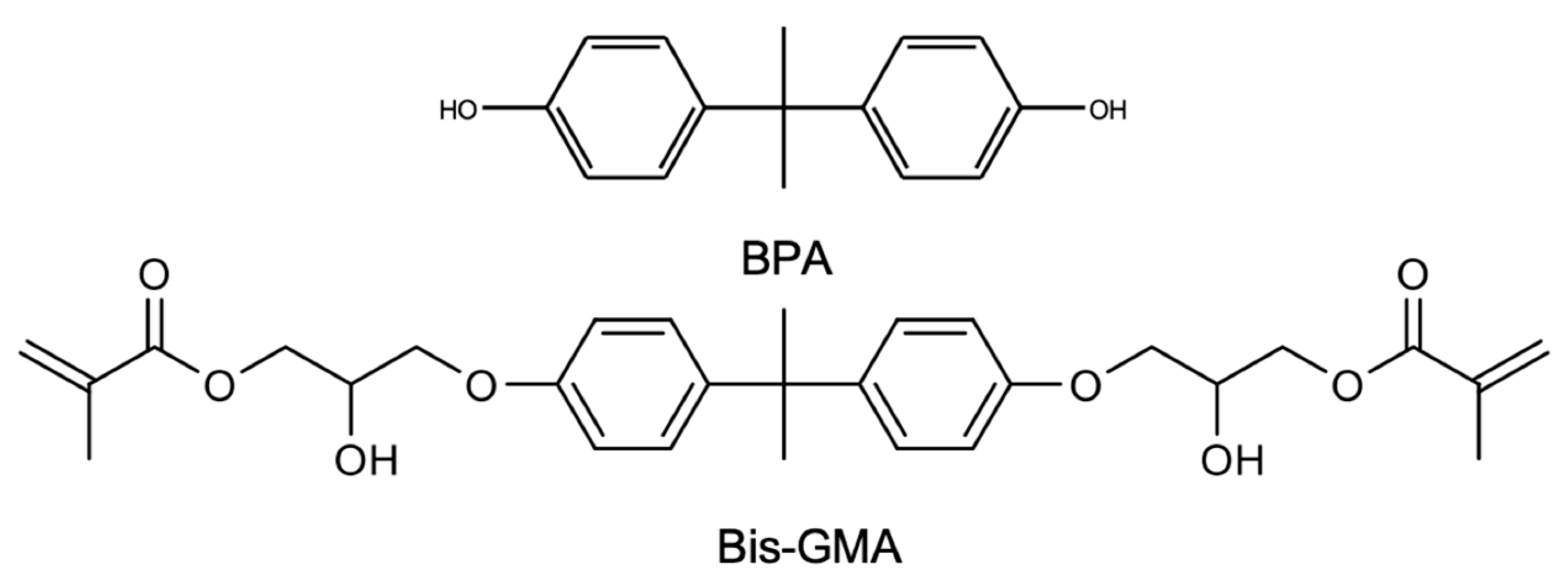
2.2. Chemicals and Reagents Used

Standards of Bisphenol A (BPA) (CAS- 80-05-7), Bisphenol A-glycidyl methacrylate (Bis-GMA) (CAS- 1565-94-2), and Bisphenol A dimethacrylate- Bis DMA (CAS- 3253-39-2) were from Sigma Aldrich (St. Louis, MO, USA); the acetonitrile and ethanol HPLC gradient grade were from Fisher Scientific (Loughborough, UK); formic acid 98–100% was from Merck (Darmstadt, Germany); ammonium acetate (97% purity) was purchased from AppliChem Panreac ITW Co. (Barcelona, Spain); and methanol (LC-MS grade) was purchased from VWR. Ultra-pure water grade was supplied by an SG Water System (Ultra Clear UV model). BPA and Bis-GMA stock solutions were prepared at 1 mg/mL in ethanol and stored at −20 °C in amber glass flasks.

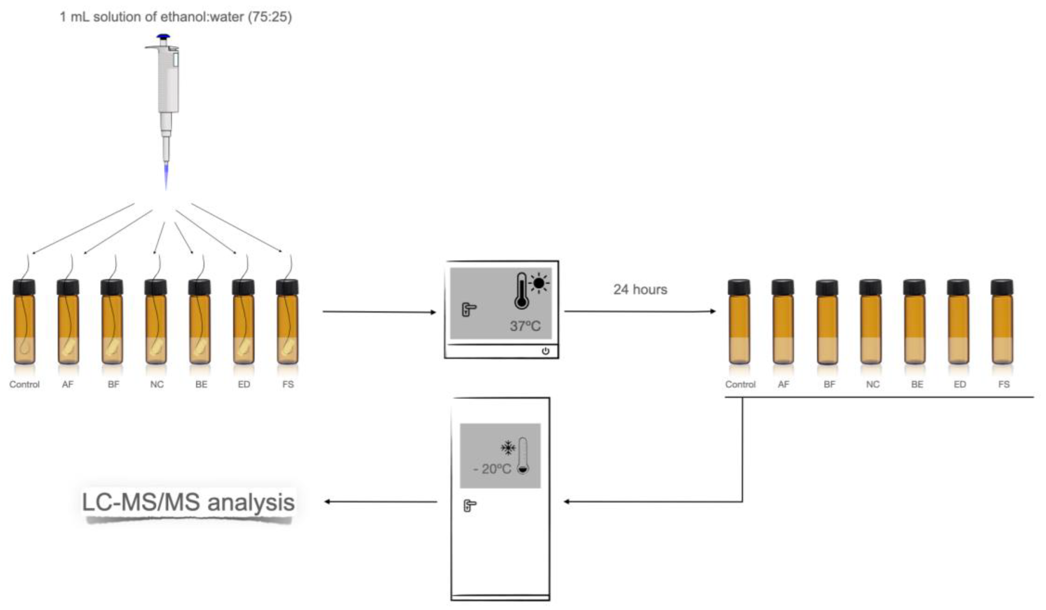
2.3. Preparation of Resin Composite Samples and Eluates

Specimen disks (6 mm diameter and 2 mm thickness) were prepared in a stainless-steel mold. A glass plate covered the top and bottom to limit oxygen inhibition, ensure smooth surfaces, and avoid excess material. Samples were polymerized for 20 s by light-curing using an LED light-curing unit (Celalux 3, High-Power LED curing-light; VOCO, Cuxhaven, Germany) at an average of 1300 mW/cm2, 450/480 nm. Prior to each polymerization, the power of the equipment was confirmed through the radiometer. Grinding was carried out under standardized conditions to simulate the clinical procedures in dental practice. Each resin composite sample was polished using silicon carbide (SiC) abrasive paper of 2400 Mesh. Each disc had its abrasive paper to avoid contamination with BPA-containing particles from previous experiments. An electronic caliper (Mitutoyo, Tokyo, Japan) was used to confirm the precise and equal sizes of the disks (Figure 2).
Consequently, the disks (n = 5 for each composite type) were immediately immersed in 1 mL of a 75% ethanol–water solution in a sealed amber glass vial. To ensure that the entire surface of the disc was in contact with the solution, the disc was held by a fisherman’s string and held in suspension in the solution (Figure 3).
A control assay was also performed using just the fisherman’s string, putting it into a vial with 1 mL of 75% ethanol–water solution. All the samples were incubated at 37 °C (Optic Ivymen system, COMECTA), and, at each 24 h (for seven days), the 75% ethanol–water solution was replaced with a fresh solution. All samples were stored at −20 °C until analysis by Liquid Chromatography (LC) analysis.

2.4. High-Performance Liquid Chromatography with UV and Fluorescence Detection (HPLC-UV/FD) Analysis

In the first approach, the samples were analyzed by High-performance liquid chromatography with UV and Fluorescence detection (HPLC-UV/FD). A Shimadzu Prominence UFLC System (Shimadzu Corporation, Tokyo, Japan) was used. It was composed of a degasser DGU-20A5, two LC-20AD pumps, a SIL-20AC autosampler, a CTO-20AC column oven, and a CBM-20A System Controller. An SPD-20A UV detector (set at 230 nm) and an RF-10AXL fluorescence detector (set at 225 nm and 310 nm for excitation and emission wavelength, respectively) were coupled to the LC system. All of the system was controlled using the LC Solution software (V. 1.24 SP1, Shimadzu). Chromatographic analysis was performed in a Luna PFP2 (150 × 4.6 mm, 3 μm) column from Phenomenex (Torrance, CA, USA) operating at 40 °C in a gradient mode at a flow rate of 0.8 mL/min. The mobile phase consisted of A) 0.1%Formic acid (in water) and B) Acetonitrile (with 0.1% formic acid). The gradient was as follows: 0–6 min 55%A:45%B; at 10 min 20%(A):80%(B) and remained at this condition until 25 min; and at 26 min the pump returned to the initial condition (55%A:45%B) and remained at this condition for 10 min to stabilize the column for the next injection. The injection volume was 20 μL.

2.5. LC-MS/MS Analysis

Sample analysis was performed by an HPLC system Waters Alliance 2695 (Waters, Milford) interfaced to a Quattro Micro triple quadrupole mass spectrometer (Waters, Manchester, UK). The chromatographic separation was achieved using a Kinetex C18 2.6 µm particle size analytical column (150 × 4.6 mm) with a Phenomenex pre-column (Tecnocroma, Portugal) at a 200 µL/min flow rate. The column was kept at 30 °C and the autosampler was maintained at room temperature (±25 °C), Figures S1 and S2 in the supplementary material. In isocratic mode, the mobile phase consisted of 90% MeOH and 10% aqueous solution of 5 mM ammonium acetate (pH 5). The total run time was 15 min. The sample injection volume was 10 μL. The MS/MS acquisition was operated in negative-ion mode with multiple reaction monitoring (MRM); the collision gas was argon 99.995% (Gasin, Portugal) with a pressure of 2.9 × 10⁻³ mbar in the collision cell. Capillary voltages of 3.0 KV were used in the negative ionization mode (Table 2). Nitrogen was used for desolvation and cone gas at the flow of 350 and 60 L/h, respectively. The desolvation temperature was set to 350 °C and the source temperature to 150 °C. Dwell times of 0.1 s/scan were selected. The data were collected using the software MassLynx4.1.
For quantification, individual standard calibration curves were performed for BPA and Bis-GMA monomers at the concentrations of 0.2, 0.4, 0.8, 3.2, and 6.4 ng/mL and 0.4, 0.8, 1.6, 12.5 and 25,0 ng/mL in 75% ethanol–water solution, respectively. The calibration curves were obtained by linear regression corresponding to the correlation between the peak area and the nominal concentration by external calibration. Quality control was also used for both monomers, at 1.6 ng/mL for BPA and 6.4 ng/mL for Bis-GMA, to evaluate method accuracy and precision. The limit of detection (LOD) and limit of quantification (LOQ) were determined by the signal-to-noise ratio (S/N = 3 for LOD and S/N = 10 for LOQ).

3. Results

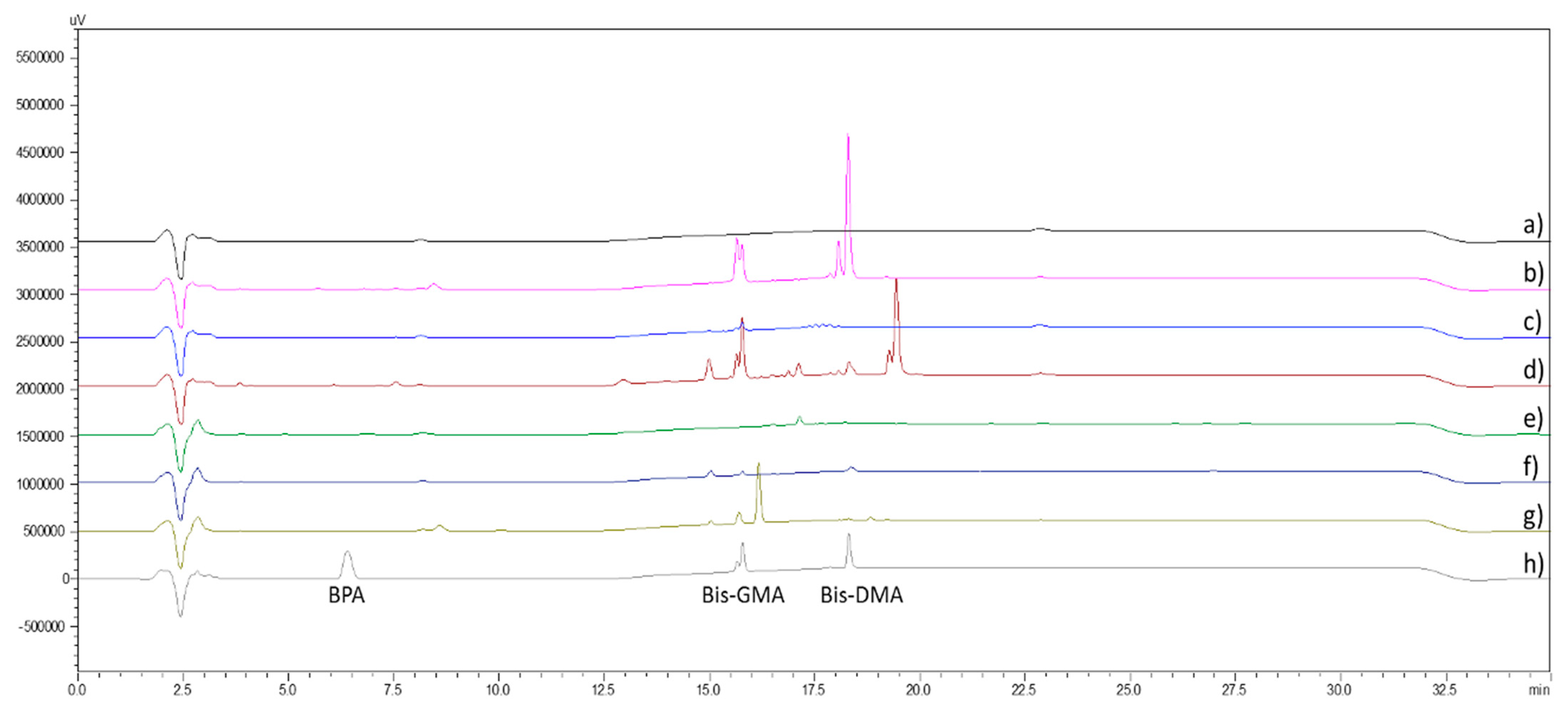
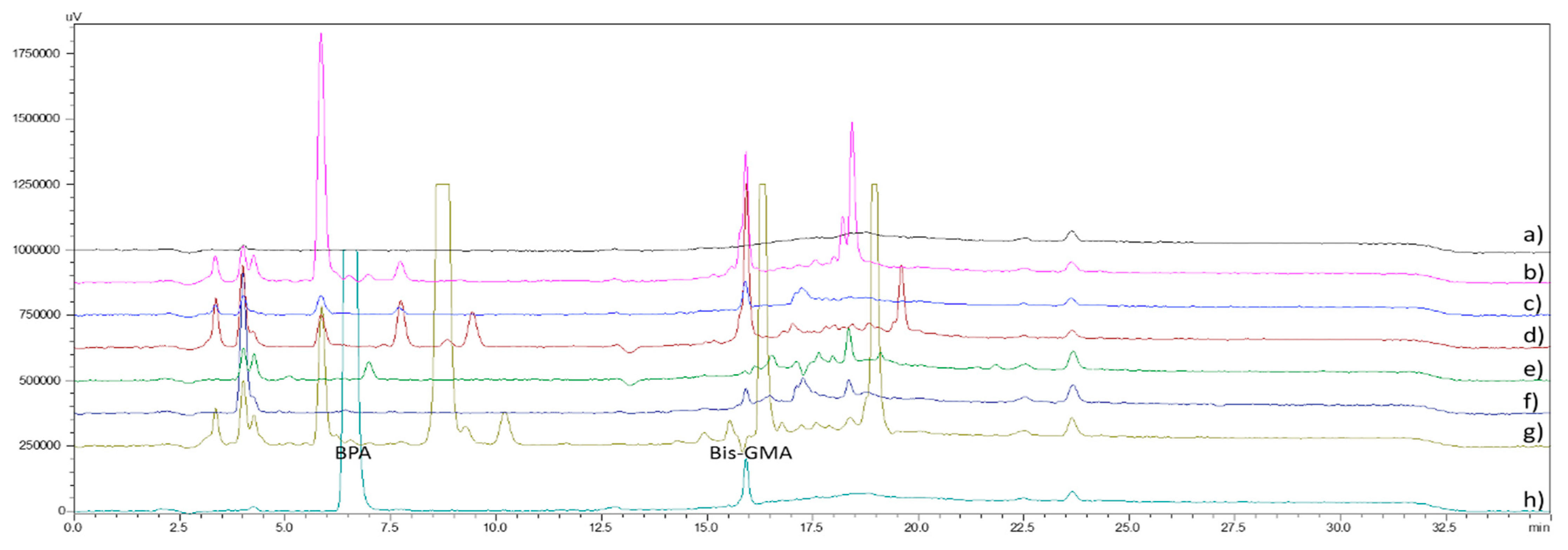
3.1. HPLC-UV/FD Analysis

In the first approach, the sample release was analyzed by LC with UV (Figure 4) and FD (Figure 5) detection. The chromatographic analysis demonstrated the different chemical patterns between the resins (Figure 4 and Figure 5). BPA and Bis-DMA were not detected in any of the six composite materials, but the chromatograms indicated the occurrence of Bis-GMA. Due to the poor selectivity and sensibility of the HPLC-UV/FD method for quantifying BPA and Bis-GMA monomers, an LC-MS/MS method was established to monitor the release studies.
The target monomers were quantified by LC-MS/MS through a validated method considering the following parameters: selectivity, linearity and range, LOD, LOQ, accuracy, recovery, and precision. The injection of the reconstituted extracts gave a coefficient of determination between 0.9914 and 0.9986 for BPA and Bis-GMA (Table 3). No carryover was observed. The LOD was 0.06 ng/mL and 0.03 ng/mL for BPA and Bis-GMA, respectively, while the LOQ was 0.2 ng/mL and 0.1 ng/mL, respectively. The method presented accuracy close to 100%.

3.2. BPA and Bis-GMA Leaching Experiment

The concentration of eluted monomers (BPA and Bis-GMA) from the six resin-based dental materials quantified by the LC-MS/MS validated method is shown in Figure 6 and Table 4.
BPA was not detected in any sample, even in the Bis-GMA resin-based samples (Figure 6 and Table 4).
As shown in Figure 6 and Table 4, only on day one was an important amount of Bis-GMA released from ED > BE > FS resins. The concentration decreased significantly over the following days, but the release persisted during the seven days. The ED and BE had similar behavior in the following days, but the FS presented the lowest and most constant Bis-GMA release.
Regarding Figure 6 and Table 4, sample analysis was only performed for the first five days for the AF, NC, and BF resins because the results remained practically null and constant regarding the release of Bis-GMA since day 3.
To our knowledge, no release studies were performed on the composite NC, an experimental brand resin-based material considered BPA-free. Despite the fact that this method was not able to detect any concentration of BPA, a minimal concentration of Bis-GMA was detected on days 1 and 2, as shown in Figure 6 and Table 4.

4. Discussion

The present study evaluated the leaching of BPA and Bis-GMA from six different resin-based dental materials. For that, a simple analytical method, without pre-concentration and/or derivatization of BPA, was established for monitoring the release of BPA and Bis-GMA in in vitro studies using a non-toxic matrix.
In vitro studies are important in analyzing human substances released because they can produce better or worse conditions while identical [23]. Several solvents, such as artificial saliva, distilled water, ethanol, methanol, and acetonitrile, have been used in studies considering the elution of monomers [32,33,34]. The results of previous reports have revealed that the type of solvent affects the amount of eluted monomer from a composite resin [35]. The oral cavity presents an environment between water and more aggressive solvents (ethanol, methanol, acetonitrile) [36]. Ethanol and ethanol/water mixtures can infiltrate the methacrylate polymer network, causing more sorption and swelling [37]. Moreover, a mixture of 75% ethanol and 25% water is not toxic for the operator in the laboratory and is considered environmentally friendly. The United States Federal Drug Administration recommends a 75% ethanol–water solution as a clinically relevant food–oral simulating liquid, and it has been used in several studies [38,39,40,41,42]. Furthermore, other authors reported that water is more similar to saliva, which would reproduce saliva and dentin fluid, the latter being the path to the dental pulp [43]. Therefore, a 75% ethanol–water solution was used in this study.
Dental resin-based materials generally consist of methacrylate monomers, inorganic filler particles (dispersed phase), photoinitiator systems, and other minor additions, including stabilizers and pigments [44,45]. Initially, the resin-based composite was based on a derivative of BPA, Bis-GMA, the most common monomer in contemporary resin materials [46]. Reducing polymerization shrinkage and enabling cross-linking during polymerization are among the advantages of Bis-GMA [47]. However, the Bis-GMA molecule is quite viscous at room temperature due to its hydroxyl groups, which increase its polarity and cause intermolecular interactions. For clinical purposes, the monomers are converted into polymers by addition polymerization, initiated mainly by light-curing. As a result, patients may be exposed to non-polymerized ‘residual’ monomers and their potential degradation products, which can leach out of composite restorations into the mouth [48,49], a process that may be accelerated by biodegradation [50] and may result in adverse health effects [51,52]. Of these, the endocrine-disrupting effect of BPA has received particular interest. Most studies monitored the release of BPA and neglected the Bis-GMA, claiming that BPA can be found in dental composites, present as an impurity from the synthesis process and/or possibly as a degradation product of BPA-based monomers [53,54] since BPA itself is not an intentional ingredient [55]. Another reason is the estrogenic effects reported for BPA but not Bis-GMA [56,57]. Although studies focus mainly on BPA, which is usually not quantified or is quantified at low concentrations, toxicity may be associated with its derivative, Bis-GMA, and other analogs. Leachable Bis-GMA monomer is considered cytotoxic and inflammatory [34,58]. In addition, high Bis-GMA doses have been associated with increased uterine wet weight and uterine collagen content in ovariectomized rats and reduced fertility in male rats [59,60].
In this work, the release was monitored by LC with a different type of detection method, but only LC-MS/MS allowed suitable conditions for the quantification of BPA and Bis-GMA. Regarding the release from the six target resin–matrix composites, the chromatograms illustrated in Figure 4 and Figure 5 show that the profile differs according to the resin type. The quantification by the validated LC-MS/MS method demonstrated that BPA release was lower than the LOD (0.06 ng/mL) from all the samples over seven days after incubation of the composite samples with 75% ethanol–water solution (Figure 6 and Table 4) while Bis-GMA was released in detectable amounts from four of the six analyzed samples: ED > BE > FS, and the NC sample, an experimental resin called BPA-free. Higher concentrations of Bis-GMA were detected in the first few days, followed by a consistently lower release in the following days for ED > BE > FS. However, for the NC sample, the presence of Bis-GMA was detected just in the first two days (Figure 6 and Table 4). Bis-GMA was quantified in levels of up to 33.46 ng/mL (ED) on the first monitoring day, a decrease in concentration was observed until day 4 (BE, FS, and ED). A slight increase in the concentration level was observed on the fifth day, followed by a trend to decrease in the following days. According to Nys et al. [61], one possible explanation for the absence of BPA is the recent development of new filler technology that binds more free resin than standard fillers, which could hinder the release of monomers and BPA. Thus, the presence of a BPA-based monomer in the raw material does not necessarily specify that BPA will be released in visible amounts from this material after light-curing. In addition, this may point out the demand for even more sensitive detection procedures [61].
The concentration of BPA-based monomers can explain the differences between the composites in the resin and their physicochemical properties (solubility in the 75% ethanol–water solution). Furthermore, the extent and rate of elution of components from composites are dependent upon the degree of conversion (DC) of monomers, the composition and solubility characteristics of the extraction solvent, and the size and chemical characteristics of the leachable species [62]. DC depends mainly on intrinsic factors, such as the chemical structure of the resin, and extrinsic factors, such as polymerization conditions [63]. To minimize sources of error, only a fully charged light-emitting diode (LED) device could be used for each material, which is common in daily dental work and superior to a halogen light-curing unit [64]. In addition, custom-made polymerization stands were used to ensure that the exact distance was maintained. It has been reported that the DC of light-polymerized resin materials is 55–80% [23,62]. There is an inverse correlation between DC and the amount of eluted monomer. The greater the extent of the polymerization reactions, the fewer residual monomers are available to elute [65]. Time is also a significant factor in monomer elution. Some studies have reported that acute monomer release occurs within 24 h [23,66]. However, some recent works have shown that monomer elution is not completed within the first 24 h and that leaching on specific monomers continues longer [23].
Finishing and polishing are also essential for eliminating the resin-rich outer layer that may be the source of unreacted monomers eluted in the oral cavity [67]. Several studies have shown different finishing and polishing procedures, such as using 12-point carbide finishing burs and Sof-Lex discs [68], which are advocated for providing the smoothest surface [68]. Therefore, they were not preferred in this study. Instead, we chose grounding at 2400 Mesh using SiC abrasive papers (Figure 2). Each disc had its abrasive paper to avoid contamination with BPA-containing particles from previous experiments. However, further in vitro investigations can focus on monomer elution using different restorative materials and finishing polishing techniques. In addition, BPA released in the oral cavity may be lower because deeper layers are often not in contact with saliva.
On the other hand, an oxygen-inhibited layer is present on the restoration surface. If not removed by finishing and polishing, it could increase BPA release compared to this in vitro study, where oxygen inhibition of polymerization was avoided by polymerizing the specimens through glass slides [69]. According to the literature, the elution of residual monomers was higher if finishing and polishing were not performed [67].
The analytical methods used in several studies may needed better specificity and sensitivity to guarantee precise detection and quantification of low levels of BPA [70], as illustrated by the high limit of detection (i.e., 100 ng/mL) reported by Noda et al. [71]. On the other hand, more information about the long-term release [72]. Typically, samples are incubated for long periods without renewal of the incubation solution at equal time intervals [73,74]. In contrast, in a study developed by Putzeys et al. [52], the long-term release of BPA-based monomers was determined in a setup with equal-interval solvent change with the release of specific monomers after an incubation period of 52 weeks. The major disadvantage of this non-specific approach is the loss of sensitivity. There is a discrepancy between different reports, which the low BPA concentration for detection can explain—either the actual absence of BPA or due to a minimal amount of BPA and BPA-based monomers in the resin. In the present study, the sensitivity of LC-MS/MS method analysis is reflected in the low LOD (0.06 ng/mL), which has a reasonable sensitivity compared with other studies. It is also important to appeal to the consideration that quantification by GC/MS may overestimate the BPA release from resin composite due to the heat used in GC/MS and the thermal stability of Bis-GMA [75]. However, making direct comparisons and drawing conclusions is not straightforward because the studies are not standardized. The differences in the composition of the resin-matrix composite could result in inconsistency in the release of the quantity and type of monomers.

5. Conclusions

This report described a simple, sensitive, and accurate method to detect low levels of BPA and Bis-GMA released from resin-based dental materials in a 75% ethanol–water solution. The results indicate that, if it is present, BPA release is below the LOD of the method. However, Bis-GMA was quantified in levels of up to 33.46 ng/mL on the first monitoring day. Bis-GMA was detected even in the NC sample, an experimental resin called “BPA-free”. Continual monitoring of in vitro and in vivo studies may be helpful to improve the quality of the resin composites and to stimulate, even more, the clinical application of BPA-free composites used in dental treatments, especially in young patients with esthetic and functional rehabilitation such as in cases of maxillary lateral incisors agenesis.
BPA-free resins are a good option, and the brands studied and marketed show that they are also free of BPA derivatives such as Bis-GMA. However, further studies are needed regarding other monomers also derived from BPA.

Supplementary Materials

The following supporting information can be downloaded at: https://www.mdpi.com/article/10.3390/separations10080455/s1, Figure S1: Liquid chromatography-tandem mass spectrometry (LC-MS/MS) analysis of PBA; Figure S2: Liquid chromatography-tandem mass spectrometry (LC-MS/MS) analysis of Bis-GMA.

Author Contributions

Conceptualization, L.L.-R., M.E.T., V.M.F.G., S.C.C. and J.O.F.; methodology, L.L.-R., M.E.T., V.M.F.G., S.C.C. and J.O.F.; validation, L.L.-R., T.P., M.E.T. and V.M.F.G.; investigation, L.L.-R., M.E.T. and V.M.F.G.; writing—original draft preparation, L.L.-R.; writing—review and editing, M.E.T. and V.M.F.G.; supervision, M.E.T. All authors have read and agreed to the published version of the manuscript.

Funding

This work is supported by CESPU–Cooperativa de Ensino Superior Politécnico e Universitário under the grant “OrthoAlignPI-4RL-IINFACTS-2019”. Additional national funding was issued under the project PTDC/CTA-AMB/6686/2020; UIDB/04423/2020 and UIDP/04423/2020 (Group of Marine Natural Products and Medicinal Chemistry—CIIMAR). Sara C. Cunha acknowledges FCT for the 2022.07841.CEECIND/CP1724/CT0014 contract. LAQV-REQUIMTE authors acknowledge financial support from PT national funds (FCT/MCTES, Fundação para a Ciência e Tecnologia and Ministério da Ciência, Tecnologia e Ensino Superior) through the project UIDB/50006/202.

Institutional Review Board Statement

Not applicable.

Informed Consent Statement

Not applicable.

Data Availability Statement

The data presented in this study are available upon reasonable request from the corresponding author.

Acknowledgments

We would like to thank the manufacturers for providing the materials: VOCO GmbH (Cuxhaven, Germany), Micerium SpA (Avegno, Italy), Coltène-Whaledent (Altstätten, Switzerland), and 3M GmbH (Seefeld, Germany).

Conflicts of Interest

The authors declare no conflict of interest.

References

  1. Araujo, E.; Perdigão, J. Anterior Veneer Restorations-An evidence-based minimal-intervention perspective. J. Adhes. Dent. 2021, 23, 91–110. [Google Scholar] [CrossRef]
  2. Hu, C.-J.; Yu, H.-C.; Chang, Y.-C. Investigation of the impact of dental care via composite resin restoration among children with attention deficit hyperactivity disorder: A registry-based nested case–control study. Healthcare 2021, 9, 803. [Google Scholar] [CrossRef]
  3. Adriana, M.V.; Camila, C.S.; Vinícius, M.C.; Hans, H.A.; Rodrigo, A.C.; Allyson, M.N.; Fernando, M.S.L. Direct guided restorations from planning to definitive restoration: A clinical report. J. Prosthet. Dent. 2023, 129, 2–6. [Google Scholar] [CrossRef]
  4. Comba, A.; Vergano, E.A.; Baldi, A.; Alovisi, M.; Pasqualini, D.; Castroflorio, T.; Stura, I.; Migliaretti, G.; Berutti, E.; Scotti, N. 5-year retrospective evaluation of direct composite restorations in orthodontically treated patients. J. Dent. 2021, 104, 103510. [Google Scholar] [CrossRef]
  5. Rocha, L.; Garcez, J.; Tiritan, M.E.; da Silva, L.F.M.; Pinho, T. Maxillary lateral incisor agenesis and microdontia: Minimally invasive symmetric and asymmetric esthetic rehabilitation. Rev. Port. Estomatol. Med. Dentária Cir. Maxilofac. 2022, 62, 41–51. [Google Scholar] [CrossRef]
  6. Krishnakumar, S.; Senthilvelan, T. Polymer composites in dentistry and orthopedic applications—A review. Mater. Today Proc. 2021, 46, 9707–9713. [Google Scholar] [CrossRef]
  7. Janda, R. Dental Resins-Material Science & Technology: Basic Level; Tredition GmbH: Hamburg, Germany, 2021; Volume 1, ISBN 9783347287846. [Google Scholar]
  8. Ferracane, J.L. Resin composite-State of the art. Dent. Mater. 2011, 27, 29–38. [Google Scholar] [CrossRef]
  9. Aminoroaya, A.; Neisiany, R.E.; Khorasani, S.N.; Panahi, P.; Das, O.; Madry, H.; Cucchiarini, M.; Ramakrishna, S. A review of dental composites: Challenges, chemistry aspects, filler influences, and future insights. Compos. Part B Eng. 2021, 216, 108852. [Google Scholar] [CrossRef]
  10. Al-Tannak, N.F.; Alzoubi, F.; Kareem, F.M.; Novotny, L. Determination of endocrine disruptor bisphenol-A leakage from different matrices of dental resin-based composite materials. Curr. Pharm. Anal. 2022, 18, 305–315. [Google Scholar] [CrossRef]
  11. Janani, K.; Teja, K.V.; Sandhya, R.; Alam, M.K.; Al-Qaisi, R.K.; Shrivastava, D.; Alnusayri, M.O.; Alkhalaf, Z.A.; Sghaireen, M.G.; Srivastava, K.C. monomer elution from three resin composites at two different time interval using high performance liquid chromatography—An in-vitro study. Polymers 2021, 13, 4395. [Google Scholar] [CrossRef]
  12. Arenholt-Bindslev, D.; Breinholt, V.; Preiss, A.; Schmalz, G. Time-related bisphenol-A content and estrogenic activity in saliva samples collected in relation to placement of fissure sealants. Clin. Oral Investig. 1999, 3, 120–125. [Google Scholar] [CrossRef]
  13. Hassan, R.; Aslam Khan, M.U.; Abdullah, A.M.; Abd Razak, S.I. A Review on current trends of polymers in orthodontics: BPA-free and smart materials. Polymers 2021, 13, 1409. [Google Scholar] [CrossRef]
  14. Lopes-Rocha, L.; Ribeiro-Gonçalves, L.; Henriques, B.; Özcan, M.; Tiritan, M.E.; Souza, J.C.M. An integrative review on the toxicity of Bisphenol A (BPA) released from resin composites used in dentistry. J. Biomed. Mater. Res. B Appl. Biomater. 2021, 109, 1942–1952. [Google Scholar] [CrossRef]
  15. European Food Safety Authority. Opinion of the Scientific Panel on food additives, flavourings, processing aids and materials in contact with food (AFC) related to 2,2-bis(4-hydroxyphenyl)propane. Eur. Food Saf. Auth. 2007, 1–75. Available online: https://www.efsa.europa.eu/en/efsajournal/pub/428 (accessed on 2 February 2020).
  16. Bisphenol, A.; ECHA. All News—ECHA (europa.eu). Available online: https://echa.europa.eu/-/group-assessment-of-bisphenols-identifies-need-for-restriction (accessed on 12 August 2023).
  17. Fenichel, P.; Chevalier, N.; Brucker-Davis, F. Bisphenol A: An endocrine and metabolic disruptor. Ann. Endocrinol. 2013, 74, 211–220. [Google Scholar] [CrossRef]
  18. Legeay, S.; Faure, S. Is bisphenol A an environmental obesogen? Fundam. Clin. Pharmacol. 2017, 31, 594–609. [Google Scholar] [CrossRef]
  19. Hessel, E.V.S.; Ezendam, J.; van Broekhuizen, F.A.; Hakkert, B.; DeWitt, J.; Granum, B.; Guzylack, L.; Lawrence, B.P.; Penninks, A.; Rooneyet, A.A.; et al. Assessment of recent developmental immunotoxicity studies with bisphenol A in the context of the 2015 EFSA t-TDI. Reprod. Toxicol. 2016, 65, 448–456. [Google Scholar] [CrossRef]
  20. Patisaul, H.B. Achieving CLARITY on bisphenol A, brain and behaviour. J. Neuroendocrinol. 2020, 32, e12730. [Google Scholar] [CrossRef]
  21. Ramírez, V.; Gálvez-Ontiveros, Y.; González-Domenech, P.J.; Baca, M.Á.; Rodrigo, L.; Rivas, A. Role of endocrine disrupting chemicals in children’s neurodevelopment. Environ. Res. 2022, 203, 111890. [Google Scholar] [CrossRef]
  22. Minatoya, M.; Kishi, R. A review of recent studies on bisphenol A and phthalate exposures and child neurodevelopment. Int. J. Environ. Res. Public Health 2021, 18, 3585. [Google Scholar] [CrossRef]
  23. Putzeys, E.; Nys SDe Cokic, S.M.; Duca, R.C.; Vanoirbeek, J.; Godderis, L.; Van Meerbeek, B.; Van Landuyt, K.L. Long-term elution of monomers from resin-based dental composites. Dent. Mater 2019, 35, 477–485. [Google Scholar] [CrossRef]
  24. Pulgar, R.; Olea-Serrano, M.F.; Novillo-Fertrell, A.; Rivas, A.; Pazos, P.; Pedraza, V.; Navajas, J.M.; Olea, N. Determination of bisphenol A and related aromatic compounds released from Bis-GMA-based composites and sealants by high performance liquid chromatography. Environ. Health Perspect. 2000, 108, 21–27. [Google Scholar] [CrossRef]
  25. Papakonstantinou, A.E.; Eliades, T.; Cellesi, F.; Watts, D.C.; Silikas, N. Evaluation of UDMA’s potential as a substitute for Bis-GMA in orthodontic adhesives. Dent. Mater. 2013, 29, 898–905. [Google Scholar] [CrossRef] [PubMed]
  26. Floyd, C.J.E.; Dickens, S.H. Network structure of Bis-GMA- and UDMA-based resin systems. Dent. Mater. 2006, 22, 1143–1149. [Google Scholar] [CrossRef]
  27. Kalra, S.; Singh, A.; Gupta, M.; Chadha, V. Ormocer: An aesthetic direct restorative material; An in vitro study comparing the marginal sealing ability of organically modified ceramics and a hybrid composite using an ormocer-based bonding agent and a conventional fifth-generation bonding agent. Contemp. Clin. Dent. 2012, 3, 48. [Google Scholar] [CrossRef]
  28. Gregor, L.; Krejci, I.; Di Bella, E.; Feilzer, A.J.; Ardu, S. Silorane, ormocer, methacrylate and compomer long-term staining susceptibility using ΔE and ΔE 00 colour-difference formulas. Odontology 2016, 104, 305–309. [Google Scholar] [CrossRef]
  29. Manhart, J.; Kunzelmann, K.H.; Chen, H.Y.; Hickel, R. Mechanical properties of new composite restorative materials. J. Biomed. Mater. Res. 2000, 53, 353–361. [Google Scholar] [CrossRef]
  30. Yap, A.U.; Wong, N.Y.; Siow, K.S. Composite cure and shrinkage associated with high intensity curing light. Oper. Dent. 2003, 28, 357–364. [Google Scholar]
  31. Silva, T.M.D.; Sales, A.L.L.S.; Pucci, C.R.; Borges, A.B.; Torres, C.R.G. The combined effect of food-simulating solutions, brushing and staining on color stability of composite resins. Acta Biomater. Odontol. Scand. 2017, 3, 1–7. [Google Scholar] [CrossRef] [PubMed]
  32. Karaarslan, E.S.; Altintas, S.; Bulbul, M.; Cebe, M.A.; Usumez, A. High performance liquid chromatography analysis of monomers from one composite resin cured with different polymerisation methods. Mater. Res. Innov. 2011, 15, 124–129. [Google Scholar] [CrossRef]
  33. Durner, J.; Spahl, W.; Zaspel, J.; Schweikl, H.; Hickel, R.; Reichl, F.-X. Eluted substances from unpolymerized and polymerized dental restorative materials and their Nernst partition coefficient. Dent. Mater. 2010, 26, 91–99. [Google Scholar] [CrossRef] [PubMed]
  34. Yap, A.U.; Han, V.T.; Soh, M.S.; Siow, K.S. Elution of leachable components from composites after LED and halogen light irradiation. Oper. Dent. 2004, 29, 448–453. [Google Scholar] [PubMed]
  35. Polydorou, O.; Huberty, C.; Wolkewitz, M.; Bolek, R.; Hellwig, E.; Kümmerer, K. The effect of storage medium on the elution of monomers from composite materials. J. Biomed. Mater. Res. Part B Appl. Biomater. 2012, 100B, 68–74. [Google Scholar] [CrossRef] [PubMed]
  36. Ferracane, J.L. Elution of leachable components from composites. J. Oral Rehabil. 1994, 21, 441–452. [Google Scholar] [CrossRef]
  37. Tabatabaee, M.H.; Mahdavi, H.; Zandi, S.; Kharrazi, M.J. HPLC analysis of eluted monomers from two composite resins cured with LED and halogen curing lights. J. Biomed. Mater. Res. Part B Appl. Biomater. 2009, 88, 191–196. [Google Scholar] [CrossRef] [PubMed]
  38. Manojlovic, D.; Radisic, M.; Vasiljevic, T.; Zivkovic, S.; Lausevic, M.; Miletic, V. Monomer elution from nanohybrid and ormocer-based composites cured with different light sources. Dent. Mater. 2011, 27, 371–378. [Google Scholar] [CrossRef]
  39. Durner, J.; Stojanovic, M.; Urcan, E.; Hickel, R.; Reichl, F.X. Influence of silver nano-particles on monomer elution from light-cured composites. Dent. Mater. 2011, 27, 631–636. [Google Scholar] [CrossRef]
  40. Šimková, M.; Tichý, A.; Dušková, M.; Bradna, P. Dental Composites–a Low-Dose Source of Bisphenol A? Physiol. Res. 2020, 69, S295–S304. [Google Scholar] [CrossRef]
  41. Lofroth, M.; Ghasemimehr, M.; Falk, A.; Steyern Vult von, P. Bisphenol A in dental materials—existence, leakage and biological effects. Heliyon 2019, 5, e01711. [Google Scholar] [CrossRef]
  42. Polydorou, O.; Trittler, R.; Hellwig, E.; Kummerer, K. Elution of monomers from two conventional dental composite materials. Dent. Mater. 2007, 23, 1535–1541. [Google Scholar] [CrossRef]
  43. Becher, R.; Wellendorf, H.; Sakhi, A.K.; Samuelsen, J.T.; Thomsen, C.; Bølling, A.K.; Kopperud, H.M. Presence and leaching of bisphenol A(BPA) from dental materials. Acta Biomater. Odontol. Scand. 2018, 4, 56–62. [Google Scholar] [CrossRef]
  44. Sideridou, I.D.; Karabela, M.M.; Vouvoudi, E.C. Physical properties of current dental nanohybrid and nanofill light-cured resin composites. Dent. Mater. 2011, 27, 598–607. [Google Scholar] [CrossRef]
  45. Sideridou, I.D.; Achilias, D.S. Elution study of unreacted Bis-GMA, TEGDMA, UDMA, and Bis-EMA from light-cured dental resins and resin composites using HPLC. J. Biomed. Mater. Res. B Appl. Biomater. 2005, 74, 617–626. [Google Scholar] [CrossRef] [PubMed]
  46. Bowen, R.L. Use of epoxy resins in restorative materials. J. Dent. Res. 1956, 35, 360–369. [Google Scholar] [CrossRef] [PubMed]
  47. He, J.; Söderling, E.; Vallittu, P.K.; Lassila, L.V.J. Preparation and Evaluation of Dental Resin with Antibacterial and Radio-Opaque Functions. Int. J. Mol. Sci. 2013, 14, 5445–5460. [Google Scholar] [CrossRef]
  48. MacAulay, M.; Tam, L.E.; Santerre, J.P.; Finer, Y. In vivo biodegradation of bisGMA and Urethane-Modified bisGMA-based resin composite materials. JDR Clin. Transl. Res. 2017, 2, 397–405. [Google Scholar] [CrossRef]
  49. Kingman, A.; Hyman, J.; Masten, S.A.; Jayaram, B.; Smith, C.; Eichmiller, F.; Arnold Michael, C.; Wong Paul, A.; Schaeffer James, M.; Solanki, S.; et al. Bisphenol A and other compounds in human saliva and urine associated with the placement of composite restorations. J. Am. Dent. Assoc. 2012, 143, 1292–1302. [Google Scholar] [CrossRef]
  50. Finer, Y.; Santerre, J.P. Biodegradation of a dental composite by esterases: Dependence on enzyme concentration and specificity. J. Biomater. Sci. Polym. Ed. 2003, 14, 837–849. [Google Scholar] [CrossRef] [PubMed]
  51. Urcan, E.; Scherthan, H.; Styllou, M.; Haertel, U.; Hickel, R.; Reichl, F.-X. Induction of DNA double-strand breaks in primary gingival fibroblasts by exposure to dental resin composites. Biomaterials 2010, 31, 2010–2014. [Google Scholar] [CrossRef]
  52. Putzeys, E.; Cokic, S.M.; Chong, H.; Smet, M.; Vanoirbeek, J.; Godderis, L.; Van Meerbeek, B.; Van Landuyt, K.L.; Duca, R.C. Simultaneous analysis of bisphenol A based compounds and other monomers leaching from resin-based dental materials by UHPLC-MS/MS. J. Sep. Sci. 2017, 40, 1063–1075. [Google Scholar] [CrossRef]
  53. Lewis, J.B.; Rueggeberg, F.A.; Lapp, C.A.; Ergle, J.W.; Schuster, G.S. Identification and characterization of estrogen-like components in commercial resin-based dental restorative materials. Clin. Oral Investig. 1999, 3, 107–113. [Google Scholar] [CrossRef]
  54. Fleisch, A.F.; Sheffield, P.E.; Chinn, C.; Edelstein, B.L.; Landrigan, P.J. Bisphenol A and Related Compounds in Dental Materials. Pediatrics 2010, 126, 760–768. [Google Scholar] [CrossRef] [PubMed]
  55. De Nys, S.; Duca, R.C.; Vervliet, P.; Covaci, A.; Boonen, I.; Elskens, M.; Vanoirbeek, J.; Godderis, L.; Van Meerbeek, B.; Van Landuyt, K.L. Bisphenol A as degradation product of monomers used in resin-based dental materials. Dent. Mater. 2021, 37, 1020–1029. [Google Scholar] [CrossRef]
  56. Bonefeld-Jørgensen, E.C.; Long, M.; Hofmeister, M.V.; Vinggaard, A.M. Endocrine-Disrupting Potential of Bisphenol A, Bisphenol A Dimethacrylate, 4-n-Nonylphenol, and 4-n-Octylphenol in vitro: New data and a brief review. Environ. Health Perspect. 2007, 115, 69–76. [Google Scholar] [CrossRef]
  57. Tarumi, H.; Imazato, S.; Narimatsu, M.; Matsuo, M.; Ebisu, S. Estrogenicity of fissure sealants and adhesive resins determined by reporter gene assay. J. Dent. Res. 2000, 79, 1838–1843. [Google Scholar] [CrossRef] [PubMed]
  58. Bakopoulou, A.; Papadopoulos, T.; Garefis, P. Molecular toxicology of substances released from resin–based dental restorative materials. Int. J. Mol. Sci. 2009, 10, 3861–3899. [Google Scholar] [CrossRef]
  59. Al-Hiyasat, A.S.; Darmani, H. In vivo effects of BISGMA—A component of dental composite—On male mouse reproduction and fertility. J. Biomed. Mater. Res. Part A 2006, 78A, 66–72. [Google Scholar] [CrossRef] [PubMed]
  60. Mariotti, A.; Söderholm, K.-J.; Johnson, S. The in vivo effects of bisGMA on murine uterine weight, nucleic acids and collagen. Eur. J. Oral Sci. 1998, 106, 1022–1027. [Google Scholar] [CrossRef]
  61. De Nys, S.; Putzeys, E.; Vervliet, P.; Covaci, A.; Boonen, I.; Elskens, M.; Vanoirbeek, J.; Godderis, L.; Van Meerbeek, B.; Van Landuyt, K.L.; et al. A novel high sensitivity UPLC-MS/MS method for the evaluation of bisphenol A leaching from dental materials. Sci. Rep. 2018, 8, 6981. [Google Scholar] [CrossRef]
  62. Pongprueksa, P.; De Munck, J.; Duca, R.C.; Poels, K.; Covaci, A.; Hoet, P.; Godderis, L.; Van Meerbeek, B.; Van Landuyt, K.L. Monomer elution in relation to degree of conversion for different types of composite. J. Dent. 2015, 43, 1448–1455. [Google Scholar] [CrossRef]
  63. Leprince, J.G.; Palin, W.M.; Hadis, M.A.; Devaux, J.; Leloup, G. Progress in dimethacrylate-based dental composite technology and curing efficiency. Dent. Mater. 2013, 29, 139–156. [Google Scholar] [CrossRef]
  64. Purushothaman, D.; Kailasam, V.; Chitharanjan, A.B. Bisphenol A release from orthodontic adhesives and its correlation with the degree of conversion. Am. J. Orthod. Dentofac. Orthop. 2015, 147, 29–36. [Google Scholar] [CrossRef] [PubMed]
  65. Rueggeberg, F.A.; Craig, R.G. Correlation of parameters used to estimate monomer conversion in a light-cured composite. J Dent. Res. 1988, 67, 932–937. [Google Scholar] [CrossRef] [PubMed]
  66. Cokic, S.M.; Duca, R.C.; De Munck, J.; Hoet, P.; Van Meerbeek, B.; Smet, M.; Godderis, L.; Van Landuyt, K.L. Saturation reduces in-vitro leakage of monomers from composites. Dent. Mater. 2018, 34, 579–586. [Google Scholar] [CrossRef] [PubMed]
  67. Bezgin, T.; Cimen, C.; Ozalp, N. Evaluation of residual monomers eluted from pediatric dental restorative materials. Biomed. Res. Int. 2021, 2021, 6316171. [Google Scholar] [CrossRef] [PubMed]
  68. Ozgünaltay, G.; Yazici, A.R.; Görücü, J. Effect of finishing and polishing procedures on the surface roughness of new tooth-coloured restoratives. J. Oral Rehabil. 2003, 30, 218–224. [Google Scholar] [CrossRef]
  69. Tichy, A.; Simkova, M.; Vrbova, R.; Roubickova, A.; Duskova, M.; Bradna, P. Bisphenol A release from dental composites and resin-modified glass ionomers under two polymerization conditions. Polymers 2022, 14, 46. [Google Scholar] [CrossRef]
  70. Hope, E.; Reed, D.R.; Moilanen, L.H. Potential confounders of bisphenol-a analysis in dental materials. Dent. Mater. 2016, 32, 961–967. [Google Scholar] [CrossRef]
  71. Noda, M.; Komatsu, H.; Sano, H. HPLC analysis of dental resin composites components. J. Biomed. Mater. Res. 1999, 47, 374–378. [Google Scholar] [CrossRef]
  72. Van Landuyt, K.L.; Nawrot, T.; Geebelen, B.; De Munck, J.; Snauwaert, J.; Yoshihara, K.; Scheers, H.; Godderis, L.; Hoet, P.; Van Meerbeek, B. How much do resin-based dental materials release? A meta-analytical approach. Dent. Mater. 2011, 27, 723–747. [Google Scholar] [CrossRef]
  73. Mourouzis, P.; Andreasidou, E.; Samanidou, V.; Tolidis, K. Short-term and long-term release of monomers from newly developed resin-modified ceramics and composite resin CAD-CAM blocks. J. Prosthet. Dent. 2020, 123, 339–348. [Google Scholar] [CrossRef]
  74. Polydorou, O.; König, A.; Hellwig, E.; Kümmerer, K. Long-term release of monomers from modern dental-composite materials. Eur. J. Oral Sci. 2009, 117, 68–75. [Google Scholar] [CrossRef] [PubMed]
  75. Deviot, M.; Lachaise, I.; Högg, C.; Durner, J.; Reichl, F.X.; Attal, J.P.; Dursun, E. Bisphenol A release from an orthodontic resin composite: A GC/MS and LC/MS study. Dent. Mater. 2018, 34, 341–354. [Google Scholar] [CrossRef] [PubMed]
Figure 1. Molecular structure of BPA and Bis-GMA.
Separations 10 00455 g001
Figure 2. Schematic illustration of composite sample preparation (d-diameter; h-height).
Separations 10 00455 g002
Figure 3. Schematic representation of the release assay.
Separations 10 00455 g003
Figure 4. Chromatograms profile from the resin samples after 24 h immersed in 1 mL of a 75% ethanol–water solution: (a) Control; (b) BE; (c) FS (d) ED; (e) AF; (f) BF; (g) NC; (h) standard (BPA, Bis-GMA, and Bis-DMA) mixture at 50 μg/mL; obtained using UV detection at 230 nm.
Separations 10 00455 g004
Figure 5. Chromatograms profile from the resin samples after 24 h immersed in 1 mL of a 75% ethanol–water solution: (a) Control; (b) BE; (c) FS; (d) ED; (e) AF; (f) BF; (g) NC; (h) standard (BPA, Bis-GMA, and Bis-GMA) mixture at 50 μg/mL; obtained using FD at λex of 225 nm and λem of 310 nm.
Separations 10 00455 g005
Figure 6. Released concentration of Bis-GMA from resin-based dental materials during five days (AF, BF, NC) and 7 days (BE, ED, FS), expressed in mean ± SD (n = 5).
Separations 10 00455 g006
Table 1. Composite materials specifications.
ResinBrandManufacturerCompositionFiller by Weight
(%)
Filler Dimension (μm)Water Sorption
(μg/mm3)
Water Solubility
(μg/mm3)
AF Admira®
Fusion
VOCO, Cuxhaven, GermanyORMOCER® resin, SiO2
Ba-Al-B-Si-glass fillers
842.5 to 3.013.4≤0.1
BFEnamel Plus HRI BIO
Function
Micerium SpA, Avegno, ItalyUDMA, TCDDMA, no co-monomers, and no Bis-GMA glass filler, high dispersion silicon dioxide, fluorine740.2 to 3.015.270.31
NCExperimental resinColtène-Whaledent, Altäsatten,
Switzerland
n.a.n.a.n.a.n.a.n.a.
BEBRILLIANT
EverGlowTM
Coltène-Whaledent, Altäsatten,
Switzerland
Bis-GMA*, TEGDMA, Bis-EMA*, ZnO, Amorphous
silica fillers
790.4 to 0.715.1<0.1
EDIPS
Empress Direct
Ivoclar Vivadent, Schaan LiechtensteinBis-GMA*, UDMA, TCDD, Ba-Al-Si-glass, YbF3, SiO2/ZrO2, MO,
Nanomodifier
780.1 to 0.319.6<0.1
FSFiltekTM Supreme XTE3M ESPE, MN, USABis-GMA*, UDMA, TEGDMA, Bis-EMA*, ZrO2/SiO2 cluster
SiO2 nano-scale fillers
72.50.6 to 20n.a.n.a.
() indicates BPA and Bis-GMA free; (*) indicates BPA-based monomers. Abbreviations: n.a.: not available; Bis-GMA: bisphenol A diglycidyl methacrylate; Bis-EMA: ethoxylated bisphenol A dimethacrylate; TEGDMA: triethylene glycol dimethacrylate; UDMA: urethane dimethacrylate; TCDDMA: tricyclodecane dimethanol dimethacrylate; TCDD: 2,3,7,8-tetrachlorodibenzo-p-dioxin.
Table 2. Selected reaction monitoring (SRM) parameters for tandem mass spectrometry analysis of target analytes.
CompoundMRM Transition (m/z)Cone Voltage (V)Collision Energy (eV)
BPA227 > 133
222 > 211
35
40
25
30
Bis-GMA513 > 277
513 > 427
513 > 496
30
30
30
11
11
11
Table 3. Method validation parameters.
LOD
(ng/mL)
LOQ
(ng/mL)
Range
(ng/mL)
Linear
Regression
r2Quality Control
(ng/mL)
Accuracy
(%)
BPA0.060.20.2–6.4y = 1804.1x − 314.550.99141.6[92.52–105.9]
Bis-GMA0.030.10.4–25y = 2539.2x − 852.620.99866.4[82.60–122.7]
LOD—Limit of detection; LOQ—Limit of quantification.
Table 4. Released concentration of BPA and Bis-GMA from resin-based dental materials over a period of 5 and 7 days. Values are expressed as mean ± SD (n = 5).
Concentration Released by Day (ng/mL) (n = 5)
Sample 1234567Total Released (ng/mL)Total Released (ng/mm3)
AFBPANDNDNDNDND--NDND
Bis-GMANDNDNDNDND--NDND
BFBPANDNDNDNDND--NDND
Bis-GMANDNDNDNDND--NDND
NCBPANDNDNDNDND--NDND
Bis-GMA0.53 ± 0.050.41 ± 0.05<LOQ<LOQ<LOQ--0.940.02
BEBPANDNDNDNDNDNDNDNDND
Bis-GMA15.46 ± 3.944.96 ± 1.043.22 ± 0.621.88 ± 0.196.51 ± 0.534.00 ± 0.434.71 ± 0.3940.750.72
FSBPANDNDNDNDNDNDNDNDND
Bis-GMA3.60 ± 0.9831.01 ± 0.0370.74 ± 0.0290.53 ± 0.0350.95 ± 0.0610.71 ± 0.0420.76 ± 0.0578.300.15
EDBPANDNDNDNDNDNDNDNDND
Bis-GMA33.46 ± 3.977.09 ± 1.214.35 ± 0.662.38 ± 0.298.95 ± 2.555.22 ± 0.645.98 ± 0.6067.431.19
ND—not detected.
Disclaimer/Publisher’s Note: The statements, opinions and data contained in all publications are solely those of the individual author(s) and contributor(s) and not of MDPI and/or the editor(s). MDPI and/or the editor(s) disclaim responsibility for any injury to people or property resulting from any ideas, methods, instructions or products referred to in the content.

