Case Report

One Clinical Picture, Two Clinical Entities: A Case Report and Literature Review of Neurofibromatosis and Hemochromatosis


1
Department of Pediatrics II, George Emil Palade University of Medicine, Pharmacy, Science and Technology of Targu-Mures, 540142 Targu Mures, Romania
2
Pediatrics Clinic II, Clinical County Mures Hospital, 540142 Targu-Mures, Romania
3
Department of Pediatrics I, George Emil Palade University of Medicine, Pharmacy, Science and Technology of Targu-Mures, 540142 Targu Mures, Romania
4
Pediatrics Clinic I, County Emergency Clinical Hospital of Târgu Mureș, 540136 Targu-Mures, Romania
5
Department of Genetics and Cellular and Molecular Biology, George Emil Palade University of Medicine, Pharmacy, Science and Technology of Targu-Mures, 540142 Targu Mures, Romania
6
Pediatric Surgery and Orthopedics Clinic, County Emergency Clinical Hospital of Târgu Mureș, 540136 Targu-Mures, Romania
*
Author to whom correspondence should be addressed.
Children 2026, 13(9), 1266; https://doi.org/10.3390/children13091266 (registering DOI)

Submission received: 9 August 2026 / Revised: 15 September 2026 / Accepted: 16 September 2026 / Published: 18 September 2026

Abstract

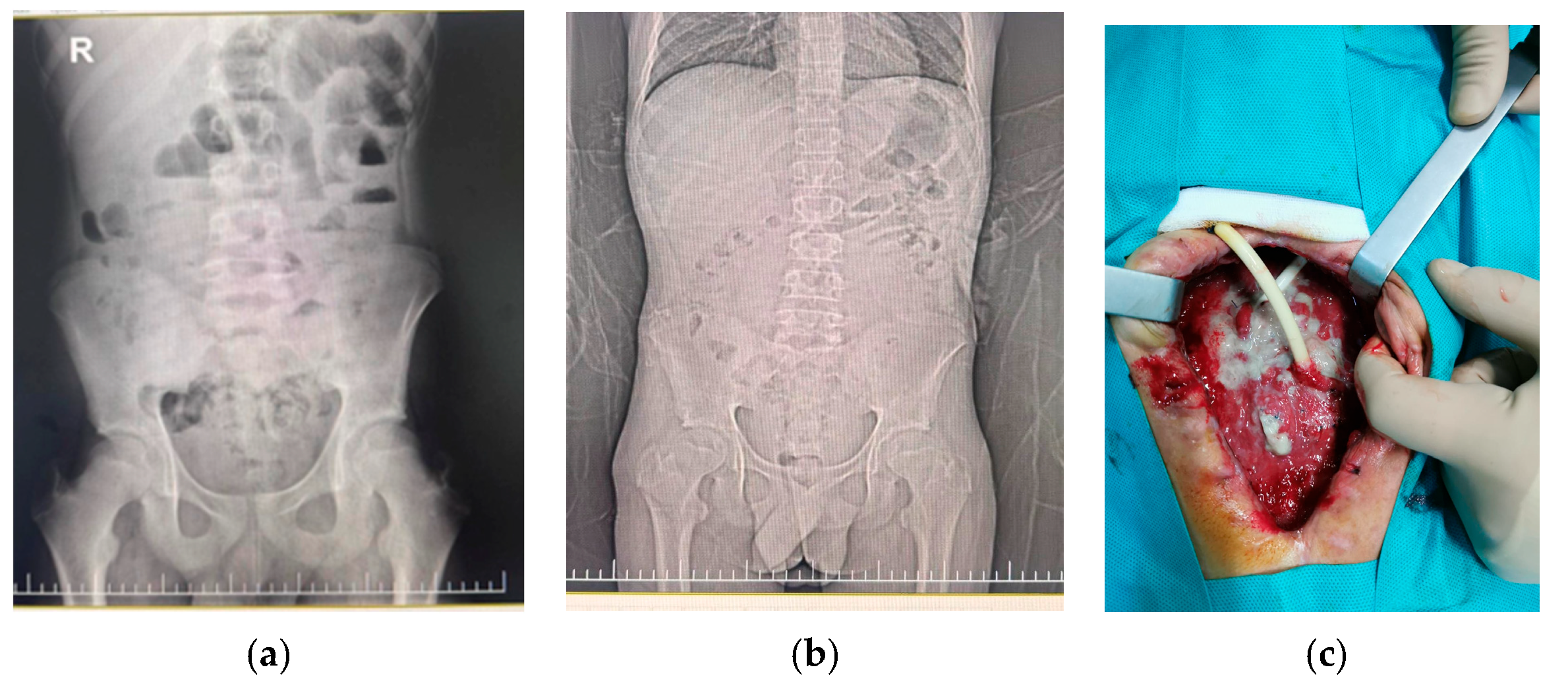
Background: Type I neurofibromatosis (NF1) represents an autosomal dominant inherited genodermatosis predisposing to tumor occurrence, caused by mutations in the NF1 gene, clinically characterized by the impairment of skin pigmentation, dermal neurofibromas, neuro-psychiatric involvement, and Lisch nodules. The initially defined diagnostic criteria were revised in 2021 to facilitate the diagnosis in young children who present only with skin pigmentation anomalies or a positive family history of NF1. Hemochromatosis (HC) is a genetic disorder consisting of systemic iron overload due to a defect in hepcidin. The mutations are commonly located in the HFE gene. The clinical picture is associated with joint pain, hyperpigmentation, hepatomegaly, etc. Genetic testing is mandatory for the diagnosis of HC. Case Presentation: The aim of this case report is to underline the importance of a multidisciplinary approach for a precise diagnosis. A 17-year-old male teenager with a past medical history of multiple abdominal surgical interventions presented to the emergency department for diarrhea and abdominal pain. The physical examination revealed weight deficit, café-au-lait macules, axillary and inguinal freckling, and mild abdominal tenderness. Laboratory evaluation revealed high CRP, mild leucocytosis and lymphopenia. The surgical consult ruled out an acute surgical cause. The abdominal X-ray, stool culture, fecal cytology, urinalysis and urine culture were negative, but ferritin level and serum iron level were very high. Ophthalmological consult described a Lisch nodule. Multiplex Ligation-dependent Probe Amplification (MLPA) genetic testing found no deletions or duplications within the NF1 gene. The clinical evolution was favorable under third-generation cephalosporin, but the ferritin and serum iron levels remain elevated. The Whole-Exome Sequencing (WES) identified a heterozygous pathogenic variant in the NF1 gene, i.e., chr17:29560075CA>C, NF1(NM_001042492.3):c.3556del; p.(Ile1186SerfsTer29); rs2151435407, but also revealed a homozygous pathogenic variant in the HFE gene, i.e., chr6:26090951C>G, HFE(NM_000410.4):c.187C>G; p.(His63Asp), rs1799945, which is associated with hereditary HC. Conclusions: The overlapping dermatological findings of both NF1 and HC might lead to diagnostic delays. The holistic approach to all pediatric patients presenting with particular hyperpigmentation involves thorough laboratory investigations, neurological, ophthalmological, cardiological and genetic examinations. In the absence of DNA Copy Number Variations (CNVs) that can be identified rapidly and cost-effectively by MLPA, sequencing is indicated to analyze small DNA variations, especially when two conditions with similar clinical pictures coexist.
Keywords: neurofibromatosis; Hemochromatosis; children; genetic testing neurofibromatosis; Hemochromatosis; children; genetic testing