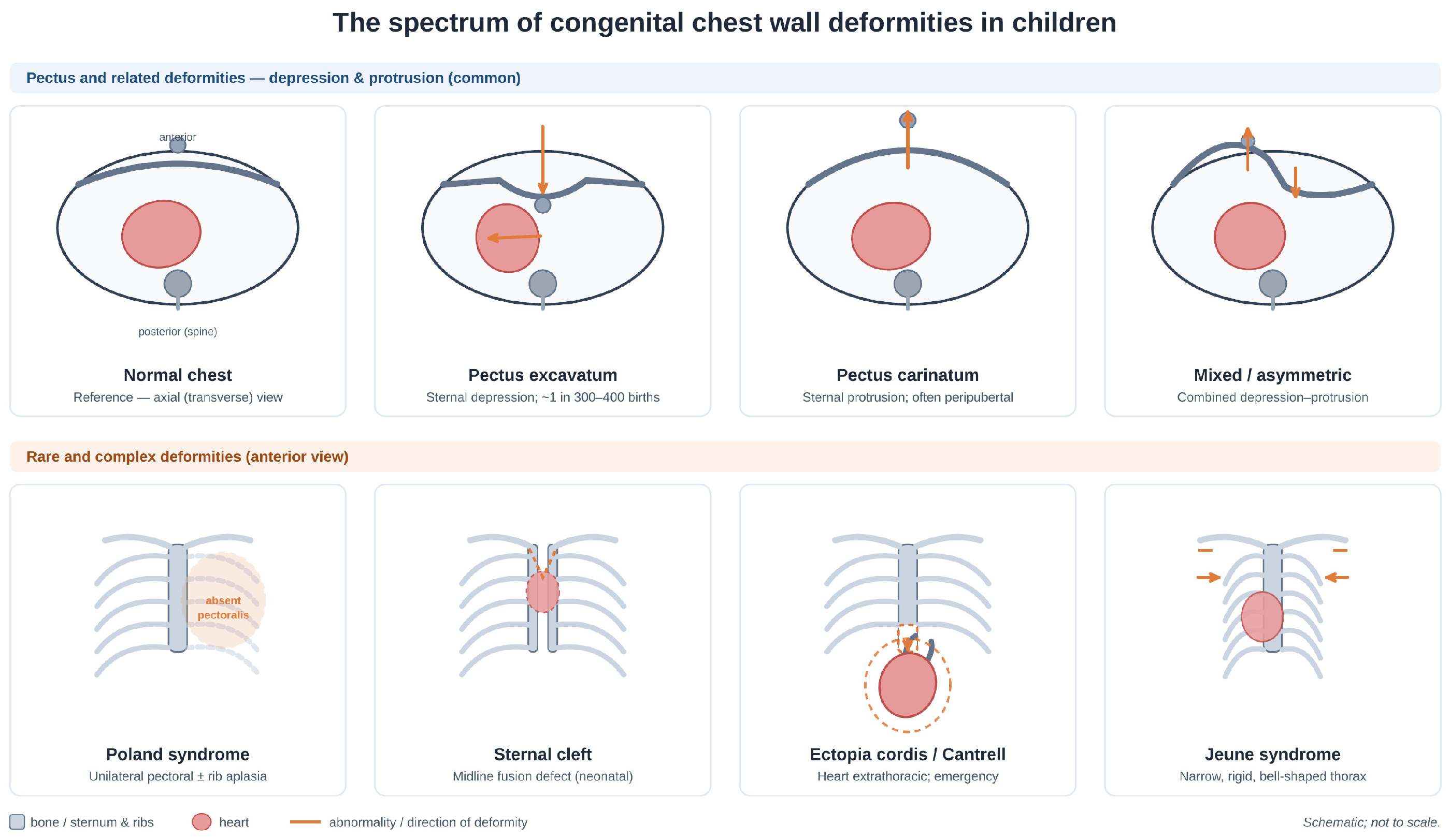
Congenital Chest Wall Deformities in Children: A Narrative Review
 , , ,
, , ,
Highlights
- Chest wall deformities in children span a wide spectrum—from pectus excavatum and carinatum (affecting ~1% of children) to rare, life-threatening anomalies—and are best understood as a genetically heterogeneous, mostly multifactorial group with clinically important syndromic associations rather than isolated cosmetic conditions.
- The most reproducible benefit of treatment lies in improved body image, self-esteem and quality of life, which are largely independent of anatomical severity, whereas a generalizable cardiopulmonary benefit of surgical correction remains unproven.
- Conservative remodelling (vacuum bell and dynamic compression bracing) should be first-line in suitable children, with minimally invasive, hybrid and modified-Ravitch surgery reserved for complex morphology.
- Surgical indications should integrate psychosocial burden alongside anatomical criteria, and early-onset deformity should prompt genetic referral and cardiovascular surveillance for associated connective-tissue disorders.
Abstract
1. Introduction
2. Methodology
3. Epidemiology
4. Genetics and Associated Syndromes
- Extracellular matrix and collagen metabolism (COL5A1, COL1A1, and COL27A1): COL5A1 regulates collagen fibril diameter, which is critical for cartilage ultrastructure; COL1A1 encodes the principal structural collagen of bone and costal cartilage; COL27A1 is cartilage-specific and essential for its structural integrity. Altered collagen composition may change the mechanical properties of the chest wall and permit abnormal growth of the chest wall.
- TGF-β/BMP signalling (SMAD4 and TGFB3): SMAD4 is the central intracellular mediator of both cascades, and TGFB3 is a key upstream ligand; together, they regulate chondrocyte proliferation, differentiation, and hypertrophy during endochondral ossification.
- Cartilage development and homeostasis (ACAN, GPR126, and GAL3ST4): Aggrecan confers compressive resistance to cartilage, GPR126 regulates chondrocyte maturation, and GAL3ST4 modifies the proteoglycans.
- RAS/MAPK growth-plate signalling (SOS1, PTPN11, and NF1): Governs chondrocyte proliferation; hyperactivation in Noonan-spectrum disorders produces abnormal rib growth and increased chest wall laxity.
- Skeletal patterning (TGDS, COL27A1, and COL1A1): Disrupted thoracic patterning or altered ossification timing may increase susceptibility.
- Transcriptional regulation (REST): A master repressor of neuronal genes in non-neuronal tissues, implicated in mesenchymal stem cell differentiation, although its mechanism in PE remains unclear.
- Neuromuscular support (BICD2): Reduced anterior chest wall support may aggravate pre-existing structural vulnerability.
5. Diagnostics and Imaging
6. Psychosocial Aspects and Cardiorespiratory Function
6.1. Psychosocial Burden
6.2. Cardiorespiratory Function
7. Conservative Treatment
7.1. Vacuum Bell Therapy for Pectus Excavatum
7.2. Dynamic Compression Bracing for Pectus Carinatum
7.3. Physiotherapy and Adjunctive Measures
8. Surgical Treatment and Techniques
9. Rare and Complex Deformities
9.1. Poland Syndrome
9.2. Sternal Clefts
9.3. Ectopia Cordis and Pentalogy of Cantrell
9.4. Jeune Syndrome (Asphyxiating Thoracic Dystrophy)
10. Conclusions
Author Contributions
Funding
Institutional Review Board Statement
Informed Consent Statement
Data Availability Statement
Acknowledgments
Conflicts of Interest
References
- Janssen, N.; Coorens, N.A.; Franssen, A.J.P.M.; Daemen, J.H.T.; Michels, I.L.; Hulsewé, K.W.E.; Vissers, Y.L.J.; de Loos, E.R. Pectus excavatum and carinatum: A narrative review of epidemiology, etiopathogenesis, clinical features, and classification. J. Thorac. Dis. 2024, 16, 1687–1701. [Google Scholar] [CrossRef] [Scilit] [PubMed]
- Pu, Y.; Jian, Y.; Huang, W.; Yang, Z. Prevalence and incidence of chest wall deformities in children below 18 years old: The first systematic review and meta-analysis. Arch. Med. Sci. 2024, 21, 1421–1431. [Google Scholar] [CrossRef] [Scilit] [PubMed]
- Buckwalter, V.J.A.; Shah, A.S. Presentation and treatment of Poland anomaly. Hand 2016, 11, 389–395. [Google Scholar] [CrossRef] [Scilit] [PubMed]
- Wojtyś, M.E.; Kordykiewicz, D.; Wójcik, J.; Tomos, P.; Kostopanagiotou, K. Consultations for Poland syndrome: The essentials for a thoracic surgeon. Medicina 2024, 60, 1178. [Google Scholar] [CrossRef] [Scilit] [PubMed]
- Fokin, A.A.; Robicsek, F. Poland’s syndrome revisited. Ann. Thorac. Surg. 2002, 74, 2218–2225. [Google Scholar] [CrossRef] [Scilit] [PubMed]
- Engum, S.A. Embryology, sternal clefts, ectopia cordis, and Cantrell’s pentalogy. Semin. Pediatr. Surg. 2008, 17, 154–160. [Google Scholar] [CrossRef] [Scilit] [PubMed]
- Creswick, H.A.; Stacey, M.W.; Kelly, R.E., Jr.; Gustin, T.; Nuss, D.; Harvey, H.; Goretsky, M.J.; Vasser, E.; Welch, J.C.; Mitchell, K.; et al. Family study of the inheritance of pectus excavatum. J. Pediatr. Surg. 2006, 41, 1699–1703. [Google Scholar] [CrossRef] [Scilit] [PubMed]
- Kotzot, D.; Schwabegger, A.H. Etiology of chest wall deformities—A genetic review for the treating physician. J. Pediatr. Surg. 2009, 44, 2004–2011. [Google Scholar] [CrossRef] [Scilit] [PubMed]
- Ranjan, R.; Imtiaz, N.; Waterhouse, B.; Paul, I.; Brunswicker, A.; Dunning, J. Genetic associations with pectus excavatum: A systematic review. Curr. Issues Mol. Biol. 2026, 48, 122. [Google Scholar] [CrossRef] [Scilit] [PubMed]
- Farina, J.M.; Olson, R.J.; Dhamija, R.; Bofferding, A.; Sekulic, A.; Egan, J.B.; Jaroszewski, D.E. The complexity of familial inheritance in pectus excavatum: A ten-family exome sequencing analysis. Genes 2024, 15, 1429. [Google Scholar] [CrossRef] [Scilit] [PubMed]
- Billar, R.J.; Manoubi, W.; Kant, S.G.; Wijnen, R.M.H.; Demirdas, S.; Schnater, J.M. Association between pectus excavatum and congenital genetic disorders: A systematic review and practical guide for the treating physician. J. Pediatr. Surg. 2021, 56, 2239–2252. [Google Scholar] [CrossRef] [Scilit] [PubMed]
- Kelly, R.E., Jr.; Goretsky, M.J.; Obermeyer, R.; Kuhn, M.A.; Redlinger, R.; Haney, T.S.; Moskowitz, A.; Nuss, D. Twenty-one years of experience with minimally invasive repair of pectus excavatum by the Nuss procedure in 1215 patients. Ann. Surg. 2010, 252, 1072–1081. [Google Scholar] [CrossRef] [Scilit] [PubMed]
- Billar, R.; Heyman, S.; Kant, S.; Wijnen, R.; Sleutels, F.; Demirdas, S.; Schnater, J.M. Early-Onset Pectus Excavatum Is More Likely to Be Part of a Genetic Variation. Eur. J. Pediatr. Surg. 2024, 34, 325–332. [Google Scholar] [CrossRef] [Scilit] [PubMed]
- Coorens, N.A.; Janssen, N.; Daemen, J.H.T.; Franssen, A.J.P.M.; Hulsewé, K.W.E.; Vissers, Y.L.J.; de Loos, E.R. Advancements in preoperative imaging of pectus excavatum: A comprehensive review. J. Thorac. Dis. 2024, 16, 696–707. [Google Scholar] [CrossRef] [Scilit] [PubMed]
- Haller, J.A.; Kramer, S.S.; Lietman, S.A. Use of CT scans in selection of patients for pectus excavatum surgery: A preliminary report. J. Pediatr. Surg. 1987, 22, 904–906. [Google Scholar] [CrossRef] [Scilit] [PubMed]
- St Peter, S.D.; Juang, D.; Garey, C.L.; Laituri, C.A.; Ostlie, D.J.; Sharp, R.J.; Snyder, C.L. A novel measure for pectus excavatum: The correction index. J. Pediatr. Surg. 2011, 46, 2270–2273. [Google Scholar] [CrossRef] [Scilit] [PubMed]
- Wharton, K.; Chun, Y.; Hunsberger, J.; Jelin, E.; Garcia, A.; Stewart, D. Successful use of an enhanced recovery after surgery pathway to improve outcomes following the Nuss procedure for pectus excavatum. J. Pediatr. Surg. 2020, 55, 1065–1071. [Google Scholar] [CrossRef] [Scilit] [PubMed]
- Mayer, O.; Campbell, R.; Cahill, P.; Redding, G. Thoracic insufficiency syndrome. Curr. Probl. Pediatr. Adolesc. Health Care 2016, 46, 72–97. [Google Scholar] [CrossRef] [Scilit] [PubMed]
- Martínez-Ferro, M. Indexes for pectus deformities. In Chest Wall Deformities and Corrective Procedures; Kolvekar, S., Pilegaard, H., Eds.; Springer: Cham, Switzerland, 2016; Chapter 7. [Google Scholar] [CrossRef] [Scilit]
- Bellía-Munzón, G.; Sanjurjo, D.; Toselli, L.; Vallee, M.; Elmo, G.; Martínez-Ferro, M. Novel index to estimate the cephalocaudal extent of the excavation in pectus excavatum: The Titanic index. J. Pediatr. Surg. 2023, 58, 605–607. [Google Scholar] [CrossRef] [Scilit] [PubMed]
- Fagelman, K.M.; Methratta, S.; Cilley, R.E.; Wilson, M.Z.; Hollenbeak, C.S. The Depression Index: An objective measure of the severity of pectus excavatum based on vertebral diameter, a morphometric correlate to patient size. J. Pediatr. Surg. 2015, 50, 1130–1133. [Google Scholar] [CrossRef] [Scilit] [PubMed]
- Kim, M.; Lee, K.Y.; Park, H.J.; Kim, H.Y.; Kang, E.Y.; Oh, Y.W.; Seo, B.K.; Je, B.K.; Choi, E.J. Development of new cardiac deformity indexes for pectus excavatum on computed tomography: Feasibility for pre- and post-operative evaluation. Yonsei Med. J. 2009, 50, 385–390. [Google Scholar] [CrossRef] [Scilit] [PubMed]
- Poston, P.M.; Patel, S.S.; Rajput, M.; Rossi, N.O.; Ghanamah, M.S.; Davis, J.E.; Turek, J.W. The Correction Index: Setting the standard for recommending operative repair of pectus excavatum. Ann. Thorac. Surg. 2014, 97, 1176–1181. [Google Scholar] [CrossRef] [Scilit] [PubMed]
- Barmash, E.A.; Mathai, B.B.; Mansfield, S.A.; Van Arendonk, K.J. Psychological impact of congenital chest wall deformities among adolescents and young adults. Children 2026, 13, 237. [Google Scholar] [CrossRef] [Scilit] [PubMed]
- Kelly, R.E.; Cash, T.F.; Shamberger, R.C.; Mitchell, K.K.; Mellins, R.B.; Lawson, M.L.; Oldham, K.; Azizkhan, R.G.; Hebra, A.V.; Nuss, D.; et al. Surgical repair of pectus excavatum markedly improves body image and perceived ability for physical activity: A multicenter study. Pediatrics 2008, 122, 1218–1222. [Google Scholar] [CrossRef] [Scilit] [PubMed]
- Sutyak, K.M.; Hebballi, N.B.; Bidart, Y.; Joly, J.M.; Broussard, M.; Christensen, H.; Mendenhall, J.; Jayarajan, N.; Anderson, I.; Young, Y.; et al. Is it time to expand the surgical criteria? The psychosocial impact of pectus excavatum in pediatrics. Surgery 2025, 185, 109546. [Google Scholar] [CrossRef] [Scilit] [PubMed]
- Ravasin, A.; Viggiano, D.; Tombelli, S.; Checchi, L.; Stefàno, P.; Voltolini, L.; Gonfiotti, A. Heart disease and pectus excavatum: An underestimated issue—Single-center experience and literature review. Life 2024, 14, 1643. [Google Scholar] [CrossRef] [Scilit] [PubMed]
- Media, A.S.; Juhl-Olsen, P.; Christensen, T.D.; Katballe, N.; Vad, H.; Petersen, R.H.; Wiggers, H.; Hansen, S.N.; Farup, J.; Overgaard, K.; et al. Cardiorespiratory fitness after correction of pectus excavatum: A systematic review with meta-analysis. Sci. Rep. 2025, 15, 26282. [Google Scholar] [CrossRef] [Scilit] [PubMed]
- Schier, F.; Bahr, M.; Klobe, E. The vacuum chest wall lifter: An innovative, non-surgical addition to the management of pectus excavatum. J. Pediatr. Surg. 2005, 40, 496–500. [Google Scholar] [CrossRef] [Scilit] [PubMed]
- Haecker, F.M. 20 years clinical application of the vacuum bell for conservative treatment of pectus excavatum—Past, present, future. J. Thorac. Dis. 2024, 16, 5285–5298. [Google Scholar] [CrossRef] [Scilit] [PubMed]
- van Braak, H.; de Beer, S.A.; al Ghouch, Y.; Zwaveling, S.; Oomen, M.W.N.; van Heurn, L.W.E.; de Jong, J.R. 15 years of vacuum bell therapy for pectus excavatum: Long-term outcomes and influencing factors. J. Pediatr. Surg. 2025, 60, 161891. [Google Scholar] [CrossRef] [Scilit] [PubMed]
- van Braak, H.; de Beer, S.A.; Zwaveling, S.; Oomen, M.W.N.; de Jong, J.R. Ravitch surgery or dynamic compression bracing for pectus carinatum: A retrospective cohort study. Ann. Thorac. Surg. 2024, 117, 144–150. [Google Scholar] [CrossRef] [Scilit] [PubMed]
- van Braak, H.; de Beer, S.A.; Twisk, J.W.R.; Zwaveling, S.; Oomen, M.W.N.; de Jong, J.R.; van Heurn, L.W.E. Improving quality of life with dynamic compression bracing in patients with pectus carinatum. J. Pediatr. Surg. 2025, 60, 161975. [Google Scholar] [CrossRef] [Scilit] [PubMed]
- Sonel Tur, B.; Genç, A. An overview of pectus deformities and rehabilitation approaches. Turk. J. Phys. Med. Rehabil. 2025, 71, 131–138. [Google Scholar] [CrossRef] [Scilit] [PubMed]
- Nuss, D.; Kelly, R.E., Jr.; Croitoru, D.P.; Katz, M.E. A 10-year review of a minimally invasive technique for the correction of pectus excavatum. J. Pediatr. Surg. 1998, 33, 545–552. [Google Scholar] [CrossRef] [Scilit] [PubMed]
- Szostawicki, M.; Kowalska, M.; Puliński, M.; Janowicz, T.; Matus, Ł.; Hermanowicz, A. Optimizing the Nuss technique: Modified approaches to pediatric pectus excavatum. Pol. Przegl. Chir. 2026, 98, 70–75. [Google Scholar] [CrossRef] [Scilit] [PubMed]
- Nasr, A.; Fecteau, A.; Wales, P.W. Comparison of the Nuss and the Ravitch procedure for pectus excavatum repair: A meta-analysis. J. Pediatr. Surg. 2010, 45, 880–886. [Google Scholar] [CrossRef] [Scilit] [PubMed]
- Ravitch, M.M. The operative treatment of pectus excavatum. Ann. Surg. 1949, 129, 429–444. [Google Scholar] [CrossRef] [Scilit] [PubMed]
- Jawitz, O.K.; Raman, V.; Thibault, D.; Yerokun, B.; Zwischenberger, B.A.; Kosinski, A.S.; Allen, M.S.; Harpole, D.H. Complications after Ravitch versus Nuss repair of pectus excavatum: A Society of Thoracic Surgeons database analysis. Surgery 2021, 169, 1493–1499. [Google Scholar] [CrossRef] [Scilit] [PubMed]
- Perdomo, D.; Leng, A.; Patel, D.; Yang, S.C.; Ha, J.S. 75 years of the Ravitch procedure: A historical report and review of outcomes. Ann. Thorac. Surg. Short Rep. 2025, 3, 808–813. [Google Scholar] [CrossRef] [Scilit] [PubMed]
- Pawlak, K.; Gąsiorowski, Ł.; Dyszkiewicz, W. Complex corrective procedure in surgical treatment of asymmetrical pectus excavatum. Kardiochir. Torakochir. Pol. 2017, 14, 110–114. [Google Scholar] [CrossRef] [Scilit] [PubMed]
- Masai, K.; Okubo, Y.; Kaseda, K.; Hishida, T.; Asakura, K. Combined Ravitch and Nuss procedure for patients with severe pectus excavatum: Technique and initial results. J. Surg. Case Rep. 2023, 2023, rjad576. [Google Scholar] [CrossRef] [Scilit] [PubMed]
- Haecker, F.M.; Krebs, T.; Kocher, G.J.; Schmid, R.A.; Sesia, S.B. Sternal elevation techniques during the minimally invasive repair of pectus excavatum. Interact. Cardiovasc. Thorac. Surg. 2019, 29, 497–502. [Google Scholar] [CrossRef] [Scilit] [PubMed]
- de Loos, E.R.; Daemen, J.H.T.; Coorens, N.A.; Maessen, J.G.; Vissers, Y.L.J.; Hulsewé, K.W.E. Sternal elevation by the crane technique during pectus excavatum repair: A quantitative analysis. JTCVS Tech. 2021, 9, 167–175. [Google Scholar] [CrossRef] [Scilit] [PubMed]
- Harrison, M.R.; Estefan-Ventura, D.; Fechter, R.; Moran, A.M., Jr.; Christensen, D. Magnetic Mini-Mover Procedure for pectus excavatum: I. Development, design, and simulations for feasibility and safety. J. Pediatr. Surg. 2007, 42, 81–85. [Google Scholar] [PubMed]
- Graves, C.E.; Hirose, S.; Raff, G.W.; Iqbal, C.W.; Imamura-Ching, J.; Christensen, D.; Fechter, R.; Kwiat, D.; Harrison, M.R. Magnetic Mini-Mover Procedure for pectus excavatum IV: FDA-sponsored multicenter trial. J. Pediatr. Surg. 2017, 52, 913–919. [Google Scholar] [CrossRef] [Scilit] [PubMed]
- Länsman, S.; Serlo, W.; Linna, O.; Pohjonen, T.; Törmälä, P.; Waris, T.; Ashammakhi, N. Treatment of pectus excavatum with bioabsorbable polylactide plates: Preliminary results. J. Pediatr. Surg. 2002, 37, 1281–1286. [Google Scholar] [CrossRef] [Scilit] [PubMed]
- Torre, M.; Jasonni, V.; Asquasciati, C.; Costanzo, S.; Romanini, M.; Varela, P. Absorbable stabilisation of the bar in minimally invasive repair of pectus excavatum. Eur. J. Pediatr. Surg. 2008, 18, 407–409. [Google Scholar] [CrossRef] [Scilit] [PubMed]
- Pilegaard, H.K.; Licht, P.B. Can absorbable stabilizers be used routinely in the Nuss procedure? Eur. J. Cardiothorac. Surg. 2009, 35, 561–564. [Google Scholar] [CrossRef] [Scilit] [PubMed]
- DiFiore, J.W.; Robertson, J.O.; Chhabada, S.; DeRoss, A.L.; Hossain, M.S.; Rincon-Cruz, L.; Sung, W. Next-day discharge after the Nuss procedure using intercostal nerve cryoablation, intercostal nerve blocks, and a perioperative ERAS pain protocol. J. Pediatr. Surg. 2022, 57, 213–218. [Google Scholar] [CrossRef] [Scilit] [PubMed]
- Keppler-Noreuil, K.M.; Adam, M.P.; Welch, J.; Muilenburg, A.; Willing, M.C. Clinical insights gained from eight new cases and review of reported cases with Jeune syndrome (asphyxiating thoracic dystrophy). Am. J. Med. Genet. A 2011, 155A, 1021–1032. [Google Scholar] [CrossRef] [Scilit] [PubMed]
- Campbell, R.M., Jr.; Smith, M.D.; Mayes, T.C.; Mangos, J.A.; Willey-Courand, D.B.; Kose, N.; Pinero, R.F.; Alder, M.E.; Duong, H.L.; Surber, J.L. The characteristics of thoracic insufficiency syndrome associated with fused ribs and congenital scoliosis. J. Bone Jt. Surg. 2003, 85, 399–408. [Google Scholar] [CrossRef] [Scilit] [PubMed]
- O’Brien, A.; Roth, M.K.; Athreya, H.; Reinker, K.; Koeck, W.; Patil, V.; Trevino, H.; Simmons, J.; Joshi, A.P.; Smith, M.D.; et al. Management of thoracic insufficiency syndrome in patients with Jeune syndrome using the 70-mm radius vertical expandable prosthetic titanium rib. J. Pediatr. Orthop. 2015, 35, 783–797. [Google Scholar] [CrossRef] [Scilit] [PubMed]


| Index | Modality | Definition/Formula | Threshold/Reference Values | Comment |
|---|---|---|---|---|
| Haller index (HI) | CT; chest radiograph; MRI | HI = T/A. T = widest internal transverse diameter of the ribcage; A = shortest distance between the vertebra and the sternum. | >3.25 conventionally indicates operative candidacy | The gold standard, but the 3.25 cut-off has never been independently validated; alternative upper limits of 2.7, 3.1 and 3.2 have been proposed. Varies with age, sex, thoracic shape and respiratory phase; ignores asymmetry and cardiac compression. Radiograph-derived HI correlates closely with CT and reduces dose [1,14,15,17]. |
| Correction index (CI/PCI) | CT; chest radiograph | A horizontal line is drawn across the anterior spine. On the same slice, the minimum distance from the posterior sternum to the anterior spine and the distance from the inner margin of the anterior chest to that line are measured; their difference is divided by the maximum prominence of the chest and multiplied by 100. | ≥10% defines the deformity; ≈28% corresponds to a Haller index of 3.25 | Independent of chest width. One of the few indices to have been independently validated [17,18]. |
| Welch index | Chest radiograph | Depression ratio D1/D2; deformity grade (1-ratio) × 10, with additions for a rib angle > 25° or a cardiothoracic ratio > 50%. | ≥5 recommended for operative repair | Graded 1–10 severity scale derived from a large historical series [19]. |
| Vertebral index (lower/upper) | Chest radiograph | Ratio of the sternovertebral distance to the sagittal diameter of the vertebral body (lower VI at the xiphisternal, upper VI at the sternomanubrial junction). | Lower VI cut-off > 27% | Lower VI is age-dependent; upper VI is age-independent; both relate sternal approximation to the spine [19]. |
| Body-manubrium (xyphoid) index | Chest radiograph | BM = ossified sternal-body length ÷ manubrium length; BMX additionally includes the xyphoid. | BM 2.16; BMX 2.73 | Requires ossified sternal segments; a short sternal body may help predict the number of bars needed [19]. |
| Frontosagittal index | Chest radiograph | (Minimum sagittal chest diameter ÷ maximum internal transverse diameter) × 100. | Preoperative cut-off < 29 | Increases after correction; a plain-radiograph alternative to CT indices [19]. |
| Titanic index (TI) | CT | Percentage of the length of the sternum lying behind the anterior costal line. | Mean 37%; >66.5% predicts the need for more than two bars (sensitivity 93%, specificity 92%) | Quantifies the cephalocaudal extent of the excavation rather than its severity at the deepest point. Correlates only weakly with HI and CI, so carries independent information. A tool for operative planning, not for establishing candidacy [20]. |
| Sternal depression index (SDI) | CT | SDI = C/B. C = maximal internal sagittal diameter of the left hemithorax; B = minimal distance from the anterior surface of the vertebral column to the posterior border of the deepest portion of the sternum. | <2.4 mild; 2.4–2.9 moderate; >2.9 severe (mean 2.7 ± 1.4) | Correlates with the cardiac rotation angle (r = 0.75). Mean absolute sternal depression in the source series was 21 ± 7 mm [19]. |
| Depression index (DI) | CT | Absolute depth of sternal depression divided by the transverse vertebral-body diameter (measured at T9–T11), the latter serving as a morphometric surrogate for patient size. | No fixed cut-off; scaled continuous measure | Independent of thoracic diameters; correlates with the Haller and correction indices and, in the original study, matched clinicians’ subjective severity ranking better than either [21]. |
| Haje width-length index (WLI) | Coronal CT | WLI = W/L, where W = maximum width of the ossified sternal body and L = its length. | Mean 0.420 in controls; >0.446 in pectus excavatum | Higher values denote a wider sternal body, with possible implications for prognosis and choice of procedure [19]. |
| Asymmetry index | CT | Asymmetry index = (R/L) × 100, where R and L are the anteroposterior distances between the anterior and posterior ribs on the right and left sides; best measured at the sternomanubrial junction. | 100 denotes a symmetric chest; departure from 100 quantifies asymmetry | Combining the Haller index with the asymmetry index improves detection of asymmetric pectus excavatum; computed alongside the Haller and correction indices in automated deep-learning pipelines [19]. |
| Cardiac compression index (CCI) | CT; MRI | CCI = transverse diameter of the heart ÷ minimum anteroposterior diameter of the heart (at the level of the xiphoid). | Higher values indicate greater cardiac compression; significant for diagnosis regardless of age | One of the cardiac deformity indices proposed by Kim et al. to bring cardiac involvement—which no purely skeletal index captures—into the assessment [22]. |
| External Haller index | 3D optical surface imaging; calipers | Analogue of the Haller index computed from the external skin-surface contour rather than internal bony landmarks. | ≥1.83 corresponds to a conventional Haller index ≥ 3.25 | Radiation-free and repeatable, well suited to follow-up during conservative treatment [14]. |
| External correction index | 3D optical surface imaging | Analogue of the correction index derived from the external skin-surface contour. | ≥15.2% corresponds to a conventional correction index ≥ 28.0% | Radiation-free; may also predict the presence of cardiac compression without CT [14]. |
| Modified Haller index (carinatum) | CT; chest radiograph | Haller index using the maximum anteroposterior length of the chest wall in place of the minimum sternovertebral distance. | No validated operative threshold | Pectus carinatum lacks a validated severity index equivalent to those for pectus excavatum; assessment remains largely clinical [1]. |
Disclaimer/Publisher’s Note: The statements, opinions and data contained in all publications are solely those of the individual author(s) and contributor(s) and not of MDPI and/or the editor(s). MDPI and/or the editor(s) disclaim responsibility for any injury to people or property resulting from any ideas, methods, instructions or products referred to in the content. |
© 2026 by the authors. Licensee MDPI, Basel, Switzerland. This article is an open access article distributed under the terms and conditions of the Creative Commons Attribution (CC BY) license.
Share and Cite
Kowalska, M.; Grabowska, H.; Szostawicki, M.; Puliński, M.; Janowicz, T.; Hermanowicz, A. Congenital Chest Wall Deformities in Children: A Narrative Review. Children 2026, 13, 1265. https://doi.org/10.3390/children13091265
Kowalska M, Grabowska H, Szostawicki M, Puliński M, Janowicz T, Hermanowicz A. Congenital Chest Wall Deformities in Children: A Narrative Review. Children. 2026; 13(9):1265. https://doi.org/10.3390/children13091265
Chicago/Turabian StyleKowalska, Małgorzata, Hanna Grabowska, Michał Szostawicki, Michał Puliński, Tomasz Janowicz, and Adam Hermanowicz. 2026. "Congenital Chest Wall Deformities in Children: A Narrative Review" Children 13, no. 9: 1265. https://doi.org/10.3390/children13091265
APA StyleKowalska, M., Grabowska, H., Szostawicki, M., Puliński, M., Janowicz, T., & Hermanowicz, A. (2026). Congenital Chest Wall Deformities in Children: A Narrative Review. Children, 13(9), 1265. https://doi.org/10.3390/children13091265