
Spectroscopic and Colorimetric Studies for Anions with a New Urea-Based Molecular Cleft


Abstract
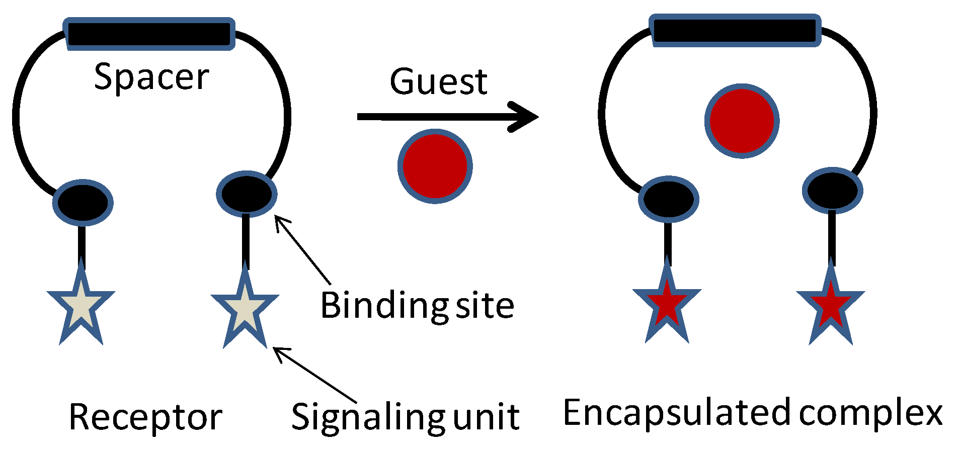
:1. Introduction
2. Materials and Methods
2.1. Materials
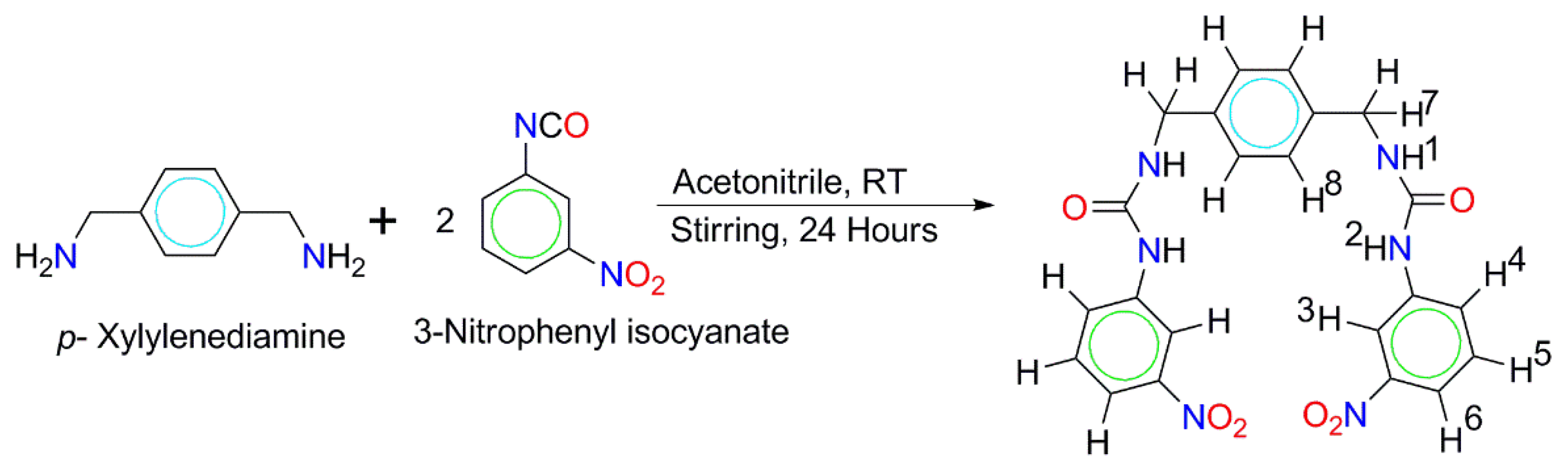
2.2. Synthesis
2.3. UV-Vis Titration Studies
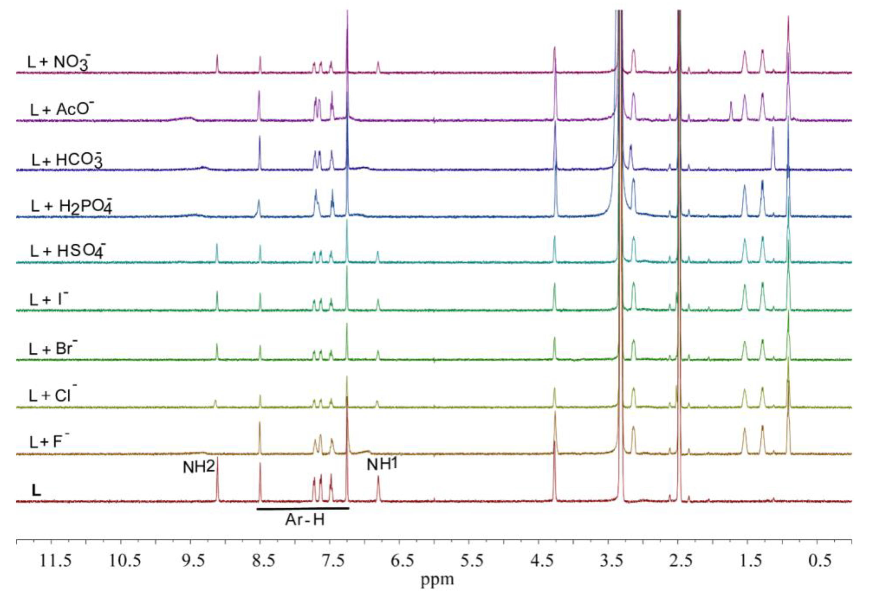
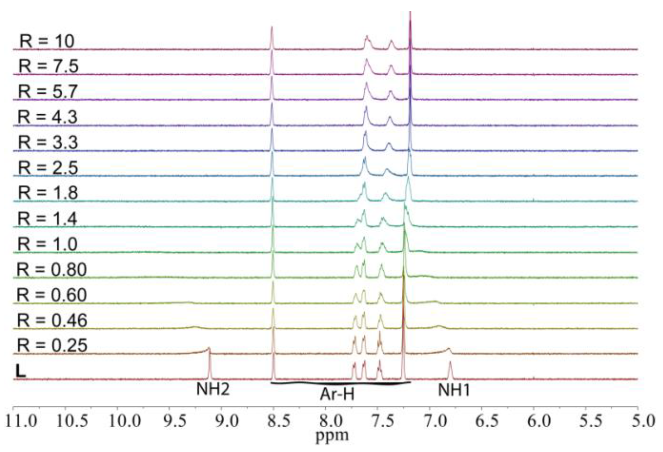
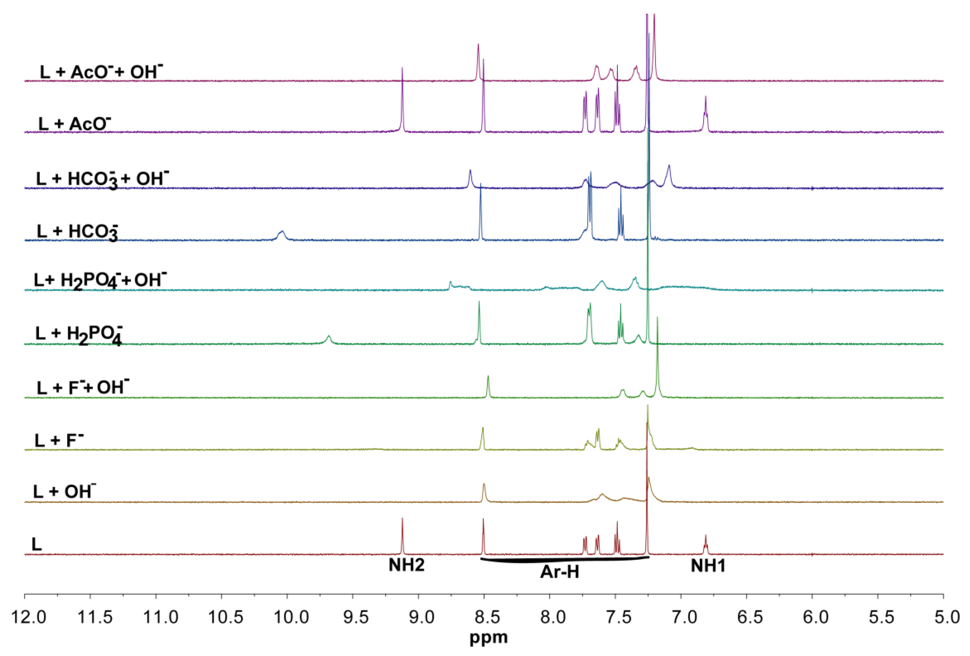
2.4. NMR Studies
3. Results
3.1. Synthesis
3.2. UV-Vis Spectroscopic Studies
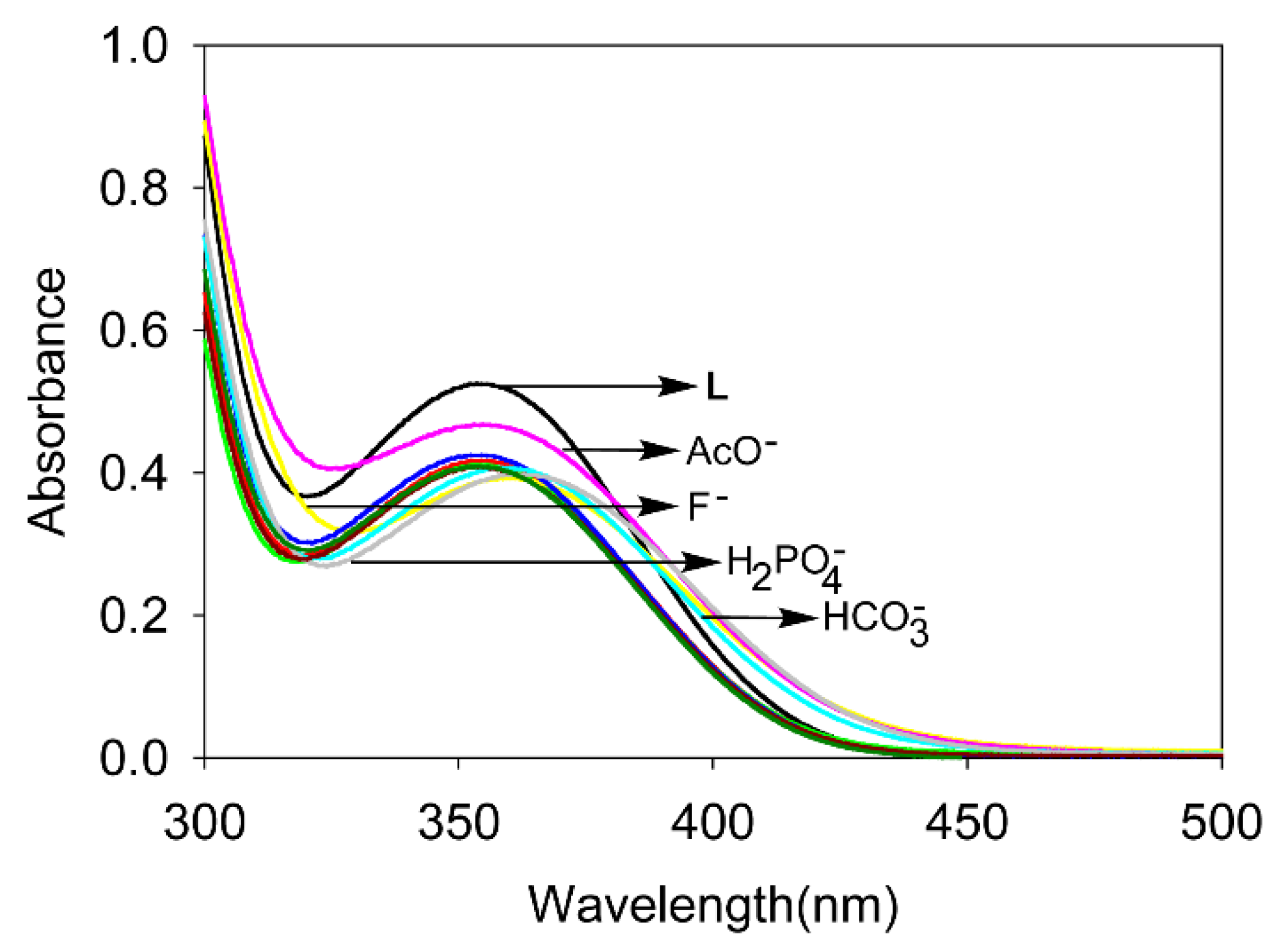
3.2.1. UV-Vis Screening
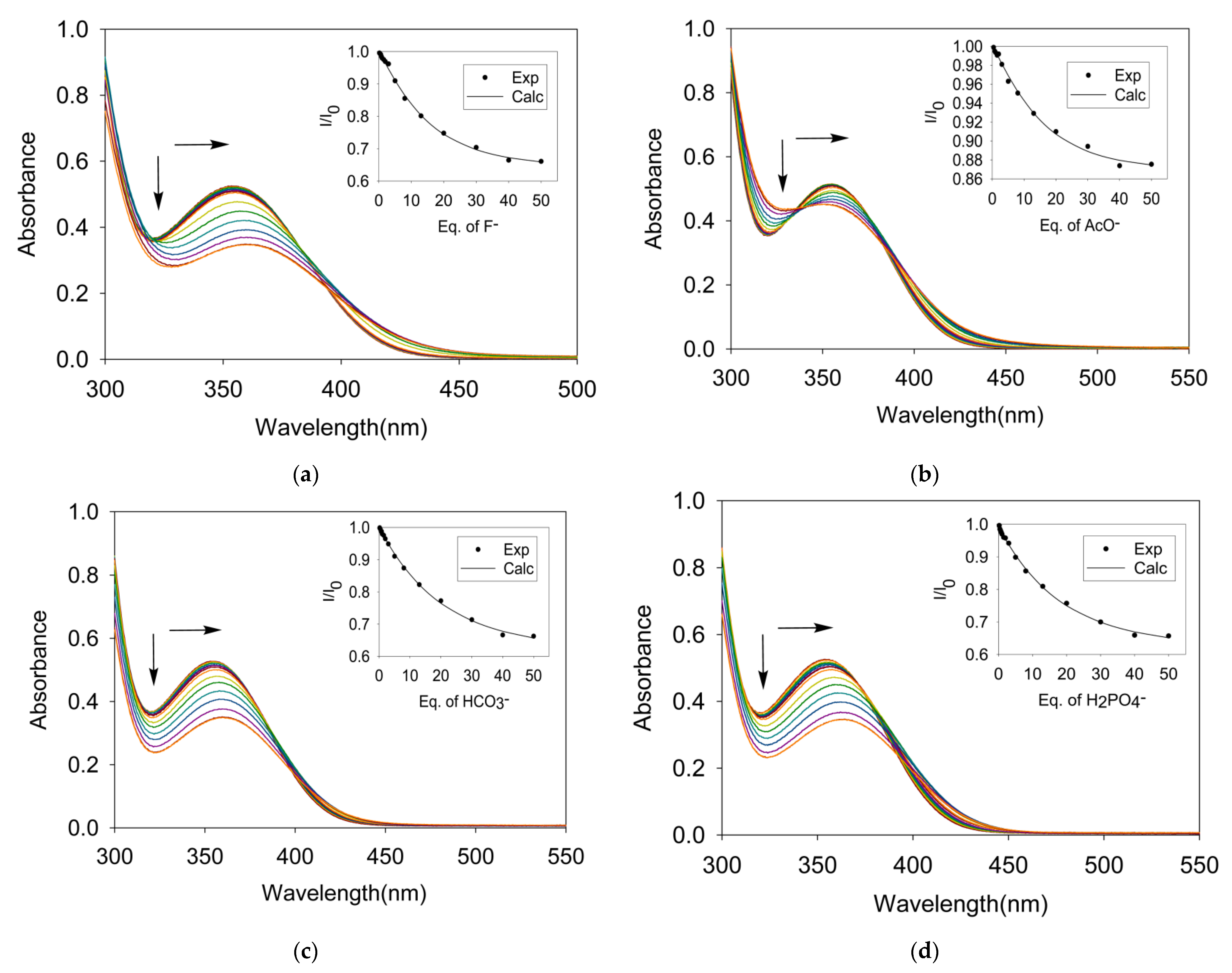
3.2.2. UV-Vis Titrations
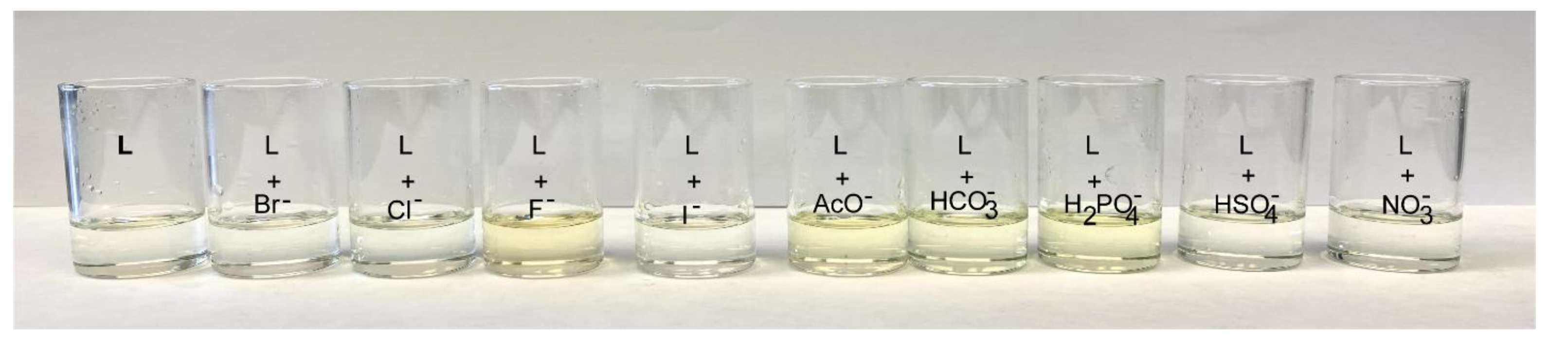
3.3. Colorimetric Study
3.4. NMR Studies
4. Conclusions
Supplementary Materials
Author Contributions
Funding
Institutional Review Board Statement
Informed Consent Statement
Data Availability Statement
Acknowledgments
Conflicts of Interest
References
- Busschaert, N.; Caltagirone, C.; Rossom, W.V.; Gale, P.A. Applications of supramolecular anion recognition. Chem. Rev. 2015, 115, 8038–8155. [Google Scholar] [CrossRef]
- Li, A.F.; Wang, J.H.; Wang, F.; Jiang, Y.B. Anion complexation and sensing using modified urea and thiourea-based receptors. Chem. Soc. Rev. 2010, 39, 3581–4008. [Google Scholar] [CrossRef] [Green Version]
- Ronchetti, R.; Moroni, G.; Carotti, A.; Gioiello, A.; Camaioni, E. Recent advances in urea- and thiourea-containing compounds: Focus on innovative approaches in medicinal chemistry and organic synthesis. RSC Med. Chem. 2021, 12, 1046–1064. [Google Scholar] [CrossRef]
- Zhang, Z.; Schreiner, P.R. (Thio) urea organocatalysis—What can be learnt from anion recognition? Chem. Soc. Rev. 2009, 38, 1187–1198. [Google Scholar] [CrossRef]
- Pal, S.; Ghosh, T.K.; Ghosh, R.; Mondal, S.; Ghosh, P. Recent advances in recognition, sensing and extraction of phosphates: 2015 onwards. Coord. Chem. Rev. 2020, 405, 213128. [Google Scholar] [CrossRef]
- Toy, D.F.; Walsh, E.F. Phosphorus Chemistry in Everyday Living, 2nd ed.; American Chemical Society: Washington, DC, USA, 1987. [Google Scholar]
- Ayoob, S.; Gupta, A.K. Fluoride in drinking water: A review on the status and stress effects. Crit. Rev. Environ. Sci. Technol. 2006, 36, 433–487. [Google Scholar] [CrossRef]
- Manna, U.; Portis, B.; Egboluche, T.K.; Nafis, M.; Hossain, M.A. Anion binding studies of urea and thiourea functionalized molecular clefts. Front. Chem. 2021, 8, 575701. [Google Scholar] [CrossRef] [PubMed]
- Tiessen, H. Phosphorus in the global environment. Plant Ecophysiol. 2008, 7, 1–7. [Google Scholar]
- Mason, C.F. Biology of Freshwater Pollution; Longman: New York, NY, USA, 1991. [Google Scholar]
- Charra, B.; Calemar, E.; Ruffet, M.; Chazot, C.; Terra, J.-C.; Valen, T.; Laurent, G. Survival as an index of adequacy of dialysis. Kidney Int. 1992, 41, 1286–1291. [Google Scholar] [CrossRef] [PubMed] [Green Version]
- Dhingra, R.; Sullivan, L.M.; Fox, C.S.; Wang, T.J.; D’Agostino, R.B., Sr.; Gaziano, J.M.; Vasan, R.S. Relations of serum phosphorus and calcium levels. Arch. Intern. Med. 2007, 167, 879–885. [Google Scholar] [CrossRef] [PubMed] [Green Version]
- Anion Coordination Chemistry; Bowman-James, K.; Bianchi, A.; Garcıá-Espaná, E. (Eds.) Wiley-VCH: Weinheim, Germany, 2011; ISBN 978-3527323708. [Google Scholar]
- Bowman-James, K. Alfred Werner Revisited: The Coordination chemistry of anions. Acc. Chem. Res. 2005, 38, 671–678. [Google Scholar] [CrossRef]
- Wenzel, M.; Hiscock, J.R.; Gale, P.A. Anion receptor chemistry: Highlights from 2010. Chem. Soc. Rev. 2012, 41, 480–520. [Google Scholar] [CrossRef] [PubMed]
- Gale, P.A.; Gunnlaugsson, T. Preface: Supramolecular chemistry of anionic species themed issue. Chem. Soc. Rev. 2010, 39, 3595–3596. [Google Scholar] [CrossRef]
- Caltagirone, C.; Gale, P.A. Anion receptor chemistry: Highlights from 2007. Chem. Soc. Rev. 2009, 38, 520–563. [Google Scholar] [CrossRef] [PubMed]
- Martinez-Manez, R.; Sancenon, F. Fluorogenic and chromogenic chemosensors and reagents for anions. Chem. Rev. 2003, 103, 4419–4476. [Google Scholar] [CrossRef]
- Wiskur, S.L.; Ait-Haddou, H.; Lavigne, J.J.; Anslyn, E.V. Teaching old indicators new tricks. Acc. Chem. Res. 2001, 34, 963–972. [Google Scholar] [CrossRef]
- Gale, P.A.; Garcìa-Garrido, S.E.; Garric, J. Anion receptors based on organic frameworks: Highlights from 2005 and 2006. Chem. Soc. Rev. 2008, 37, 151–190. [Google Scholar] [CrossRef]
- Gómez-Vega, J.; Moreno-Corral, R.A.; Santacruz Ortega, H.; Corona-Martínez, D.O.; Höpfl, H.; Sotelo-Mundo, R.R.; OchoaTerán, A.; Escobar-Picos, R.E.; Ramírez-Ramírez, J.Z.; JuárezSánchez, O.; et al. Anion, cation and ion-pair recognition by bis-urea based receptors containing a polyether bridge. Supramol. Chem. 2019, 31, 322–335. [Google Scholar] [CrossRef]
- Khansari, M.E.; Hasan, M.H.; Johnson, C.R.; Williams, N.A.; Powell, D.R.; Tandon, R.; Wong, B.M.; Hossain, M.A. Anion complexation studies of 3-Nitrophenyl-substituted tripodal thiourea receptor: A naked-eye detection of sulfate via fluoride displacement assay. ACS Omega 2017, 2, 9057–9066. [Google Scholar] [CrossRef] [Green Version]
- Nishizawa, S.; Bühlmsnn, P.; Iwaho, M.; Umezawa, Y. Anion recognition by urea and thiourea groups: Remarkably simple neutral receptors for dihydrogenphosphate. Tetrahedron. Lett. 1995, 36, 6483–6486. [Google Scholar] [CrossRef]
- Emgenbroich, M.; Borrelli, C.; Shinde, S.; Lazraq, I.; Vilela, F.; Hall, A.J.; Oxelbark, J.; De Lorenzi, E.; Courtois, J.; Simenova, A.; et al. A phosphotyrosine-imprinted polymer receptor for the recognition of tyrosine phosphorylated peptides. Chem.-Eur. J. 2008, 14, 9516–9529. [Google Scholar] [CrossRef]
- Caltagirone, C.; Bazzicalupi, C.; Isaia, F.; Light, M.E.; Lippolis, V.; Montis, R.; Murgia, S.; Olivari, M.; Picci, G. A new family of bis-ureidic receptors for pyrophosphate optical sensing. Org. Biomol. Chem. 2013, 11, 2445–2451. [Google Scholar] [CrossRef]
- Olivari, M.; Montis, R.; Karagiannidis, L.E.; Horton, P.N.; Mapp, L.K.; Coles, S.J.; Light, M.E.; Gale, P.A.; Caltagirone, C. Anion complexation, transport and structural studies of a series of bis-methylurea compounds. Dalton Trans. 2015, 44, 2138–2149. [Google Scholar] [CrossRef]
- Gillen, D.M.; Hawes, C.S.; Gunnlaugsson, T. Solution-state anion recognition, and structural studies of a series of electron-rich meta-phenylene bis(phenylurea) receptors and their self-assembled structures. J. Org. Chem. 2018, 83, 10398–10408. [Google Scholar] [CrossRef]
- Niedbała, P.; Dąbrowa, K.; Cholewiak-Janusz, A.; Jurczak, J. Solution and Solid State Studies of Urea Derivatives of DITIPIRAM Acting as Powerful Anion Receptors. Molecules 2021, 26, 1788. [Google Scholar] [CrossRef]
- Ravikumar, I.; Ghosh, P. Recognition and separation of sulfate anions. Chem. Soc. Rev. 2012, 41, 3077–3098. [Google Scholar] [CrossRef]
- Dutta, R.; Ghosh, P. Artificial receptors for nitrate: A comprehensive overview. Chem. Comm. 2015, 51, 9070–9084. [Google Scholar] [CrossRef] [PubMed]
- Portis, B.; Mirchi, A.; Khansari, M.E.; Pramanik, A.; Johnson, C.R.; Powell, D.R.; Leszczynski, J.; Hossain, M.A. An ideal C3-symmetric sulfate complex: Molecular recognition of oxoanions by m-nitrophenyl- and pentafluorophenyl-functionalized hexaurea receptors. ACS Omega 2017, 2, 5840–5849. [Google Scholar] [CrossRef] [PubMed] [Green Version]
- Turner, D.R.; Paterson, M.J.; Steed, J.W.A. Conformationally flexible, urea-based tripodal anion receptor: Solid-state, solution, and theoretical studies. J. Org. Chem. 2005, 71, 1598–1608. [Google Scholar] [CrossRef] [PubMed]
- Qureshi, N.; Yufit, D.S.; Steed, K.M.; Howard, J.A.K.; Steed, J.W. Anion hydrogen bonding from a ‘revealed’ urea ligand. CrystEngComm 2016, 18, 5333–5337. [Google Scholar] [CrossRef] [Green Version]
- Pandurangan, K.; Kitchen, J.A.; Blasco, S.; Boyle, E.M.; Fitzpatrick, B.; Feeney, M.; Kruger, P.E.; Gunnlaugsson, T. Unexpected self-sorting self-assembly formation of a [4:4] sulfate: Ligand cage from a preorganized tripodal urea ligand. Angew. Chem. Int. Ed. 2015, 54, 4566–4570. [Google Scholar] [CrossRef]
- Morozov, B.S.; Ravi, A.; Oshchepkov, A.S.; Rüffer, T.; Lang, H.; Kataev, E.A. Helix-like receptors for perrhenate recognition forming hydrogen bonds with all four oxygen ttoms. Chemosensors 2021, 9, 93. [Google Scholar] [CrossRef]
- Basaran, I.; Wang, X.; Alamgir, A.; Wang, J.; Haque, S.A.; Zhang, Y.; Powell, D.R.; Leszczynski, J.; Hossain, M.A. Synthesis and anion binding properties of a urea-based molecular cleft. Tetrahedron. Let. 2015, 56, 657–661. [Google Scholar] [CrossRef]
- Bregovi’c, V.B.; Basari´c, N.; Mlinari´c-Majerski, K. Anion binding with urea and thiourea derivatives. Coord. Chem. Rev. 2015, 295, 80–124. [Google Scholar] [CrossRef]
- Jia, C.; Zuo, W.; Zhang, D.; Yang, X.-J.; Wu, B. Anion recognition by oligo-(thio)urea-based receptors. Chem. Comm. 2016, 52, 9614–9627. [Google Scholar] [CrossRef] [PubMed]
- Pflugrath, J.W.; Quiocho, F.A. Sulphate sequestered in the sulphate-binding protein of Salmonella typhimurium is bound solely by hydrogen bonds. Nature 1985, 314, 257–260. [Google Scholar] [CrossRef] [PubMed]
- Wang, Z.; Luecke, H.; Yao, N.; Quiocho, F.A. A low energy short hydrogen bond in very high resolution structures of protein receptor-phosphate complexes. Nat. Struct. Biol. 1997, 4, 519–522. [Google Scholar] [CrossRef]
- Kaur, N.; Kaur, G.; Fegade, U.A.; Singh, A.; Sahoo, S.K.; Kuwar, A.S.; Singh, N. Anion sensing with chemosensors having multiple –NH recognition units. Trends Anal. Chem. 2017, 95, 86–109. [Google Scholar] [CrossRef]
- Haque, S.A.; Bolhofner, R.L.; Wong, B.M.; Hossain, M.A. Colorimetric and optical discrimination of halides by a simple chemosensor. RSC Adv. 2015, 5, 38733–38741. [Google Scholar] [CrossRef] [Green Version]
- Portis, B.; Mirchi, A.; Hasan, M.H.; Khansari, M.E.; Johnson, C.; Leszczynski, J.; Tandon, R.; Hossain, M.A. Cleft-induced ditopic binding of spherical halides with a hexaurea receptor. Chem. Select 2020, 5, 1401–1409. [Google Scholar] [CrossRef]
- Rhaman, M.M.; Hasan, M.H.; Ali, Z.A.; Powell, D.R.; Tandon, R.; Wong, B.M.; Hossain, M.A. Charge-density induced discrimination of halides with a rigid dinuclear copper(II) complex. Mol. Syst. Des. Eng. 2020, 5, 996–1002. [Google Scholar] [CrossRef]
- Hynes, M.J. EQNMR: A computer program for the calculation of stability constants from nuclear magnetic resonance chemical shift data. Dalton Trans. 1993, 2, 311–312. [Google Scholar] [CrossRef]
- Duke, R.M.; McCabe, T.; Schmitt, W.; Gunnlaugsson, T. Recognition and sensing of biologically relevant anions in alcohol and mixed alcohol–aqueous solutions using charge neutral cleft-like glycol-derived pyridyl–amidothiourea receptors. J. Org. Chem. 2012, 77, 3115–3126. [Google Scholar] [CrossRef]
- Farrugia, K.N.; Makuc, D.; Podborska, A.; Szaciłowski, K.; Plavec, J.; Magri, D.C. UV-visible and 1H–15N NMR spectroscopic studies of colorimetric thiosemicarbazide anion sensors. Org. Biomol. Chem. 2015, 13, 1662–1672. [Google Scholar] [CrossRef] [PubMed] [Green Version]
- Chen, C.-L.; Lin, T.-P.; Chen, Y.-S.; Sun, S.-S. Probing receptor–anion interactions by ratiometric chemosensors containing pyrrolecarboxamide interacting sites. Eur. J. Org. Chem. 2007, 2007, 3999–4010. [Google Scholar] [CrossRef]
- Shang, X.-F.; Xu, X.-F.; Lin, H.; Shao, J.; Lin, H.-K. Studies on synthesis and anion recognition properties of (3′-nitrobenzo)[2,3-d]- (3″-nitrobenzo)[9,10-d]-1,4,8,11- tetraazacyclotetradecane-5,7,12,14-tetraone. J. Mol. Recognit. 2007, 20, 139–144. [Google Scholar] [CrossRef] [PubMed]
- Hofmeister, F. Zur Lehre von der Wirkung der Salze. Arch. Exp. Pathol. Pharmacol. 1888, 24, 247–260. [Google Scholar] [CrossRef] [Green Version]
- Khansari, M.E.; Johnson, C.R.; Basaran, I.; Nafis, A.; Wang, J.; Leszczynski, J.; Hossain, M.A. Synthesis and anion binding studies of tris(3- aminopropyl)amine-based tripodal urea and thiourea receptors: Proton transfer-induced selectivity for hydrogen sulfate over sulfate. RSC Adv. 2015, 5, 17606–17614. [Google Scholar] [CrossRef]
- Jia, C.; Wu, B.; Li, S.; Huang, X.; Zhao, Q.; Li, Q.S.; Yang, X.J. Highly efficient extraction of sulfate ions with a tripodal hexaurea receptor. Angew. Chem. Int. Ed. 2011, 50, 486–490. [Google Scholar] [CrossRef]


| Sample | λmax (/nm) | ∆λ (/nm) |
|---|---|---|
| Free L | 354.4 | |
| L + F− | 360 | 5.6 |
| L + Cl− | 355.5 | 1.1 |
| L + Br− | 356 | 1.6 |
| L + I− | 354.5 | 0.1 |
| L + HSO4− | 355.5 | 1.1 |
| L + H2PO4− | 365 | 10.6 |
| L + HCO− | 360 | 5.6 |
| L + AcO− | 355.6 | 1.2 |
| L + NO3− | 355.5 | 1.1 |
| Anions | log K (UV-Vis) a | log K (1HNMR) b | ||||
|---|---|---|---|---|---|---|
| log K1 | log K2 | log β = log K1K2 | log K1 | log K2 | log β = log K1K2 | |
| F− | 2.12 | 3.12 | 5.24 | c | c | c |
| Cl− | 2.17 | 2.25 | 4.42 | 2.15 | 2.33 | 4.48 |
| Br− | 2.16 | 2.05 | 4.21 | d | d | d |
| I− | 2.27 | 1.87 | 4.04 | d | d | d |
| HSO4− | 2.16 | 1.61 | 3.77 | d | d | d |
| H2PO4− | 2.23 | 2.48 | 4.71 | 2.17 | 2.46 | 4.63 |
| HCO3− | 2.12 | 2.46 | 4.58 | 2.11 | 2.43 | 4.54 |
| AcO− | 1.98 | 2.99 | 4.97 | 2.01 | 2.95 | 4.96 |
| NO3− | 1.99 | 1.55 | 3.54 | d | d | d |
Publisher’s Note: MDPI stays neutral with regard to jurisdictional claims in published maps and institutional affiliations. |
© 2021 by the authors. Licensee MDPI, Basel, Switzerland. This article is an open access article distributed under the terms and conditions of the Creative Commons Attribution (CC BY) license (https://creativecommons.org/licenses/by/4.0/).
Share and Cite
Kundu, S.; Egboluche, T.K.; Yousuf, Z.; Hossain, M.A. Spectroscopic and Colorimetric Studies for Anions with a New Urea-Based Molecular Cleft. Chemosensors 2021, 9, 287. https://doi.org/10.3390/chemosensors9100287
Kundu S, Egboluche TK, Yousuf Z, Hossain MA. Spectroscopic and Colorimetric Studies for Anions with a New Urea-Based Molecular Cleft. Chemosensors. 2021; 9(10):287. https://doi.org/10.3390/chemosensors9100287
Chicago/Turabian StyleKundu, Sanchita, Tochukwu Kevin Egboluche, Zehra Yousuf, and Md. Alamgir Hossain. 2021. "Spectroscopic and Colorimetric Studies for Anions with a New Urea-Based Molecular Cleft" Chemosensors 9, no. 10: 287. https://doi.org/10.3390/chemosensors9100287
APA StyleKundu, S., Egboluche, T. K., Yousuf, Z., & Hossain, M. A. (2021). Spectroscopic and Colorimetric Studies for Anions with a New Urea-Based Molecular Cleft. Chemosensors, 9(10), 287. https://doi.org/10.3390/chemosensors9100287