Article

COC Chip-Integrated Zinc Finger Protein Array for PCR-Free Detection of RASSF1A Promoter Methylation

1
Department of Innovative Drug Discovery and Development, College of Pharmacy, Keimyung University, Daegu 42601, Republic of Korea
2
Microsystem and BioMEMS Laboratory, University of Cincinnati, Cincinnati, OH 45221, USA
3
Department of BioNanotechnology, Gachon University, Seongnam-si 13120, Republic of Korea
*
Author to whom correspondence should be addressed.
Chemosensors 2026, 14(7), 162; https://doi.org/10.3390/chemosensors14070162
Submission received: 23 May 2026 / Revised: 5 July 2026 / Accepted: 10 July 2026 / Published: 13 July 2026
(This article belongs to the Section (Bio)chemical Sensing)

Abstract

The detection of RASSF1A (Ras-associated domain family 1 isoform A) promoter methylation in body fluids can offer a powerful tool for the early diagnosis of bladder cancer. Zinc finger proteins (ZFPs) serve as sequence-specific recognition elements for targeting double-stranded DNA sequences. Here, we report a cyclic olefin copolymer (COC) chip-integrated ZFP array-based molecular sensor that bypasses the need for bisulfite conversion and PCR amplification to recognize the specific site of DNA methylation in the RASSF1A promoter. Building upon the SEER-LAC (SEquence-Enabled Reassembly of β-Lactamase) framework, we engineered a dual-recognition split-enzyme system in which a COC chip-immobilized ZFP array confers sequence specificity while a co-recruited methyl-binding domain (MBD) enforces methylation-dependent gating, together driving the proximity-induced reconstitution of functional β-lactamase at methylated target loci. Accordingly, this sensor specifically reassembles and restores enzymatic activity only in the presence of specific methylated DNA in the RASSF1A promoter region. We demonstrate that this dual-component array effectively differentiates methylation status with high specificity. Given its rapid turnaround and non-PCR-based mechanism, this system can be well-suited for developing diagnostic assays for bladder cancer, offering a potential alternative to conventional epigenetic screening methods.

1. Introduction

Cancer is one of the leading causes of death in the United States and is associated with significant mortality and morbidity worldwide. Among various cancers, bladder cancer is the fifth most common cancer, with an estimated 400,000 new cases annually worldwide and 212,546 deaths in 2020 [1]. One of the distinctive features of bladder cancer is that 70–80% of newly diagnosed cases are non-invasive (no muscle invasion), and approximately 50% of them recur, accompanied by an advanced stage of disease and poor prognosis [2,3]. Bladder cancers will then need recurrence monitoring with repeated urine cytology, a standard invasive diagnostic method [3]. However, the gold-standard invasive method has significant limitations, especially for detecting low-grade tumors, because it is costly and often uncomfortable for patients [4,5,6,7]. These limitations highlight the need for new approaches to bladder cancer detection and progression with reliable, non-invasive biomarkers that can be detected in liquid biopsies such as urine, and that may improve the early detection and monitoring of bladder cancer [7].
Among the molecular alterations associated with bladder carcinogenesis, epigenetic changes have attracted considerable attention because they frequently occur at an early stage of tumor development [8]. In particular, aberrant DNA methylation within CpG islands in gene promoter regions is a well-established mechanism of transcriptional silencing in cancer [9]. Such methylation-mediated inactivation is especially relevant for tumor suppressor genes, which normally regulate cell-cycle progression, apoptosis, and genomic stability [9]. RASSF1A (Ras-associated domain family 1 isoform A) is a well-characterized tumor suppressor gene that is frequently inactivated by promoter hypermethylation in bladder cancer [7,10,11]. Because tumor-derived DNA is released into body fluids, detection of promoter-specific methylation in RASSF1A represents a promising strategy for the non-invasive molecular diagnosis of bladder cancer.
Despite the diagnostic potential of DNA methylation markers, their routine clinical application remains limited by the constraints of conventional analytical methods. Current methods for the detection of DNA methylation biomarkers involve bisulfite treatment where the cytosines in the DNA are converted to uracil residues, and the modified DNA is used as a template in a real-time PCR [12]. The amplicons are treated with primers of the known sequence to detect the presence of the biomarker of interest [13]. This is also called combined bisulfite restriction analysis (CoBRA) and methylation-specific PCR (MSP) [12,14], which are labor-intensive and time-consuming. Another major problem associated with bisulfite treatment is obtaining false-positive results due to incomplete bisulfite treatment [15].
Recent advances in DNA-based biosensors have been driven by the integration of diverse transduction modalities—including electrochemical, optical, electrochemiluminescent, and photoelectrochemical platforms—with functional nanomaterials, which together enable multiplexed and point-of-care detection [16,17]. Nonetheless, translating these hybrid systems into practical biosensing platforms remains challenging because of difficulties in achieving reproducible DNA immobilization and the poor electrical conductivity of certain nanomaterial supports [18], prompting the development of signal amplification strategies such as gold nanoparticle-assisted enhancement and the high DNA-loading capacity of porous nanomaterials [19,20]. Despite these advances, achieving high selectivity while maintaining simplified assay design and practical applicability remains an important challenge.
To overcome these critical problems, a zinc finger protein (ZFP)-based DNA detection platform without chemical conversion or amplification can prove to be much more useful and efficient, because it is a rapid and non-PCR-based technique for the detection of the desired sequences of dsDNA with high specificity [21]. The Cys2-His2 zinc finger (ZF) domain is one of the most prevalent DNA-binding modules in eukaryotic genomes; each ~30-amino-acid unit folds into a ββα architecture stabilized by the tetrahedral coordination of zinc via two conserved cysteine and two histidine residues, augmented by hydrophobic core packing [22,23]. Each ZF is capable of detecting three nucleotides in the major groove of the double-stranded (ds) DNA [24]. Modular assembly enables the efficient construction of a tandem array of multi-fingers [25,26]. Critically, a ZFP array comprising six Cys2-His2 fingers engages a contiguous 18-nucleotide recognition sequence—a binding footprint that exceeds the theoretical minimum required for unique genomic addressability, thereby enabling unambiguous locus-specific targeting within the RASSF1A promoter region without cross-reactivity. We previously demonstrated the utility of a ZFP array integrated with the SEER-LAC (SEquence Enabled Reassembly of β-Lactamase) system, a split-protein reporter platform in which signal generation depends on target-guided reassembly [21]. This architecture offers a useful framework for sequence-specific DNA detection without PCR amplification. The specific sequences of dsDNA were detected by ZFPs with high specificity and selectivity [21].
For application to epigenetic cancer diagnostics, a methyl-binding domain (MBD), which selectively recognizes methylated CpG sites in dsDNA, must be incorporated. Thus, combining sequence-specific recognition by a ZFP array with methylation-sensitive binding by a MBD would enable the development of a dual-recognition biosensing platform for promoter-specific methylation analysis. In this design, the ZFP array anchors the system to the target RASSF1A promoter sequence, while the MBD simultaneously interrogates the methylation status of the bound DNA. As the primary capture element of the dual-recognition sensing platform, engineered ZFPs are immobilized on a cyclic olefin copolymer (COC) chip—a thermoplastic substrate offering negligible autofluorescence, high optical transmittance, and manufacturing scalability—thereby establishing a solid-phase interface at which sequence-specific target capture, methylation interrogation, and proximity-dependent β-lactamase reassembly are spatially co-localized.
In the present study, we developed a rapid, non-PCR-based detection platform using an engineered ZFP/MBD array on the COC chip to identify methylated DNA within the promoter region of RASSF1A associated with bladder cancer. Our results demonstrate that the ZFP array, in combination with MBDs, can distinguish methylated from non-methylated RASSF1A DNA with promoter specificity. These findings have two important implications. First, they support the feasibility of a rapid epigenetic detection strategy that avoids the technical limitations associated with conventional bisulfite-PCR-based assays. Second, they provide a targeted framework for the non-invasive detection of bladder cancer–related methylation biomarkers, supporting the broader development of molecular diagnostic approaches that may reduce reliance on invasive surveillance procedures.

2. Materials and Methods

2.1. Construction, Expression, and Purification of RASSF1A Zinc Finger Proteins (ZFPs)

All ZFPs were constructed using a modular assembly method utilizing the Barbas set of modules in a modified Sp1C framework [26]. The DNA coding region for each ZFP was commercially synthesized by Bio Basic (Amherst, NY, USA). Both ZFPs (RASSF1A 150 and RASSF1A 407) were subcloned between XmaI and HindIII restriction sites of pMAL-c2X Lac A–stx2 236, replacing the C-terminal stx2-236 ZFP previously constructed. An N-terminal maltose-binding protein tag was used as a purification tag. The reagents used for protein purification were purchased from Sigma-Aldrich (St. Louis, MO, USA). Proteins were expressed in E. coli BL21 (Invitrogen, Carlsbad, CA, USA) upon induction with 1 mM Isopropyl β-D-1-thiogalactopyranoside (IPTG) after the cell culture attained an OD600 of 0.6–0.8 for 3 h at 37 °C. Cell pellets were resuspended in Zinc buffer A (ZBA: 10 mM Tris base, 90 mM KCl, 1 mM MgCl2 100 μM ZnCl2 at pH 7.5) + 5 mM dithiothreitol (DTT) and 50 μg RNase A. After sonication, soluble proteins in the cell lysate were passed through an amylose resin column equilibrated with ZBA + 5 mM DTT. The column was washed twice with 2 M NaCl in ZBA and ZBA + 1 mM tris(2-carboxyethyl) phosphine (TCEP), respectively. The proteins were eluted in 10 mM maltose + 1 mM TCEP in ZBA. The purity and molecular weight of the eluted ZFPs were assessed using sodium dodecyl sulphate polyacrylamide gel electrophoresis (SDS-PAGE). Protein concentration was analyzed with the Bradford assay. Proteins were stored in ZBA + 1 mM TCEP solution at 4 °C until use.

2.2. Construction, Expression, and Purification of Methyl-Binding Domain (MBD)

The DNA coding regions for MBD1 and MBD2 were commercially synthesized from Bio Basic. Both MBD1 and MBD2 were subcloned between AgeI and BamHI sites of pMAL-c2X stx2-233-LacB, replacing the N-terminal stx2-233 ZFP. An N-terminal maltose-binding protein was used as a purification tag. Proteins were expressed and purified as ZFPs.

2.3. Fabrication of the COC Chip

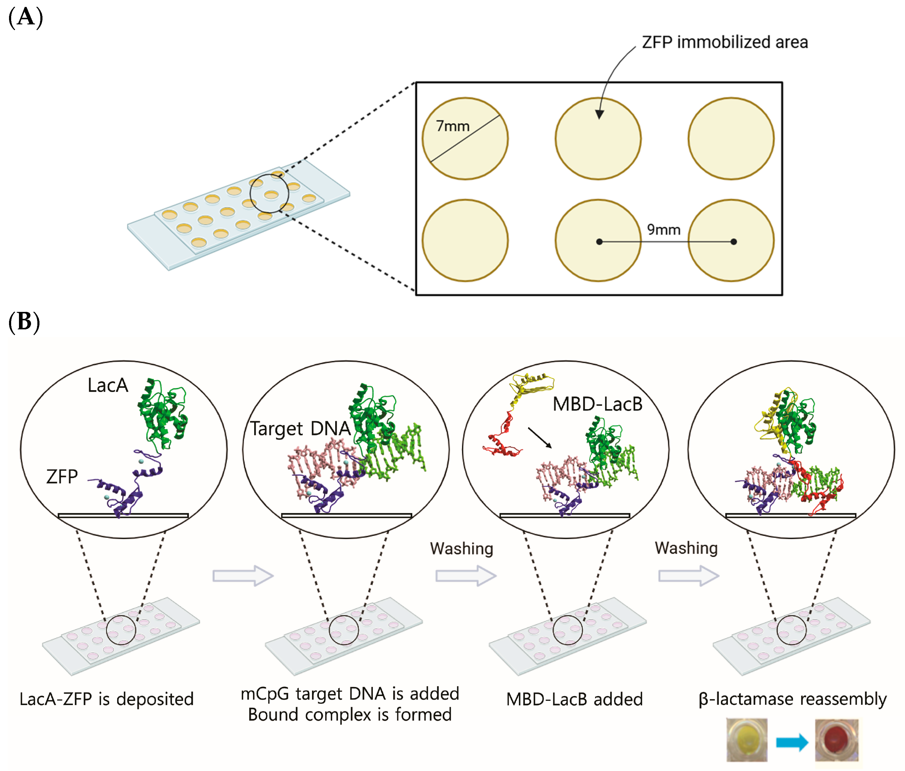
COC chips (TOPAS® 5013-S, Topas Advanced Polymers, Florence, KY, USA) were fabricated by injection molding as previously described in our previous publication [27]. Briefly, polished COC disks (1 mm thickness and 76.2 mm diameter) were produced and patterned with circular reaction sites (7 mm diameter, 9 mm center-to-center spacing) corresponding to the layout of a standard 96-well microplate (Figure 1A). Before use, the chips were incubated in phosphate-buffered saline (PBS) for 15–45 min, air-dried, and the masking tape was removed prior to surface functionalization.

2.4. ZFP Array and DNA Binding

DNA oligonucleotides were annealed as follows. Complementary pairs of forward and reverse oligonucleotides were prepared by heating at 95 °C for 10 min, slowly cooled down to 55 °C at a rate of 1 °C per 40 s, and then incubated for 15 min at 55 °C. Subsequently, the oligonucleotides were cooled to 4 °C at a rate of 1 °C per 40 s to form the double-stranded DNA. The sequences of oligonucleotides are provided in Figure S1 of the Supplementary Information.
ZFP immobilization and reaction area were confined by placing a silicone gasket (Grace Bio-Labs, Bend, OR, USA) with a diameter of 6 mm and a well depth of 1 mm onto the COC surface before arraying ZFPs. 5 μL of purified Lac A-ZFP RASSF1A at a concentration of 2.5 μM was immobilized on the COC surface and incubated for 40 min until dry. 10 μL of DNA at different concentrations was added to different wells and allowed to bind to the ZFP for 20 min. The gasket was removed before washing the slide with 50 mM KCl in ZBA, followed by 0.05% Tween-20 in ZBA. 10 μL of purified MBD-Lac B was added to the ZFP array and incubated for 20 min to allow for binding methylated DNA that was complexed with LacA-ZFP RASSF1A. The slide was washed with ZBA + 50 mM KCl and ZBA + 0.05% Tween-20, followed by air-drying. After placing the slide onto a 96-well plate and aligning the arrays with the wells, 20 μL of 1 mM nitrocefin (Calbiochem, San Diego, CA, USA) was added immediately to the ZFP array before monitoring the absorbance at 486 nm in a microplate reader (Molecular Devices, Sunnyvale, CA, USA). All experiments were repeated in triplicate, and the results are presented as the mean ± standard deviation (SD).

2.5. Electrophoretic Mobility Shift Assay (EMSA)

EMSA experiments were performed as previously described by our group [28]. Briefly, biotin-labeled double-stranded DNA targets were incubated with purified ZFPs in ZFP binding buffer for 1.5 h at room temperature. DNA–protein complexes were resolved on 10% native polyacrylamide gels, transferred to nylon membranes, UV crosslinked, and detected using the LightShift™ Chemiluminescent EMSA Kit (Pierce, Rockford, IL, USA). Images were acquired using an AlphaImager HP imaging system (ProteinSimple, San Jose, CA, USA).

3. Results and Discussion

3.1. ZFP Array on the COC Chip

Conventional methods for DNA methylation analysis, including methylation-specific PCR (MSP) and other bisulfite-based assays, offer high analytical sensitivity but require bisulfite conversion and PCR amplification [29,30]. These procedures increase assay complexity and may introduce DNA degradation, incomplete bisulfite conversion, amplification bias, and contamination-related errors, particularly when analyzing limited or fragmented DNA samples [29,30]. To address these limitations, we developed a PCR- and bisulfite-free sensing platform based on the combined use of ZFPs and MBDs integrated into a COC chip. Unlike MSP and bisulfite-based methods, the proposed platform directly recognizes native double-stranded DNA through simultaneous sequence-specific and methylation-specific binding without chemical conversion or enzymatic amplification. This dual-recognition strategy simplifies the analytical workflow, preserves native DNA integrity, and provides a promising foundation for rapid epigenetic analysis and future point-of-care cancer diagnostics.
As shown in the schematic representation (Figure 1B), our detection strategy is based on a bipartite recognition logic: the ZFP anchors the specific genetic sequence, while the MBD acts as an epigenetic sensor. In the presence of the methylated RASSF1A promoter, both domains are localized to the same DNA fragment, triggering the reassembly of LacA and LacB enzymatic fragments. For unmethylated DNA, the lack of MBD affinity leads to its dissociation during washing, thereby providing a clear distinction between methylated and unmethylated epigenetic profiles through a significant differential in signal intensity.
The mCpG-SEER-Lac platform operates through a multi-layered molecular detection strategy in which signal generation is contingent upon the simultaneous fulfillment of three independent recognition criteria. First, the ZFP module confers sequence specificity by selectively binding to the defined target sequence within the RASSF1A promoter region, thereby anchoring the biosensor complex to the correct DNA locus. Second, the MBD provides epigenetic specificity by recognizing and engaging mCpG dinucleotides at the target site; this architecture spatially pre-orients the MBD in proximity to the methylated CpG locus, thereby lowering the energy barrier for target engagement [31] and enhancing overall epigenetic recognition efficiency. Third, spatial and geometric specificity is imposed by the strict helical phasing requirement of the split-β-lactamase fragments (LacA and LacB), wherein correct rotational alignment—governed by the 36°/bp displacement of B-form DNA—is a prerequisite for enzymatic reconstitution and downstream signal output.
The analytical sensitivity and the ability to discriminate between methylated and unmethylated DNA can be influenced by binding kinetics and washing stringency. By immobilizing the ZFP onto the cyclic olefin copolymer (COC) chip via hydrophobic interactions, we ensured the structural accessibility of the protein domains for target capture. The drying step in ZFP immobilization follows the protocol used in our previous SEER-LAC-based ZFP array studies on PEG hydrogel slides and COC chips [21,27]. In both studies, immobilized ZFPs retained sequence-specific DNA-binding activity after drying, as demonstrated by dose-dependent detection signals comparable to those reported for engineered ZFPs. Because signal generation in the present system requires sequence-specific DNA binding by the immobilized ZFPs to reconstitute split β-lactamase activity, the strong and specific colorimetric responses observed here provide functional evidence that the surface-drying step did not significantly compromise probe bioactivity. The optimized incubation period facilitates robust protein–DNA recognition, while subsequent washing steps effectively eliminate non-specific, unbound DNA molecules. This selective enrichment of target DNA on the sensor surface creates a high-fidelity interface, providing a well-defined platform for the subsequent recruitment of the MBD to the methylated CpG sites.
Cyclic olefin copolymer (COC) has emerged as a material of choice for lab-on-a-chip applications, owing to a compelling combination of optical, chemical, and fabrication properties [32,33]. COC exhibits exceptional optical transparency across a broad spectrum, from the UV (~240 nm) through the visible range, coupled with inherently low autofluorescence [32,33,34]. Its high light transmittance (>90%) renders it functionally comparable to glass, yet it retains the processing flexibility of a thermoplastic, enabling cost-effective, high-throughput manufacturing via hot embossing or injection molding with feature resolution down to the micrometer scale [33,34]. From a chemical standpoint, COC demonstrates excellent resistance to a wide range of aqueous solutions, acids, and polar organic solvents [33]. Crucially for biological applications, COC is biocompatible and exhibits low non-specific protein adsorption, thereby preserving the reliability and sensitivity of on-chip biochemical assays [34,35]. Taken together, these attributes make COC an ideal substrate for constructing robust, disposable, and optically transparent microfluidic platforms intended for molecular diagnostics, nucleic acid analysis, and point-of-care applications [33]. In this study, the COC chip was fabricated to serve as the substrate surface on which LacA-ZFP was deposited within a confined area of a silicone gasket on the chip.
Both ZFP RASSF1A 150 and 407 are engineered to recognize specific sequences in the RASSF1A promoter region. With three GNN-targeting zinc finger recognition modules, a tripartite recognition mode is distributed across the 18 bp cognate DNA sequence (Table 1). The binding affinity (Kd) of ZFP RASSF1A 407 was determined to be 4.7 nM, whereas that of ZFP RASSF1A 150 was 253.1 nM based on the Electrophoretic Mobility Shift Assay (EMSA) (Supplementary Information). This can be attributed to all three GNN domains of ZFP RASSF1A 407 being strong binders within the Kd of 1~2 nM [23], exhibiting strong binding affinity for their respective target DNA sequences.

3.2. Detection of Methylated CpG and Target Sites

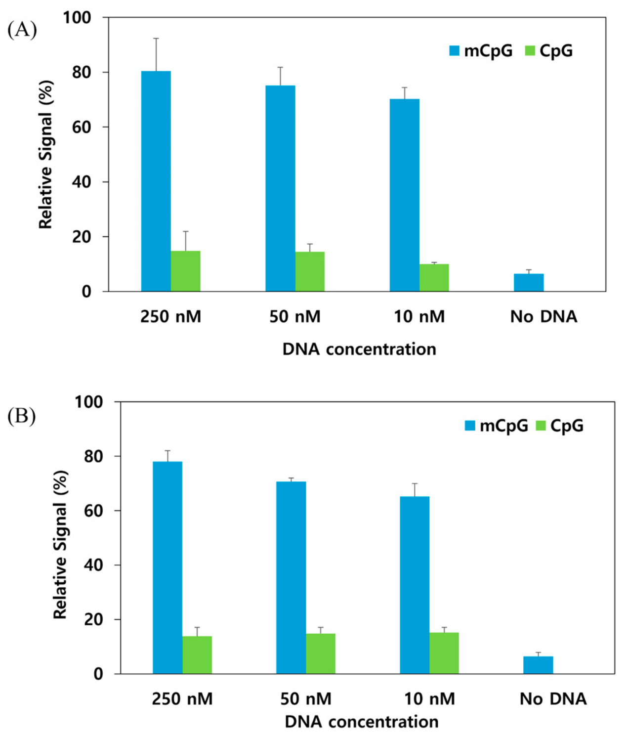
When LacA-ZFP RASSF1A 150 binds the 18 bp target site and MBD1-LacB recognizes the methylated site simultaneously, that triggers the reassembly of the enzymatic fragments of LacA and LacB, which generates the color response from yellow to red in the enzymatic assay upon hydrolysis of the colorimetric substrate. In contrast, the signal change was significantly lower when incubated with the unmethylated DNA, as shown in Figure 2. This result demonstrates that both MBD1 and MBD2 were able to selectively recognize the methylated site. The analytical performance of the ZFP-MBD biosensor demonstrated robust capacity for epigenetic discrimination, yielding a significantly elevated signal in the presence of methylated RASSF1A promoter DNA compared to its unmethylated counterpart. This high signal intensity is attributed to the synergistic dual-recognition mechanism; the immobilized ZFP capture probe ensures sequence-specific tethering of the target DNA to the COC chip surface, while the MBD-linked detection probe selectively targets the methylated CpG motifs. Conversely, the minimal signal observed with unmethylated DNA highlights the exquisite specificity of the MBD, which would remain in the solution phase in the absence of methyl groups, thereby preventing the enzymatic reconstruction of LacA and LacB. This contrast in signal output confirms that our platform can effectively ‘gate’ the detection process, ensuring that the presence of the target sequence alone is insufficient for signal generation without the requisite epigenetic modification. The substantial signal divergence between methylated and unmethylated templates further underscores the platform’s high analytical specificity for detecting epigenetic modifications.
As shown in Figure 2 and Figure 3, the data also show a dose-dependent signal for the methylated target. A linear regression of the tested concentrations yielded an R2 value of 0.98 (y = 7.3 log10 x + 62.8). A concentration-dependent signal was observed over the tested concentration range. Because this study was designed as a proof-of-concept using three target concentrations, the analytical working range has not yet been comprehensively established and will be investigated in future studies. Future studies will focus on improving analytical sensitivity through optimization of protein immobilization, microfluidic design, and signal transduction, to extend the platform toward clinically relevant diagnostic applications.
While our platform currently demonstrates a dynamic range of 10–250 nM, we recognize that tumor-derived cfDNA biomarkers in clinical urine samples are often present at substantially lower concentrations, creating a sensitivity gap for future clinical translation. However, this comparison warrants caution, as many amplification-free biosensors achieve femtomolar-to-attomolar sensitivity mainly under simplified buffer conditions rather than complex biological matrices [36,37], and PCR-based platforms such as CRISPR-Cas13a assays reach attomolar sensitivity only with an obligatory pre-amplification step, indicating that such sensitivity reflects amplification efficiency rather than intrinsic detection chemistry [38]. To narrow this gap while preserving the PCR-free nature of our assay, future work will explore amplification-free signal-enhancement strategies, such as nanostructured electrodes and enzyme-mediated catalytic amplification, to improve the limit of detection without compromising simplicity or turnaround time.
Figure 2. Detection of methylated CpG and target sites by (A) MBD1-ZFP RASSF1A 150 and (B) MBD2-ZFP RASSF1A 407 at different DNA concentrations of 250, 50, and 10 nM.
Chemosensors 14 00162 g002
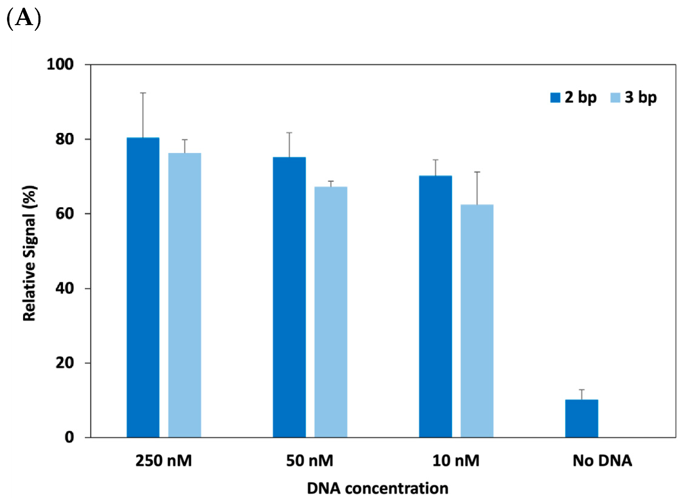
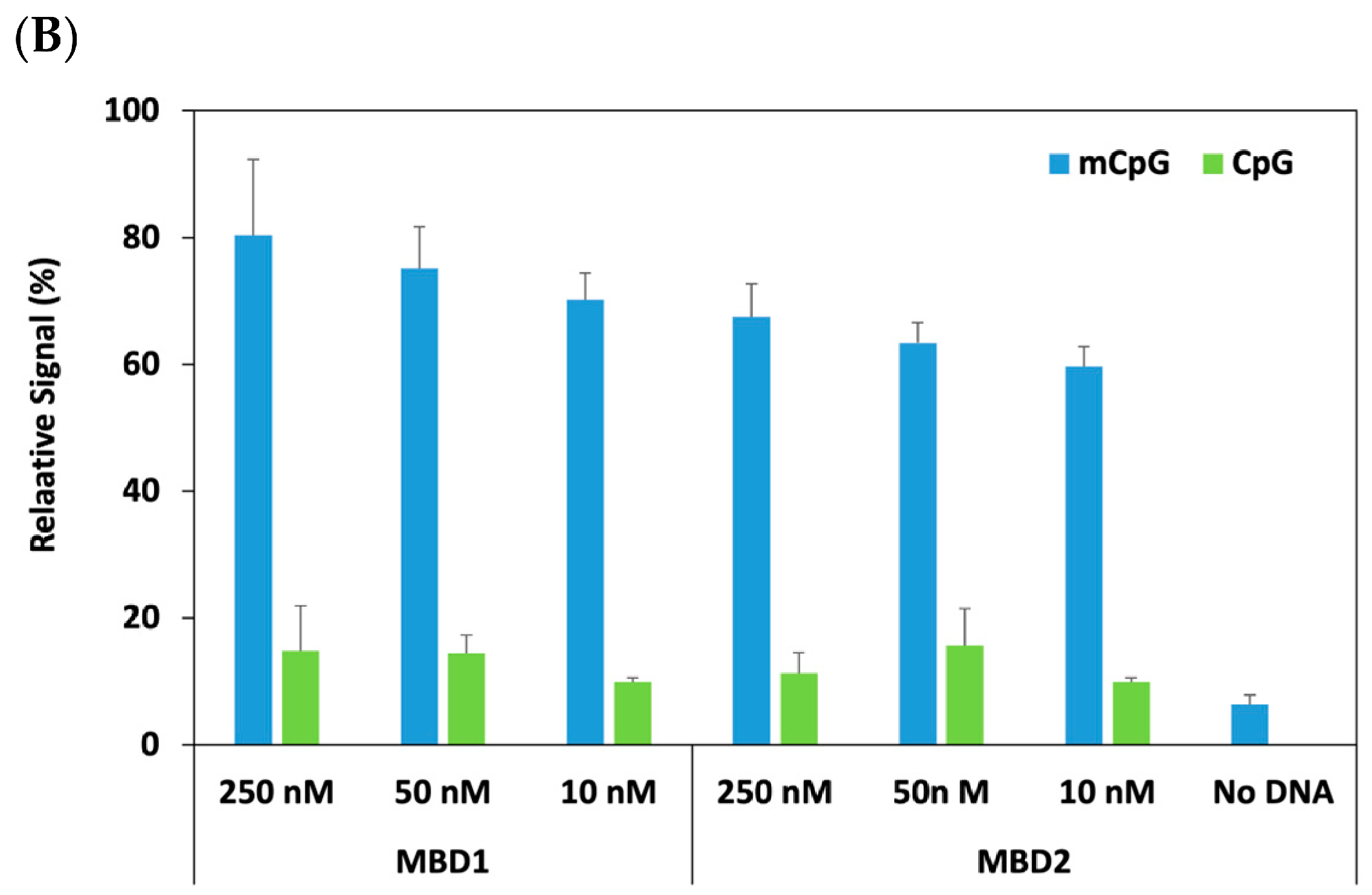
Figure 3. (A) Effect of inter-ligand spacing (2 bp vs. 3 bp) on the co-localization of MBD1 and ZFP RASSF1A on methylated DNA templates. (B) Comparative analysis of MBD1- and MBD2-mediated reassembly efficiency in the presence of ZFP RASSF1A 150.
Chemosensors 14 00162 g003aChemosensors 14 00162 g003b

3.3. Spatial Alignment of Dual-Recognition Modules on Target DNA

We investigated the effect of helical phasing by adjusting the base-pair interval between the primary recognition sites. Porter et al. evaluated the effect of distance and proximity of the mCpG-SEER system using a series of DNA targets with different spacing between the mCpG and Zif268 binding sites in the range of 0~13 bps [39]. They found that the 2 bp spacing between the mCpG and ZFP target site resulted in the maximum relative signal [39]. Building upon this finding, two oligonucleotides with different nucleotide intervals between the mCpG and the ZF target site were used: one was positioned 2 bp apart, and the other was 3 bp apart. The 3 bp spacing was included to provide slightly more spacing for better accommodation of a larger protein because our ZFP is a six-finger, which is bigger than the size of a three-finger Zif268. The experimental data in Figure 3A reveal that a 2 bp interval provides higher reassembly efficiency than a 3 bp spacing. This suggests that the 2 bp configuration positions the ZFP and MBD in a spatial orientation that optimizes the encounter frequency of the LacA and LacB fragments. Due to the helical nature of the DNA duplex, a single base-pair shift introduces a substantial rotational displacement (approximately 36°) [40]. This topological shift would likely move the ZFP target site out of the “optimal window” for proximity-driven interaction, creating a geometric mismatch that may hinder the spontaneous association of the split-enzyme domains.
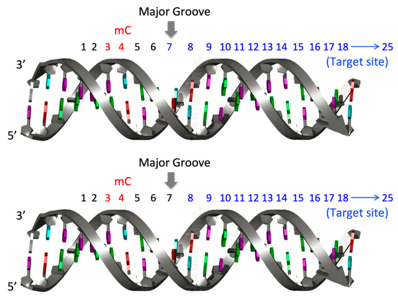
To facilitate the functional reassembly of the split-enzyme fragments of β-lactamase, the assay architecture was designed to accommodate simultaneous occupancy of the DNA major groove by both the MBD and ZFP. As shown in Figure 3A, the proximity-driven reconstitution of β-lactamase can be dependent on the inter-ligand spacing. Optimal enzymatic activity was observed at a 2 bp interval between the mCpG and the ZFP target sequence. Increasing this distance to 3 bp decreased the signal, suggesting that the increased spatial separation exceeds the effective reach required for LacA and LacB interaction. Furthermore, in the context of the 39 bp oligonucleotide, the reduced signal suggests that the target sites may adopt unfavorable orientations—potentially sequestered within the center of a DNA loop or at the distal ends of the major groove—thereby creating steric or distance-related barriers to enzymatic reconstitution.

3.4. Efficiency of MBD1 and MBD2

Selective recognition of methylated DNA by the MBD is mediated through conserved arginine and tyrosine residues, which establish base-specific contacts with the mCpG dinucleotide [31]. MBD1 and MBD2 both show binding to mammalian mCpG [41]. However, many other experiments also suggest that MBD1 and MBD2 show a higher preference for mCpG than other methyl-binding proteins [42]. This was the reason for selecting MBD1 and MBD2 proteins for our study. The efficiency of MBD1 and MBD2 proteins was compared using ZFP RASSF1A 150 in the mCpG-SEER-Lac system. In Figure 3B, MBD1 showed more effective binding results in the mCpG-SEER-Lac system than MBD2. However, MBD2 was still able to recognize the methylated site, suggesting that in our system, both MBD1 and MBD2 were capable of distinguishing methylated from non-methylated sites. This result is supported by the study of Badran et al. [42], where different MBDs, including MBD1 and MBD2, were compared to evaluate the global CpG methylation status. In their study, MBD1 was identified as the most selective domain to discriminate between the mCpG site and its unmethylated counterpart, with MBD2 being the second most sensitive one [42].

3.5. Influence of Target and mCpG Site Configuration on Signal Reassembly Efficiency

We postulate that positioning the ZFP target site on the antisense strand ensures that both the sequence-specific anchor and the methylated cytosine (mC) are co-localized within the DNA major groove. This spatial arrangement is a prerequisite for the functional reassembly of LacA and LacB fragments. Conversely, a shift in orientation would likely sequester one of the recognition motifs toward the minor groove, significantly increasing the inter-domain distance and precluding enzymatic reconstitution. Furthermore, the length of the oligonucleotide may alter the helical phasing, potentially rotating the mCpG site out of the major groove. This would physically obstruct MBD binding, as the protein’s conserved arginine residues must access the major groove to establish critical bidentate hydrogen bonds and hydrophobic interactions with the guanine base of the mCpG dinucleotide [43]. To ensure that the recognition elements were correctly oriented within the DNA major groove, we engineered a 39 bp oligonucleotide with a 12 bp nonspecific leader sequence (Figure 4). Given that the helical pitch of B-DNA is approximately 10.5 bp per turn, this leader sequence ensures that the subsequent mCpG and antisense ZFP target sites are positioned on the accessible face of the double helix. Our experimental results indicate that a 2 bp interval between these sites provides relatively better reassembly efficiency compared to a 3 bp spacing. This suggests that the 2 bp configuration minimizes the spatial separation between the LacA and LacB fragments, facilitating a more productive enzymatic reconstitution.

3.6. Specificity of the CpG-SEER-Lac System

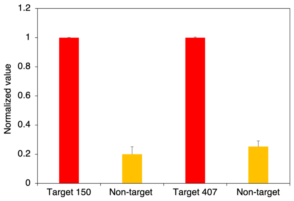
The analytical specificity of the ZFP RASSF1A 150 and 407 was evaluated within the MBD1-paired biosensor framework. This analysis confirmed the system’s capacity to discriminately recognize the target RASSF1A promoter sequence, ensuring that signal generation is strictly dependent on the high-affinity co-localization of both the sequence-specific ZFP and the methyl-specific MBD1 domain. As shown in Figure 5, incubation with non-target DNA resulted in a significantly attenuated signal compared to the methylated RASSF1A target. The inclusion of a methylated non-target DNA control demonstrates that methylation alone does not produce a positive signal. The negligible response observed for the methylated non-target sequence indicates that successful detection requires both sequence-specific recognition by the ZFP and methylation-specific recognition by the MBD, thereby supporting the proposed dual-recognition mechanism. This high degree of specificity validates the engineered ZFP-MBD framework as a reliable tool for distinguishing the target epigenetic biomarker from a complex genomic background.

4. Conclusions

In conclusion, the ZFP array with the mCpG SEER-Lac system on the COC chip provides an efficient sensing platform to detect a desired methylated DNA. Simultaneous recognition by both MBD and ZFP serves as an excellent detector system that binds to the specific methylated site on the target DNA [42]. Immobilization of the ZFP on the chip and the washing step after incubation for DNA-ZFP binding allow elimination of unbound DNA molecules and availability of the ZFP-DNA complex. This enhances the accessibility of the MBD to methylated CpG dinucleotides, facilitating high-affinity epigenetic recognition at the target locus. The ZFP array confers robust signal discrimination between methylated and unmethylated DNA. Such results underscore the potential of this assay as a PCR-free and bisulfite-independent diagnostic tool, capable of detecting epigenetic biomarkers. Thus, compared with the microfluidic PCR chip, the proposed platform offers direct, amplification-free recognition of methylated dsDNA without bisulfate conversion, thereby simplifying the analytical workflow while providing simultaneous sequence- and methylation-specific detection.
Signal generation requires the simultaneous satisfaction of three molecular recognition requirements: correct DNA sequence, CpG methylation, and appropriate DNA helical geometry for proximity-dependent β-lactamase reassembly. Consequently, the platform establishes a highly discriminative tool for epigenetic profiling of cancer-associated promoter hypermethylation. In the future, we will establish a panel with multiple genes, such as RASSF1A, APC, and p14ARF that can serve as a highly reliable set of biomarkers to detect bladder cancer. The insights garnered from the present methodology will inform a subsequent study aiming at validating the platform’s performance in complex, real-world biological matrices. Future efforts will focus on translating this biosensing architecture into a fully integrated microfluidic device, with particular emphasis on optimizing assay robustness, sample compatibility, and analytical throughput to meet the stringent requirements of clinical deployment.
Given the emerging approaches for defining the sequence preferences of DNA-binding proteins and the modular nature of the SEER architecture, the ZFP array with mCpG-SEER-Lac offers a versatile platform for directly assessing promoter-specific DNA methylation on the chip. Accordingly, this method may enable the development of new diagnostic reagents to advance studies of epigenetic regulation and to support early cancer detection.

Supplementary Materials

The following supporting information can be downloaded at: https://www.mdpi.com/article/10.3390/chemosensors14070162/s1, Figure S1: Partial sequence of the RASSF1A promoter region containing methylated CpG dinucleotides; Table S1: Target oligonucleotides used to test the dependence of mCpG-ZFP RASSF1 target site spacing on signal generation. The ZFP binding site and mCpG are highlighted in green and red, respectively; Table S2: Oligonucleotide sequences used in EMSA (electromobility shift assay) with ZFP binding sites highlighted in blue; Figure S2: Illustration of EMSA (electromobility shift assay) of engineered zinc finger proteins (ZFPs) to target different sequences within the RASSF1 gene. (A) RASSF1A 150, (B) RASSF1A 407. The ZFP concentrations (nM) are given at the top. The top bands indicate the ZFP and DNA-bound complex, and the bottom bands show free DNA.

Author Contributions

Conceptualization, M.-S.K.; methodology, M.-S.K., S.G. and C.H.A.; validation, H.Y.J. and S.G.; formal analysis, H.Y.J. and M.-S.K.; investigation, H.Y.J. and S.G.; data curation, M.-S.K., N.C. and M.T.H.; writing—original draft preparation, M.-S.K.; writing—review and editing, M.-S.K., N.C. and M.T.H.; supervision, M.-S.K. and C.H.A.; funding acquisition, M.-S.K. All authors have read and agreed to the published version of the manuscript.

Funding

This research was funded by the Bisa Research Grant of Keimyung University in No. 20250582.

Institutional Review Board Statement

Not applicable.

Informed Consent Statement

Not applicable.

Data Availability Statement

The original contributions presented in this study are included in the article and Supplementary Materials. Further inquiries can be directed to the corresponding authors.

Acknowledgments

This work was supported by the Bisa Research Grant of Keimyung University (Grant No. 20250582).

Conflicts of Interest

The authors declare no conflicts of interest.

References

  1. Varchulova Novakova, Z.; Kuniakova, M.; Ziaran, S.; Harsanyi, S. Molecular Biomarkers of Bladder Cancer: A Mini-Review. Physiol. Res. 2023, 72, S247–S256. [Google Scholar] [CrossRef] [PubMed]
  2. Miyake, M.; Nishimura, N.; Fujii, T.; Fujimoto, K. Recent advancements in the diagnosis and treatment of non-muscle invasive bladder cancer: Evidence update of surgical concept, risk stratification, and BCG-treated disease. Int. J. Urol. 2023, 30, 944–957. [Google Scholar] [CrossRef] [PubMed]
  3. Aydin, M.E.; Aykac, A.; Kaya, C.; Sungur, M. Comparative analysis of EAU, AUA, and NCCN guidelines for the management of non-muscle invasive bladder cancer. BMC Urol. 2026, 26, 38. [Google Scholar] [CrossRef] [PubMed]
  4. Tomiyama, E.; Fujita, K.; Hashimoto, M.; Uemura, H.; Nonomura, N. Urinary markers for bladder cancer diagnosis: A review of current status and future challenges. Int. J. Urol. 2024, 31, 208–219. [Google Scholar] [CrossRef] [PubMed]
  5. Babjuk, M.; Burger, M.; Capoun, O.; Cohen, D.; Comperat, E.M.; Dominguez Escrig, J.L.; Gontero, P.; Liedberg, F.; Masson-Lecomte, A.; Mostafid, A.H.; et al. European Association of Urology Guidelines on Non-muscle-invasive Bladder Cancer (Ta, T1, and Carcinoma in Situ). Eur. Urol. 2022, 81, 75–94. [Google Scholar] [CrossRef] [PubMed]
  6. Mossanen, M.; Wang, Y.; Szymaniak, J.; Tan, W.S.; Huynh, M.J.; Preston, M.A.; Trinh, Q.D.; Sonpavde, G.; Kibel, A.S.; Chang, S.L. Evaluating the cost of surveillance for non-muscle-invasive bladder cancer: An analysis based on risk categories. World J. Urol. 2019, 37, 2059–2065. [Google Scholar] [CrossRef] [PubMed]
  7. Yang, Z.; Song, F.; Zhong, J. Urinary Biomarkers in Bladder Cancer: FDA-Approved Tests and Emerging Tools for Diagnosis and Surveillance. Cancers 2025, 17, 3425. [Google Scholar] [CrossRef] [PubMed]
  8. Kanai, Y. Molecular pathological approach to cancer epigenomics and its clinical application. Pathol. Int. 2024, 74, 167–186. [Google Scholar] [CrossRef] [PubMed]
  9. Bhootra, S.; Jill, N.; Shanmugam, G.; Rakshit, S.; Sarkar, K. DNA methylation and cancer: Transcriptional regulation, prognostic, and therapeutic perspective. Med. Oncol. 2023, 40, 71. [Google Scholar] [CrossRef] [PubMed]
  10. Raos, D.; Ulamec, M.; Katusic Bojanac, A.; Bulic-Jakus, F.; Jezek, D.; Sincic, N. Epigenetically inactivated RASSF1A as a tumor biomarker. Bosn. J. Basic Med. Sci. 2021, 21, 386–397. [Google Scholar] [CrossRef] [PubMed]
  11. Pietrusinski, M.; Kepczynski, Ƚ.; Jedrzejczyk, A.; Borkowska, E.; Traczyk-Borszynska, M.; Constantinou, M.; Kaƚuzewski, B.; Borowiec, M. Detection of bladder cancer in urine sediments by a hypermethylation panel of selected tumor suppressor genes. Cancer Biomark. 2017, 18, 47–59. [Google Scholar] [CrossRef] [PubMed]
  12. Jeddi, F.; Faghfuri, E.; Mehranfar, S.; Soozangar, N. The common bisulfite-conversion-based techniques to analyze DNA methylation in human cancers. Cancer Cell Int. 2024, 24, 240. [Google Scholar] [CrossRef] [PubMed]
  13. Massen, M.; Lommen, K.; Wouters, K.A.D.; Vandersmissen, J.; van Criekinge, W.; Herman, J.G.; Melotte, V.; Schouten, L.J.; van Engeland, M.; Smits, K.M. Technical considerations in PCR-based assay design for diagnostic DNA methylation cancer biomarkers. Clin. Epigenet. 2022, 14, 56. [Google Scholar] [CrossRef] [PubMed]
  14. Olkhov-Mitsel, E.; Bapat, B. Strategies for discovery and validation of methylated and hydroxymethylated DNA biomarkers. Cancer Med. 2012, 1, 237–260. [Google Scholar] [CrossRef] [PubMed]
  15. Gong, T.; Borgard, H.; Zhang, Z.; Chen, S.; Gao, Z.; Deng, Y. Analysis and Performance Assessment of the Whole Genome Bisulfite Sequencing Data Workflow: Currently Available Tools and a Practical Guide to Advance DNA Methylation Studies. Small Methods 2022, 6, e2101251. [Google Scholar] [CrossRef] [PubMed]
  16. Behyar, M.B.; Nilghaz, A.; Hasanzadeh, M.; Shadjou, N. Recent progresses and challenges on mesoporous silica nanoparticles for DNA-based biosensors and diagnostics. TrAC Trends Anal. Chem. 2024, 178, 117846. [Google Scholar] [CrossRef]
  17. Malecka, K.; Stachyra, A.; Góra-Sochacka, A.; Sirko, A.; Zagórski-Ostoja, W.; Radecka, H.; Radecki, J. Electrochemical genosensor based on disc and screen printed gold electrodes for detection of specific DNA and RNA sequences derived from Avian Influenza Virus H5N1. Sens. Actuators B Chem. 2016, 224, 290–297. [Google Scholar] [CrossRef]
  18. Kellett, A.; Molphy, Z.; Slator, C.; McKee, V.; Farrell, N.P. Molecular methods for assessment of non-covalent metallodrug-DNA interactions. Chem. Soc. Rev. 2019, 48, 971–988. [Google Scholar] [CrossRef] [PubMed]
  19. Li, Y.; Wang, W.; Gong, H.; Xu, J.; Yu, Z.; Wei, Q.; Tang, D. Graphene-coated copper-doped ZnO quantum dots for sensitive photoelectrochemical bioanalysis of thrombin triggered by DNA nanoflowers. J. Mater. Chem. B 2021, 9, 6818–6824. [Google Scholar] [CrossRef] [PubMed]
  20. Saadaoui, M.; Fernandez, I.; Luna, G.; Diez, P.; Campuzano, S.; Raouafi, N.; Sanchez, A.; Pingarron, J.M.; Villalonga, R. Label-free electrochemical genosensor based on mesoporous silica thin film. Anal. Bioanal. Chem. 2016, 408, 7321–7327. [Google Scholar] [CrossRef] [PubMed]
  21. Kim, M.S.; Stybayeva, G.; Lee, J.Y.; Revzin, A.; Segal, D.J. A zinc finger protein array for the visual detection of specific DNA sequences for diagnostic applications. Nucleic Acids Res. 2011, 39, e29. [Google Scholar] [CrossRef] [PubMed]
  22. Kim, M.S.; Kini, A.G. Engineering and Application of Zinc Finger Proteins and TALEs for Biomedical Research. Mol. Cells 2017, 40, 533–541. [Google Scholar] [CrossRef] [PubMed]
  23. Sander, J.D.; Zaback, P.; Joung, J.K.; Voytas, D.F.; Dobbs, D. An affinity-based scoring scheme for predicting DNA-binding activities of modularly assembled zinc-finger proteins. Nucleic Acids Res. 2009, 37, 506–515. [Google Scholar] [CrossRef] [PubMed]
  24. Liu, Q.; Segal, D.J.; Ghiara, J.B.; Barbas, C.F., 3rd. Design of polydactyl zinc-finger proteins for unique addressing within complex genomes. Proc. Natl. Acad. Sci. USA 1997, 94, 5525–5530. [Google Scholar] [CrossRef] [PubMed]
  25. Gonzalez, B.; Schwimmer, L.J.; Fuller, R.P.; Ye, Y.; Asawapornmongkol, L.; Barbas, C.F., 3rd. Modular system for the construction of zinc-finger libraries and proteins. Nat. Protoc. 2010, 5, 791–810. [Google Scholar] [CrossRef] [PubMed]
  26. Bhakta, M.S.; Segal, D.J. The generation of zinc finger proteins by modular assembly. Methods Mol. Biol. 2010, 649, 3–30. [Google Scholar] [CrossRef] [PubMed]
  27. Ha, D.T.; Ghosh, S.; Ahn, C.H.; Segal, D.J.; Kim, M.S. Pathogen-specific DNA sensing with engineered zinc finger proteins immobilized on a polymer chip. Analyst 2018, 143, 4009–4016. [Google Scholar] [CrossRef] [PubMed]
  28. Ha, D.T.; Nguyen, V.T.; Kim, M.S. Graphene Oxide-Based Simple and Rapid Detection of Antibiotic Resistance Gene via Quantum Dot-Labeled Zinc Finger Proteins. Anal. Chem. 2021, 93, 8459–8466. [Google Scholar] [CrossRef] [PubMed]
  29. Yousefi, P.D.; Suderman, M.; Langdon, R.; Whitehurst, O.; Davey Smith, G.; Relton, C.L. DNA methylation-based predictors of health: Applications and statistical considerations. Nat. Rev. Genet. 2022, 23, 369–383. [Google Scholar] [CrossRef] [PubMed]
  30. Adampourezare, M.; Hasanzadeh, M.; Seidi, F. Optical bio-sensing of DNA methylation analysis: An overview of recent progress and future prospects. RSC Adv. 2022, 12, 25786–25806. [Google Scholar] [CrossRef] [PubMed]
  31. Scarsdale, J.N.; Webb, H.D.; Ginder, G.D.; Williams, D.C., Jr. Solution structure and dynamic analysis of chicken MBD2 methyl binding domain bound to a target-methylated DNA sequence. Nucleic Acids Res. 2011, 39, 6741–6752. [Google Scholar] [CrossRef] [PubMed]
  32. Liu, J.; Chen, C.F.; Tsao, C.W.; Chang, C.C.; Chu, C.C.; DeVoe, D.L. Polymer microchips integrating solid-phase extraction and high-performance liquid chromatography using reversed-phase polymethacrylate monoliths. Anal. Chem. 2009, 81, 2545–2554. [Google Scholar] [CrossRef] [PubMed]
  33. Nge, P.N.; Rogers, C.I.; Woolley, A.T. Advances in microfluidic materials, functions, integration, and applications. Chem. Rev. 2013, 113, 2550–2583. [Google Scholar] [CrossRef] [PubMed]
  34. Liedert, R.; Amundsen, L.K.; Hokkanen, A.; Maki, M.; Aittakorpi, A.; Pakanen, M.; Scherer, J.R.; Mathies, R.A.; Kurkinen, M.; Uusitalo, S.; et al. Disposable roll-to-roll hot embossed electrophoresis chip for detection of antibiotic resistance gene mecA in bacteria. Lab Chip 2012, 12, 333–339. [Google Scholar] [CrossRef] [PubMed]
  35. Laib, S.; MacCraith, B.D. Immobilization of biomolecules on cycloolefin polymer supports. Anal. Chem. 2007, 79, 6264–6270. [Google Scholar] [CrossRef] [PubMed]
  36. Williamson, P.; Piskunen, P.; Ijas, H.; Butterworth, A.; Linko, V.; Corrigan, D.K. Signal Amplification in Electrochemical DNA Biosensors Using Target-Capturing DNA Origami Tiles. ACS Sens. 2023, 8, 1471–1480. [Google Scholar] [CrossRef] [PubMed]
  37. Wang, C.; Wang, W.; Xu, Y.; Zhao, X.; Li, S.; Qian, Q.; Mi, X. Tetrahedral DNA Framework-Programmed Electrochemical Biosenors with Gold Nanoparticles for Ultrasensitive Cell-Free DNA Detection. Nanomaterials 2022, 12, 666. [Google Scholar] [CrossRef] [PubMed]
  38. Gootenberg, J.S.; Abudayyeh, O.O.; Lee, J.W.; Essletzbichler, P.; Dy, A.J.; Joung, J.; Verdine, V.; Donghia, N.; Daringer, N.M.; Freije, C.A.; et al. Nucleic acid detection with CRISPR-Cas13a/C2c2. Science 2017, 356, 438–442. [Google Scholar] [CrossRef] [PubMed]
  39. Porter, J.R.; Stains, C.I.; Segal, D.J.; Ghosh, I. Split beta-lactamase sensor for the sequence-specific detection of DNA methylation. Anal. Chem. 2007, 79, 6702–6708. [Google Scholar] [CrossRef] [PubMed]
  40. Arnott, S.; Hukins, D.W.L. Optimised parameters for A-DNA and B-DNA. Biochem. Biophys. Res. Commun. 1972, 47, 1504–1509. [Google Scholar] [CrossRef] [PubMed]
  41. Hendrich, B.; Bird, A. Identification and characterization of a family of mammalian methyl-CpG binding proteins. Mol. Cell. Biol. 1998, 18, 6538–6547. [Google Scholar] [CrossRef] [PubMed]
  42. Badran, A.H.; Furman, J.L.; Ma, A.S.; Comi, T.J.; Porter, J.R.; Ghosh, I. Evaluating the global CpG methylation status of native DNA utilizing a bipartite split-luciferase sensor. Anal. Chem. 2011, 83, 7151–7157. [Google Scholar] [CrossRef] [PubMed]
  43. Zou, X.; Ma, W.; Solov’yov, I.A.; Chipot, C.; Schulten, K. Recognition of methylated DNA through methyl-CpG binding domain proteins. Nucleic Acids Res. 2012, 40, 2747–2758. [Google Scholar] [CrossRef] [PubMed]
Figure 1. (A) The image of the COC spotted array chip. (B) Schematic representation of the ZFP-MBD sensing array on a COC chip. The capture probe (LacA-ZFP) is immobilized on the COC surface to recognize a specific target site on the double-stranded DNA. The detection probe (MBD-LacB) selectively binds to methylated CpG sites adjacent to the ZFP target sequence. This dual-binding event brings the split-enzyme fragments, LacA and LacB, into proximity, facilitating the functional reassembly of β-lactamase. The restored β-lactamase activity is subsequently quantified via colorimetric substrate hydrolysis, resulting in a color change from yellow to red.
Chemosensors 14 00162 g001
Figure 4. dsDNA oligonucleotides highlighting an 18 bp of the ZFP target site (blue) and a methylated CpG dinucleotide (red) with two (top) and three (bottom) bp spacing between the methylated and target sites, respectively.
Chemosensors 14 00162 g004
Figure 5. The specificity of ZFPs RASSF1A 150 and 407 within the MBD1-paired biosensor framework. The target sequence of ZFP150 was used as a non-target DNA for ZFP407 and vice versa.
Chemosensors 14 00162 g005
Table 1. Sequences of zinc finger recognition modules and their corresponding 3 bp DNA subsites, and the Kd values of zinc finger proteins.
ZFPFinger 6Finger 5Finger 4Finger 3Finger 2Finger 1Kd (nM)
Target site
RASSF1A 150
GAG
RSDNLVR
GGA
DPGHLVR
AGG
RSDHLTN
AAG
RKDNLKN
GGC
DPGHLVR
AAG
RKDNLKN
253.1
Target site
RASSF1A 407
GAG
RSDNLVR
CTG
RNDALTE
CGG
RSDKLTE
GAG
RSDNLVR
CTG
RNDALTE
GCA
QSGDLRR
4.7
Disclaimer/Publisher’s Note: The statements, opinions and data contained in all publications are solely those of the individual author(s) and contributor(s) and not of MDPI and/or the editor(s). MDPI and/or the editor(s) disclaim responsibility for any injury to people or property resulting from any ideas, methods, instructions or products referred to in the content.