1. Introduction
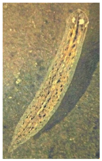
Planaria are flatworms belonging to the phylum
Platyhelminthes, class
Rhabditophora, and order
Tricladida [
1]. These invertebrates are bilaterally symmetrical, triploblastic, and acoelomate, and are commonly found in freshwater habitats such as ponds, lakes, and rivers, although some species inhabit saltwater or terrestrial environments [
2,
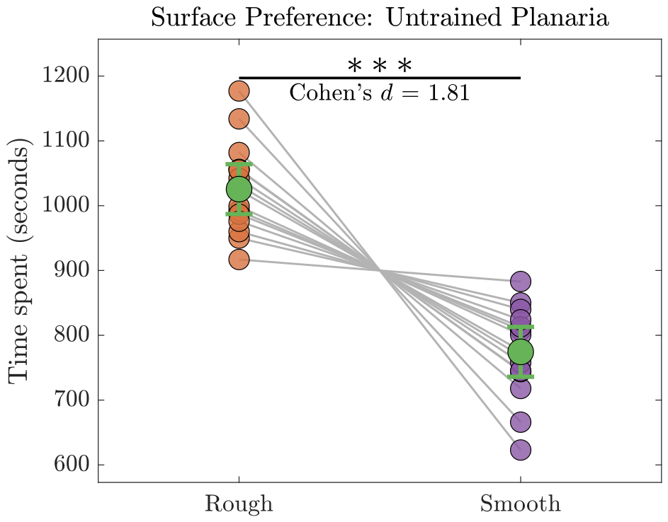
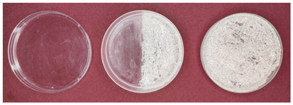
3]. Planaria exhibit distinct behavioral preferences, including sensitivity to light, electric and magnetic fields, chemical gradients, and surface textures, typically favoring rough over smooth surfaces [
4,
5,
6,
7]. These inherent behavioral tendencies make planaria an ideal system for investigating how environmental cues shape behavior and for exploring the neural mechanisms underlying learning and memory.
A defining feature of planaria is their extraordinary regenerative capacity. They reproduce asexually via binary fission, and any fragment of a planarian can regenerate into a complete, fully functional organism [
2,
8,
9,
10,
11,
12,
13,
14]. This remarkable ability is primarily attributed to a high concentration of pluripotent stem cells, called neoblasts, which constitute roughly 25% of the organism [
11,
13,
15]. In addition to their regenerative properties, planaria possess a primitive “brain” located in the anterior region, within the cephalic ganglion, which contains specialized sensory structures and nerve cells [
1,
7,
16]. Despite its simplicity, the planarian nervous system exhibits many structural and neurochemical features similar to vertebrates, including the presence of nearly all mammalian neurotransmitters, such as dopamine, and comparable receptor systems [
17,
18,
19]. These similarities allow researchers to investigate fundamental neural processes in a simple organism while maintaining relevance to more complex species.
Planaria are particularly well suited for the present study because of their remarkable ability to regenerate missing body structures, including the head and nervous system, after amputation. This feature makes them an especially appropriate model for investigating whether reward-associated memory can persist in body fragments following loss of the cephalic ganglion. In addition, their responses to environmental and chemical stimuli can be observed directly, supporting their use in behavioral studies of drug-induced effects, toxicology, and neuropharmacology [
17]. Thus, in the context of the present work, the choice of planaria is motivated primarily by their biological suitability for examining memory retention across regeneration.
Planaria have been widely employed in studies of drug-induced behavioral changes and addiction-like phenomena [
20,
21]. Exposure to psychoactive substances—including cocaine, nicotine, opioids, amphetamines, cannabinoids, and sucrose—can elicit conditioned behaviors that parallel addiction-related responses observed in vertebrates, including withdrawal symptoms [
21,
22,
23]. In humans, genetic and epigenetic factors substantially contribute to addiction vulnerability, with approximately 40–60% of risk estimated to be heritable [
24]. For instance, children of alcohol abusers are three- to five-fold more likely to develop addictive behaviors than children of non-alcoholic parents [
24]. These findings highlight the relevance of conserved biological mechanisms across species. Because planaria possess neurotransmitter systems and neural circuits analogous to those in mammals, they provide a valuable model for investigating the fundamental processes underlying addiction-like behavior and reward [
19,
25].
Sucrose, a naturally rewarding substance, has been shown to induce addiction-like behavior in planaria, with neurochemical responses resembling those elicited by drugs of abuse [
23]. Repeated exposure to a sucrose solution can establish a conditioned place preference (CPP), in which planaria associate a previously non-preferred surface with a rewarding stimulus [
6]. This Pavlovian conditioning paradigm provides a window into associative learning, demonstrating how environmental cues become linked with rewarding experiences. Planaria are also capable of acquiring long-term memory, which can persist even after amputation and subsequent brain regeneration [
4,
26,
27]. Some studies have proposed that memory can be transferred through cannibalism or RNA-mediated mechanisms, although these claims remain debated [
28,
29,
30]. Collectively, these observations raise an important question: is reward-associated memory confined to the cephalic ganglion, or can it also be stored and expressed in posterior, brainless segments following amputation?
Primary Aim: The main objective of this study was to investigate whether sucrose-induced reward-associated memory is retained in both anterior (head-containing) and posterior (brainless) segments of planaria following amputation. If addiction-like behavior involves non-cephalic or distributed memory mechanisms, the posterior fragment may retain the learned preference even in the absence of the cephalic ganglion. We hypothesized that the brainless posterior fragment would preserve reward-associated memory and exhibit a CPP comparable to that of the anterior segment.
Secondary Aim: We also investigated whether disrupting dopaminergic signaling with a D1 dopamine receptor antagonist could prevent the acquisition of a sucrose-induced CPP [
19,
25]. Importantly, we tested whether D1 antagonism is sufficient to block reward-associated memory formation in both anterior and posterior segments, representing a novel aspect of this study. We hypothesized that planaria exposed to a D1 antagonist would fail to develop a CPP, indicating that dopamine-dependent reinforcement is necessary for the formation of sucrose-associated memory across both fragments.
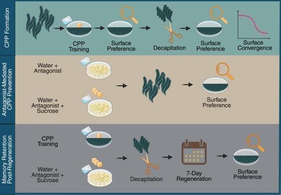
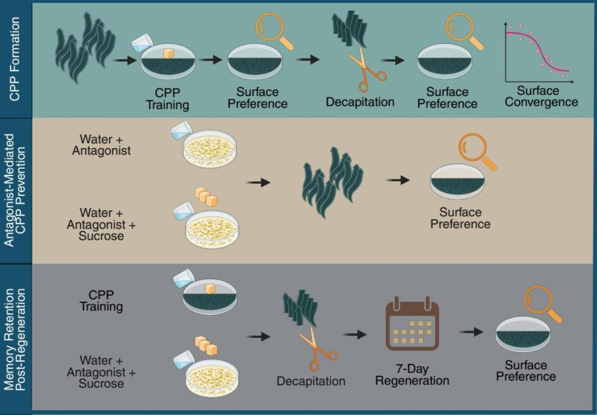
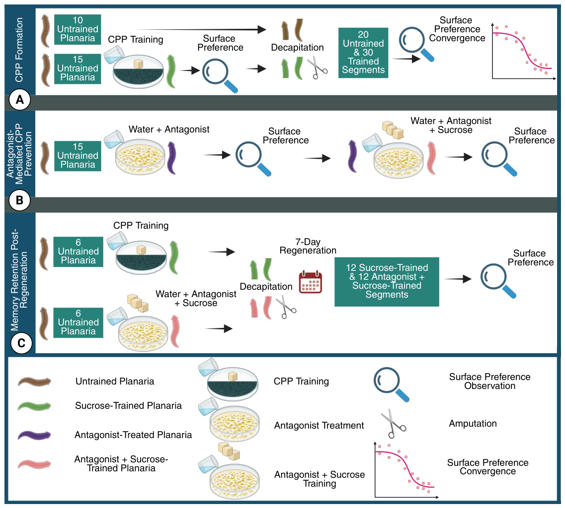
Approach: To address these objectives, we first verified the innate surface preferences of intact and fragmented planaria. Next, we trained planaria in a CPP paradigm using a sucrose solution paired with a previously non-preferred surface. Following amputation, we assessed whether posterior fragments retained the conditioned preference. Finally, we evaluated the effect of D1 antagonism during CPP training in both intact and amputated fragments. This experimental design allows for an examination of the segmental requirements for the retention of addiction-like behavior and the identification of dopaminergic signaling in reward-associated learning.
Taken together, these studies demonstrate that planaria provide a powerful model for examining distributed memory storage, addiction-like behavior, and dopaminergic modulation. By investigating how memory and addiction-like behavior persist outside of the cephalic ganglion, this work aims to provide novel insights into the molecular and cellular mechanisms underlying associative learning and reward across species, highlighting the broader significance of simple invertebrate models in understanding complex neurobiological processes.
4. Discussions
4.1. Methodological Considerations
We acknowledge that the cohort sizes used in the present study are modest. However, such sample sizes are not unusual in the planarian behavioral literature, where experiments frequently rely on either small groups of worms observed collectively or limited numbers of individually tracked animals. For example, Paskin et al. used 10 groups of 6 worms per condition to study wavelength-dependent phototactic behavior [
39]. Talbot and Schötz reported locomotor analyses using
n = 5 worms for intra-worm variability and
for broader characterization of wild-type locomotion [
40]. Similarly, Pagán et al. quantified seizure-like responses using group sizes ranging from approximately 4–14 worms depending on the assay [
41]. More broadly, the planarian model has been widely adopted because it is experimentally tractable, low cost, and easy to maintain [
42].
Accordingly, the sample sizes used here are consistent with prior work in this model system rather than unusually small by planarian research standards [
39,
40,
41,
42]. Although the cohort size was modest, the interpretability of the present data is strengthened by the observed effect sizes. Statistical power is influenced not only by sample size but also by the magnitude of the underlying effect, such that moderate-to-large effects support the sensitivity of the design to detect biologically meaningful differences. In this context, effect sizes in the medium-to-large range suggest that the principal findings are unlikely to reflect sampling variability alone. Thus, while small or nonsignificant effects should be interpreted cautiously, the observed effect sizes support the view that the study was sufficiently sensitive to detect the main effects of interest.
4.2. Acquisition of Reward-Associated Place Preference
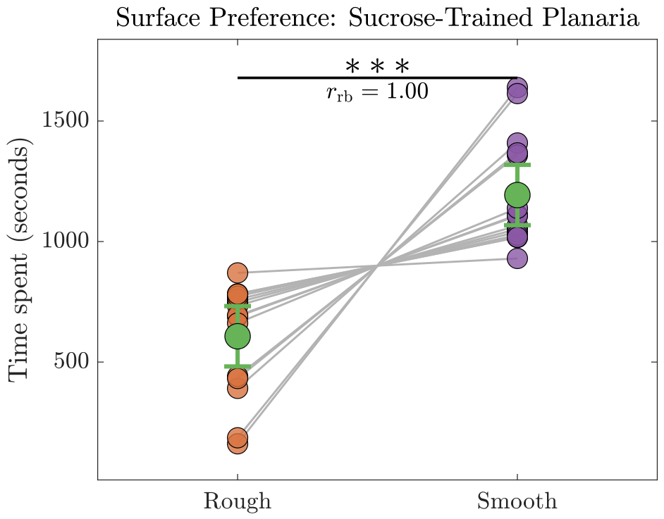
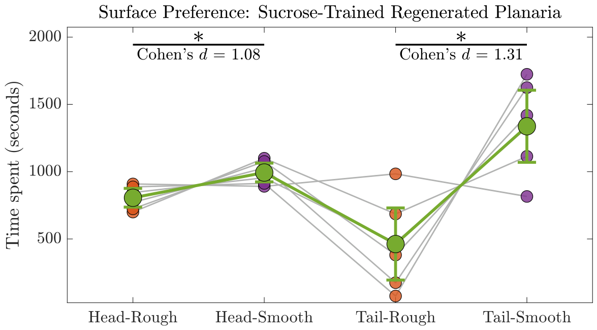
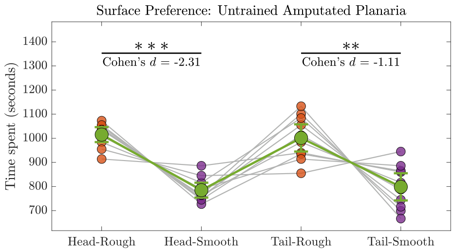
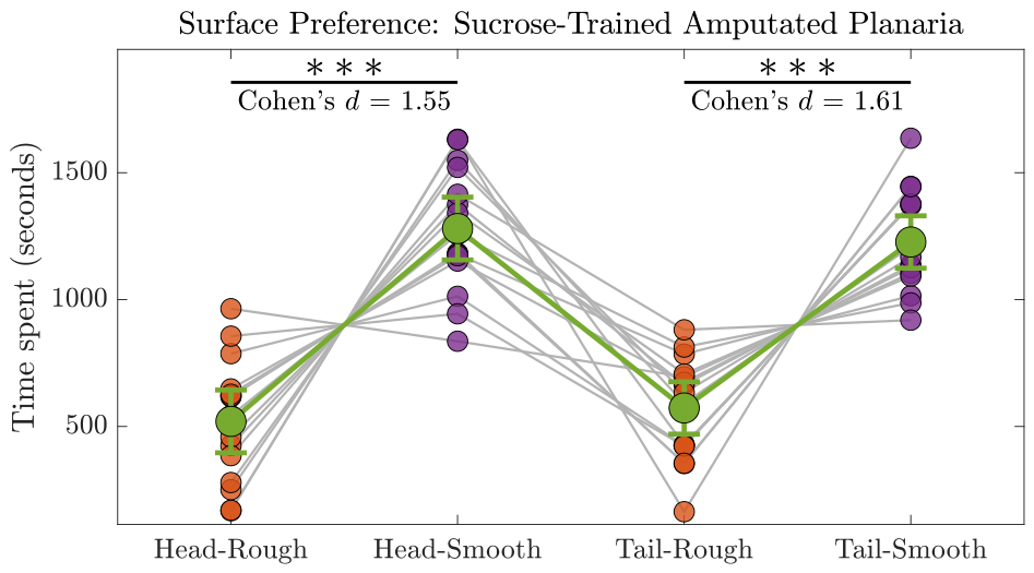
The present study demonstrates that planaria can acquire a robust conditioned place preference when sucrose is repeatedly paired with an initially non-preferred surface. Untrained animals displayed a natural bias toward the rough surface, whereas trained animals reversed this preference after repeated association of sucrose with the smooth surface. This shift indicates that the smooth surface acquired positive motivational value through associative learning and further supports the utility of planaria as a tractable model for studying reward-related behavior.
This finding is in line with earlier work showing that sucrose can function as a rewarding stimulus in planaria and can induce addiction-like behavioral responses [
6,
23]. In the present study, however, the significance of the intact-animal CPP extends beyond simply reproducing prior observations. Establishing a clear and reproducible conditioned preference in intact animals was essential because the central aim of the study was to determine whether a previously acquired reward-associated behavioral state could remain detectable after amputation and regeneration. The intact CPP results therefore serve as the necessary conceptual and experimental foundation for interpreting all subsequent findings.
More broadly, the successful induction of CPP in this paradigm suggests that reward learning in planaria can be sufficiently stable to serve as a meaningful probe of memory persistence. This point is important because not every behavioral change necessarily reflects a consolidated learned state. A transient motivational bias would provide only limited insight into regeneration-related retention. By contrast, the sustained reversal of the innate surface preference observed here indicates that the conditioned state was strong enough to justify examining whether it could later survive tissue loss and re-emerge in regenerated animals.
4.3. Reward Exposure as a Determinant of CPP Formation
The present findings further indicate that the establishment of conditioned place preference depends not only on the availability of reward, but also on the manner in which that reward is delivered. Although sucrose clearly acts as a reinforcing stimulus, prolonged exposure did not reliably generate conditioned preference in the initial training paradigm, whereas shorter and more discrete daily exposures produced a stable CPP. This suggests that reward-driven learning in planaria is shaped not simply by reward magnitude, but by the temporal structure of reward presentation.
One possible interpretation is that prolonged exposure diminishes the salience of the conditioned context or introduces competing physiological effects that weaken the association between the environment and the rewarding stimulus. By contrast, shorter exposures may preserve the distinctiveness of the learning episode and thereby facilitate stronger contextual encoding. In this sense, the results suggest that effective conditioning in planaria depends on a balance between reward intensity and reward timing, a principle that resonates broadly with reinforcement-based learning across model systems.
This issue is especially relevant to the logic of the present study. Because the central question concerned whether a conditioned reward preference could persist through regeneration, it was necessary first to define training conditions capable of producing a robust and reproducible behavioral state. Optimizing the reward exposure paradigm therefore did more than improve conditioning efficiency; it created the essential experimental framework for testing whether a consolidated reward-associated preference could later remain detectable after major anatomical disruption.
4.4. Post-Regeneration Persistence of Reward-Associated Memory
The most consequential finding of the present study is that the reward-associated preference established before amputation remained evident after regeneration. This observation provides the clearest evidence that a conditioned behavioral state can survive not only tissue loss due to amputation, but also the extensive anatomical and functional reorganization that accompanies regeneration. The conditioned preference remained evident across both stages: it was detectable as an early behavioral bias immediately after amputation and was subsequently re-expressed after regeneration in both anterior- and posterior-derived animals.
This post-regeneration persistence is central to the broader interpretation of the study because it directly addresses a long-standing question in planarian memory research: whether previously acquired behavioral information can remain accessible after the organism has undergone substantial physical reconstruction. The present findings indicate that sucrose conditioning leaves behind a durable trace that continues to influence behavior after regeneration is complete. From a behavioral standpoint, this suggests that the information underlying reward-associated learning is sufficiently stable to outlast the loss of major body structures and to shape the phenotype of regenerated animals.
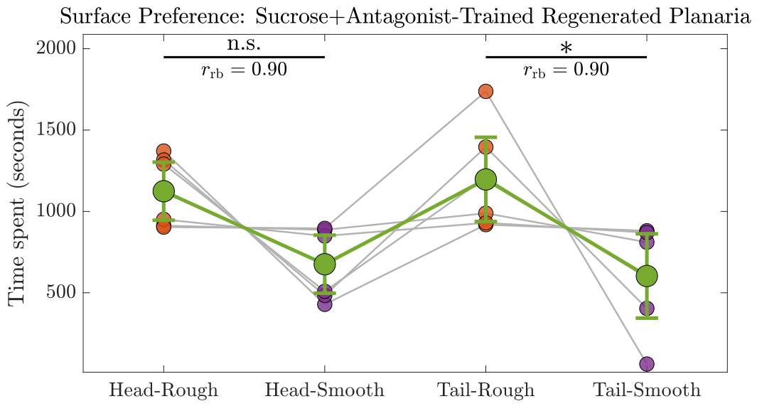
Particularly striking is the persistence of the conditioned preference in tail-derived regenerates. Because posterior fragments initially lack the original cephalic ganglion, their later expression of the learned sucrose-associated preference argues strongly against the view that reward memory is stored exclusively in the head. Although the present data do not identify the precise biological substrate of this persistence, they provide compelling evidence that the ability to re-express the conditioned behavior is not solely dependent on continuous preservation of the original brain. This is the central conceptual advance of the study and the reason the post-regeneration findings carry the greatest interpretive weight.
These results extend the broader literature on memory retention in planaria by showing that reward-associated learning, and not only simple environmental conditioning, can remain behaviorally accessible after regeneration [
4,
27,
43]. This is especially important because sucrose-induced CPP is widely regarded as a proxy for addiction-like reward behavior in this model. The present findings therefore suggest that motivationally relevant learned states may persist across profound biological disruption, highlighting planaria as a particularly informative system for studying how reward memories are preserved, transformed, or re-expressed during regeneration.
4.5. Implications for Distributed Memory Storage
The persistence of conditioned preference in regenerated animals has important implications for how memory may be organized in planaria. If animals regenerated from posterior fragments can later express a reward-associated preference acquired before amputation, then the relevant memory-related information cannot be assumed to reside exclusively within the original cephalic ganglion. Rather, the findings support the broader possibility that information necessary for behavioral re-expression is distributed across the organism or can be preserved in forms that do not depend on uninterrupted maintenance of the original brain architecture.
This interpretation is consistent with increasing interest in distributed and non-canonical mechanisms of memory storage in regenerative systems [
9,
13,
44]. Several possibilities could, in principle, account for the present findings. Memory-related information may persist within peripheral neural structures such as longitudinal nerve cords and associated sensory circuitry. Alternatively, it may be retained through stable molecular or epigenetic states in non-neural tissues, RNA-mediated signaling pathways, or regeneration-associated programs capable of reconstructing previously acquired behavioral tendencies. Although the present study does not discriminate among these alternatives, it provides a strong behavioral basis for taking such mechanisms seriously.
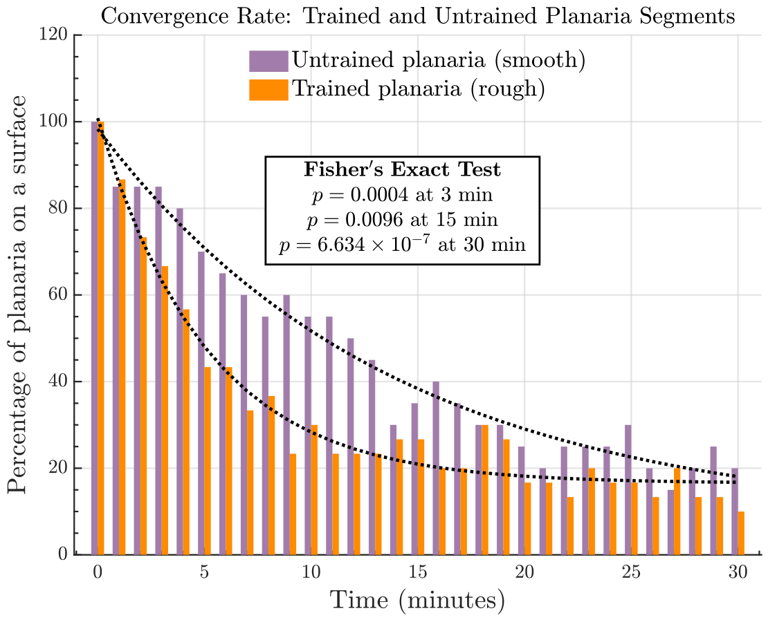
The immediate post-amputation convergence dynamics provide an important complementary perspective on the retention of reward-associated behavior. Rather than merely influencing the eventual distribution of fragments across surfaces, prior sucrose conditioning appeared to shape the trajectory by which anterior and posterior fragments reoriented immediately after amputation. Fragments derived from conditioned planaria converged more readily toward the reward-associated smooth surface, whereas those from untrained animals continued to exhibit behavior more consistent with the innate rough-surface bias. This pattern is conceptually significant because it suggests that the influence of prior learning remained detectable not only after regeneration, but already during the acute phase following tissue disruption. In this view, conditioning did not simply alter the final behavioral outcome; it appeared to modulate the early directional dynamics of surface selection immediately after amputation. Although the post-regeneration phenotype provides the strongest evidence for persistence of reward-associated memory, these convergence-rate findings imply that the behavioral trace of prior conditioning may remain accessible across successive stages of biological disruption and recovery. Considered together, the immediate post-amputation and post-regeneration observations strengthen the broader interpretation that reward-associated memory in planaria is supported by mechanisms that are not exclusively dependent on the intact cephalic ganglion.
More broadly, these findings carry conceptual significance because sucrose engages dopaminergic and opioid signaling pathways in a manner analogous to addictive substances in vertebrates [
6,
19,
25,
45,
46]. The ability of a sucrose-associated behavioral state to persist through regeneration suggests that addiction-like reward memories may be encoded in a biologically robust manner that is not readily erased by structural disruption. In this respect, planaria offer a uniquely tractable framework for studying how reward-related internal states are maintained across major anatomical change and may help illuminate broader principles of memory organization that extend beyond this model.
4.6. Dopaminergic Regulation of Reward Learning Across Regeneration
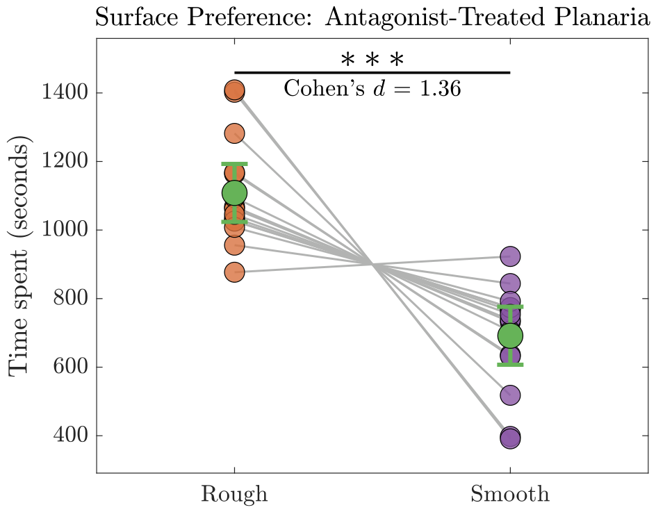
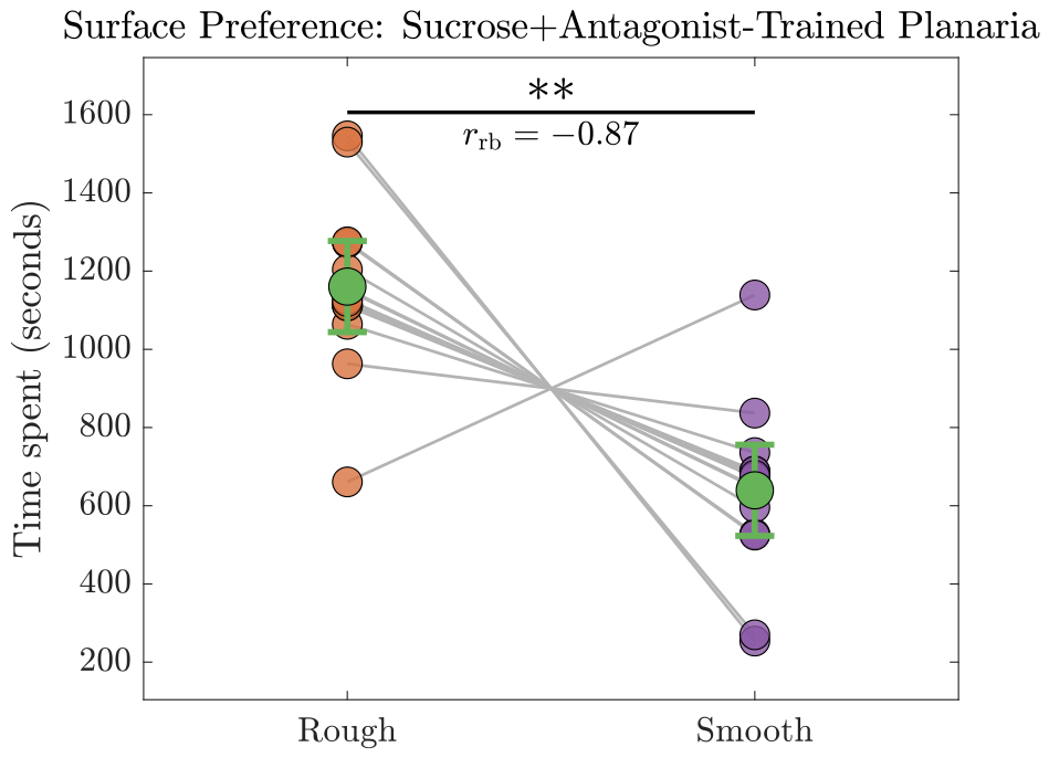
The antagonist experiments provide an important mechanistic complement to the regeneration findings by showing that dopaminergic signaling is required for establishment of the sucrose-associated behavioral state that later persists after regeneration. When a D1 dopamine antagonist was present during conditioning, planaria failed to acquire the sucrose-associated preference and instead retained their natural surface bias. This indicates that D1-mediated dopaminergic reinforcement is necessary for the reward-paired context to acquire motivational significance in this paradigm, consistent with previous work implicating dopamine in CPP-like behavior and addiction-related responses in planaria [
6,
19,
23,
25].
The regenerated antagonist-treated animals are particularly informative because they largely preserved the innate rough-surface preference rather than the learned smooth-surface preference observed in the sucrose-trained regenerated group. This contrast is important because it indicates that the post-regeneration phenotype is shaped by the learning conditions present during the original conditioning phase rather than emerging as a nonspecific consequence of regeneration itself. Regeneration alone did not generate an arbitrary or stereotyped behavioral outcome; instead, the regenerated phenotype reflected whether dopaminergic reinforcement had been available at the time the reward association was formed.
This finding strengthens the interpretation that D1-dependent signaling is required for establishing conditioned reward preference that is subsequently preserved and re-expressed after regeneration. If the reward-associated state fails to form under dopamine receptor blockade, then there is no conditioned phenotype to be re-expressed following tissue reconstruction. In this sense, the antagonist experiments show not only that dopamine is important for immediate reward learning, but also that it is necessary for establishing the kind of long-lasting internal state that can subsequently survive major biological disruption.
The fact that this blockade of conditioned preference remained evident after regeneration further sharpens the interpretation of the study. The persistence of the innate preference in antagonist-treated regenerates suggests that dopaminergic signaling is required for creating the reward-associated trace that is later preserved across regeneration. Thus, the antagonist findings do more than replicate prior work on dopamine and CPP; they directly link dopaminergic reinforcement to the enduring expression of reward-associated behavior in a regenerative context. This makes D1-mediated signaling a particularly relevant target for future investigation of how addiction-like behavioral states are encoded, stabilized, and later re-expressed.
4.7. Limitations and Future Directions
Despite the strength of the post-regeneration findings, several limitations warrant consideration. First, although the persistence of conditioned preference in tail-derived regenerates argues against memory storage being confined exclusively to the cephalic ganglion, the present behavioral data do not identify the biological substrate responsible for this persistence. The observed retention could reflect the continued influence of peripheral neural structures, molecular or epigenetic states in non-neural tissues, RNA-mediated signaling, neoblast-associated processes, or regeneration-dependent re-establishment of a previously acquired behavioral program. Because the study relied on behavioral readouts alone, it cannot distinguish among these mechanistic possibilities.
Second, the immediate post-amputation experiments require careful interpretation. Fragments in the immediate aftermath of amputation remain in a disrupted physiological state, and their behavior may therefore reflect acute injury responses, stress-related effects, residual neural activity, or early regenerative signaling in addition to any memory-related process. For this reason, the immediate fragment data should be regarded as supportive rather than definitive, whereas the regenerated-animal experiments provide the strongest basis for concluding that reward-associated behavior can persist across regeneration. The major interpretive emphasis of the present study therefore rests on the post-regeneration phenotype rather than on the immediate post-amputation observations.
Third, the behavioral paradigms differed across experiments. The fragment-switching assays were quantified at the group level, whereas the intact-animal CPP assays were assessed at the individual level. These paradigms capture different dimensions of behavior and are therefore not directly quantitatively interchangeable. As a result, comparisons across intact, immediate post-amputation, and regenerated states should be interpreted primarily at the conceptual rather than strictly numerical level. Although the overall behavioral patterns are mutually informative, they do not establish one-to-one equivalence across all experimental contexts.
The modest sample size also remains an important limitation. Although the observed behavioral patterns were coherent and biologically interpretable, larger cohorts would strengthen confidence in the generalizability of these findings and improve precision in estimating the robustness of the effects. In addition, while the antagonist results strongly support disruption of reward learning, the present design does not fully exclude the possibility that dopaminergic blockade may also influence movement-related or exploratory behaviors that secondarily affect surface occupancy. Future experiments incorporating more refined locomotor analysis will therefore be useful in distinguishing specific effects on reward learning from broader behavioral consequences.
Future studies should examine memory retention across multiple post-amputation time points in order to better separate acute injury-related effects from stable post-regeneration behavioral expression. Automated tracking and higher-resolution behavioral analysis would further improve sensitivity and reduce potential observer bias. At the mechanistic level, molecular and cellular follow-up studies—including transcriptomic, epigenetic, and fragment-specific profiling approaches—will be required to determine how reward-associated information is preserved and later re-expressed after regeneration [
10,
11,
12,
44,
47]. It will also be important to investigate the contribution of additional dopaminergic receptors and other neurotransmitter systems to the acquisition, maintenance, and disruption of reward-associated memory [
19,
25]. Collectively, such work may clarify how addiction-like behavioral states are encoded and sustained in regenerative systems, with broader relevance to general principles of memory and reward biology [
20,
21,
43].
5. Conclusions
The present study demonstrates that sucrose can induce a robust conditioned place preference in planaria and, more importantly, that this reward-associated behavioral state can persist after amputation and subsequent regeneration. Both immediate post-amputation observations and post-regeneration behavioral testing support the interpretation that the learned sucrose-associated preference is not dependent exclusively on the continuous presence of the original cephalic ganglion. The retention and later re-expression of conditioned preference in animals regenerated from posterior fragments therefore provide behavioral evidence consistent with distributed or non-cephalic mechanisms of memory storage. These findings extend the planarian memory literature by showing that reward-associated learning, including an addiction-like behavioral phenotype, can remain detectable despite major anatomical disruption and reconstruction.
The study further shows that dopaminergic signaling is essential for the formation of this persistent reward-associated state. Blocking D1 dopamine receptors during training prevented the acquisition of sucrose-induced conditioned place preference and abolished its later expression after regeneration, indicating that dopamine-dependent reinforcement is required not only for immediate reward learning but also for establishing the enduring trace that survives regenerative remodeling. Taken together, these results highlight planaria as a powerful tool for investigating how reward memories are encoded, stabilized, and re-expressed outside conventional centralized brain frameworks. More broadly, the findings provide a foundation for future mechanistic studies aimed at identifying the molecular, cellular, and neural substrates that support distributed memory storage in regenerative organisms.