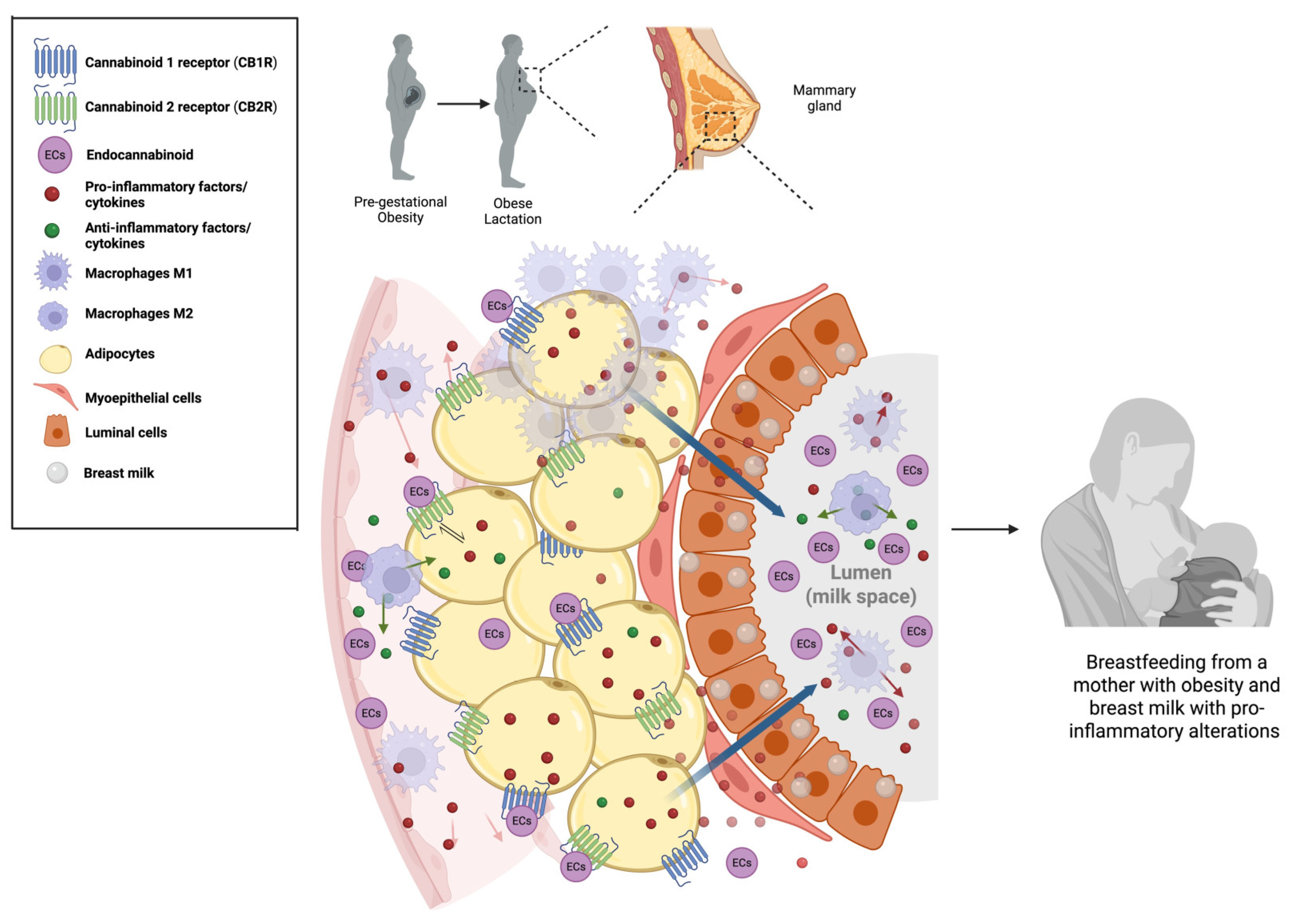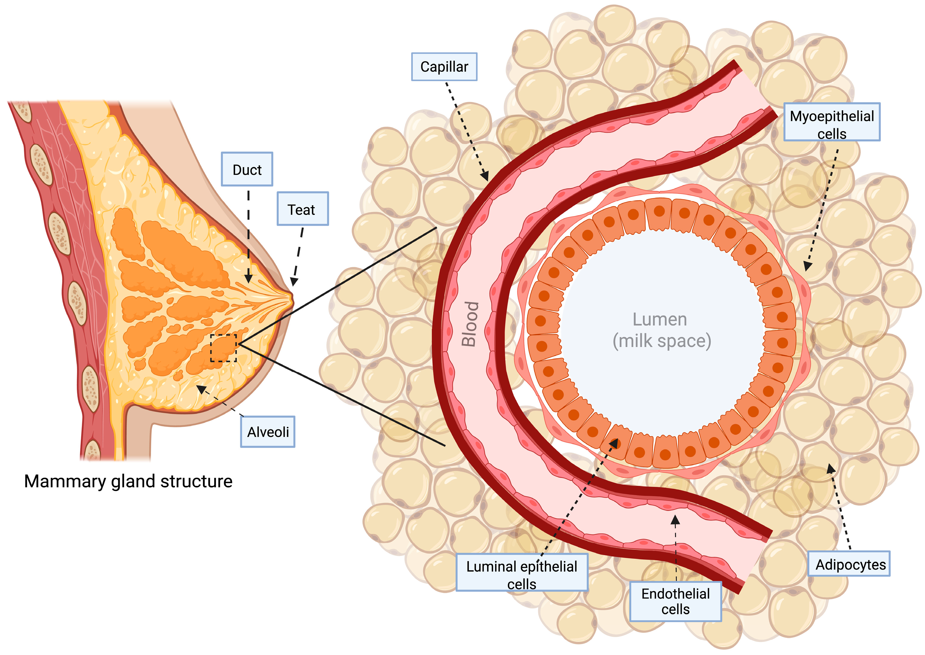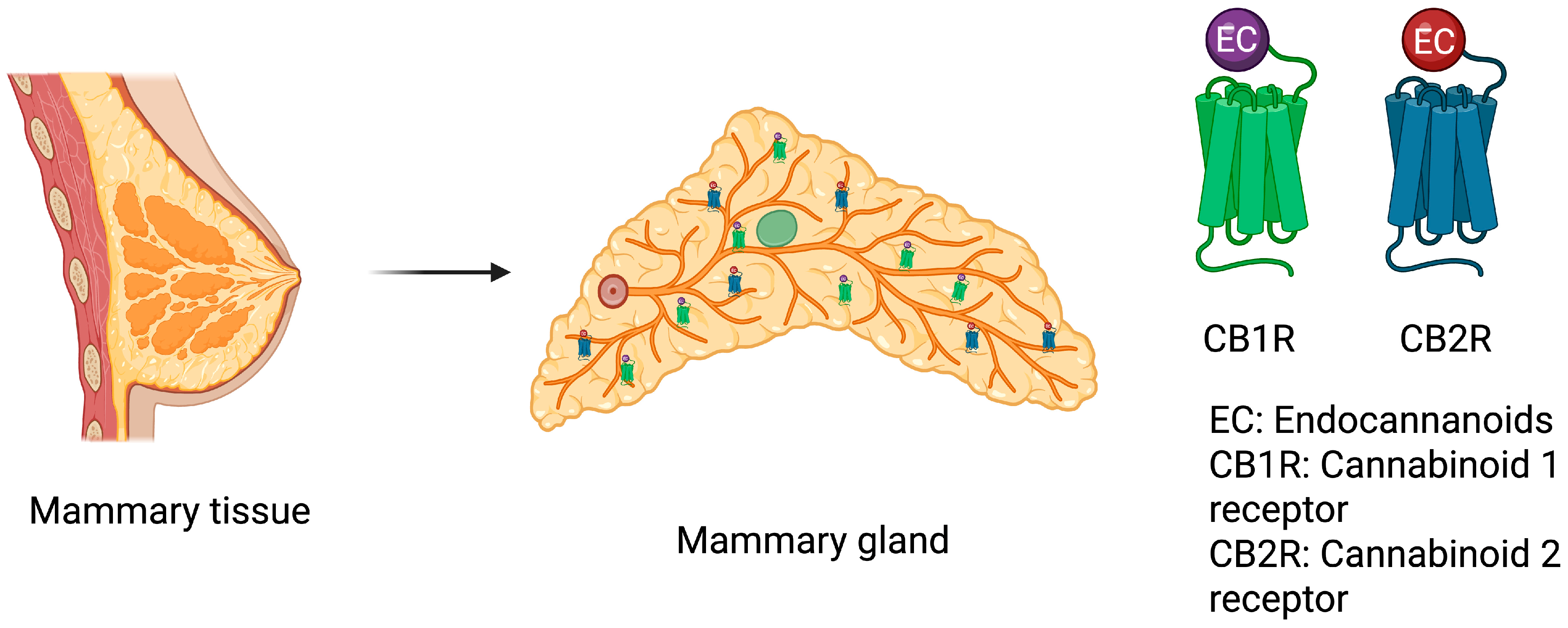
Exploring the Endocannabinoid System’s Influence on Mammary Tissue and Breast Milk Inflammation in Maternal Chronic Obesity


Abstract
1. Introduction
2. Review Methods
2.1. Methodology Used to Quantify Endocannabinoids in the Breast Milk
2.2. Assessment of Study Quality and Risk of Bias
3. Mammary Adipose Tissue Inflammation and Crown Like-Structure Development
4. Inflammatory Characteristics of Breast Milk Derived from Overweight and Obese Women
5. Endocannabinoid System as a Source of Inflammation in Obesity
6. Endocannabinoid Components in the Mammary Gland, Breast Milk, and Obesity
7. Conclusions and Future Perspectives
7.1. Future Perspectives
7.2. Limitations
Author Contributions
Funding
Institutional Review Board Statement
Informed Consent Statement
Data Availability Statement
Acknowledgments
Conflicts of Interest
References
- Purnell, J.Q. Definitions, Classification, and Epidemiology of Obesity. In Endotext; MDText.com, Inc.: South Dartmouth, MA, USA, 2023. [Google Scholar]
- Fantuzzi, G. Adipose Tissue, Adipokines, and Inflammation. J. Allergy Clin. Immunol. 2005, 115, 911–919. [Google Scholar] [CrossRef] [Scilit]
- Khanna, D.; Khanna, S.; Khanna, P.; Kahar, P.; Patel, B.M. Obesity: A Chronic Low-Grade Inflammation and Its Markers. Cureus 2022, 14, e22711. [Google Scholar] [CrossRef] [Scilit]
- Powell-Wiley, T.M.; Poirier, P.; Burke, L.E.; Després, J.P.; Gordon-Larsen, P.; Lavie, C.J.; Lear, S.A.; Ndumele, C.E.; Neeland, I.J.; Sanders, P.; et al. Obesity and Cardiovascular Disease: A Scientific Statement from the American Heart Association. Circulation 2021, 143, E984–E1010. [Google Scholar] [CrossRef] [Scilit]
- Spencer, L.; Rollo, M.; Hauck, Y.; MacDonald-Wicks, L.; Wood, L.; Hutchesson, M.; Giglia, R.; Smith, R.; Collins, C. The Effect of Weight Management Interventions That Include a Diet Component on Weight-Related Outcomes in Pregnant and Postpartum Women: A Systematic Review Protocol. JBI Database Syst. Rev. Implement. Rep. 2015, 13, 88–98. [Google Scholar] [CrossRef] [Scilit]
- Lim, S.; Harrison, C.; Callander, E.; Walker, R.; Teede, H.; Moran, L. Addressing Obesity in Preconception, Pregnancy, and Postpartum: A Review of the Literature. Curr. Obes. Rep. 2022, 11, 405–414. [Google Scholar] [CrossRef] [Scilit]
- MINSAL. Nutrición y Alimentación en la Gestante; MINSAL: Santiago, Chile, 2019. [Google Scholar]
- Álvarez, D.; Muñoz, Y.; Ortiz, M.; Maliqueo, M.; Chouinard-Watkins, R.; Valenzuela, R. Impact of Maternal Obesity on the Metabolism and Bioavailability of Polyunsaturated Fatty Acids during Pregnancy and Breastfeeding. Nutrients 2020, 13, 19. [Google Scholar] [CrossRef] [Scilit]
- Hrolfsdottir, L.; Schalkwijk, C.G.; Birgisdottir, B.E.; Gunnarsdottir, I.; Maslova, E.; Granström, C.; Strøm, M.; Olsen, S.F.; Halldorsson, T.I. Maternal Diet, Gestational Weight Gain, and Inflammatory Markers during Pregnancy. Obesity 2016, 24, 2133–2139. [Google Scholar] [CrossRef] [Scilit]
- Liang, W.; Qi, Y.; Yi, H.; Mao, C.; Meng, Q.; Wang, H.; Zheng, C. The Roles of Adipose Tissue Macrophages in Human Disease. Front. Immunol. 2022, 13, 908749. [Google Scholar] [CrossRef] [Scilit] [PubMed]
- Weisberg, S.P.; McCann, D.; Desai, M.; Rosenbaum, M.; Leibel, R.L.; Ferrante, A.W. Obesity Is Associated with Macrophage Accumulation in Adipose Tissue. J. Clin. Investig. 2003, 112, 1796–1808. [Google Scholar] [CrossRef] [Scilit] [PubMed]
- Longo, M.; Zatterale, F.; Naderi, J.; Parrillo, L.; Formisano, P.; Raciti, G.A.; Beguinot, F.; Miele, C. Adipose Tissue Dysfunction as Determinant of Obesity-Associated Metabolic Complications. Int. J. Mol. Sci. 2019, 20, 2358. [Google Scholar] [CrossRef] [Scilit] [PubMed]
- Yao, J.; Wu, D.; Qiu, Y. Adipose Tissue Macrophage in Obesity-Associated Metabolic Diseases. Front. Immunol. 2022, 13, 977485. [Google Scholar] [CrossRef] [Scilit]
- Cottam, D.R.; Mattar, S.G.; Barinas-Mitchell, E.; Eid, G.; Kuller, L.; Kelley, D.E.; Schauer, P.R. The Chronic Inflammatory Hypothesis for the Morbidity Associated with Morbid Obesity: Implications and Effects of Weight Loss. Obes. Surg. 2004, 14, 589–600. [Google Scholar] [CrossRef] [Scilit]
- Duffaut, C.; Galitzky, J.; Lafontan, M.; Bouloumié, A. Unexpected Trafficking of Immune Cells within the Adipose Tissue during the Onset of Obesity. Biochem. Biophys. Res. Commun. 2009, 384, 482–485. [Google Scholar] [CrossRef] [Scilit] [PubMed]
- Johnson, A.R.; Justin Milner, J.; Makowski, L. The Inflammation Highway: Metabolism Accelerates Inflammatory Traffic in Obesity. Immunol. Rev. 2012, 249, 218. [Google Scholar] [CrossRef] [Scilit] [PubMed]
- Xu, H.; Barnes, G.T.; Yang, Q.; Tan, G.; Yang, D.; Chou, C.J.; Sole, J.; Nichols, A.; Ross, J.S.; Tartaglia, L.A.; et al. Chronic Inflammation in Fat Plays a Crucial Role in the Development of Obesity-Related Insulin Resistance. J. Clin. Investig. 2003, 112, 1821–1830. [Google Scholar] [CrossRef]
- Cagnina, A.; Chabot, O.; Davin, L.; Lempereur, M.; Maréchal, P.; Oury, C.; Lancellotti, P. Atherosclerosis, an inflammatory disease. Rev. Med. Liege 2022, 77, 302–309. [Google Scholar] [PubMed]
- Ruggiero, A.D.; Key, C.C.C.; Kavanagh, K. Adipose Tissue Macrophage Polarization in Healthy and Unhealthy Obesity. Front. Nutr. 2021, 8, 625331. [Google Scholar] [CrossRef] [Scilit]
- Thrum, S.; Sommer, M.; Raulien, N.; Gericke, M.; Massier, L.; Kovacs, P.; Krasselt, M.; Landgraf, K.; Körner, A.; Dietrich, A.; et al. Macrophages in Obesity Are Characterised by Increased IL-1β Response to Calcium-Sensing Receptor Signals. Int. J. Obes. 2022, 46, 1883–1891. [Google Scholar] [CrossRef] [Scilit]
- Thomas, D.; Apovian, C. Macrophage Functions in Lean and Obese Adipose Tissue. Metabolism 2017, 72, 120–143. [Google Scholar] [CrossRef] [Scilit]
- Maliniak, M.L.; Cheriyan, A.M.; Cheriyan, A.M.; Sherman, M.E.; Liu, Y.; Liu, Y.; Gogineni, K.; Gogineni, K.; Liu, J.; He, J.; et al. Detection of Crown-like Structures in Breast Adipose Tissue and Clinical Outcomes among African-American and White Women with Breast Cancer. Breast Cancer Res. 2020, 22, 65. [Google Scholar] [CrossRef] [Scilit]
- Maliniak, M.L.; Miller-Kleinhenz, J.; Cronin-Fenton, D.P.; Lash, T.L.; Gogineni, K.; Janssen, E.A.M.; McCullough, L.E. Crown-Like Structures in Breast Adipose Tissue: Early Evidence and Current Issues in Breast Cancer. Cancers 2021, 13, 2222. [Google Scholar] [CrossRef] [Scilit]
- Chang, M.C.; Eslami, Z.; Ennis, M.; Goodwin, P.J. Crown-like Structures in Breast Adipose Tissue of Breast Cancer Patients: Associations with CD68 Expression, Obesity, Metabolic Factors and Prognosis. npj Breast Cancer 2021, 7, 97. [Google Scholar] [CrossRef] [Scilit]
- Cinti, S.; Mitchell, G.; Barbatelli, G.; Murano, I.; Ceresi, E.; Faloia, E.; Wang, S.; Fortier, M.; Greenberg, A.S.; Obin, M.S. Adipocyte Death Defines Macrophage Localization and Function in Adipose Tissue of Obese Mice and Humans. J. Lipid Res. 2005, 46, 2347–2355. [Google Scholar] [CrossRef] [Scilit]
- Faria, S.S.; Corrêa, L.H.; Heyn, G.S.; de Sant’Ana, L.P.; Almeida, R.D.N.; Magalhães, K.G. Obesity and Breast Cancer: The Role of Crown-Like Structures in Breast Adipose Tissue in Tumor Progression, Prognosis, and Therapy. J. Breast Cancer 2020, 23, 233. [Google Scholar] [CrossRef] [Scilit] [PubMed]
- McNally, S.; Stein, T. Overview of Mammary Gland Development: A Comparison of Mouse and Human. In Methods in Molecular Biology; Humana Press: New York, NY, USA, 2017; Volume 1501, pp. 1–17. [Google Scholar] [CrossRef] [Scilit]
- Biswas, S.K.; Banerjee, S.; Baker, G.W.; Kuo, C.Y.; Chowdhury, I. The Mammary Gland: Basic Structure and Molecular Signaling during Development. Int. J. Mol. Sci. 2022, 23, 3883. [Google Scholar] [CrossRef] [Scilit] [PubMed]
- Visvader, J.E. Keeping Abreast of the Mammary Epithelial Hierarchy and Breast Tumorigenesis. Genes Dev. 2009, 23, 2563–2577. [Google Scholar] [CrossRef] [Scilit]
- Cristea, S.; Polyak, K. Dissecting the Mammary Gland One Cell at a Time. Nat. Commun. 2018, 9, 2473. [Google Scholar] [CrossRef] [Scilit]
- Froń, A.; Orczyk-Pawiłowicz, M. Understanding the Immunological Quality of Breast Milk in Maternal Overweight and Obesity. Nutrients 2023, 15, 5016. [Google Scholar] [CrossRef] [Scilit]
- Andres, A.C.; Djonov, V. The Mammary Gland Vasculature Revisited. J. Mammary Gland Biol. Neoplasia 2010, 15, 319–328. [Google Scholar] [CrossRef] [Scilit]
- Gieniec, K.A. Mammary Gland Architecture under the Lens: How Visualising Tissues in 3D Provides Deeper Insights into Structure and Function. Biochim. Biophys. Acta—Mol. Cell Res. 2022, 1869, 119283. [Google Scholar] [CrossRef] [Scilit]
- Howard, B.A.; Gusterson, B.A. Human Breast Development. J. Mammary Gland. Biol. Neoplasia 2000, 5, 119–137. [Google Scholar] [CrossRef] [Scilit]
- Stevenson, A.J.; Vanwalleghem, G.; Stewart, T.A.; Condon, N.D.; Lloyd-Lewis, B.; Marino, N.; Putney, J.W.; Scott, E.K.; Ewing, A.D.; Davis, F.M. Multiscale Imaging of Basal Cell Dynamics in the Functionally Mature Mammary Gland. Proc. Natl. Acad. Sci. USA 2020, 117, 26822–26832. [Google Scholar] [CrossRef] [Scilit]
- Masedunskas, A.; Chena, Y.; Stussman, R.; Weigert, R.; Mather, I.H. Kinetics of Milk Lipid Droplet Transport, Growth, and Secretion Revealed by Intravital Imaging: Lipid Droplet Release Is Intermittently Stimulated by Oxytocin. Mol. Biol. Cell 2017, 28, 935–946. [Google Scholar] [CrossRef] [Scilit] [PubMed]
- Hillers-Ziemer, L.E.; Arendt, L.M. Weighing the Risk: Effects of Obesity on the Mammary Gland and Breast Cancer Risk. J. Mammary Gland Biol. Neoplasia 2020, 25, 115–131. [Google Scholar] [CrossRef] [Scilit]
- Luzardo-Ocampo, I.; Dena-Beltrán, J.L.; Ruiz-Herrera, X.; Ocampo-Ruiz, A.L.; Martínez de la Escalera, G.; Clapp, C.; Macotela, Y. Obesity-Derived Alterations in the Lactating Mammary Gland: Focus on Prolactin. Mol. Cell Endocrinol. 2023, 559, 111810. [Google Scholar] [CrossRef] [Scilit]
- Chamberlin, T.; D’Amato, J.V.; Arendt, L.M. Obesity Reversibly Depletes the Basal Cell Population and Enhances Mammary Epithelial Cell Estrogen Receptor Alpha Expression and Progenitor Activity. Breast Cancer Res. 2017, 19, 128. [Google Scholar] [CrossRef] [Scilit]
- Li, J.; Wu, H.; Liu, Y.; Yang, L. High Fat Diet Induced Obesity Model Using Four Strains of Mice: Kunming, C57BL/6, BALB/c and ICR. Exp. Anim. 2020, 69, 326–335. [Google Scholar] [CrossRef] [Scilit]
- Martins, T.; Castro-Ribeiro, C.; Lemos, S.; Ferreira, T.; Nascimento-Gonçalves, E.; Rosa, E.; Oliveira, P.A.; Antunes, L.M. Murine Models of Obesity. Obesities 2022, 2, 127–147. [Google Scholar] [CrossRef] [Scilit]
- Kamikawa, A.; Ichii, O.; Yamaji, D.; Imao, T.; Suzuki, C.; Okamatsu-Ogura, Y.; Terao, A.; Kon, Y.; Kimura, K. Diet-Induced Obesity Disrupts Ductal Development in the Mammary Glands of Nonpregnant Mice. Dev. Dyn. 2009, 238, 1092–1099. [Google Scholar] [CrossRef] [Scilit] [PubMed]
- Flint, D.J.; Travers, M.T.; Barber, M.C.; Binart, N.; Kelly, P.A. Diet-Induced Obesity Impairs Mammary Development and Lactogenesis in Murine Mammary Gland. Am. J. Physiol. Endocrinol. Metab. 2005, 288, 1179–1187. [Google Scholar] [CrossRef] [Scilit]
- Jevitt, C. Pregnancy Complicated by Obesity: Midwifery Management. J. Midwifery Women’s Health 2009, 54, 445–451. [Google Scholar] [CrossRef] [Scilit]
- Chang, Y.S.; Glaria, A.A.; Davie, P.; Beake, S.; Bick, D. Breastfeeding Experiences and Support for Women Who Are Overweight or Obese: A Mixed-methods Systematic Review. Matern. Child. Nutr. 2020, 16, e12865. [Google Scholar] [CrossRef] [Scilit]
- Buckley, S.; Uvnäs-Moberg, K.; Pajalic, Z.; Luegmair, K.; Ekström-Bergström, A.; Dencker, A.; Massarotti, C.; Kotlowska, A.; Callaway, L.; Morano, S.; et al. Maternal and Newborn Plasma Oxytocin Levels in Response to Maternal Synthetic Oxytocin Administration during Labour, Birth and Postpartum—A Systematic Review with Implications for the Function of the Oxytocinergic System. BMC Pregnancy Childbirth 2023, 23, 137. [Google Scholar] [CrossRef] [Scilit]
- Rassie, K.; Dhungana, R.R.; Mousa, A.; Teede, H.; Joham, A.E. Maternal Metabolic Conditions as Predictors of Breastfeeding Outcomes: Insights from an Australian Cohort Study. Acta Obstet. Gynecol. Scand. 2024, 103, 1570–1583. [Google Scholar] [CrossRef] [Scilit]
- Nommsen-Rivers, L.A.; Chantry, C.J.; Peerson, J.M.; Cohen, R.J.; Dewey, K.G. Delayed Onset of Lactogenesis among First-Time Mothers Is Related to Maternal Obesity and Factors Associated with Ineffective Breastfeeding. Am. J. Clin. Nutr. 2010, 92, 574–584. [Google Scholar] [CrossRef] [Scilit]
- Rasmussen, K.M.; Hilson, J.A.; Kjolhede, C.L. Obesity May Impair Lactogenesis II. J. Nutr. 2001, 131, 3009S–3011S. [Google Scholar] [CrossRef] [Scilit] [PubMed]
- Gaitán, A.V.; Wood, J.A.T.; Zhang, F.; Makriyannis, A.; Lammi-Keefe, C.J. Endocannabinoid Metabolome Characterization of Transitional and Mature Human Milk. Nutrients 2018, 10, 1294. [Google Scholar] [CrossRef] [Scilit]
- Datta, P.; Melkus, M.W.; Rewers-Felkins, K.; Patel, D.; Bateman, T.; Baker, T.; Hale, T.W. Human Milk Endocannabinoid Levels as a Function of Obesity and Diurnal Rhythm. Nutrients 2021, 13, 2297. [Google Scholar] [CrossRef] [Scilit]
- Pontes, T.F.; Reis, G.; Santos, G.R.C.; Pereira, H.M.G.; Kac, G.; Ferreira, A.L.L.; Trevenzoli, I.H. Maternal Obesity and Excessive Gestational Weight Gain Influence Endocannabinoid Levels in Human Milk Across Breastfeeding: Potential Implications for Offspring Development. Nutrients 2025, 17, 1344. [Google Scholar] [CrossRef] [Scilit]
- Wood, J.T.; Durham, H.A.; Vadivel, S.K.; Makriyannis, A.; Lammi-Keefe, C.J. Postpartum Changes in the Endocannabinoid Metabolome of Human Breast Milk. FASEB J. 2013, 27, 629.15. [Google Scholar] [CrossRef] [Scilit]
- Csatári-Kovács, R.; Röszer, T.; Csősz, É. Comparative Analysis of Omega-3, Omega-6, and Endocannabinoid Content of Human, Cattle, Goat, and Formula Milk. Foods 2025, 14, 1786. [Google Scholar] [CrossRef] [Scilit] [PubMed]
- Vaysse, C.; Lomo, J.; Garred, O.; Fjeldheim, F.; Lofteroed, T.; Schlichting, E.; McTiernan, A.; Frydenberg, H.; Husoy, A.; Lundgren, S.; et al. Inflammation of Mammary Adipose Tissue Occurs in Overweight and Obese Patients Exhibiting Early-Stage Breast Cancer. npj Breast Cancer 2017, 3, 19. [Google Scholar] [CrossRef] [Scilit]
- Zangani, D.; Darcy, K.M.; Shoemaker, S.; Ip, M.M. Adipocyte-Epithelial Interactions Regulate the in Vitro Development of Normal Mammary Epithelial Cells. Exp. Cell Res. 1999, 247, 399–409. [Google Scholar] [CrossRef] [Scilit]
- Brenot, A.; Hutson, I.; Harris, C. Epithelial-Adipocyte Interactions Are Required for Mammary Gland Development, but Not for Milk Production or Fertility. Dev. Biol. 2020, 458, 153–163. [Google Scholar] [CrossRef] [Scilit]
- Gregor, M.F.; Misch, E.S.; Yang, L.; Hummasti, S.; Inouye, K.E.; Lee, A.H.; Bierie, B.; Hotamisligil, G.S. The Role of Adipocyte XBP1 in Metabolic Regulation during Lactation. Cell Rep. 2013, 3, 1430–1439. [Google Scholar] [CrossRef] [Scilit]
- Berger, N.A. Crown-like Structures in Breast Adipose Tissue from Normal Weight Women: Important Impact. Cancer Prev. Res. 2017, 10, 223–225. [Google Scholar] [CrossRef] [Scilit]
- Harris, A.R.; Hughes, J.D.; Lawrence, W.R.; Lenz, P.; Franklin, J.; Bhawsar, P.M.S.; Dorsey, T.H.; Rossi, E.L.; Pichardo, C.M.; Pichardo, M.S.; et al. Neighborhood Environment, DNA Methylation, and Presence of Crown-Like Structures of the Breast. JAMA Netw. Open 2025, 8, e2461334. [Google Scholar] [CrossRef] [Scilit] [PubMed]
- Murano, I.; Barbatelli, G.; Parisani, V.; Latini, C.; Muzzonigro, G.; Castellucci, M.; Cinti, S. Dead Adipocytes, Detected as Crown-like Structures, Are Prevalent in Visceral Fat Depots of Genetically Obese Mice. J. Lipid Res. 2008, 49, 1562–1568. [Google Scholar] [CrossRef] [Scilit]
- Hausman, D.B.; DiGirolamo, M.; Bartness, T.J.; Hausman, G.J.; Martin, R.J. The Biology of White Adipocyte Proliferation. Obes. Rev. 2001, 2, 239–254. [Google Scholar] [CrossRef] [Scilit]
- Trayhurn, P.; Wood, I.S. Adipokines: Inflammation and the Pleiotropic Role of White Adipose Tissue. Br. J. Nutr. 2004, 92, 347–355. [Google Scholar] [CrossRef] [Scilit]
- Colleluori, G.; Perugini, J.; Barbatelli, G.; Cinti, S. Mammary Gland Adipocytes in Lactation Cycle, Obesity and Breast Cancer. Rev. Endocr. Metab. Disord. 2021, 22, 241–255. [Google Scholar] [CrossRef] [Scilit]
- Zhang, Z.; Scherer, P.E. Adipose Tissue: The Dysfunctional Adipocyte—A Cancer Cell’s Best Friend. Nat. Rev. Endocrinol. 2018, 14, 132–134. [Google Scholar] [CrossRef] [Scilit]
- Seo, B.R.; Bhardwaj, P.; Choi, S.; Gonzalez, J.; Eguiluz, R.C.A.; Wang, K.; Mohanan, S.; Morris, P.G.; Du, B.; Zhou, X.K.; et al. Obesity-Dependent Changes in Interstitial ECM Mechanics Promote Breast Tumorigenesis. Sci. Transl. Med. 2015, 7, 301ra130. [Google Scholar] [CrossRef] [Scilit]
- Pasarica, M.; Gowronska-Kozak, B.; Burk, D.; Remedios, I.; Hymel, D.; Gimble, J.; Ravussin, E.; Bray, G.A.; Smith, S.R. Adipose Tissue Collagen VI in Obesity. J. Clin. Endocrinol. Metab. 2009, 94, 5155–5162. [Google Scholar] [CrossRef] [Scilit]
- Khan, I.T.; Bule, M.; Ullah, R.; Nadeem, M.; Asif, S.; Niaz, K. The Antioxidant Components of Milk and Their Role in Processing, Ripening, and Storage: Functional Food. Vet. World 2019, 12, 12–33. [Google Scholar] [CrossRef] [Scilit]
- Kalarikkal, S.M.; Pfleghaar, J.L. Breastfeeding. In StatPearls; StatPearls Publishing: Treasure Island, FL, USA, 2024. [Google Scholar]
- World Health Organization. Obesity and Overweight. Available online: https://www.who.int/news-room/fact-sheets/detail/obesity-and-overweight (accessed on 5 August 2025).
- Eisha, S.; Joarder, I.; Wijenayake, S.; Mcgowan, P.O. Nutritive Bioactive Components in Maternal Milk and Offspring Development: A Scoping Review Non-Nutritive Bioactive Components in Maternal Milk and Offspring Development: A Scoping Review. J. Dev. Orig. Health Dis. 2022, 13, 665–673. [Google Scholar] [CrossRef] [Scilit] [PubMed]
- Wijenayake, S.; Martz, J.; Lapp, H.E.; Storm, J.A.; Champagne, F.A.; Kentner, A.C. The Contributions of Parental Lactation on Offspring Development: It’s Not Udder Nonsense! Horm. Behav. 2023, 153, 105375. [Google Scholar] [CrossRef] [Scilit] [PubMed]
- Dieterich, C.M.; Felice, J.P.; O’Sullivan, E.; Rasmussen, K.M. Breastfeeding and Health Outcomes for the Mother-Infant Dyad. Pediatr. Clin. N. Am. 2013, 60, 31–48. [Google Scholar] [CrossRef] [Scilit]
- Lyons, K.E.; Ryan, C.A.; Dempsey, E.M.; Ross, R.P.; Stanton, C. Breast Milk, a Source of Beneficial Microbes and Associated Benefits for Infant Health. Nutrients 2020, 12, 1039. [Google Scholar] [CrossRef] [Scilit]
- Mantzorou, M.; Papandreou, D.; Pavlidou, E.; Papadopoulou, S.K.; Tolia, M.; Mentzelou, M.; Poutsidi, A.; Antasouras, G.; Vasios, G.K.; Giaginis, C. Maternal Gestational Diabetes Is Associated with High Risk of Childhood Overweight and Obesity: A Cross-Sectional Study in Pre-School Children Aged 2–5 Years. Medicina 2023, 59, 455. [Google Scholar] [CrossRef] [Scilit]
- Bode, L.; Raman, A.; Murch, S.; Rollins, N.; Gordon, J. Understanding the Mother-Breastmilk-Infant “Triad”. Science 2020, 367, 1070–1072. [Google Scholar] [CrossRef] [Scilit]
- Edwards, P.D.; Lavergne, S.G.; Mccaw, L.K.; Wijenayake, S.; Boonstra, R.; Mcgowan, P.O.; Holmes, M.M. Maternal Effects in Mammals: Broadening Our Understanding of Offspring Programming Offspring Phenotype Androgens Photoperiod Melatonin Microbiome Immunity Maternal Milk Exosomes MicroRNA. Front. Neuroendocrinol. 2021, 62, 100924. [Google Scholar] [CrossRef] [Scilit]
- Lucas, A. Programming by Early Nutrition: An Experimental Approach. J. Nutr. 1998, 128, 401S–406S. [Google Scholar] [CrossRef] [Scilit]
- Lokossou, G.A.G.; Kouakanou, L.; Schumacher, A.; Zenclussen, A.C. Human Breast Milk: From Food to Active Immune Response with Disease Protection in Infants and Mothers. Front. Immunol. 2022, 13, 849012. [Google Scholar] [CrossRef] [Scilit] [PubMed]
- Leghi, G.E.; Netting, M.J.; Middleton, P.F.; Wlodek, M.E.; Geddes, D.T.; Muhlhausler, B.S. The Impact of Maternal Obesity on Human Milk Macronutrient Composition: A Systematic Review and Meta-Analysis. Nutrients 2020, 12, 934. [Google Scholar] [CrossRef] [Scilit]
- Sims, C.R.; Lipsmeyer, M.E.; Turner, D.E.; Andres, A. Human Milk Composition Differs by Maternal BMI in the First 9 Months Postpartum. Am. J. Clin. Nutr. 2020, 112, 548–557. [Google Scholar] [CrossRef] [Scilit]
- Ahuja, S.; Boylan, M.; Hart, S.L.; Román-Shriver, C.; Spallholz, J.E.; Pence, B.C.; Sawyer, B.G. Glucose and Insulin Levels Are Increased in Obese and Overweight Mothers’ Breast-Milk. Food Nutr. Sci. 2011, 2, 201–206. [Google Scholar] [CrossRef]
- Cheema, A.S.; Stinson, L.F.; Rea, A.; Lai, C.T.; Payne, M.S.; Murray, K.; Geddes, D.T.; Gridneva, Z. Human Milk Lactose, Insulin, and Glucose Relative to Infant Body Composition during Exclusive Breastfeeding. Nutrients 2021, 13, 3724. [Google Scholar] [CrossRef] [Scilit] [PubMed]
- Houghton, M.R.; Gracey, M.; Burke, V.; Bottrell, C.; Spargo, R.M. Breast Milk Lactoferrin Levels in Relation to Maternal Nutritional Status. J. Pediatr. Gastroenterol. Nutr. 1985, 4, 230–233. [Google Scholar] [CrossRef] [Scilit]
- Hua, M.C.; Su, H.M.; Yao, T.C.; Liao, S.L.; Tsai, M.H.; Su, K.W.; Chen, L.C.; Lai, S.H.; Chiu, C.Y.; Yeh, K.W.; et al. The Association between Human Milk Fatty Acid Composition in Mothers with an Elevated Body Mass Index and Infant Growth Changes. Clin. Nutr. 2024, 43, 203–210. [Google Scholar] [CrossRef] [Scilit]
- Panagos, P.G.; Vishwanathan, R.; Penfield-Cyr, A.; Matthan, N.R.; Shivappa, N.; Wirth, M.D.; Hebert, J.R.; Sen, S. Breastmilk from Obese Mothers Has Pro-Inflammatory Properties and Decreased Neuroprotective Factors. J. Perinatol. 2016, 36, 284–290. [Google Scholar] [CrossRef] [Scilit]
- De La Garza Puentes, A.; Alemany, A.M.; Chisaguano, A.M.; Goyanes, R.M.; Castellote, A.I.; Torres-Espínola, F.J.; García-Valdés, L.; Escudero-Marín, M.; Segura, M.T.; Campoy, C.; et al. The Effect of Maternal Obesity on Breast Milk Fatty Acids and Its Association with Infant Growth and Cognition—The PREOBE Follow-Up. Nutrients 2019, 11, 2154. [Google Scholar] [CrossRef] [Scilit]
- Tekin-Guler, T.; Koc, N.; Kara-Uzun, A.; Fisunoglu, M. The Association of Pre-Pregnancy Obesity and Breast Milk Fatty Acids Composition and the Relationship of Postpartum Maternal Diet, Breast Milk Fatty Acids and Infant Growth. Breastfeed. Med. 2023, 18, 475–482. [Google Scholar] [CrossRef] [Scilit] [PubMed]
- Simon Sarkadi, L.; Zhang, M.; Muránszky, G.; Vass, R.A.; Matsyura, O.; Benes, E.; Vari, S.G. Fatty Acid Composition of Milk from Mothers with Normal Weight, Obesity, or Gestational Diabetes. Life 2022, 12, 1093. [Google Scholar] [CrossRef] [Scilit]
- Erliana, U.D.; Fly, A.D. The Function and Alteration of Immunological Properties in Human Milk of Obese Mothers. Nutrients 2019, 11, 1284. [Google Scholar] [CrossRef] [Scilit] [PubMed]
- Whitaker, K.M.; Marino, R.C.; Haapala, J.L.; Foster, L.; Smith, K.D.; Teague, A.M.; Jacobs, D.R.; Fontaine, P.L.; McGovern, P.M.; Schoenfuss, T.C.; et al. Associations of Maternal Weight Status Before, During, and After Pregnancy with Inflammatory Markers in Breast Milk. Obesity 2017, 25, 2092–2099. [Google Scholar] [CrossRef] [Scilit]
- Young, B.E.; Patinkin, Z.W.; Pyle, L.; de la Houssaye, B.; Davidson, B.S.; Geraghty, S.; Morrow, A.L.; Krebs, N. Markers of Oxidative Stress in Human Milk Do Not Differ by Maternal BMI But Are Related to Infant Growth Trajectories. Matern. Child Health J. 2017, 21, 1367–1376. [Google Scholar] [CrossRef] [Scilit] [PubMed]
- Collado, M.C.; Laitinen, K.; Salminen, S.; Isolauri, E. Maternal Weight and Excessive Weight Gain during Pregnancy Modify the Immunomodulatory Potential of Breast Milk. Pediatr. Res. 2012, 72, 77–85. [Google Scholar] [CrossRef] [Scilit]
- Silver, R.J. The Endocannabinoid System of Animals. Animals 2019, 9, 686. [Google Scholar] [CrossRef] [Scilit]
- Bazwinsky-Wutschke, I.; Zipprich, A.; Dehghani, F. Endocannabinoid System in Hepatic Glucose Metabolism, Fatty Liver Disease, and Cirrhosis. Int. J. Mol. Sci. 2019, 20, 2516. [Google Scholar] [CrossRef] [Scilit]
- Murillo-Rodríguez, E.; Budde, H.; Veras, A.B.; Rocha, N.B.; Telles-Correia, D.; Monteiro, D.; Cid, L.; Yamamoto, T.; Machado, S.; Torterolo, P. The Endocannabinoid System May Modulate Sleep Disorders in Aging. Curr. Neuropharmacol. 2020, 18, 97–108. [Google Scholar] [CrossRef] [Scilit] [PubMed]
- Garani, R.; Watts, J.J.; Mizrahi, R. Endocannabinoid System in Psychotic and Mood Disorders, a Review of Human Studies. Prog. Neuro-Psychopharmacol. Biol. Psychiatry 2021, 106, 110096. [Google Scholar] [CrossRef] [Scilit]
- Bourke, S.L.; Schlag, A.K.; O’Sullivan, S.E.; Nutt, D.J.; Finn, D.P. Cannabinoids and the Endocannabinoid System in Fibromyalgia: A Review of Preclinical and Clinical Research. Pharmacol. Ther. 2022, 240, 108216. [Google Scholar] [CrossRef] [Scilit]
- Finn, D.P.; Haroutounian, S.; Hohmann, A.G.; Krane, E.; Soliman, N.; Rice, A.S.C. Cannabinoids, the Endocannabinoid System, and Pain: A Review of Preclinical Studies. Pain 2021, 162, S5–S25. [Google Scholar] [CrossRef] [Scilit] [PubMed]
- Barrie, N.; Manolios, N. The Endocannabinoid System in Pain and Inflammation: Its Relevance to Rheumatic Disease. Eur. J. Rheumatol. 2017, 4, 210–218. [Google Scholar] [CrossRef] [Scilit]
- Kruk-Slomka, M.; Dzik, A.; Budzynska, B.; Biala, G. Endocannabinoid System: The Direct and Indirect Involvement in the Memory and Learning Processes—A Short Review. Mol. Neurobiol. 2017, 54, 8332–8347. [Google Scholar] [CrossRef] [Scilit] [PubMed]
- Lunardi, P.; de Souza, L.W.; dos Santos, B.; Popik, B.; de Oliveira Alvares, L. Effect of the Endocannabinoid System in Memory Updating and Forgetting. Neuroscience 2020, 444, 33–42. [Google Scholar] [CrossRef] [Scilit]
- Almogi-Hazan, O.; Or, R. Cannabis, the Endocannabinoid System and Immunity-the Journey from the Bedside to the Bench and Back. Int. J. Mol. Sci. 2020, 21, 4448. [Google Scholar] [CrossRef] [Scilit]
- Pagano, C.; Rossato, M.; Vettor, R. Endocannabinoids, Adipose Tissue and Lipid Metabolism. J. Neuroendocrinol. 2008, 20, 124–129. [Google Scholar] [CrossRef] [Scilit]
- Di Marzo, V. The Endocannabinoid System in Obesity and Type 2 Diabetes. Diabetologia 2008, 51, 1356–1367. [Google Scholar] [CrossRef] [Scilit]
- Lu, H.C.; Mackie, K. Review of the Endocannabinoid System. Biol. Psychiatry Cogn. Neurosci. Neuroimaging 2021, 6, 607–615. [Google Scholar] [CrossRef] [Scilit]
- Alvheim, A.R.; Malde, M.K.; Osei-Hyiaman, D.; Hong Lin, Y.; Pawlosky, R.J.; Madsen, L.; Kristiansen, K.; Frøyland, L.; Hibbeln, J.R. Dietary Linoleic Acid Elevates Endogenous 2-AG and Anandamide and Induces Obesity. Obesity 2012, 20, 1984–1994. [Google Scholar] [CrossRef] [Scilit] [PubMed]
- Liu, J.; Wang, L.; Harvey-White, J.; Osei-Hyiaman, D.; Razdan, R.; Gong, Q.; Chan, A.C.; Zhou, Z.; Huang, B.X.; Kim, H.Y.; et al. A Biosynthetic Pathway for Anandamide. Proc. Natl. Acad. Sci. USA 2006, 103, 13345–13350. [Google Scholar] [CrossRef] [Scilit] [PubMed]
- Bryk, M.; Chwastek, J.; Kostrzewa, M.; Mlost, J.; Pędracka, A.; Starowicz, K. Alterations in Anandamide Synthesis and Degradation during Osteoarthritis Progression in an Animal Model. Int. J. Mol. Sci. 2020, 21, 7381. [Google Scholar] [CrossRef] [Scilit]
- Silvestri, C.; Di Marzo, V. The Endocannabinoid System in Energy Homeostasis and the Etiopathology of Metabolic Disorders. Cell Metab. 2013, 17, 475–490. [Google Scholar] [CrossRef] [Scilit]
- Maccarrone, M.; Di Marzo, V.; Gertsch, J.; Grether, U.; Howlett, A.C.; Hua, T.; Makriyannis, A.; Piomelli, D.; Ueda, N.; van der Stelt, M. Goods and Bads of the Endocannabinoid System as a Therapeutic Target: Lessons Learned after 30 Years. Pharmacol. Rev. 2023, 75, 885–958. [Google Scholar] [CrossRef] [Scilit]
- Clayton, P.; Subah, S.; Venkatesh, R.; Hill, M.; Bogoda, N. Palmitoylethanolamide: A Potential Alternative to Cannabidiol. J. Diet. Suppl. 2023, 20, 505–530. [Google Scholar] [CrossRef] [Scilit]
- Romano, A.; Coccurello, R.; Giacovazzo, G.; Bedse, G.; Moles, A.; Gaetani, S. Oleoylethanolamide: A Novel Potential Pharmacological Alternative to Cannabinoid Antagonists for the Control of Appetite. BioMed Res. Int. 2014, 2014, 203425. [Google Scholar] [CrossRef] [Scilit]
- Zou, S.; Kumar, U. Cannabinoid Receptors and the Endocannabinoid System: Signaling and Function in the Central Nervous System. Int. J. Mol. Sci. 2018, 19, 833. [Google Scholar] [CrossRef] [Scilit]
- Mallat, A.; Teixeira-Clerc, F.; Deveaux, V.; Manin, S.; Lotersztajn, S. The Endocannabinoid System as a Key Mediator during Liver Diseases: New Insights and Therapeutic Openings. Br. J. Pharmacol. 2011, 163, 1432–1440. [Google Scholar] [CrossRef] [Scilit]
- Farooqi, T.; Bhuyan, D.J.; Low, M.; Sinclair, J.; Leonardi, M.; Armour, M. Cannabis and Endometriosis: The Roles of the Gut Microbiota and the Endocannabinoid System. J. Clin. Med. 2023, 12, 7071. [Google Scholar] [CrossRef] [Scilit]
- Schönke, M.; Martinez-Tellez, B.; Rensen, P.C. Role of the Endocannabinoid System in the Regulation of the Skeletal Muscle Response to Exercise. Curr. Opin. Pharmacol. 2020, 52, 52–60. [Google Scholar] [CrossRef] [Scilit]
- Rabino, M.; Mallia, S.; Castiglioni, E.; Rovina, D.; Pompilio, G.; Gowran, A. The Endocannabinoid System and Cannabidiol: Past, Present, and Prospective for Cardiovascular Diseases. Pharmaceuticals 2021, 14, 936. [Google Scholar] [CrossRef] [Scilit]
- Myers, M.N.; Abou-Rjeileh, U.; Chirivi, M.; Parales-Girón, J.; Lock, A.L.; Tam, J.; Zachut, M.; Contreras, G.A. Cannabinoid-1 Receptor Activation Modulates Lipid Mobilization and Adipogenesis in the Adipose Tissue of Dairy Cows. J. Dairy. Sci. 2023, 106, 3650–3661. [Google Scholar] [CrossRef] [Scilit] [PubMed]
- Walker, O.L.S.; Holloway, A.C.; Raha, S. The Role of the Endocannabinoid System in Female Reproductive Tissues. J. Ovarian Res. 2019, 12, 3. [Google Scholar] [CrossRef] [Scilit]
- The Human Protein Atlas CNR2 Protein Expression Summary. Available online: https://www.proteinatlas.org/ENSG00000188822-CNR2 (accessed on 1 April 2024).
- The Human Protein Atlas CNR1 Protein Expression Summary. Available online: https://www.proteinatlas.org/ENSG00000118432-CNR1 (accessed on 1 April 2024).
- Jung, K.M.; Lin, L.; Piomelli, D. The Endocannabinoid System in the Adipose Organ. Rev. Endocr. Metab. Disord. 2022, 23, 51–60. [Google Scholar] [CrossRef] [Scilit] [PubMed]
- Rakotoarivelo, V.; Sihag, J.; Flamand, N. Role of the Endocannabinoid System in the Adipose Tissue with Focus on Energy Metabolism. Cells 2021, 10, 1279. [Google Scholar] [CrossRef] [Scilit]
- Tam, J.; Szanda, G.; Drori, A.; Liu, Z.; Cinar, R.; Kashiwaya, Y.; Reitman, M.L.; Kunos, G. Peripheral Cannabinoid-1 Receptor Blockade Restores Hypothalamic Leptin Signaling. Mol. Metab. 2017, 6, 1113–1125. [Google Scholar] [CrossRef] [Scilit]
- Sidibeh, C.O.; Pereira, M.J.; Lau Börjesson, J.; Kamble, P.G.; Skrtic, S.; Katsogiannos, P.; Sundbom, M.; Svensson, M.K.; Eriksson, J.W. Role of Cannabinoid Receptor 1 in Human Adipose Tissue for Lipolysis Regulation and Insulin Resistance. Endocrine 2017, 55, 839–852. [Google Scholar] [CrossRef] [Scilit] [PubMed]
- Ruhl, T.; Karthaus, N.; Kim, B.S.; Beier, J.P. The Endocannabinoid Receptors CB1 and CB2 Affect the Regenerative Potential of Adipose Tissue MSCs. Exp. Cell Res. 2020, 389, 111881. [Google Scholar] [CrossRef] [Scilit]
- Schulz, P.; Hryhorowicz, S.; Rychter, A.M.; Zawada, A.; Słomski, R.; Dobrowolska, A.; Krela-Kaźmierczak, I. What Role Does the Endocannabinoid System Play in the Pathogenesis of Obesity? Nutrients 2021, 13, 373. [Google Scholar] [CrossRef] [Scilit]
- Eid, B.G.; Neamatallah, T.; Binmahfouz, L.S.; Bagher, A.M.; Alamoudi, A.J.; Aldawsari, H.M.; Hanafy, A.; Hasan, A.; El-Bassossy, H.M.; Abdel-Naim, A.B.; et al. Effects of the CB1 Receptor Antagonists AM6545 and AM4113 on Metabolic Syndrome-Induced Prostatic Hyperplasia in Rats. Biomol. Biomed. 2023, 23, 1069–1078. [Google Scholar] [CrossRef] [Scilit]
- Krott, L.M.; Piscitelli, F.; Heine, M.; Borrino, S.; Scheja, L.; Silvestri, C.; Heeren, J.; Di Marzo, V. Endocannabinoid Regulation in White and Brown Adipose Tissue Following Thermogenic Activation. J. Lipid Res. 2016, 57, 464–473. [Google Scholar] [CrossRef] [Scilit]
- Jager, G.; Witkamp, R.F. The Endocannabinoid System and Appetite: Relevance for Food Reward. Nutr. Res. Rev. 2014, 27, 172–185. [Google Scholar] [CrossRef] [Scilit]
- Joshi, N.; Onaivi, E.S. Endocannabinoid System Components: Overview and Tissue Distribution. In Recent Advances in Cannabinoid Physiology and Pathology. Advances in Experimental Medicine and Biology; Springer: Cham, Switzerland, 2019; Volume 1162, pp. 1–12. [Google Scholar] [CrossRef] [Scilit]
- Jordan, C.J.; Xi, Z.X. Progress in Brain Cannabinoid CB2 Receptor Research: From Genes to Behavior. Neurosci. Biobehav. Rev. 2019, 98, 208–220. [Google Scholar] [CrossRef] [Scilit] [PubMed]
- Tortora, C.; Di Paola, A.; Argenziano, M.; Creoli, M.; Marrapodi, M.M.; Cenni, S.; Tolone, C.; Rossi, F.; Strisciuglio, C. Effects of CB2 Receptor Modulation on Macrophage Polarization in Pediatric Celiac Disease. Biomedicines 2022, 10, 874. [Google Scholar] [CrossRef] [Scilit]
- Whiting, Z.M.; Yin, J.; de la Harpe, S.M.; Vernall, A.J.; Grimsey, N.L. Developing the Cannabinoid Receptor 2 (CB2) Pharmacopoeia: Past, Present, and Future. Trends Pharmacol. Sci. 2022, 43, 754–771. [Google Scholar] [CrossRef] [Scilit]
- Pellati, F.; Borgonetti, V.; Brighenti, V.; Biagi, M.; Benvenuti, S.; Corsi, L. Cannabis Sativa, L. and Nonpsychoactive Cannabinoids: Their Chemistry and Role against Oxidative Stress, Inflammation, and Cancer. BioMed Res. Int. 2018, 2018, 1691428. [Google Scholar] [CrossRef] [Scilit] [PubMed]
- Maccarrone, M. Missing Pieces to the Endocannabinoid Puzzle. Trends Mol. Med. 2020, 26, 263–272. [Google Scholar] [CrossRef] [Scilit] [PubMed]
- Matias, I.; Belluomo, I.; Cota, D. The Fat Side of the Endocannabinoid System: Role of Endocannabinoids in the Adipocyte. Cannabis Cannabinoid Res. 2016, 1, 176–185. [Google Scholar] [CrossRef] [Scilit]
- Miralpeix, C.; Reguera, A.C.; Fosch, A.; Zagmutt, S.; Casals, N.; Cota, D.; Rodríguez-Rodríguez, R. Hypothalamic Endocannabinoids in Obesity: An Old Story with New Challenges. Cell Mol. Life Sci. 2021, 78, 7469–7490. [Google Scholar] [CrossRef] [Scilit]
- Di Marzo, V. Targeting the Endocannabinoid System: To Enhance or Reduce? Nat. Rev. Drug Discov. 2008, 7, 438–455. [Google Scholar] [CrossRef] [Scilit] [PubMed]
- Fajardo, L.; Sanchez, P.; Salles, J.; Rigaudière, J.P.; Patrac, V.; Caspar-Bauguil, S.; Bergoglgio, C.; Moro, C.; Walrand, S.; Le Bacquer, O. Inhibition of the Endocannabinoid System Reverses Obese Phenotype in Aged Mice and Partly Restores Skeletal Muscle Function. Am. J. Physiol. Endocrinol. Metab. 2023, 324, E176–E184. [Google Scholar] [CrossRef] [Scilit]
- Qamri, Z.; Preet, A.; Nasser, M.W.; Bass, C.E.; Leone, G.; Barsky, S.H.; Ganju, R.K. Synthetic Cannabinoid Receptor Agonists Inhibit Tumor Growth and Metastasis of Breast Cancer. Mol. Cancer Ther. 2009, 8, 3117–3129. [Google Scholar] [CrossRef] [Scilit]
- Han, L.; Lu, S.N.; Nishimura, T.; Kobayashi, K. Regulatory Roles of Dopamine D2 Receptor in Milk Protein Production and Apoptosis in Mammary Epithelial Cells. Exp. Cell Res. 2024, 439, 114090. [Google Scholar] [CrossRef] [Scilit]
- Tundidor, I.; Seijo-Vila, M.; Blasco-Benito, S.; Rubert-Hernández, M.; Moreno-Bueno, G.; Bindila, L.; de la Rosa, R.F.; Guzmán, M.; Sánchez, C.; Pérez-Gómez, E. Fatty Acid Amide Hydrolase Drives Adult Mammary Gland Development by Promoting Luminal Cell Differentiation. bioRxiv 2023. [Google Scholar] [CrossRef] [Scilit]
- Battista, N.; Sergi, M.; Montesano, C.; Napoletano, S.; Compagnone, D.; Maccarrone, M. Analytical Approaches for the Determination of Phytocannabinoids and Endocannabinoids in Human Matrices. Drug Test Anal. 2014, 6, 7–16. [Google Scholar] [CrossRef] [Scilit]
- Lam, P.M.W.; Marczylo, T.H.; Konje, J.C. Simultaneous Measurement of Three N-Acylethanolamides in Human Bio-Matrices Using Ultra Performance Liquid Chromatography-Tandem Mass Spectrometry. Anal. Bioanal. Chem. 2010, 398, 2089–2097. [Google Scholar] [CrossRef] [Scilit] [PubMed]
- Schuel, H.; Burkman, L.J.; Lippes, J.; Crickard, K.; Forester, E.; Piomelli, D.; Giuffrida, A. N-Acylethanolamines in Human Reproductive Fluids. Chem. Phys. Lipids 2002, 121, 211–227. [Google Scholar] [CrossRef] [Scilit] [PubMed]
- Wu, J.; Gouveia-Figueira, S.; Domellöf, M.; Zivkovic, A.M.; Nording, M.L. Oxylipins, Endocannabinoids, and Related Compounds in Human Milk: Levels and Effects of Storage Conditions. Prostaglandins Other Lipid Mediat. 2016, 122, 28–36. [Google Scholar] [CrossRef] [Scilit]
- Di Marzo, V.; Sepe, N.; De Petrocellis, L.; Berger, A.; Crozier, G.; Fride, E.; Mechoulam, R. Trick or Treat from Food Endocannabinoids? Nature 1998, 396, 636. [Google Scholar] [CrossRef] [Scilit] [PubMed]
- Fride, E.; Ginzburg, Y.; Breuer, A.; Bisogno, T.; Di Marzo, V.; Mechoulam, R. Critical Role of the Endogenous Cannabinoid System in Mouse Pup Suckling and Growth. Eur. J. Pharmacol. 2001, 419, 207–214. [Google Scholar] [CrossRef] [Scilit] [PubMed]


Disclaimer/Publisher’s Note: The statements, opinions and data contained in all publications are solely those of the individual author(s) and contributor(s) and not of MDPI and/or the editor(s). MDPI and/or the editor(s) disclaim responsibility for any injury to people or property resulting from any ideas, methods, instructions or products referred to in the content. |
© 2026 by the authors. Licensee MDPI, Basel, Switzerland. This article is an open access article distributed under the terms and conditions of the Creative Commons Attribution (CC BY) license.
Share and Cite
Elgueta, S.; Sánchez, N.; Valdivia, P.; Contreras-Duarte, S. Exploring the Endocannabinoid System’s Influence on Mammary Tissue and Breast Milk Inflammation in Maternal Chronic Obesity. Biomolecules 2026, 16, 201. https://doi.org/10.3390/biom16020201
Elgueta S, Sánchez N, Valdivia P, Contreras-Duarte S. Exploring the Endocannabinoid System’s Influence on Mammary Tissue and Breast Milk Inflammation in Maternal Chronic Obesity. Biomolecules. 2026; 16(2):201. https://doi.org/10.3390/biom16020201
Chicago/Turabian StyleElgueta, Sebastián, Natalia Sánchez, Pascuala Valdivia, and Susana Contreras-Duarte. 2026. "Exploring the Endocannabinoid System’s Influence on Mammary Tissue and Breast Milk Inflammation in Maternal Chronic Obesity" Biomolecules 16, no. 2: 201. https://doi.org/10.3390/biom16020201
APA StyleElgueta, S., Sánchez, N., Valdivia, P., & Contreras-Duarte, S. (2026). Exploring the Endocannabinoid System’s Influence on Mammary Tissue and Breast Milk Inflammation in Maternal Chronic Obesity. Biomolecules, 16(2), 201. https://doi.org/10.3390/biom16020201