Review

Dual Role of CACNA1C/CaV1.2 in Cardiac and Neuropsychiatric Disease

by
David Königstein
1,†,
Theresa M. Kisko
2,3,†,
Susanne Michels
4,5,*,
Judith Alferink
6,7,
Markus Wöhr
2,3,8,9,
Carsten Culmsee
1,9 and
Jens Kockskämper
1,*
1
Institute of Pharmacology and Clinical Pharmacy, Faculty of Pharmacy, Marburg University, Karl-von-Frisch-Str. 2 K|03, 35043 Marburg, Germany
2
Social and Affective Neuroscience Research Group, Laboratory of Biological Psychology, Research Unit Brain and Cognition, Faculty of Psychology and Educational Sciences, KU Leuven, 3000 Leuven, Belgium
3
Leuven Brain Institute, KU Leuven, 3000 Leuven, Belgium
4
A.I.Virtanen Institute for Molecular Sciences, University of Eastern Finland, 70211 Kuopio, Finland
5
Institute for Biomedicine, Eurac Research, 39100 Bolzano, Italy
6
Department of Psychiatry, University of Münster, 48149 Münster, Germany
7
Cells in Motion Interfaculty Cluster, University of Münster, 48149 Münster, Germany
8
Social and Affective Neuroscience, Experimental and Biological Psychology, Faculty of Psychology, Marburg University, 35037 Marburg, Germany
9
Center for Mind, Brain and Behavior (CMBB), Marburg University, 35032 Marburg, Germany
*
Authors to whom correspondence should be addressed.
†
These authors contributed equally to this work.
Biomolecules 2026, 16(10), 1459; https://doi.org/10.3390/biom16101459
Submission received: 31 July 2026 / Revised: 19 September 2026 / Accepted: 1 October 2026 / Published: 7 October 2026
(This article belongs to the Special Issue The Role of Calcium Signaling in Cardiac and Skeletal Muscle)

Abstract

The CACNA1C gene encodes the α1C subunit of voltage-gated CaV1.2 calcium channels, serving many pivotal functions throughout the body, including in the heart and brain. Clinical and genetic evidence implicates altered expression and/or function of CACNA1C/CaV1.2 in both cardiac and neuropsychiatric disease, including hypertension, cardiac arrhythmias, coronary artery disease, schizophrenia, bipolar disorder, depression, and autism. Gain-of-function mutations in CACNA1C cause Timothy syndrome, a potentially lethal multi-organ disorder with severe cardiac and neuropsychiatric defects. Conversely, the complete loss of Cacna1c is lethal in mice. The development of viable mouse and rat models with Cacna1c haploinsufficiency has allowed us to study the consequences of reduced expression and function of Cacna1c/CaV1.2 for heart and brain function from the molecular and cellular to the behavioral level. We review recent findings from these rodent models, revealing (i) alterations in behavior and (ii) in neuronal and non-neuronal function, as well as (iii) an impaired ability of the heart and its cardiomyocytes to adapt to (sympathetic) stress. Results lend further support to the notion that CACNA1C/CaV1.2 dysfunction represents a common link between cardiac and neuropsychiatric disorders and that appropriately targeting CACNA1C/CaV1.2 in selected patients may hold the promise of treating both conditions effectively with a single drug.

1. CACNA1C/CaV1.2 in Physiology and Pathology

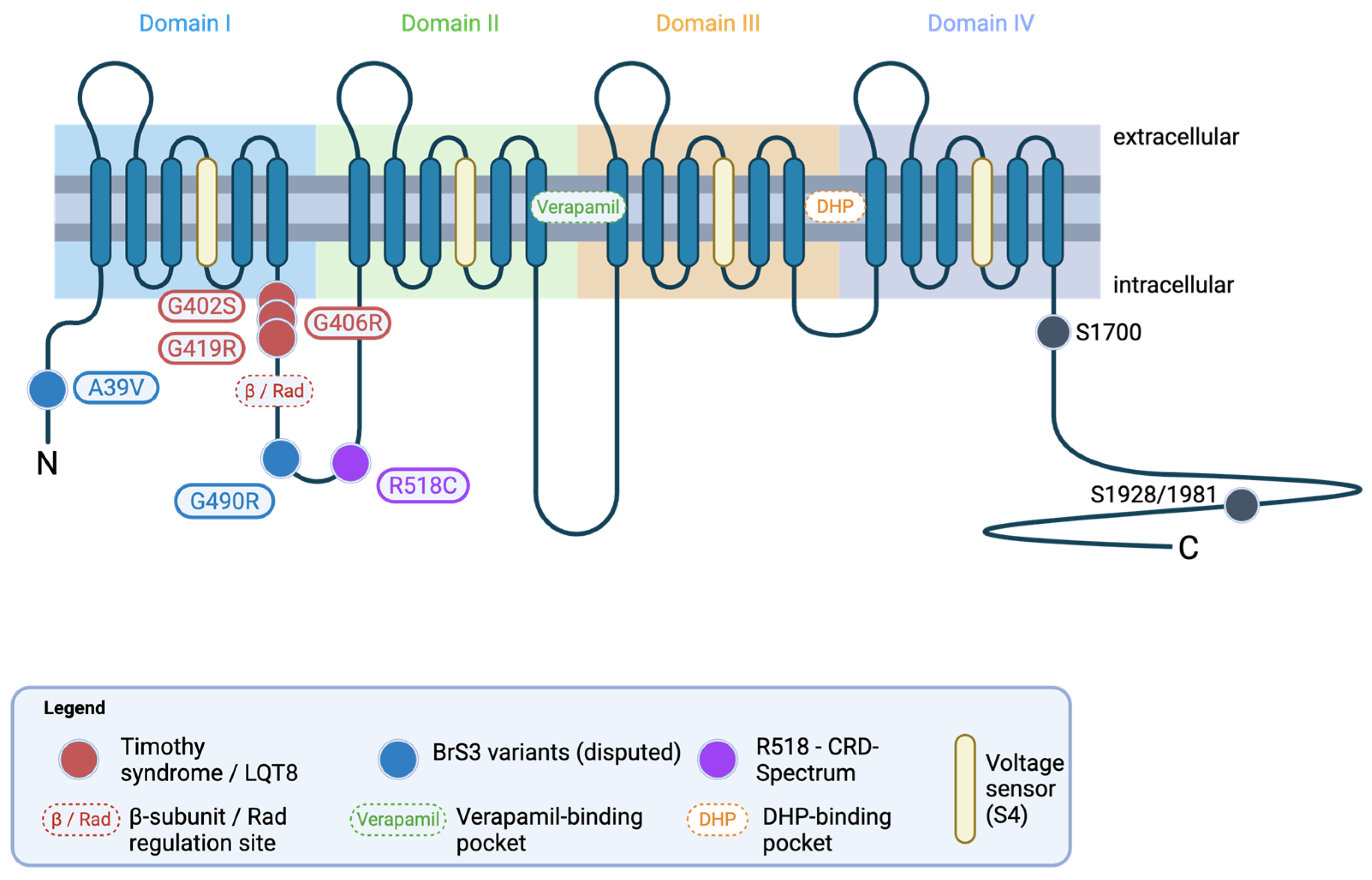
The CACNA1C gene, located on chromosome 12p13.33, encodes the pore-forming α1C subunit of the voltage-gated L-type calcium channel CaV1.2. Functional CaV1.2 channels are heteromeric protein complexes composed of the pore-forming α1C subunit together with auxiliary β and α2δ subunits and constitutively associated calmodulin (CaM) molecules, which are required for functional expression and cell surface trafficking [1,2]. The major channel characteristics including voltage sensor (S4 segments), ion selectivity, and pharmacological drug-binding sites reside in the α1C subunit, which consists of four domains (I-IV) with six transmembrane segments each (Figure 1). Similar to other L-type channels, CaV1.2 generates long-lasting calcium currents in response to strong membrane depolarization and is sensitive to blockade by dihydropyridines (DHP, such as nifedipine), phenylalkylamines (such as verapamil) and benzothiazepines (such as diltiazem) [3].
CaV1.2 is expressed throughout the human body and it is involved in the regulation of a multitude of pivotal processes. For example, CaV1.2 is found (1) in neurons in the nervous system, where it regulates various aspects of neuronal activity including gene expression and synaptic plasticity; (2) in cardiomyocytes in the heart, where it is critically involved in the regulation of heart rate, atrioventricular (AV) conduction and cardiac contractility; (3) in arterial smooth muscle cells, where it regulates arterial diameter and, hence, tissue perfusion and blood pressure; (4) in intestinal smooth muscle cells, where it regulates motility; or (5) in hormone-producing cells, like β-cells of the pancreas or chromaffin cells of the adrenal medulla, where it contributes to hormone secretion [3,4]. Not surprisingly, therefore, complete (homozygous) loss of Cacna1c is lethal in mice before embryonic day 14.5 [5]. Moreover, cardiomyocyte-restricted homozygous deletion of Cacna1c in adult mice is lethal within less than two weeks owing to a massive loss of cardiac contractility [6]. On the other hand, a gain-of-function mutation in CaV1.2 causes Timothy syndrome (TS), a severe multi-organ disorder characterized by cardiac and neuropsychiatric abnormalities including lethal cardiac arrhythmias, congenital heart disease, cognitive abnormalities and autism, but also by immune deficiency and hypoglycemia [7]. Thus, mammalian life is not feasible without CaV1.2 and major alterations in its expression or function cause severe disease that affects various organ systems, most of all heart and brain.
Figure 1. Domain structure and membrane topology of CaV1.2. CaV1.2 is a single integral membrane protein consisting of four domains (I–IV) with six transmembrane segments (S1–S6) each. The S4 voltage sensors are highlighted in yellow. Cryo-EM structures of human CaV1.2 in apo and ligand-bound conformations [8,9] have resolved the major drug-binding pockets: Dihydropyridines (DHP) bind a III–IV fenestration on the lipid-facing surface. Verapamil and other phenylalkylamines occlude the inner pore from the cytoplasmic side; this binding mode has been characterized in CaV1.1 cryo-EM structures and modeled for CaV1.2 by homology [10]. Variant mapping onto this schematic places gain-of-function variants (red; G406R, G402S, G419R) predominantly in proximity to the S6 segment of domain I (the activation gate); loss-of-function variants (blue; A39V, G490R) localize to the cytoplasmic N-terminal or I–II linker regions; the mixed variant R518C (purple) sits in the I–II linker. The β-subunit/Rad interaction site on the I-II linker mediates β-adrenergic regulation; two candidate PKA phosphorylation sites on the C-terminal tail are indicated: the proximal S1700 site and the distal S1928/S1981 site. Created in BioRender. Königstein, D. (2026) https://BioRender.com/w0fm4tk.
Biomolecules 16 01459 g001

2. CACNA1C/CaV1.2 in Neuropsychiatric Disorders and Behavior

2.1. Genetic Evidence Implicating CACNA1C in Neuropsychiatric Disorders

The first evidence linking CaV1.2 to neuropsychiatric phenotypes emerged from investigations of TS, a rare multisystem disorder resulting from de novo gain-of-function mutations in CACNA1C [7]. TS is characterized by cardiac arrhythmias and a distinct neuropsychiatric phenotype including cognitive abnormalities and autism. The exonic G406R missense mutation (Figure 1) leads to increased CaV1.2 channel opening at subthreshold potentials as well as reduced voltage-dependent inactivation resulting in elevated calcium entry. Since its first introduction, the spectrum of phenotypes in TS has expanded to include more variants and affected organ systems. The high variability in the expression of CACNA1C and the resultant physical, physiological and behavioral outcomes has led to a call for a standardized definition for individuals presenting with pathogenic variants that may be linked to CACNA1C [11,12].
In addition to rare coding variants, genome-wide association studies (GWAS) have identified multiple common neuropsychiatric risk-associated single-nucleotide polymorphisms (SNPs) with a substantial genetic overlap between different diagnoses. Over the past two decades, CACNA1C has emerged as one of the most robust and reproducible risk loci across various neuropsychiatric disorders including schizophrenia, bipolar disorder, major depressive disorder, attention deficit-hyperactivity disorder, and autism spectrum disorder [13]. Several psychiatric risk-associated SNPs have been found within non-coding regions of CACNA1C, most prominently the intronic variant rs1006737 (risk allele A, global minor allele frequency 0.30, odds ratio ~1.15) [14,15]. These variants are enriched in intron 3, which harbors regulatory elements that physically interact with the CACNA1C promoter through chromatin looping. Neuropsychiatric risk alleles within this region disrupted enhancer–promoter communication, thereby decreasing transcriptional activity and reducing CACNA1C mRNA expression [16]. Functional studies of rs1006737 report heterogeneous effects on CACNA1C mRNA abundance, with evidence for both up- and downregulation [17,18,19]. This variability likely reflects context-dependent regulation influenced by developmental stage, brain region, cellular identity, and potentially isoform-specific expression patterns. Given the extensive alternative splicing of CACNA1C, it is also plausible that neuropsychiatric risk variants influence exon usage or isoform composition rather than total gene expression, although this possibility remains insufficiently characterized [20]. Collectively, these findings support a model in which common non-coding variants modulate disease susceptibility through subtle regulatory effects on CACNA1C expression and isoform balance, in contrast to rare coding mutations that produce pronounced alterations in channel function and high disease penetrance.

2.2. Cacna1c Rodent Models of Neuropsychiatric Disorders

Rodent models, most often mice and rats, provide a powerful translational tool for investigating the behavioral consequences of genetic variation associated with neuropsychiatric disorders [21]. There exists a broad array of standardized paradigms used to assess phenotypes relevant to neuropsychiatric disease, spanning emotional, cognitive, and social domains. Several key characteristics can be assessed across these domains to help build a more thorough phenotypic profile. For example, anxiety and locomotor-related behaviors are commonly evaluated using open-field assessment, a light–dark box, and/or an elevated plus maze [21,22]. Depression-relevant phenotypes are assessed most often using a tail-suspension task or the well-known forced swim test [23,24]. Morris water maze and spatial learning tasks can be used to evaluate learning and memory and other cognitive traits [25,26,27]. Emotional processing and associative learning are investigated using fear conditioning paradigms, in which freezing behavior, and more recently 22 kHz ultrasonic vocalizations (USVs)—thought to be indices of negative affect—are the primary readouts [28,29]. In addition, assays for social interaction and socio-affective communication, in the form of the three-chamber social approach task or direct reciprocal social interaction during social play, together with the assessment of concomitant emission of USVs, have become increasingly important for exhibiting phenotypes relevant to autism spectrum disorder and other neuropsychiatric conditions with marked social behavior and communication deficits [30,31,32,33].
Accordingly, Cacna1c rodent models have been examined across many of these domains and exhibit phenotypes encompassing alterations in anxiety, emotional learning, social behavior, socio-affective communication through USVs, and cognitive function.

2.3. In Vivo Behavioral Findings in Cacna1c Mouse Models

Several of the results obtained in mouse models for Cacna1c have corroborated findings seen in human CACNA1C data, linking anxiety and anti-depressive behavioral endophenotypes associated with neuropsychiatric disorders [34,35]. Translational relevance provided by mouse models provides insight into the role of Cacna1c in the neurocircuitry of neuropsychiatric disorders.
Initial studies in Cacna1c mouse models, commonly generated on C57BL/6 backgrounds, have established the central role of CaV1.2 in the regulation of affective, cognitive, and social behaviors, with evidence implicating prefrontal cortex–amygdala–hippocampal circuitry [36]. Constitutive and region-specific reductions and deletions in Cacna1c expression levels in adult males and females consistently produce anxiety-like phenotypes [34,37,38,39,40]. In contrast, in a recent transgenic Cacna1c mouse model with increased expression levels of CaV1.2, the adult mice exhibited increased risk taking and higher exploratory levels of behavior [41]. Findings related to depression-like behaviors have been less consistent, with several studies reporting antidepressant-like responses in the forced swim and tail suspension tests [34,40,42], while the overexpression of CaV1.2 resulted in no depression-like behavior in the Porsolt forced swim test [41]. Reduced Cacna1c expression appears to strongly attenuate psychostimulant-induced locomotion in adult females but is also evident in males, indicating the role of CaV1.2 in mesolimbic dopamine-dependent behaviors [34,43]. Moreover, the mouse model for CaV1.2 gain-of-function characterizing TS has displayed characteristic ASD-like traits analogous to those seen to be at the core of ASD, including social and learning impairments, repetitive behavior patterns and altered vocalizations [37,44,45,46]. In contrast, the pan-neuronal overexpression of CaV1.2 resulted in no deficits in social behavior in adult mice with both sexes able to distinguish a mouse from an object and intact abilities to distinguish a familiar mouse [41].
Evidence from fear conditioning paradigms has been variable, although altered fear processing, aversive learning and emotional memory processing have been reported following both loss- and gain-of-function manipulations. While some studies report minimal or no impairment, TS, characterized by gain-of-function models in adult male mice, shows enhanced contextual and cue-associated fear memory persistence [37], supporting the role of CaV1.2 in aversive learning and emotional memory encoding relevant to stress- and anxiety-related pathologies. However, in the transgenic Cacna1c overexpression mouse model [41], the authors reported a failure in long-term memory consolidation in the adult mutant mice. Although TS knock-in mice suggest that gain-of-function CaV1.2 mutations can produce distinct behavioral phenotypes relative to loss-of-function models, broader interpretations of CACNA1C function are complicated by heterogeneous findings across studies, including variability in behavioral directionality and molecular expression patterns [44].
Cacna1c has also been implicated in hippocampal-dependent cognition, with forebrain- and hippocampus-dependent deletions producing impairments in adult mice in terms of spatial learning and long-term memory, particularly under conditions of increased demand [47,48]. In contrast, adult male TS knock-in mice show relatively intact acquisition but impaired cognitive flexibility, suggesting that gain- versus loss-of-function manipulations differentially affect distinct aspects of learning and executive control [37]. More recently, integrated behavioral analysis has further refined these observations. In a cell-type- and circuit-specific Cacna1c deletion model targeting dopamine D1 receptor-expressing neurons, dissociable effects on emotional and cognitive domains were found, with enhanced or prolonged contextual or cued fear memory, alongside selective impairments in spatial memory performance that were dependent on sex-gene dosage [49]. Female mice showed enhanced aversive memory with partial Cacna1c loss, while males required a complete loss of Cacna1c expression to show enhanced fear behaviors [49]. For males, impairment in spatial learning was found after both partial and complete Cacna1c expression loss, while no effect was observed in females [49]. These findings extend previous work by highlighting that both the affected neuronal populations and biological sex critically shape Cacna1c-related behavioral phenotypes. Alterations in the CaV1.2 channel consistently report sex-dependent effects across domains [34,50].
Alterations in social behavior in adult mice are reported, although findings remain heterogeneous. Forebrain and prefrontal Cacna1c deletions have been associated with reduced social preferences in some studies [42,50,51], while constitutive heterozygous models show no consistent social deficits and may even exhibit enhanced sociability in certain paradigms [37]. In the recent Cacna1c deletion model targeting dopamine D1 receptor-expressing neurons [49], no effect on adult social behavior was observed in males or females during a three-chamber social interaction test. In contrast, adult male mice with deletion of Cacna1c in CaMKII-expressing cells do show impaired social preference [43], highlighting the importance of Cacna1c expression in specific cell types and the role of sex in determining behavioral effects. More broadly, social phenotypes such as these are often interpreted within a neuropsychiatric framework spanning disorders characterized by altered social motivation and interaction, including autism spectrum-related characteristics among broader affective and psychotic spectrum conditions. Together, mouse models have helped establish a broader behavioral profile for Cacna1c across affective, cognitive and social domains, albeit with variability across manipulations and tasks with most conducted in adult mice. Given their greater suitability for complex behavioral phenotyping and more complex social repertoire from an early developmental age, rat models have increasingly been used to further probe and refine these Cacna1c-associated phenotypes.

2.4. In Vivo Behavioral Findings in the Cacna1c Haploinsufficient Rat Model

Comprehensive behavioral studies on the Cacna1c haploinsufficient (Cacna1c+/−) rat model have been conducted to investigate its effects on social, emotional, and cognitive behaviors over time. Similar to the mouse, behavioral measures in the Cacna1c rat model align with core symptoms of human neuropsychiatric disorders, such as those linked to CACNA1C by GWAS [52]. In a series of studies, a comprehensive gene x environmental approach was applied to assess several behavioral domains across the lifespan in a genetic rat model haploinsufficient for the cross-disorder risk gene Cacna1c on a Sprague–Dawley background. Of prominent note, alterations in socio-affective behavior and communication appear to be key traits in the Cacna1c rodent model during early life [31,53,54,55] and adulthood [56,57]. Evidence for the role of Cacna1c haploinsufficiency has also been shown through several studies focusing on cognitive and emotional processing [58,59,60,61].

2.5. Social Behavior and Communication

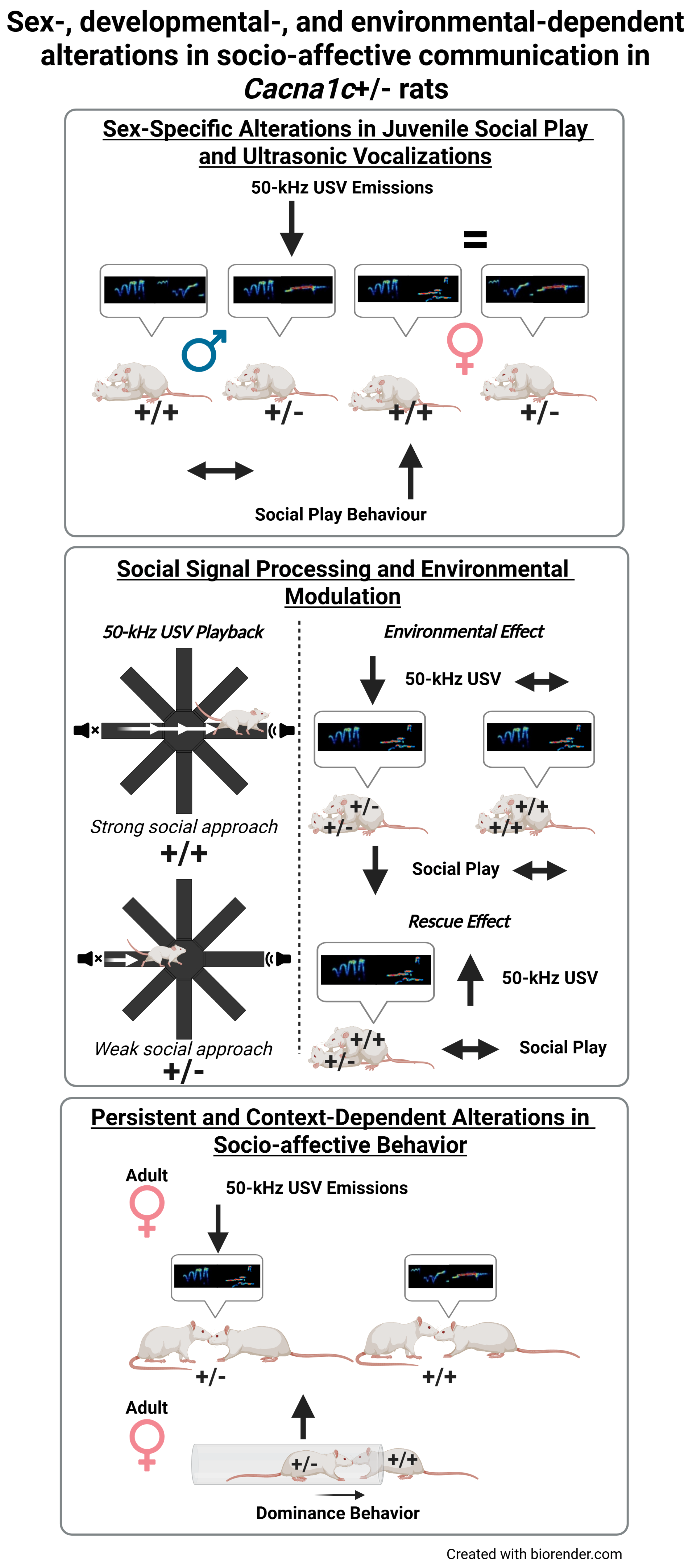
Notable social and communication alterations have been repeatedly observed in Cacna1c+/− rats (Figure 2). These occur in both the sender and receiver as both adults and juveniles. Evidence from several studies have shown that Cacna1c haploinsufficiency leads to deficits in socio-affective communication, affecting both 22 kHz and 50 kHz USVs (for review see: [62]). Juvenile and adult rats produce two types of USV [63,64]. The 22 kHz “alarm calls”, usually evoked by aversive stimuli or exposure to predators, typically lead to behavioral inhibition [65,66], and 50 kHz “rat laughter” calls are associated with positive affect and rewarding experiences such as social play and mating [67,68,69]. The 50 kHz USVs are characterized by a high level of complexity and contain a variety of subtypes ranging from two to fourteen [70,71,72]. The emission and response to these prominent socio-affective stimuli appear to be altered as a result of the alteration in CaV1.2 channel expression.
In juvenile rats, a highly prominent feature of their social repertoire involves social play behavior [73]. When examining social play and concomitant 50 kHz USV emission, sex-specific effects are evident. Male Cacna1c+/− rats show typical social play behaviors but emit far fewer 50 kHz USVs compared to wildtype controls, whereas pairs of juvenile Cacna1c+/− female rats exhibited masculinized social play—meaning a higher rate of play than what is typically observed in female rats—but had comparable 50 kHz USV emission rates to the female wildtype pairs [31,53]. While not emitting a higher number of 50 kHz calls, further analysis revealed that pairs of Cacna1c+/− females had a distinct 50 kHz USV subtype emission pattern resembling that of male wildtype controls [54]. Upon further investigation, it was determined that environmental factors in addition to genotype strongly influenced these outcomes for juvenile females. Particularly, it was found that in juvenile Cacna1c+/− females raised without wildtype cage mates (i.e., only with same genotype cage mates) there was a stark reduction in social play and emission of 50 kHz USVs compared to the pairs of wildtype females from same genotype cages. Interestingly, this reduction in play and 50 kHz USVs in Cacna1c+/− females is restored by providing the opportunity to engage and play with a wildtype play partner, demonstrating a prominent rescue effect for wildtype cage mates and playmates [55].
As adults, female Cacna1c+/− rats displayed mild social differences, showing more dominant behavior in tube tests, consistent with masculinized juvenile play behavior, but without increased aggression [56]. Adult females likewise showed a general reduction in 50 kHz USV emission during social interactions [56], suggesting that Cacna1c haploinsufficiency produces developmental alterations in social communication and socio-affective behaviors that are sex-specific and highly sensitive to environmental influences.
Complementing effects found in the sender, meaning the rats emitting USVs, Cacna1c+/− rats correspondingly showed changes as receivers of social signals. To assess communicative functions of USVs, a playback paradigm can be utilized [74]. Through this technique, strong social approach behavior is elicited in response to the playback of natural 50 kHz USVs, thought to be an indication of social preference, motivation and sociability, while the presentation of natural 22 kHz USVs typically leads to behavioral inhibition, which is in line with alarm and danger signals [74]. In Cacna1c+/− juvenile rats, social approach behavior triggered by the presentation of 50 kHz USVs was reduced in both the male and female Cacna1c+/− rats; however, effects were more pronounced in males [53]. Moreover, after USV playback ended the wildtype controls continued to display searching behavior whereas the Cacna1c+/− rats did not, suggesting a diminished social motivation. In line with these findings, Wöhr et al. (2020) [57] recently showed that Cacna1c+/− rats display alterations in emotional processing in response to aversive communication signals. In Cacna1c+/− males, but not Cacna1c+/− females, there was a reduction in behavioral inhibition to the presentation of 22 kHz USV playback while being exposed to predator urine. This suggests that Cacna1c haploinsufficiency may result in lower motivation or possibly diminished capabilities to respond appropriately to socio-affective communication signals [57].
Taken together these findings indicate that Cacna1c haploinsufficiency affects both the production and perception of socio-affective signals, with sex and environmental factors further shaping these phenotypes. The findings from the studies outlined above are highly relevant to neuropsychiatric disorders characterized by disturbance in social functioning, motivation and emotional regulation.
Beyond the alterations in social communication and social behavior, accumulating evidence suggests that changes extend into broader domains of emotional processing and reward-related behaviors, with more subtle effects observed on cognition and learning.

2.6. Emotional Processing, Reward Learning and Cognition

Consistent with the notion that alterations in socio-affective signaling extend beyond social interactions alone, evidence from emotional and cognitive paradigms suggests a more subtle and heterogeneous behavioral profile in Cacna1c+/− rats. Increased anxiety behaviors have been reported by Moon et al. (2025) [75] in adult male and female rats with a global reduction in CaV1.2 expression. In addition, heightened fear responses in fear conditioning paradigms have been reported in adult Cacna1c+/− rats relative to wildtype controls [76,77], supporting the role of CaV1.2 in emotional learning and stress-related processing. Similarly, the aforementioned study showing that male Cacna1c+/− rats displayed reduced behavioral inhibition in response to 22 kHz USV playback during predator odor exposure, further indicates altered processing of aversive socio-affective signals.
In line with findings in mice, alterations in reward-related behaviors have been observed in haploinsufficient Cacna1c rat models. During sign- and goal-tracking paradigms, female Cacna1c+/− rats emitted fewer than 50 kHz USVs and displayed a shift toward goal-tracking behavior, suggesting changes in incentive salience and motivational processes [61]. In the cognitive domain, male Cacna1c+/− rats exhibited intact spatial learning but mild impairments in cognitive flexibility during reversal learning, deficits that were ameliorated by environmental enrichment [58,59]. Similar impairments in reversal learning have been reported by Sykes et al. (2019) [76] and Moon et al. (2020) [77]. By contrast, post-weaning social isolation affected novel object recognition irrespective of genotype, whereas hippocampal overexpression of microRNA-499-5p abolished novel object preference in male Cacna1c+/− rats, suggesting that microRNAs may modulate neuroplasticity and behavioral outcomes [60,78,79]. Finally, although Gasalla et al. (2023) [80] observed no differences in Pavlovian extinction learning, distinct regional brain activation patterns were detected in adult male Cacna1c+/− rats, implying altered neural processing despite preserved behavioral performance.

2.7. Summary of Cacna1c Rodent Models

In summary, Cacna1c haploinsufficiency in rats appears to preferentially affect socio-affective communication and emotional processing, with comparatively subtle and task-dependent effects on cognition. Collectively, these findings suggest the role of CaV1.2 in the integration of social, motivational, and affective information, indicating that disrupted socio-affective signal processing may represent a particular sensitive behavioral consequence of reduced Cacna1c expression.
Despite these insights, several limitations should be considered when interpreting the findings form Cacna1c rodent models. Interspecies differences in brain development, behavioral repertoire, and Cacna1c regulation limit direct extrapolation to humans and may contribute to differences between mouse and rat models. Interpretation is further complicated by the type of genetic manipulation employed, as global or constitutive reductions in Cacna1c expression affect multiple cell types and circuits throughout development, whereas region- and cell-specific models can reveal more selective effects. Behavioral assays likewise provide measures of specific behavioral processes rather than direct equivalents of human psychiatric symptoms and have limited predictive value in isolation. Finally, the magnitude of manipulation in haploinsufficient, knockout, or gain-of-function models differs from common human CACNA1C variants identified through GWAS, which typically exert modest effects within a broader polygenic and environmental context. The rodent models, therefore, should primarily be considered mechanistic tools for identifying biological and behavioral processes sensitive to altered CaV1.2 signaling rather than direct representations of human CACNA1C-associated neuropsychiatric risk.

3. Cellular Mechanisms of CACNA1C/CaV1.2 Dysfunction in the Nervous System

3.1. Subcellular Localization and Functions of CaV1.2 in Neurons

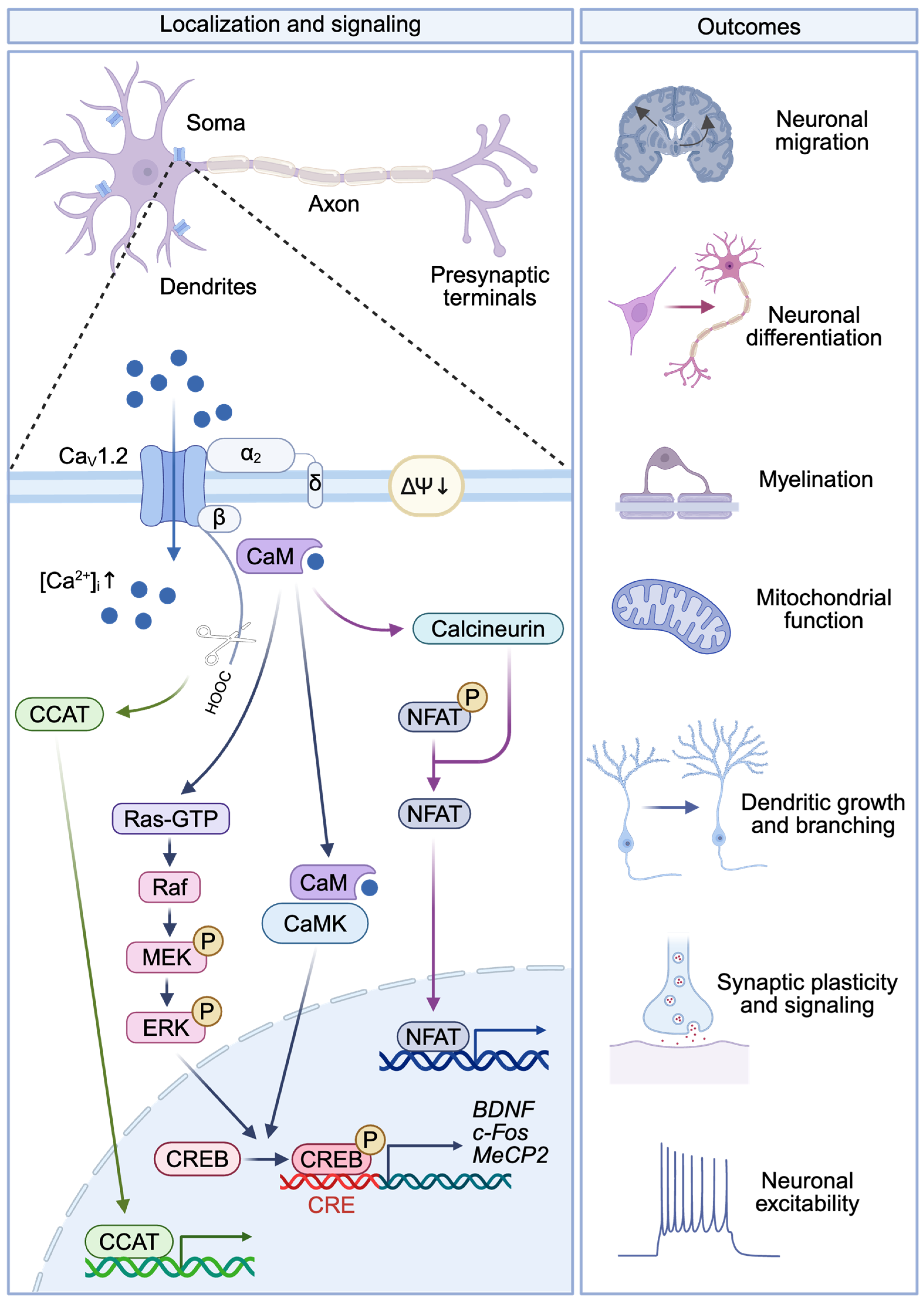
CaV1.2 represents the predominant L-type calcium channel (LTCC) subtype in the mammalian brain, accounting for approximately 80% of neuronal LTCCs [81,82]. Within neurons, CaV1.2 is primarily localized in somato-dendritic and postsynaptic regions, where it occupies a strategic position for coupling electrical activity to intracellular calcium signals that subsequently regulate transcriptional, structural, and synaptic processes (Figure 3) [83,84]. Consequently, CaV1.2 functions as a critical mediator of excitation–transcription coupling, linking neuronal activity to long-term cellular adaptations and biological processes increasingly implicated in the pathophysiology of neuropsychiatric disorders.

3.2. CaV1.2-Mediated Intracellular Signaling and Gene Regulation in Neurons

One of the most extensively characterized functions of CaV1.2 is the initiation of signaling cascades that convert transient electrical activity into sustained changes in gene expression (Figure 3). Following membrane depolarization, calcium entering through CaV1.2 binds to channel-associated calmodulin (CaM), triggering the activation of multiple intracellular signaling pathways. Among the best-established pathways are the Ca2+/CaM-dependent protein kinase (CaMK) cascade and the Ras/mitogen-activated protein kinase (MAPK)/extracellular signal-regulated kinase (ERK) pathway [85]. The activation of these signaling networks culminates in phosphorylation of the transcription factor cyclic adenosine monophosphate (cAMP) response element-binding protein (CREB), a master regulator of activity-dependent gene expression involved in neuronal plasticity, learning, and memory. CREB-dependent transcription constitutes one of the most robust downstream consequences of LTCC activation. Genes regulated through this pathway include brain-derived neurotrophic factor (BDNF), c-Fos, methyl-CpG-binding protein 2 (MeCP2), and numerous plasticity-related transcripts that influence neuronal maturation and synaptic remodeling [85,86,87]. Notably, the autism-associated CaV1.2G406R mutant exhibits constitutive CREB activation and spontaneous transcription of activity-dependent genes (c-Fos, MeCP2), even in the absence of depolarizing stimuli, suggesting that excessive CaV1.2 signaling can disrupt transcriptional homeostasis [88].
In addition to kinase-mediated pathways, CaV1.2 activates calcium-dependent phosphatase signaling. Ca2+/CaM-dependent activation of calcineurin promotes dephosphorylation and nuclear translocation of nuclear factor of activated T cells (NFAT) transcription factors, thereby regulating dendritic growth, synaptic plasticity and memory formation [89]. Furthermore, the proteolytic processing of the channel can generate a C-terminal fragment known as CCAT (calcium channel-associated transcription regulator), which translocates to the nucleus and directly regulates genes involved in neuronal excitability and signaling [90].

3.3. Role of CaV1.2 in Neuronal Migration and Differentiation

Calcium signaling regulates numerous developmental processes, including neuronal migration and differentiation, i.e., through cytoskeletal activity, making CaV1.2 an important determinant of cortical organization. Evidence from TS models indicates that excessive CaV1.2 activity disrupts migration of excitatory and inhibitory neurons. Prenatal expression of the TS mutation CaV1.2G406R in mice impairs radial migration of layer 2/3 excitatory neurons, whereas postnatal normalization of calcium influx can partially rescue these defects [91]. Human-induced pluripotent stem cell (iPSC)-derived forebrain assembloids have additionally demonstrated defects in tangential migration of TS inhibitory interneurons, characterized by shorter saltation length and increased saltation frequency that can be corrected by LTCC inhibition with nimodipine [92].
CaV1.2 also influences the differentiation of neural progenitors. Studies using neuronal cultures differentiated from TS patient-derived iPSCs and mice overexpressing CaV1.2G406R in neurons have demonstrated reduced numbers of lower-layer cells expressing SATB2, thereby decreasing the fraction of callosal projection neurons [93,94]. Conversely, Cacna1c knock-out increased SATB2 expression in the developing cortex of mice, indicating that calcium influx through CaV1.2 contributes to developmental programs controlling neuronal identity [93].

3.4. Regulation of Neuronal Morphology, Synaptic Plasticity, and Circuit Function by CaV1.2

CaV1.2-mediated calcium signaling exerts profound effects on neuronal morphology and connectivity. In both rodent and human TS models, mutant channels cause activity-dependent dendritic retraction and reduce the number of dendritic branches. These structural abnormalities are independent of calcium flux through TS-CaV1.2 and associated with activation of GTPase RhoA signaling that governs cytoskeletal remodeling [95]. In addition to dendritic development, CaV1.2 regulates axonal growth and connectivity. Expression of gain-of-function CaV1.2 channels in cortical layer 2/3 excitatory neurons diminished axonal arborization in the contralateral hemisphere [96]. Furthermore, enhanced maturation of oligodendrocyte progenitor cells isolated from cortex and increased axon myelination in the corpus callosum have been reported for TS mice [97].
At the synaptic level, CaV1.2 contributes to neuronal excitability and activity-dependent plasticity. Forebrain-specific deletion of Cacna1c leads to increased presynaptic excitatory inputs as well as higher frequency and amplitude of miniature excitatory postsynaptic currents in layer 5 pyramidal neurons, suggesting a shift in the excitation-inhibition balance, a circuit feature frequently implicated in neuropsychiatric disorders [42]. Moreover, CaV1.2 is required for specific forms of hippocampal long-term potentiation. Loss of CaV1.2 function impairs MAPK/CREB-mediated transcription in CA1 pyramidal neurons, linking channel activity to molecular mechanisms underlying spatial memory formation [47].

3.5. Impact of CaV1.2 on Mitochondrial Function, Oxidative Stress, and Neuronal Survival

Recent studies have extended the role of CaV1.2 beyond neuronal signaling and development to the regulation of mitochondrial homeostasis and cellular resilience. In immortalized mouse hippocampal neurons, both pharmacological LTCC inhibition and siRNA-mediated reduction in Cacna1c expression protect against glutamate-induced oxidative damage, preserving mitochondrial membrane potential, maintaining ATP levels, and preventing cell death [98,99]. These findings suggest that CaV1.2 occupies a critical position at the intersection of neuronal activity, energy metabolism, and stress responses. Dysregulated calcium entry may therefore contribute not only to developmental abnormalities and synaptic dysfunction but also to cellular vulnerability under pathological conditions. However, since this is a developing field of research, further validation of these results is still needed.

3.6. Potential Role of CaV1.2 Signaling in Neuroinflammatory and Peripheral Immune Signatures

In contrast to excitable cells such as neurons, cardiac and smooth muscle, where LTCCs are primarily activated by membrane depolarization, the mechanisms governing LTCC activity in non-excitable cells such as microglia and peripheral immune cells have not yet been fully elucidated and therefore represent a still evolving research field [100,101].
In microglia, the brain’s resident immune cells, intracellular calcium dynamics regulate phagocytosis, oxidative burst, inflammasome activation, and subsequent cytokine release. These signals arise from a network of purinergic receptors, store-operated calcium entry, transient receptor potential channels, intracellular stores, and ryanodine receptors (RyRs) [102,103]. LTCC-related pathways may thus contribute to microglial phenotype and function under inflammatory conditions [101]. Although CaV1.2 is not regarded as the dominant calcium entry route in resting microglia, it may act as context-dependent modulator of reactive states. Several studies combining channel detection with genetic manipulation provide evidence for CaV1.2 expression and activity in microglia. Espinosa-Parrilla et al. [104] showed that activated BV2 microglia express both CaV1.2 and CaV1.3 (and that nifedipine reduced TNF and NO release without affecting their phagocytotic capacity or viability). CaV1.2 activity has further been proposed to functionally regulate intracellular calcium fluctuations that affect microglial activation, migration, and the subsequent release of pro-inflammatory cytokines [101]. Direct evidence is provided by demonstrating that pharmacological blockade or knockdown of microglial CaV1.2 can disrupt microglial balance, promoting neurotoxic states, accelerating neurodegeneration, and exacerbating neuroinflammation in a murine model of Parkinson’s disease [105].
Simultaneously, findings suggest that CACNA1C/CaV1.2 expression in astrocytes may help to maintain the structural and functional integrity of the blood–brain barrier (BBB). In brain-specific Cacna1c-deficient mice, the deficiency or dysfunction in astrocytic CaV1.2 triggers severe astrocyte reactivity, causing upregulation of aquaporin-4 at astrocytic endfeet, destabilizing the BBB, and inducing a local increase in chemokines and inflammatory mediators [106].
In addition, the immunological influence of CACNA1C/CaV1.2 extends beyond the CNS, driving systemic immune responses that engage in cross-talk with neural pathology. As described before, pathogenic mutations, such as the gain-of-function variants underlying TS, have been linked to peripheral immune deficiencies and systemic cytokine dysregulation alongside severe neurodevelopmental deficits [7,100]. Although it results from rare, highly penetrant variants, it illustrates how altered CaV1.2 function can affect multiple organ systems, including the CNS and the peripheral immune system.
CaV1.2 expression has been found in several cell types of both the innate and the adaptive immune system [100,107]. Human dendritic cells, for example, express CaV1.2 functionally coupled to RyRs. In these cells, LTCC–RyR signaling has been shown to induce upregulation of CD83 and MHC class II surface molecules, evidencing the role of CaV1.2 in dendritic cell maturation and antigen presentation [108]. It has further been proposed that CaV1.2 (or related LTCC components) may contribute to T-cell-receptor-induced calcium signaling, Th2 differentiation, and cytokine production, since CaV1 knockdown reduced Th2 activation and ameliorated experimental asthma in mice [109,110]. However, this view of the role of CaV1.2 in immune cells has been challenged by findings demonstrating that voltage-gated calcium channels (CaV1.1 [111]; CaVβ [112]) do not function as ion channels in T cells [113] and regulate cellular function independently of voltage-gated ion flux [112] through protein interactions, scaffolding or modulation of gene expression (CaV1.1 [114]; CaVβ [115]). As the topic of CaV biology in peripheral immune cells lies beyond the scope of the present review, readers are referred to comprehensive reviews in the field [100,107,116,117,118]. Whether CaV1.2 similarly mediates non-canonical signaling or scaffolding functions in immune cells remains to be established.
When astrocytic CACNA1C/CaV1.2 dysfunction compromises the BBB, this peripheral–central divide collapses. Concomitantly, enhanced dendritic cell maturation may promote T-cell activation and release of pro-inflammatory mediators. Soluble inflammatory factors and, in severe cases of BBB disruption, also peripheral immune cells like monocytes and neutrophils, may gain access to brain parenchyma. This infiltration could amplify local neuroinflammation, reshape peripheral immune differentiation by microglia-secreted inflammatory mediators and thus create a feedback loop of chronic systemic immune activation. Consequently, CACNA1C/CaV1.2 can be regarded as an immunomodulatory hub [100], whose dysregulation couples local glial reactivity with systemic immune dysfunction, thereby potentially modulating the onset and progression of neuropsychiatric and neurodegenerative disorders [106]. Whether CACNA1C/CaV1.2 regulation associated with immune cell reactivity in neural disease can be exploited therapeutically needs to be further clarified in appropriate studies on disease-relevant genetic variants and targeted pharmacological interventions.

3.7. Summary of CaV1.2 Dysfunction in Neuronal and Non-Neuronal Cells in the CNS

CaV1.2 functions as a central molecular hub linking neuronal activity to intracellular signaling, gene transcription, cellular development, and circuit plasticity (Figure 3). Genetic evidence from TS and large-scale GWAS has firmly established CACNA1C as a major neuropsychiatric risk gene, while mechanistic studies have revealed how altered CaV1.2 signaling can influence neuronal migration, differentiation, dendritic architecture, synaptic plasticity, and cell survival. Further, CACNA1C mutations have been linked to altered immune responses in non-excitable cells such as in microglia and astrocytes, as well as to pathology-associated peripheral immune signatures, suggesting CaV1.2 is an immunomodulatory hub of neuro-immune cross-talk in neurodevelopmental and neuropsychiatric disorders. Together, these findings support a model in which perturbations of CaV1.2-dependent signaling could represent a convergent mechanism underlying vulnerability to neurodevelopmental and neuropsychiatric disorders. However, important limitations remain, particularly regarding the cell-type-specific functions of CaV1.2 in glia, its relationship to mitochondrial dysfunction, and the mechanisms by which altered CaV1.2 signaling intersects with immune pathways. The extent to which these processes represent primary drivers, downstream consequences, or interacting components of disease pathology has yet to be fully elucidated and warrants further investigation.

4. CACNA1C/CaV1.2 in the Heart

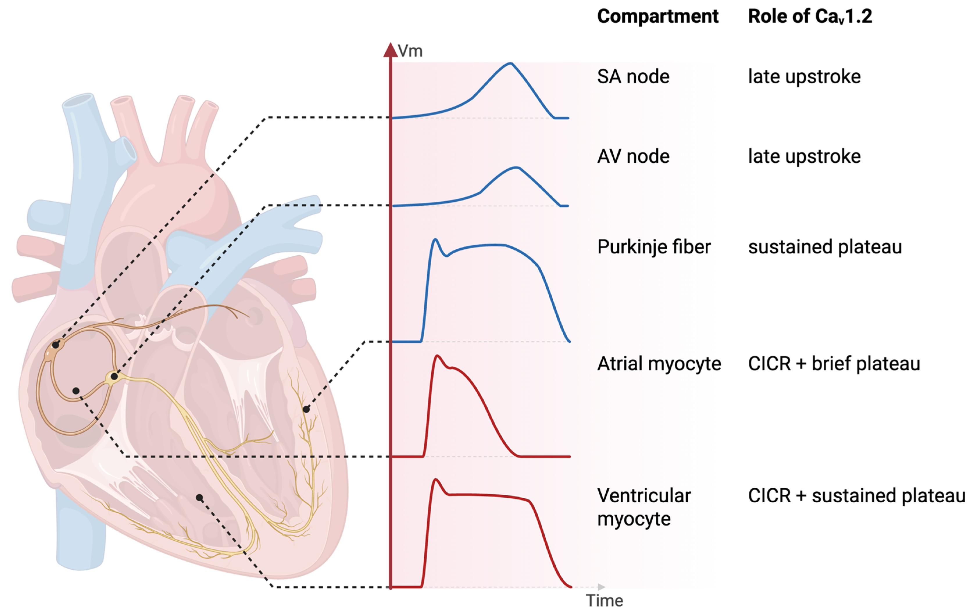
CaV1.2 is expressed throughout the mammalian heart, but its roles vary by cardiac compartment [3,4] (Figure 4), shaping how CACNA1C variants may cause disease. By converting membrane depolarization into calcium entry, CaV1.2 supports nodal impulse formation and, in working myocardium, sustains the action potential (AP) plateau and couples excitation to contraction. In the sinoatrial (SA) node, CaV1.2 is recruited mainly during the later phase of the upstroke of the AP. In the atrioventricular (AV) node, CaV1.2 contributes to the slow upstroke of the AP. Thus, LTCC blockers like verapamil impair SA and AV nodal APs and exert negative-chronotropic and negative-dromotropic effects. These effects have led to the use of cardiac-specific LTCC blockers (verapamil, diltiazem) as anti-arrhythmic drugs (AADs) [119,120], e.g., in patients with atrial fibrillation (AF). Cardiac Purkinje myocytes exhibit a sustained plateau phase (similar to ventricular myocytes), and this plateau phase is largely mediated by CaV1.2 [121]. In atrial and ventricular myocytes, CaV1.2 triggers calcium-induced calcium release (CICR) and, hence, is essential for cardiac excitation–contraction coupling. Interference with this process, e.g., by LTCC blockers, results in impaired cardiac contractility (negative-inotropic effect), an effect that prohibits the use of cardiac-specific LTCC blockers (class IV AADs) in patients with heart failure (HF) (with reduced ejection fraction) [119,122].
In summary, CaV1.2 exerts various pivotal functions in both pacemaker cells and working cardiomyocytes, which makes the heart particularly vulnerable to alterations in CaV1.2 expression and function.
The cardiac phenotype of CACNA1C dysfunction is variant- and mechanism-dependent rather than uniform. Gain-of-function variants underlying TS and isolated long QT syndrome type 8 (LQT8) constitute the strongest gene–disease evidence for CACNA1C in cardiac disease. By contrast, loss-of-function alleles and the proposed CACNA1C–Brugada syndrome association remain mechanistically plausible but weakly supported at the gene level; each reported variant therefore needs to be assessed individually, using clinical presentation, family segregation and functional data. Across these mechanistic axes, regulatory layers that dynamically tune CaV1.2 function—for example the β-adrenergic signaling node—help explain why reduced channel expression does not necessarily mean reduced basal current density [6,123,124] and why biophysically identical variants can produce different clinical phenotypes (TS1 vs. TS2) [125].

4.1. Gain-of-Function Variants: Timothy Syndrome and Long QT Syndrome 8

4.1.1. Timothy Syndrome (TS): A Splice-Variant-Graded Severity Spectrum

The best-characterized cardiac manifestation of multisystem CACNA1C dysfunction is TS [7]. The canonical TS1 mutation, G406R in the alternatively spliced exon 8A, markedly impairs voltage-dependent inactivation of CaV1.2, producing a sustained inward calcium current that prolongs the cardiac AP and underlies QT prolongation and arrhythmia susceptibility. Clinically, QTc typically exceeds 500 ms, accompanied by a prolonged isoelectric ST segment, functional 2:1 AV block, T-wave alternans, and a high risk of torsade-de-pointes and ventricular fibrillation. In contrast to the predominantly cardiac gain-of-function phenotypes discussed later, TS1 is defined by multisystem involvement, including neuropsychiatric features (see above), syndactyly, immune dysregulation and intermittent hypoglycemia.
TS2 is caused by the identical G406R substitution in exon 8 rather than exon 8A and carries an even more severe cardiac phenotype [125]. The severity gradient is quantitative and follows splice-isoform abundance: the dominant cardiac CaV1.2 splice variant (exon 8) comprises ≈80% of cardiac transcripts [125,126]. The minor isoform (exon 8A/α1C-b) is predominantly expressed in vascular smooth muscle and developmentally enriched in neurons. Splicing between exons 8 and 8A is regulated by polypyrimidine tract-binding protein (PTB) and its neuronal homolog nPTB [126]. A TS1 (exon 8A) mutation therefore affects a small fraction of cardiac CaV1.2; a TS2 (exon 8) mutation affects the dominant pool—hence the earlier-onset and more lethal phenotype of TS2. The same inactivation defect therefore produces different severities depending on how much of cardiac CaV1.2 it affects.
Like the exon-8 G406R variant, G402S markedly impairs voltage-dependent inactivation. Because exon 8-containing CaV1.2 transcripts predominate in the heart (~80% of cardiac transcripts), G402S is associated with a severe cardiac TS2 phenotype with marked QT prolongation and life-threatening ventricular arrhythmias [125,127]. G419R, by contrast, represents a mechanistically distinct gain-of-function variant. Rather than impairing inactivation, G419R produces an increase in peak calcium current density and a hyperpolarizing shift in activation gating linked to calmodulin-dependent regulation, thereby enhancing CaV1.2 activity at physiological membrane potentials [128,129]. Clinically, G419R has been associated with TS features together with left ventricular non-compaction and ventricular pre-excitation via multiple accessory pathways, a phenotype not previously reported in TS. In a second family, an alternative nucleotide substitution at the same codon (c.1255G>A) produced the same p.G419R amino-acid change and was associated with familial transmission and survival into middle adulthood [130]. Together, G402S and G419R illustrate that pathogenic CACNA1C gain of function can arise through distinct gating abnormalities and can produce a broad spectrum of clinical severity.
The recent international Delphi-consensus statement of the Timothy Syndrome Alliance [12] retains the designations TS1 (syndactyly—featuring classical exon-8A p.G406R phenotype) and TS2 (hip dysplasia—featuring exon-8 variants, including p.G406R and p.G402S as in Splawski’s original definition), while introducing “CACNA1C-Related Disorder (CRD)” as an umbrella term for syndromic CACNA1C variants that do not fulfill the classical TS constellation. Attenuated familial presentations, such as the G419R phenotype reported by Fanucci et al. (2025) [130], also fall within this expanded framework. The CRD terminology thus accommodates an increasingly broad phenotypic and genetic spectrum, including recently reported variants such as p.C1021R [131].
Borbás et al. (2022) [132] systematically reviewed 59 TS, 6 cardiac-only TS (COTS), and 20 isolated LQT8 index cases, documenting significantly earlier onset and higher mortality in TS than in either COTS or LQT8. Congenital heart defects—including patent ductus arteriosus, ventricular septal defects, and tetralogy of Fallot—recur in the case literature, although precise frequencies are difficult to establish given disease rarity and probable publication bias toward severe presentations. Delinière et al. (2023) [127] further characterized the G402S variant, and Han et al. (2019) [133] and Jiang and Zhang (2023) [134] reviewed the mechanistic and clinical spectrum from cell to bedside. Across the spectrum, impaired channel inactivation is the shared molecular lesion, whereas clinical expression is modulated by splice-isoform context, variant-specific biophysics, and likely genetic background.

4.1.2. Cardiac-Only TS and Isolated LQT8

Not all gain-of-function variants produce the complete TS phenotype. Some cause severe QT prolongation, structural abnormalities, and sudden death without multisystem developmental features; the extra-cardiac TS phenotype appears to depend on mutation-specific biophysical properties and their developmental timing. Boczek et al. (2015) [135] identified R518C in a pedigree with long QT (LQT) interval, hypertrophic cardiomyopathy, congenital heart disease, and sudden cardiac death, revealing a mixed gain- and loss-of-function biophysical profile—a finding that remains mechanistically unresolved. One possibility is that the variant has differential effects on distinct gating states or in cellular contexts not fully captured by heterologous expression systems. Gakenheimer-Smith et al. (2021) [136] documented AF and sick sinus syndrome in carriers of the same variant across four generations. This finding illustrates both within-pedigree variability and the extension of CACNA1C dysfunction to supraventricular and conduction tissue, consistent with the established role of CaV1.2 in SA pacemaker APs and atrial AP duration. Fukuyama et al. (2014) [137] identified five novel mutations in 2.5% of LQT syndrome (LQTS) probands, with phenotypes ranging from mild QT prolongation to near-TS severity. Wemhöner et al. (2015) [138] helped define non-syndromic LQT8 as a clinical entity distinct from TS. Whether the current three-tier classification (TS/COTS/LQT8) adequately captures the data, or whether a grouping based on the underlying molecular mechanism would be more informative, remains an open question.
Taken together, CACNA1C gain-of-function disease is not uniform and best understood by the specific biophysical defect and the developmental context in which that defect operates.

4.2. Loss-of-Function Variants: Brugada Syndrome Type 3 and Short QT Syndrome

Loss-of-function variants reduce L-type calcium current and produce an electrophysiologically distinct phenotype. Antzelevitch et al. (2007) [139] identified CACNA1C mutations (A39V, G490R) in patients with ST segment elevation, short QT intervals (≤360 ms), and sudden cardiac death, leading to the historical designation Brugada syndrome type 3 (BrS3). Subsequent gene–disease validity reappraisal has substantially weakened this association. SCN5A remains the only gene with definitive evidence for BrS causality, whereas CACNA1C and other historically implicated BrS genes are considered to have disputed gene–disease validity [140,141]. Consistent with this reassessment, the historically prominent variant G490R occurs in population databases at a frequency that is difficult to reconcile with a rare, highly penetrant arrhythmia syndrome.
The proposed mechanism is physiologically plausible: reduced L-type calcium current could increase repolarization heterogeneity and thereby favor an arrhythmogenic substrate. However, mechanistic plausibility alone does not establish gene-level causality, and current evidence does not support CACNA1C as an established monogenic cause of classic BrS. This contrasts with the CACNA1C–Timothy syndrome/LQT8 spectrum, for which gain-of-function variants are supported by substantially stronger genetic, functional and mechanistic evidence.

4.3. CACNA1C Haploinsufficiency and Regulatory Control of CaV1.2

4.3.1. Cardiac Consequences of Cacna1c Haploinsufficiency

The human cardiovascular consequences of isolated CACNA1C haploinsufficiency remain poorly defined, and most data derive from rodent models. As mentioned before, constitutive homozygous loss of Cacna1c is lethal in mice before embryonic day 14.5 [5]. Using a cardiomyocyte-specific, tamoxifen-inducible Cre approach (α-MHC-MerCreMer × Cacna1c-floxed mice, with loxP sites flanking exon 2; 6–7-week-old mice of either sex), Rosati et al. (2011) [6] showed that adult homozygous deletion of Cacna1c is lethal within ≈12 days, with myocardial contractility falling to ≈14% of control. This has restricted in vivo work on chronically reduced expression of CaV1.2 to heterozygous (Cacna1c+/−) and tissue-restricted conditional models.
Two constitutive heterozygous models have been used extensively in this context: a global Cacna1c+/− mouse [142] and a global Cacna1c+/− rat carrying a zinc-finger-nuclease-generated 4 bp deletion in exon 6 that results in a premature stop codon [123,124]. In the rat studies discussed here, female animals aged 9–15 months were analyzed [123,124]. Both models are viable and do not appear to exhibit excess mortality. Their cardiac phenotype—at least under basal, non-stressed conditions—is surprisingly mild [123,142], in particular when considering the many pivotal functions of CaV1.2 in the heart and the severe cardiac phenotype observed in TS (including lethal arrhythmias). The heart rate in Cacna1c+/− mice is slightly increased. The heart weight to body weight ratio is normal in Cacna1c+/− rats [123] or increases only at older ages in Cacna1c+/− mice [142]. Cardiac contractility (as judged from echocardiography or sarcomere/cell shortening measurements in ventricular myocytes) is slightly decreased (mice [142]) or unchanged (rats [123]). Similarly, ventricular myocyte calcium transients and SR calcium load are only slightly decreased (mice) or essentially unchanged (rats). Finally, and in line with the contractility and calcium data, L-type calcium current is either only slightly reduced (mice) or unchanged (rats). These observations imply that—in both rodent models—the hearts and cardiomyocytes must have evolved mechanisms to compensate for the loss of one Cacna1c allele and retain (almost) normal function.
In both cases, these compensatory mechanisms converge on altered intracellular calcium regulation and altered kinase or phosphatase signaling to maintain cellular function, but they come at the expense of compromised responses to stress. In Cacna1c+/– mice [142], paradigms of pathological or physiological stress, i.e., pressure overload by aortic constriction, chronic isoproterenol (a β-adrenergic receptor agonist) infusion, or forced swimming, caused exaggerated hypertrophy, ventricular dysfunction, and dilation. This cardiac phenotype could be explained by elevated neurohumoral stress resulting (1) in sensitized SR calcium release with leaky type-2 ryanodine receptors (RyR2s) with augmented phosphorylation at Ser-2808 and Ser-2814 to compensate for reduced calcium influx via CaV1.2 and (2) in augmented calcineurin signaling to drive the exaggerated hypertrophy response [142]. On the other hand, cardiomyocytes from Cacna1c+/– rats exhibited an impaired sympathetic stress response [123]. Here, the isoproterenol-induced increase in cytosolic calcium transients and contractility was blunted due to elevated PKA-dependent phosphorylation of RyR2 (at Ser-2808) and of CaV1.2 (at Ser-1928; rat/rabbit numbering used here; Ser-1927 in mouse/guinea-pig; the corresponding distal C-terminal residue is annotated as Ser-1981 in canonical human α1C, UniProt Q13936-1, based on the sequence alignment in [143]; see also Figure 1) under baseline conditions, resulting in a compromised sympathetic reserve [123,124].
The conceptual lesson from the Cacna1c+/– mouse and rat models is that the heart exhibits remarkable plasticity in compensating for reduced Cacna1c expression to retain an apparently (close to) normal function under basal, non-stressed conditions [6,123,142] and that physiological and pathological stressors unveil a cardiac phenotype characterized by an inability to respond adequately to sympathetic and pressure overload stress [123,142]. Whether the rodent models generalize to the human heart remains unknown.

4.3.2. Regulatory Control of CaV1.2: β-Adrenergic Regulation and Beyond

The clinical phenotype of a CACNA1C variant depends not only on its intrinsic biophysical effect but on regulatory layers that dynamically shape channel availability, gating, and expression. Four axes are most relevant in the cardiac setting: β-adrenergic regulation, CaMKII-mediated facilitation, redox modulation, and transcriptional/post-transcriptional control. Rather than reviewing each axis in mechanistic depth, this and the following section highlight how these regulatory layers intersect with a central paradox of Cacna1c haploinsufficiency: loss of one Cacna1c allele would predict a proportional halving of CaV1.2 expression and sarcolemmal L-type calcium influx, yet CaV1.2/α1C protein falls by only ≈20–40% and peak L-type calcium current is either essentially unchanged or reduced by only ≈25% across Cacna1c+/− mouse and rat models [6,123,124,142].
Physiologically, sympathetic activation is arguably the most important means of augmenting cardiac contractility, and this is achieved largely by β-adrenergic stimulation of L-type calcium current and elevated cytosolic calcium transients [144]. β-adrenergic activation leads to a huge increase in L-type calcium current through PKA-dependent phosphorylation of the channel macromolecular complex. The field has shifted from an α1C-centered phosphorylation model (at Ser-1700/Thr-1704 and Ser-1928/1981, Figure 1) toward a Rad-centered model in which PKA phosphorylation of the RGK protein Rad releases its inhibitory grip on channel gating [145,146,147,148], mechanistically analogous to PKA phosphorylation of phospholamban relieving the inhibition of SERCA during β-adrenergic stimulation. In 4SA-Rad knock-in mice, prevention of Rad phosphorylation markedly attenuates β-adrenergic augmentation of Ca2+ influx and contractility [147,149,150]. The additional contribution of direct α1C phosphorylation, however, particularly at Ser-1700/Thr-1704, remains an option [151,152]. In conclusion, PKA-dependent phosphorylation of Rad is now widely accepted as the primary mediator of L-type calcium current elevation during β-adrenergic stimulation in cardiomyocytes. Direct α1C phosphorylation by PKA, on the other hand, may serve a secondary modulatory role in L-type calcium current regulation.
Beyond β-adrenergic regulation, CaV1.2 activity is further shaped by additional means including CaMKII-dependent facilitation and redox modulation acting through α1C thiol oxidation and CaMKII activation, which becomes pathologic in heart failure [153,154,155]. Total channel abundance is further set by transcriptional and post-transcriptional inputs, notably mineralocorticoid-driven Cacna1c promoter switching [156] and miR-328/miR-499/miR-155 activity in AF [157,158,159]. It remains unknown though whether any of these axes is operating in hearts of animals with Cacna1c haploinsufficiency.

4.3.3. The Paradox of Preserved Calcium Current in Cacna1c Haploinsufficiency

A central mechanistic puzzle in Cacna1c haploinsufficiency is that loss of one gene copy does not produce a proportional loss of CaV1.2 expression and L-type calcium current. Multiple independent studies on mouse and rat models converge on a mismatch across the gene-to-current pathway in adult ventricular myocytes: In a mouse model, mRNA was reduced to ≈60% and CaV1.2 protein to ≈80% of control, while L-type current was not significantly reduced [6]. In another mouse model, CaV1.2 protein was reduced to ≈60% of control, i.e., by ≈40%, yet L-type current was reduced by only ≈25% [142]. Finally, in a Cacna1c+/− rat model, CaV1.2 protein was reduced to ≈70% of control, but neither L-type current nor sarcolemmal calcium influx were changed [123,124].
Several cellular mechanisms could contribute to this compensation, and a further observation frames the altered local regulatory context in which they operate:
(i) Post-transcriptional or proteostatic buffering: Rosati et al. (2011) [6] proposed that the mismatch reflects a passive bottleneck: one of the biosynthetic steps downstream of transcription becomes saturated—for example, ER folding capacity or the pool of β and α2δ auxiliary subunits—so that halving mRNA does not translate into halving protein. Faster translation or slower protein degradation could also contribute, but neither has been demonstrated in Cacna1c+/− hearts.
(ii) Preferential preservation of the functional surface pool. Reduced total CaV1.2 protein need not translate into a proportional reduction in sarcolemmal, dyadically localized channels: preferential membrane retention, altered turnover of surface-resident channels, increased clustering, altered trafficking or shifts in auxiliary subunit stoichiometry could each preserve the gating-competent channel count in the membrane [160,161]. Königstein et al. (2024) [124] found unchanged total CaVβ2 protein in Cacna1c+/− rat ventricles. This means that—because total α1C expression is reduced—the bulk CaVβ2-to-α1C abundance ratio is increased. Whether this alters subunit availability, channel assembly, or surface delivery remains unknown. The general possibility of a preserved functional surface pool remains untested and would require quantitative surface protein/channel measurements.
(iii) Altered local PKA-mediated phosphorylation of Cav1.2 and Rad in the vicinity of the channel: As mentioned above, Rad is an important regulator of channel activity. Rad holds a fraction of surface CaV1.2 channels in a low open probability state, and PKA phosphorylation of Rad releases this brake. Thus, increased Rad phosphorylation would recruit these “reserve” channels. In the Cacna1c+/− mouse, reduced L-type current and contractile performance were accompanied by mild tachycardia, and metoprolol (a β-adrenergic receptor blocker) attenuated the disease phenotype, consistent with compensatory neuroendocrine activation [142]. Such β-adrenergic drive could, in principle, increase tonic Rad phosphorylation and recruit reserve channels; however, Rad phosphorylation and its contribution to basal current have not been examined directly in Cacna1c+/− myocardium.
Along these lines, basal PKA phosphorylation of α1C at Ser-1928 was found to be increased in Cacna1c+/− rat ventricles [124]. While phosphorylation of this site might not be required for β-adrenergic augmentation of the L-type calcium current in cardiomyocytes [162], it nevertheless serves as a sensitive readout for local PKA activity in the vicinity of the channel and hence suggests that, under basal conditions, CaV1.2-mediated calcium influx is normalized in the face of reduced channel number, possibly by elevated PKA-dependent phosphorylation of the CaV1.2 macromolecular complex, including Rad.
Taken together, these findings converge on a consistent phenotype in cardiomyocytes: basal CaV1.2 function, i.e., L-type calcium current, is largely preserved despite reduced Cacna1c expression due to an elevated basal PKA-mediated phosphorylation of the channel and presumably Rad; this basal localized PKA “hyperphosphorylation” leaves a less sympathetic reserve for channel regulation. Hence, during acute sympathetic stress, the cardiomyocytes are unable to recruit a sufficient number of additional channels—due to an already exhausted PKA phosphorylation reserve—resulting in impaired contractile function. The phenotypic manifestations of this exhausted reserve are consistent across models: exaggerated hypertrophy, ventricular dysfunction and dilation after pressure overload, chronic isoproterenol infusion or forced swimming in the Cacna1c+/− mouse [142], and blunted inotropic and Ca2+-transient responses to isoproterenol in the Cacna1c+/− rat, despite preserved basal function [123,124]. The concept of an exhausted sympathetic reserve thus provides a mechanistic framework linking preserved basal function to altered cardiac responses under stress in Cacna1c haploinsufficiency.

5. Clinical Perspective: Killing Two Birds with One Stone by Targeting CACNA1C/CaV1.2?

In clinical practice, there are mutual associations between neuropsychiatric disorders, like major depression, bipolar disorder, autism or schizophrenia, and many cardiovascular diseases including hypertension, coronary artery disease (CAD), HF or AF. For example, depression may increase the risk for CAD, myocardial infarction, HF or AF; vice versa, patients with AF or CAD have an increased risk of developing depression [163,164,165,166,167,168]. Patients with schizophrenia have reduced life expectancy (by 10–20 years); this is caused, in large part, by increased cardiac mortality, with mortality rates for CAD, myocardial infarction, HF, and cardiac arrhythmias being increased by a factor of about 2–3 each [169,170]. Likewise, genetic evidence strongly implicates CACNA1C variants in many neuropsychiatric disorders as well as cardiovascular disease (as already discussed above). Data from rodent models largely confirm that altered expression and function of CaV1.2 may affect both neurological function and behavior as well as cardiac function. This suggests that CaV1.2 may be a common mechanistic link between neuropsychiatric and cardiac disorders and that appropriate targeting of CaV1.2 may be used to treat both neuropsychiatric and cardiac disease in a given (selected) patient at the same time.
LTCC blockers (CCBs) have been used clinically for decades to treat cardiac arrhythmias (as class IV AADs like verapamil and diltiazem), CAD (as anti-anginal drugs, DHPs and non-DHPs) and hypertension (as anti-hypertensive drugs, DHPs and non-DHPs). Thus, there is ample clinical evidence regarding their desired effects but also their unwanted side effects, and both can be explained well by their inhibition of LTCCs in the respective tissues and cell types. These include (1) negative chronotropic, dromotropic and inotropic effects in the heart due to inhibition of LTCCs in SA and AV nodal pacemaker cells and ventricular myocytes; (2) the dilation of coronary arteries due to inhibition of LTCCs in coronary artery smooth muscle cells; and (3) reduction in blood pressure due to inhibition of LTCCs in arterial smooth muscle cells of the peripheral circulation. The (4) latter effect on arterial smooth muscle cells may also facilitate the development of flush and ankle edema, frequently observed side effects of CCBs. Finally, they include (5) obstipation caused by impaired intestinal motility due to inhibition of LTCCs in intestinal smooth muscle cells. Whether CCBs also act in the brain to inhibit LTCCs in neurons and non-neuronal cells and whether this might be exploited clinically to treat neurological or neuropsychiatric disorders is less clear though. Clinical studies in the psychiatric setting conducted so far often suffer from limitations such as small sample size, potential indication bias, or being retrospective and observational and the overall evidence in support of the clinical utility of CCBs in such settings remains controversial with some studies hinting at a potential benefit while others do not confirm any such effect or even report adverse outcomes. For example, six double-blind trials found no evidence for an effect of verapamil in acute mania [171]. However, RCTs were small (157 patients in total) and of short duration. In the four trials in which verapamil was compared to lithium, lithium treatment was found not to be superior to verapamil [171]. In a Mendelian randomization study, genetically proxied CCBs showed no protective effect on mental disorders [172]. Some large observational studies, however, reveal more promising results. In a study with >140,000 individuals from the Swedish population with bipolar disorder, schizophrenia or nonaffective psychosis, use of CCBs was associated with reduced rates of psychiatric hospitalization and self-harm in all three subgroups [173]. In two studies with individuals from the Finnish population with either bipolar disorder (>60,000) or schizophrenia (>61,000), the use of CCBs was associated with a reduced risk of hospitalization due to affective symptoms [174] or a reduced risk of psychiatric rehospitalization [175], respectively. Interestingly, in both studies, the effect was consistently present for DHPs but absent for verapamil, while diltiazem showed mixed results [174,175]. In a study with patients from Scotland, the use of CCB monotherapy (as compared to monotherapy with other antihypertensive drugs) was associated with increased hospital admission rates for mood disorders, mainly major depressive disorder [176]. Similarly, a recent meta-analysis also found an association between the use of CCBs and the risk of depression (albeit very small with an odds ratio of 1.06) [177]. By contrast, in a nationwide study in Denmark, the use of a few antihypertensive drugs, including the CCBs amlodipine and verapamil, was associated with decreased risk of depression [178]. Some studies also looked into the potential role of brain penetrance of the CCBs in distinguishing between non-brain-penetrant DHPs (mainly amlodipine) and brain-penetrant DHPs (most other DHPs including felodipine, isradipine, nifedipine and nitrendipine). Again, results were mixed. One study revealed that brain-penetrant DHPs were associated with a reduced incidence of several psychiatric disorders (e.g., schizophrenia, affective disorder, major depressive disorder) when compared with amlodipine [179]. By contrast, another study did not find an association between the type of DHP (brain-penetrant versus non-brain-penetrant) and the risk of incident psychiatric disorder (schizophrenia, schizoaffective disorder, major depressive disorder, bipolar disorder) [180]. Thus, these mixed results argue against the notion that CCBs might be suitable as a general treatment for a broad range of psychiatric disorders. On the other hand, they do not rule out the possibility that novel, more selective CCBs might be of considerable value for treating certain psychiatric disorders. Afterall, currently available CCBs are neither selective for CACNA1C/CaV1.2 nor do they act on specific brain circuits. It is most likely, that a multitude of factors may decide about the potential beneficial effects of a given CCB in a given patient including (but not limited to) the class of the CCB used (DHPs, phenylalkylamines (verapamil), benzothiazepines (diltiazem), or novel, more selective CCBs to be developed) and its pharmacokinetics (e.g., crossing of the BBB), genetic background of the patient (e.g., CACNA1C variant present), type of neuropsychiatric disorder, and comorbidities present. Clearly, a better understanding of these factors is mandatory in order to identify patients in whom certain CCBs could be used to treat certain neuropsychiatric disorders successfully, but also to identify patients in whom certain CCBs should be avoided because they might be harmful.

6. Conclusions

CACNA1C/CaV1.2 acts as a voltage-gated LTCC in cardiomyocytes and neurons. Its proper function is critical for maintaining normal cardiac and brain physiology. Consequently, dysfunctional CACNA1C/CaV1.2 has been implicated in cardiac disease (e.g., arrhythmias), where activation of CaV1.2 channels is directly involved in the action potential of pacemaker cells and cells of the working myocardium, and neuropsychiatric disorders as diverse as schizophrenia, bipolar disorder, major depressive disorder, attention deficit-hyperactivity disorder, and autism spectrum disorder. At the cellular level, in the brain, CaV1.2 links neuronal electrical activity with intracellular signaling, gene transcription, cellular development and circuit plasticity. Recent evidence suggests that CaV1.2 also serves pivotal functions in non-neuronal cells in the brain, including astrocytes and microglia, where it may be involved in maintaining the integrity of the BBB and in regulating microglial activation, respectively. Similarly, CaV1.2 or CaV1.1 have been found in peripheral immune cells. The mode of activation of CaV1.2 in electrically non-excitable cells is still not well-understood and some studies suggest that it may rather act as a signaling hub or by other means.
Rodent models with global heterozygous or cell-type- or region-specific knockout of Cacna1c represent valuable tools to study the specific roles of CaV1.2 in heart and brain function as well as its potential involvement in cardiac and neuropsychiatric disorders. Behavioral studies in Cacna1c+/− mouse and rat models have revealed, in part, age- and sex-dependent alterations in socio-affective communication and emotional processing (including anxiety-like behavior) as well as more subtle cognitive alterations. The cellular neuronal mechanisms underlying these behavioral alterations, however, often remain elusive. Moreover, the cardiac phenotype of the same models indicates altered cardiac stress responses, lending support to the general notion that altered expression and/or function of CACNA1C/CaV1.2 is sufficient to cause both cardiac and behavioral/neuropsychiatric abnormalities. However, whether—and to what extent—these findings can be translated to human pathophysiology remains an open question.
Currently available LTCC blockers (mainly DHPs, diltiazem, verapamil), which are used clinically to treat cardiovascular disease (hypertension, CAD, arrhythmias), have been evaluated for their potential usefulness in patients with neuropsychiatric disorders with mixed results. The available LTCC blockers, however, are neither selective for CaV1.2, nor do they act on specific organs or brain circuits. More selective CaV1.2-modulating drugs may be required for this purpose. In addition, large, randomized, placebo-controlled, prospective clinical trials are needed to provide more robust evidence for—or against—the usefulness and efficacy of a certain calcium channel blocker (or modulator) in a certain neuropsychiatric disorder and patient.

Author Contributions

Writing—original draft preparation, D.K., T.M.K., S.M., J.A., C.C., J.K.; writing—review and editing, all authors; visualization, D.K., T.M.K., S.M., M.W., J.K.; supervision, M.W., C.C., J.K.; project administration, S.M., J.K. All authors have read and agreed to the published version of the manuscript.

Funding

This research was funded by the Flexi Funds Programme of Forschungscampus Mittelhessen (FCMH) [Projektnummer 2018_1_1_2 to C.C. and J.K.]. Our research on Cacna1c haploinsufficiency in rats has benefited from long-term support by grants from the Deutsche Forschungsgemeinschaft (DFG, German Research Foundation) to M.W. (WO 1732/4-1, WO 1732/4-2, Project-ID 290878970—RTG 2271; Project-ID 422744262—CRC 289; Project-ID 521379614—CRC 393), and grants from the Fonds Wetenschappelijk Onderzoek—Vlaanderen (FWO; Research Foundation—Flanders) to M.W. (G0C0522N, G0G4421N, I007022N). S.M. receives funding by the European Union (MSCA Postdoctoral Fellowship Psy-hiPSC microglia). The work of J.A. was supported by NEURON-ERA-NET 2021 (MINERVA—Grant 01EW2209B) and in part by the SFB/TRR 393 consortium from the DFG (subproject A05). The work of C.C. was supported by NEURON-ERA-NET 2021 (MINERVA—Grant 01EW2209A) and in part by the SFB/TRR 393 consortium from the DFG (Project ID 521379614; subprojects B04,B08).

Institutional Review Board Statement

Not applicable.

Informed Consent Statement

Not applicable.

Data Availability Statement

No new data were created or analyzed in this study. Data sharing is not applicable to this article.

Acknowledgments

During the preparation of this manuscript, the authors used, in part, Perplexity (Perplexity AI, https://www.perplexity.ai/, accessed on 18 September 2026) to support initial literature search and DeepL Write (DeepL SE, Cologne, Germany, https://www.deepl.com/en, accessed on 18 September 2026) for language editing and improvement of grammar and style. All identified references were retrieved as primary sources and independently verified by the authors, who take full responsibility for the content of this publication.

Conflicts of Interest

The authors declare no conflicts of interest. The funders had no role in the design of the study; in the interpretation of data; in the writing of the manuscript; or in the decision to publish the study.

Abbreviations

The following abbreviations are used in this manuscript:
AADAnti-arrhythmic drug
AFAtrial fibrillation
APAction potential
AVAtrioventricular
BBBBlood–brain barrier
CACNA1CGene encoding the α1C subunit of the CaV1.2 channel
CADCoronary artery disease
CaMCalmodulin
CCBCalcium channel blocker
CICRCalcium-induced calcium release
CNSCentral nervous system
CREBCyclic adenosine monophosphate response element-binding protein
DHPDihydropyridine(s)
ERKExtracellular signal-regulated kinase
GWASGenome-wide association studies
HFHeart failure
LQTLong QT syndrome
LTCCL-type calcium channel(s)
MAPKMitogen-activated protein kinase
PKAProtein kinase A
RyRRyanodine receptor
SASinoatrial
SNPsSingle-nucleotide polymorphisms
TSTimothy syndrome
USVUltrasonic vocalization

References

  1. Catterall, W.A.; Perez-Reyes, E.; Snutch, T.P.; Striessnig, J. International Union of Pharmacology. XLVIII. Nomenclature and structure-function relationships of voltage-gated calcium channels. Pharmacol. Rev. 2005, 57, 411–425. [Google Scholar] [CrossRef] [Scilit] [PubMed]
  2. Dolphin, A.C. Calcium channel auxiliary α2δ and β subunits: Trafficking and one step beyond. Nat. Rev. Neurosci. 2012, 13, 542–555. [Google Scholar] [CrossRef] [Scilit] [PubMed]
  3. Zamponi, G.W.; Striessnig, J.; Koschak, A.; Dolphin, A.C. The Physiology, Pathology, and Pharmacology of Voltage-Gated Calcium Channels and Their Future Therapeutic Potential. Pharmacol. Rev. 2015, 67, 821–870. [Google Scholar] [CrossRef] [Scilit] [PubMed]
  4. Hofmann, F.; Flockerzi, V.; Kahl, S.; Wegener, J.W. L-type Cav1.2 calcium channels: From in vitro findings to in vivo function. Physiol. Rev. 2014, 94, 303–326. [Google Scholar] [CrossRef] [Scilit] [PubMed]
  5. Seisenberger, C.; Specht, V.; Welling, A.; Platzer, J.; Pfeifer, A.; Kuhbandner, S.; Striessnig, J.; Klugbauer, N.; Feil, R.; Hofmann, F. Functional embryonic cardiomyocytes after disruption of the L-type α1C (Cav1.2) calcium channel gene in the mouse. J. Biol. Chem. 2000, 275, 39193–39199. [Google Scholar] [CrossRef] [Scilit] [PubMed]
  6. Rosati, B.; Yan, Q.; Lee, M.S.; Liou, S.R.; Ingalls, B.; Foell, J.; Kamp, T.J.; McKinnon, D. Robust L-type calcium current expression following heterozygous knockout of the Cav1.2 gene in adult mouse heart. J. Physiol. 2011, 589, 3275–3288. [Google Scholar] [CrossRef] [Scilit] [PubMed]
  7. Splawski, I.; Timothy, K.W.; Sharpe, L.M.; Decher, N.; Kumar, P.; Bloise, R.; Napolitano, C.; Schwartz, P.J.; Joseph, R.M.; Condouris, K.; et al. CaV1.2 calcium channel dysfunction causes a multisystem disorder including arrhythmia and autism. Cell 2004, 119, 19–31. [Google Scholar] [CrossRef] [Scilit] [PubMed]
  8. Gao, S.; Yao, X.; Chen, J.; Huang, G.; Fan, X.; Xue, L.; Li, Z.; Wu, T.; Zheng, Y.; Huang, J.; et al. Structural basis for human Cav1.2 inhibition by multiple drugs and the neurotoxin calciseptine. Cell 2023, 186, 5363–5374.e16. [Google Scholar] [CrossRef] [Scilit] [PubMed]
  9. Wei, Y.; Yu, Z.; Wang, L.; Li, X.; Li, N.; Bai, Q.; Wang, Y.; Li, R.; Meng, Y.; Xu, H.; et al. Structural bases of inhibitory mechanism of CaV1.2 channel inhibitors. Nat. Commun. 2024, 15, 2772. [Google Scholar] [CrossRef] [Scilit] [PubMed]
  10. Li, W.; Shi, G. How CaV1.2-bound verapamil blocks Ca2+ influx into cardiomyocyte: Atomic level views. Pharmacol. Res. 2019, 139, 153–157. [Google Scholar] [CrossRef] [Scilit] [PubMed]
  11. Cipriano, L.; Piscopo, R.; Aiello, C.; Novelli, A.; Iolascon, A.; Piscopo, C. Expanding the Phenotype of the CACNA1C-Associated Neurological Disorders in Children: Systematic Literature Review and Description of a Novel Mutation. Children 2024, 11, 541. [Google Scholar] [CrossRef] [Scilit] [PubMed]
  12. Underwood, J.F.G.; Timothy, K.W.; Tyroll, H.; Levy, R.J.; Dick, I.E.; Pitt, G.S.; Tunbridge, E.M.; Baban, A.; Wilkinson, G.; Hall, N.A.L.; et al. Timothy syndrome and CACNA1C-Related Disorder: First international language and management guidelines consensus statement. Eur. J. Hum. Genet. 2026. [Google Scholar] [CrossRef] [Scilit] [PubMed]
  13. Cross-Disorder Group of the Psychiatric Genomics Consortium. Identification of risk loci with shared effects on five major psychiatric disorders: A genome-wide analysis. Lancet 2013, 381, 1371–1379. [Google Scholar] [CrossRef] [Scilit] [PubMed]
  14. Ferreira, M.A.; O’Donovan, M.C.; Meng, Y.A.; Jones, I.R.; Ruderfer, D.M.; Jones, L.; Fan, J.; Kirov, G.; Perlis, R.H.; Green, E.K.; et al. Collaborative genome-wide association analysis supports a role for ANK3 and CACNA1C in bipolar disorder. Nat. Genet. 2008, 40, 1056–1058. [Google Scholar] [CrossRef] [Scilit] [PubMed]
  15. Green, E.K.; Grozeva, D.; Jones, I.; Jones, L.; Kirov, G.; Caesar, S.; Gordon-Smith, K.; Fraser, C.; Forty, L.; Russell, E.; et al. The bipolar disorder risk allele at CACNA1C also confers risk of recurrent major depression and of schizophrenia. Mol. Psychiatry 2010, 15, 1016–1022. [Google Scholar] [CrossRef] [Scilit] [PubMed]
  16. Roussos, P.; Mitchell, A.C.; Voloudakis, G.; Fullard, J.F.; Pothula, V.M.; Tsang, J.; Stahl, E.A.; Georgakopoulos, A.; Ruderfer, D.M.; Charney, A.; et al. A role for noncoding variation in schizophrenia. Cell Rep. 2014, 9, 1417–1429. [Google Scholar] [CrossRef] [Scilit] [PubMed]
  17. Bigos, K.L.; Mattay, V.S.; Callicott, J.H.; Straub, R.E.; Vakkalanka, R.; Kolachana, B.; Hyde, T.M.; Lipska, B.K.; Kleinman, J.E.; Weinberger, D.R. Genetic variation in CACNA1C affects brain circuitries related to mental illness. Arch. Gen. Psychiatry 2010, 67, 939–945. [Google Scholar] [CrossRef] [Scilit] [PubMed]
  18. Gershon, E.S.; Grennan, K.; Busnello, J.; Badner, J.A.; Ovsiew, F.; Memon, S.; Alliey-Rodriguez, N.; Cooper, J.; Romanos, B.; Liu, C. A rare mutation of CACNA1C in a patient with bipolar disorder, and decreased gene expression associated with a bipolar-associated common SNP of CACNA1C in brain. Mol. Psychiatry 2014, 19, 890–894. [Google Scholar] [CrossRef] [Scilit] [PubMed]
  19. Yoshimizu, T.; Pan, J.Q.; Mungenast, A.E.; Madison, J.M.; Su, S.; Ketterman, J.; Ongur, D.; McPhie, D.; Cohen, B.; Perlis, R.; et al. Functional implications of a psychiatric risk variant within CACNA1C in induced human neurons. Mol. Psychiatry 2015, 20, 162–169. [Google Scholar] [CrossRef] [Scilit] [PubMed]
  20. Harrison, P.J.; Husain, S.M.; Lee, H.; Los Angeles, A.; Colbourne, L.; Mould, A.; Hall, N.A.L.; Haerty, W.; Tunbridge, E.M. CACNA1C (CaV1.2) and other L-type calcium channels in the pathophysiology and treatment of psychiatric disorders: Advances from functional genomics and pharmacoepidemiology. Neuropharmacology 2022, 220, 109262. [Google Scholar] [CrossRef] [Scilit] [PubMed]
  21. Cryan, J.F.; Holmes, A. The ascent of mouse: Advances in modelling human depression and anxiety. Nat. Rev. Drug Discov. 2005, 4, 775–790. [Google Scholar] [CrossRef] [Scilit] [PubMed]
  22. Weiss, S.M.; Lightowler, S.; Stanhope, K.J.; Kennett, G.A.; Dourish, C.T. Measurement of anxiety in transgenic mice. Rev. Neurosci. 2000, 11, 59–74. [Google Scholar] [CrossRef] [Scilit] [PubMed]
  23. Bergner, C.L.; Smolinsky, A.N.; Hart, P.C.; Dufour, B.D.; Egan, R.J.; Laporte, J.L.; Kalueff, A.V. Mouse models for studying depression-like states and antidepressant drugs. In Mouse Models for Drug Discovery; Methods in Molecular Biology; Humana Press: New York, NY, USA, 2010; Volume 602, pp. 267–282. [Google Scholar] [CrossRef] [Scilit] [PubMed]
  24. Porsolt, R.D.; Bertin, A.; Jalfre, M. Behavioral despair in mice: A primary screening test for antidepressants. Arch. Int. Pharmacodyn. Ther. 1977, 229, 327–336. [Google Scholar] [PubMed]
  25. Morellini, F. Spatial memory tasks in rodents: What do they model? Cell Tissue Res. 2013, 354, 273–286. [Google Scholar] [CrossRef] [Scilit] [PubMed]
  26. Morris, R.G. Spatial localization does not require the presence of local cues. Learn. Motiv. 1981, 12, 239–260. [Google Scholar] [CrossRef] [Scilit]
  27. Olton, D.S.; Samuelson, R.J. Remembrance of places passed: Spatial memory in rats. J. Exp. Psychol. Anim. Behav. Process. 1976, 2, 97–116. [Google Scholar] [CrossRef]
  28. Fendt, M.; Fanselow, M.S. The neuroanatomical and neurochemical basis of conditioned fear. Neurosci. Biobehav. Rev. 1999, 23, 743–760. [Google Scholar] [CrossRef] [Scilit] [PubMed]
  29. Wohr, M.; Borta, A.; Schwarting, R.K. Overt behavior and ultrasonic vocalization in a fear conditioning paradigm: A dose-response study in the rat. Neurobiol. Learn. Mem. 2005, 84, 228–240. [Google Scholar] [CrossRef] [Scilit] [PubMed]
  30. Silverman, J.L.; Yang, M.; Lord, C.; Crawley, J.N. Behavioural phenotyping assays for mouse models of autism. Nat. Rev. Neurosci. 2010, 11, 490–502. [Google Scholar] [CrossRef] [Scilit] [PubMed]
  31. Kisko, T.M.; Braun, M.D.; Michels, S.; Witt, S.H.; Rietschel, M.; Culmsee, C.; Schwarting, R.K.W.; Wohr, M. Sex-dependent effects of Cacna1c haploinsufficiency on juvenile social play behavior and pro-social 50-kHz ultrasonic communication in rats. Genes Brain Behav. 2020, 19, e12552. [Google Scholar] [CrossRef] [Scilit] [PubMed]
  32. Gzielo, K.; Potasiewicz, A.; Litwa, E.; Piotrowska, D.; Popik, P.; Nikiforuk, A. The Effect of Maternal Immune Activation on Social Play-Induced Ultrasonic Vocalization in Rats. Brain Sci. 2021, 11, 344. [Google Scholar] [CrossRef] [Scilit] [PubMed]
  33. Wohr, M.; Scattoni, M.L. Behavioural methods used in rodent models of autism spectrum disorders: Current standards and new developments. Behav. Brain Res. 2013, 251, 5–17. [Google Scholar] [CrossRef] [Scilit] [PubMed]
  34. Dao, D.T.; Mahon, P.B.; Cai, X.; Kovacsics, C.E.; Blackwell, R.A.; Arad, M.; Shi, J.; Zandi, P.P.; O’Donnell, P.; Knowles, J.A.; et al. Mood Disorder Susceptibility Gene CACNA1C Modifies Mood-Related Behaviors in Mice and Interacts with Sex to Influence Behavior in Mice and Diagnosis in Humans. Biol. Psychiatry 2010, 68, 801–810. [Google Scholar] [CrossRef] [Scilit] [PubMed]
  35. Kircher, T.; Wohr, M.; Nenadic, I.; Schwarting, R.; Schratt, G.; Alferink, J.; Culmsee, C.; Garn, H.; Hahn, T.; Muller-Myhsok, B.; et al. Neurobiology of the major psychoses: A translational perspective on brain structure and function—The FOR2107 consortium. Eur. Arch. Psychiatry Clin. Neurosci. 2019, 269, 949–962. [Google Scholar] [CrossRef] [Scilit] [PubMed]
  36. Kabir, Z.D.; Lee, A.S.; Rajadhyaksha, A.M. L-type Ca2+ channels in mood, cognition and addiction: Integrating human and rodent studies with a focus on behavioural endophenotypes. J. Physiol. 2016, 594, 5823–5837. [Google Scholar] [CrossRef] [Scilit] [PubMed]
  37. Bader, P.L.; Faizi, M.; Kim, L.H.; Owen, S.F.; Tadross, M.R.; Alfa, R.W.; Bett, G.C.; Tsien, R.W.; Rasmusson, R.L.; Shamloo, M. Mouse model of Timothy syndrome recapitulates triad of autistic traits. Proc. Natl. Acad. Sci. USA 2011, 108, 15432–15437. [Google Scholar] [CrossRef] [Scilit] [PubMed]
  38. Lee, A.S.; Ra, S.; Rajadhyaksha, A.M.; Britt, J.K.; De Jesus-Cortes, H.; Gonzales, K.L.; Lee, A.; Moosmang, S.; Hofmann, F.; Pieper, A.A.; et al. Forebrain elimination of cacna1c mediates anxiety-like behavior in mice. Mol. Psychiatry 2012, 17, 1054–1055. [Google Scholar] [CrossRef] [Scilit] [PubMed]
  39. Smedler, E.; Louhivuori, L.; Romanov, R.A.; Masini, D.; Dehnisch Ellstrom, I.; Wang, C.; Caramia, M.; West, Z.; Zhang, S.; Rebellato, P.; et al. Disrupted Cacna1c gene expression perturbs spontaneous Ca2+ activity causing abnormal brain development and increased anxiety. Proc. Natl. Acad. Sci. USA 2022, 119, e2108768119. [Google Scholar] [CrossRef] [Scilit] [PubMed]
  40. Loganathan, S.; Menegaz, D.; Delling, J.P.; Eder, M.; Deussing, J.M. Cacna1c deficiency in forebrain glutamatergic neurons alters behavior and hippocampal plasticity in female mice. Transl. Psychiatry 2024, 14, 421. [Google Scholar] [CrossRef] [Scilit] [PubMed]
  41. Parent, R.; Lin, R.L.; Ouillette, L.; Glass, E.; Burns, H.; Uhler, M.D.; Case, S.L.; Thibault, O.; Murphy, G.G. The Impact of Overexpression of the Mouse Ortholog of CACNA1C on Behavior and Cortical Dynamics. Biol. Psychiatry Glob. Open Sci. 2025, 5, 100537. [Google Scholar] [CrossRef] [Scilit] [PubMed]
  42. Kabir, Z.D.; Che, A.; Fischer, D.K.; Rice, R.C.; Rizzo, B.K.; Byrne, M.; Glass, M.J.; De Marco Garcia, N.V.; Rajadhyaksha, A.M. Rescue of impaired sociability and anxiety-like behavior in adult cacna1c-deficient mice by pharmacologically targeting eIF2α. Mol. Psychiatry 2017, 22, 1096–1109. [Google Scholar] [CrossRef] [Scilit] [PubMed]
  43. Terrillion, C.E.; Dao, D.T.; Cachope, R.; Lobo, M.K.; Puche, A.C.; Cheer, J.F.; Gould, T.D. Reduced levels of Cacna1c attenuate mesolimbic dopamine system function. Genes Brain Behav. 2017, 16, 495–505. [Google Scholar] [CrossRef] [Scilit] [PubMed]
  44. Marcantoni, A.; Calorio, C.; Hidisoglu, E.; Chiantia, G.; Carbone, E. Cav1.2 channelopathies causing autism: New hallmarks on Timothy syndrome. Pflug. Arch. Eur. J. Physiol. 2020, 472, 775–789. [Google Scholar] [CrossRef] [Scilit] [PubMed]
  45. Herold, K.G.; Hussey, J.W.; Dick, I.E. CACNA1C-Related Channelopathies. In Voltage-Gated Ca2+ Channels: Pharmacology, Modulation and their Role in Human Disease; Handbook of Experimental Pharmacology; Springer: Cham, Switzerland, 2023; Volume 279, pp. 159–181. [Google Scholar] [CrossRef] [Scilit] [PubMed]
  46. Kabitzke, P.A.; Brunner, D.; He, D.; Fazio, P.A.; Cox, K.; Sutphen, J.; Thiede, L.; Sabath, E.; Hanania, T.; Alexandrov, V.; et al. Comprehensive analysis of two Shank3 and the Cacna1c mouse models of autism spectrum disorder. Genes Brain Behav. 2018, 17, 4–22. [Google Scholar] [CrossRef] [Scilit] [PubMed]
  47. Moosmang, S.; Haider, N.; Klugbauer, N.; Adelsberger, H.; Langwieser, N.; Muller, J.; Stiess, M.; Marais, E.; Schulla, V.; Lacinova, L.; et al. Role of hippocampal Cav1.2 Ca2+ channels in NMDA receptor-independent synaptic plasticity and spatial memory. J. Neurosci. 2005, 25, 9883–9892. [Google Scholar] [CrossRef] [Scilit] [PubMed]
  48. White, J.A.; McKinney, B.C.; John, M.C.; Powers, P.A.; Kamp, T.J.; Murphy, G.G. Conditional forebrain deletion of the L-type calcium channel CaV1.2 disrupts remote spatial memories in mice. Learn. Mem. 2008, 15, 1–5. [Google Scholar] [CrossRef] [Scilit] [PubMed]
  49. Walsh, J.D.; Scala-Chavez, D.; Lee, A.S.; Martinez-Rivera, A.; Rajadhyaksha, A.M. Deletion of Cacna1c (CaV1.2) in D1-expressing cells elicits divergent sex-specific effects on aversive and spatial memories. Front. Cell. Neurosci. 2026, 20, 1776165. [Google Scholar] [CrossRef] [Scilit] [PubMed]
  50. Klomp, A.J.; Plumb, A.; Mehr, J.B.; Madencioglu, D.A.; Wen, H.; Williams, A.J. Neuronal deletion of CaV1.2 is associated with sex-specific behavioral phenotypes in mice. Sci. Rep. 2022, 12, 22152. [Google Scholar] [CrossRef] [Scilit] [PubMed]
  51. Dedic, N.; Pohlmann, M.L.; Richter, J.S.; Mehta, D.; Czamara, D.; Metzger, M.W.; Dine, J.; Bedenk, B.T.; Hartmann, J.; Wagner, K.V.; et al. Cross-disorder risk gene CACNA1C differentially modulates susceptibility to psychiatric disorders during development and adulthood. Mol. Psychiatry 2018, 23, 533–543. [Google Scholar] [CrossRef] [Scilit] [PubMed]
  52. Nestler, E.J.; Hyman, S.E. Animal models of neuropsychiatric disorders. Nat. Neurosci. 2010, 13, 1161–1169. [Google Scholar] [CrossRef] [Scilit] [PubMed]
  53. Kisko, T.M.; Braun, M.D.; Michels, S.; Witt, S.H.; Rietschel, M.; Culmsee, C.; Schwarting, R.K.W.; Wohr, M. Cacna1c haploinsufficiency leads to pro-social 50-kHz ultrasonic communication deficits in rats. Dis. Model. Mech. 2018, 11, dmm034116. [Google Scholar] [CrossRef] [Scilit] [PubMed]
  54. Kisko, T.M.; Schwarting, R.K.W.; Wohr, M. Sex differences in the acoustic features of social play-induced 50-kHz ultrasonic vocalizations: A detailed spectrographic analysis in wild-type Sprague-Dawley and Cacna1c haploinsufficient rats. Dev. Psychobiol. 2021, 63, 262–276. [Google Scholar] [CrossRef] [Scilit] [PubMed]
  55. Bogdan, R.; Kayumova, R.; Schwarting, R.K.W.; Wohr, M.; Kisko, T.M. Wildtype peers rescue social play and 50-kHz ultrasonic vocalization deficits in juvenile female Cacna1c heterozygous rats. Front. Behav. Neurosci. 2023, 17, 1190272. [Google Scholar] [CrossRef] [Scilit] [PubMed]
  56. Redecker, T.M.; Kisko, T.M.; Schwarting, R.K.W.; Wohr, M. Effects of Cacna1c haploinsufficiency on social interaction behavior and 50-kHz ultrasonic vocalizations in adult female rats. Behav. Brain Res. 2019, 367, 35–52. [Google Scholar] [CrossRef] [Scilit] [PubMed]
  57. Wohr, M.; Willadsen, M.; Kisko, T.M.; Schwarting, R.K.W.; Fendt, M. Sex-dependent effects of Cacna1c haploinsufficiency on behavioral inhibition evoked by conspecific alarm signals in rats. Prog. Neuropsychopharmacol. Biol. Psychiatry 2020, 99, 109849. [Google Scholar] [CrossRef] [Scilit] [PubMed]
  58. Braun, M.D.; Kisko, T.M.; Vecchia, D.D.; Andreatini, R.; Schwarting, R.K.W.; Wohr, M. Sex-specific effects of Cacna1c haploinsufficiency on object recognition, spatial memory, and reversal learning capabilities in rats. Neurobiol. Learn. Mem. 2018, 155, 543–555. [Google Scholar] [CrossRef] [Scilit] [PubMed]
  59. Braun, M.D.; Kisko, T.M.; Witt, S.H.; Rietschel, M.; Schwarting, R.K.W.; Wohr, M. Long-term environmental impact on object recognition, spatial memory and reversal learning capabilities in Cacna1c-haploinsufficient rats. Hum. Mol. Genet. 2019, 28, 4113–4131. [Google Scholar] [CrossRef] [Scilit] [PubMed]
  60. Martins, H.C.; Gilardi, C.; Sungur, A.O.; Winterer, J.; Pelzl, M.A.; Bicker, S.; Gross, F.; Kisko, T.M.; Malikowska-Racia, N.; Braun, M.D.; et al. Bipolar-associated miR-499-5p controls neuroplasticity by downregulating the Cav1.2 subunit CACNB2. EMBO Rep. 2022, 23, e54420. [Google Scholar] [CrossRef] [Scilit] [PubMed]
  61. Sangarapillai, N.; Wohr, M.; Schwarting, R.K.W. Appetitive 50 kHz calls in a pavlovian conditioned approach task in Cacna1c haploinsufficient rats. Physiol. Behav. 2022, 250, 113795. [Google Scholar] [CrossRef] [Scilit] [PubMed]
  62. Wohr, M.; Kisko, T.M.; Schwarting, R.K.W. Social Behavior and Ultrasonic Vocalizations in a Genetic Rat Model Haploinsufficient for the Cross-Disorder Risk Gene Cacna1c. Brain Sci. 2021, 11, 724. [Google Scholar] [CrossRef] [Scilit] [PubMed]
  63. Brudzynski, S.M. Ethotransmission: Communication of emotional states through ultrasonic vocalization in rats. Curr. Opin. Neurobiol. 2013, 23, 310–317. [Google Scholar] [CrossRef] [Scilit] [PubMed]
  64. Wohr, M.; Schwarting, R.K. Affective communication in rodents: Ultrasonic vocalizations as a tool for research on emotion and motivation. Cell Tissue Res. 2013, 354, 81–97. [Google Scholar] [CrossRef] [Scilit] [PubMed]
  65. Blanchard, R.J.; Blanchard, D.C.; Agullana, R.; Weiss, S.M. Twenty-two kHz alarm cries to presentation of a predator, by laboratory rats living in visible burrow systems. Physiol. Behav. 1991, 50, 967–972. [Google Scholar] [CrossRef] [Scilit] [PubMed]
  66. Fendt, M.; Brosch, M.; Wernecke, K.E.A.; Willadsen, M.; Wohr, M. Predator odour but not TMT induces 22-kHz ultrasonic vocalizations in rats that lead to defensive behaviours in conspecifics upon replay. Sci. Rep. 2018, 8, 11041. [Google Scholar] [CrossRef] [Scilit] [PubMed]
  67. Panksepp, J. Beyond a joke: From animal laughter to human joy? Science 2005, 308, 62–63. [Google Scholar] [CrossRef] [Scilit] [PubMed]
  68. Knutson, B.; Burgdorf, J.; Panksepp, J. Anticipation of play elicits high-frequency ultrasonic vocalizations in young rats. J. Comp. Psychol. 1998, 112, 65–73. [Google Scholar] [CrossRef] [Scilit] [PubMed]
  69. Sales, G.D. Ultrasound and aggressive behaviour in rats and other small mammals. Anim. Behav. 1972, 20, 88–100. [Google Scholar] [CrossRef] [Scilit] [PubMed]
  70. Burgdorf, J.; Panksepp, J.; Moskal, J.R. Frequency-modulated 50 kHz ultrasonic vocalizations: A tool for uncovering the molecular substrates of positive affect. Neurosci. Biobehav. Rev. 2011, 35, 1831–1836. [Google Scholar] [CrossRef] [Scilit] [PubMed]
  71. Wright, J.M.; Gourdon, J.C.; Clarke, P.B. Identification of multiple call categories within the rich repertoire of adult rat 50-kHz ultrasonic vocalizations: Effects of amphetamine and social context. Psychopharmacology 2010, 211, 1–13. [Google Scholar] [CrossRef] [Scilit] [PubMed]
  72. Pereira, M.; Andreatini, R.; Schwarting, R.K.; Brenes, J.C. Amphetamine-induced appetitive 50-kHz calls in rats: A marker of affect in mania? Psychopharmacology 2014, 231, 2567–2577. [Google Scholar] [CrossRef] [Scilit] [PubMed]
  73. Vanderschuren, L.J.; Achterberg, E.J.; Trezza, V. The neurobiology of social play and its rewarding value in rats. Neurosci. Biobehav. Rev. 2016, 70, 86–105. [Google Scholar] [CrossRef] [Scilit] [PubMed]
  74. Wohr, M.; Schwarting, R.K. Ultrasonic communication in rats: Can playback of 50-kHz calls induce approach behavior? PLoS ONE 2007, 2, e1365. [Google Scholar] [CrossRef] [Scilit] [PubMed]
  75. Moon, A.L.; Mawson, E.R.; Gasalla, P.; Wilkinson, L.S.; Dwyer, D.M.; Hall, J.; Thomas, K.L. Reduced Gene Dosage of the Psychiatric Risk Gene Cacna1c Is Associated with Impairments in Hypothalamic-Pituitary-Adrenal Axis Activity in Rats. Int. J. Mol. Sci. 2025, 26, 5547. [Google Scholar] [CrossRef] [Scilit] [PubMed]
  76. Sykes, L.; Haddon, J.; Lancaster, T.M.; Sykes, A.; Azzouni, K.; Ihssen, N.; Moon, A.L.; Lin, T.E.; Linden, D.E.; Owen, M.J.; et al. Genetic Variation in the Psychiatric Risk Gene CACNA1C Modulates Reversal Learning Across Species. Schizophr. Bull. 2019, 45, 1024–1032. [Google Scholar] [CrossRef] [Scilit] [PubMed]
  77. Moon, A.L.; Brydges, N.M.; Wilkinson, L.S.; Hall, J.; Thomas, K.L. Cacna1c Hemizygosity Results in Aberrant Fear Conditioning to Neutral Stimuli. Schizophr. Bull. 2020, 46, 1231–1238. [Google Scholar] [CrossRef] [Scilit] [PubMed]
  78. Forstner, A.J.; Hofmann, A.; Maaser, A.; Sumer, S.; Khudayberdiev, S.; Muhleisen, T.W.; Leber, M.; Schulze, T.G.; Strohmaier, J.; Degenhardt, F.; et al. Genome-wide analysis implicates microRNAs and their target genes in the development of bipolar disorder. Transl. Psychiatry 2015, 5, e678. [Google Scholar] [CrossRef] [Scilit] [PubMed]
  79. Martins, H.C.; Schratt, G. MicroRNA-dependent control of neuroplasticity in affective disorders. Transl. Psychiatry 2021, 11, 263. [Google Scholar] [CrossRef] [Scilit] [PubMed]
  80. Gasalla, P.; Manahan-Vaughan, D.; Dwyer, D.M.; Hall, J.; Mendez-Couz, M. Characterisation of the neural basis underlying appetitive extinction & renewal in Cacna1c rats. Neuropharmacology 2023, 227, 109444. [Google Scholar] [CrossRef] [Scilit] [PubMed]
  81. Hell, J.W.; Westenbroek, R.E.; Warner, C.; Ahlijanian, M.K.; Prystay, W.; Gilbert, M.M.; Snutch, T.P.; Catterall, W.A. Identification and differential subcellular localization of the neuronal class C and class D L-type calcium channel alpha 1 subunits. J. Cell Biol. 1993, 123, 949–962. [Google Scholar] [CrossRef] [Scilit] [PubMed]
  82. Sinnegger-Brauns, M.J.; Huber, I.G.; Koschak, A.; Wild, C.; Obermair, G.J.; Einzinger, U.; Hoda, J.C.; Sartori, S.B.; Striessnig, J. Expression and 1,4-dihydropyridine-binding properties of brain L-type calcium channel isoforms. Mol. Pharmacol. 2009, 75, 407–414. [Google Scholar] [CrossRef] [Scilit] [PubMed]
  83. Berridge, M.J. Calcium signalling and psychiatric disease: Bipolar disorder and schizophrenia. Cell Tissue Res. 2014, 357, 477–492. [Google Scholar] [CrossRef] [Scilit] [PubMed]
  84. Simms, B.A.; Zamponi, G.W. Neuronal voltage-gated calcium channels: Structure, function, and dysfunction. Neuron 2014, 82, 24–45. [Google Scholar] [CrossRef] [Scilit] [PubMed]
  85. Ma, H.; Khaled, H.G.; Wang, X.; Mandelberg, N.J.; Cohen, S.M.; He, X.; Tsien, R.W. Excitation-transcription coupling, neuronal gene expression and synaptic plasticity. Nat. Rev. Neurosci. 2023, 24, 672–692. [Google Scholar] [CrossRef] [Scilit] [PubMed]
  86. Tabuchi, A.; Nakaoka, R.; Amano, K.; Yukimine, M.; Andoh, T.; Kuraishi, Y.; Tsuda, M. Differential activation of brain-derived neurotrophic factor gene promoters I and III by Ca2+ signals evoked via L-type voltage-dependent and N-methyl-D-aspartate receptor Ca2+ channels. J. Biol. Chem. 2000, 275, 17269–17275. [Google Scholar] [CrossRef] [Scilit] [PubMed]
  87. West, A.E.; Chen, W.G.; Dalva, M.B.; Dolmetsch, R.E.; Kornhauser, J.M.; Shaywitz, A.J.; Takasu, M.A.; Tao, X.; Greenberg, M.E. Calcium regulation of neuronal gene expression. Proc. Natl. Acad. Sci. USA 2001, 98, 11024–11031. [Google Scholar] [CrossRef] [Scilit] [PubMed]
  88. Servili, E.; Trus, M.; Sajman, J.; Sherman, E.; Atlas, D. Elevated basal transcription can underlie timothy channel association with autism related disorders. Prog. Neurobiol. 2020, 191, 101820. [Google Scholar] [CrossRef] [Scilit] [PubMed]
  89. Graef, I.A.; Mermelstein, P.G.; Stankunas, K.; Neilson, J.R.; Deisseroth, K.; Tsien, R.W.; Crabtree, G.R. L-type calcium channels and GSK-3 regulate the activity of NF-ATc4 in hippocampal neurons. Nature 1999, 401, 703–708. [Google Scholar] [CrossRef] [Scilit] [PubMed]
  90. Gomez-Ospina, N.; Tsuruta, F.; Barreto-Chang, O.; Hu, L.; Dolmetsch, R. The C terminus of the L-type voltage-gated calcium channel CaV1.2 encodes a transcription factor. Cell 2006, 127, 591–606. [Google Scholar] [CrossRef] [Scilit] [PubMed]
  91. Kamijo, S.; Ishii, Y.; Horigane, S.I.; Suzuki, K.; Ohkura, M.; Nakai, J.; Fujii, H.; Takemoto-Kimura, S.; Bito, H. A Critical Neurodevelopmental Role for L-Type Voltage-Gated Calcium Channels in Neurite Extension and Radial Migration. J. Neurosci. 2018, 38, 5551–5566. [Google Scholar] [CrossRef] [Scilit] [PubMed]
  92. Birey, F.; Li, M.Y.; Gordon, A.; Thete, M.V.; Valencia, A.M.; Revah, O.; Pasca, A.M.; Geschwind, D.H.; Pasca, S.P. Dissecting the molecular basis of human interneuron migration in forebrain assembloids from Timothy syndrome. Cell Stem Cell 2022, 29, 248–264 e7. [Google Scholar] [CrossRef] [Scilit] [PubMed]
  93. Panagiotakos, G.; Haveles, C.; Arjun, A.; Petrova, R.; Rana, A.; Portmann, T.; Pasca, S.P.; Palmer, T.D.; Dolmetsch, R.E. Aberrant calcium channel splicing drives defects in cortical differentiation in Timothy syndrome. eLife 2019, 8, e51037. [Google Scholar] [CrossRef] [Scilit] [PubMed]
  94. Pasca, S.P.; Portmann, T.; Voineagu, I.; Yazawa, M.; Shcheglovitov, A.; Pasca, A.M.; Cord, B.; Palmer, T.D.; Chikahisa, S.; Nishino, S.; et al. Using iPSC-derived neurons to uncover cellular phenotypes associated with Timothy syndrome. Nat. Med. 2011, 17, 1657–1662. [Google Scholar] [CrossRef] [Scilit] [PubMed]
  95. Krey, J.F.; Pasca, S.P.; Shcheglovitov, A.; Yazawa, M.; Schwemberger, R.; Rasmusson, R.; Dolmetsch, R.E. Timothy syndrome is associated with activity-dependent dendritic retraction in rodent and human neurons. Nat. Neurosci. 2013, 16, 201–209. [Google Scholar] [CrossRef] [Scilit] [PubMed]
  96. Nakagawa-Tamagawa, N.; Kirino, E.; Sugao, K.; Nagata, H.; Tagawa, Y. Involvement of Calcium-Dependent Pathway and β Subunit-Interaction in Neuronal Migration and Callosal Projection Deficits Caused by the Cav1.2 I1166T Mutation in Developing Mouse Neocortex. Front. Neurosci. 2021, 15, 747951. [Google Scholar] [CrossRef] [Scilit] [PubMed]
  97. Cheli, V.T.; Santiago Gonzalez, D.A.; Zamora, N.N.; Lama, T.N.; Spreuer, V.; Rasmusson, R.L.; Bett, G.C.; Panagiotakos, G.; Paez, P.M. Enhanced oligodendrocyte maturation and myelination in a mouse model of Timothy syndrome. Glia 2018, 66, 2324–2339. [Google Scholar] [CrossRef] [Scilit] [PubMed]
  98. Michels, S.; Ganjam, G.K.; Martins, H.; Schratt, G.M.; Wohr, M.; Schwarting, R.K.W.; Culmsee, C. Downregulation of the psychiatric susceptibility gene Cacna1c promotes mitochondrial resilience to oxidative stress in neuronal cells. Cell Death Discov. 2018, 4, 54. [Google Scholar] [CrossRef] [Scilit] [PubMed]
  99. Michels, S.; Wohr, M.; Schwarting, R.K.; Culmsee, C. Psychiatric risk gene Cacna1c determines mitochondrial resilience against oxidative stress in neurons. Cell Death Dis. 2018, 9, 645. [Google Scholar] [CrossRef] [Scilit] [PubMed]
  100. Boller, A.L.; Ruland, T.; Dantas, R.L.; Michels, S.; Dannlowski, U.; Scheu, S.; Baune, B.T.; Culmsee, C.; Alferink, J. The role of the risk gene CACNA1C in neuroinflammation and peripheral immunity in autism spectrum disorder. Brain Behav. Immun. 2025, 129, 709–723. [Google Scholar] [CrossRef] [Scilit] [PubMed]
  101. Hopp, S.C. Targeting microglia L-type voltage-dependent calcium channels for the treatment of central nervous system disorders. J. Neurosci. Res. 2021, 99, 141–162. [Google Scholar] [CrossRef] [Scilit] [PubMed]
  102. Garaschuk, O.; Verkhratsky, A. Calcium Signalling in Microglia. In Microglia; Advances in Neurobiology; Springer: Cham, Switzerland, 2024; Volume 37, pp. 123–133. [Google Scholar] [CrossRef] [Scilit] [PubMed]
  103. Moller, T. Calcium signaling in microglial cells. Glia 2002, 40, 184–194. [Google Scholar] [CrossRef] [Scilit] [PubMed]
  104. Espinosa-Parrilla, J.F.; Martinez-Moreno, M.; Gasull, X.; Mahy, N.; Rodriguez, M.J. The L-type voltage-gated calcium channel modulates microglial pro-inflammatory activity. Mol. Cell. Neurosci. 2015, 64, 104–115. [Google Scholar] [CrossRef] [Scilit] [PubMed]
  105. Wang, X.; Saegusa, H.; Huntula, S.; Tanabe, T. Blockade of microglial Cav1.2 Ca2+ channel exacerbates the symptoms in a Parkinson’s disease model. Sci. Rep. 2019, 9, 9138. [Google Scholar] [CrossRef] [Scilit] [PubMed]
  106. Koh, Y.; Noterman-Soulinthavong, M.; Bangalore, A.; Kandjoze, U.P.; Bud, Z.; Noel, K.L.; Lee, H.; Franke, K.; Cintron-Perez, C.J.; Rajadhyaksha, A.M.; et al. Astrocytic abnormalities in brain-specific Cacna1c-deficient mice: Implications for BBB impairment in neuropsychiatric diseases associated with CACNA1C mutations. Channels 2025, 19, 2523788. [Google Scholar] [CrossRef] [Scilit] [PubMed]
  107. Feske, S.; Wulff, H.; Skolnik, E.Y. Ion channels in innate and adaptive immunity. Annu. Rev. Immunol. 2015, 33, 291–353. [Google Scholar] [CrossRef] [Scilit] [PubMed]
  108. Vukcevic, M.; Spagnoli, G.C.; Iezzi, G.; Zorzato, F.; Treves, S. Ryanodine receptor activation by Cav1.2 is involved in dendritic cell major histocompatibility complex class II surface expression. J. Biol. Chem. 2008, 283, 34913–34922. [Google Scholar] [CrossRef] [Scilit] [PubMed]
  109. Cabral, M.D.; Paulet, P.E.; Robert, V.; Gomes, B.; Renoud, M.L.; Savignac, M.; Leclerc, C.; Moreau, M.; Lair, D.; Langelot, M.; et al. Knocking down Cav1 calcium channels implicated in Th2 cell activation prevents experimental asthma. Am. J. Respir. Crit. Care Med. 2010, 181, 1310–1317. [Google Scholar] [CrossRef] [Scilit] [PubMed]
  110. Robert, V.; Triffaux, E.; Paulet, P.E.; Guery, J.C.; Pelletier, L.; Savignac, M. Protein kinase C-dependent activation of CaV1.2 channels selectively controls human TH2-lymphocyte functions. J. Allergy Clin. Immunol. 2014, 133, 1175–1183. [Google Scholar] [CrossRef] [Scilit] [PubMed]
  111. Matza, D.; Badou, A.; Klemic, K.G.; Stein, J.; Govindarajulu, U.; Nadler, M.J.; Kinet, J.P.; Peled, A.; Shapira, O.M.; Kaczmarek, L.K.; et al. T Cell Receptor Mediated Calcium Entry Requires Alternatively Spliced Cav1.1 Channels. PLoS ONE 2016, 11, e0147379. [Google Scholar] [CrossRef] [Scilit] [PubMed][Green Version]
  112. Erdogmus, S.; Concepcion, A.R.; Yamashita, M.; Sidhu, I.; Tao, A.Y.; Li, W.; Rocha, P.P.; Huang, B.; Garippa, R.; Lee, B.; et al. Cavβ1 regulates T cell expansion and apoptosis independently of voltage-gated Ca2+ channel function. Nat. Commun. 2022, 13, 2033. [Google Scholar] [CrossRef] [Scilit] [PubMed]
  113. Cahalan, M.D. Voltage-gated Ca2+ channel proteins do not function as ion channels in T cells. Sci. Signal. 2022, 15, eabq5594. [Google Scholar] [CrossRef] [Scilit] [PubMed]
  114. Matza, D.; Badou, A.; Kobayashi, K.S.; Goldsmith-Pestana, K.; Masuda, Y.; Komuro, A.; McMahon-Pratt, D.; Marchesi, V.T.; Flavell, R.A. A scaffold protein, AHNAK1, is required for calcium signaling during T cell activation. Immunity 2008, 28, 64–74. [Google Scholar] [CrossRef] [Scilit] [PubMed]
  115. Badou, A.; Jha, M.K.; Matza, D.; Mehal, W.Z.; Freichel, M.; Flockerzi, V.; Flavell, R.A. Critical role for the beta regulatory subunits of Cav channels in T lymphocyte function. Proc. Natl. Acad. Sci. USA 2006, 103, 15529–15534. [Google Scholar] [CrossRef] [Scilit] [PubMed]
  116. Matza, D.; Flavell, R.A. Roles of Cav channels and AHNAK1 in T cells: The beauty and the beast. Immunol. Rev. 2009, 231, 257–264. [Google Scholar] [CrossRef] [Scilit] [PubMed]
  117. Badou, A.; Jha, M.K.; Matza, D.; Flavell, R.A. Emerging roles of L-type voltage-gated and other calcium channels in T lymphocytes. Front. Immunol. 2013, 4, 243. [Google Scholar] [CrossRef] [Scilit] [PubMed]
  118. Wang, Y.; Tao, A.; Vaeth, M.; Feske, S. Calcium regulation of T cell metabolism. Curr. Opin. Physiol. 2020, 17, 207–223. [Google Scholar] [CrossRef] [Scilit] [PubMed]
  119. Merino, J.L.; Tamargo, J.; Blomstrom-Lundqvist, C.; Boriani, G.; Crijns, H.; Dobrev, D.; Goette, A.; Hohnloser, S.H.; Naccarelli, G.V.; Reiffel, J.A.; et al. Practical compendium of antiarrhythmic drugs: A clinical consensus statement of the European Heart Rhythm Association of the European Society of Cardiology. Europace 2025, 27, euaf076. [Google Scholar] [CrossRef] [Scilit] [PubMed]
  120. Zeppenfeld, K.; Tfelt-Hansen, J.; de Riva, M.; Winkel, B.G.; Behr, E.R.; Blom, N.A.; Charron, P.; Corrado, D.; Dagres, N.; de Chillou, C.; et al. 2022 ESC Guidelines for the management of patients with ventricular arrhythmias and the prevention of sudden cardiac death. Eur. Heart J. 2022, 43, 3997–4126. [Google Scholar] [CrossRef] [Scilit] [PubMed]
  121. Varro, A.; Tomek, J.; Nagy, N.; Virag, L.; Passini, E.; Rodriguez, B.; Baczko, I. Cardiac transmembrane ion channels and action potentials: Cellular physiology and arrhythmogenic behavior. Physiol. Rev. 2021, 101, 1083–1176. [Google Scholar] [CrossRef] [Scilit] [PubMed]
  122. McDonagh, T.A.; Metra, M.; Adamo, M.; Gardner, R.S.; Baumbach, A.; Bohm, M.; Burri, H.; Butler, J.; Celutkiene, J.; Chioncel, O.; et al. 2021 ESC Guidelines for the diagnosis and treatment of acute and chronic heart failure. Eur. Heart J. 2021, 42, 3599–3726. [Google Scholar] [CrossRef] [Scilit] [PubMed]
  123. Fender, H.; Walter, K.; Kiper, A.K.; Plackic, J.; Kisko, T.M.; Braun, M.D.; Schwarting, R.K.W.; Rohrbach, S.; Wohr, M.; Decher, N.; et al. Calcium Handling Remodeling Underlies Impaired Sympathetic Stress Response in Ventricular Myocardium from Cacna1c Haploinsufficient Rats. Int. J. Mol. Sci. 2023, 24, 9795. [Google Scholar] [CrossRef] [Scilit] [PubMed]
  124. Konigstein, D.; Fender, H.; Plackic, J.; Kisko, T.M.; Wohr, M.; Kockskamper, J. Altered Protein Kinase A-Dependent Phosphorylation of Cav1.2 in Left Ventricular Myocardium from Cacna1c Haploinsufficient Rat Hearts. Int. J. Mol. Sci. 2024, 25, 13713. [Google Scholar] [CrossRef] [Scilit] [PubMed]
  125. Splawski, I.; Timothy, K.W.; Decher, N.; Kumar, P.; Sachse, F.B.; Beggs, A.H.; Sanguinetti, M.C.; Keating, M.T. Severe arrhythmia disorder caused by cardiac L-type calcium channel mutations. Proc. Natl. Acad. Sci. USA 2005, 102, 8089–8096. [Google Scholar] [CrossRef] [Scilit] [PubMed]
  126. Munyao, W.; Rahman, M.M.; Sabzanov, S.A.; Chu, E.H.; Wang, R.; Wang, Z.; Yu, Y.; Ruggiu, M. Alternative Splicing and CaV-Associated Channelopathies. WIREs RNA 2025, 16, e70016. [Google Scholar] [CrossRef] [Scilit] [PubMed]
  127. Deliniere, A.; Haddad, C.; Herrera-Siklody, C.; Hermida, A.; Pruvot, E.; Bressieux-Degueldre, S.; Millat, G.; Janin, A.; Hermida, J.S.; Asatryan, B.; et al. Phenotypic Characterization of Timothy Syndrome Caused by the CACNA1C p.Gly402Ser Variant. Circ. Genom. Precis. Med. 2023, 16, 280–282. [Google Scholar] [CrossRef] [Scilit] [PubMed]
  128. Kelu Bisabu, K.; Zhao, J.; Mokrane, A.E.; Segura, E.; Marsolais, M.; Grondin, S.; Naas, E.; Gagnon, J.; Cadrin-Tourigny, J.; Aguilar, M.; et al. Novel Gain-of-Function Variant in CACNA1C Associated with Timothy Syndrome, Multiple Accessory Pathways, and Noncompaction Cardiomyopathy. Circ. Genom. Precis. Med. 2020, 13, e003123. [Google Scholar] [CrossRef] [Scilit] [PubMed]
  129. Zhao, J.; Segura, E.; Marsolais, M.; Parent, L. A CACNA1C variant associated with cardiac arrhythmias provides mechanistic insights in the calmodulation of L-type Ca2+ channels. J. Biol. Chem. 2022, 298, 102632. [Google Scholar] [CrossRef] [Scilit] [PubMed]
  130. Fanucci, V.; Nayak, H.M.; Kohli, U. CACNA1C c.1255G>A Variant Is Associated with an Atypical Timothy Syndrome Phenotype. JACC Case Rep. 2025, 30, 103854. [Google Scholar] [CrossRef] [Scilit] [PubMed]
  131. Jiang, C.; Zhou, Y.; Zhang, S.; Lu, X.; Li, X.; Zhang, Y. A Chinese infant with CACNA1C p.C1021R variant expands clinical phenotype of CACNA1C-related disorder. Mol. Genet. Metab. Rep. 2026, 48, 101338. [Google Scholar] [CrossRef] [Scilit] [PubMed]
  132. Borbas, J.; Vamos, M.; Hategan, L.; Hanak, L.; Farkas, N.; Szakacs, Z.; Csupor, D.; Tel, B.; Kupo, P.; Csanyi, B.; et al. Geno- and phenotypic characteristics and clinical outcomes of CACNA1C gene mutation associated Timothy syndrome, “cardiac only” Timothy syndrome and isolated long QT syndrome 8: A systematic review. Front. Cardiovasc. Med. 2022, 9, 1021009. [Google Scholar] [CrossRef] [Scilit] [PubMed]
  133. Han, D.; Xue, X.; Yan, Y.; Li, G. Dysfunctional Cav1.2 channel in Timothy syndrome, from cell to bedside. Exp. Biol. Med. 2019, 244, 960–971. [Google Scholar] [CrossRef] [Scilit] [PubMed]
  134. Jiang, C.; Zhang, Y. Current updates on arrhythmia within Timothy syndrome: Genetics, mechanisms and therapeutics. Expert Rev. Mol. Med. 2023, 25, e17. [Google Scholar] [CrossRef] [Scilit] [PubMed]
  135. Boczek, N.J.; Ye, D.; Jin, F.; Tester, D.J.; Huseby, A.; Bos, J.M.; Johnson, A.J.; Kanter, R.; Ackerman, M.J. Identification and Functional Characterization of a Novel CACNA1C-Mediated Cardiac Disorder Characterized by Prolonged QT Intervals with Hypertrophic Cardiomyopathy, Congenital Heart Defects, and Sudden Cardiac Death. Circ. Arrhythmia Electrophysiol. 2015, 8, 1122–1132. [Google Scholar] [CrossRef] [Scilit] [PubMed]
  136. Gakenheimer-Smith, L.; Meyers, L.; Lundahl, D.; Menon, S.C.; Bunch, T.J.; Sawyer, B.L.; Tristani-Firouzi, M.; Etheridge, S.P. Expanding the phenotype of CACNA1C mutation disorders. Mol. Genet. Genom. Med. 2021, 9, e1673. [Google Scholar] [CrossRef] [Scilit] [PubMed]
  137. Fukuyama, M.; Wang, Q.; Kato, K.; Ohno, S.; Ding, W.G.; Toyoda, F.; Itoh, H.; Kimura, H.; Makiyama, T.; Ito, M.; et al. Long QT syndrome type 8: Novel CACNA1C mutations causing QT prolongation and variant phenotypes. Europace 2014, 16, 1828–1837. [Google Scholar] [CrossRef] [Scilit] [PubMed]
  138. Wemhoner, K.; Friedrich, C.; Stallmeyer, B.; Coffey, A.J.; Grace, A.; Zumhagen, S.; Seebohm, G.; Ortiz-Bonnin, B.; Rinne, S.; Sachse, F.B.; et al. Gain-of-function mutations in the calcium channel CACNA1C (Cav1.2) cause non-syndromic long-QT but not Timothy syndrome. J. Mol. Cell. Cardiol. 2015, 80, 186–195. [Google Scholar] [CrossRef] [Scilit] [PubMed]
  139. Antzelevitch, C.; Pollevick, G.D.; Cordeiro, J.M.; Casis, O.; Sanguinetti, M.C.; Aizawa, Y.; Guerchicoff, A.; Pfeiffer, R.; Oliva, A.; Wollnik, B.; et al. Loss-of-function mutations in the cardiac calcium channel underlie a new clinical entity characterized by ST-segment elevation, short QT intervals, and sudden cardiac death. Circulation 2007, 115, 442–449. [Google Scholar] [CrossRef] [Scilit] [PubMed]
  140. Hosseini, S.M.; Kim, R.; Udupa, S.; Costain, G.; Jobling, R.; Liston, E.; Jamal, S.M.; Szybowska, M.; Morel, C.F.; Bowdin, S.; et al. Reappraisal of Reported Genes for Sudden Arrhythmic Death: Evidence-Based Evaluation of Gene Validity for Brugada Syndrome. Circulation 2018, 138, 1195–1205. [Google Scholar] [CrossRef] [Scilit] [PubMed]
  141. Bhardwaj, P.; Stavnem, D.; Jacobsen, S.B.; Winkel, B.G.; Tfelt-Hansen, J. Current perspectives on risk prediction and genetic basis of Brugada syndrome. Front. Cardiovasc. Med. 2025, 12, 1722105. [Google Scholar] [CrossRef] [Scilit] [PubMed]
  142. Goonasekera, S.A.; Hammer, K.; Auger-Messier, M.; Bodi, I.; Chen, X.; Zhang, H.; Reiken, S.; Elrod, J.W.; Correll, R.N.; York, A.J.; et al. Decreased cardiac L-type Ca2+ channel activity induces hypertrophy and heart failure in mice. J. Clin. Investig. 2012, 122, 280–290. [Google Scholar] [CrossRef] [Scilit] [PubMed]
  143. Yoo, R.; Haji-Ghassemi, O.; Bader, M.; Xu, J.; McFarlane, C.; Van Petegem, F. Crystallographic, kinetic, and calorimetric investigation of PKA interactions with L-type calcium channels and Rad GTPase. J. Biol. Chem. 2025, 301, 108039. [Google Scholar] [CrossRef] [Scilit] [PubMed]
  144. Papa, A.; Kushner, J.; Marx, S.O. Adrenergic Regulation of Calcium Channels in the Heart. Annu. Rev. Physiol. 2022, 84, 285–306. [Google Scholar] [CrossRef] [Scilit] [PubMed]
  145. Liu, G.; Papa, A.; Katchman, A.N.; Zakharov, S.I.; Roybal, D.; Hennessey, J.A.; Kushner, J.; Yang, L.; Chen, B.X.; Kushnir, A.; et al. Mechanism of adrenergic CaV1.2 stimulation revealed by proximity proteomics. Nature 2020, 577, 695–700. [Google Scholar] [CrossRef] [Scilit] [PubMed]
  146. Papa, A.; Kushner, J.; Hennessey, J.A.; Katchman, A.N.; Zakharov, S.I.; Chen, B.X.; Yang, L.; Lu, R.; Leong, S.; Diaz, J.; et al. Adrenergic CaV1.2 Activation via Rad Phosphorylation Converges at α1C I-II Loop. Circ. Res. 2021, 128, 76–88. [Google Scholar] [CrossRef] [Scilit] [PubMed]
  147. Papa, A.; Zakharov, S.I.; Katchman, A.N.; Kushner, J.S.; Chen, B.X.; Yang, L.; Liu, G.; Jimenez, A.S.; Eisert, R.J.; Bradshaw, G.A.; et al. Rad regulation of CaV1.2 channels controls cardiac fight-or-flight response. Nat. Cardiovasc. Res. 2022, 1, 1022–1038. [Google Scholar] [CrossRef] [Scilit] [PubMed]
  148. Katz, M.; Subramaniam, S.; Chomsky-Hecht, O.; Tsemakhovich, V.; Flockerzi, V.; Klussmann, E.; Hirsch, J.A.; Weiss, S.; Dascal, N. Reconstitution of β-adrenergic regulation of CaV1.2: Rad-dependent and Rad-independent protein kinase A mechanisms. Proc. Natl. Acad. Sci. USA 2021, 118, e2100021118. [Google Scholar] [CrossRef] [Scilit] [PubMed]
  149. Papa, A.; del Rivero Morfin, P.J.; Chen, B.X.; Yang, L.; Katchman, A.N.; Zakharov, S.I.; Liu, G.; Bohnen, M.S.; Zheng, V.; Katz, M.; et al. A membrane-associated phosphoswitch in Rad controls adrenergic regulation of cardiac calcium channels. J. Clin. Investig. 2024, 134, e176943. [Google Scholar] [CrossRef] [Scilit] [PubMed]
  150. Elmore, G.; Ahern, B.M.; McVay, N.M.; Barker, K.W.; Lohano, S.S.; Ali, N.; Sebastian, A.; Andres, D.A.; Satin, J.; Levitan, B.M. The C-terminus of Rad is required for membrane localization and L-type calcium channel regulation. J. Gen. Physiol. 2024, 156, e202313518. [Google Scholar] [CrossRef] [Scilit] [PubMed]
  151. Wang, L.; Chen, Y.; Li, J.; Westenbroek, R.; Philyaw, T.; Zheng, N.; Scott, J.D.; Liu, Q.; Catterall, W.A. Anchored PKA synchronizes adrenergic phosphoregulation of cardiac CaV1.2 channels. J. Biol. Chem. 2024, 300, 107656. [Google Scholar] [CrossRef] [Scilit] [PubMed]
  152. Hovey, L.; Gamal El-Din, T.M.; Catterall, W.A. Convergent regulation of CaV1.2 channels by direct phosphorylation and by the small GTPase RAD in the cardiac fight-or-flight response. Proc. Natl. Acad. Sci. USA 2022, 119, e2208533119. [Google Scholar] [CrossRef] [Scilit] [PubMed]
  153. Muralidharan, P.; Cserne Szappanos, H.; Ingley, E.; Hool, L.C. The cardiac L-type calcium channel α subunit is a target for direct redox modification during oxidative stress—The role of cysteine residues in the alpha interacting domain. Clin. Exp. Pharmacol. Physiol. 2017, 44, 46–54. [Google Scholar] [CrossRef] [Scilit] [PubMed]
  154. Mattiazzi, A.; Bers, D.M. CaMKII in the Heart: From Homeostasis to Pathology. Annu. Rev. Physiol. 2026, 88, 99–128. [Google Scholar] [CrossRef] [Scilit] [PubMed]
  155. Orfali, R.; Gamal El-Din, A.H.; Karthick, V.; Lamis, E.; Xiao, V.; Ramanishka, A.; Alwatban, A.; Alkhamees, O.; Alaseem, A.; Nam, Y.W.; et al. Modulation of Redox-Sensitive Cardiac Ion Channels. Antioxidants 2025, 14, 836. [Google Scholar] [CrossRef] [Scilit] [PubMed]
  156. Mesquita, T.R.; Auguste, G.; Falcon, D.; Ruiz-Hurtado, G.; Salazar-Enciso, R.; Sabourin, J.; Lefebvre, F.; Viengchareun, S.; Kobeissy, H.; Lechene, P.; et al. Specific Activation of the Alternative Cardiac Promoter of Cacna1c by the Mineralocorticoid Receptor. Circ. Res. 2018, 122, e49–e61. [Google Scholar] [CrossRef] [Scilit] [PubMed]
  157. Lu, Y.; Zhang, Y.; Wang, N.; Pan, Z.; Gao, X.; Zhang, F.; Zhang, Y.; Shan, H.; Luo, X.; Bai, Y.; et al. MicroRNA-328 contributes to adverse electrical remodeling in atrial fibrillation. Circulation 2010, 122, 2378–2387. [Google Scholar] [CrossRef] [Scilit] [PubMed]
  158. Ling, T.Y.; Wang, X.L.; Chai, Q.; Lu, T.; Stulak, J.M.; Joyce, L.D.; Daly, R.C.; Greason, K.L.; Wu, L.Q.; Shen, W.K.; et al. Regulation of cardiac CACNB2 by microRNA-499: Potential role in atrial fibrillation. BBA Clin. 2017, 7, 78–84. [Google Scholar] [CrossRef] [Scilit] [PubMed]
  159. Wang, J.; Ye, Q.; Bai, S.; Chen, P.; Zhao, Y.; Ma, X.; Bai, C.; Liu, Y.; Xin, M.; Zeng, C.; et al. Inhibiting microRNA-155 attenuates atrial fibrillation by targeting CACNA1C. J. Mol. Cell. Cardiol. 2021, 155, 58–65. [Google Scholar] [CrossRef] [Scilit] [PubMed]
  160. Westhoff, M.; Dixon, R.E. Mechanisms and Regulation of Cardiac CaV1.2 Trafficking. Int. J. Mol. Sci. 2021, 22, 5927. [Google Scholar] [CrossRef] [Scilit] [PubMed]
  161. Dixon, R.E. Nanoscale Organization, Regulation, and Dynamic Reorganization of Cardiac Calcium Channels. Front. Physiol. 2021, 12, 810408. [Google Scholar] [CrossRef] [Scilit] [PubMed]
  162. Lemke, T.; Welling, A.; Christel, C.J.; Blaich, A.; Bernhard, D.; Lenhardt, P.; Hofmann, F.; Moosmang, S. Unchanged β-Adrenergic Stimulation of Cardiac L-type Calcium Channels in Cav1.2 Phosphorylation Site S1928A Mutant Mice. J. Biol. Chem. 2008, 283, 34738–34744. [Google Scholar] [CrossRef] [Scilit] [PubMed]
  163. Pina, I.L.; Di Palo, K.E.; Ventura, H.O. Psychopharmacology and Cardiovascular Disease. J. Am. Coll. Cardiol. 2018, 71, 2346–2359. [Google Scholar] [CrossRef] [Scilit] [PubMed]
  164. Sbolli, M.; Fiuzat, M.; Cani, D.; O’Connor, C.M. Depression and heart failure: The lonely comorbidity. Eur. J. Heart Fail. 2020, 22, 2007–2017. [Google Scholar] [CrossRef] [Scilit] [PubMed]
  165. Headrick, J.P.; Peart, J.N.; Budiono, B.P.; Shum, D.H.K.; Neumann, D.L.; Stapelberg, N.J.C. The heartbreak of depression: ‘Psycho-cardiac’ coupling in myocardial infarction. J. Mol. Cell Cardiol. 2017, 106, 14–28. [Google Scholar] [CrossRef] [Scilit] [PubMed]
  166. Vaccarino, V.; Badimon, L.; Bremner, J.D.; Cenko, E.; Cubedo, J.; Dorobantu, M.; Duncker, D.J.; Koller, A.; Manfrini, O.; Milicic, D.; et al. Depression and coronary heart disease: 2018 position paper of the ESC working group on coronary pathophysiology and microcirculation. Eur. Heart J. 2020, 41, 1687–1696. [Google Scholar] [CrossRef] [Scilit] [PubMed]
  167. Vrints, C.; Andreotti, F.; Koskinas, K.C.; Rossello, X.; Adamo, M.; Ainslie, J.; Banning, A.P.; Budaj, A.; Buechel, R.R.; Chiariello, G.A.; et al. 2024 ESC Guidelines for the management of chronic coronary syndromes. Eur. Heart J. 2024, 45, 3415–3537. [Google Scholar] [CrossRef] [Scilit] [PubMed]
  168. Van Gelder, I.C.; Rienstra, M.; Bunting, K.V.; Casado-Arroyo, R.; Caso, V.; Crijns, H.; De Potter, T.J.R.; Dwight, J.; Guasti, L.; Hanke, T.; et al. 2024 ESC Guidelines for the management of atrial fibrillation developed in collaboration with the European Association for Cardio-Thoracic Surgery (EACTS): Developed by the task force for the management of atrial fibrillation of the European Society of Cardiology (ESC), with the special contribution of the European Heart Rhythm Association (EHRA) of the ESC. Endorsed by the European Stroke Organisation (ESO). Eur. Heart J. 2024, 45, 3314–3414. [Google Scholar] [CrossRef] [Scilit] [PubMed]
  169. Westman, J.; Eriksson, S.V.; Gissler, M.; Hallgren, J.; Prieto, M.L.; Bobo, W.V.; Frye, M.A.; Erlinge, D.; Alfredsson, L.; Osby, U. Increased cardiovascular mortality in people with schizophrenia: A 24-year national register study. Epidemiol. Psychiatr. Sci. 2018, 27, 519–527. [Google Scholar] [CrossRef] [Scilit] [PubMed]
  170. Refisch, A.; Komatsuzaki, S.; Ungelenk, M.; Schumann, A.; Chung, H.Y.; Schilling, S.S.; Jantzen, W.; Schroder, S.; Nothen, M.M.; Muhleisen, T.W.; et al. Analysis of CACNA1C and KCNH2 Risk Variants on Cardiac Autonomic Function in Patients with Schizophrenia. Genes 2022, 13, 2132. [Google Scholar] [CrossRef] [Scilit] [PubMed]
  171. Cipriani, A.; Saunders, K.; Attenburrow, M.J.; Stefaniak, J.; Panchal, P.; Stockton, S.; Lane, T.A.; Tunbridge, E.M.; Geddes, J.R.; Harrison, P.J. A systematic review of calcium channel antagonists in bipolar disorder and some considerations for their future development. Mol. Psychiatry 2016, 21, 1324–1332. [Google Scholar] [CrossRef] [Scilit] [PubMed]
  172. Fan, B.; Zhao, J.V. Genetic proxies for antihypertensive drugs and mental disorders: Mendelian randomization study in European and East Asian populations. BMC Med. 2024, 22, 6. [Google Scholar] [CrossRef] [Scilit] [PubMed]
  173. Hayes, J.F.; Lundin, A.; Wicks, S.; Lewis, G.; Wong, I.C.K.; Osborn, D.P.J.; Dalman, C. Association of Hydroxylmethyl Glutaryl Coenzyme A Reductase Inhibitors, L-Type Calcium Channel Antagonists, and Biguanides with Rates of Psychiatric Hospitalization and Self-Harm in Individuals with Serious Mental Illness. JAMA Psychiatry 2019, 76, 382–390. [Google Scholar] [CrossRef] [Scilit] [PubMed]
  174. Lintunen, J.; Lahteenvuo, M.; Tanskanen, A.; Tiihonen, J.; Taipale, H. Allopurinol, dipyridamole and calcium channel blockers in the treatment of bipolar disorder—A nationwide cohort study. J. Affect. Disord. 2022, 313, 43–48. [Google Scholar] [CrossRef] [Scilit] [PubMed]
  175. Lintunen, J.; Lahteenvuo, M.; Tiihonen, J.; Tanskanen, A.; Taipale, H. Adenosine modulators and calcium channel blockers as add-on treatment for schizophrenia. npj Schizophr. 2021, 7, 1. [Google Scholar] [CrossRef] [Scilit] [PubMed]
  176. Boal, A.H.; Smith, D.J.; McCallum, L.; Muir, S.; Touyz, R.M.; Dominiczak, A.F.; Padmanabhan, S. Monotherapy with Major Antihypertensive Drug Classes and Risk of Hospital Admissions for Mood Disorders. Hypertension 2016, 68, 1132–1138. [Google Scholar] [CrossRef] [Scilit] [PubMed]
  177. Tewari, J.; Qidwai, K.A.; Tewari, A.; Rana, A.; Tewari, V.; Singh, V.; Tiwari, R.; Maheshwari, A.; Verma, N.; Jha, H.; et al. Association between antihypertensive drug use and the risk of depression: A systematic review and network meta-analysis. J. Hum. Hypertens. 2025, 39, 246–253. [Google Scholar] [CrossRef] [Scilit] [PubMed]
  178. Kessing, L.V.; Rytgaard, H.C.; Ekstrom, C.T.; Torp-Pedersen, C.; Berk, M.; Gerds, T.A. Antihypertensive Drugs and Risk of Depression: A Nationwide Population-Based Study. Hypertension 2020, 76, 1263–1279. [Google Scholar] [CrossRef] [Scilit] [PubMed]
  179. Colbourne, L.; Harrison, P.J. Brain-penetrant calcium channel blockers are associated with a reduced incidence of neuropsychiatric disorders. Mol. Psychiatry 2022, 27, 3904–3912. [Google Scholar] [CrossRef] [Scilit] [PubMed]
  180. Kern, D.M.; Bohn, J.; Maher, M.P.; Dymshyts, D.; Shoaibi, A. Lack of association between the brain penetrance of calcium channel blockers and the incidence of neuropsychiatric outcomes: A retrospective, multidatabase cohort study. BMJ Open 2026, 16, e100282. [Google Scholar] [CrossRef] [Scilit] [PubMed]
Figure 2. Schematic summary of sex-, developmental-, and environmental-dependent alterations in socio-affective communication observed in Cacna1c+/− rats. The (top panel) illustrates the sex-specific juvenile phenotype, in which male Cacna1c+/− rats exhibit reduced ultrasonic vocalizations (USVs) emission despite normal levels of social play, whereas female Cacna1c+/− rats display increased social play together with altered 50 kHz USV subtype profiles. The (middle panel) summarizes alterations in social signal processing and environmental modulation, including reduced social approach to 50 kHz USV playback and the influence of the social environment on female social play and 50 kHz USV emission, with restoration of these behaviors following interaction with wildtype playmates. The (bottom panel) highlights the persistence of these phenotypes into adulthood, where female Cacna1c+/− rats show reduced 50 kHz USV emission during social interactions together with increased social dominance behavior. Symbols indicate changes relative to wildtype controls (↑ increase, ↓ decrease, ↔ no change, = equivalent). Created in BioRender. Woehr, M. (2026) https://BioRender.com/1363hgp.
Biomolecules 16 01459 g002
Figure 3. Subcellular localization, intracellular signaling and functional roles of CaV1.2 in neurons. Cav1.2 channels are located primarily in the postsynaptic dendritic processes and somata and couple calcium influx to neuronal gene transcription via several intracellular signaling cascades (left side). This places Cav1.2 at a key position to regulate and influence various neuronal functions and outcomes (right side). For further details see text. BDNF, brain-derived neurotrophic factor; [Ca2+]i, intracellular calcium concentration; CaM, calmodulin; CaMK, CaM-dependent protein kinase; CCAT, calcium channel-associated transcription regulator; CREB, cyclic adenosine monophosphate (cAMP) response element-binding protein; ΔΨ, membrane potential; ERK, extracellular signal-regulated kinase; MeCP2, methyl-CpG-binding protein 2; MEK, MAPK/ERK; NFAT, nuclear factor of activated T cells; P, phosphorylated form. Created in BioRender. Königstein, D. (2026) https://BioRender.com/389grih.
Biomolecules 16 01459 g003
Figure 4. Compartment-specific AP morphologies and roles of CaV1.2 in the mammalian heart. Left, schematic cardiac anatomy and conduction pathways. Middle, representative action potential shapes of nodal and conduction system cells (blue) and atrial and ventricular myocytes of the working myocardium (red); traces are schematic and not to scale. Right, principal compartment-specific contributions of CaV1.2. For further details see text. Abbreviations: AV, atrioventricular; CICR, calcium-induced calcium release; SA, sinoatrial. Created in BioRender. Königstein, D. (2026) https://BioRender.com/su70kpt.
Biomolecules 16 01459 g004
Disclaimer/Publisher’s Note: The statements, opinions and data contained in all publications are solely those of the individual author(s) and contributor(s) and not of MDPI and/or the editor(s). MDPI and/or the editor(s) disclaim responsibility for any injury to people or property resulting from any ideas, methods, instructions or products referred to in the content.