Review

Brain Vascular Microenvironments in Cancer Metastasis

by
Lucas E. Tobar
1,2,
Rae H. Farnsworth
1,2 and
Steven A. Stacker
1,2,3,*
1
Peter MacCallum Cancer Centre, Melbourne, VIC 3000, Australia
2
Sir Peter MacCallum Department of Oncology, University of Melbourne, Parkville, VIC 3010, Australia
3
Department of Surgery, Royal Melbourne Hospital, University of Melbourne, Parkville, VIC 3050, Australia
*
Author to whom correspondence should be addressed.
Biomolecules 2022, 12(3), 401; https://doi.org/10.3390/biom12030401
Submission received: 23 December 2021 / Revised: 27 February 2022 / Accepted: 1 March 2022 / Published: 4 March 2022
(This article belongs to the Special Issue The Biology of Cell Metastasis)

Abstract

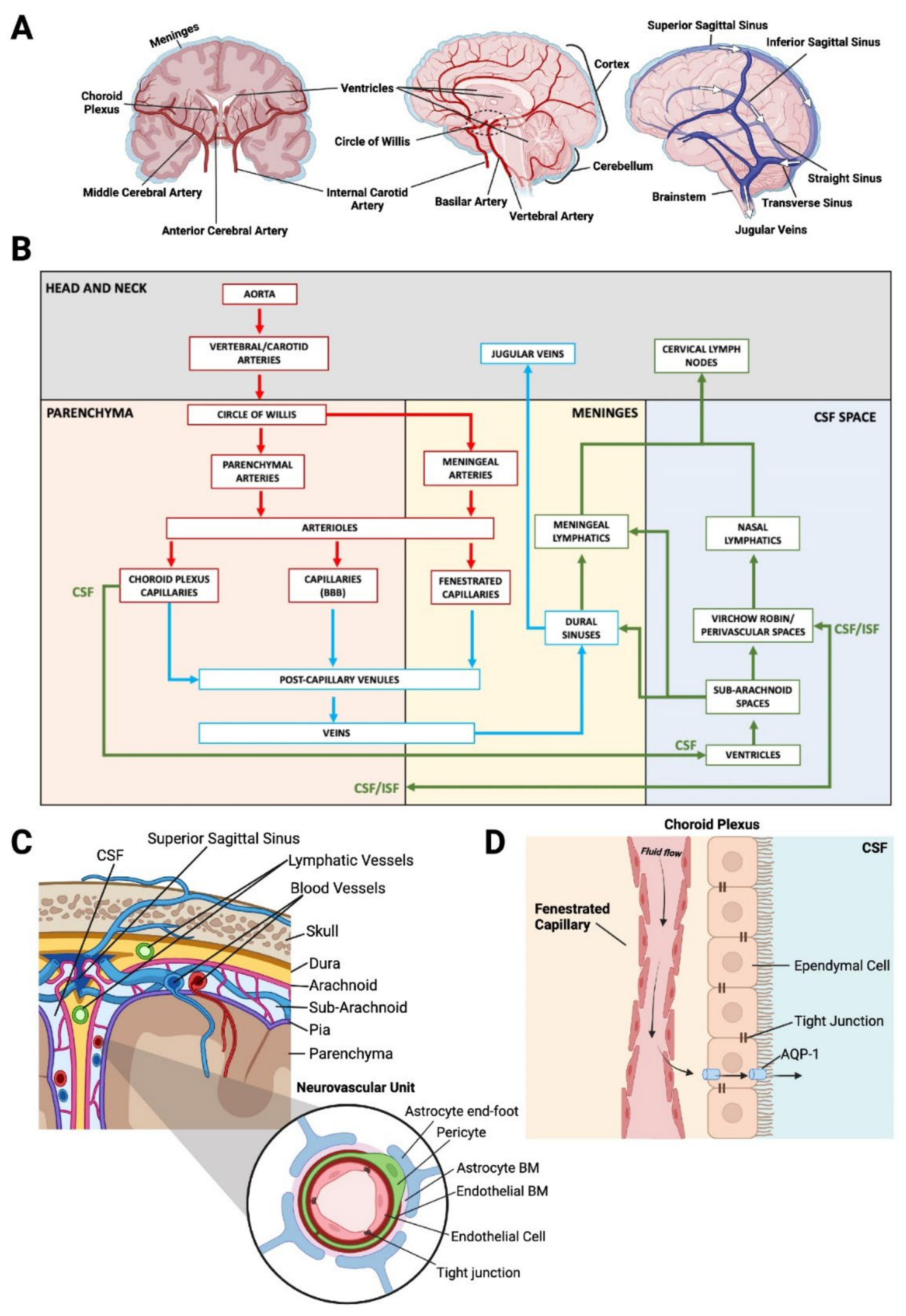
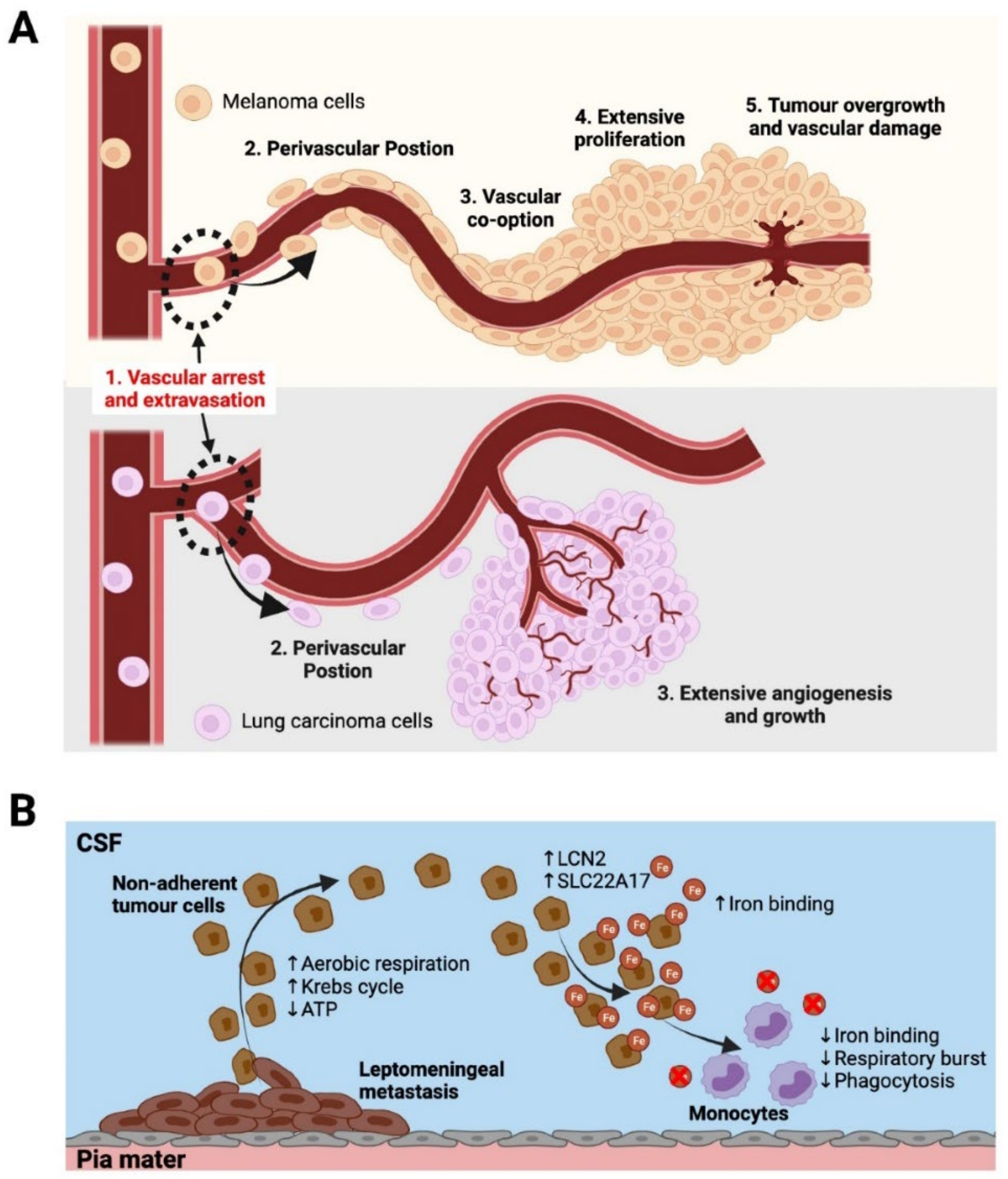
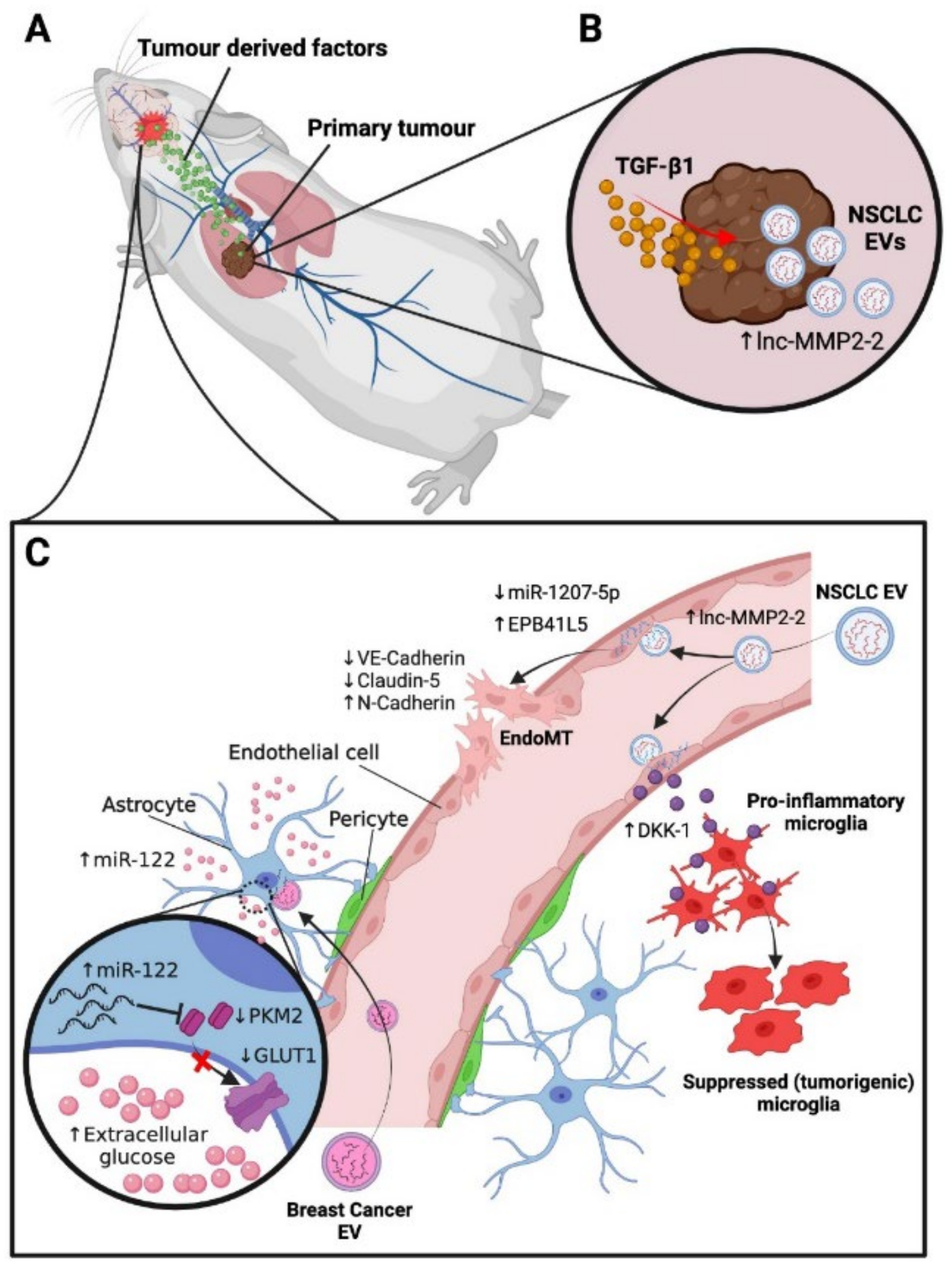
Primary tumours, particularly from major solid organs, are able to disseminate into the blood and lymphatic system and spread to distant sites. These secondary metastases to other major organs are the most lethal aspect of cancer, accounting for the majority of cancer deaths. The brain is a frequent site of metastasis, and brain metastases are often fatal due to the critical role of the nervous system and the limited options for treatment, including surgery. This creates a need to further understand the complex cell and molecular biology associated with the establishment of brain metastasis, including the changes to the environment of the brain to enable the arrival and growth of tumour cells. Local changes in the vascular network, immune system and stromal components all have the potential to recruit and foster metastatic tumour cells. This review summarises our current understanding of brain vascular microenvironments, fluid circulation and drainage in the context of brain metastases, as well as commenting on current cutting-edge experimental approaches used to investigate changes in vascular environments and alterations in specialised subsets of blood and lymphatic vessel cells during cancer spread to the brain.
Keywords: metastasis; brain; leptomeninges; blood vessels; lymphatic vessels; stroma; microenvironment metastasis; brain; leptomeninges; blood vessels; lymphatic vessels; stroma; microenvironment