Article

Redox-Dependent Copper Ion Modulation of Amyloid-β (1-42) Aggregation In Vitro

Department of Biology and Biological Engineering, Chalmers University of Technology, 412 96 Gothenburg, Sweden
*
Author to whom correspondence should be addressed.
Biomolecules 2020, 10(6), 924; https://doi.org/10.3390/biom10060924
Submission received: 15 May 2020 / Revised: 12 June 2020 / Accepted: 16 June 2020 / Published: 18 June 2020
(This article belongs to the Special Issue Toxic and Essential Metals in Human Health and Disease)

Abstract

:
Plaque deposits composed of amyloid-β (Aβ) fibrils are pathological hallmarks of Alzheimer’s disease (AD). Although copper ion dyshomeostasis is apparent in AD brains and copper ions are found co-deposited with Aβ peptides in patients’ plaques, the molecular effects of copper ion interactions and redox-state dependence on Aβ aggregation remain elusive. By combining biophysical and theoretical approaches, we here show that Cu2+ (oxidized) and Cu+ (reduced) ions have opposite effects on the assembly kinetics of recombinant Aβ(1-42) into amyloid fibrils in vitro. Cu2+ inhibits both the unseeded and seeded aggregation of Aβ(1-42) at pH 8.0. Using mathematical models to fit the kinetic data, we find that Cu2+ prevents fibril elongation. The Cu2+-mediated inhibition of Aβ aggregation shows the largest effect around pH 6.0 but is lost at pH 5.0, which corresponds to the pH in lysosomes. In contrast to Cu2+, Cu+ ion binding mildly catalyzes the Aβ(1-42) aggregation via a mechanism that accelerates primary nucleation, possibly via the formation of Cu+-bridged Aβ(1-42) dimers. Taken together, our study emphasizes redox-dependent copper ion effects on Aβ(1-42) aggregation and thereby provides further knowledge of putative copper-dependent mechanisms resulting in AD.

Graphical Abstract

1. Introduction

Alzheimer’s disease (AD) is an incurable and fatal neurodegenerative condition resulting from the progressive death of neurons in the brain and the concomitant loss of cognitive functions, and which is associated with protein aggregation and deposition [1]. AD is the most common form of adult dementia, mainly afflicting people over the age of 65 [2]. The prevalence of the disease is expected to rise worldwide due to population ageing, and unless there are new disease-modifying treatments, a projected 130 million people may be affected by 2050 [3].
AD is pathologically, genetically, and biochemically strongly linked to amyloid formation and the deposition of amyloid-β (Aβ) peptides into extracellular senile plaques [1,4,5]. Aβ peptides are generated by the proteolytic cleavage of the so-called amyloid precursor protein (APP) by β- and γ-secretases [6]. This occurs in the acidic environment of endosomes [7], where Aβ can also aggregate [8] and accumulate during early neuronal dysfunction and prior to the deposition of extracellular plaques [9]. Aβ peptides can differ in size. Even though the 40 amino acid Aβ(1-40) variant is most abundant, the two residues longer Aβ(1-42) variant is more aggregation-prone [10] and also the main protein constituent of extracellular amyloid plaques [11]. Probing Aβ(1-42) self-assembly mechanisms and the influence of biological as well as pharmacological extrinsic aggregation modifiers is therefore important. Recent progress in the development of models that mechanistically describe how amyloid fibrils are formed [12,13] has enabled the determination of rate constants for different microscopic reaction steps (Figure 1a) through the analysis of amyloid kinetics data. In the case of Aβ(1-42), this analysis has revealed that secondary nucleation is the dominant mechanism of new aggregate formation [14]. The same methods have also been used to rationalize the inhibitory effects of chaperones [15] and antibodies [16], and the catalytic effect of small molecules [17] and lipids [18].
Metal ions and imbalances in metal ion homeostasis are considered to play pivotal roles in neurodegeneration [19,20,21], contributing to oxidative stress responses [22,23] as well as directly to protein aggregation [24,25,26,27]. Along this line, metal ions—including iron, zinc, and copper (Cu)—are found to co-deposit with Aβ fibrils in the core of plaques [28,29,30], and interactions between Aβ and redox-active metal ions (e.g., Fe and Cu) can drive the production of hydrogen peroxide [31,32,33], superoxide anions [34], and hydroxyl radicals [35], thus potentially contributing directly to the oxidative stress that is observed in AD [36]. Based on this type of data, metal chelators have been proposed as future AD therapeutics [37], and favorable effects on Aβ accumulation and toxicity both in vitro and in vivo have been reported [38]. However, the literature on how metal ions affect Aβ peptide aggregation is inconclusive in many respects, with reports on both enhancing and inhibitory effects.
Dysregulated Cu homeostasis has been associated with AD [19,39,40,41,42] and is manifested as elevated Cu levels in plasma [19,41] but diminished levels of Cu in the brain, especially in the amygdala and hippocampal regions [40]. The concentrations of total Cu in amyloid plaques (400 μM) and the synaptic cleft (15 μM) are higher than the extracellular concentration in the brain (0.2–1.7 μM) [21]; the former may contribute to the reported imbalances. The redox ability of Cu enables it to cycle between Cu+ and Cu2+ states, underlying its function in many enzymes. The Cu2+ form is predominant under oxidizing conditions which also prevail in many locations where Aβ is abundant, such as the extracellular space and the oxidizing and low pH environment of endosomes and lysosomes [43]. In the context of AD, Cu2+ is therefore the most frequently studied redox form. Cu+, which is mainly bound to proteins in the cytosol where the environment is highly reducing, has been less explored, but the high reduction potential found for Cu when bound to Aβ suggests that Cu+:Aβ may be present at oxidative stress and/or pathological conditions. Probing the effects of both the redox states of Cu on Aβ aggregation is therefore important.
There are several biophysical studies on the effect of Cu2+ on Aβ aggregation, but the results are divergent. The majority of papers suggest that Cu2+ inhibits Aβ aggregation [44,45,46,47,48,49,50], a property shared with, for example, Zn2+ [51]. However, accelerating effects have also been suggested [52,53,54], for example under mildly acidic conditions [24]. Cu2+ has also been reported to selectively induce the formation of cytotoxic Aβ(1-42) oligomers under conditions where Zn2+ did not [55], thereby putatively contributing specifically to toxicity. In addition, Cu2+ have been reported to modulate Aβ integrity, for example by inducing the aggregation-inhibitory dityrosine dimerization in the presence of H2O2 or ascorbate [52], or by mediating hydrolytic cleavage in presence of large excess of antioxidants [56], emphasizing that this metal has multiple effects. The role of Cu+ in Aβ(1-42) aggregation has been less studied, but one report suggested, based on transmission electron microscopy, that both Cu+ and Cu2+ could prevent Aβ fibril formation [57].
Aβ peptides coordinate both Cu2+ and Cu+ ions via N-terminal residues (Figure 1a), but there is significant disparity in their binding affinities and exact coordination geometries. For Cu2+, dissociation constants are reportedly in the picomolar to low nanomolar range [58], and both 1:1 and 2:1 binding stoichiometries have been reported, albeit with significantly weaker binding of the second Cu2+ ion [59,60,61,62]. Aβ binds Cu2+ primarily via 3N1O coordination, resulting in the formation of an N-terminal loop via the engagement of the N-terminus, the carboxyl group on Asp1, and two imidazole nitrogens (on His6, His13, or His 14) or Asp1 and all three histidines (Figure 1b,c) [60,63,64,65,66]. The binding affinity [67], coordination geometry [64,66,68,69], and stoichiometry [70] of Cu2+ is sensitive to pH. A loss of coordination or weak affinity at pH 5.0 has been reported [67], as well as the successive transition towards 4N coordination above pH 8.0 [66]. Probing how Aβ aggregation proceeds as a function of pH is therefore important, particularly in light of the pathological aggregation of Aβ(1-42) in endolysosomes [8,9] where metals, including Cu, are conspicuously abundant [71]. Cu2+ can bind monomeric, oligomeric, and fibrillar Aβ states with comparable affinity [63,64], and it was reported that the level of Cu2+ bound to Aβ(1-40) did not change during aggregation [63]. This is consistent with the fact that residues 1-14 of Aβ(1-42) (to which Cu coordinates) remain flexible and are not incorporated in the amyloid fibril core (Figure 1b). Cu+ binds Aβ in a bidentate linear 2N coordination engaging two N-imidazoles (Figure 1d), most likely His13 and His14 [65]. Thus, Cu+ binding can occur without loop formation, rendering the N-terminus more flexible compared to when binding Cu2+. The affinity of Cu+ is reported to be in the femtomolar range, suggesting that Cu+ binds tighter than Cu2+ to Aβ [65,72]. Notably, there are no reports on the interactions of Cu+ with Aβ oligomers or fibrils.
In this study, we revisit the question of how Cu influences Aβ(1-42) amyloid formation, using Thioflavin-T assays with monomeric recombinant peptides as the starting material [75], a procedure that we [18] and others [14] have shown to result in highly reproducible aggregation kinetics. Moreover, by taking advantage of recent theoretical developments to model protein aggregation kinetics [14], enabling detailed molecular and mechanistic understanding of amyloid formation catalysis and inhibition as mentioned above, this study reveals new quantitative information on how Cu2+ and Cu+ affect discrete microscopic events during Aβ(1-42) self-assembly, resulting in different macroscopic effects on aggregation by the two forms of Cu. We also explore the pH-dependent effects of Cu2+ ions on Aβ(1-42) aggregation, addressing the pathological consequences of alterations in pH during endolysosomal processing, which may relate to the earliest Aβ aggregation events [73,74]. Our main finding is that Cu2+ and Cu+ have opposite effects on Aβ(1-42) aggregation. Cu2+ primarily interferes with fibril elongation, whereas Cu+ instead catalyzes primary nucleation. We discuss the results in relation to the Cu coordination geometries and the putative effects of Cu on Aβ-mediated neurotoxicity in the context of AD.

2. Materials and Methods

2.1. Protein Expression and Purification

Recombinant Aβ(1-42) was expressed as a fusion protein with the solubility tag NT* and purified as described in [76,77], but with some modification. The plasmid encoding for NT-Aβ(1-42) (a kind gift from Dr. Henrik Biverstål, Karolinska Institute) was transfected into E. coli (BL21) cells and expressed overnight using 0.5 mM IPTG induction at OD600~0.7. Bacterial cells were harvested by centrifugation and lysed in 20 mM Tris-HCl, 8 M urea, pH 8.0, by sonication. The sonicated lysate was filtered through a 0.22 µM syringe filter (VWR, Radnor, US) and loaded onto a HisPrep FF 16/10 column (GE Healthcare, Chicago, US) equilibrated with 20 mM Tris-HCl, 8 M urea, pH 8.0. The NT-Aβ(1-42) was eluted with 300 mM imidazole in the same buffer and then dialyzed against 5 L 20 mM sodium phosphate at 4 °C, with 1.5 mM 1,4-dithiothreitol (DTT) and 0.5 mM EDTA, pH 8.0. After 2 hrs of dialysis, 1:20 molar equivalents of tobacco etch virus (TEV) protease (produced as previously described [78]) was added to the NT-Aβ(1-42), followed by quiescent incubation over night at 4 °C. Following the TEV digestion, the solution was injected onto a HiLoad 16/600 Superdex 30 pg (GE Healthcare, Chicago, US) size exclusion column equilibrated with 20 mM sodium phosphate, pH 8.0, and monomeric Aβ(1-42) was isolated as a single peak. The monomeric peptides were immediately aliquoted, freeze dried, and stored at −20 °C until further use.

2.2. Aβ(1-42) Aggregation Assays

The Aβ(1-42) aggregation was monitored by thioflavin-T (ThT) fluorescence. Prior to each kinetic experiment, lyophilized Aβ(1-42) was dissolved in 6 M guanidine hydrochloride (GuHCl), followed by incubation on ice for 20 min. The monomeric Aβ(1-42) solution was purified by size-exclusion chromatography using a 10/300 Superdex 75 (GE Healthcare, Chicago, IL, US) column and equilibrated with 20 mM sodium phosphate, pH 8.0, to minimize isoelectric precipitation and reduce the aggregation rate so that the samples could be transferred to microtiter plates without significant aggregation occurring, following a common experimental protocol [14,79,80]. The Aβ(1-42) concentration was determined from the integrated peak area using an extinction coefficient of ε280 = 1280 M−1cm−1. The monomeric Aβ(1-42) solution was immediately transferred to ice to prevent any aggregation before starting the experiments. The Aβ(1-42) monomers were diluted in phosphate buffers or citrate-phosphate buffers to obtain a specific solution pH, as indicated in the text and figure legends (see Table S1 for detailed buffer compositions). 5 µM thioflavin-T (ThT, Sigma, St. Louis, MO, US) (three times recrystallized in tetrahydrofuran (THF) to remove impurities) was added from a 0.5 mM stock solution. The Aβ(1-42) solutions were supplemented with CuCl2 (to obtain Cu2+) or CuCl2 pre-mixed with a 5x molar excess of freshly prepared 1,4-dithiothreitol (DTT, Sigma, St. Louis, MO, US) to generate Cu+, according to a previously established protocol [81,82,83]. The peptide solutions were thereafter added to the wells of 96-well black half-area, low binding, transparent bottom microtiter plates (Corning, #3881, Corning, NY, US). The total volume in each well was always 70 µL. The plates were sealed with adhesive film (BIO-RAD, Hercules, US) to prevent sample evaporation and the ThT emission was measured from the bottom of the plate using a Fluostar Optima or Fluostar Omega plate reader (BMG Labtech, Ortenberg, Germany ) with a 440 ± 10 nm bandpass filter for excitation, and a 490 ± 10 nm bandpass filter for emission. All measurements were performed in triplicate (n = 3) and under strictly quiescent conditions (no shaking) at 37 °C and were repeated at least 3 times.

2.3. Seeds Preparation

Fibril seeds for seeded aggregation experiments were formed by setting up the above-described kinetic experiment with 2.6 µM Aβ(1-42) in 20 mM sodium phosphate buffer, pH 8.0, and 5 µM ThT. All seeds were prepared in the absence of Cu. The seeds were collected from the wells after the completion of the experiment and transferred into low-binding 1.5 mL tubes and then added to samples based on the monomer-equivalent concentration.

2.4. Analysis and Fitting of ThT Kinetic Curves

Kinetic data acquired in the presence of increasing amounts of Cu+ and Cu2+ were analyzed based on the previously described integrated rate laws for Aβ(1-42) amyloid formation and using a model including secondary nucleation [14,84]. The theoretical curves of the kinetic evolution of aggregates were globally fitted to the experimental ThT data (see Section 2.2) using the AmyloFit web interface [85] to obtain quantitative estimates of the rate constants. For Cu2+, we used a seeded model to determine individually the rate constants for primary nucleation (kn), elongation (k+), and secondary nucleation (k2), keeping one parameter at a time variable (i.e., dependent on the Cu2+ concentration) and the others as global constants (i.e., independent of the Cu2+ concentration). All the fits were conducted using a 10-basin hop algorithm with errors and using normalized ThT kinetics data, as described by Meisl et al. [85]. For Cu+, we used a non-seeded model to determine the convoluted rate constants k+kn and k+k2.

2.5. Dot-Blot Assay

Dot-blot assays were performed using the anti-amyloid fibril antibody LOC (Millipore, Burlington, MA, US, [86]), specific for amyloid fibrils. 20 µL of each sample containing Aβ(1-42) with or without Cu2+ or Cu+ was taken directly from the aggregation reactions and added to a 96-well dot-blot hybridization manifold (Scie-plas Ltd., Cambridge, UK). Vacuum pressure was applied to deposit the samples onto an LF-PVDF membrane (Trans-Blot, BIO-RAD, Hercules, CA, US). The membrane was dried and blocked with 30 mL of 5% skimmed milk in phosphate buffer saline with Tween-20 (PBS-T) and incubated for 1 h at room temperature. The membrane was then incubated for 30 min at room temperature with 30 mL of a primary antibody (1:100,000) dissolved in BSA/PBS-T. After three washes (5 min each time) with PBS-T, the membrane was incubated with a 1:20,000 dilution HRP-conjugated goat anti-rabbit secondary antibody (Sigma, St. Louis, MO, US). Thereafter, the membrane was washed three times with PBS-T (15 min × 1, 5 min × 2). The resulting dots were developed using an enhanced chemiluminescence solution (ECL, GE healthcare, Chicago, IL, US) together with the chemiluminescence detection system of a ChemiDocTM gel scanner (BIO-RAD, Hercules, CA, US). The densiometric analysis was performed using the instrument’s built-in software and the region-of-interest function.

2.6. Transmission Electron Microscopy (TEM)

TEM images were taken to examine the morphological appearance of Aβ(1-42) amyloid aggregates in the absence and presence of Cu. 10 µL of the sample taken at the end-point of the kinetic aggregation experiment was deposited onto glow-charged, formvar-coated, carbon-stabilized copper EM grids (Agar scientific, S138, Essex, UK) and allowed to settle for 10 min. The grids were thereafter stained with aqueous phosphotungstate (1% PTA), blotted with filter paper (Whatman, Grade 40, GE Healthcare, Chicago, IL, US), and allowed to dry before imaging [87]. High-contrast images were obtained with a TALOS L120C TEM (Thermo Fisher Scientific, Waltham, MA, US) at an accelerating voltage of 120 kV using a 4 × 4 k CMOS Ceta camera (Thermo Fisher Scientific, Waltham, MA, US). The magnification was set to 17,500x–300,000x.

3. Results and Discussion

3.1. Cu2+ Inhibits Aβ(1-42) Amyloid Formation

To explore the effect of Cu2+ ions on the aggregation of Aβ(1-42) into fibrils, we used Thioflavin-T (ThT) fluorescence to measure the amyloid formation kinetics. ThT binds to the regular β-sheet structure of amyloid fibrils (but does not associate with unstructured oligomers), and this increases dramatically its fluorescence quantum yield [88]. At stoichiometric amounts, ThT has been shown to have a negligible effect on the aggregation kinetics [14], and it is therefore a good in situ reporter of amyloid fibril formation. Recombinant Aβ(1-42) was monomerized by size exclusion chromatography (SEC) (Figure S1) immediately prior to each experiment to avoid the presence of pre-formed aggregates [75,85]. The purified peptides were allowed to aggregate under quiescent conditions in the absence or presence of CuCl2, such that the experiment probed the Cu2+:Aβ ratios of 0.5:1, 1:1, 1.5:1 and 2:1. The results (Figure 2a, Figure S2a) show that presence of Cu2+ inhibits the aggregation of Aβ(1-42) in a dose-dependent manner, resulting in both longer aggregation half-times and reduced maximum levels of ThT emission. We confirmed that the inhibitory effect is specific to Cu2+ by demonstrating that another divalent metal ion (Mg2+) had no, or a very marginal effect, even when added in 30x molar excess (Figure S3).
Using the conformation-sensitive anti-amyloid fibril antibody LOC [86] to detect fibrils, the dot-blot analysis reveals that the fibril yield decreases with increasing Cu2+ concentration (Figure 2b, Figure S4). A densiometric analysis suggests a ~90% reduction in fibrils at the highest Cu2+ concentration (Figure S5a). This confirms that the decrease in ThT intensity reflects inhibition and is not merely an effect of Cu2+-mediated fluorescence quenching [89]. Control experiments showed, however, that some Cu-mediated quenching also occurs; 34% of the ThT emission in a sample with pre-formed Aβ(1-42) fibrils was lost at an Cu2+:Aβ ratio of 1:1, but the effect was reversible upon the addition of EDTA (Figure S6). This is consistent with the result that the ThT emission at a 1:1 ratio decreased more than expected from the reduction in the LOC-detected fibril yield (Figure S5a).
Our findings are in good qualitative agreement with several other biophysical studies [44,47,48,49,50], and thus add to the growing consensus that Cu2+ ions inhibit Aβ(1-42) aggregation. Whilst a few studies report that Cu2+ ions induce the formation of amorphous aggregates of Aβ(1-42) [48,90], the TEM analysis revealed the formation of mature amyloid fibrils at both sub- and super-stoichiometric Cu2+ concentrations (Figure 2c,d) in our case.

3.2. pH and Salt Dependence of the Cu2+-Mediated Inhibition of Aβ(1-42) Amyloid Formation

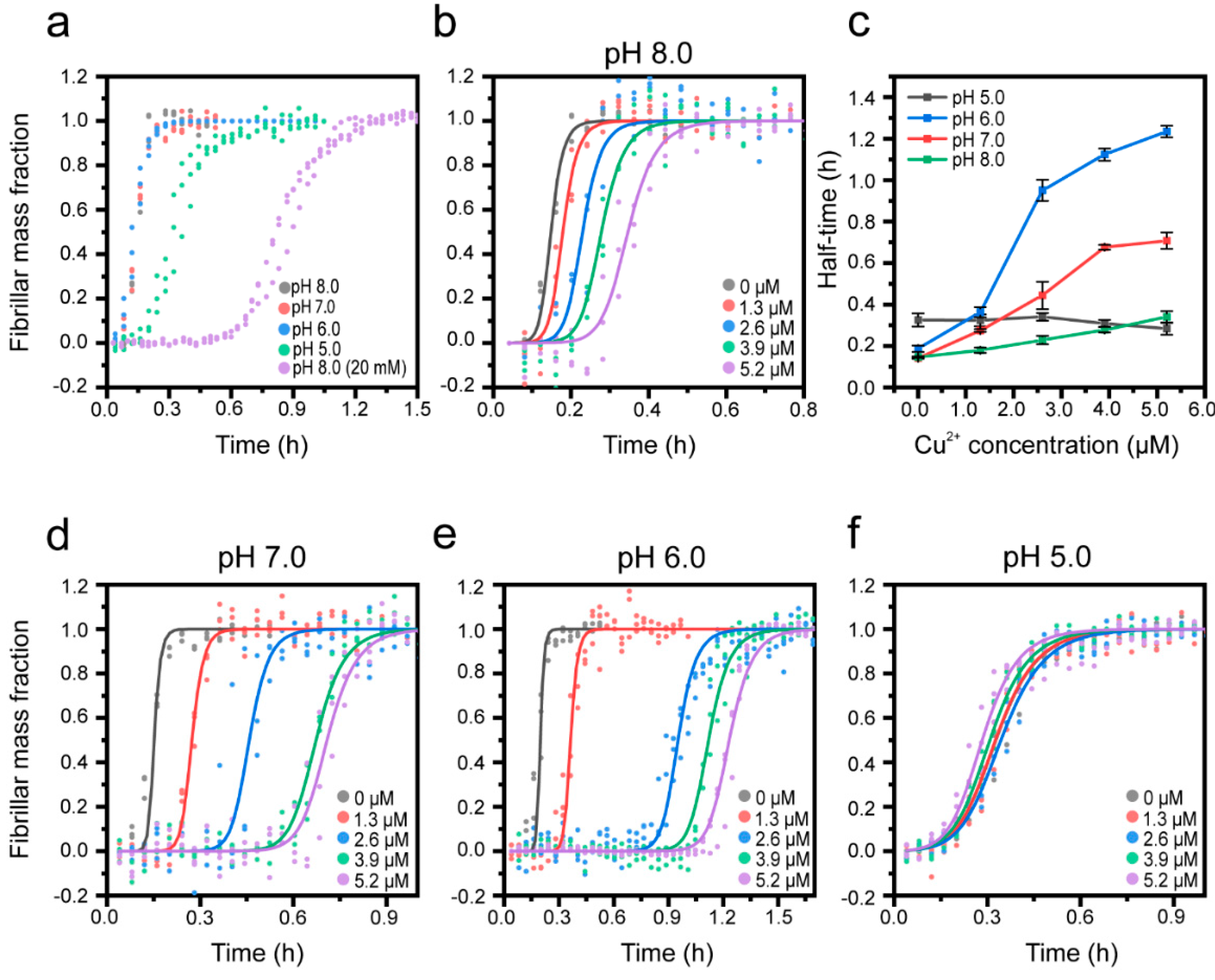
Next, we explored the effect of Cu2+ on Aβ(1-42) aggregation at different pHs to mimic the biological conditions that prevail in the endolysosomal pathway, where early accumulation of Aβ aggregates may occur [9]. By successively reducing the pH from 8.0 to 5.0, we also explored the effect of protonation of Cu-coordinating histidines (pKa ~ 6) in the Aβ(1-42) N-terminus. To cover this pH range, a citrate-phosphate buffer was used, which required an increase in the ionic strength to 200 mM compared to the data presented in Figure 2 in order to obtain sufficient buffering capacity at pH 5.0. This in itself decreased the half-time of Aβ(1-42) aggregation from 0.82 ± 0.03 h (20 mM) to 0.20 ± 0.01 h (200 mM) (Figure 3a), consistent with previous observations of salt effects [91,92] and a recent study demonstrating that increased electrostatic screening enhances several mechanistic steps in the self-assembly of Aβ(1-42) [93]. We found that Cu2+ slows down Aβ(1-42) aggregation also in 200 mM salt, pH 8.0, (Figure 3b), but the effect is weaker (1.4 times increase in the half-time at 5.2 μM of Cu2+ (Figure 3c) compared to a 4.6 times increase at 20 mM of salt (Figure 2a)). This suggests that the catalyzing effects of electrostatic screening on Aβ(1-42) aggregation may to some extent outcompete Cu2+-mediated inhibitory actions.
The effects of pH on the intrinsic aggregation kinetics of Aβ(1-42) are shown in Figure 3a, revealing no or very modest changes above Aβ(1-42)’s isoelectric point (pI = 5.3, [94]), whereas at pH 5.0 the aggregation rate increases, which is also reflected by the change in the aggregation halftimes (0.32 ± 0.05 h in pH 5.0, compared to 0.2 ± 0.01 h at pH 8.0) (Figure 3c). Upon lowering the pH from 8.0 (Figure 3b) to 7.0 or 6.0 (Figure 3d,e), we observed that the Cu2+-mediated inhibition became increasingly potent; the aggregation curves display extended lag phases and significantly increased half-times (Figure 3c), particularly at Cu2+:Aβ ratios equal to or exceeding 1:1. This behavior could relate to differences in the coordination mode and binding stoichiometry of the Cu2+:Aβ complex, which has been shown to be pH-dependent in several studies [64,66,68,69]. Particularly, the binding of a second Cu2+ ion appears more prominent in mildly acidic (pH 6.6) conditions [70], which could explain the sudden increase in the degree of inhibition of aggregation that we observe at a Cu2+:Aβ ratio of 1:1 and above at pH 7.0 and 6.0, but not at pH 8.0. The lack of effect of Cu2+ additions at pH 5.0 (Figure 3f) is most likely due to a loss of binding, related to the full protonation of His residues [67], even if one report suggests a weak Cu2+ binding to Aβ monomers at this pH [66]. This result further implies that Aβ(1-42) may release Cu2+ during endolysosomal acidification following endocytic uptake [95], and that this in turn could lead to both aggravated aggregation of Aβ and putative toxicity due to free Cu2+ ions.

3.3. Cu2+-Mediated Inhibition of Aβ(1-42) Aggregation Affects the Fibril Elongation Step

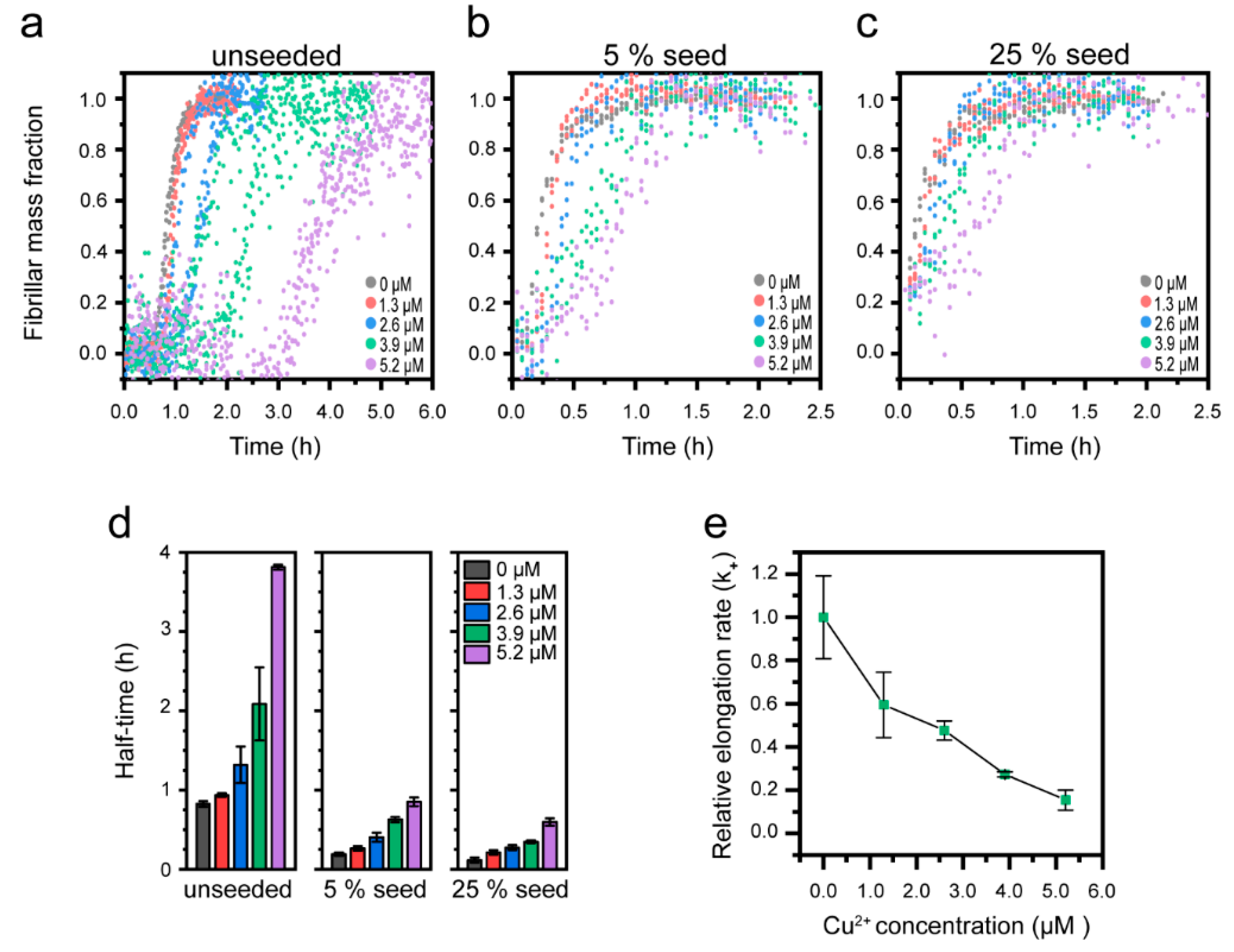
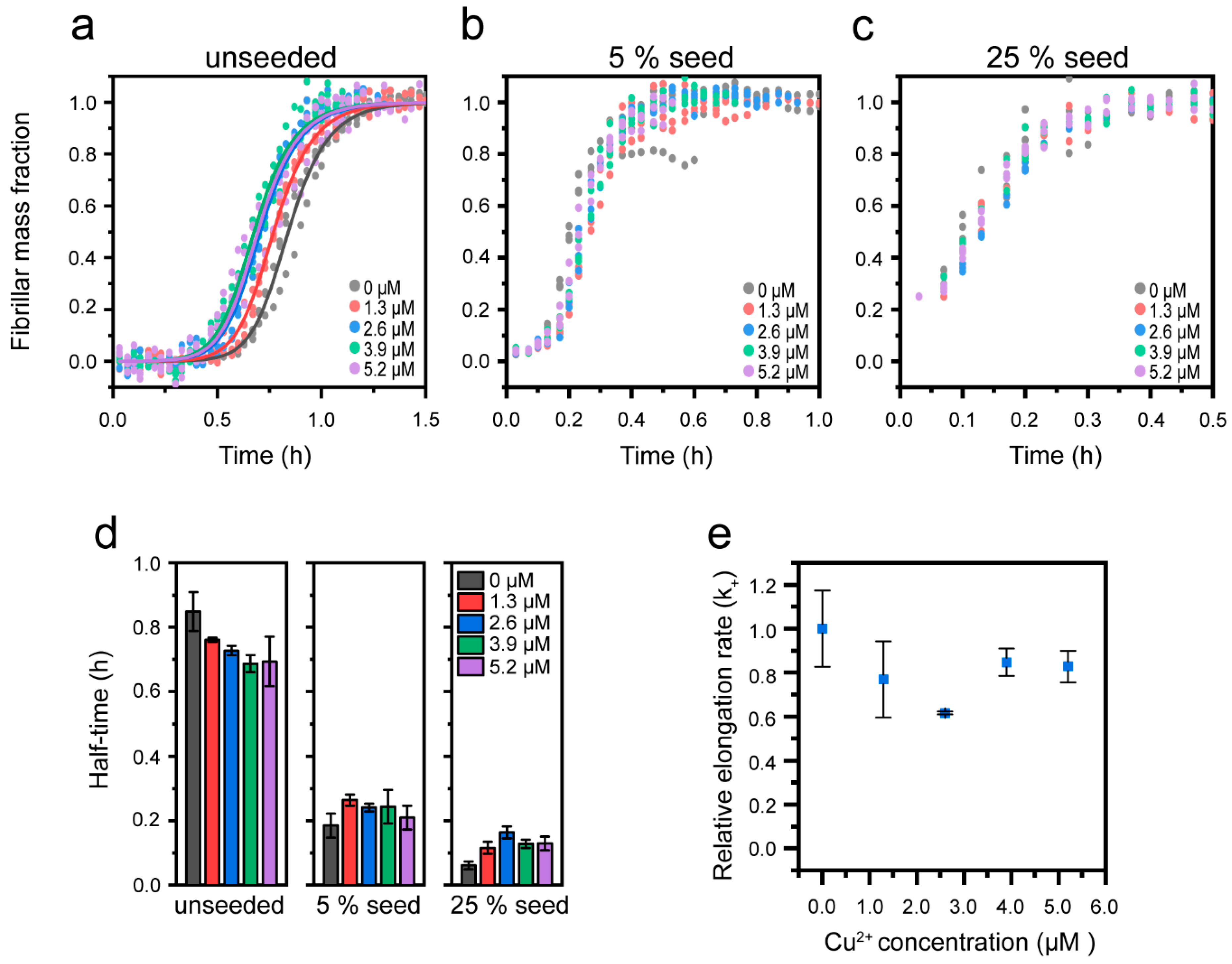
It has been shown in several independent studies that the rate-limiting step in unseeded aggregation of Aβ(1-42) is secondary nucleation [14,18,51]. Aggregation modulators can, however, act on different mechanistic steps [96]. To investigate how Cu2+ affects the mechanism of Aβ(1-42) aggregation, we returned to pH 8.0 and low ionic strength conditions and explored how the aggregation kinetics of Aβ(1-42) were affected by the addition of pre-formed fibril seeds (prepared in the absence of Cu2+). Figure 4a shows the kinetic profiles of 2.6 µM Aβ(1-42) with increasing Cu2+ and no added seeds (the normalized data from Figure S2a). Figure 4b,c shows the same experiment in the presence of 5 mol% or 25 mol% Aβ(1-42) fibril seeds (mol% on a monomer basis). It is clear that even at strongly seeded conditions (25%), the aggregation of Aβ(1-42) is reduced in a dose-dependent manner by Cu2+. This is further visualized by the half-time dependence on the Cu2+ concentration, as shown in Figure 4d and Figure S7. The distinct concentration-dependent increase in half-time with 5% and 25% seeds strongly suggests that Cu2+-mediated inhibition is not due to a reduction in the primary nucleation rate, since the addition of seeds bypasses this reaction step [97]. Instead, the retardation of Aβ(1-42) aggregation by Cu2+ appears to be associated with inhibition of secondary processes and/or elongation. It has been shown that elongation can be distinguished from secondary nucleation at strongly seeded conditions where fibril elongation dominates over all nucleation processes [17].
Since our data show that a significant effect of Cu2+ on the aggregation half-times remains with 25% seeds, we proceeded to estimate the elongation rates by fitting a linear curve to the initial growth phase (up to 50% fibril mass fraction) under strongly (25%) seeded conditions (Figure S8a), as described by Abelein et al. [51]. This analysis revealed a Cu2+ concentration-dependent decrease in the relative elongation rate (Figure 4e), supporting the notion that Cu2+ inhibition acts on fibril elongation. Similar observations have been made in studies on the effect of Zn2+ on the aggregation of Aβ(1-40) [51].
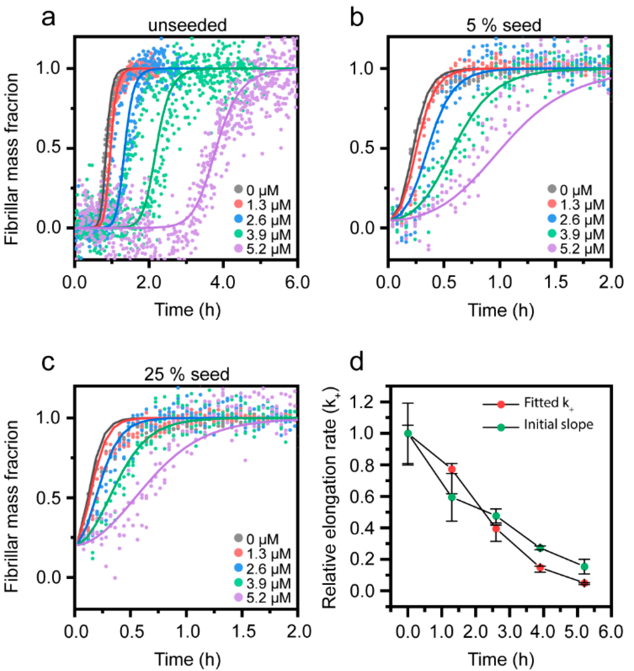
To quantitatively determine the elongation rate constants together with the rates for other steps in the reaction, we performed a global fitting to the combined data sets of unseeded and seeded ThT emission kinetics in the Amylofit web interface, using a secondary nucleation model for seeded amyloid growth [85]. This allowed us to independently determine the microscopic rate constants for primary nucleation (kn), elongation (k+), and secondary nucleation (k2). The elongation rate constant (k+) was probed as a fitting parameter, (allowed to vary with the Cu2+ concentration, but kept constant across seeding concentrations), and the two other rate constants were fitted as fixed global parameters. The resulting fits are shown in Figure 5a–c, and the associated rate constants are given in Table 1. We repeated the analysis with, respectively, kn and k2 as fitting parameters (Figure S9), further substantiating that Cu2+ cannot inhibit the primary nucleation step of the Aβ(1-42) aggregation reaction (Figure S9a–c).
From a mathematical perspective, it is more difficult to distinguish between the fits for secondary nucleation inhibition (k2, Figure S9d–f) and elongation inhibition (k+, Figure 5a–c), but the latter (which also has a slightly better goodness of fit, 0.0206 vs 0.0214; the corresponding parameter for the rejected kn fit was 0.0265) captures better the initial lagging behavior at high Cu2+ ratios. Furthermore, increasing the Cu2+ concentration results in longer lag times together with successively decreased steepness of the slopes of the aggregation kinetic curves (Figure 4a), which is expected for elongation inhibitors [16]. The change in the elongation rate constant (k+) with increasing Cu2+ concentration determined from the fitting (Figure 5e, Table 1) overlaps very well with the relative elongation rate constants estimated using the initial slope of the kinetic profiles (Figure 4e). We therefore conclude that Cu2+ inhibits the Aβ(1-42) aggregation by interfering with the fibril elongation step. In order to compare the fitted rate constants for the Aβ(1-42) aggregation in the absence of Cu2+ with previously published values, we calculated the combined rate constants knk+ and k2k+ from the data given in Table 1. This yielded 2.5 × 10⁷ M−2h−2 and 8.6 × 10¹⁸ M−3h−2 for knk+ and k2k+, in good agreement with previous reports [14,51], demonstrating the robustness of this Aβ(1-42) aggregation system.
Our results show that Cu2+ reduces the elongation rate of Aβ(1-42) by 60% at a 1:1 Cu2+:Aβ ratio and by 95% at a 2:1 ratio. These values can be compared to the elongation inhibitory effect of Zn2+ on Aβ(1-40), which resulted in an 80% reduction in the rate constant at sub-stoichiometric concentrations (2.5:20 molar ratio) [51] and to the extracellular chaperone clusterin, which elicits the strong inhibition of elongation even at molar ratios as low as 1:100 [80]. In comparison, the Cu2+-mediated inhibition appears to be weaker.
Fibril elongation occurs via monomer addition to the growing fibril end, and the inhibition could therefore be envisioned to result from Cu2+ binding to the attaching monomer, the growing fibril, or both, especially since Cu2+ has been reported to bind both Aβ(1-42) monomers and fibrils with a comparable affinity [63]. Most studies suggest that elongation occurs via templating interactions, where the misfolding rearrangement is aided by monomer contacts to the fibril end [98,99,100]. Molecular dynamics simulations have suggested that it is the N-terminal part of the Aβ peptide that forms the initial contact with the growing fibril end [101]. It can therefore be envisioned that the Cu2+ interaction, promoting loop-formation in the N-terminus of either the free Aβ(1-42) monomer or the Aβ(1-42) entities at the fibril end (Figure 1a), imposes conformational restrictions or charge repulsions that impede monomers from attaching to the fibril ends.

3.4. Effect of Cu+ on Aβ(1-42) Aggregation

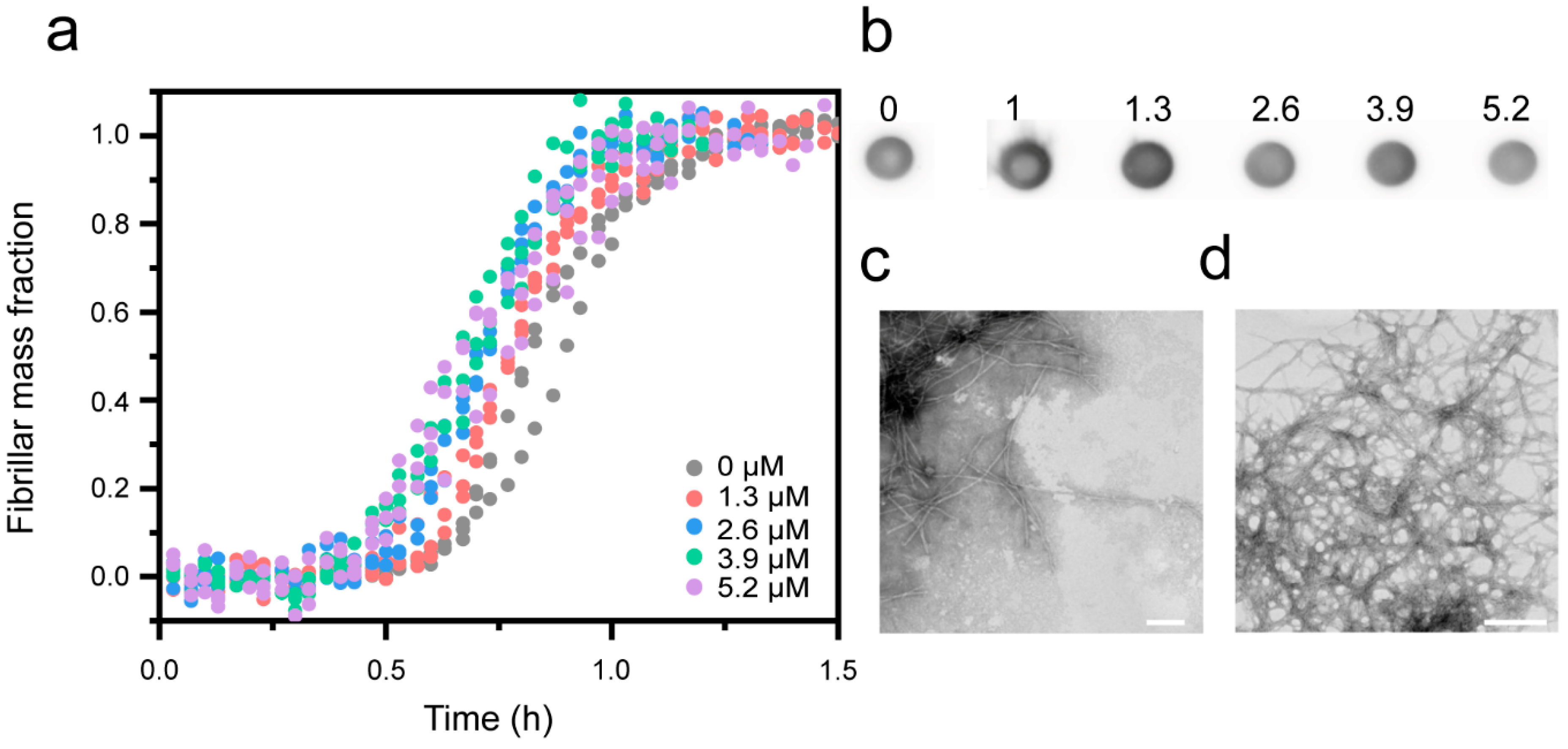
Since copper can exist in, and cycle between, two redox states (Cu+ and Cu2+), we also studied the effect of Cu+ on the aggregation kinetics of Aβ peptides. Notably, this has not been explored previously, in part because Cu+ is difficult to work with in aired solutions. To create and keep Cu+ in solution, we used the reducing agent dithiothreitol (DTT), in similarity to work with Cu+ chaperones [82]. The aggregation of Aβ(1-42) in presence of CuCl2 and a 5x molar excess of DTT to Cu (creating Cu+) resulted in a modest but reproducible concentration-dependent increase in the aggregation rate of Aβ(1-42) (Figure 6a) (for two additional replicates of the experiments, see supporting Figure S10), thus opposite to the effect of Cu2+. Control experiments showed that DTT on its own had no effect (Figure S11). The kinetic results were confirmed by dot blot (Figure 6b, Figure S4), showing no decrease in the fibril yield in the presence of Cu+. TEM imaging revealed the presence of typical amyloid fibril aggregates at both sub- and super stochiometric Cu+:Aβ ratios (Figure 6c,d), similar to those observed with Cu2+, suggesting that copper ions and redox state have no major effect on the morphology of formed amyloid fibrils.

3.5. Cu+ Enhances Aβ(1-42) Aggregation by Catalysis of Primary Nucleation

Cu+ has been reported to bind Aβ(1-42) via bidentate coordination to imidazoles of His13 and His14, located adjacent to the N-terminal side of the first β-sheet in the fibril core, but His6 may also be involved (Figure 1d, [65]). In order to rationalize how this leads to the enhancement of Aβ(1-42) aggregation, we performed seeded experiments analogous to those explained in Section 3.3.
We find that whilst Cu+ shortens the half-time of unseeded Aβ(1-42) aggregation from 0.84 ± 0.06 to 0.69 ± 0.07 (Figure 7a,d), there is no significant effect of Cu+ on seeded reactions (Figure 7b–d), suggesting that secondary pathways are not affected. Consistently, Cu+ had no effect on the initial slope of the kinetic traces under strongly seeded (25%) conditions, and therefore not on the relative elongation rate constants (Figure 7e and Figure S7b). Furthermore, the observed characteristic shortening of the lag time, without apparent alterations to the steepness of the slope of the aggregation curves [97], strongly supports that the Cu+-mediated catalysis of the Aβ aggregation acts on primary nucleation. Although in aired solutions, Cu+ will over time oxidize to Cu2+, it has been previously shown that under reducing conditions (1-fold excess ascorbate) Cu+ remains stable for at least 1.5 h [65], which is longer than the experimental time span in Figure 6a.
We globally fitted the unseeded Aβ(1-42) aggregation data in the presence of Cu+ using the Amylofit web interface [85] and a secondary nucleation model for unseeded amyloid growth. The fit (solid lines in Figure 7a) shows that the mild aggregation-enhancing effect of Cu+ is well-described by an increase in the combined primary nucleation and elongation rate constant knk+; the resulting combined rate constants from the fitting are shown in Table 2.
There are relatively few studies investigating the Cu+–Aβ interaction, but due to the preferred linear coordination geometry involving His13 and His14 [65,102], it is conceivable that Cu+ cannot simultaneously bind these two ligands in the fibrillar state (due to steric restrictions imposed by the alternating directions of side-chain residues in or near the cross-β core (Figure 1b)). However, it is plausible that Cu+ may interact with His13 and His14 residues on different monomers in the amyloid fibrils. Our data would then be consistent with a model in which Cu+ catalyzes the formation of Aβ(1-42) dimers, which are the minimal elongation-competent Aβ(1-42) units [14], thus promoting primary nucleation.

4. Conclusions

This study explores the effect of copper ions on Aβ(1-42) amyloid formation in vitro, identifying effects on the overall aggregation rate as well as on discrete mechanistic steps of the aggregation pathway (illustrated in Figure 8). Our results extend the growing body of literature, suggesting that Cu2+, the major extracellular copper form [21], acts in an inhibitory manner on fibril formation [44,45,46,47,48,49,50]. We solidify this view by seeding experiments and kinetic analyses, which conclusively show how Cu2+ primarily impedes fibril elongation at both sub- and super-stoichiometric ratios and in a concentration range that is relevant to the conditions that prevail in the brain. The Cu2+-mediated inhibition is modest in comparison to other elongation inhibitors, including Zn2+ (see above and [51]). This is consistent with observations of the significant accumulation of Cu2+ in AD plaques [21], and suggests that Cu2+ impedes, rather than completely blocks, Aβ(1-42) aggregation. Although the inhibition of Aβ(1-42) aggregation is a tractable goal to limit the onset and progression of AD, it is important to recognize that elongation inhibition, under conditions where primary and secondary nucleation proceeds, can instead drive the formation of small fibrils and oligomers [12,16]. It is therefore possible that Cu2+, despite its ability to reduce the amount of mature fibrils formed, as we report here, contributes to Aβ toxicity in the brain. Furthermore, although we describe a strong stoichiometry-dependent inhibitory effect of Cu2+ at a mildly acidic pH, we also report a loss of function at pH 5.0, a condition that prevails in late endosomes and lysosomes and which may drive Aβ aggregation in intraneuronal locations of the brain.
Furthermore, we demonstrate that the effect of Cu is dependent on the redox state by including the first experimental evidence that Cu+ also has an Aβ(1-42) aggregation-modulatory effect. Through kinetic analysis and seeded experiments, we report that Cu+ accelerates fibril formation by mild catalysis of primary nucleation, possibly related to Cu+-mediated dimer formation. This finding is important, particularly in relation to observations that the redox cycling of Aβ-bound Cu can contribute to the generation of reactive oxygen species in the brain [32]. Our results suggest that such reactions, which result in Cu+ bound to Aβ, may also accelerate aggregation, thereby constituting a dual toxic mechanism. Altogether, our study sheds more light on the possible pathological relevance of Cu in Alzheimer’s disease and suggests a complex pivotal role of Cu in Aβ(1-42) aggregation modulation that warrants further investigation in biological in vivo environments. The therapeutic value of Cu–Aβ(1-42) interactions should also be taken into account in future strategies to develop disease-modifying treatments.

Supplementary Materials

The following supplementary results are available online at https://www.mdpi.com/2218-273X/10/6/924/s1; Figure S1: Size-exclusion (SEC) purification of Aβ(1-42) monomers; Figure S2: Reproducibility of Aβ(1-42) aggregation kinetics; Figure S3: Aβ(1-42) amyloid formation in presence of MgCl2; Figure S4: Dot blot; Figure S5: Relative ratio of fibrillar content at different Cu concentrations; Figure S6: Quenching of ThT by Cu2+; Figure S7: Half-time of Aβ(1-42) aggregation in presence of Cu2+; Figure S8: Initial slope analysis of 25% seeded aggregation curves; Figure S9: Fitting of Aβ(1-42) amyloid kinetic data in presence of Cu2+ to models of primary nucleation and secondary nucleation inhibition; Figure S10: Amyloid formation in presence of Cu+- extra data; Figure S11: Dithiotreitol (DTT) has no effect on Aβ(1-42) amyloid formation; Table S1: Buffer specifications.

Author Contributions

Conceptualization, N.S. and E.K.E.; Formal analysis, N.S., D.B. and E.K.E.; Funding acquisition, E.K.E.; Investigation, N.S., D.B. and I.H.; Methodology, N.S., D.B., I.H., P.W.-S. and E.K.E.; Resources, E.K.E.; Visualization, N.S., D.B. and E.K.E.; Writing—original draft, N.S., D.B., P.W.-S. and E.K.E.; Writing—review and editing, N.S., D.B., I.H., P.W.-S. and E.K.E. All authors have read and agreed to the published version of the manuscript.

Funding

This research was funded by grants to E.K.E from the Swedish Research Council (grant number 2016-03902) and Chalmers Excellence Initiative Nano”. N.S is an Excellence PhD student funded by Chalmers Nano program. P.W.S. acknowledges funding from the Wallenberg Scholar Program and the Swedish Research Council (grant number 2019-03673).

Acknowledgments

We thank Henrik Biverstål at the Karolinska Institute for help with the setup of the recombinant Aβ(1-42) production and the gift of the Aβ(1-42)-encoding plasmid. Emma Lorentzon is acknowledged for help with TEM image acquisition. These images were recorded using instruments and technical support from the Centre for Cellular Imaging (CCI) core facility at Gothenburg University.

Conflicts of Interest

The authors declare no conflict of interest.

References

  1. Masters, C.L.; Simms, G.; Weinman, N.A.; Multhaup, G.; McDonald, B.L.; Beyreuther, K. Amyloid plaque core protein in Alzheimer disease and Down syndrome. Proc. Natl. Acad. Sci. USA 1985, 82, 4245–4249. [Google Scholar] [CrossRef] [PubMed] [Green Version]
  2. Hebert, L.E.; Weuve, J.; Scherr, P.A.; Evans, D.A. Alzheimer disease in the United States (2010–2050) estimated using the 2010 census. Neurology 2013, 80, 1778–1783. [Google Scholar] [CrossRef] [PubMed] [Green Version]
  3. Prince, M.J. World Alzheimer Report 2015: The Global Impact of Dementia: An Analysis of Prevalence, Incidence, Cost and Trends; Alzheimer’s Disease International: London, UK, 2015. [Google Scholar]
  4. Glenner, G.G.; Wong, C.W. Alzheimer’s disease: Initial report of the purification and characterization of a novel cerebrovascular amyloid protein. Biochem. Biophys. Res. Commun. 1984, 120, 885–890. [Google Scholar] [CrossRef]
  5. Hardy, J.; Allsop, D. Amyloid deposition as the central event in the aetiology of Alzheimer’s disease. Trends Pharmacol. Sci. 1991, 12, 383–388. [Google Scholar] [CrossRef]
  6. LaFerla, F.M.; Green, K.N.; Oddo, S. Intracellular amyloid-β in Alzheimer’s disease. Nat. Rev. Neurosci. 2007, 8, 499–509. [Google Scholar] [CrossRef] [PubMed]
  7. Sannerud, R.; Esselens, C.; Ejsmont, P.; Mattera, R.; Rochin, L.; Tharkeshwar, A.K.; De Baets, G.; De Wever, V.; Habets, R.; Baert, V. Restricted location of PSEN2/γ-secretase determines substrate specificity and generates an intracellular Aβ pool. Cell 2016, 166, 193–208. [Google Scholar] [CrossRef] [PubMed] [Green Version]
  8. Esbjörner, E.K.; Chan, F.; Rees, E.; Erdelyi, M.; Luheshi, L.M.; Bertoncini, C.W.; Kaminski, C.F.; Dobson, C.M.; Schierle, G.S.K. Direct observations of amyloid β self-assembly in live cells provide insights into differences in the kinetics of Aβ (1–40) and Aβ (1–42) aggregation. Chem. Biol. 2014, 21, 732–742. [Google Scholar] [CrossRef] [PubMed] [Green Version]
  9. Gouras, G.K.; Tsai, J.; Naslund, J.; Vincent, B.; Edgar, M.; Checler, F.; Greenfield, J.P.; Haroutunian, V.; Buxbaum, J.D.; Xu, H. Intraneuronal Aβ42 accumulation in human brain. Am. J. Pathol. 2000, 156, 15–20. [Google Scholar] [CrossRef]
  10. Jarrett, J.T.; Berger, E.P.; Lansbury, P.T., Jr. The carboxy terminus of the. beta. amyloid protein is critical for the seeding of amyloid formation: Implications for the pathogenesis of Alzheimer’s disease. Biochemistry 1993, 32, 4693–4697. [Google Scholar] [CrossRef] [PubMed]
  11. Murphy, M.P.; LeVine, H., III. Alzheimer’s disease and the amyloid-β peptide. J. Alzheimer’s Dis. 2010, 19, 311–323. [Google Scholar] [CrossRef] [PubMed] [Green Version]
  12. Linse, S. Monomer-dependent secondary nucleation in amyloid formation. Biophys. Rev. 2017, 9, 329–338. [Google Scholar] [CrossRef] [PubMed] [Green Version]
  13. Meisl, G.; Yang, X.; Frohm, B.; Knowles, T.P.; Linse, S. Quantitative analysis of intrinsic and extrinsic factors in the aggregation mechanism of Alzheimer-associated Aβ-peptide. Sci. Rep. 2016, 6, 18728. [Google Scholar] [CrossRef] [PubMed]
  14. Cohen, S.I.; Linse, S.; Luheshi, L.M.; Hellstrand, E.; White, D.A.; Rajah, L.; Otzen, D.E.; Vendruscolo, M.; Dobson, C.M.; Knowles, T.P. Proliferation of amyloid-β42 aggregates occurs through a secondary nucleation mechanism. Proc. Natl. Acad. Sci. USA 2013, 110, 9758–9763. [Google Scholar] [CrossRef] [PubMed] [Green Version]
  15. Arosio, P.; Michaels, T.C.; Linse, S.; Månsson, C.; Emanuelsson, C.; Presto, J.; Johansson, J.; Vendruscolo, M.; Dobson, C.M.; Knowles, T.P. Kinetic analysis reveals the diversity of microscopic mechanisms through which molecular chaperones suppress amyloid formation. Nat. Commun. 2016, 7, 10948. [Google Scholar] [CrossRef] [Green Version]
  16. Munke, A.; Persson, J.; Weiffert, T.; De Genst, E.; Meisl, G.; Arosio, P.; Carnerup, A.; Dobson, C.M.; Vendruscolo, M.; Knowles, T.P. Phage display and kinetic selection of antibodies that specifically inhibit amyloid self-replication. Proc. Natl. Acad. Sci. USA 2017, 114, 6444–6449. [Google Scholar] [CrossRef] [PubMed] [Green Version]
  17. Limbocker, R.; Chia, S.; Ruggeri, F.S.; Perni, M.; Cascella, R.; Heller, G.T.; Meisl, G.; Mannini, B.; Habchi, J.; Michaels, T.C. Trodusquemine enhances Aβ 42 aggregation but suppresses its toxicity by displacing oligomers from cell membranes. Nat. Commun. 2019, 10, 225. [Google Scholar] [CrossRef] [PubMed] [Green Version]
  18. Lindberg, D.J.; Wesén, E.; Björkeroth, J.; Rocha, S.; Esbjörner, E.K. Lipid membranes catalyse the fibril formation of the amyloid-β (1–42) peptide through lipid-fibril interactions that reinforce secondary pathways. Biochim. Biophys. Acta (BBA)-Biomembr. 2017, 1859, 1921–1929. [Google Scholar] [CrossRef]
  19. Bush, A.I. The metallobiology of Alzheimer’s disease. Trends Neurosci. 2003, 26, 207–214. [Google Scholar] [CrossRef]
  20. Li, F.; Calingasan, N.Y.; Yu, F.; Mauck, W.M.; Toidze, M.; Almeida, C.G.; Takahashi, R.H.; Carlson, G.A.; Flint Beal, M.; Lin, M.T. Increased plaque burden in brains of APP mutant MnSOD heterozygous knockout mice. J. Neurochem. 2004, 89, 1308–1312. [Google Scholar] [CrossRef]
  21. Smith, D.G.; Cappai, R.; Barnham, K.J. The redox chemistry of the Alzheimer’s disease amyloid β peptide. Biochim. Biophys. Acta (BBA)-Biomembr. 2007, 1768, 1976–1990. [Google Scholar] [CrossRef] [Green Version]
  22. Huang, X.; Atwood, C.S.; Hartshorn, M.A.; Multhaup, G.; Goldstein, L.E.; Scarpa, R.C.; Cuajungco, M.P.; Gray, D.N.; Lim, J.; Moir, R.D. The Aβ peptide of Alzheimer’s disease directly produces hydrogen peroxide through metal ion reduction. Biochemistry 1999, 38, 7609–7616. [Google Scholar] [CrossRef] [PubMed]
  23. Huang, X.; Moir, R.D.; Tanzi, R.E.; Bush, A.I.; Rogers, J.T. Redox-active metals, oxidative stress, and Alzheimer’s disease pathology. Ann. N. Y. Acad. Sci. 2004, 1012, 153–163. [Google Scholar] [CrossRef]
  24. Atwood, C.S.; Moir, R.D.; Huang, X.; Scarpa, R.C.; Bacarra, N.M.E.; Romano, D.M.; Hartshorn, M.A.; Tanzi, R.E.; Bush, A.I. Dramatic aggregation of Alzheimer Aβ by Cu (II) is induced by conditions representing physiological acidosis. J. Biol. Chem. 1998, 273, 12817–12826. [Google Scholar] [CrossRef] [PubMed] [Green Version]
  25. Bush, A.I.; Pettingell, W.H.; Multhaup, G.; d Paradis, M.; Vonsattel, J.-P.; Gusella, J.F.; Beyreuther, K.; Masters, C.L.; Tanzi, R.E. Rapid induction of Alzheimer A beta amyloid formation by zinc. Science 1994, 265, 1464–1467. [Google Scholar] [CrossRef] [PubMed]
  26. Esler, W.P.; Stimson, E.R.; Jennings, J.M.; Ghilardi, J.R.; Mantyh, P.W.; Maggio, J.E. Zinc-induced aggregation of human and rat β-amyloid peptides in vitro. J. Neurochem. 1996, 66, 723–732. [Google Scholar] [CrossRef]
  27. Mantyh, P.W.; Ghilardi, J.R.; Rogers, S.; DeMaster, E.; Allen, C.J.; Stimson, E.R.; Maggio, J.E. Aluminum, iron, and zinc ions promote aggregation of physiological concentrations of β-amyloid peptide. J. Neurochem. 1993, 61, 1171–1174. [Google Scholar] [CrossRef] [PubMed]
  28. Beauchemin, D.; Kisilevsky, R. A method based on ICP-MS for the analysis of Alzheimer’s amyloid plaques. Anal. Chem. 1998, 70, 1026–1029. [Google Scholar] [CrossRef] [PubMed]
  29. Dong, J.; Atwood, C.S.; Anderson, V.E.; Siedlak, S.L.; Smith, M.A.; Perry, G.; Carey, P.R. Metal binding and oxidation of amyloid-β within isolated senile plaque cores: Raman microscopic evidence. Biochemistry 2003, 42, 2768–2773. [Google Scholar] [CrossRef]
  30. Miller, L.M.; Wang, Q.; Telivala, T.P.; Smith, R.J.; Lanzirotti, A.; Miklossy, J. Synchrotron-based infrared and X-ray imaging shows focalized accumulation of Cu and Zn co-localized with β-amyloid deposits in Alzheimer’s disease. J. Struct. Biol. 2006, 155, 30–37. [Google Scholar] [CrossRef]
  31. Curtain, C.C.; Ali, F.; Volitakis, I.; Cherny, R.A.; Norton, R.S.; Beyreuther, K.; Barrow, C.J.; Masters, C.L.; Bush, A.I.; Barnham, K.J. Alzheimer’s disease amyloid-β binds copper and zinc to generate an allosterically ordered membrane-penetrating structure containing superoxide dismutase-like subunits. J. Biol. Chem. 2001, 276, 20466–20473. [Google Scholar] [CrossRef] [PubMed] [Green Version]
  32. Huang, X.; Cuajungco, M.P.; Atwood, C.S.; Hartshorn, M.A.; Tyndall, J.D.; Hanson, G.R.; Stokes, K.C.; Leopold, M.; Multhaup, G.; Goldstein, L.E. Cu (II) potentiation of Alzheimer Aβ neurotoxicity correlation with cell-free hydrogen peroxide production and metal reduction. J. Biol. Chem. 1999, 274, 37111–37116. [Google Scholar] [CrossRef] [PubMed] [Green Version]
  33. Opazo, C.; Huang, X.; Cherny, R.A.; Moir, R.D.; Roher, A.E.; White, A.R.; Cappai, R.; Masters, C.L.; Tanzi, R.E.; Inestrosa, N.C. Metalloenzyme-like activity of Alzheimer’s disease β-amyloid Cu-dependent catalytic conversion of dopamine, cholesterol, and biological reducing agents to neurotoxic H2O2. J. Biol. Chem. 2002, 277, 40302–40308. [Google Scholar] [CrossRef] [PubMed] [Green Version]
  34. Reybier, K.; Ayala, S.; Alies, B.; Rodrigues, J.V.; Bustos Rodriguez, S.; La Penna, G.; Collin, F.; Gomes, C.M.; Hureau, C.; Faller, P. Free Superoxide is an Intermediate in the Production of H2O2 by Copper (I)-Aβ Peptide and O2. Angew. Chem. Int. Ed. 2016, 55, 1085–1089. [Google Scholar] [CrossRef] [PubMed]
  35. Guilloreau, L.; Combalbert, S.; Sournia-Saquet, A.; Mazarguil, H.; Faller, P. Redox Chemistry of Copper–Amyloid-β: The Generation of Hydroxyl Radical in the Presence of Ascorbate is Linked to Redox-Potentials and Aggregation State. ChemBioChem 2007, 8, 1317–1325. [Google Scholar] [CrossRef] [PubMed]
  36. Mathys, Z.K.; White, A.R. Copper and Alzheimer’s Disease. In Neurotoxicity of Metals; Springer: Berlin/Heidelberg, Germany, 2017; pp. 199–216. [Google Scholar]
  37. Santos, M.A.; Chand, K.; Chaves, S. Recent progress in multifunctional metal chelators as potential drugs for Alzheimer’s disease. Coord. Chem. Rev. 2016, 327, 287–303. [Google Scholar] [CrossRef]
  38. Matlack, K.E.; Tardiff, D.F.; Narayan, P.; Hamamichi, S.; Caldwell, K.A.; Caldwell, G.A.; Lindquist, S. Clioquinol promotes the degradation of metal-dependent amyloid-β (Aβ) oligomers to restore endocytosis and ameliorate Aβ toxicity. Proc. Natl. Acad. Sci. USA 2014, 111, 4013–4018. [Google Scholar] [CrossRef] [PubMed] [Green Version]
  39. Adlard, P.A.; Bush, A.I. Metals and Alzheimer’s disease. J. Alzheimer’s Dis. 2006, 10, 145–163. [Google Scholar] [CrossRef] [PubMed]
  40. Deibel, M.; Ehmann, W.; Markesbery, W. Copper, iron, and zinc imbalances in severely degenerated brain regions in Alzheimer’s disease: Possible relation to oxidative stress. J. Neurol. Sci. 1996, 143, 137–142. [Google Scholar] [CrossRef]
  41. Squitti, R.; Ghidoni, R.; Simonelli, I.; Ivanova, I.D.; Colabufo, N.A.; Zuin, M.; Benussi, L.; Binetti, G.; Cassetta, E.; Rongioletti, M. Copper dyshomeostasis in Wilson disease and Alzheimer’s disease as shown by serum and urine copper indicators. J. Trace Elem. Med. Biol. 2018, 45, 181–188. [Google Scholar] [CrossRef]
  42. Xu, J.; Begley, P.; Church, S.J.; Patassini, S.; McHarg, S.; Kureishy, N.; Hollywood, K.A.; Waldvogel, H.J.; Liu, H.; Zhang, S. Elevation of brain glucose and polyol-pathway intermediates with accompanying brain-copper deficiency in patients with Alzheimer’s disease: Metabolic basis for dementia. Sci. Rep. 2016, 6, 27524. [Google Scholar] [CrossRef] [PubMed]
  43. Austin, C.D.; Wen, X.; Gazzard, L.; Nelson, C.; Scheller, R.H.; Scales, S.J. Oxidizing potential of endosomes and lysosomes limits intracellular cleavage of disulfide-based antibody–drug conjugates. Proc. Natl. Acad. Sci. USA 2005, 102, 17987–17992. [Google Scholar] [CrossRef] [PubMed] [Green Version]
  44. Bolognin, S.; Messori, L.; Drago, D.; Gabbiani, C.; Cendron, L.; Zatta, P. Aluminum, copper, iron and zinc differentially alter amyloid-Aβ1–42 aggregation and toxicity. Int. J. Biochem. Cell Biol. 2011, 43, 877–885. [Google Scholar] [CrossRef] [PubMed]
  45. Brzyska, M.; Trzesniewska, K.; Wieckowska, A.; Szczepankiewicz, A.; Elbaum, D. Electrochemical and Conformational Consequences of Copper (CuI and CuII) Binding to β-Amyloid (1–40). ChemBioChem 2009, 10, 1045–1055. [Google Scholar] [CrossRef]
  46. Dai, X.; Sun, Y.; Gao, Z.; Jiang, Z. Copper enhances amyloid-β peptide neurotoxicity and non β-aggregation: A series of experiments conducted upon copper-bound and copper-free amyloid-β peptide. J. Mol. Neurosci. 2010, 41, 66–73. [Google Scholar] [CrossRef] [PubMed]
  47. House, E.; Mold, M.; Collingwood, J.; Baldwin, A.; Goodwin, S.; Exley, C. Copper Abolishes the β-Sheet Secondary Structure of Preformed Amyloid Fibrils of Amyloid-β 42. J. Alzheimer’s Dis. 2009, 18, 811–817. [Google Scholar] [CrossRef] [Green Version]
  48. Mold, M.; Ouro-Gnao, L.; Wieckowski, B.M.; Exley, C. Copper prevents amyloid-β 1–42 from forming amyloid fibrils under near-physiological conditions in vitro. Sci. Rep. 2013, 3, 1256. [Google Scholar] [CrossRef] [Green Version]
  49. Yoshiike, Y.; Tanemura, K.; Murayama, O.; Akagi, T.; Murayama, M.; Sato, S.; Sun, X.; Tanaka, N.; Takashima, A. New insights on how metals disrupt amyloid β-aggregation and their effects on amyloid-β cytotoxicity. J. Biol. Chem. 2001, 276, 32293–32299. [Google Scholar] [CrossRef] [Green Version]
  50. Zou, J.; Kajita, K.; Sugimoto, N. Cu2+ Inhibits the Aggregation of Amyloid β-Peptide (1–42) in vitro. Angew. Chem. Int. Ed. 2001, 40, 2274–2277. [Google Scholar] [CrossRef]
  51. Abelein, A.; Gräslund, A.; Danielsson, J. Zinc as chaperone-mimicking agent for retardation of amyloid β peptide fibril formation. Proc. Natl. Acad. Sci. USA 2015, 112, 5407–5412. [Google Scholar] [CrossRef] [Green Version]
  52. Gu, M.; Bode, D.C.; Viles, J.H. Copper redox cycling inhibits Aβ fibre formation and promotes fibre fragmentation, while generating a dityrosine Aβ dimer. Sci. Rep. 2018, 8, 1–14. [Google Scholar] [CrossRef] [PubMed] [Green Version]
  53. Sarell, C.J.; Wilkinson, S.R.; Viles, J.H. Substoichiometric levels of Cu2+ ions accelerate the kinetics of fiber formation and promote cell toxicity of amyloid-β from Alzheimer disease. J. Biol. Chem. 2010, 285, 41533–41540. [Google Scholar] [CrossRef] [PubMed] [Green Version]
  54. Smith, D.P.; Ciccotosto, G.D.; Tew, D.J.; Fodero-Tavoletti, M.T.; Johanssen, T.; Masters, C.L.; Barnham, K.J.; Cappai, R. Concentration dependent Cu2+ induced aggregation and dityrosine formation of the Alzheimer’s disease amyloid-β peptide. Biochemistry 2007, 46, 2881–2891. [Google Scholar] [CrossRef] [PubMed] [Green Version]
  55. Sharma, A.K.; Pavlova, S.T.; Kim, J.; Kim, J.; Mirica, L.M. The effect of Cu 2+ and Zn 2+ on the Aβ 42 peptide aggregation and cellular toxicity. Metallomics 2013, 5, 1529–1536. [Google Scholar] [CrossRef] [PubMed] [Green Version]
  56. Yang, J.; Zhang, X.; Zhu, Y.; Lenczowski, E.; Tian, Y.; Yang, J.; Zhang, C.; Hardt, M.; Qiao, C.; Tanzi, R.E. The double-edged role of copper in the fate of amyloid beta in the presence of anti-oxidants. Chem. Sci. 2017, 8, 6155–6164. [Google Scholar] [CrossRef] [PubMed] [Green Version]
  57. Du, X.; Wang, Z.; Zheng, Y.; Li, H.; Ni, J.; Liu, Q. Inhibitory effect of selenoprotein P on Cu+/Cu2+-induced Aβ42 aggregation and toxicity. Inorg. Chem. 2014, 53, 1672–1678. [Google Scholar] [CrossRef]
  58. Faller, P.; Hureau, C. Bioinorganic chemistry of copper and zinc ions coordinated to amyloid-β peptide. Dalton Trans. 2009, 1080–1094. [Google Scholar] [CrossRef]
  59. Garzon-Rodriguez, W.; Yatsimirsky, A.K.; Glabe, C.G. Binding of Zn (II), Cu (II), and Fe (II) ions to alzheimer’s Aß peptide studied by fluorescence. Bioorganic Med. Chem. Lett. 1999, 9, 2243–2248. [Google Scholar] [CrossRef]
  60. Guilloreau, L.; Damian, L.; Coppel, Y.; Mazarguil, H.; Winterhalter, M.; Faller, P. Structural and thermodynamical properties of Cu II amyloid-β16/28 complexes associated with Alzheimer’s disease. JBIC J. Biol. Inorg. Chem. 2006, 11, 1024–1038. [Google Scholar] [CrossRef]
  61. Jiang, D.; Men, L.; Wang, J.; Zhang, Y.; Chickenyen, S.; Wang, Y.; Zhou, F. Redox reactions of copper complexes formed with different β-amyloid peptides and their neuropathalogical relevance. Biochemistry 2007, 46, 9270–9282. [Google Scholar] [CrossRef] [Green Version]
  62. Tõugu, V.; Tiiman, A.; Palumaa, P. Interactions of Zn (II) and Cu (II) ions with Alzheimer’s amyloid-beta peptide. Metal ion binding, contribution to fibrillization and toxicity. Metallomics 2011, 3, 250–261. [Google Scholar] [CrossRef]
  63. Karr, J.W.; Szalai, V.A. Cu (II) binding to monomeric, oligomeric, and fibrillar forms of the Alzheimer’s disease amyloid-β peptide. Biochemistry 2008, 47, 5006–5016. [Google Scholar] [CrossRef] [PubMed]
  64. Sarell, C.J.; Syme, C.D.; Rigby, S.E.; Viles, J.H. Copper (II) binding to amyloid-β fibrils of Alzheimer’s disease reveals a picomolar affinity: Stoichiometry and coordination geometry are independent of Aβ oligomeric form. Biochemistry 2009, 48, 4388–4402. [Google Scholar] [CrossRef] [PubMed]
  65. Shearer, J.; Szalai, V.A. The amyloid-β peptide of Alzheimer’s disease binds CuI in a linear bis-his coordination environment: Insight into a possible neuroprotective mechanism for the amyloid-β peptide. J. Am. Chem. Soc. 2008, 130, 17826–17835. [Google Scholar] [CrossRef] [PubMed] [Green Version]
  66. Syme, C.D.; Nadal, R.C.; Rigby, S.E.; Viles, J.H. Copper Binding to the Amyloid-β (Aβ) Peptide Associated with Alzheimer’s Disease Folding, Coordination Geometry, ph Dependence, Stoichiometry, and Affinity of aβ-(1–28): Insights from a Range of Complementary Spectroscopic Techniques. J. Biol. Chem. 2004, 279, 18169–18177. [Google Scholar] [CrossRef] [Green Version]
  67. Bin, Y.; Chen, S.; Xiang, J. pH-dependent kinetics of copper ions binding to amyloid-β peptide. J. Inorg. Biochem. 2013, 119, 21–27. [Google Scholar] [CrossRef]
  68. Ma, Q.F.; Hu, J.; Wu, W.H.; Liu, H.D.; Du, J.T.; Fu, Y.; Wu, Y.W.; Lei, P.; Zhao, Y.F.; Li, Y.M. Characterization of copper binding to the peptide amyloid-β (1–16) associated with Alzheimer’s disease. Biopolym. Orig. Res. Biomol. 2006, 83, 20–31. [Google Scholar] [CrossRef]
  69. Yako, N.; Young, T.R.; Jones, J.M.C.; Hutton, C.A.; Wedd, A.G.; Xiao, Z. Copper binding and redox chemistry of the Aβ16 peptide and its variants: Insights into determinants of copper-dependent reactivity. Metallomics 2017, 9, 278–291. [Google Scholar] [CrossRef] [Green Version]
  70. Atwood, C.S.; Scarpa, R.C.; Huang, X.; Moir, R.D.; Jones, W.D.; Fairlie, D.P.; Tanzi, R.E.; Bush, A.I. Characterization of Copper Interactions with Alzheimer Amyloid β Peptides: Identification of an Attomolar-Affinity Copper Binding Site on Amyloid β1-42. J. Neurochem. 2000, 75, 1219–1233. [Google Scholar] [CrossRef]
  71. Lawrence, R.E.; Zoncu, R. The lysosome as a cellular centre for signalling, metabolism and quality control. Nat. Cell Biol. 2019, 21, 133–142. [Google Scholar] [CrossRef] [PubMed]
  72. Furlan, S.; Hureau, C.; Faller, P.; La Penna, G. Modeling the Cu+ Binding in the 1− 16 Region of the Amyloid-β Peptide Involved in Alzheimer’s Disease. J. Phys. Chem. B 2010, 114, 15119–15133. [Google Scholar] [CrossRef]
  73. Colvin, M.T.; Silvers, R.; Ni, Q.Z.; Can, T.V.; Sergeyev, I.; Rosay, M.; Donovan, K.J.; Michael, B.; Wall, J.; Linse, S. Atomic resolution structure of monomorphic Aβ42 amyloid fibrils. J. Am. Chem. Soc. 2016, 138, 9663–9674. [Google Scholar] [CrossRef] [PubMed] [Green Version]
  74. Wälti, M.A.; Ravotti, F.; Arai, H.; Glabe, C.G.; Wall, J.S.; Böckmann, A.; Güntert, P.; Meier, B.H.; Riek, R. Atomic-resolution structure of a disease-relevant Aβ (1–42) amyloid fibril. Proc. Natl. Acad. Sci. USA 2016, 113, E4976–E4984. [Google Scholar] [CrossRef] [PubMed] [Green Version]
  75. Hellstrand, E.; Boland, B.; Walsh, D.M.; Linse, S. Amyloid β-protein aggregation produces highly reproducible kinetic data and occurs by a two-phase process. ACS Chem. Neurosci. 2010, 1, 13–18. [Google Scholar] [CrossRef] [PubMed] [Green Version]
  76. Abelein, A.; Chen, G.; Kitoka, K.; Aleksis, R.; Oleskovs, F.; Sarr, M.; Landreh, M.; Pahnke, J.; Nordling, K.; Kronqvist, N. High-yield Production of Amyloid-β Peptide Enabled by a Customized Spider Silk Domain. Sci. Rep. 2020, 10, 1–10. [Google Scholar] [CrossRef] [PubMed] [Green Version]
  77. Kronqvist, N.; Sarr, M.; Lindqvist, A.; Nordling, K.; Otikovs, M.; Venturi, L.; Pioselli, B.; Purhonen, P.; Landreh, M.; Biverstål, H. Efficient protein production inspired by how spiders make silk. Nat. Commun. 2017, 8, 1–15. [Google Scholar] [CrossRef] [PubMed] [Green Version]
  78. Tropea, J.E.; Cherry, S.; Waugh, D.S. Expression and Purification of Soluble His 6-tagged TEV Protease. In High Throughput Protein Expression and Purification; Springer: Berlin/Heidelberg, Germany, 2009; pp. 297–307. [Google Scholar]
  79. Cohen, S.I.; Arosio, P.; Presto, J.; Kurudenkandy, F.R.; Biverstål, H.; Dolfe, L.; Dunning, C.; Yang, X.; Frohm, B.; Vendruscolo, M. A molecular chaperone breaks the catalytic cycle that generates toxic Aβ oligomers. Nat. Struct. Mol. Biol. 2015, 22, 207. [Google Scholar] [CrossRef]
  80. Scheidt, T.; Łapińska, U.; Kumita, J.R.; Whiten, D.R.; Klenerman, D.; Wilson, M.R.; Cohen, S.I.; Linse, S.; Vendruscolo, M.; Dobson, C.M. Secondary nucleation and elongation occur at different sites on Alzheimer’s amyloid-β aggregates. Sci. Adv. 2019, 5, eaau3112. [Google Scholar] [CrossRef] [PubMed] [Green Version]
  81. Niemiec, M.S.; Dingeldein, A.P.; Wittung-Stafshede, P. T versus D in the MTCXXC motif of copper transport proteins plays a role in directional metal transport. JBIC J. Biol. Inorg. Chem. 2014, 19, 1037–1047. [Google Scholar] [CrossRef] [PubMed]
  82. Niemiec, M.S.; Dingeldein, A.P.; Wittung-Stafshede, P. Enthalpy-entropy compensation at play in human copper ion transfer. Sci. Rep. 2015, 5, 10518. [Google Scholar] [CrossRef] [PubMed] [Green Version]
  83. Niemiec, M.S.; Weise, C.F.; Wittung-Stafshede, P. In vitro thermodynamic dissection of human copper transfer from chaperone to target protein. PLoS ONE 2012, 7, e36102. [Google Scholar] [CrossRef] [PubMed] [Green Version]
  84. Knowles, T.P.; Waudby, C.A.; Devlin, G.L.; Cohen, S.I.; Aguzzi, A.; Vendruscolo, M.; Terentjev, E.M.; Welland, M.E.; Dobson, C.M. An analytical solution to the kinetics of breakable filament assembly. Science 2009, 326, 1533–1537. [Google Scholar] [CrossRef] [PubMed] [Green Version]
  85. Meisl, G.; Kirkegaard, J.B.; Arosio, P.; Michaels, T.C.; Vendruscolo, M.; Dobson, C.M.; Linse, S.; Knowles, T.P. Molecular mechanisms of protein aggregation from global fitting of kinetic models. Nat. Protoc. 2016, 11, 252. [Google Scholar] [CrossRef]
  86. Kayed, R.; Head, E.; Sarsoza, F.; Saing, T.; Cotman, C.W.; Necula, M.; Margol, L.; Wu, J.; Breydo, L.; Thompson, J.L. Fibril specific, conformation dependent antibodies recognize a generic epitope common to amyloid fibrils and fibrillar oligomers that is absent in prefibrillar oligomers. Mol. Neurodegener. 2007, 2, 18. [Google Scholar] [CrossRef] [PubMed] [Green Version]
  87. Indi, S.; Rao, K. Copper-and iron-induced differential fibril formation in α-synuclein: TEM study. Neurosci. Lett. 2007, 424, 78–82. [Google Scholar]
  88. Levine, H., III. Thioflavine T interaction with synthetic Alzheimer’s disease β-amyloid peptides: Detection of amyloid aggregation in solution. Protein Sci. 1993, 2, 404–410. [Google Scholar] [CrossRef] [PubMed]
  89. Lindberg, D.J.; Wranne, M.S.; Gatty, M.G.; Westerlund, F.; Esbjörner, E.K. Steady-state and time-resolved Thioflavin-T fluorescence can report on morphological differences in amyloid fibrils formed by Aβ (1-40) and Aβ (1-42). Biochem. Biophys. Res. Commun. 2015, 458, 418–423. [Google Scholar] [CrossRef] [PubMed] [Green Version]
  90. Jun, S.; Saxena, S. The Aggregated State of Amyloid-β Peptide In Vitro Depends on Cu2+ Ion Concentration. Angew. Chem. Int. Ed. 2007, 46, 3959–3961. [Google Scholar] [CrossRef]
  91. Klement, K.; Wieligmann, K.; Meinhardt, J.; Hortschansky, P.; Richter, W.; Fändrich, M. Effect of different salt ions on the propensity of aggregation and on the structure of Alzheimer’s Aβ (1-40) amyloid fibrils. J. Mol. Biol. 2007, 373, 1321–1333. [Google Scholar] [CrossRef]
  92. Lindberg, D.J.; Esbjörner, E.K. Detection of amyloid-β fibrils using the DNA-intercalating dye YOYO-1: Binding mode and fibril formation kinetics. Biochem. Biophys. Res. Commun. 2016, 469, 313–318. [Google Scholar] [CrossRef]
  93. Meisl, G.; Yang, X.; Dobson, C.M.; Linse, S.; Knowles, T.P. Modulation of electrostatic interactions to reveal a reaction network unifying the aggregation behaviour of the Aβ42 peptide and its variants. Chem. Sci. 2017, 8, 4352–4362. [Google Scholar] [CrossRef] [Green Version]
  94. Antzutkin, O.N. Amyloidosis of Alzheimer’s Aβ peptides: Solid-state nuclear magnetic resonance, electron paramagnetic resonance, transmission electron microscopy, scanning transmission electron microscopy and atomic force microscopy studies. Magn. Reson. Chem. 2004, 42, 231–246. [Google Scholar] [CrossRef] [PubMed]
  95. Wesén, E.; Jeffries, G.D.; Dzebo, M.M.; Esbjörner, E.K. Endocytic uptake of monomeric amyloid-β peptides is clathrin-and dynamin-independent and results in selective accumulation of Aβ (1–42) compared to Aβ (1–40). Sci. Rep. 2017, 7, 1–14. [Google Scholar] [CrossRef]
  96. Törnquist, M.; Michaels, T.C.; Sanagavarapu, K.; Yang, X.; Meisl, G.; Cohen, S.I.; Knowles, T.P.; Linse, S. Secondary nucleation in amyloid formation. Chem. Commun. 2018, 54, 8667–8684. [Google Scholar] [CrossRef] [PubMed] [Green Version]
  97. Cohen, S.I.; Vendruscolo, M.; Dobson, C.M.; Knowles, T.P. From macroscopic measurements to microscopic mechanisms of protein aggregation. J. Mol. Biol. 2012, 421, 160–171. [Google Scholar] [CrossRef] [PubMed]
  98. Bacci, M.; Vymětal, J.í.; Mihajlovic, M.; Caflisch, A.; Vitalis, A. Amyloid β fibril elongation by monomers involves disorder at the tip. J. Chem. Theory Comput. 2017, 13, 5117–5130. [Google Scholar] [CrossRef] [PubMed]
  99. Cannon, M.J.; Williams, A.D.; Wetzel, R.; Myszka, D.G. Kinetic analysis of beta-amyloid fibril elongation. Anal. Biochem. 2004, 328, 67–75. [Google Scholar] [CrossRef] [PubMed]
  100. Vettore, N.; Buell, A.K. Thermodynamics of amyloid fibril formation from chemical depolymerization. Phys. Chem. Chem. Phys. 2019, 21, 26184–26194. [Google Scholar] [CrossRef] [PubMed]
  101. Gurry, T.; Stultz, C.M. Mechanism of amyloid-β fibril elongation. Biochemistry 2014, 53, 6981–6991. [Google Scholar] [CrossRef] [PubMed]
  102. Feaga, H.A.; Maduka, R.C.; Foster, M.N.; Szalai, V.A. Affinity of Cu+ for the copper-binding domain of the amyloid-β peptide of Alzheimer’s disease. Inorg. Chem. 2011, 50, 1614–1618. [Google Scholar] [CrossRef] [PubMed]
Figure 1. Amyloid formation and copper coordination by amyloid-β (Aβ)(1-42). (a) Schematic illustration of the reaction steps involved in nucleated amyloid fibril formation, including primary nucleation, elongation, secondary nucleation, and fragmentation. The kinetics of the aggregation of Aβ(1-42) alone is dominated by the generation of new aggregates through secondary nucleation (bold text), whereas fragmentation (shadowed) plays no significant role. (b) Aβ(1-42) primary structure with proposed amyloid fibril core β-sheets, according to recent high-resolution structural models indicated by blue boxes [73,74]. Note that one model reports β-sheet formation also at the N-terminus, albeit without participation in the formation of the core. Coordination sites for Cu2+ and Cu+ are indicated by red and yellow dots, respectively. (c,d) Consensus coordination mode of (c) Cu2+ and (d) Cu+, with respective ligation partners [65].
Biomolecules 10 00924 g001
Figure 2. Aβ(1-42) amyloid formation at pH 8.0 in the presence of Cu2+. (a) Amyloid formation kinetics of 2.6 µM Aβ(1-42) measured by thioflavin-T (ThT) (5 µM) fluorescence in the presence of indicated concentrations of CuCl2. Data are shown as representative curves; the full set of replicates (n = 3) are shown in Figure S2a. (b) Dot blot showing LOC-positive Aβ(1-42) species at the endpoint of the kinetic experiments shown in (a). The full, uncropped image of the dot-blot membrane is shown in Figure S4. (c,d) TEM images of 2.6 µM Aβ(1-42) amyloid fibrils formed in the presence of (c) 1.3 µM and (d) 5.2 µM Cu2+. The scale bars represent 200 nm.
Biomolecules 10 00924 g002
Figure 3. Cu2+ inhibition of Aβ(1-42) amyloid formation at different pHs. (a) Normalized kinetic profiles of 2.6 µM Aβ(1-42) at various pHs. (b) Normalized kinetic profiles of 2.6 µM Aβ(1-42) in a 200 mM sodium phosphate buffer, pH 8.0, and in the presence of Cu2+. (c) Half-times of the aggregation of 2.6 µM Aβ(1-42) at various pHs in the presence of increasing concentrations of Cu2+. (df) Normalized kinetic profiles of 2.6 µM Aβ(1-42) in the presence of increasing concentrations of Cu2+, recorded in 200 mM citrate-phosphate buffers adjusted to (d) pH 7.0, (e) pH 6.0, and (f) pH 5.0.
Biomolecules 10 00924 g003
Figure 4. Effect of Cu2+ on seeded Aβ(1-42) amyloid formation at pH 8.0 (a–c) Normalized kinetic profiles of 2.6 µM Aβ(1-42) in presence of indicated concentrations of Cu2+ in reactions with (a) no seeds, (b) 5% seeds, and (c) 25% seeds. (d) Half-times of Aβ(1-42) aggregation, extracted from the data in (ac). Error bars represent the standard deviation (n = 3). (e) Relative elongation rate of 2.6 µM Aβ(1–42) in presence of 25% seeds as a function of Cu2+ concentration, as estimated using a linear approximation of the kinetic slope up until a fibril mass fraction of 0.5. All the rate constants are relative to that of 2.6 µM of Aβ(1-42) in absence of Cu2+.
Biomolecules 10 00924 g004
Figure 5. Fitting of the Aβ(1-42) aggregation kinetics in the presence of Cu2+ at pH 8.0 to mathematical models of amyloid formation. (ac) Normalized kinetic profiles of 2.6 µM Aβ(1-42) at pH 8.0 corresponding to the data in Figure 4. Solid lines are the global fits to data using a model for Cu2+-dependent variation in the elongation rate constant (k+). (d) Relative change in the elongation rate constant (k+) determined from the fitting of the data in (ac) compared to the relative rate determined via the linear approximation of the initial slope at 25% seeds in Figure 4e. All the rate constants are relative to that of 2.6 µM of Aβ(1-42) in the absence of Cu2+.
Biomolecules 10 00924 g005
Figure 6. Aβ(1-42) amyloid formation in the presence of Cu+ at pH 8.0. (a) Normalized kinetic profiles of 2.6 µM Aβ(1-42), measured by ThT (5 µM) fluorescence, in the presence of indicated concentrations of CuCl2 and 5x molar excess 1,4-dithiothreitol (DTT) to generate Cu+. The non-normalized dataset is shown in Figure S2b. (b) Dot blot showing LOC-positive Aβ(1-42) species at the endpoint of the kinetic experiments shown in (a). The full, uncropped image of the dot-blot membrane is shown in Figure S4. (c,d) TEM images of 2.6 µM Aβ(1-42) amyloid fibrils formed in the presence of (c) 1.3 µM and (d) 5.2 µM Cu2+. The scale bars represent 200 nm.
Biomolecules 10 00924 g006
Figure 7. Effect of Cu+ on seeded Aβ(1-42) amyloid formation at pH 8.0. (ac) Normalized kinetic profiles of 2.6 µM Aβ(1-42) in presence of indicated concentrations of Cu2+ in reactions with (a) no seeds, (b) 5% seeds, and (c) 25% seeds. (d) Half-times of Aβ(1-42) aggregation extracted from the data in (ac). Error bars represent the standard deviation (n = 3). (e) Relative elongation rate of 2.6 µM Aβ(1-42) in the presence of 25% seeds as a function of Cu2+ concentration, as estimated using a linear approximation of the kinetic slope up until a fibril mass fraction of 0.5. All the rate constants are relative to that of 2.6 µM Aβ(1-42) in the absence of Cu2+. The solid lines in (a) are the global fits to data using a model for Cu2+-dependent variation in the combined rate constant for primary nucleation and elongation (knk+).
Biomolecules 10 00924 g007
Figure 8. Schematic model showing the differential effects of Cu2+ and Cu+ on the amyloid formation of Aβ(1-42), with Cu2+ acting in an inhibitory manner on elongation and Cu+ mildly affecting primary nucleation.
Biomolecules 10 00924 g008
Table 1. Rate constants for Aβ(1-42) aggregation in the presence of Cu2+. Rate constants were determined by fitting the seeded kinetic data with k+ as a dependent variable and kn and k2 as globally fitted fixed parameters (e.g., elongation inhibition). The fitted values for kn and k2 were 1.2 × 10 1 9.2 × 10 3 + 1.9 × 10 2 M 1 h 1 and 4.1 × 10 10 3.5 × 10 9 + 6.7 × 10 9 M 2 h 1 respectively. The upper/lower errors define the interval in which the rate constant variation does not significantly changing the goodness of fit [85].
[Cu2+]k+Upper/Lower Error
(µM)(M−1h−1)(M−1h−1)
02.1 × 10⁸+1.1 × 10⁷
−4.2 × 10⁷
1.31.6 × 106+7.5 × 10⁶
−3.2 × 10⁷
2.68.2 × 107+4.5 × 10⁶
−1.7 × 10⁷
3.93.1 × 107+1.6 × 10⁶
−6.2 × 10⁶
5.21.0 × 107+5.3 × 10⁵
−2.1 × 10⁶
Table 2. Rate constants for Aβ(1-42) aggregation in presence of Cu+. Rate constants were determined by fitting unseeded kinetic data and setting the combined rate constant knk+ as the dependent variable and the combined rate constant k2k+ as a globally fitted fixed parameter (e.g., primary nucleation catalysis). The fitted value for k+k2 was 4.9 × 10 18 1.1 × 10 17 + 6.8 × 10 16 M 3 h 2 . The upper/lower errors define the interval in which the rate constant variation does not significantly change the goodness of fit [85].
[Cu+]k+knUpper/Lower Error
(µM)(M−2h−2)(M−2h−2)
05.5 × 10⁸+5.6 × 10⁷
−3.9 × 10⁷
1.31.4 × 109+1.3 × 10⁸
−1.0 × 10⁸
2.63.2 × 109+2.6 × 10⁸
−2.1 × 10⁸
3.94.7 × 109+4.3 × 10⁸
−2.8 × 10⁸
5.23.7 × 109+3.0 × 10⁸
−2.4 × 10⁸