Article

In Vitro and In Vivo Antioxidant Activity of Agave sisalana Agro-Industrial Residue

by
Stella Maria Andrade Gomes Barreto
,
Cesar Orlando Muñoz Cadavid
,
Rafael Amir de Oliveira Moura
,
Giovanna Melo Martins Silva
,
Samara Vitória Ferreira de Araújo
,
Jean Antônio Aderaldo da Silva Filho
,
Hugo Alexandre Oliveira Rocha
,
Riva de Paula Oliveira
,
Raquel Brandt Giordani
and
Márcio Ferrari
*
College of Pharmacy, Federal University of Rio Grande do Norte, Natal 59012-570, Brazil
*
Author to whom correspondence should be addressed.
Biomolecules 2020, 10(10), 1435; https://doi.org/10.3390/biom10101435
Submission received: 31 July 2020 / Revised: 25 September 2020 / Accepted: 6 October 2020 / Published: 12 October 2020
(This article belongs to the Special Issue Biomolecules from Plant Residues)

Abstract

:
Agave sisalana agro-industrial residue has considerable potential against damage associated with oxidative stress and skin aging. This study aims to demonstrate, in vitro and in vivo, the potential of Agave sisalana agro-industrial residue as a safe and effective alternative for the prevention of damage caused by oxidative stress and aging. The antioxidant activity was evaluated in vitro (total antioxidant capacity, reducing power, DPPH radical scavenging, metal chelating (Fe2+ and Cu2+), and hydroxyl radical scavenging) and in vivo using the Caenorhabditis elegans organism model. The extract showed in vitro antioxidant activity in all tests performed. Tests with C. elegans showed that the extract was able to reduce the intracellular levels of reactive oxygen species (ROS) and increase the survival rate of worms. A downregulation of gst-4::GFP expression suggests a direct action against free radicals. Agave sisalana agro-industrial residue extract (AsRE) can therefore be considered as a source of antioxidant biomolecules, and the use of this agro-industrial residue in a new production process can lead to sustainability and socioeconomic development.

1. Introduction

Agave belongs to the family Agavaceae and is widely distributed in tropical and subtropical regions of the world, and, due to their ability to grow in dry lands and their several potential applications, plants of this genus have been called “plants of the century” [1].
The species Agave sisalana, commonly known as sisal, is a species that originates from Mexico and is widely cultivated in the Northeast region of Brazil [2]. This species presents a great socioeconomic importance for these regions because it is the main source of hard fiber worldwide, and is used for the production of threads and ropes [3] that are used in the agriculture and marine industries due to their resistance and durability [4,5]. Furthermore, the cultivation of A. sisalana has a significant socioeconomic role, especially for families that live around the productive chain. Those families have their jobs and main incomes based on crop management, crop harvesting, crop defibration, fiber processing, industrialization and/or handicrafts production [6].
Approximately 96.0% of the gross weight of A. sisalana leaves used to obtain fibers is discarded as agro-industrial residue with no commercial value and prospectively can cause environmental damage [3,4]. However, this residue is reported as being a promising source of biomolecules with potential for food, cosmetic and pharmaceutical applications. Among these biomolecules, the most commonly studied are saponins [1] and phenolic compounds [7]. The main saponins found in Agave species are sapogenins [3,8,9] and saponins linked to glycosidic structures [3]. Furthermore, these species also synthesize flavonoids, homoisoflavonoids, and phenolic acids as the main phenolic compounds [7]. In addition, it is important to highlight that agro-industrial A. sisalana residue has a high content of sugars (~65%) [10], these being mainly pectins [4], and heteropolysaccharides containing galactose, glucose, mannose, and rhamnose [5,11], and, as well as these, arabinose and ribose were also found [11].
Oxidative stress (OS) results from an imbalance between the production of reactive species and their degradation, which is associated with different physiological disorders [12]. In addition, OS causes cumulative damage in molecules (proteins, lipids, DNA, RNA, carbohydrates) in the human body, which accelerates aging [13].
Therefore, antioxidant biomolecules can act against the process involving OS by fighting or preventing its damage due to their antioxidant activity. These antioxidant biomolecules can act as an intracellular modulator of OS, acting either directly with reactive species, through the transfer of hydrogen atoms, electron transfer and/or chelation of transition metals [14], or indirectly through the activation of the phase II detoxifiers, which are the major line of defense against oxidative stress and its damage [15]. Based on this rationale, different studies have been carried out aiming to evaluate the intrinsic potential of extracts and biomolecules as potential antioxidants through in vitro spectrophotometric assays and in vivo models such as C. elegans [16,17,18].
The nematode C. elegans is used to investigate the processes involving aging and resistance to oxidative stress, and is preferred because of its short life cycle, rapid reproduction, easy handling and relatively clear mechanism of life extension. C. elegans is the model organism most used in research for these purposes as it is an alternative that demonstrates rapid safety and efficacy in the screening of drugs and biomolecules [17,18,19].
Previous studies using polysaccharides [5,11] from the residue of A. sisalana showed antioxidant activity in vitro. However, the process to obtain these molecules involves several steps, which makes it expensive. In addition, this is not an ecofriendly process, although extracts are bioactive agents and can be obtained using an ecofriendly process.
Data about in vitro and in vivo safety from residue of A. sisalana extract has been presented in previous articles. Araldi et al. [20] evaluated the cytotoxic activity and genotoxic/clastogenic potential in Vero cells, human lymphocytes, and mice. These authors showed that several extracts from A. sisalana did not present genotoxicity in Vero cells and mice. Therefore, A. sisalana extracts can lead to DNA breaks in human lymphocytes but did not promote DNA damage in mice bone marrow. However, a previous study from our own group showed that hydroethanolic extract from residue of A. sisalana did not affect the mammals’ cell viability. Accordingly, in a primary irritability test, it was dropped on the healthy skin of volunteers (men and women, aged between 22 and 46 years old) and after 72 h, the authors did not find signs of irritability on the skin of volunteers [10].
Therefore, given the growing demand for safe and effective compounds and biomolecules for oral and topical use, this study aims to demonstrate, in vitro and in vivo, the potential of A. sisalana agro-industrial residue as a safe and effective alternative to prevent aging and oxidative stress.

2. Materials and Methods

2.1. Materials

Aluminum chloride, quercetin, vanillin, diosgenin, ascorbic acid, 1,1-Diphenyl-2-picrylhydrazyl (DPPH), 2,7-diclorodihydrofluorescein diacetate (H2DCFDA), butylated hydroxytoluene (BHT), tert-butylhydroperoxide (TBHP), trichloroacetic acid (TCA), ferrozine, and potassium ferricyanide, were purchased from Sigma-Aldrich (St. Louis, MO, USA). The following were also purchased for use: (Ethylenedinitrilo) tetraacetic acid (EDTA) (Neon, São Paulo, SP, Brazil), ferric chloride (Merk, Darmstadt, Hesse, Germany), sulfuric acid (Labsynth, São Paulo, SP, Brazil), sodium phosphate (Quimiobras, Rio de Janeiro, RJ, Brazil), ammonium molybdate (Vetec, Jaragua do Sul, SC, Brazil), Luria Bertani Medium (Kasvi, São José do Pinhais, PR, Brazil), yeast fermente (Kasvi, São José do Pinhais, PR, Brazil), Nematode Growth Medium (NGM): bacteriological agar and pepton (Kasvi, São José do Pinhais, PR, Brazil), cholesterol (Vetec, Jaragua do Sul, SC, Brazil), calcium chloride (Synth, Diadema, SP, Brazil), magnesium sulfate (Sigma-Aldrich, São Paulo, SP, Brazil), and sodium chloride (Sigma-Aldrich, St. Louis, MO, USA).

2.2. Plant Material

Sisaltec Industry (Natal, RN, Brazil) supplied the sisal residue that was collected from a farm directly from a decortication machine in the city of Pedra Grande, Rio Grande do Norte State, Brazil (5°07′56.3″ S 35°54′31.7″ W). The plant material was identified by a botanist (Msc. Alan de Araújo Roque) and the voucher specimen was deposited at the Herbarium of the Federal University of Rio Grande do Norte (UFRN) under number #20544.

2.3. Extract Production

The natural fiber (sisal) was mechanically removed from the A. sisalana leaves and the residual material from this process (residue) was pressed, providing a liquid portion. The liquid portion was added to ethanol 95 °GL at a 1:3 (v/v) residue/solvent ratio and stored at 4 °C for 24 h [10]. Subsequently, the liquid extract was filtrated, concentrated, and lyophilized (Christ Alpha 1–2 LD, Osterode am Harz, Germany). The A. sisalana agro-industrial residue extract obtained was named AsRE. The yield was calculated from the initial amount of liquid portion according to Equation (1).
Yield (%) = (mass of lyophilized sample/mass of liquid portion) × 100

2.4. Chemical Characterization

2.4.1. Total Flavonoids

Total flavonoid was determined by the aluminum chloride coloration method [21]. Briefly, 1.0 mL of AsRE (1.0 mg/mL) was added to 1.0 mL aluminum chloride solution (2.5% w/v). The reaction mixture was incubated at room temperature, for 60 min, in the dark. Absorbance was measured at 425 nm using a spectrophotometer (Biospectro SP-220, Curitiba, Brazil). The total flavonoids were calculated with a standard curve made with the quercetin standard as a reference (2.0, 5.0, 10.0, 15.0, 20.0, 25.0 and 30.0 µg/mL). The assay was realized in triplicate and the results expressed as µg Eq quercetin (EQ)/mg of AsRE.

2.4.2. Total Saponins

The concentration of total saponins was determined by vanillinsulfuric acid method as described by Ribeiro et al. [22]. Thus, 250 µL of sample (10.0 mg/mL diluted in methanol 80% w/v) was added to 250 µL of ethanolic solution of vanillin 8% (w/v) and 2.5 mL of sulfuric acid 72% (v/v). The resulting acid solution was kept at 60 °C for 10 min. Finally, the absorbance at 544 nm was registered using a spectrophotometer (Biospectro SP-220, Curitiba, Brazil). The standard curve was made using a stock solution of diosgenin (0.50 g/L). The assay was realized in triplicate and the results expressed as percentage.

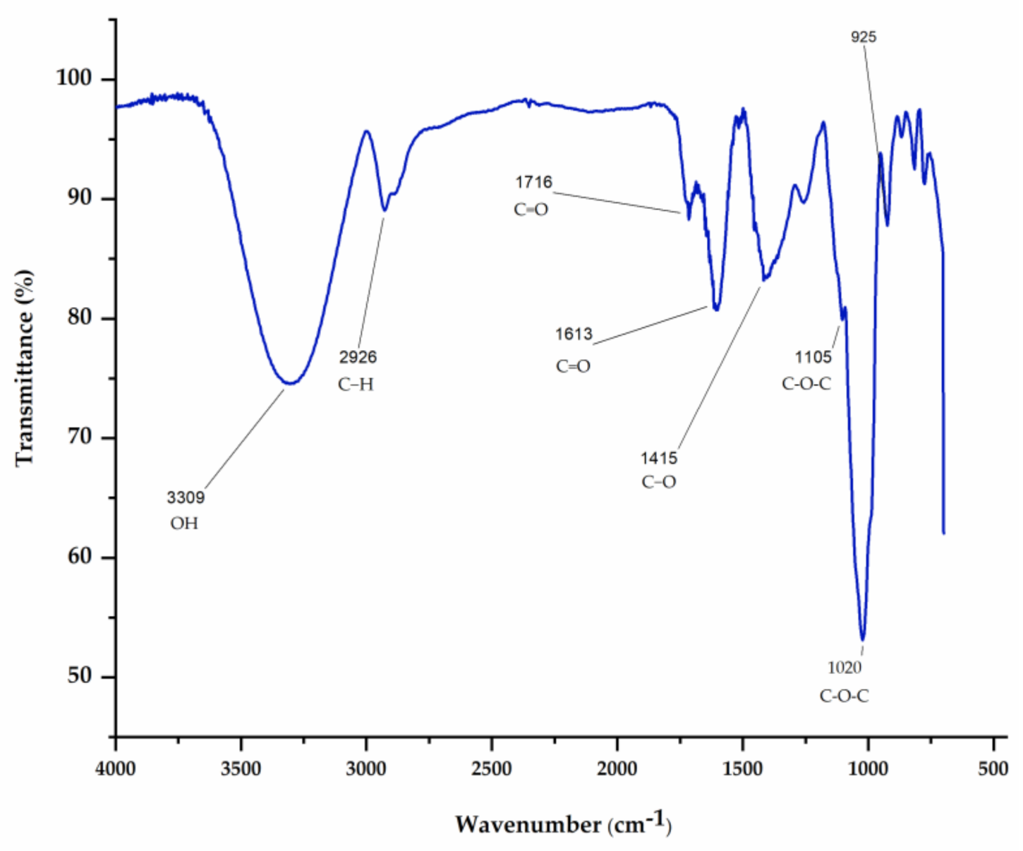
2.4.3. Fourier Transform InfraRed (FTIR) Spectroscopy

The FTIR was recorded in an IR Prestige-21 spectrophotometer (Shimadzu, Kyoto, Japan) with a frequency range of 700 to 4000 cm−1 and a resolution of 4 cm−1. AsRE (10 mg) was mixed with spectroscopic-grade potassium bromide powder and then pressed into 1.0 mm pellets and submitted to FTIR analyses.

2.5. In Vitro Antioxidant Activity

A single antioxidant assay is not sufficient to comprehensively assess the antioxidant capacity of extracts [23]. Therefore, in order to evaluate the activity of the extract at different stages of the oxidative cascade (initiation, propagation and termination), the antioxidant activity of the extract was evaluated by six different methods: total antioxidant capacity, reducing power, 1,1-Diphenyl-2-picrylhydrazyl (DPPH) radical scavenging test, cupric and ferric chelating, and hydroxyl radical scavenging. The IC50 (concentration required for 50% inhibition) value was determined from the plotted graph of reducing power, scavenging activity (DPPH and Hydroxyl radical) and metal chelating (Cupric chelating and ferric chelating) against the different concentrations of AsRE according to Equation (2).
IC50 value = (50-b/a)

2.5.1. Total Antioxidant Capacity (TAC)

The antioxidant capacity assay was assessed according to the method described by Costa et al. [24]. AsRE, together with a reagent solution (0.6 M sulfuric acid, 28 mM sodium phosphate and 4 mM ammonium molybdate), was incubated at 95 °C for 90 min. The mixture was cooled, and the absorbance measured at 695 nm against a blank. The antioxidant capacity was expressed as ascorbic acid equivalent.

2.5.2. Reducing Power

The reducing power was evaluated as described previously [24]. Briefly, 4.0 mL of reaction mixture, containing different sample concentrations (0.05, 0.1, 0.25, 0.5, and 1.0 mg/mL) in phosphate buffer (0.2 M, pH 6.6), was incubated with potassium ferricyanide (1% w/v) at 50 °C for 20 min. The reaction was stopped by trichloroacetic acid solution (10% w/v). Afterwards, the solution was mixed with distilled water and ferric chloride solution (0.1% w/v). The absorbance was measured at 700 nm against a blank. The mixture was stirred, and the absorbance (700 nm) measured using a microplate reader (BioTek, Winooski, VT, USA). Results were expressed as the percentage of activity observed for 0.1 mg/mL (highest activity) ascorbic acid.

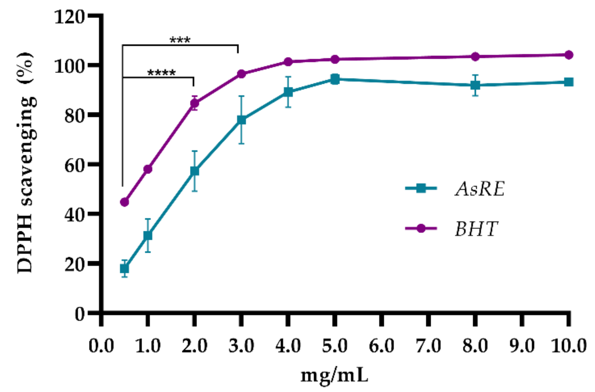
2.5.3. 1,1-Diphenyl-2-Picrylhydrazyl (DPPH) Radical Scavenging Activity

AsRE’s ability to scavenge 1,1-Diphenyl-2-picrylhydrazyl (DPPH) free radicals was determined using the method described by Brand-Williams et al. [25]. Briefly, 1.0 mL of different concentrations of AsRE (0.25, 0.5, 1.0, 2.0, 4.0, 8.0 and 10.0 mg/mL) were added to 2.0 mL of 0.1 mM DPPH ethanolic solution. After 30 min at room temperature, the absorbances were measured at 517 nm. Butylated hydroxytoluene (BHT) (0.25, 0.5, 1.0, 2.0, 4.0, 8.0 and 10.0 mg/mL) was used as a positive control. The results were expressed as a percentage of radical scavenging according to Equation (3).
DPPH radical scavenging (%) = [1 − (As/A0)] × 100
where As = absorbance of the sample; A0 = absorbance of the control.

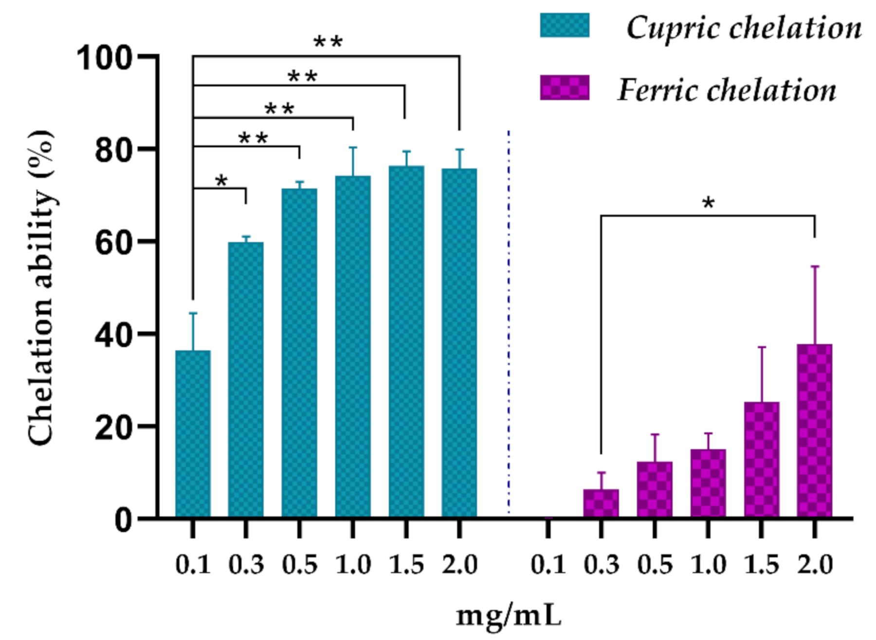
2.5.4. Cupric Chelating

The cupric chelating was evaluated as described previously [26]. Different concentrations of AsRE (0.1, 0.3, 0.5, 1.0, 1.5, and 2.0 mg/mL) were added to reagent solution (pyracotechol (4.0 mM) and cupric sulfate II pentahydrate (0.2 mM)) and measured at 632 nm. EDTA was used as a positive control (0.2–6.0 µg/mL). The results were expressed as a percentage of cupric chelation according to Equation (4).
Cupric chelating (%) = [(A0 − As)/A0] × 100
where As = absorbance of the sample; A0 = absorbance of the control.

2.5.5. Ferric Chelating

The ferric chelating effects of AsRE were evaluated as described previously by Decker and Welch [27]. Different concentrations (0.1, 0.3, 0.5, 1.0, 1.5, and 2.0 mg/mL) were added to reagent solution (FeCl2 2.0 mM, Ferrozine 5.0 mM) and incubated at room temperature for 10 min at a wavelength of 562 nm. EDTA was used as a positive control (0.005–0.05 µg/mL). The results were expressed as a percentage of iron ion chelating according to Equation (5).
Ferric chelating (%) = [(A0 − As)/A0] × 100
where As = absorbance of the sample; A0 = absorbance of the control.

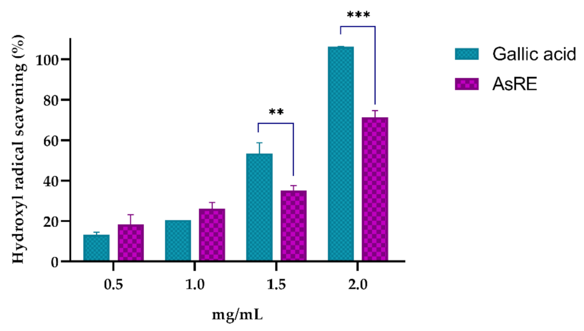
2.5.6. Hydroxyl Radical Scavenging Activity

The hydroxyl radical scavenging activity was evaluated using Fenton’s reaction as described previously [28]. Different concentrations of the AsRE (0.5, 1.0, 1.5 and 2.0 mg/mL) were added to reagent solution (10.0 mM iron sulphate, 10.0 mM EDTA, 2.0 mM sodium salicylate, 30% H2O2) and incubated at 37 °C for 1 h. Gallic acid (0.5, 1.0, 1.5 and 2.0 mg/mL) was used as positive control. The hydroxyl radical was investigated by monitoring absorbance at 510 nm. The results were expressed as a percentage of the scavenging of radicals according to Equation (6).
Hydroxyl radical scavenging (%) = [1 − (A0 − As)/(A0 − Ab)] × 100
where As = absorbance of the sample; A0 = absorbance of the control; Ab = absorbance of the blank.

2.6. Caenorhabditis elegans Maintenance Strains and Extract Treatment

The N2 (wild type) strain of C. elegans was used, which was cultivated in the Nematode Growth Medium (NGM) with Escherichia coli OP50 and maintained at 20 °C [29]. Worms were synchronized by treating pregnant hermaphrodites with lysis solution (hypochlorite 2% and NaOH 10 mM) in order to obtain worms at the L1 stage. AsRE was added to the E. coli OP50 fluid at a final concentration of 1.0, 3.0 and 5.0 mg/mL. The fluid was added to the surface NGM [17].

2.7. Bacterial Growth Assay

The bacterial growth of E. coli OP50 was assessed by optical density (OD) readings on the spectrophotometer (Biochrom Libra S22, Holliston, MA, USA). E. coli OP50 was cultivated into sterilized liquid medium with or without the different concentrations of AsRE (1.0, 3.0 and 5.0 mg/mL) in Luria–Bertani medium (LB). The nematodes were inoculated with the E. coli OP50 and separated into groups, respectively. The results expressed the relative levels. The initial values of optical density (OD600) to E. coli OP50 were compared at each point with the OD600 at the zero point of each condition. The samples were transferred to a rocking shaker at 37 °C (Marconi MA 420, Piracicaba, Brazil), and values measured once every 1 h up to 5 h.

2.8. Toxicity of Agave sisalana Extract Against C. elegans

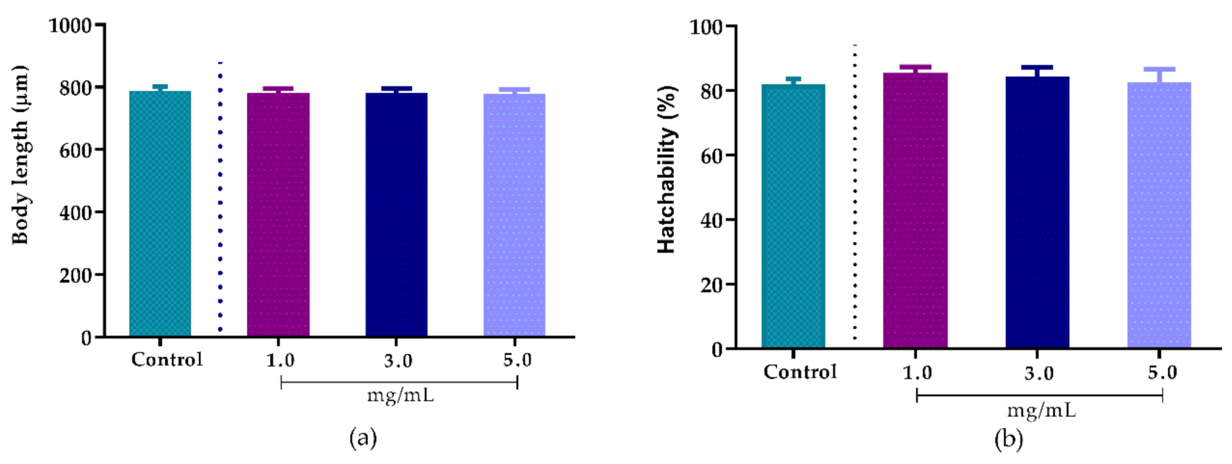
2.8.1. Effect of A. sisalana Agro-Industrial Residue Extract on Body Length

The worms synchronized at the L1 stage cultivated in NGM medium plates were separated into groups and submitted to treatment with AsRE at 1.0, 3.0 and 5.0 mg/mL for 48 h at 20 °C. In the control group, nematodes were cultured on the NGM with the E. coli OP50 fluid. In total, 30 worms from each group were photographed and body length measured using the ImageJ software (ImageJ 1.51j8-https://imagej.nih.gov/ij/). The experiment was performed in triplicate and results presented as mean area ± standard error of the mean (SEM).

2.8.2. Effect of Agave sisalana Agro-Industrial Residue Extract on Hatchability

The effect of AsRE on the egg hatching of wild type C. elegans was evaluated for hatchability percentage. Embryos resistant to treatment with lysis solution were collected and added to the surface NGM medium in treatment groups at 1.0, 3.0 and 5.0 mg/mL (in E. coli OP50) and the control group only with E. coli OP50. After 24 h at 20 °C, hatchability percentage was calculated to a ratio of progeny number over brood size. Experiments were performed in triplicate and results presented as mean area ± standard error of the mean (SEM).

2.9. In Vivo Antioxidant Activity of Agave sisalana Agro-Industrial Residue Extract Using C. elegans as a Model Organism

2.9.1. Intracellular Accumulation of Reactive Oxygen Species (ROS) in C. elegans

H2DCF-DA is converted into a non-fluorescent intermediate form that can later be oxidized by ROS to a fluorescent compound, DCF, in the intracellular medium. The fluorescence intensity of this oxidative derivative is correlated with the amount of intracellular ROS in a living organism. Wild type C. elegans from the L1 stage were treated with or without (control group) the different concentrations of AsRE (1.0, 3.0 and 5.0 mg/mL) for 48 h at 20 °C. Next, approximately 40 worms per group were transferred to a 96-well plate containing 50 μM H2DCFDA (2.7-dichlorofluorescein-diacetate) in PBS. Fluorescence quantification was performed on GloboMax®-mult detection system (Promega, Madison, WI, USA), with excitation at 490 nm and emission at 510–570 nm. Six readings were taken, with an interval of 30 min between each reading. The experiment was performed in three independent experiments, performed in triplicate and results presented as mean area ± standard error of the mean (SEM).

2.9.2. Oxidative Stress Survival Assay

Wild type worms from the L1 stage were treated with AsRE at concentrations of 1.0, 3.0 and 5.0 mg/mL for 48 h at 20 °C. Subsequently, 50 nematodes per group were transferred to plates with NGM solid medium containing 10 mM tert-butyl hydroperoxide (TBHP). The fraction of surviving nematodes was evaluated for up to 33 h using a stereoscopic microscope by probing the nematodes. This assay was repeated three times with a total number of 150 worms/group and results presented as mean area ± standard error of the mean (SEM).

2.9.3. Expression Levels of the Oxidative Resistance Relative Gene

CL2166 mutant worms carrying an inducible green fluorescence protein (GFP) reporter for gst-4 (gst-4::GFP) were maintained on NGM plates with or without (control) different concentrations of AsRE (1.0, 3.0 and 5.0 mg/mL) for 48 h. The gene expression was determined by fluorescence microscopy (Olympus BX51, BR). The fluorescence intensity was quantified using the ImageJ software (ImageJ 1.51 j8).

2.10. Statistical Analysis

All experiments were performed three times. Statistical analysis was performed using GraphPad Prism 8.0.2 (https://www.graphpad.com/scientificsoftware/prism/). Student’s t test was used to compare pairs of groups, as well as a one-way ANOVA followed by Tukey’s post test. For the hydroxyl radical scavenging test, a two-way way ANOVA followed by Sidak’s multiple comparisons was used. Survival curves were analyzed by the log-rank (Mantel–Cox) test.

3. Results

3.1. Chemical Characterization

Saponins and polyphenols are the most widely studied classes of the Agave genus. After the extractive process, the yield for AsRE was 7.08%. The results obtained showed that saponins comprise 18.01% ± 1.81 of AsRE while the concentration of total flavonoids is 0.92 ± 0.26 µg EQ/mg. The infrared spectra of AsRE displayed some characteristic bands compatible with polysaccharides, saponins and phenolics (Figure 1).

3.2. In Vitro Antioxidant Activity

The total antioxidant capacity test (TAC) showed a high activity for AsRE (91.75 mg Eq AA/g). The reducing power assay showed AsRE (1.0 mg/mL) with an activity of 49.13% ± 2.03 in comparison to ascorbic acid (100 µg). When AsRE was assessed for its ability to scavenge DPPH radical scavenging, it showed that it was able to scavenge the radicals in a concentration-dependent manner up to a concentration of 5.0 mg/mL with maximum values of 94.40% ± 1.94 (Figure 2). Starting at the concentration of 4.0 mg/mL and higher concentrations, there is no significant difference between AsRE and the positive control used.
In relation to the metal chelating capacity, the extract showed a high cupric chelating power (70% at 0.5 mg/mL). Furthermore, for ferric chelating activity, the extract (1.0 mg/mL) showed 15.10% ± 3.44 chelating activity (Figure 3).
AsRE demonstrated hydroxyl radical scavenging in a concentration-dependent manner, reaching 71.38% ± 3.25 at 2.0 mg/mL (Figure 4).
The results presented demonstrate the potential of the extract as an antioxidant raw material with possible intrinsic performances in different phases of the oxidative cascade. The IC50 values obtained for antioxidant tests can be seen in Table 1.

3.3. Bacterial Growth Assay

Before the in vivo antioxidant tests, the effect of the extract on the growth of E. coli OP50 bacteria, used as a food source, was evaluated. The toxicity against E. coli OP50 can lead to an increase in the lifespan of C. elegans [17]. The AsRE (1.0, 3.0 and 5.0 mg/mL) did not exhibit antibacterial activity against E. coli OP50.

3.4. Toxicity of Agave sisalana Extract Against C. elegans

After 48 h of exposure to different concentrations (1.0, 3.0 and 5.0 mg/mL) of the extract, no significant changes were observed in terms of body length (Figure 5a) or hatchability (Figure 5b), indicating that the fractions do not present toxicity to the nematodes.

3.5. In Vivo Antioidant Activity

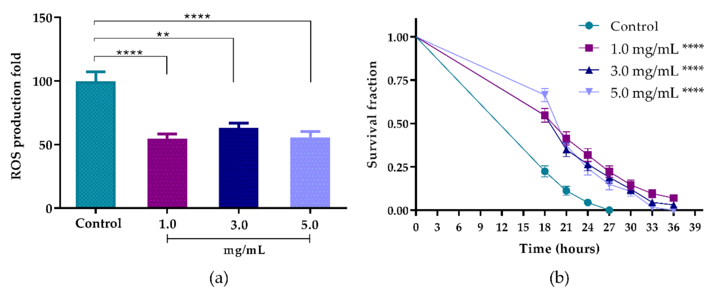
3.5.1. Intracellular Accumulation of ROS in C. elegans

Nematodes treated with 1.0, 3.0 and 5.0 mg/mL of the extract showed a reduction in intracellular ROS levels of 45.18% (p < 0.0001), 36.84% (p < 0.01) and 44.19% (p < 0.0001), respectively (Figure 6a). However, no statistical differences were observed among the AsRE at different concentrations.

3.5.2. Oxidative Stress Survival Assay

The reduction in intracellular levels of ROS enabled us to evaluate the ability of the extract to increase the survival fraction of worms against the acute stress induced (TBHP). The extract promoted a significant change to the right in the survival curve under acute oxidative stress induced at the concentrations evaluated when compared to the control (p < 0.0001) (Figure 6b). The increase in average life span when compared to the control group (p < 0.0001) can be seen in Table 2. The OS process leads to cumulative damage to biomolecules that promote the aging process and consequently the survival time of C. elegans [13]. Therefore, the extension of the lifespan is strongly associated with enhanced resistance to stress [15]. Thus, we suggest that there is an increase in the survival fraction with a reduction in the ROS levels promoted by the extract.

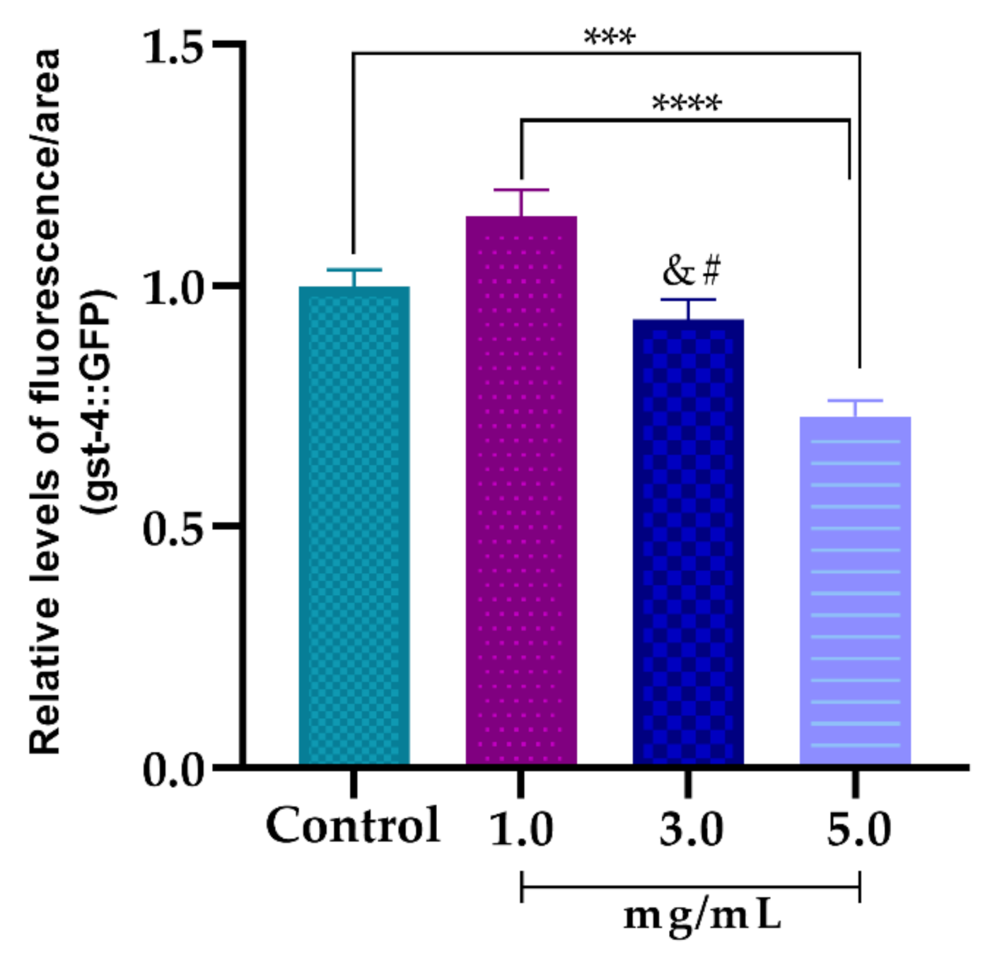
3.5.3. Effect of AsRE on the Expression Levels of the Oxidative Stress Relative Gene (gst-4::GFP)

The CL2166 worms were previously treated with 1.0, 3.0 and 5.0 mg/mL of AsRE to assess their ability to modulate the expression of genes related to oxidative stress resistance (gst-4::GFP). A dose-dependent profile was observed in the negative expression of the gst-4::GFP gene (Figure 7). However, when compared to the control, only the concentration of 5.0 mg/mL showed a significant reduction in the expression of gst-4::GFP, reducing the fluorescence intensity by 27.04% ± 0.04.

4. Discussion

The search for new molecules with biological activity has been intensified in studies with plant extracts due to their diverse and complex chemical constitution. In a previous study, our group showed that A. sisalana extract contained polyphenols (2.53%) and sugars (65%) [10]. Now, in this present study, to advance the chemical characterization of this extract, the total saponins and total flavonoids were analyzed. The infrared analyses also indicated the presence of sugar (OH, C-H, C-O-C, C-O, C=O) [23,30,34,35], saponins (CO, C-O-C, C=O) [32,33], and flavonoids [36] in AsRE.
It is interesting to highlight that among the phenolic compounds of the genus Agave, flavonoids, homoisoflavonoids and phenolic acids were the most common phenolic compounds found [7]. Therefore, we hypothesize that the low amount of total flavonoids could indicate a richness of phenolic acid in the extract. Furthermore, the presence of saponin characterization data was an expected result regarding the historical importance and occurrence of these metabolites in the genus. Taken together, these features are compatible with the expected chemical profile of A. sisalana hydroethanolic extract and these biomolecules demonstrated a strong correlation with biological activities, such as antioxidant activity [5,8].
In particular, molecules with antioxidant capacity are used in order to combat the damage caused by oxidative stress and to prevent aging [37,38]. Many studies with extracts and their components have demonstrated the correlation between antioxidant activity and primary and secondary metabolites of plants such as polyphenols [39,40], saponins [8] and polysaccharides [41,42,43,44].
Phenolic compounds are constituted of one or more aromatic rings containing variable hydroxyl groups, and are, therefore, potentially capable of stabilizing reactive species by forming resonance-stabilized phenoxy radicals [45]. The activity of saponins, on the other hand, has been related to their glycosylated C-3 hydroxyl group [8], while polysaccharides can donate atoms and stop chain reactions by turning radicals into stable non-harmful molecules. According to Huang et al. [44], the carboxyl and carbonyl groups, present in polysaccharides, decrease the dissociation energy of the O-H bond, which results in increased proton release, potentiating the antioxidant activity of these compounds.
The antioxidant activity of AsRE was evaluated in three stages: initiation (Total Antioxidant Capacity, power reducing and DPPH radical scavenging), propagation (cupric and ferric chelating), and termination (hydroxyl radical scavenging) [46].
The total antioxidant capacity test showed an activity for the AsRE of 91.75 mg EqAA/g, which is considered high antioxidant activity [24]. Moreover, AsRE showed high reducing power when compared with positive control ascorbic acid (49.13% ± 2.03). A previous study using isolated polysaccharides of agro-industrial A. sisalana showed a power reduction of 37% compared to ascorbic acid [5]. Reducing properties are generally associated with the presence of biomolecules, which exhibit antioxidant activity by scavenging effect on the reactive species [17], donating hydrogen atoms to break chain reactions [5].
AsRE showed high DPPH radical scavenging activity when compared to the positive control (Figure 2). Polysaccharides isolated from this material demonstrated the ability to interact with oxidative processes acting as an antioxidant agent [5,11]. Furthermore, Ribeiro et al. [8] pointed out a more significant activity for the extract when compared to the isolated compounds. These authors highlighted the low ratio of saponins isolated from the leaves of A. sisalana with the elimination of DPPH radicals (IC50 > 10.0 mg/mL), while the extract containing saponins and polyphenols showed results (IC50 = 1.4 mg/mL) similar to those observed for AsRE (IC50 = 1.44 mg/mL). Therefore, it is possible to imply that the diversified chemical composition of this plant, when compared to isolated molecules, may favor a greater antioxidant potential. The significant results of the total antioxidant capacity and reducing power in vitro indicate that AsRE has the ability to interact with the systems and give electrons to minimize the attack of radicals [47], which correlates with the ability to eliminate radicals [5].
AsRE demonstrated chelating activity of cupric and iron (Figure 3). Several studies showed plant extracts with iron and/or cupric chelating activity. These authors relate this chelating activity to the presence of molecules such as polyphenols [48] and polysaccharides [5,24,42,49]. The evaluation of the metal chelating potential is important due to transition metals, free of redox activity, such as Cu2+ and Fe2+, which can be extremely pro-oxidant, meaning that they can react with hydrogen peroxide and catalyze the formation of hydroxyl radical through the Fenton reaction [37]. Therefore, when AsRE acts as a metal chelator, it is indirectly combating the damage caused by the hydroxyl radical. However, there are two types of antioxidant mechanism against the hydroxyl radical: one suppresses the generation of the hydroxyl radical and the other scavenges the hydroxyl radicals generated [24]. Due to this, the potential of AsRE for radical hydroxyl scavenging was also evaluated (Figure 4). The scavenging ability of AsRE for hydroxyl radicals increased with the concentration within the range of 0.5 e 2.0 mg/mL. The rate that AsRE and gallic acid scavenged hydroxyl radicals at 2.0 mg/mL was 71.38% and 100%, respectively. Zhang et al. [5] showed the scavenging effect on the hydroxyl radical by polysaccharides of A. sisalana residue. However, the activity found for these metabolics was around 33% at 4.0 mg/mL. Biomolecules and extracts of other sources also demonstrated activity for scavenging hydroxyl radicals. Cheng et al. [50] showed the activity of flavonoids from Carex meyeriana on scavenging the hydroxyl radical. Melocactus zehntneri ethanolic extract containing phenolic compounds, saponins and polysaccharides showed 100% hydroxyl scavenging activity for polar extract (using 100 µg/mL), whereas the non-polar extracts showed an activity of around 20% [46]. Therefore, AsRE exerted their antioxidant activity in three stages: initiation, propagation and termination.
Considering that AsRE showed excellent in vitro antioxidant activity, the nematode C. elegans was used to evaluate the in vivo antioxidant potential of AsRE. In addition, after the results obtained in the in vitro tests and knowing that high concentrations are used in tests with these worms due to the presence of a thick cuticle [18], the concentrations 1.0, 3.0 and 5.0 mg/mL were used in the tests in vivo.
In first step, bacterial Growth of E. coli OP50 was evaluated. The worms feed on E. coli OP50 and the inhibition of this bacteria can lead to an increase in the lifespan of C. elegans [17]. Consequently, if the extract caused the death of the bacteria or growth inhibition, the worms should be fed with the dead bacteria so that the increase in the lifespan does not interfere with the results of the antioxidant tests performed with this model. However, AsRE did not interfere with the bacterial growth of E. coli OP50, so it was not necessary to feed them with dead bacteria. The non-toxicity of AsRE corroborates the results obtained by Santos et al. [51], which showed that hydroethanolic extracts from A. sisalana leaves and residue have no relevant activity against the bacteria tested. Similar results were observed by Ribeiro et al. [8] when evaluating saponins from A. sisalana to E. coli.
An anthelmintic effect, in vitro, against goat gastrointestinal nematodes of A. sisalana agro-industrial residue has been demonstrated by Domingues et al. [52]. Therefore, the ovicidal and larvicidal in vitro effect of A. sisalana was shown by Botura et al. [53] using gastrointestinal nematodes of goats. However, AsRE did not affect the development of C. elegans nematodes.
The results for the antioxidant capacity (Figure 6) obtained with the C. elegans model reinforce the results observed in vitro. AsRE was able to act as an antioxidant by reducing the intracellular levels of ROS in wild-type worms and by increasing survival rates after they had been induced to strong oxidative stress. ROS are highly reactive, usually produced by the cellular aerobic metabolism that can increase dramatically when organisms are exposed to different stress conditions [54].
The C. elegans results involving the reduction in intracellular levels of ROS are commonly associated with an increase in the rate of survival of these animals, when induced to stress. Different studies have shown plant extracts with antioxidant activity capable of reducing ROS levels and increasing the C. elegans survival rate. Azevedo et al. [16] demonstrated that the aqueous extract of Uncaria tomentosa leaves was also able to reduce ROS levels and increase the survival rate of C. elegans. Panax notoginseng ethanol extracts also increased lifespan and protected C. elegans against oxidative stress. Similar results were observed by Ribeiro et al. [18] when using ethanol extract from Myrciaria tenella leaves. Moreover, a correlation between stress resistance and lifespan extension has been attributed to polyphenols [13,55] and saponins [56].
However, antioxidants can act directly to reduce ROS levels [16] or to increase resistance to oxidative stress, inducing the transcription of cytoprotective proteins [57]. To understand the mechanisms by which AsRE can act against oxidative stress, glutathione S-transferase (gst-4) gene expression levels were evaluated. gst-4 is a phase II detoxification enzyme that catalyzes the conjugation of several exogenous and endogenous electrophiles with the reduced form of glutathione, resulting in the detoxification of ROS [56].
AsRE promoted a reduction in expression of gst-4 levels in a dose-dependent manner (Figure 7). The capacity to suppress expression of gst-4 is widely interpreted as proof of their ability to reduce oxidative stress in C. elegans [16,56]. Accordingly, it could be suggested that the reduction in intracellular levels of ROS associated with increased resistance to stress and downregulation of gst-4::GFP expression is related to the direct activity of the AsRE against ROS.
AsRE was able to reduce the production of intracellular ROS and increase the mean lifespan of wild-type C. elegans under stress conditions. Since oxidative stress plays a role in many diseases and contributes to aging [12], AsRE may lead to the production of a new raw material against oxidative stress and aging in the near future. This agro-industrial residue can be considered a potential renewable, sustainable, and environmentally friendly source. Hence, it is possible to reincorporate it into the industry in new production processes, which can also generate extra income and contribute to socioeconomic development in semi-arid regions.

5. Conclusions

A. sisalana agro-industrial residue extract has a diverse chemical composition and proven antioxidant activity. In this study, the antioxidant evaluation, in vitro and in vivo, using the C. elegans organism model showed evidence that the extract can efficiently eliminate reactive species in vitro and in vivo, reducing endogenous levels of reactive species and promoting an increased survival rate of C. elegans under stress, acting directly on the elimination of reactive species. However, new tests should be carried out to explain the relationship between in vivo and in vitro assays. A. sisalana agro-industrial residue extract can be considered a potential antioxidant raw material for different applications in the food, cosmetic and pharmaceutical industries. We can also point out as advantages of this studied extract the ease and availability of obtaining biomass and the fact that the production process of the extract is industrially scalable, fundamental conditions for it to become commercial. This enables the prospect of introducing to the agroindustry a renewable process that promotes sustainability and socioeconomic development and brings benefits to current and future generations.

Author Contributions

Conceptualization, S.M.A.G.B. and C.O.M.C.; Data curation, R.A.d.O.M. and G.M.M.S.; Formal analysis, S.M.A.G.B.; Funding acquisition, M.F.; Investigation, S.M.A.G.B., R.A.d.O.M., S.V.F.d.A. and J.A.A.d.S.F.; Methodology, S.M.A.G.B. and C.O.M.C.; Resources, H.A.O.R., R.d.P.O., R.B.G. and M.F.; Supervision, R.B.G. and M.F.; Writing—original draft, S.M.A.G.B.; Writing—review and editing, R.G.B. and M.F. All authors have read and agreed to the published version of the manuscript.

Funding

This study was financed by the Coordenação de Aperfeiçoamento de Pessoal de Nível Superior—Brasil (CAPES), Finance Code 001, and Universidade Federal do Rio Grande do Norte (UFRN).

Acknowledgments

The authors are thankful to the Universidade Federal do Rio Grande do Norte (UFRN), Conselho Nacional de Desenvolvimento Científico e Tecnológico (CNPq) and Instituto Nacional de Ciência e Tecnologia em Biodiversidade e Produtos Naturais (INCT-BioNat).

Conflicts of Interest

The authors declare no conflict of interest.

References

  1. Sidana, J.; Singh, B.; Sharma, O.P. Phytochemistry Saponins of Agave: Chemistry and bioactivity. Phytochemistry 2016, 130, 22–46. [Google Scholar] [CrossRef] [PubMed]
  2. Gutiérrez, A.; Rodríguez, I.M.; del Río, J.C. Chemical composition of lipophilic extractives from sisal (Agave sisalana) fibers. Ind. Crops Prod. 2008, 28, 81–87. [Google Scholar] [CrossRef] [Green Version]
  3. Santos, J.D.G.; Vieira, I.J.C.; Braz-Filho, R.; Branco, A. Chemicals from Agave sisalana biomass: Isolation and identification. Int. J. Mol. Sci. 2015, 16, 8761–8771. [Google Scholar] [CrossRef] [PubMed] [Green Version]
  4. Maran, J.P.; Priya, B. Ultrasound-assisted extraction of pectin from sisal waste. Carbohydr. Polym. 2014, 115, 732–738. [Google Scholar] [CrossRef]
  5. Zhang, X.; Liu, L.; Lin, C. Isolation, structural characterization and antioxidant activity of a neutral polysaccharide from Sisal waste. Food Hydrocoll. 2014, 39, 10–18. [Google Scholar] [CrossRef]
  6. Martin, A.R.; Martins, M.A.; Mattoso, L.H.C.; Silva, O.R.R.F. Caracteriza̧ão química e estrutural de fibra de sisal da variedade Agave sisalana. Polimeros 2009, 19, 40–46. [Google Scholar] [CrossRef] [Green Version]
  7. Almaraz-Abarca, N.; Delgado-Alvarado, E.A.; Ávila-Reyes, J.A.; Uribe-Soto, J.N.; González-Valdez, L.S. The Phenols of the Genus Agave (Agavaceae). J. Biomater. Nanobiotechnol. 2013, 04, 9–16. [Google Scholar] [CrossRef] [Green Version]
  8. Ribeiro, B.D.; Alviano, D.S.; Barreto, D.W.; Coelho, M.A.Z. Functional properties of saponins from sisal (Agave sisalana) and juá (Ziziphus joazeiro): Critical micellar concentration, antioxidant and antimicrobial activities. Colloids Surfaces A Physicochem. Eng. Asp. 2013, 436, 736–743. [Google Scholar] [CrossRef]
  9. Santos, J.D.G.; Branco, A. GC-MS Characterisation of Sapogenins from Sisal Waste and a Method to Isolate Pure Hecogenin. BioResources 2014, 9, 1325–1333. [Google Scholar] [CrossRef]
  10. Barreto, S.M.A.G.; Maia, M.S.; Benicá, A.M.; de Assis, H.R.B.S.; Leite-Silva, V.R.; da Rocha-Filho, P.A.; de Negreiros, M.M.F.; de Oliveira Rocha, H.A.; Ostrosky, E.A.; Lopes, P.S.; et al. Evaluation of in vitro and in vivo safety of the by-product of Agave sisalana as a new cosmetic raw material: Development and clinical evaluation of a nanoemulsion to improve skin moisturizing. Ind. Crops Prod. 2017, 108, 470–479. [Google Scholar] [CrossRef]
  11. Zhang, X.; Liu, L.; Lin, C. Structural features, antioxidant and immunological activity of a new polysaccharide (SP1) from sisal residue. Int. J. Biol. Macromol. 2013, 59, 184–191. [Google Scholar] [CrossRef] [PubMed]
  12. Sies, H. Oxidative Stress: Concept and Some Practical Aspects. Antioxidants 2020, 9, 852. [Google Scholar] [CrossRef] [PubMed]
  13. Grünz, G.; Haas, K.; Soukup, S.; Klingenspor, M.; Kulling, S.E.; Daniel, H.; Spanier, B. Structural features and bioavailability of four flavonoids and their implications for lifespan-extending and antioxidant actions in C. elegans. Mech. Ageing Dev. 2012, 133, 1–10. [Google Scholar] [CrossRef] [PubMed]
  14. Leopoldini, M.; Russo, N.; Toscano, M. The molecular basis of working mechanism of natural polyphenolic antioxidants. Food Chem. 2011, 125, 288–306. [Google Scholar] [CrossRef]
  15. De Oliveira Caland, R.B.; Cadavid, C.O.M.; Carmona, L.; Peña, L.; De Paula Oliveira, R. Pasteurized orange juice rich in carotenoids protects caenorhabditis elegans against oxidative stress and β-amyloid toxicity through direct and indirect mechanisms. Oxid. Med. Cell. Longev. 2019, 2019. [Google Scholar] [CrossRef] [PubMed] [Green Version]
  16. Azevedo, B.C.; Roxo, M.; Borges, M.C.; Peixoto, H.; Crevelin, E.J.; Bertoni, B.W.; Contini, S.H.T.; Lopes, A.A.; França, S.C.; Pereira, A.M.S.; et al. Antioxidant Activity of an Aqueous Leaf Extract from Uncaria tomentosa and Its Major Alkaloids Mitraphylline and Isomitraphylline in Caenorhabditis elegans. Molecules 2019, 24, 3299. [Google Scholar] [CrossRef] [Green Version]
  17. Yuan, Y.; Kang, N.; Li, Q.; Zhang, Y.; Liu, Y.; Tan, P. Study of the Effect of Neutral Polysaccharides from Rehmannia glutinosa on Lifespan of Caenorhabditis elegans. Molecules 2019, 24, 1–15. [Google Scholar] [CrossRef] [PubMed] [Green Version]
  18. Ribeiro, A.R.C.; da Silva Cordeiro, M.L.; Silva, L.M.P.; Cadavid, C.O.M.; de Oliveira Caland, R.B.; Fernandes-Negreiros, M.M.; Queiroz, M.F.; da Silva Barbosa, J.; Aragão, C.F.S.; Zucolotto, S.M.; et al. Myrciaria tenella (DC.) O. Berg (myrtaceae) leaves as a source of antioxidant compounds. Antioxidants 2019, 8, 310. [Google Scholar] [CrossRef] [Green Version]
  19. Artal-Sanz, M.; de Jong, L.; Tavernarakis, N. Caenorhabditis elegans: A versatile platform for drug discovery. Biotechnol. J. 2006, 1, 1405–1418. [Google Scholar] [CrossRef]
  20. Araldi, R.P.; dos Santos, M.O.; Barbon, F.F.; Manjerona, B.A.; Meirelles, B.R.; de Oliva Neto, P.; da Silva, P.I.; dos Santos, L.; Camargo, I.C.C.; de Souza, E.B. Analysis of antioxidant, cytotoxic and mutagenic potential of Agave sisalana Perrine extracts using Vero cells, human lymphocytes and mice polychromatic erythrocytes. Biomed. Pharmacother. 2018, 98, 873–885. [Google Scholar] [CrossRef]
  21. Chinedum, E.; Sanni, S.; Theressa, N.; Ebere, A. Effect of domestic cooking on the starch digestibility, predicted glycemic indices, polyphenol contents and alpha amylase inhibitory properties of beans (Phaseolis vulgaris) and breadfruit (Treculia africana). Int. J. Biol. Macromol. 2018, 106, 200–206. [Google Scholar] [CrossRef] [PubMed]
  22. Ribeiro, B.D.; Barreto, D.W.; Coelho, M.A.Z. Use of micellar extraction and cloud point preconcentration for valorization of saponins from sisal (Agave sisalana) waste. Food Bioprod. Process. 2015, 94, 601–609. [Google Scholar] [CrossRef]
  23. Feng, S.; Cheng, H.; Xu, Z.; Feng, S.; Yuan, M.; Huang, Y.; Liao, J.; Ding, C. Antioxidant and anti-aging activities and structural elucidation of polysaccharides from Panax notoginseng root. Process Biochem. 2019, 78, 189–199. [Google Scholar] [CrossRef]
  24. Costa, L.S.; Fidelis, G.P.; Cordeiro, S.L.; Oliveira, R.M.; Sabry, D.A.; Câmara, R.B.G.; Nobre, L.T.D.B.; Costa, M.S.S.P.; Almeida-Lima, J.; Farias, E.H.C.; et al. Biological activities of sulfated polysaccharides from tropical seaweeds. Biomed. Pharmacother. 2010, 64, 21–28. [Google Scholar] [CrossRef] [PubMed]
  25. Brand-Williams, W.; Cuvelier, M.E.; Berset, C. Use of a Free Radical Method to Evaluate Antioxidant Activity. LWT Food Sci. Technol. 1995, 28, 25–30. [Google Scholar] [CrossRef]
  26. Anton, A. Colorimetric Estimation of Aluminum with Pyrocatechol Violet. Anal. Chem. 1960, 32, 725–726. [Google Scholar] [CrossRef]
  27. Decker, E.A.; Welch, B. Role of Ferritin as a Lipid Oxidation Catalyst in Muscle Food. J. Agric. Food Chem. 1990, 674–677. [Google Scholar] [CrossRef]
  28. Sminorff, N.; Cumbes, Q.J. Hydroxyl radical scavenging activity of compatible solutes. Phytochemistry 1989, 28, 1057–1060. [Google Scholar]
  29. Brenner, S. Nature’s gift to science (Nobel lecture). ChemBioChem 2003, 4, 683–687. [Google Scholar] [CrossRef]
  30. Qu, Y.; Li, C.; Zhang, C.; Zeng, R.; Fu, C. Optimization of infrared-assisted extraction of Bletilla striata polysaccharides based on response surface methodology and their antioxidant activities. Carbohydr. Polym. 2016, 148, 345–353. [Google Scholar] [CrossRef]
  31. Wang, L.C.; Zhang, K.; Di, L.Q.; Liu, R.; Wu, H. Isolation and structural elucidation of novel homogenous polysaccharide from Mactra veneriformis. Carbohydr. Polym. 2011, 86, 982–987. [Google Scholar] [CrossRef]
  32. Ulaganathan, V.; Del Castillo, L.; Webber, J.L.; Ho, T.T.M.; Ferri, J.K.; Krasowska, M.; Beattie, D.A. The influence of pH on the interfacial behaviour of Quillaja bark saponin at the air-solution interface. Colloids Surfaces B Biointerfaces 2019, 176, 412–419. [Google Scholar] [CrossRef] [PubMed]
  33. Almutairi, M.S.; Ali, M. Direct detection of saponins in crude extracts of soapnuts by FTIR. Nat. Prod. Res. 2015, 29, 1271–1275. [Google Scholar] [CrossRef] [PubMed]
  34. Fidelis, G.P.; Camara, R.B.G.; Queiroz, M.F.; Costa, M.S.S.P.; Santos, P.C.; Rocha, H.A.O.; Costa, L.S. Proteolysis, NaOH and ultrasound-enhanced extraction of anticoagulant and antioxidant sulfated polysaccharides from the edible seaweed, Gracilaria birdiae. Molecules 2014, 19, 18511–18526. [Google Scholar] [CrossRef] [Green Version]
  35. Galinari, É.; Sabry, D.A.; Sassaki, G.L.; Macedo, G.R.; Passos, F.M.L.; Mantovani, H.C.; Rocha, H.A.O. Chemical structure, antiproliferative and antioxidant activities of a cell wall α-D-mannan from yeast Kluyveromyces marxianus. Carbohydr. Polym. 2017, 157, 1298–1305. [Google Scholar] [CrossRef]
  36. Abbas, O.; Compère, G.; Larondelle, Y.; Pompeu, D.; Rogez, H.; Baeten, V. Phenolic compound explorer: A mid-infrared spectroscopy database. Vib. Spectrosc. 2017, 92, 111–118. [Google Scholar] [CrossRef]
  37. Reiter, R.J.; Tan, D.X.; Rosales-Corral, S.; Galano, A.; Zhou, X.J.; Xu, B. Mitochondria: Central Organelles for Melatonin?s Antioxidant and Anti-Aging Actions. Molecules 2018, 23, 1–25. [Google Scholar] [CrossRef] [Green Version]
  38. Birch-Machin, M.A.; Bowman, A. Oxidative stress and ageing. Br. J. Dermatol. 2016, 175, 26–29. [Google Scholar] [CrossRef] [Green Version]
  39. Xiang, J.; Apea-bah, F.B.; Ndolo, V.U.; Katundu, M.C.; Beta, T. Profile of phenolic compounds and antioxidant activity of finger millet varieties. Food Chem. 2019, 275, 361–368. [Google Scholar] [CrossRef]
  40. Stojiljković, D.; Arsić, I.; Tadić, V. Extracts of wild apple fruit (Malus sylvestris (L.) Mill., Rosaceae) as a source of antioxidant substances for use in production of nutraceuticals and cosmeceuticals. Ind. Crops Prod. 2016, 80, 165–176. [Google Scholar] [CrossRef]
  41. Zhao, H.; Li, J.; Zhang, J.; Wang, X.; Hao, L.; Jia, L. Purification, in vitro antioxidant and in vivo anti-aging activities of exopolysaccharides by Agrocybe cylindracea. Int. J. Biol. Macromol. 2017, 102, 351–357. [Google Scholar] [CrossRef] [PubMed]
  42. Chou, C.H.; Sung, T.J.; Hu, Y.N.; Lu, H.Y.; Yang, L.C.; Cheng, K.C.; Lai, P.S.; Hsieh, C.W. Chemical analysis, moisture-preserving, and antioxidant activities of polysaccharides from Pholiota nameko by fractional precipitation. Int. J. Biol. Macromol. 2019, 131, 1021–1031. [Google Scholar] [CrossRef] [PubMed]
  43. Li, Q.; Wang, W.; Zhu, Y.; Chen, Y.; Zhang, W.; Yu, P.; Mao, G.; Zhao, T.; Feng, W.; Yang, L.; et al. Structural elucidation and antioxidant activity a novel Se-polysaccharide from Se-enriched Grifola frondosa. Carbohydr. Polym. 2017, 161, 42–52. [Google Scholar] [CrossRef] [PubMed]
  44. Huang, F.; Zhang, R.; Dong, L.; Guo, J.; Deng, Y.; Yi, Y.; Zhang, M. Antioxidant and antiproliferative activities of polysaccharide fractions from litchi pulp. Food Funct. 2015, 6, 2598–2606. [Google Scholar] [CrossRef]
  45. Dudonné, S.; Vitrac, X.; Coutiére, P.; Woillez, M.; Mérillon, J.M. Comparative study of antioxidant properties and total phenolic content of 30 plant extracts of industrial interest using DPPH, ABTS, FRAP, SOD, and ORAC assays. J. Agric. Food Chem. 2009, 57, 1768–1774. [Google Scholar] [CrossRef]
  46. Aquino-Martins, V.G.d.Q.; de Melo, L.F.M.; Silva, L.M.P.; de Lima, T.R.T.; Queiroz, M.F.; Viana, R.L.S.; Zucolotto, S.M.; Andrade, V.S.; Rocha, H.A.O.; Scortecci, K.C. In vitro antioxidant, anti-biofilm, and solar protection activities of melocactus zehntneri (Britton & rose) pulp extract. Antioxidants 2019, 8, 439. [Google Scholar] [CrossRef] [Green Version]
  47. Zhang, Z.; Wang, F.; Wang, X.; Liu, X.; Hou, Y.; Zhang, Q. Extraction of the polysaccharides from five algae and their potential antioxidant activity in vitro. Carbohydr. Polym. 2010, 82, 118–121. [Google Scholar] [CrossRef]
  48. Granato, D.; Shahidi, F.; Wrolstad, R.; Kilmartin, P.; Melton, L.D.; Hidalgo, F.J.; Miyashita, K.; Van Camp, J.; Alasalvar, C.; Ismail, A.B.; et al. Antioxidant activity, total phenolics and flavonoids contents: Should we ban in vitro screening methods ? Food Chem. 2018, 264, 471–475. [Google Scholar] [CrossRef]
  49. Melo-Silveira, R.F.; Fidelis, G.P.; Viana, R.L.S.; Soeiro, V.C.; Da Silva, R.A.; Machado, D.; Costa, L.S.; Ferreira, C.V.; Rocha, H.A.O. Antioxidant and antiproliferative activities of methanolic extract from a neglected agricultural product: Corn cobs. Molecules 2014, 19, 5360–5378. [Google Scholar] [CrossRef] [Green Version]
  50. Cheng, X.; Cheng, Y.; Zhang, N.; Zhao, S.; Cui, H.; Zhou, H. Purification of flavonoids from Carex meyeriana Kunth based on AHP and RSM: Composition analysis, antioxidant, and antimicrobial activity. Ind. Crops Prod. 2020, 157, 112900. [Google Scholar] [CrossRef]
  51. Santos, J.D.G.; Branco, A.; Silva, A.F.; Pinheiro, C.S.R.; Neto, G.; Uetanabaro, A.P.T.; Queiroz, S.R.O.D.; Osuna, J.T. A Antimicrobial activity of Agave sisalana. Afr. J. Biotechnol. 2009, 8, 6181–6184. [Google Scholar] [CrossRef] [Green Version]
  52. Domingues, L.F.; Botura, M.B.; Da Cruz, A.C.F.G.; Yuki, C.C.; Da Silva, G.D.; Costa, M.S.; Murphy, G.; Moreira, E.L.T.; Meneses, I.D.S.; De Almeida, M.D.G.Á.R.; et al. Evaluation of anthelmintic activity of liquid waste of Agave sisalana (sisal) in goats. Rev. Bras. Parasitol. Vet. 2010, 19, 270–272. [Google Scholar] [CrossRef] [PubMed] [Green Version]
  53. Botura, M.B.; dos Santos, J.D.G.; da Silva, G.D.; de Lima, H.G.; de Oliveira, J.V.A.; de Almeida, M.A.O.; Batatinha, M.J.M.; Branco, A. In vitro ovicidal and larvicidal activity of Agave sisalana Perr. (sisal) on gastrointestinal nematodes of goats. Vet. Parasitol. 2013, 192, 211–217. [Google Scholar] [CrossRef] [PubMed] [Green Version]
  54. Son, Y.; Cheong, Y.-K.; Kim, N.-H.; Chung, H.-T.; Kang, D.G.; Pae, H.-O. Mitogen-Activated Protein Kinases and Reactive Oxygen Species: How Can ROS Activate MAPK Pathways? J. Signal Transduct. 2011, 2011, 1–6. [Google Scholar] [CrossRef] [PubMed]
  55. Peixoto, H.; Roxo, M.; Silva, E.; Valente, K.; Braun, M.; Wang, X.; Wink, M. Bark extract of the amazonian tree endopleura uchi (humiriaceae) extends lifespan and enhances stress resistance in caenorhabditis elegans. Molecules 2019, 24, 915. [Google Scholar] [CrossRef] [Green Version]
  56. Roxo, M.; Peixoto, H.; Wetterauer, P.; Lima, E.; Wink, M. Piquiá Shells (Caryocar villosum): A Fruit by-Product with Antioxidant and Antiaging Properties in Caenorhabditis elegans. Oxid. Med. Cell Longev. 2020, 2020, 1–19. [Google Scholar] [CrossRef]
  57. De Bonomo, L.F.; Silva, D.N.; Boasquivis, P.F.; Paiva, F.A.; Guerra, J.F.D.C.; Martins, T.A.F.; De Jesus Torres, Á.G.; De Paula, I.T.B.R.; Caneschi, W.L.; Jacolot, P.; et al. Açaí (Euterpe oleracea Mart.) modulates oxidative stress resistance in Caenorhabditis elegans by direct and indirect mechanisms. PLoS ONE 2014, 9. [Google Scholar] [CrossRef] [Green Version]
Figure 1. Infrared spectra of A. sisalana agro-industrial residue extract (AsRE); bands at: 3309 attributed to a hydroxyl stretching vibration [23]; 2926 attributed to the C−H asymmetric stretching vibration [30]; 1020 attributed to C−O stretching vibrations [31] and 1105 and 1020 attributed to C−O−C [32]; 1716 attributed to C=O [33]; 1415 cm−1 and 1613 cm−1 corresponded to C−O [23] and the free carboxyl group, respectively [34,35].
Biomolecules 10 01435 g001
Figure 2. DPPH radical scavenging assay; AsRE = A. sisalana agro-industrial residue extract; *** p < 0.001 and **** p < 0.0001, by two-way ANOVA, by one-way ANOVA, followed by a Tukey’s post-test. The assay was carried out in three independent experiments and performed in triplicate. The results were expressed as mean ± DP.
Biomolecules 10 01435 g002
Figure 3. Metal chelating capacity of A. sisalana agro-industrial residue extract. Cupric and ferric chelating activity. * p < 0.05 and ** p < 0.01 by one-way ANOVA. The assay was carried out in three independent experiments, performed in triplicate. The results were expressed as mean ± DP. For cupric chelating, the positive control used (EDTA) reached maximum inhibition from the concentration at 0.03 µg. For ferric chelating, the positive control used (EDTA) reached maximum inhibition from the concentration at 5.0 µg.
Biomolecules 10 01435 g003
Figure 4. Antioxidant potential of A. sisalana agro-industrial residue extract. AsRE = A. sisalana agro-industrial residue extract; ** p < 0.01 and *** p < 0.001 by two-way ANOVA, followed by a Sidak’s multiple comparisons test. The assay was carried out in three independent experiments, performed in triplicate. The results were expressed as mean ± DP.
Biomolecules 10 01435 g004
Figure 5. Effect of Agave sisalana agro-industrial residue extract on worm development. (a) Body length after treatments with 1.0, 3.0 and 5.0 mg/mL of extract; (b) Hatchability percentage after treatments with 1.0, 3.0 and 5.0 mg/mL of extract.
Biomolecules 10 01435 g005
Figure 6. Intracellular levels of ROS and survival rate curve of wild-type N2 worms treated with AsRE. (a) ROS production levels after treatment with 1.0, 3.0 and 5.0 mg/mL of AsRE. (b) Survival fraction curves of C. elegans under stress conditions after treatment with 1.0, 3.0 and 5.0 mg/mL of AsRE; ** p < 0.01 compared control with 3.0 mg of treatment; **** p < 0.0001 compared control with 1.0 and 3.0 mg of treatment; by one-way ANOVA, followed by a Tukey’s post-test; (b) Survival fraction after treatment with 1.0, 3.0 and 5.0 mg/mL of extract. Survival was measured every 3 h at 25 °C; **** p < 0.0001, compared to the control with worms treated by log-rank (Mantel–Cox) test.
Biomolecules 10 01435 g006
Figure 7. Effect of A. sisalana agro-industrial residue extract (AsRE) on the expression levels of the oxidative stress relative gene gst-4::GFP; *** p < 0.001 compared control with 5.0 mg/mL of treatment; **** p < 0.0001 compared treatment of 1.0 mg and 5.0 mg/mL; # = ** p < 0.01 compared treatment of 3.0 and 1.0 mg/mL; & = * p < 0.05 compared treatment of 3.0 and 5.0 mg/mL; by one-way ANOVA, followed by a Tukey’s post-test. The assay was performed in duplicate and the results expressed as mean ± DP.
Biomolecules 10 01435 g007
Table 1. IC50 values of Agave sisalana agro-industrial residue extract obtained from antioxidant tests.
Antioxidant TestsIC5O Value (mg/mL)
Reducing power1.02
DPPH radical scavenging1.44
Cupric chelating0.16
Ferric chelating2.76
Hydroxyl radical scavenging1.61
Table 2. Evaluation of resistance to oxidative stress in nematodes treated with the extract.
Concentrations
(mg/mL)
Mean Survival
(hours ± SEM)
% Mean Survival Time Variation vs. Untreatedp Value (Long Rank)
Extract vs. Untreated
n 1
Control19.14 ± 0.19--165 (3)
AsRE1.023.23 ± 0.4921.37<0.0001159 (3)
3.022.55 ± 0.4717.82<0.0001154 (3)
5.022.66 ± 0.4418.39<0.0001152 (3)
Where: n 1 = number of animals used in experiment; AsRE = A. sisalana agro-industrial residue extract.

