1. Introduction
The global prevalence of thyroid dysfunction—including both hypo- and hyperthyroidism—has risen steadily in recent decades, with estimates ranging from 5% to 30% depending on region and diagnostic criteria [
1]. This increase has been variously attributed to aging populations, environmental factors, and advances in diagnostic tools, such as ultrasound (US) imaging technology [
2,
3,
4]. Structural abnormalities, such as thyroid nodules, are also highly frequent, detected in up to 60% of adults when sensitive ultrasonography is applied [
5,
6]. Although most nodules are benign, approximately 5–10% harbor malignancy, emphasizing the clinical need for precise risk stratification [
7,
8].
The thyroid gland secretes thyroxine (T
4) and triiodothyronine (T
3), under the control of thyroid-stimulating hormone (TSH), which play central roles in metabolic regulation, thermogenesis, cardiovascular performance, and neurocognitive function. Dysregulation of this axis—whether biochemical or structural—can have multisystemic consequences. Hypothyroidism can lead to dyslipidemia, fatigue, and cognitive decline, whereas hyperthyroidism may cause arrhythmia, bone loss, and muscle weakness [
1]. Because such manifestations are often subtle, the early detection of thyroid dysfunction and nodular changes is crucial for preventing irreversible complications and improving quality of life [
5,
9].
US is generally preferred for thyroid imaging due to its minimal or absent risk, high sensitivity, non-invasive nature, and ability to detect small lesions invisible to palpation. The American College of Radiology Thyroid Imaging Reporting and Data System (ACR TI-RADS) assigns malignancy-risk categories based on various criteria—including echogenicity, margins, and calcifications—standardizing sonographic assessment [
10]. This approach enhances diagnostic accuracy and minimizes unnecessary fine-needle aspirations (FNA) [
11]. US-based risk stratification systems are primarily driven by structural imaging features; while thyroid hormone levels also reflect functional status, the relationship between structural and biochemical markers remains incompletely understood. Future studies that combine biochemical and imaging data are therefore necessary.
In Saudi Arabia, thyroid disorders reflect global patterns but occur with higher prevalence, especially among women. Community-based and hospital studies indicate that between 25–40% of Saudi individuals exhibit some kind of thyroid disease, with subclinical hypothyroidism being the most prevalent among participants [
12,
13]. Recent records reveal that thyroid cancer ranks among the top three malignancies nationwide and is the second most common cancer in women [
14]. Research in several regions shows that iodine-deficient areas persist in southern and western regions despite salt iodization initiatives, leading to endemic goiter and nodular illness [
15]. Together, these findings highlight a regional need for precise, integrative diagnostic frameworks that combine laboratory and imaging markers.
Several studies have explored the measurement of serum TSH as a potential predictor of malignancy in thyroid nodules. However, most have relied on cytological or histopathological outcomes, rather than standardized US-based risk stratification systems such as TI-RADS. As a result, whether thyroid hormone status is consistently associated with imaging-defined risk categories, independent of cytological confirmation, remains uncertain. In addition, evidence is limited in Middle Eastern populations, where patterns of iodine intake and the prevalence of autoimmune thyroid disease may differ. This highlights an important knowledge gap regarding whether biochemical thyroid function meaningfully corresponds with US-based nodule risk in routine clinical practice.
Accordingly, the present study aims to investigate the relationship between thyroid hormone levels—specifically TSH and free T4 (FT4)—and anatomical US features of thyroid nodules as classified by TI-RADS among patients attending an endocrinology clinic in Tabuk, Saudi Arabia. By retrospectively analyzing records of real-world data, this study seeks to determine whether thyroid hormonal status is independently associated with standardized US risk stratification, addressing a gap between biochemical and imaging-based approaches in routine thyroid evaluation.
4. Discussion
Thyroid disorders are a major global health issue with a steadily rising prevalence over the past two decades. Recent reports suggest that nearly one in five adults may have some type of thyroid dysfunction, ranging from mild to more serious forms of hypo- or hyperthyroidism [
16]. The increased incidence has been attributed to improved diagnostic sensitivity, environmental exposure to endocrine disruptors, and aging populations [
2,
3,
4]. Epidemiological patterns in Saudi Arabia mirror global trends, but show higher female predominance and increased rates of subclinical hypothyroidism [
14]. Increasing rates and subclinical manifestations underscore the importance of integrating biochemical and imaging tools for early detection and management.
US is well established as the primary clinical method for assessing thyroid morphology. Its sensitivity and non-invasive application make it an essential complement to clinical and laboratory evaluations [
17]. High-resolution sonography allows accurate measurement of thyroid size, delineation of parenchymal texture, and identification of small nodules undetectable by palpation. Systems such as TI-RADS enable standardized malignancy-risk stratification, guiding biopsy decisions and follow-up strategies [
10]. Normal thyroid tissue typically appears homogeneous and moderately echogenic relative to adjacent neck muscle; abnormal findings such as hypoechogenicity, microcalcifications, or irregular margins suggest inflammatory or neoplastic changes. Nodule size remains clinically relevant, as lesions exceeding 10 mm with suspicious sonographic features often warrant cytological evaluation [
7].
Most participants in our study had euthyroid or subclinical hypothyroid function, with predominantly benign-appearing nodules categorized as TI-RADS 2–3. This distribution aligns with prior population-based research reporting that 60–70% of thyroid nodules detected by ultrasound are benign [
5,
18]. Although we observed no significant correlation between TSH or FT4 levels and TI-RADS categories, there was a subtle trend toward higher TI-RADS grades in participants with mildly elevated TSH. This observation is consistent with earlier studies suggesting a link between elevated serum TSH and an increased risk of malignancy [
9]. To reduce potential confounding, multivariable regression models were adjusted for age, sex, and BMI, factors known to influence thyroid hormone regulation and nodule prevalence. The inclusion of these covariates did not meaningfully affect the lack of association between TI-RADS classification and thyroid hormone levels; this indicates that the observed independence between structural imaging findings and biochemical parameters was not attributable to these demographic or metabolic variables.
In the multivariable regression analysis, male sex was independently associated with lower log-transformed TSH levels after adjustment for age and BMI. This finding is consistent with epidemiological data demonstrating a higher prevalence of elevated TSH and thyroid dysfunction among women, particularly in relation to autoimmune thyroid disorders [
1,
12,
19]. The observed association likely reflects established sex-related biological and immunological differences rather than a direct causal influence of sex itself. However, given the predominance of female participants in this cohort, this result should be interpreted cautiously. In contrast, no significant adjusted predictors of FT4 were identified. This may indicate that FT4 levels remained relatively stable across demographic and structural variables in this sample, potentially reflecting tighter physiological regulation of circulating thyroid hormone compared to the more sensitive pituitary feedback response reflected by TSH [
1,
16].
Nevertheless, BMI demonstrated a statistically significant but modest association with thyroid hormone levels, showing a small positive correlation with TSH and a small negative correlation with FT4 (ρ = −0.20). However, the magnitude of these associations was small and accounted for only a minor proportion of the variability in hormone levels. Accordingly, while statistically significant, the clinical relevance of BMI in relation to thyroid function within this cohort appears limited. These findings align with previous reports indicating subtle alterations in the hypothalamic–pituitary–thyroid axis in individuals with higher levels of adiposity, possibly mediated by leptin signaling or chronic low-grade inflammation [
19]. However, the small effect size we observed suggests that BMI is a contributing factor to, rather than a primary driver of, variability in thyroid hormone levels. The observed correlation (ρ = 0.20) corresponds to approximately 4% small effect, reinforcing that body mass accounts for only a small fraction of hormonal variability.
The lack of a statistically significant association between thyroid hormone levels and TI-RADS classification has important clinical implications. Specifically, it indicates that biochemical euthyroidism does not exclude the presence of structurally suspicious nodules; US-based risk assessment cannot be inferred from hormone levels alone. In clinical practice, a reliance on thyroid function tests alone may fail to identify patients with morphologically high-risk nodules. These findings support the view that laboratory evaluation and imaging assessment serve complementary, rather than interchangeable, roles in thyroid nodule evaluation.
The absence of a strong link between thyroid hormone levels and US findings suggests that hormonal and structural changes in the thyroid may progress independently, particularly during the early stages of disease. FNA results from a subset of patients showed an almost significant trend by which higher TI-RADS grades were associated with abnormal cytology findings. This pattern is consistent with earlier regional findings [
11], suggesting that TI-RADS retains moderate predictive validity even in heterogeneous endocrine populations. However, the lack of statistical significance—likely due to the small number of patients who underwent FNA in this sample—emphasizes the need for larger studies to confirm these associations. The trend between higher TI-RADS categories and abnormal cytology warrants further investigation in larger samples. These findings highlight that thyroid nodule morphology may not directly mirror hormonal function, reinforcing the need for integrated assessment using both imaging and laboratory parameters.
Overall, our findings provide valuable insight from a Saudi population. They show an expected close link between BMI and thyroid hormone levels, while confirming that US findings—specifically TI-RADS classifications—do not always match hormonal status. Clinically, this means that normal hormone levels cannot rule out structural thyroid abnormalities, highlighting the need for both biochemical and imaging assessments. In addition, these results underline the importance of local data, since the pattern and frequency of thyroid disorders in Saudi Arabia are likely affected by regional iodine intake and demographic characteristics.
In summary, this study adds valuable insight to the current evidence on thyroid dysfunctions by combining US findings with hormone measurements in real clinical cases. While our results did not show a significant relationship between hormone levels and TI-RADS scores, the observed patterns highlight the complex nature of thyroid regulation. These findings indicate the value of integrating laboratory testing with standardized US assessment to provide a clearer and more complete understanding of thyroid function.
5. Limitations
This study has several limitations that should be acknowledged. First, its retrospective, cross-sectional design limits our ability to infer causality between thyroid hormone levels and US-based structural changes. We observed associations, but we cannot determine whether hormonal fluctuations precede or follow nodular development. A prospective follow-up would be more informative in clarifying this temporal relationship.
Second, although the sample size (n = 102) was sufficient for descriptive and exploratory analyses, it was relatively small, and FNA results were only available from a limited number of participants. This may have limited our ability to detect small-to-moderate associations between thyroid hormone levels, cytological findings, and TI-RADS categories. Based on an alpha level of 0.05 and a two-tailed test, a sample of this size provides approximately 80% statistical power to identify correlations of r = 0.27 or higher. Consequently, weaker associations may have gone undetected, raising the possibility of a type II error. Future studies involving larger, multicenter cohorts are required to determine whether subtle relationships exist between biochemical thyroid parameters and US-based risk stratification.
Although TI-RADS classification was performed by experienced radiologists using standardized ACR criteria, interobserver agreement was not formally assessed. Therefore, some degree of variability in US interpretation cannot be excluded. The data were also collected from a single tertiary hospital in Tabuk, which may not fully reflect the broader Saudi population. Regional factors such as iodine intake, genetic background, and environmental exposures could all influence thyroid function and structure.
Third, some potentially important confounding factors—including iodine status and medication history—were not consistently recorded in the hospital database. In addition, data on thyroid autoimmunity markers, including anti-TPO and anti-Tg antibodies, were not consistently available. This limitation restricts our ability to determine whether the relatively high prevalence of subclinical hypothyroidism in this cohort is attributable to underlying autoimmune thyroiditis, iodine-related factors, or other metabolic influences. Future studies that incorporate comprehensive autoimmune profiling would help clarify the underlying causes of mild thyroid dysfunction in this population.
Despite these limitations, this study provides a useful reflection of real clinical practice and adds valuable local data that complement larger international research. The close agreement between our findings and those from other studies strengthens the credibility of our results. Together, these data underscore the importance of combining laboratory tests with imaging evaluation in routine thyroid assessment.
Author Contributions
A.H.A. (corresponding author) conceptualized the study, supervised the project, and led manuscript drafting and final editing. A.A.A., S.S.A., A.A. (Ahmed Alghamdi), and M.A.A. contributed to data collection and data organization. A.B., A.A. (Adnan Alahmadi), and N.A. contributed to manuscript review, interpretation of findings, and critical revision. All authors have read and agreed to the published version of the manuscript.
Funding
This research received no specific grant from any funding agency in the public, commercial, or not-for-profit sectors.
Institutional Review Board Statement
This study was reviewed and approved by the Institutional Review Board (IRB) of the University of Tabuk (approval code: UT-782-346-2025, approval date: 21 December 2025) and was facilitated by the Research Ethics Committee of King Fahad Specialist Hospital, Tabuk. As this was a retrospective review of routinely collected clinical records, the requirement for informed consent was waived. All extracted data were anonymized prior to analysis and handled in accordance with the Declaration of Helsinki.
Informed Consent Statement
Not applicable. This manuscript does not include identifiable individual data, images, or personal information.
Data Availability Statement
The dataset used in this study is derived from hospital electronic medical records and contains sensitive clinical information. Therefore, it is not publicly available. De-identified data may be made available from the corresponding author upon reasonable request, subject to institutional approvals and data-sharing regulations.
Acknowledgments
The authors would like to thank the University of Tabuk and King Fahad Specialist Hospital for their support and cooperation in facilitating this study. We also acknowledge the radiology and laboratory staff for their assistance with ultrasound examinations and laboratory testing. We sincerely thank all patients whose records contributed to this work.
Conflicts of Interest
The authors declare no conflicts of interest.
References
- Taylor, P.N.; Albrecht, D.; Scholz, A.; Gutierrez-Buey, G.; Lazarus, J.H.; Dayan, C.M.; Okosieme, O.E. Global epidemiology of hyperthyroidism and hypothyroidism. Nat. Rev. Endocrinol. 2018, 14, 301–316. [Google Scholar] [CrossRef] [PubMed]
- Gao, B.; Yang, T.; Li, G. Clinical management of thyroid dysfunction in the aging population. Aging Res. 2025, 3, 9340048. [Google Scholar] [CrossRef]
- Gupta, M.; Desai, K.; Thanadar, R.; Kotwal, A.; Chindris, A.-M. Thyroid: Year in Review 2024–2025. Endocr. Pract. 2026, 32, 280–285. [Google Scholar] [CrossRef] [PubMed]
- Street, M.E.; Shulhai, A.-M.; Petraroli, M.; Patianna, V.; Donini, V.; Giudice, A.; Gnocchi, M.; Masetti, M.; Montani, A.G.; Rotondo, R. The impact of environmental factors and contaminants on thyroid function and disease from fetal to adult life: Current evidence and future directions. Front. Endocrinol. 2024, 15, 1429884. [Google Scholar] [CrossRef] [PubMed]
- Tran, N.Q.; Le, B.H.; Hoang, C.K.; Nguyen, H.-T.; Thai, T.T. Prevalence of Thyroid Nodules and Associated Clinical Characteristics: Findings from a Large Sample of People Undergoing Health Checkups at a University Hospital in Vietnam. Rocky Mt. Health Plans 2023, 16, 899–907. [Google Scholar] [CrossRef] [PubMed]
- Wong, R.; Farrell, S.G.; Grossmann, M. Thyroid nodules: Diagnosis and management. Med. J. Aust. 2018, 209, 92–98. [Google Scholar] [CrossRef] [PubMed]
- Kant, R.; Davis, A.; Verma, V. Thyroid nodules: Advances in evaluation and management. Am. Fam. Physician 2020, 102, 298–304. [Google Scholar] [PubMed]
- Russ, G.; Bonnema, S.J.; Erdogan, M.F.; Durante, C.; Ngu, R.; Leenhardt, L. European Thyroid Association guidelines for ultrasound malignancy risk stratification of thyroid nodules in adults: The EU-TIRADS. Eur. Thyroid J. 2017, 6, 225–237. [Google Scholar] [CrossRef] [PubMed]
- Golbert, L.; de Cristo, A.P.; Faccin, C.S.; Farenzena, M.; Folgierini, H.; Graudenz, M.S.; Maia, A.L. Serum TSH levels as a predictor of malignancy in thyroid nodules: A prospective study. PLoS ONE 2017, 12, e0188123. [Google Scholar] [CrossRef] [PubMed]
- Tessler, F.N.; Middleton, W.D.; Grant, E.G. Thyroid Imaging Reporting and Data System (TI-RADS): A User’s Guide. Radiology 2018, 287, 29–36. [Google Scholar] [CrossRef] [PubMed]
- Abou Shaar, B.; Meteb, M.; El-Karim, G.A.; Almalki, Y. Reducing the number of unnecessary thyroid nodule biopsies with the American College of Radiology (ACR) Thyroid Imaging Reporting and Data System (TI-RADS). Cureus 2022, 14, e23118. [Google Scholar] [CrossRef] [PubMed]
- Al Shahrani, A.S.; El-Metwally, A.; Al-Surimi, K.; Salih, S.B.; Saleh, Y.; Al-Shehri, A.; Ali, A. The epidemiology of thyroid diseases in the Arab world: A systematic review. J. Public Health Epidemiol. 2016, 8, 17–26. [Google Scholar]
- Alqahtiani, N.M.; Alramadhan, Z.T.; Kurdi, A.N.; Alhelal, A.A.; Aljaafar, F.A.; AlDairam, W.H.; Alyaqoub, Z.A.; Sharufana, A.S.; Abdalla, A.A. Hypothyroidism in Saudi Arabia; Prevalence, risk factors, and its relation with Diabetes Mellitus. Arch. Pharm. Pract. 2020, 11, 56–62. [Google Scholar]
- Alameer, E.S. Pediatric thyroid cancer in Saudi Arabia: A literature review of current trends in management and outcomes. Gland Surg. 2024, 13, 1076. [Google Scholar] [CrossRef] [PubMed]
- Abbag, F.I.; Abu-Eshy, S.A.; Mahfouz, A.A.; Al-Fifi, S.A.; El-Wadie, H.; Abdallah, S.M.; Musa, M.G.; Devansan, C.S.; Patel, A. Iodine-deficiency disorders in the Aseer region, south-western Saudi Arabia: 20 years after the national survey and universal salt iodization. Public Health Nutr. 2015, 18, 2523–2529. [Google Scholar] [CrossRef] [PubMed]
- Moini, J.; Pereira, K.; Samsam, M. Epidemiology of Thyroid Disorders; Elsevier: Amsterdam, The Netherlands, 2020; Available online: https://books.google.com/books?hl=ar&lr=&id=7RXIDwAAQBAJ&oi=fnd&pg=PP1&dq=Taylor,+P.+N.,+et+al.+(2020).+Global+epidemiology+of+thyroid+disorders:+An+update.+Nature+Reviews&ots=Zu_6g09oLt&sig=ctlzy8XBa1lJaNnX0XFZ4jX3XG4 (accessed on 8 January 2026).
- Boers, T.; Braak, S.J.; Rikken, N.E.T.; Versluis, M.; Manohar, S. Ultrasound imaging in thyroid nodule diagnosis, therapy, and follow-up: Current status and future trends. J. Clin. Ultrasound 2023, 51, 1087–1100. [Google Scholar] [CrossRef] [PubMed]
- Guth, S.; Theune, U.; Aberle, J.; Galach, A.; Bamberger, C.M. Very high prevalence of thyroid nodules detected by high frequency (13 MHz) ultrasound examination. Eur. J. Clin. Investig. 2009, 39, 699–706. [Google Scholar] [CrossRef] [PubMed]
- Bambini, F.; Gatta, E.; D’Alessio, R.; Dondi, F.; Pignata, G.; Pirola, I.; Bertagna, F.; Cappelli, C. Thyroid disease and autoimmunity in obese patients: A narrative review. Endokrynol. Pol. 2023, 74, 576–590. [Google Scholar] [CrossRef] [PubMed]
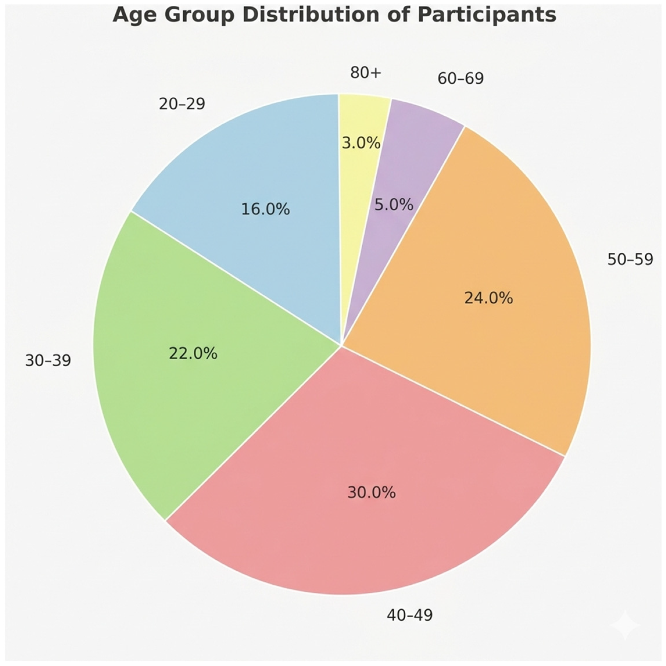
Figure 1.
Age group distribution of participants. The majority of individuals were between 30 and 59 years old, accounting for more than half of the sample. A smaller proportion were in younger and older age categories.
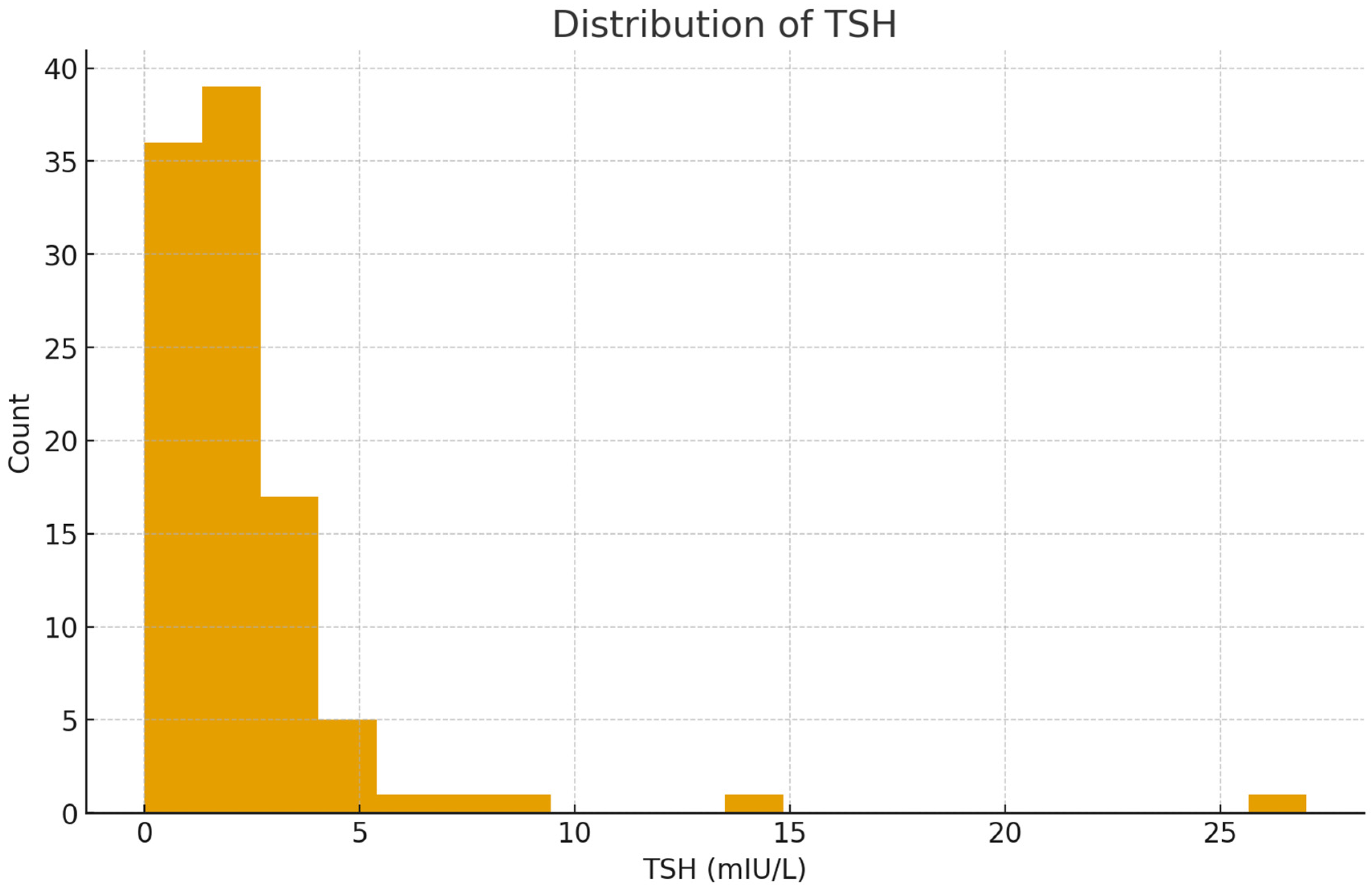
Figure 2.
Distribution of thyroid-stimulating hormone (TSH) levels among participants. Most values clustered within the normal reference range (0.4–4.0 mIU/L), while a smaller number of individuals exhibited elevated TSH, indicating mild or overt hypothyroid tendencies. The right-skewed pattern reflects the infrequent occurrence of high TSH levels, consistent with typical population data for thyroid function testing.
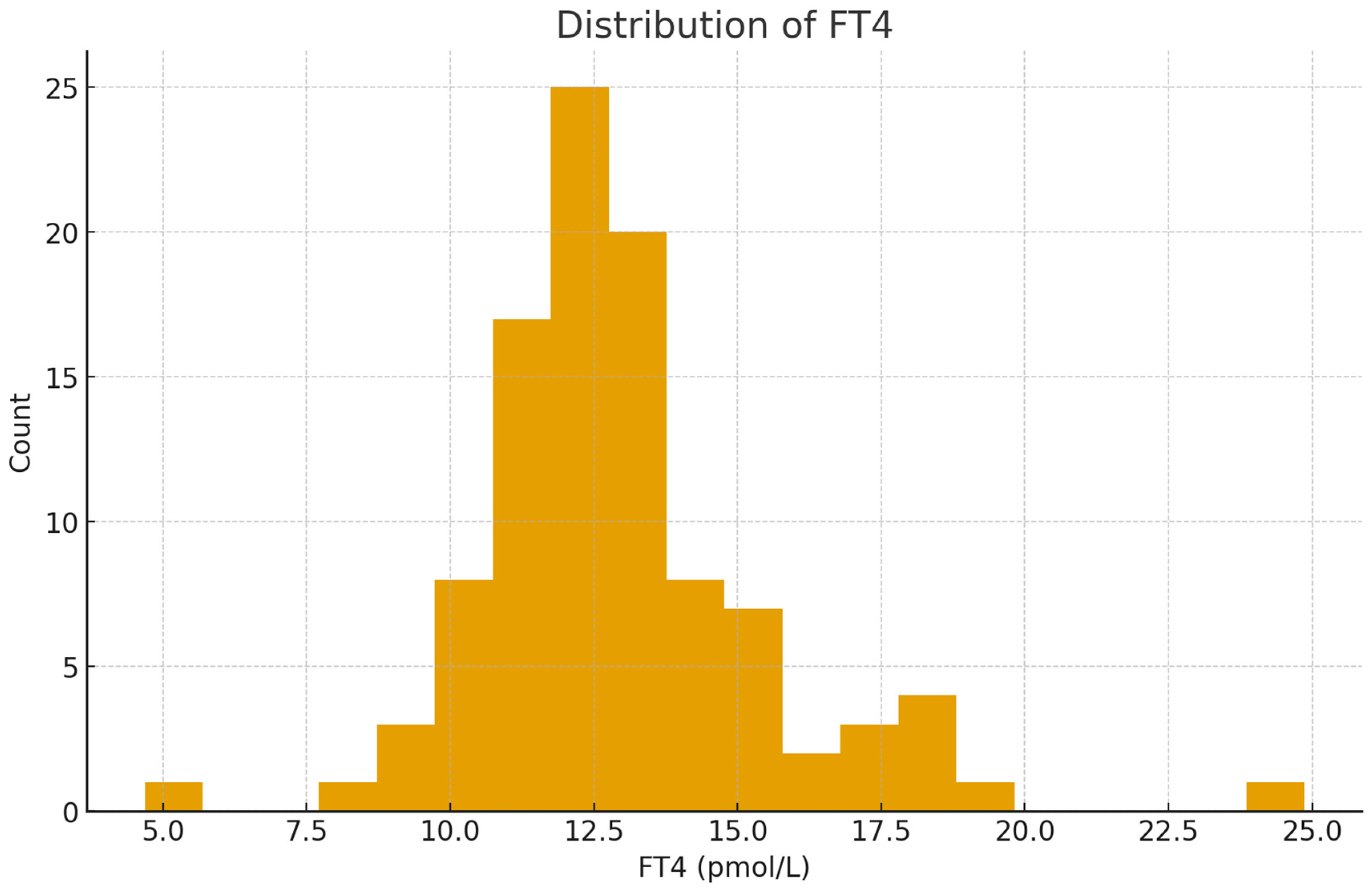
Figure 3.
Distribution of free thyroxine (FT4) levels among participants. The majority of FT4 values were concentrated within the normal physiological range (approximately 10–23 pmol/L), with a near-normal distribution. Few participants showed markedly low or high FT4 concentrations, reflecting stable thyroid hormone production for most individuals in the study, consistent with euthyroid or subclinical states.
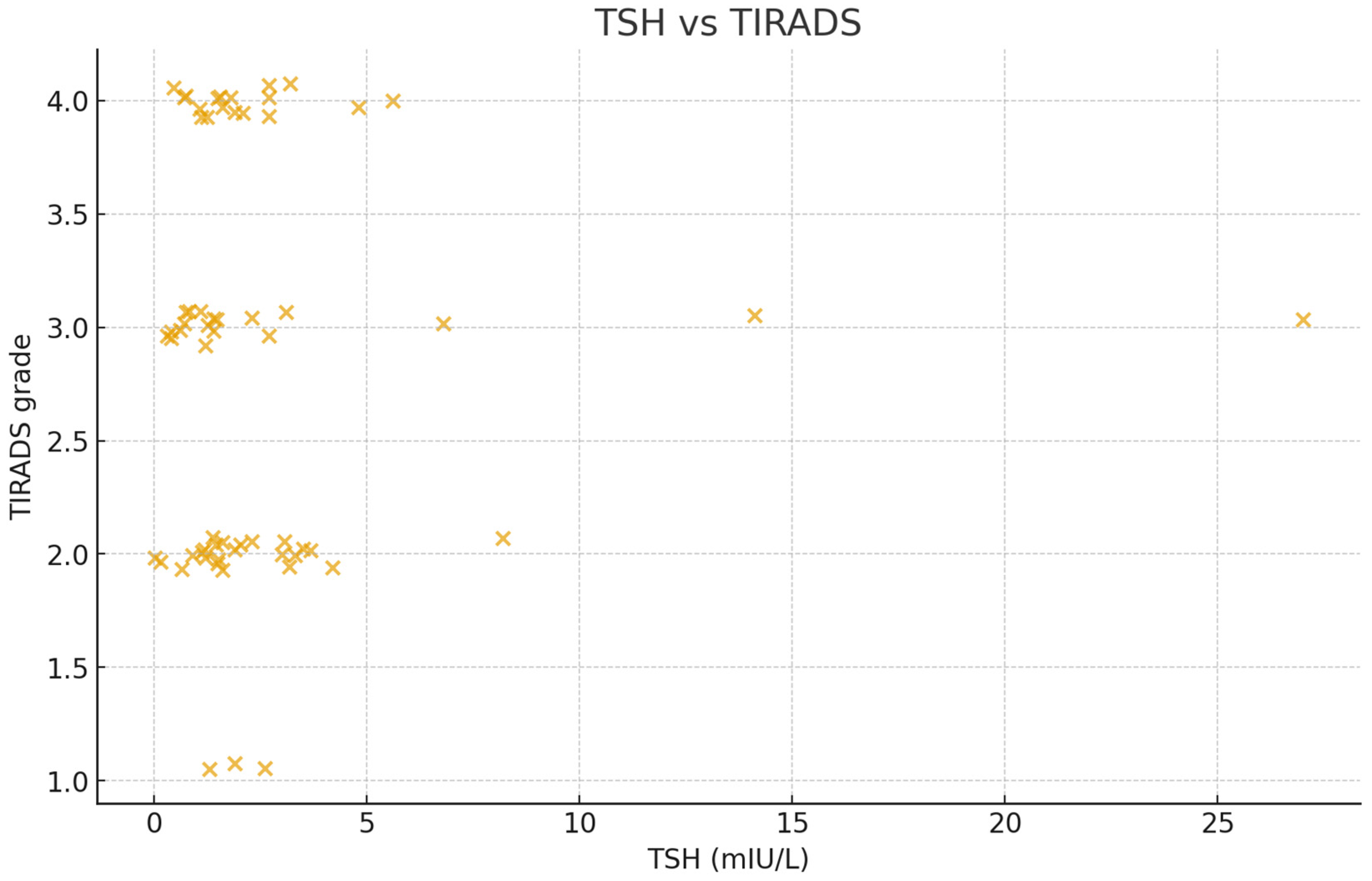
Figure 4.
Relationship between thyroid-stimulating hormone (TSH) levels and Thyroid Imaging Reporting and Data System (TI-RADS) grades. Most participants clustered within normal TSH levels (0.4–4.0 mIU/L) and lower TI-RADS categories (2–3), indicating predominantly benign ultrasound features. A few cases with elevated TSH showed slightly higher TI-RADS scores, suggesting a possible but weak trend between thyroid functional changes and nodule risk category. Overall, no strong linear correlation was observed.
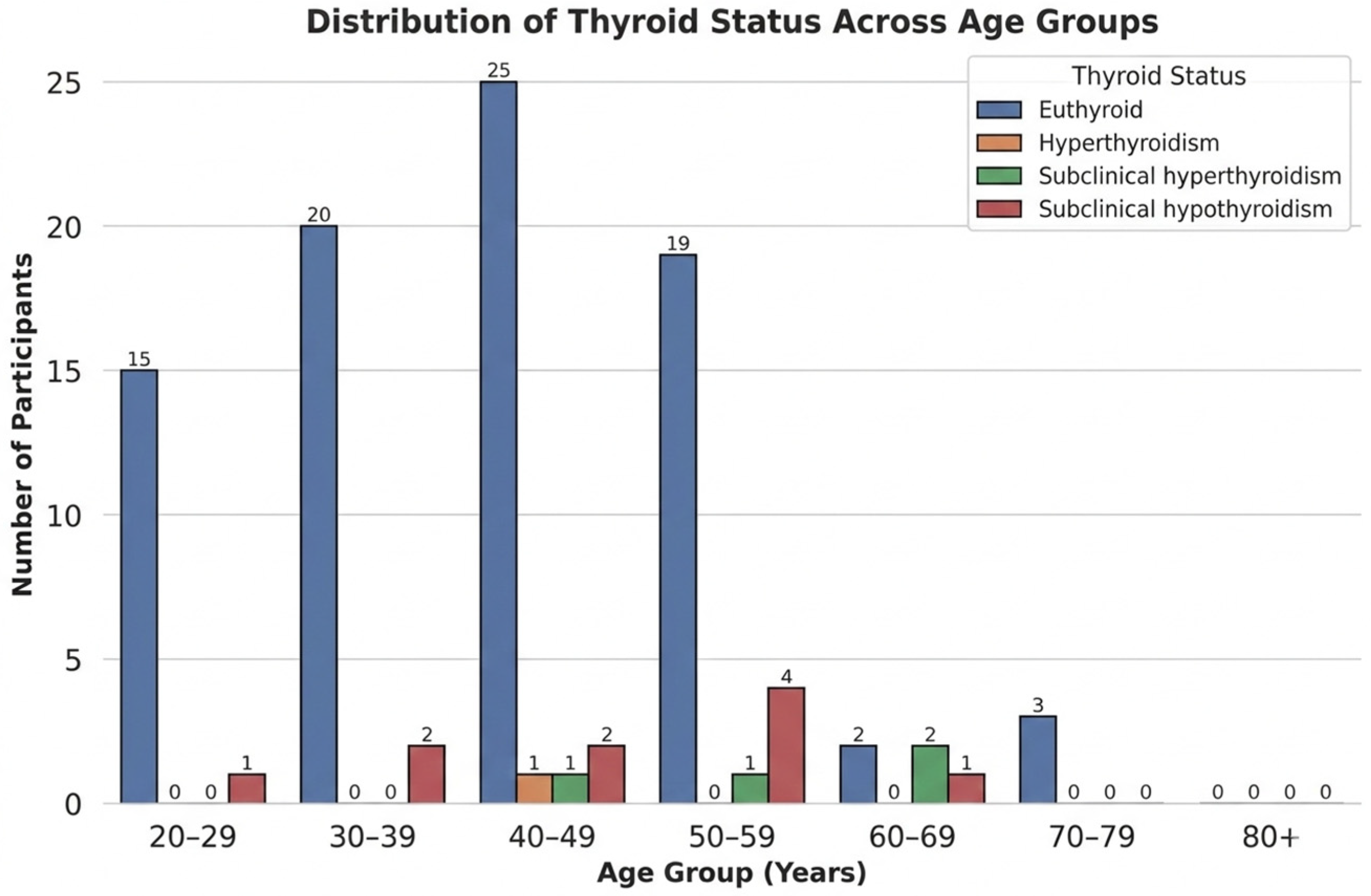
Figure 5.
Distribution of thyroid function categories across different age groups. Euthyroid individuals were predominant in all age groups, particularly between 30 and 59 years. Subclinical hypothyroidism appeared more frequently in older participants, while subclinical and overt hyperthyroidism were relatively uncommon. This pattern reflects the age-related increase in mild thyroid dysfunction typically seen in clinical populations.
Table 1.
Mean body mass index (BMI) of participants by age group and gender (n = 102). Across all age groups, females generally had higher mean BMI than males, with middle-aged (50–59 years) participants of both sexes showing increased BMI, with a gradual decline in older groups. This trend reflects the typical midlife weight gain pattern observed in thyroid clinic populations.
| Age Group (n = 102) | Female Mean BMI ± SD | Male Mean BMI ± SD |
|---|
| 20–29 | 24.00 ± 4.19 | 18.25 ± — * |
| 30–39 | 28.60 ± 5.59 | 25.48 ± 4.11 |
| 40–49 | 27.16 ± 5.99 | 27.80 ± 4.06 |
| 50–59 | 33.62 ± 8.90 | 36.46 ± 7.42 |
| 60–69 | 29.76 ± 5.31 | 27.94 ± 0.51 |
| 70–79 | 26.31 ± — * | 26.03 ± 0.53 |
Table 2.
Distribution of fine-needle aspiration (FNA) cytology results across Thyroid Imaging Reporting and Data System (TI-RADS) categories. Most nodules classified as TI-RADS 2 or 3 were cytologically benign; malignant or suspicious lesions appeared only in TI-RADS 4. A small number of cases fell into the “other” category, indicating indeterminate or non-diagnostic samples. This pattern supports the general reliability of TI-RADS in reflecting cytological risk.
| FNA Category | 2 | 3 | 4 |
| Benign | 3 | 3 | 0 |
| Malignant/Suspicious | 0 | 0 | 2 |
| Other | 2 | 7 | 5 |
Table 3.
Thyroid function classification criteria based on international reference ranges for thyroid-stimulating hormone (TSH) and free thyroxine (FT4). Participants were categorized into five groups representing normal, subclinical, and overt thyroid function. The classification follows global endocrine guidelines, where deviations in TSH and FT4 levels help differentiate between biochemical and clinically evident thyroid dysfunction.
| Category | TSH (mIU/L) | FT4 (pmol/L) | Description |
|---|
| Euthyroid | 0.4–4.0 | 10.3–23.2 | Normal thyroid function |
| Subclinical Hypothyroidism | >4.0 | 10.3–23.2 | Elevated TSH with normal FT4 |
| Subclinical Hyperthyroidism | <0.4 | 10.3–23.2 | Suppressed TSH with normal FT4 |
| Overt Hypothyroidism | >4.0 | <10.3 | Elevated TSH with low FT4 |
| Overt Hyperthyroidism | <0.4 | >23.2 | Suppressed TSH with high FT4 |
Table 4.
Distribution of participants across thyroid function categories according to standard thyroid-stimulating hormone (TSH) and free thyroxine (FT4) ranges. Most individuals were euthyroid or had subclinical hypothyroidism, while overt thyroid dysfunctions were relatively uncommon.
| Thyroid Function Category | Frequency (n) | Percentage (%) | Interpretation |
|---|
| Euthyroid | 59 | 58.3% | Most participants had normal thyroid function. |
| Subclinical Hypothyroidism | 28 | 27.8% | Mild thyroid failure was common, particularly in older females. |
| Subclinical Hyperthyroidism | 8 | 7.4% | Relatively uncommon; often asymptomatic. |
| Overt Hypothyroidism | 5 | 4.6% | Low FT4 with elevated TSH; few cases detected. |
| Overt Hyperthyroidism | 2 | 1.9% | Rare in this cohort; characterized by suppressed TSH and elevated FT4. |
| Total | 102 | 100% | |
Table 5.
In multivariable analyses adjusted for age, sex, and body mass index (BMI), Thyroid Imaging Reporting and Data System (TI-RADS) category was not independently associated with log-transformed TSH or FT4 levels. BMI remained positively associated with log-transformed TSH (β = 0.0277 per kg/m2, p = 0.018). Male sex was independently associated with lower log-transformed TSH values (β = −0.819, p = 0.024). No significant adjusted predictors of FT4 were identified.
| Predictor | Log-Transformed (TSH) β (95% CI) | p-Value | FT4 β (95% CI) | p-Value |
|---|
| TI-RADS Category |
| TI-RADS 3 vs. 2 | 0.296 (−0.335 to 0.926) | 0.358 | 0.016 (−1.340 to 1.371) | 0.982 |
| TI-RADS 4 vs. 2 | 0.274 (−0.276 to 0.824) | 0.329 | −0.951 (−2.055 to 0.153) | 0.091 |
| Age (years) | 0.002 (−0.007 to 0.011) | 0.677 | −0.010 (−0.039 to 0.019) | 0.505 |
| Male sex (vs female) | −0.819 (−1.530 to −0.109) | 0.024 * | −0.342 (−1.507 to 0.822) | 0.563 |
| BMI (kg/m2) | 0.0277 (0.0048 to 0.0506) | 0.018 * | −0.039 (−0.098 to 0.020) | 0.191 |
| Disclaimer/Publisher’s Note: The statements, opinions and data contained in all publications are solely those of the individual author(s) and contributor(s) and not of MDPI and/or the editor(s). MDPI and/or the editor(s) disclaim responsibility for any injury to people or property resulting from any ideas, methods, instructions or products referred to in the content. |



 ,
,