Review

Neurotrophic Factors: Emerging Biology and Therapeutic Applications for Cardiovascular Diseases

1
School of Exercise and Health, Shanghai University of Sport, 188 Hengren Road, Yangpu District, Shanghai 200438, China
2
State Key Laboratory of Genetics and Development of Complex Phenotypes, Fudan University, Shanghai 200438, China
*
Authors to whom correspondence should be addressed.
Metabolites 2026, 16(1), 58; https://doi.org/10.3390/metabo16010058
Submission received: 20 November 2025 / Revised: 19 December 2025 / Accepted: 7 January 2026 / Published: 9 January 2026

Abstract

Cardiovascular diseases (CVDs) have emerged as a common health problem. However, despite their prevalence, little progress has been made in their treatment. In recent years, neurotrophic factors (NTFs) have been discovered to exert cardioprotective functions for CVDs. NTFs can modulate vascular integrity, myocardial remodeling, angiogenesis, and autonomic regulation, playing the roles of maintaining cardiovascular homeostasis and influencing disease progression. Under pathological conditions, the supplement of NTFs can induce substantial adaptations to mitigate adverse cardiac responses. Several NTFs have been investigated in this regard. This review briefly elaborates on present insights into the expression, signaling pathways, and regulatory effects of NTFs on the development of CVDs, and also discusses emerging therapeutic strategies based on NTFs, ranging from exercise to advanced modalities including stem cell therapy, gene transfer, recombinant protein therapy and NTF mimetics, among which the mimetics and exercise interventions emerge as the most promising avenues for clinical translation.

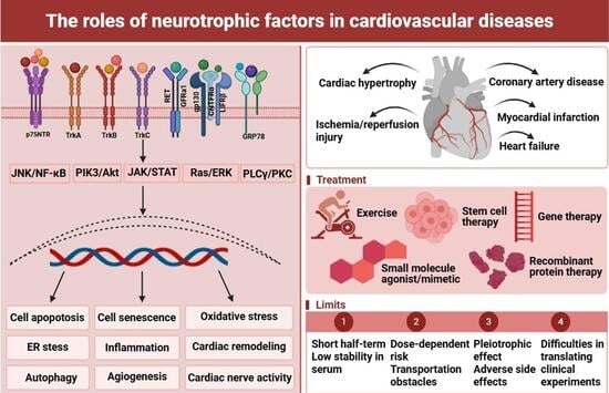
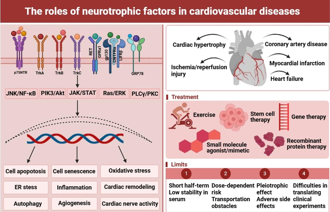
Graphical Abstract

1. Introduction

Cardiovascular diseases (CVDs) are one of the predominant contributors to worldwide disease burden and mortality, becoming a critical and ongoing global health priority [1]. Projections reveal a substantial upward trajectory in the global burden of CVDs from 2025 to 2050. Over this period, the prevalence of CVDs is anticipated to rise by 90.0%, while crude mortality and crude disability-adjusted life years are, respectively, projected to increase by 73.4% and 54.7%. As a result, the annual number of cardiovascular deaths is expected to rise from 20.5 million in 2025 to 35.6 million by 2050 [2]. Although dietary and lifestyle interventions can effectively reduce the prevalence and mortality, there are low rates of long-term successes [3,4]. Therefore, many drugs have been developed for treating CVDs. However, pharmacotherapy is usually accompanied by a change in patients’ status (e.g., pregnancy, cost, side effects), which might finally not be sufficient to correct clinical symptoms for some individuals [5]. Therefore, an urgent need exists for novel cardiovascular therapeutics.
Neurotrophic factors (NTFs) are secreted proteins derived from nervous tissues that regulate neurite outgrowth, neuronal differentiation, survival, and communication between neurons and their target tissues [6]. They include primarily the neurotrophin (NT) family, the glial-derived neurotrophic factor (GDNF) family, ciliary neurotrophic factor (CNTF), and the recently identified cerebral dopamine neurotrophic factor (CDNF)/mesencephalic astrocyte-derived neurotrophic factor (MANF) family [7,8]. Like the brain, several tissues also abundantly express NTFs, including heart, liver, pancreas, kidneys, and skeletal muscles and intestine, which can explain why NTFs can modulate the function of non-neuronal tissues through autocrine and paracrine mechanisms [9,10,11]. Levels of NTFs markedly change in humans during cellular stress, such as in neurodegenerative diseases, metabolic syndromes, cancers, immune diseases and CVDs. Significant alterations in NTF levels can indicate adverse responses to cardiac rehabilitation in patients with CVDs [12]. NTFs contribute to cardiac remodeling, regulation of angiogenesis, cardiomyocyte contractility, and maintenance of overall cardiac integrity in the cardiovascular system [13]. In particular, neurotrophin signaling through the tropomyosin receptor kinase (Trk) family receptors is indispensable for the development of embryonic cardiomyocytes, as demonstrated by severe cardiac defects observed in mice lacking neurotrophins or Trk family receptors. However, the involvement of NTFs with heterogeneous sympathetic innervation might increase susceptibility to ventricular arrhythmias and sudden cardiac death. Therefore, NTFs represent promising therapeutic targets for cardiac conditions.
In this review, we highlight NTFs’ production and secretion as well as what is known about their signaling. We also synthesize current preclinical and clinical findings providing evidence that NTFs might be an effective drug target. The review will further critically evaluate the therapeutic strategies that have been applied, including their current progress and existing challenges. Finally, we summarize and critically analyze the roles of various NTFs in the progression of CVDs in both animal models and humans.

2. Biological Functions of Neurotrophic Factors

The nerve growth factor (NGF) family of growth factors, collectively known as neurotrophins (NTs), regulates extensive biological functions and was initially recognized for its roles in neuronal survival, growth, and plasticity. Conventional NT family members comprise brain-derived neurotrophic factor (BDNF), NGF, NT-3, and NT-4/5, with NT-6 and NT-7 later identified as novel neurotrophins [14]. A hallmark of NTs is their initial synthesis as a precursor protein that must be proteolytically processed, either intracellularly or in the extracellular environment. Current evidence indicates that the NT-6 and NT-7 genes are restricted to fish lineages, with no definite homologs in mammalian or avian species [15]. NTs exert their effects through two distinct receptor types: the receptors of the Trk family and the p75 neurotrophin receptor (p75NTR). Owing to the differences in binding affinity and microenvironment, ligand–receptor interactions display distinct specificities: NGF selectively activates TrkA, whereas BDNF and NT-4/5 bind to TrkB. NT-3 primarily engages TrkC with high affinity but also stimulates TrkA and TrkB, albeit with reduced efficiency [16]. In contrast, p75NTR interacts with all NTs, though with relatively low affinity [16].
GDNF, neurturin, persephin, and artemin together form the GDNF family. Although these proteins exhibit limited amino acid sequence homology, they all function as homodimers to activate the receptor tyrosine kinase rearranged during transfection (RET). Similarly to NTs, the GDNF family signals through two receptor systems: RET and GDNF family receptor alpha (GFRa), which are glycosylphos-phoinositol (GPI)-linked receptors [17,18]. Specifically, GDNF signals through GFRa1, NRTN through GFRa2, ARTN via GFRa3, and PSPN through GFRa4 [19]. Notably, GFRa2 has been shown to regulate cardiomyocyte differentiation through a RET-independent signaling pathway [20]. Although all co-receptors are capable of interacting with RET, ligand specificity is largely determined by their differential binding affinities. Cross-signaling has been demonstrated. For instance, GDNF has the ability to activate GFRa2, while NRTN utilizes GFRa1 [21]. Furthermore, GPI-anchored co-receptors, while initially membrane-bound, can be cleaved and subsequently function as soluble co-receptors, thereby extending their role in signaling. Consequently, when soluble co-receptors are present, GDNF family ligands retain the capacity to activate RET even in cells that do not naturally express these co-receptors. GDNF is initially synthesized as a precursor protein and subsequently processed into its active form through sequential cleavage events [22]. By binding to its receptors, active GDNF triggers multiple downstream pathways controlling neuronal development, survival, and neuron–tissue communication [23]. In addition, akin to MANF/CDNF, GDNF can modulate endoplasmic reticulum (ER) stress and mitigate protein aggregation and inflammation [24]. Dysregulation of GDNF synthesis and signaling has been reported in a variety of pathological conditions, as well as in the contexts of aging, exercise, and addiction [25,26].
Originally identified in chick embryo ciliary neuron, CNTF possesses demonstrated roles in promoting neuronal differentiation, neurite outgrowth, and cell viability [27]. Functionally, CNTF regulates oxidative stress, inflammation, and other cellular processes to support regeneration and provide protective effects in the central nervous system (CNS) [28]. Mechanistically, CNTF first binds to its specific receptor α subunit (CNTFRα), which then recruits glycoprotein 130 (gp130) and leukemia inhibitory factor receptor β (LIFRβ) to assemble a heterotrimeric receptor complex [29]. CNTF binding to its receptors mainly activates the Janus kinase (JAK)/signal transducer and activator of transcription (STAT) signaling pathway, along with additional cascades such as the phosphoinositide 3-kinase (PI3K)/serine/threonine-specific protein kinase (Akt) or mitogen-activated extracellular signal-regulated kinase (MEK)/extracellular regulated protein kinase (ERK) pathways, mitogen-activated protein kinases (MAPK), AMP-activated protein kinase (AMPK), depending on the cell type [30,31]. For instance, PI3K/Akt pathway triggered by CNTF has a crucial impact in both neuronal and neurite growth, and mediates the CNTF’s role in glucose uptake within skeletal muscle tissue [32].
MANF was initially discovered as an ER-localized protein with protective functions for dopamine neurons, and CDNF is a structural homolog of MANF, both of which constitute a novel family of evolutionarily conserved neurotrophic factors [24,33]. Despite their neurotrophic properties, MANF and CDNF do not share any sequence similarities with traditional NTFs. They exhibit dual functions: acting both as an extracellular trophic factor and as an ER-resident protein [34]. Neuroplastin (NPTN) has been identified as a cell surface receptor for MANF, mediating its effects [35]. Binding of MANF to NPTN regulates ER stress-mediated cell death and inflammatory response through the activation of the NF-κB signaling pathway [35]. However, CDNF has not yet been found to bind to any cell surface receptors. Notably, proper protein folding within the myocardial ER is essential for maintaining cardiac function. Various pathological conditions, such as cardiac hypertrophy, ischemia, heart failure (HF) that induce ER stress, triggers the accumulation of unfolded or misfolded protein, which ultimately leads to the unfolded protein response (UPR) [36]. The UPR originally plays a pro-survival role, whereas under prolonged stress conditions, sustained UPR activation can switch to promote cell death [37]. The present studies have demonstrated that MANF and CDNF can modulate ER stress-induced UPR and supports protein homeostasis [38,39]. However, the potential mechanisms by which MANF and CDNF reduce the activation of UPR remain unclear, although there are some relevant researchers suggesting a connection to glucose regulatory protein 78 (GRP78) [40]. MANF interacts with GRP78 in the ER and reduced ER calcium levels facilitates the secretion of MANF. MANF then binds to the nucleotide binding domain of GRP78 [41,42]. MANF also inhibits ADP dissociation from GRP78 and impedes ATP binding. The activity stabilizes GRP78-client protein complexes, a step to promote their transfer to downstream [43]. Consequently, depletion of MANF is predicted to destabilize these complexes, rendering cells more prone to activating the UPR. Conversely, MANF overexpression likely reinforces complex stability and attenuates UPR induction. Nevertheless, one study has challenged this paradigm, asserting that MANF and GRP78 interact directly regardless of calcium levels [44]. Therefore, further investigations into MANF and the related protein CDNF are required to elucidate their precise roles as negative regulators of the UPR across diverse ER stress contexts.

3. The Roles of Neurotrophic Factors in Cardiovascular Pathology

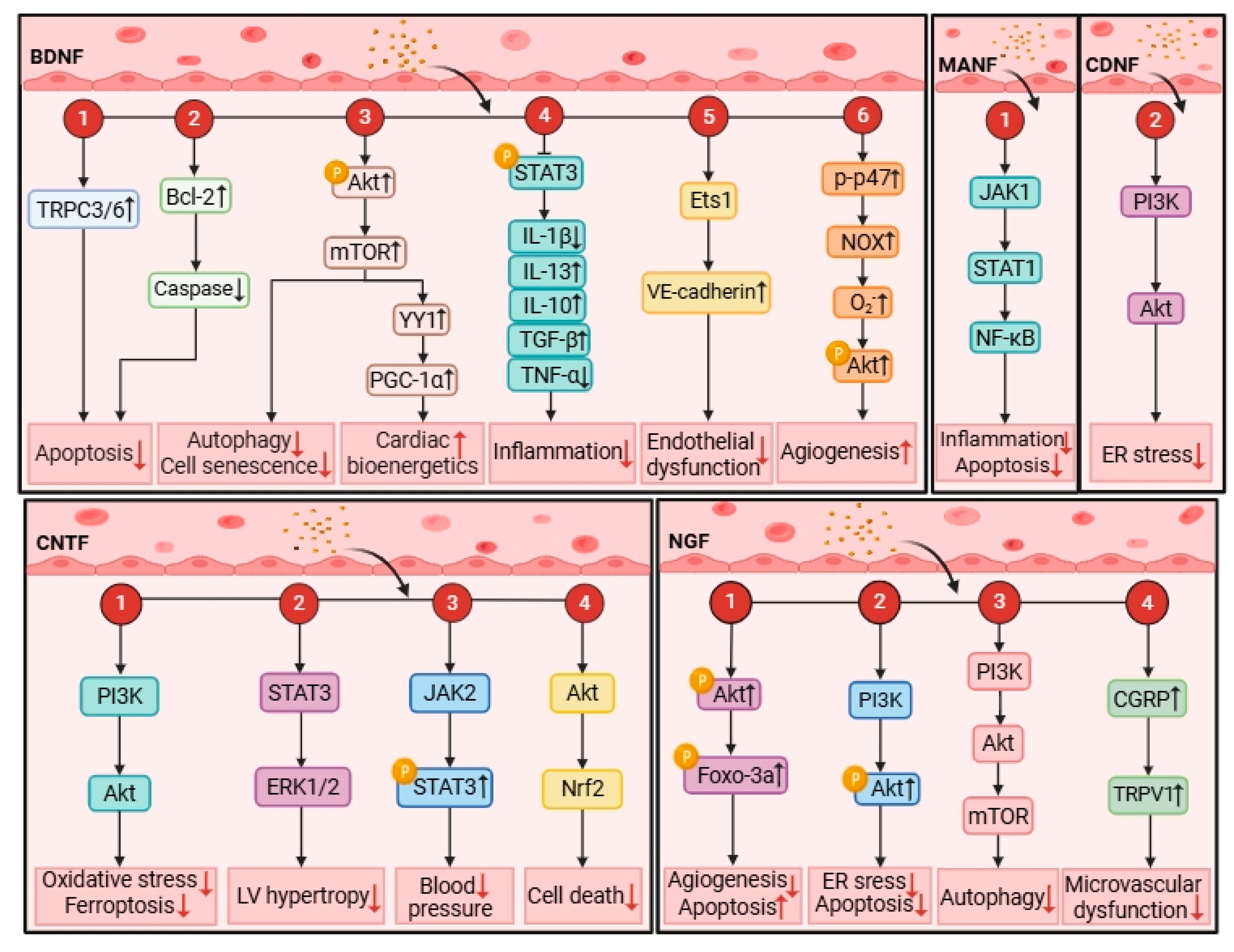
NTFs are weakly expressed in healthy human hearts, while their expressions significantly change in cardiomyocytes and cardiac tissues responding to cardiovascular injury (Table 1). The exact cell types contributing to the change are not known, but might be endothelial cells, smooth muscle cells, platelets, macrophages and cardiomyocytes [45,46,47]. The alterations of circulating and cardiac NTF concentrations are established biomarkers for atherosclerosis, hypoxic–ischemic damage, heart failure, cardiac hypertrophy and endothelial dysfunction [48]. As has been demonstrated extensively, NTFs are biomarkers associated with unfavorable prognosis of CVDs. Thus, subsequent sections will examine NTFs’ potential roles regarding CVDs (Figure 1).

3.1. Coronary Artery Disease

BDNF reflects the lipid imbalance, inflammatory state and stenosis degree in patients with coronary artery disease (CAD), and it is negatively correlated with the levels of representative molecules [49]. The upregulation of BDNF, with elevated levels of negative signaling regulators and diminished TrkB expression in perivascular fat, indicates a compromised or completely absent vascular BDNF signaling pathway in patients with coronary atherosclerosis [50]. Of note, the heightened expression of BDNF serves to mitigate the atherosclerosis in diabetes mellitus patients by promoting M2 macrophage polarization through the activation of the CAMP response element binding protein/BDNF/TrkB signaling cascade while simultaneously suppressing STAT3 pathway [46]. Additionally, BDNF binding to TrkB plays a crucial role in preserving endothelial integrity throughout the atherosclerotic process by boosting Ets1-driven vascular endothelial cadherin (VE-cadherin) expression and enhancing smooth muscle cell function, allowing both mechanisms to act together in the development of CAD [51]. Plasma BDNF levels were enhanced, and BDNF expression was elevated in coronary arteries in patients with unstable angina (UA) [52]. Paradoxically, plasma BDNF contents are significantly lower in patients with CAD or in those with stable angina (SA) and acute myocardial infarction (AMI), and reduce along with clinical acuity and disease severity [53]. However, the change in plasma BDNF levels only in CAD patients or in those with SA and AMI remains greatly controversial. On the one hand, BDNF was demonstrated to be increased within the initial hours after AMI, and positively correlated with the severity of subsequent HF [54]. On the other hand, decreased BDNF concentrations were linked to adverse cardiac remodeling after AMI [55]. According to these findings, either a protective or deleterious effect of BDNF on CAD has been proposed. Nevertheless, all these studies classified BDNF concentrations just based on the clinical manifestations in CAD patients, disregarding the morphological characteristics of their coronary plaques. Therefore, differences in plaque features may account for the conflicting BDNF patterns observed in previous studies [56,57]. Interestingly, elevated plasma BDNF levels are independently associated with a vulnerable plaque phenotype, irrespective of clinical CAD presentations [48]. The phenomenon is demonstrated to be related to increased oxidative stress by BDNF [52,53]. In addition, inflammatory responses, platelets activation and endothelial dysfunction particularly in patients with CAD can also affect or be affected by BDNF [57,58]. Thus, all these elements could act as modulators that must be taken into account when assessing BDNF contents and its association with plaque vulnerability [59,60]. However, further investigations are needed to deeply illustrate the role of BDNF in coronary plaque activity and stability.
Although there are a few studies describing the effects of NGF, it has been implicated in the development of CAD. The elevated levels of NGF levels positively correlate with disease activity, inflammatory state, and plaque instability [61]. By binding to its TrkA and p75NTR, NGF acts as a dual driver. On one hand, it facilitates smooth muscle cells’ migration and proliferation, and impairs endothelial function, which contributes to early plaque formation and later vascular remodeling [62]. On the other hand, it activates mast cells and macrophages to exacerbate inflammatory responses, accelerating plaque progression [63]. Briefly, NGF serves as a negative regulatory molecule when modulating the CAD’s progression.

3.2. Myocardial Infarction

The BDNF-TrkB axis suppresses myocardial apoptosis and mitigates cardiac ischemic injury through modulating transient receptor potential canonical 3/6 (TRPC3/6) channel [54]. BDNF also upregulates b-cell lymphoma-2 (Bcl-2) expression and downregulates cysteinyl aspartate specific proteinase 3 (caspase-3) activity to suppress apoptosis [54]. However, the upregulation of miR-195 in ischemic cardiomyocytes can facilitate pro-apoptotic effects via targeting Bcl-2 [64]. Notably, dysregulation of the brain–heart axis has been increasingly implicated in the onset and progression of myocardial infarction (MI) [65,66,67]. Cardiac afferent nerve fibers transmit the ischemic signals of MI to the brain, leading to the activation of various signaling pathways in the CNS. Activation of these CNS pathways enhances BDNF expression within the brain, then increases in circulating BDNF in turn confers protective effects on the heart during ischemic injury [68]. Increases in BDNF contribute to improved survival and attenuated left ventricular (LV) remodeling, an effect mediated through the reduction in early inflammatory response and pathological angiogenesis. Furthermore, the BDNF Val66Met gene polymorphism leads to adverse cardiac remodeling after MI and causes pro-inflammatory M1-like phenotypes in both mouse and human macrophages [55]. Simultaneously, it is also associated with poorer functional recovery, including greater susceptibility to depression and anxiety, impaired cardiac autonomic modulation, and attenuated improvement in left ventricular ejection fraction, indicating BDNF polymorphism are closely connected with the prognosis of CAD [69,70]. BDNF exerts pro-angiogenic effects through activating Akt, thereby promoting the generation of reactive oxygen species (ROS) derived from nicotinamide adenine dinucleotide phosphate hydrogen (NADPH) oxidase, and subsequently facilitating angiogenic tube formation [58]. Of note, BDNF and vascular endothelial growth factor (VEGF) can cooperate to facilitate tube formation [71]. Moreover, short-term BDNF stimulation promotes angiogenesis and tube formation by inducing moderate ROS production. In contrast, prolonged exposure reduces cell viability due to the excessive ROS. Beyond activating Akt in vascular endothelial cells through the TrkB-dependent mechanism, BDNF also promotes neovascularization through the recruitment of bone marrow-derived cells [72,73]. Development of cardiac sympathetic heterogeneity after MI contributes to ventricular arrhythmias and sudden cardiac death. BDNF plays protective roles by preferentially binding p75NTR to stimulate sympathetic denervation in the peri-infarct region and inhibits hyperinnervation in the distal peri-infarct area [74]. The downregulation of miR-18a elevates BDNF expression, thereby inhibiting the Akt/mammalian target of rapamycin (mTOR) signal pathway [75]. Consequently, this induction of autophagy and suppression of cell senescence confers protection against acute MI [75].
NGF with its high-affinity receptor TrkA activates the Akt/Forkhead box-O transcription factors (Foxo-3a) signaling pathway, thereby promoting angiogenesis and suppressing apoptosis [73,76]. Following MI, the infarct site exhibits a significant elevation in NGF levels accompanied by the upregulation of growth-associated protein 43 (GAP43), a key driver of post-MI cardiac nerve sprouting [77]. NGF and GAP43 are retrogradely transported to the left stellate ganglion, thereby inducing sympathetic hyperinnervation [77,78]. Individual variability in NGF expression may be partly responsible for differences in nerve sprouting and arrhythmia susceptibility after MI. However, empagliflozin can mitigate these adverse effects by inhibiting the NGF/TrkA pathway [79]. Of note, proNGF binds p75NTR to promote pathological changes in the microvasculature that ultimately result in cardiomyopathy after fatal MI [80].
CNTF can markedly reduce oxidative stress, ferroptosis, and cardiac dysfunction in MI-induced cardiac remodeling via the PI3K/Akt signaling in murine models [81]. Cardiac glucose metabolism in leptin-resistant mouse hearts can be restored after acute MI via CNTF-mediated STAT3/PI3K/Akt signal pathway [82]. CNTF also reverses LV hypertrophy in leptin-deficient and leptin-resistant obese mice, involving a reduction in ventricular septal thickness, posterior wall thickness, and LV mass [82]. Furthermore, both CNTF and leptin have been demonstrated to activate the STAT3 and ERK1/2 signaling pathways [83].
Collectively, these studies show that BDNF, NGF and CNTF serve a protective role in MI through specific cellular-protein-receptor interactions.

3.3. Heart Failure

Emerging clinical evidence indicates an inverse relationship between circulating BDNF levels and lethality in patients with HF, wherein diminished BDNF concentrations are associated with poor survival prognosis and cardiac remodeling [84]. Consistently, a recent systematic review and meta-analysis specifically evaluated circulating BDNF levels in HF patients compared with healthy individuals, and discussed its potential as a diagnostic, stratification, or prognostic biomarker, demonstrating that BDNF is significantly lower in HF patients [85]. Notably, BDNF levels reduce in HF, while an increase in acute HF may be associated with inflammatory response. BDNF loss triggers chronic ischemic HF and TrkB agonists can counteract ischemic LV dysfunction via replenishing myocardial BDNF. β3 adrenergic receptor stimulation increases cardiac BDNF levels and plays a crucial role in post-ischemic cardiac BDNF production [86]. BDNF binding to TrkB initiates a calcium/calmodulin-dependent protein kinase II (CaMKII)-dependent signaling cascade, acting with β-adrenergic signaling to enhance myocardial Ca2+ cycling, thereby augmenting cardiac contraction and relaxation [87]. Interestingly, a parallel study revealed that the modulation of cardiac contractility by BDNF is facilitated through the truncated TrkB. T1 receptors present on cardiomyocytes, a discrepancy likely explained by differences in cardiac phenotypes between the mouse models used in these studies [88]. One study has demonstrated that BDNF upregulates peroxisome proliferator-activated receptor γ coactivator 1α (PGC-1α) expression and recovers cardiac bioenergetics through activating AKT/mTOR/Yin Yang 1 (YY1) pathway in response to HF, whereas exercise can work synergistically with BDNF [89]. Moreover, the downregulation of silent information regulator 1 in cardiomyocytes activates nuclear factor kappa-B (NF-κB), which upregulates miR-155 [90]. Elevated miR-155 expression suppresses BDNF production, thereby contributing to impaired ventricular function in HF [91].
NGF promotes cardiac regeneration by stimulating cardiomyocyte proliferation rather than possessing anti-apoptotic effect, thereby reducing the incidence of HF and mortality [92]. Studies indicate that abnormal cardiac sympathetic nerves are observed in chronic HF, along with reduced expression of NGF and NT-3, whereas there is increased expression of BDNF and CNTF [93]. Differential expression of cardiac NTFs may explain two features of sympathetic dysfunction in chronic HF: sympathetic loss and nerve terminal transformation [94]. The absence of NGF and NT-3 may serve as the initial trigger for altered cardiac sympathetic function, while the upregulation of BDNF and CNTF may promote the shift in sympathetic nerves from a balanced norepinephrine (NE) storage/release/uptake mechanism to a predominantly NE-releasing mechanism [95].

3.4. Ischemia/Reperfusion Injury

Ischemia/reperfusion (I/R) injury can dysregulate sarcoplasmic reticulum in the heart and calcium of the ER, thus leading to MANF secretion [96]. The present study hypothesizes that elevated MANF alleviates ER stress through the inhibiting JAK 1/STAT1/NF-κB signal pathway to mitigate cardiomyocyte apoptosis [97]. The JAK1/STAT1 signaling pathway can regulate inflammation and LV remodeling subsequent to MI, ultimately mitigating I/R injury in cardiac tissues [98]. Similarly to MANF, CDNF binding to KDEL activates the PI3K/Akt signal pathway following myocardial I/R to mitigate ER stress, restore the calcium transient, and reduce the infarct area [99]. However, the lack of N-glycosylation on the CDNF protein reduces the protein stability and promotes cardiomyocyte apoptosis through downregulating the PI3K/Akt pathway [100]. In addition, BDNF and NT-3 have been implicated in protection against ischemia and I/R-induced cardiomyocyte death. One in vivo study demonstrates that the adrenoceptor beta 2 agonist and caveolin-3 can potentiate the activity of the BDNF/TrkB and cyclic adenosine 2′,3′-monophosphate/protein kinase signaling pathways in the diabetic heart [101]. Meanwhile, another study has revealed that NT-3 reduces apoptosis via the ERK-Bim signaling pathway and promotes angiogenesis [102]. Consistent with these findings, NGF binding to TrkA protects against I/R injury by alleviating ER stress-induced cardiomyocyte apoptosis through the activation of the PI3K/Akt signaling pathway [103]. NGF also inhibits autophagy during myocardial I/R by the activation of its downstream PI3K/Akt/mTOR signaling. Simultaneously, NGF can enhance cardiac recovery by upregulating transient receptor potential vanilloid-1 (TRPV1) and promoting the synthesis and release of endogenous calcitonin gene-related peptide (CGRP) [104,105]. As mitochondrial homeostasis is disrupted, CNTF prevents myocardial cells against oxygen-glucose deprivation followed by re-oxygenation by Akt-Nuclear factor erythroid 2-related factor 2 signaling pathway to avoid ischemic injury [106,107].

3.5. Cardiac Hypertrophy

Hypertension is a prevalent cause of cardiac hypertrophy, predominantly affecting left ventricular hypertrophy [108]. Elevated circulating BDNF concentrations are associated with an increased risk of hypertension and cardiometabolic dysfunction in middle-aged and elderly adults, while endothelial BDNF expression is found to decrease [109]. These findings imply that the elevated serum/plasma BDNF observed in hypertension is potentially not from the endothelial cells [108]. BDNF can intervene cardiovascular stress response, affect heart rate and regulate blood pressure in which effects on blood pressure fluctuations could be achieved through renin-angiotensin signal transduction pathway. In addition, CNTF also modulates blood pressure by regulating angiotensin II (Ang II)-induced pressure response through a JAK2/STAT3-dependent mechanism, suggesting that CNTF can effectively prevent the occurrence of hypertension [110]. Of note, cardiac hypertrophy promotes the expression of MANF, then MANF protects the heart through maintaining the glycolysis-oxidative phosphorylation balance and mitochondrial homeostasis by interacting with the pro-apoptotic protein bcl2-associated X (BAX) to inhibit mitochondrial translocation, thereby reducing mitochondrial damage and cardiomyocyte death [111]. Meanwhile, MANF effectively counteracts the hypertrophic effects triggered by α1-adrenergic receptor agonist phenylephrine. While the precise mechanisms behind MANF’s anti-hypertrophic actions remains to be elucidated, MANF demonstrates protective capabilities when added to cultured cells, at least some of its benefits mediated through a cell-surface receptor (e.g., NPTN) [35,40].

4. Therapeutic Approaches

Having distinct mechanisms of action, NTFs facilitate cardiomyocyte repair, safeguard cardiac tissue, and bolster cellular function. Additionally, they trigger receptor activation that initiates cascades of reactions following cardiac damage, contributing to enhanced cardiac functions and improved cardiac remodeling. Given all these characteristics of NTFs, the following section will focus on the exploration of NTFs with therapeutic effects that can be applied in CVDs (Table 2).

4.1. Exercise

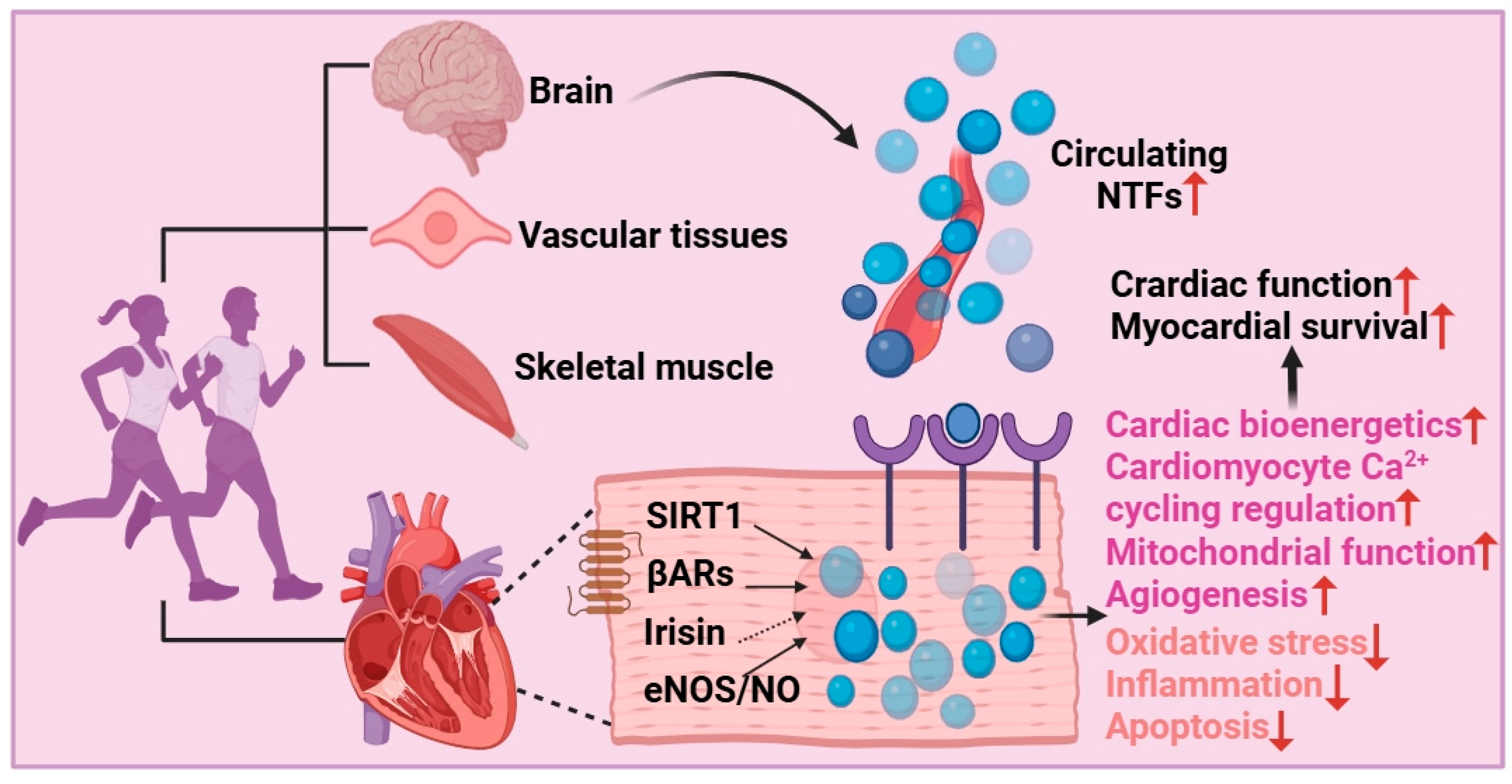
Exercise has emerged as a prevalent approach for the prevention and management of chronic diseases [117,118,119]. A large body of research has supported that exercise exerts considerable effects on NTFs. Exercise mitigates the decline in ejection fraction (EF) and the elevation in LV end-diastolic pressure (LVEDP) and increases stroke volume and cardiac index following MI [113]. The improvement of EF but not LVEDP is mediated through the activation of the BDNF-TrkB signal pathway and its downstream effectors Ca2+/CaMKII and Akt in the noninfarct LV area [113]. High-intensity interval training (HIIT), resistance training (RT), and combined exercise elicit more pronounced alterations in NT levels relative to baseline measurements [120,121]. Interestingly, both the combined training and RT regimens induce greater modifications in BDNF, NT-3, and NT-4/5 compared to the HIIT group [122]. Consistently, both continuous training regimens and HIIT elevate circulating levels of BDNF and GDNF, and the increases are consistently greater after HIIT protocols compared to continuous training regimens [123]. Moreover, serum BDNF concentrations have been observed to increase in response to acute and chronic aerobic exercise, indicating that aerobic but not strength/resistance training generally increases peripheral BDNF concentrations [124,125]. Long-term aerobic exercise also can increase NT-4/5 expression. Notably, according to studies, increased serum BDNF induced by acute exercise may be connected with the increased number of platelets entering the bloodstream after their release from the spleen, and plasma proBDNF in response to high-intensity exercise is released from skeletal muscle [47,125]. In fact, exercise triggers the circulating increase in BDNF levels through several tissue sources including lungs, intestinal tissue, myocardial cells, endothelial cells, skeletal muscle, peripheral neurons, platelets, and the brain [124]. Nevertheless, evidence about the effect of exercise on circulating BDNF levels is limited and controversial, and more investigations are urgently needed to bridge the research gap.
Therefore, when formulating exercise programs for patients with heart disorders, NTFs could be used as biomarkers and various characteristics must be taken into consideration [126,127] (Figure 2). However, most studies emphasize the relationship between exercise and NTs, leaving other NTFs largely unexplored. Expanding this scope to encompass MANF and CDNF could provide novel insights into exercise-induced secretion of NTFs. In response to adverse effects, such as ER stress, the expression of MANF and CDNF is upregulated, which in turn can alleviate ER stress. In parallel, CDNF also reduces protein aggregation and attenuates inflammation. Although there are no relevant studies, future investigations can focus on how different exercise intensities influence the circulating levels of MANF and CDNF and what functions the alterations can exert.

4.2. Small Molecule Agonist/Mimetic

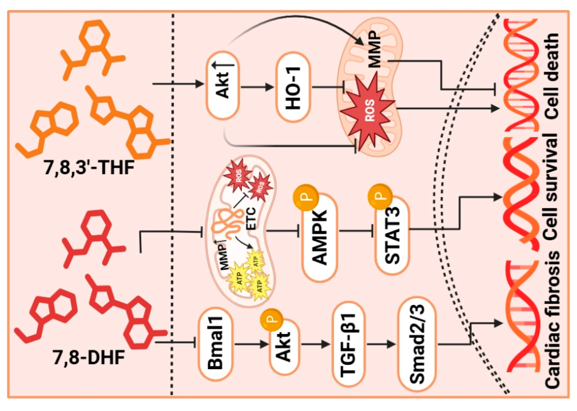
TrkB receptor agonist can enhance ischemic LV dysfunction by restoring myocardial BDNF content. Thus, targeting the TrkB receptor agonist has emerged as a promising therapeutic strategy to restore BDNF-dependent neurotrophic support to the ischemic myocardium [128]. 7,8-Dihydroflavone (7,8-DHF) acts as both a TrkB receptor agonist and mimic of BDNF [129,130]. Emerging evidence indicates that 7,8-DHF effectively reduces cardiac fibrosis as well as maintains healthy circadian rhythms, partly stemming from its ability to suppress the brain and muscle Arnt-like protein 1 (Bmal1)/Akt signaling pathway [131]. Furthermore, 7,8-DHF has been shown to have mito-protective effects on rotenone (Rot)-induced cardiomyocyte toxicity. These effects are a result of the inhibition of mitochondrial dysfunction and the promotion of the nuclear translocation of phosphorylated STAT3 [112]. Similarly, 7,8-DHF also can enhance cardiac function and restore the expression of related proteins by activating the Akt signaling pathway against doxorubicin (Dox)-induced injury and cell death [114]. Of note, 7,8-DHF is orally bioactive and processes therapeutic efficacy when BDNF signaling is deficient [130]. In addition, 7,8,3′-Trihydroxyflavone (7,8,3′-THF) is another BDNF mimic. Both 7,8-DHF and 7,8,3′-THF have the ability to facilitate the recovery of palmitic acid (PA)-induced mitochondrial damage by maintaining the function of Akt-dependent mitochondria [115]. In conclusion, these studies demonstrate that TrkB receptor agonists ameliorate various cardiac pathologies primarily through the restoration of mitochondrial function and activation of the Akt signaling pathway (Figure 3).
However, the translation of 7,8-DHF to clinical application has a series of critical hurdles. First, due to its poor pharmacokinetics, characterized by low oral bioavailability and rapid metabolism, this behavior hinders the maintenance of therapeutic concentrations at lesion sites [130]. Furthermore, it is important that the dosage of 7,8-DHF must be validated strictly as over-activation of the TrkB signaling may pose devastating consequences, like off-target risks [130]. At the same time, long-term TrkB activation also leads to unknown risks, such as tumorigenesis and cardiovascular hyperplasia [132]. Therefore, the progression from bench to bedside will depend on structural optimization of the compound, the development of precise delivery systems, and in-depth mechanistic studies.

4.3. Gene Therapy

Harnessing viral or non-viral vectors for gene transfer has been the mainstay of medical therapy. NTFs gene transfer leads to functional improvement and normal development with the heart [133]. A particular investigation compared the impact of elevated cardiac NGF levels using two distinct methodologies: direct intramyocardial administration of an adeno associated virus (AAV) 2 vector and whole-body AAV9 delivery [134]. The results demonstrated that NGF gene transfer successfully shielded cardiac tissue from damage associated with diabetic cardiomyopathy (DCM), regardless of the method employed [134]. However, the overexpression of CNTF in cardiomyocytes mediated by AAV9 exacerbates apoptosis, cardiac fibrosis, and inflammatory response, suggesting that CNTF gene delivery may fail to improve adverse cardiac remodeling in diabetic mice [116]. Additionally, intrathecal lentivirus-mediated interference RNA targeted at reducing NGF gene expression attenuates myocardial I/R injury in the rat model, which is achieved through the inhibition of TRPV1/Akt/ERK signal pathway [104].

4.4. Stem Cell Therapy

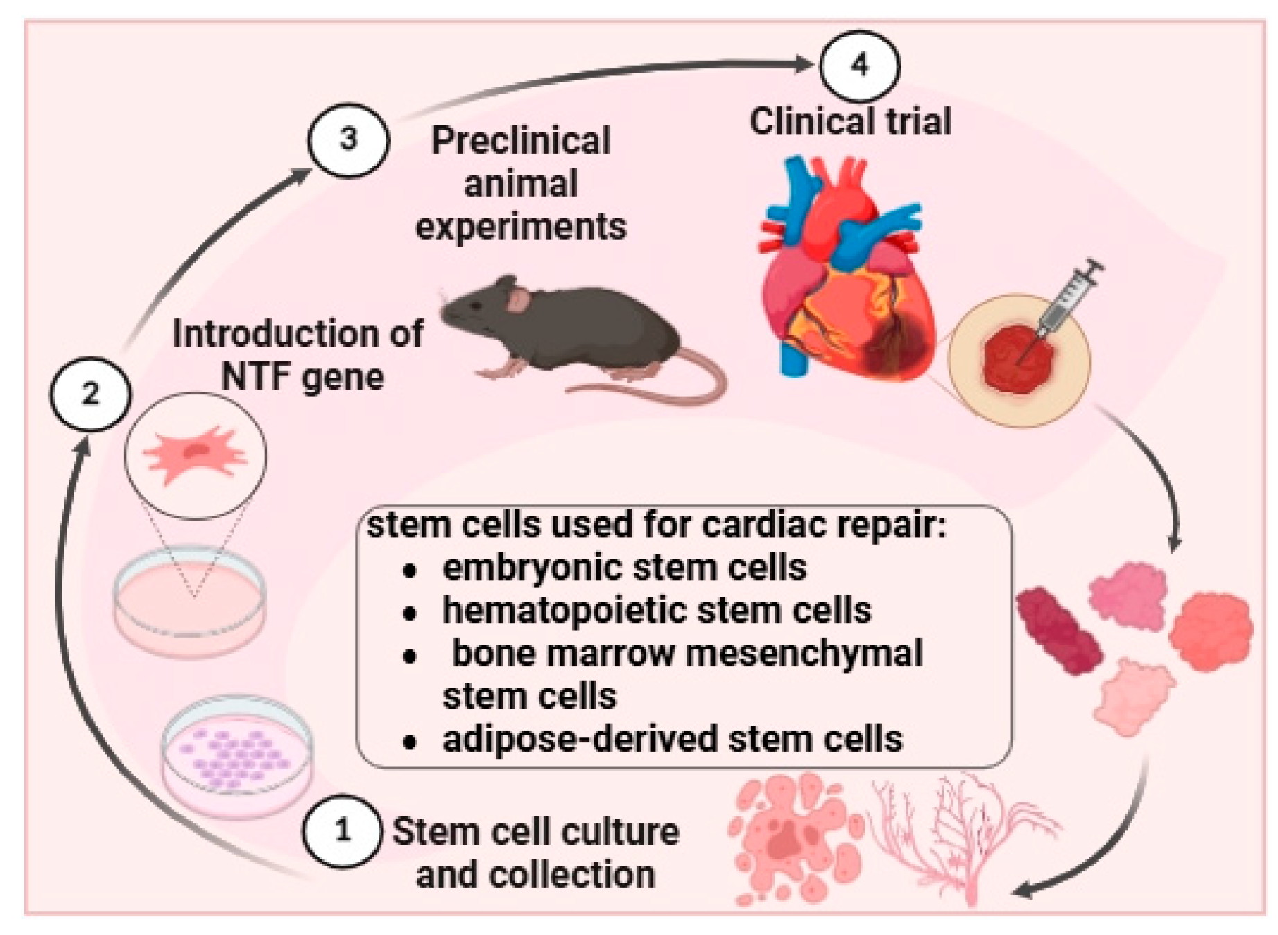
Autologous and allogeneic cell transplantation has been applied in cardiac generation, substantially mitigating concerns related to immune rejection [135]. Current stem cell types for cardiac repair are embryonic stem cells, hematopoietic stem cells, bone marrow-derived mesenchymal stem cells, and adipose-derived stem cells [136] (Figure 4). Notably, the exposure to NTF can rejuvenate aged human multipotent mesenchymal stromal cells (hMSCs) [137]. Transplantation of these NTF-rejuvenated hMSCs improves cardiac function, primarily by suppressing apoptosis and stimulating angiogenic responses [138]. The expression of NGF significantly increases after mesenchymal stem cells transplantation, which will affect sympathetic remodeling and the electrophysiological properties after MI [137]. NTF-rejuvenated stem cells can serve as an effectively dual product for integrating cellular and genetic treatments to enhance aged heart recovery following cardiac ischemia. However, long-term observation is needed to investigate potential side effects and efficacy.

4.5. Recombinant Protein Therapy

The administration of recombined BDNF can elevate myocardial BDNF levels, thereby enhancing survival rate and improving cardiac function, with a concomitant reduction in oxidative stress and myocardial apoptosis [139]. Recombined BDNF also effectively ameliorates the compromised exercise capacity observed in mice with MI-induced HF, through enhancing fatty acid oxidation by activating theAMPKα-peroxisome proliferator-activated receptor gamma coactivator 1-alpha signaling pathway [140]. The underlying mechanism resembles that of exercise training, thus BDNF is considered a potential therapeutic target for enhancing exercise capacity following HF. Consequently, therapeutic intervention with BDNF, whether as a monotherapy or in conjunction with exercise training, is a promising strategy in this context [141]. Furthermore, recombinant MANF treatment also decreases infarct size and inflammation in I/R injury. The knockdown of endogenous MANF will increase cell death, but adding recombinant MANF restores the protective effect. However, the clinical application of recombinant NTFs is constrained by certain limitations [139,142]. Currently, the primary challenge includes the short half-life, poor targeting, poor stability, expensive cost and delivery barriers. However, the production of recombinant NTFs is of great biotechnological interest. Notably, one study has demonstrated that recombinant BDNF can be efficiently produced through the engineering of Escherichia coli, representing an approach with high yield and low cost to producing and purifying recombinant NTFs [45]. In addition, there are other new strategies that are widely concerned, including nanotechnology, protein engineering, and fusion technology [143,144,145]. Although these methods can achieve the goal of producing a stable and biologically active protein, more researchers are still needed for pharmaceutical application.

5. Conclusions

NTFs have garnered attention for elucidating connections between CVD pathogenesis and therapeutic approaches, while offering broad applications in disease management. Among these, BDNF and MANF seem to hold the greatest translational promise, reflecting their roles in cardioprotection. Current studies indicate that NTFs essentially safeguard cardiomyocytes in the heart through three key mechanisms. First, NTFs directly participate in mediating cardiac normal contractility and relaxation. Second, NTFs mitigate the detrimental impacts of fibrosis, hypertrophy, apoptosis, cell death, oxidative stress and inflammation, effectively preserving cardiomyocytes. Finally, NTFs promote angiogenesis and maintain endothelial function. All three factors foster neuro-immune–cardiovascular interaction under cardiac stress, which are intricately related to CVD development. Meanwhile, in contrast to pathological conditions, studies have demonstrated that some certain NTFs, such as BDNF, NT-3, NT4/5, and GDNF, can be stimulated to increase secretion through exercise. Additionally, multiple transcription factors are involved in the regulation of ER stress-related diseases. Thus, elucidating the targets of MANF/CDNF in the context of ER stress in the heart represents a significant step into therapeutic strategies, particularly their interactions with activating transcription factor 6.
With respect to clinical translation, due to NTFs being able to be produced and isolated using external plasmids, it is feasible to manufacture recombinant NTFs on an industrial scale, even gene-engineered cells. Moreover, exploring the development of small molecular agonist/mimetics is feasible. Notably, beyond the use of a single NTF, further investigations can focus on the benefits of multiple factor synergistic approaches, especially in stem cell therapies. In these ways, people can be supplemented with some deficient NTFs in a specialized manner.
Looking ahead, several research aspects should be taken into account. To begin with, finding proper methods of differentiating biological differences between the precursor/mature form and detecting each NTF concentration will be critical to ensure accurate interpretation of therapeutic efficacy. Moreover, due to the different NTFs’ properties, integrative approaches hold promise for synergistically enhancing NTF signaling and improving intervention results. For instance, exercise combined with specific NTF supplements or stem cell therapy combined with gene therapy can aid more efficient treatment outcomes. In addition, optimizing the design of emerging NTF vectors is required for evaluating durability, immune responses, and assessing long-term safety. Furthermore, it is important to identify better predictive biomarkers to stratify patient subgroups, enabling them to most likely benefit from NTF-targeted interventions.
However, the understanding of the roles of NTFs in CVDs remains limited, with the precise molecular pathways still shrouded and not already completely observed. Therefore, the application of NTF products in CVDs needs more research to identify additional targets and optimize delivery approaches, providing standards for clinical applications.

Author Contributions

Y.L.: Writing—original draft preparation, writing—review and editing, investigation, conceptualization, visualization. H.Z.: Writing—review and editing. F.Y.: Writing—review and editing. T.L.: Supervision and conceptualization. D.J.: Writing—review and editing, supervision, conceptualization. R.W.: Supervision, conceptualization. All authors have read and agreed to the published version of the manuscript.

Funding

This work was supported by a grant from Shanghai Baiyulan Talent Program Pujiang Project Funding (ID: 24PJC067) and research project of Shanghai University of Sport.

Institutional Review Board Statement

Not applicable.

Informed Consent Statement

Not applicable.

Data Availability Statement

No new data were created or analyzed in this study.

Conflicts of Interest

The authors declare no conflicts of interest.

Abbreviations

BDNFBrain-derived neurotrophic factor
7,8-DHF7,8-Dihydroxyflavone
RotRotenone
NONitric oxide
eNOSEndothelial nitric oxide synthase
DMASDiabetes mellitus-accelerated atherosclerosis
TrkTropomyosin receptor kinase 
DoxDoxorubicin
MAPKMitogen-activated protein kinase
ERKExtracellular regulated protein kinase
PAPalmitic acid
7,8,3′-THF7,8,3′-Trihydroxyflavone
DCMDiabetic cardiomyopathy
FoxoForkhead box-O transcription factors
AktSerine/threonine-specific protein kinase
I/RIschemia/reperfusion
EREndoplasmic reticulum
PI3KPhosphatidylinositol 3-kinase
NGFNerve growth factor
LDHLactate dehydrogenase 
CNTFCiliary neurotrophic factor
AAVAdeno associated virus
MANFMesencephalic astrocyte-derived neurotrophic factor
JAKJanus kinase
STATSignal transducer and activator of transcription
NF-κBNuclear factor kappa-B
CDNFCerebral dopamine neurotrophic factor
TRPCTransient receptor potential canonical
CaspaseCysteinyl aspartate specific proteinase
mTORMammalian target of rapamycin
VE-cadherin Vascular endothelial cadherin
p75NTRp75 neurotrophin receptor
NTNeurotrophin
NTFNeurotrophic factor
GFRaGDNF family receptor alpha
GPIGlycosylphos-phoinositol
RETRearranged during Transfection
gp130Glycoprotein 130
NPTNNeuroplastin
HFHeart failure
UPRUnfolded protein response
GRP78Glucose regulatory protein 78 
CADCoronary artery disease 
CNSCentral nervous system
MIMyocardial infarction
LVLeft ventricle
Bcl-2B-cell lymphoma-2
ROSReactive oxygen species
NADPH Nicotinamide adenine dinucleotide phosphate hydrogen
VEGFVascular endothelial growth factor
GAP-43Growth-associated protein 43
CaMKIICalcium/calmodulin-dependent protein kinase II 
NENorepinephrine
Ang IIAngiotensin II
HIITHigh-intensity interval training
RTResistance training
VSMCVascular smooth muscle cell
hMSCsHuman multipotent mesenchymal stromal cells
Bmal1Brain and muscle Arnt-like protein 1
TRPV1Transient receptor potential vanilloid-1
CGRPCalcitonin gene-related peptide
YY1Yin Yang 1 
PGC-1αPeroxisome proliferator-activated receptor γ coactivator 1α
SAStable angina 
AMIAcute myocardial infarction
AMPKAMP-activated protein kinase
UAUnstable angina

References

  1. Timmis, A.; Vardas, P.; Townsend, N.; Torbica, A.; Katus, H.; De Smedt, D.; Gale, C.P.; Maggioni, A.P.; Petersen, S.E.; Huculeci, R.; et al. European Society of Cardiology: Cardiovascular disease statistics 2021. Eur. Heart J. 2022, 43, 716–799. [Google Scholar] [CrossRef]
  2. Chong, B.; Jayabaskaran, J.; Jauhari, S.M.; Chan, S.P.; Goh, R.; Kueh, M.T.W.; Li, H.; Chin, Y.H.; Kong, G.; Anand, V.V.; et al. Global burden of cardiovascular diseases: Projections from 2025 to 2050. Eur. J. Prev. Cardiol. 2025, 32, 1001–1015. [Google Scholar] [CrossRef]
  3. Arage, G.; Dekkers, K.F.; Rašo, L.M.; Hammar, U.; Ericson, U.; Larsson, S.C.; Engel, H.; Baldanzi, G.; Pertiwi, K.; Sayols-Baixeras, S.; et al. Plasma metabolite profiles of meat intake and their association with cardiovascular disease risk: A population-based study in Swedish cohorts. Metab. Clin. Exp. 2025, 168, 156188. [Google Scholar] [CrossRef] [PubMed]
  4. Wu, J.; Feng, Y.; Zhao, Y.; Guo, Z.; Liu, R.; Zeng, X.; Yang, F.; Liu, B.; Gu, J.; Tarimo, C.S.; et al. Lifestyle behaviors and risk of cardiovascular disease and prognosis among individuals with cardiovascular disease: A systematic review and meta-analysis of 71 prospective cohort studies. Int. J. Behav. Nutr. Phys. Act. 2024, 21, 42. [Google Scholar] [CrossRef]
  5. Nestelberger, T.; Kaiser, C.; Jeger, R. Drug-coated balloons in cardiovascular disease: Benefits, challenges, and clinical applications. Expert Opin. Drug Deliv. 2020, 17, 201–211. [Google Scholar] [CrossRef]
  6. Castrén, E.; Antila, H. Neuronal plasticity and neurotrophic factors in drug responses. Mol. Psychiatry 2017, 22, 1085–1095. [Google Scholar] [CrossRef]
  7. Renko, J.M.; Bäck, S.; Voutilainen, M.H.; Piepponen, T.P.; Reenilä, I.; Saarma, M.; Tuominen, R.K. Mesencephalic Astrocyte-Derived Neurotrophic Factor (MANF) Elevates Stimulus-Evoked Release of Dopamine in Freely-Moving Rats. Mol. Neurobiol. 2018, 55, 6755–6768. [Google Scholar] [CrossRef] [PubMed]
  8. Lindahl, M.; Saarma, M.; Lindholm, P. Unconventional neurotrophic factors CDNF and MANF: Structure, physiological functions and therapeutic potential. Neurobiol. Dis. 2017, 97, 90–102. [Google Scholar] [CrossRef]
  9. Severini, C. Neurotrophic Factors in Health and Disease. Cells 2022, 12, 47. [Google Scholar] [CrossRef] [PubMed]
  10. Allen, S.J.; Watson, J.J.; Shoemark, D.K.; Barua, N.U.; Patel, N.K. GDNF, NGF and BDNF as therapeutic options for neurodegeneration. Pharmacol. Ther. 2013, 138, 155–175. [Google Scholar] [CrossRef]
  11. Wei, M.; Wu, T.; Chen, N. Bridging neurotrophic factors and bioactive peptides to Alzheimer’s disease. Ageing Res. Rev. 2024, 94, 102177. [Google Scholar] [CrossRef]
  12. Fukushima, A.; Kinugawa, S.; Homma, T.; Masaki, Y.; Furihata, T.; Yokota, T.; Matsushima, S.; Takada, S.; Kadoguchi, T.; Oba, K.; et al. Serum brain-derived neurotropic factor level predicts adverse clinical outcomes in patients with heart failure. J. Card. Fail. 2015, 21, 300–306. [Google Scholar] [CrossRef]
  13. Matsuda, S.; Fujita, T.; Kajiya, M.; Takeda, K.; Shiba, H.; Kawaguchi, H.; Kurihara, H. Brain-derived neurotrophic factor induces migration of endothelial cells through a TrkB-ERK-integrin αVβ3-FAK cascade. J. Cell. Physiol. 2012, 227, 2123–2129. [Google Scholar] [CrossRef]
  14. Metsis, M. Genes for neurotrophic factors and their receptors: Structure and regulation. Cell. Mol. Life Sci. CMLS 2001, 58, 1014–1020. [Google Scholar] [CrossRef]
  15. Götz, R.; Köster, R.; Winkler, C.; Raulf, F.; Lottspeich, F.; Schartl, M.; Thoenen, H. Neurotrophin-6 is a new member of the nerve growth factor family. Nature 1994, 372, 266–269. [Google Scholar] [CrossRef]
  16. Conroy, J.N.; Coulson, E.J. High-affinity TrkA and p75 neurotrophin receptor complexes: A twisted affair. J. Biol. Chem. 2022, 298, 101568. [Google Scholar] [CrossRef]
  17. Treanor, J.J.; Goodman, L.; de Sauvage, F.; Stone, D.M.; Poulsen, K.T.; Beck, C.D.; Gray, C.; Armanini, M.P.; Pollock, R.A.; Hefti, F.; et al. Characterization of a multicomponent receptor for GDNF. Nature 1996, 382, 80–83. [Google Scholar] [CrossRef] [PubMed]
  18. Airaksinen, M.S.; Saarma, M. The GDNF family: Signalling, biological functions and therapeutic value. Nat. Rev. Neurosci. 2002, 3, 383–394. [Google Scholar] [CrossRef] [PubMed]
  19. Bonafina, A.; Trinchero, M.F.; Ríos, A.S.; Bekinschtein, P.; Schinder, A.F.; Paratcha, G.; Ledda, F. GDNF and GFRα1 Are Required for Proper Integration of Adult-Born Hippocampal Neurons. Cell Rep. 2019, 29, 4308–4319.e4. [Google Scholar] [CrossRef] [PubMed]
  20. Ishida, H.; Saba, R.; Kokkinopoulos, I.; Hashimoto, M.; Yamaguchi, O.; Nowotschin, S.; Shiraishi, M.; Ruchaya, P.; Miller, D.; Harmer, S.; et al. GFRA2 Identifies Cardiac Progenitors and Mediates Cardiomyocyte Differentiation in a RET-Independent Signaling Pathway. Cell Rep. 2016, 16, 1026–1038. [Google Scholar] [CrossRef]
  21. Buj-Bello, A.; Adu, J.; Piñón, L.G.; Horton, A.; Thompson, J.; Rosenthal, A.; Chinchetru, M.; Buchman, V.L.; Davies, A.M. Neurturin responsiveness requires a GPI-linked receptor and the Ret receptor tyrosine kinase. Nature 1997, 387, 721–724. [Google Scholar] [CrossRef] [PubMed]
  22. Lin, L.F.; Doherty, D.H.; Lile, J.D.; Bektesh, S.; Collins, F. GDNF: A glial cell line-derived neurotrophic factor for midbrain dopaminergic neurons. Science 1993, 260, 1130–1132. [Google Scholar] [CrossRef]
  23. Wang, M.; Han, X.; Zha, W.; Wang, X.; Liu, L.; Li, Z.; Shi, Y.; Kan, X.; Wang, G.; Gao, D.; et al. GDNF Promotes Astrocyte Abnormal Proliferation and Migration Through the GFRα1/RET/MAPK/pCREB/LOXL2 Signaling Axis. Mol. Neurobiol. 2022, 59, 6321–6340. [Google Scholar] [CrossRef]
  24. Lindholm, P.; Saarma, M. Cerebral dopamine neurotrophic factor protects and repairs dopamine neurons by novel mechanism. Mol. Psychiatry 2022, 27, 1310–1321. [Google Scholar] [CrossRef] [PubMed]
  25. Baloh, R.H.; Johnson, J.P.; Avalos, P.; Allred, P.; Svendsen, S.; Gowing, G.; Roxas, K.; Wu, A.; Donahue, B.; Osborne, S.; et al. Transplantation of human neural progenitor cells secreting GDNF into the spinal cord of patients with ALS: A phase 1/2a trial. Nat. Med. 2022, 28, 1813–1822. [Google Scholar] [CrossRef] [PubMed]
  26. Soret, R.; Schneider, S.; Bernas, G.; Christophers, B.; Souchkova, O.; Charrier, B.; Righini-Grunder, F.; Aspirot, A.; Landry, M.; Kembel, S.W.; et al. Glial Cell-Derived Neurotrophic Factor Induces Enteric Neurogenesis and Improves Colon Structure and Function in Mouse Models of Hirschsprung Disease. Gastroenterology 2020, 159, 1824–1838.e17. [Google Scholar] [CrossRef]
  27. Si, Z.P.; Wang, G.; Han, S.S.; Jin, Y.; Hu, Y.X.; He, M.Y.; Brand-Saberi, B.; Yang, X.; Liu, G.S. CNTF and Nrf2 Are Coordinately Involved in Regulating Self-Renewal and Differentiation of Neural Stem Cell During Embryonic Development. iScience 2019, 19, 303–315. [Google Scholar] [CrossRef]
  28. Pasquin, S.; Sharma, M.; Gauchat, J.F. Ciliary neurotrophic factor (CNTF): New facets of an old molecule for treating neurodegenerative and metabolic syndrome pathologies. Cytokine Growth Factor Rev. 2015, 26, 507–515. [Google Scholar] [CrossRef]
  29. Rose-John, S. Interleukin-6 Family Cytokines. Cold Spring Harb. Perspect. Biol. 2018, 10, a028415. [Google Scholar] [CrossRef]
  30. Murakami, M.; Kamimura, D.; Hirano, T. Pleiotropy and Specificity: Insights from the Interleukin 6 Family of Cytokines. Immunity 2019, 50, 812–831. [Google Scholar] [CrossRef]
  31. Wen, R.; Tao, W.; Li, Y.; Sieving, P.A. CNTF and retina. Prog. Retin. Eye Res. 2012, 31, 136–151. [Google Scholar] [CrossRef]
  32. Askvig, J.M.; Watt, J.A. The MAPK and PI3K pathways mediate CNTF-induced neuronal survival and process outgrowth in hypothalamic organotypic cultures. J. Cell Commun. Signal. 2015, 9, 217–231. [Google Scholar]
  33. Liu, Y.Y.; Huo, D.; Zeng, L.T.; Fan, G.Q.; Shen, T.; Zhang, T.M.; Cai, J.P.; Cui, J. Mesencephalic astrocyte-derived neurotrophic factor (MANF): Structure, functions and therapeutic potential. Ageing Res. Rev. 2022, 82, 101763. [Google Scholar] [CrossRef] [PubMed]
  34. Pakarinen, E.; Lindholm, P.; Saarma, M.; Lindahl, M. CDNF and MANF regulate ER stress in a tissue-specific manner. Cell. Mol. Life Sci. CMLS 2022, 79, 124. [Google Scholar] [CrossRef]
  35. Yagi, T.; Asada, R.; Kanekura, K.; Eesmaa, A.; Lindahl, M.; Saarma, M.; Urano, F. Neuroplastin Modulates Anti-inflammatory Effects of MANF. iScience 2020, 23, 101810. [Google Scholar] [CrossRef]
  36. Inigo, J.R.; Chandra, D. The mitochondrial unfolded protein response (UPR(mt)): Shielding against toxicity to mitochondria in cancer. J. Hematol. Oncol. 2022, 15, 98. [Google Scholar] [CrossRef]
  37. Wang, M.; Kaufman, R.J. Protein misfolding in the endoplasmic reticulum as a conduit to human disease. Nature 2016, 529, 326–335. [Google Scholar] [CrossRef] [PubMed]
  38. Arrieta, A.; Blackwood, E.A.; Stauffer, W.T.; Santo Domingo, M.; Bilal, A.S.; Thuerauf, D.J.; Pentoney, A.N.; Aivati, C.; Sarakki, A.V.; Doroudgar, S.; et al. Mesencephalic astrocyte-derived neurotrophic factor is an ER-resident chaperone that protects against reductive stress in the heart. J. Biol. Chem. 2020, 295, 7566–7583. [Google Scholar] [CrossRef]
  39. Lõhelaid, H.; Saarma, M.; Airavaara, M. CDNF and ER stress: Pharmacology and therapeutic possibilities. Pharmacol. Ther. 2024, 254, 108594. [Google Scholar] [CrossRef]
  40. Glembotski, C.C. Functions for the cardiomyokine, MANF, in cardioprotection, hypertrophy and heart failure. J. Mol. Cell. Cardiol. 2011, 51, 512–517. [Google Scholar] [CrossRef] [PubMed]
  41. Tang, Q.; Li, Y.; He, J. MANF: An emerging therapeutic target for metabolic diseases. Trends Endocrinol. Metab. TEM 2022, 33, 236–246. [Google Scholar] [CrossRef]
  42. Graewert, M.A.; Volkova, M.; Jonasson, K.; Määttä, J.A.E.; Gräwert, T.; Mamidi, S.; Kulesskaya, N.; Evenäs, J.; Johnsson, R.E.; Svergun, D.; et al. Structural basis of CDNF interaction with the UPR regulator GRP78. Nat. Commun. 2024, 15, 8175. [Google Scholar] [CrossRef]
  43. Kovaleva, V.; Yu, L.Y.; Ivanova, L.; Shpironok, O.; Nam, J.; Eesmaa, A.; Kumpula, E.P.; Sakson, S.; Toots, U.; Ustav, M.; et al. MANF regulates neuronal survival and UPR through its ER-located receptor IRE1α. Cell Rep. 2023, 42, 112066. [Google Scholar] [CrossRef] [PubMed]
  44. Eesmaa, A.; Yu, L.Y.; Göös, H.; Nõges, K.; Kovaleva, V.; Hellman, M.; Zimmermann, R.; Jung, M.; Permi, P.; Varjosalo, M.; et al. The cytoprotective protein MANF promotes neuronal survival independently from its role as a GRP78 cofactor. J. Biol. Chem. 2021, 296, 100295. [Google Scholar] [CrossRef]
  45. Li, J.N.; Luo, R.Y.; Luo, C.; Hu, Z.L.; Zha, A.H.; Shen, W.Y.; Li, Q.; Li, H.; Fu, D.; Dai, R.P. Brain-Derived Neurotrophic Factor Precursor Contributes to a Proinflammatory Program in Monocytes/Macrophages After Acute Myocardial Infarction. J. Am. Heart Assoc. 2023, 12, e028198. [Google Scholar] [CrossRef] [PubMed]
  46. Bi, C.; Fu, Y.; Li, B. Brain-derived neurotrophic factor alleviates diabetes mellitus-accelerated atherosclerosis by promoting M2 polarization of macrophages through repressing the STAT3 pathway. Cell Signal. 2020, 70, 109569. [Google Scholar] [CrossRef] [PubMed]
  47. Boukhatem, I.; Fleury, S.; Welman, M.; Le Blanc, J.; Thys, C.; Freson, K.; Best, M.G.; Würdinger, T.; Allen, B.G.; Lordkipanidzé, M. The brain-derived neurotrophic factor prompts platelet aggregation and secretion. Blood Adv. 2021, 5, 3568–3580. [Google Scholar] [CrossRef]
  48. Kaess, B.M.; Preis, S.R.; Lieb, W.; Beiser, A.S.; Yang, Q.; Chen, T.C.; Hengstenberg, C.; Erdmann, J.; Schunkert, H.; Seshadri, S.; et al. Circulating brain-derived neurotrophic factor concentrations and the risk of cardiovascular disease in the community. J. Am. Heart Assoc. 2015, 4, e001544. [Google Scholar] [CrossRef]
  49. Swardfager, W.; Herrmann, N.; Marzolini, S.; Saleem, M.; Shammi, P.; Oh, P.I.; Albert, P.R.; Daigle, M.; Kiss, A.; Lanctôt, K.L. Brain derived neurotrophic factor, cardiopulmonary fitness and cognition in patients with coronary artery disease. Brain Behav. Immun. 2011, 25, 1264–1271. [Google Scholar] [CrossRef]
  50. Zierold, S.; Buschmann, K.; Gachkar, S.; Bochenek, M.L.; Velmeden, D.; Hobohm, L.; Vahl, C.F.; Schäfer, K. Brain-Derived Neurotrophic Factor Expression and Signaling in Different Perivascular Adipose Tissue Depots of Patients with Coronary Artery Disease. J. Am. Heart Assoc. 2021, 10, e018322. [Google Scholar] [CrossRef]
  51. Jiang, H.; Huang, S.; Li, X.; Li, X.; Zhang, Y.; Chen, Z.Y. Tyrosine kinase receptor B protects against coronary artery disease and promotes adult vasculature integrity by regulating Ets1-mediated VE-cadherin expression. Arterioscler. Thromb. Vasc. Biol. 2015, 35, 580–588. [Google Scholar] [CrossRef][Green Version]
  52. Ejiri, J.; Inoue, N.; Kobayashi, S.; Shiraki, R.; Otsui, K.; Honjo, T.; Takahashi, M.; Ohashi, Y.; Ichikawa, S.; Terashima, M.; et al. Possible role of brain-derived neurotrophic factor in the pathogenesis of coronary artery disease. Circulation 2005, 112, 2114–2120. [Google Scholar] [CrossRef] [PubMed]
  53. Amadio, P.; Cosentino, N.; Eligini, S.; Barbieri, S.; Tedesco, C.C.; Sandrini, L.; Zarà, M.; Fabiocchi, F.; Niccoli, G.; Magnani, G.; et al. Potential Relation Between Plasma BDNF Levels and Human Coronary Plaque Morphology. Diagnostics 2021, 11, 1010. [Google Scholar] [CrossRef] [PubMed]
  54. Hang, P.; Zhao, J.; Cai, B.; Tian, S.; Huang, W.; Guo, J.; Sun, C.; Li, Y.; Du, Z. Brain-derived neurotrophic factor regulates TRPC3/6 channels and protects against myocardial infarction in rodents. Int. J. Biol. Sci. 2015, 11, 536–545. [Google Scholar] [CrossRef] [PubMed]
  55. Sandrini, L.; Castiglioni, L.; Amadio, P.; Werba, J.P.; Eligini, S.; Fiorelli, S.; Zarà, M.; Castiglioni, S.; Bellosta, S.; Lee, F.S.; et al. Impact of BDNF Val66Met Polymorphism on Myocardial Infarction: Exploring the Macrophage Phenotype. Cells 2020, 9, 1084. [Google Scholar] [CrossRef]
  56. Montone, R.A.; Camilli, M.; Del Buono, M.G.; Russo, M.; Rinaldi, R.; Canonico, F.; Pedicino, D.; Severino, A.; D’Amario, D.; Trani, C.; et al. Brain-derived neurotrophic factor in patients with acute coronary syndrome. Transl. Res. J. Lab. Clin. Med. 2021, 231, 39–54. [Google Scholar]
  57. Liu, X.; Ma, J.; Ma, L.; Liu, F.; Zhang, C.; Zhang, Y.; Ni, M. Overexpression of tissue factor induced atherothrombosis in apolipoprotein E-/- mice via both enhanced plaque thrombogenicity and plaque instability. J. Mol. Cell. Cardiol. 2019, 127, 1–10. [Google Scholar] [CrossRef]
  58. Usui, T.; Naruo, A.; Okada, M.; Hayabe, Y.; Yamawaki, H. Brain-derived neurotrophic factor promotes angiogenic tube formation through generation of oxidative stress in human vascular endothelial cells. Acta Physiol. 2014, 211, 385–394. [Google Scholar] [CrossRef]
  59. Medina-Leyte, D.J.; Zepeda-García, O.; Domínguez-Pérez, M.; González-Garrido, A.; Villarreal-Molina, T.; Jacobo-Albavera, L. Endothelial Dysfunction, Inflammation and Coronary Artery Disease: Potential Biomarkers and Promising Therapeutical Approaches. Int. J. Mol. Sci. 2021, 22, 3850. [Google Scholar] [CrossRef]
  60. Müller, K.A.; Chatterjee, M.; Rath, D.; Geisler, T. Platelets, inflammation and anti-inflammatory effects of antiplatelet drugs in ACS and CAD. Thromb. Haemost. 2015, 114, 498–518. [Google Scholar] [CrossRef]
  61. Chaldakov, G.N.; Stankulov, I.S.; Fiore, M.; Ghenev, P.I.; Aloe, L. Nerve growth factor levels and mast cell distribution in human coronary atherosclerosis. Atherosclerosis 2001, 159, 57–66. [Google Scholar] [CrossRef]
  62. Yaneva-Sirakova, T.; Traykov, L.; Karamfiloff, K.; Petrov, I.; Hristova, J.; Vassilev, D. Neurotrophins in carotid atherosclerosis and stenting. Ann. Med. 2023, 55, 335–341. [Google Scholar] [CrossRef] [PubMed]
  63. Ohashi, K.; Enomoto, T.; Joki, Y.; Shibata, R.; Ogura, Y.; Kataoka, Y.; Shimizu, Y.; Kambara, T.; Uemura, Y.; Yuasa, D.; et al. Neuron-derived neurotrophic factor functions as a novel modulator that enhances endothelial cell function and revascularization processes. J. Biol. Chem. 2014, 289, 14132–14144. [Google Scholar] [CrossRef]
  64. Hang, P.; Sun, C.; Guo, J.; Zhao, J.; Du, Z. BDNF-mediates Down-regulation of MicroRNA-195 Inhibits Ischemic Cardiac Apoptosis in Rats. Int. J. Biol. Sci. 2016, 12, 979–989. [Google Scholar] [CrossRef] [PubMed]
  65. Valenza, G.; Matić, Z.; Catrambone, V. The brain-heart axis: Integrative cooperation of neural, mechanical and biochemical pathways. Nat. Rev. Cardiol. 2025, 22, 537–550. [Google Scholar] [CrossRef] [PubMed]
  66. Battaglini, D.; Robba, C.; Lopes da Silva, A.; Dos Santos Samary, C.; Leme Silva, P.; Dal Pizzol, F.; Pelosi, P.; Rocco, P.R.M. Brain-heart interaction after acute ischemic stroke. Crit. Care 2020, 24, 163. [Google Scholar] [CrossRef]
  67. Sposato, L.A.; Hilz, M.J.; Aspberg, S.; Murthy, S.B.; Bahit, M.C.; Hsieh, C.Y.; Sheppard, M.N.; Scheitz, J.F. Post-Stroke Cardiovascular Complications and Neurogenic Cardiac Injury: JACC State-of-the-Art Review. J. Am. Coll. Cardiol. 2020, 76, 2768–2785. [Google Scholar] [CrossRef]
  68. Okada, S.; Yokoyama, M.; Toko, H.; Tateno, K.; Moriya, J.; Shimizu, I.; Nojima, A.; Ito, T.; Yoshida, Y.; Kobayashi, Y.; et al. Brain-derived neurotrophic factor protects against cardiac dysfunction after myocardial infarction via a central nervous system-mediated pathway. Arterioscler. Thromb. Vasc. Biol. 2012, 32, 1902–1909. [Google Scholar]
  69. Zhao, M.; Chen, L.; Yang, J.; Han, D.; Fang, D.; Qiu, X.; Yang, X.; Qiao, Z.; Ma, J.; Wang, L.; et al. BDNF Val66Met polymorphism, life stress and depression: A meta-analysis of gene-environment interaction. J. Affect. Disord. 2018, 227, 226–235. [Google Scholar] [CrossRef]
  70. Kang, H.J.; Bae, K.Y.; Kim, S.W.; Shin, I.S.; Hong, Y.J.; Ahn, Y.; Jeong, M.H.; Yoon, J.S.; Kim, J.M. BDNF val66met polymorphism and depressive disorders in patients with acute coronary syndrome. J. Affect. Disord. 2016, 194, 1–8. [Google Scholar] [CrossRef]
  71. Deindl, E. Mechanistic insights into the functional role of vascular endothelial growth factor and its signalling partner brain-derived neurotrophic factor in angiogenic tube formation. Acta Physiol. 2014, 211, 268–270. [Google Scholar] [CrossRef]
  72. Kermani, P.; Rafii, D.; Jin, D.K.; Whitlock, P.; Schaffer, W.; Chiang, A.; Vincent, L.; Friedrich, M.; Shido, K.; Hackett, N.R.; et al. Neurotrophins promote revascularization by local recruitment of TrkB+ endothelial cells and systemic mobilization of hematopoietic progenitors. J. Clin. Investig. 2005, 115, 653–663. [Google Scholar] [CrossRef]
  73. Joki, Y.; Ohashi, K.; Yuasa, D.; Shibata, R.; Kataoka, Y.; Kambara, T.; Uemura, Y.; Matsuo, K.; Hayakawa, S.; Hiramatsu-Ito, M.; et al. Neuron-derived neurotrophic factor ameliorates adverse cardiac remodeling after experimental myocardial infarction. Circulation. Heart Fail. 2015, 8, 342–351. [Google Scholar] [CrossRef]
  74. Lorentz, C.U.; Parrish, D.C.; Alston, E.N.; Pellegrino, M.J.; Woodward, W.R.; Hempstead, B.L.; Habecker, B.A. Sympathetic denervation of peri-infarct myocardium requires the p75 neurotrophin receptor. Exp. Neurol. 2013, 249, 111–119. [Google Scholar] [CrossRef] [PubMed]
  75. Lin, B.; Feng, D.; Xu, J. Cardioprotective effects of microRNA-18a on acute myocardial infarction by promoting cardiomyocyte autophagy and suppressing cellular senescence via brain derived neurotrophic factor. Cell Biosci. 2019, 9, 38. [Google Scholar] [CrossRef]
  76. Meloni, M.; Caporali, A.; Graiani, G.; Lagrasta, C.; Katare, R.; Van Linthout, S.; Spillmann, F.; Campesi, I.; Madeddu, P.; Quaini, F.; et al. Nerve growth factor promotes cardiac repair following myocardial infarction. Circ. Res. 2010, 106, 1275–1284. [Google Scholar] [CrossRef]
  77. Zhou, S.; Chen, L.S.; Miyauchi, Y.; Miyauchi, M.; Kar, S.; Kangavari, S.; Fishbein, M.C.; Sharifi, B.; Chen, P.S. Mechanisms of cardiac nerve sprouting after myocardial infarction in dogs. Circ. Res. 2004, 95, 76–83. [Google Scholar] [CrossRef] [PubMed]
  78. Saygili, E.; Schauerte, P.; Küppers, F.; Heck, L.; Weis, J.; Weber, C.; Schwinger, R.H.; Hoffmann, R.; Schröder, J.W.; Marx, N.; et al. Electrical stimulation of sympathetic neurons induces autocrine/paracrine effects of NGF mediated by TrkA. J. Mol. Cell. Cardiol. 2010, 49, 79–87. [Google Scholar] [CrossRef]
  79. Jing, Y.; Ding, Y.; Fu, H.; Li, T.; Long, T.; Ye, Q. Empagliflozin ameliorates ventricular arrhythmias by inhibiting sympathetic remodeling via nerve growth factor/tyrosine kinase receptor A pathway inhibition. J. Cardiovasc. Med. 2024, 25, 664–673. [Google Scholar] [CrossRef]
  80. Siao, C.J.; Lorentz, C.U.; Kermani, P.; Marinic, T.; Carter, J.; McGrath, K.; Padow, V.A.; Mark, W.; Falcone, D.J.; Cohen-Gould, L.; et al. ProNGF, a cytokine induced after myocardial infarction in humans, targets pericytes to promote microvascular damage and activation. J. Exp. Med. 2012, 209, 2291–2305. [Google Scholar] [CrossRef] [PubMed]
  81. Wang, J.; Wu, N.; Zhang, J.; Li, X.; Hu, Y.; Dai, J.; Shen, C.; Chen, X. Ciliary neurotrophic factor attenuates myocardial infarction-induced oxidative stress and ferroptosis via PI3K/Akt signaling. J. Mol. Histol. 2025, 56, 90. [Google Scholar] [CrossRef]
  82. Witham, W.; Yester, K.; O’Donnell, C.P.; McGaffin, K.R. Restoration of glucose metabolism in leptin-resistant mouse hearts after acute myocardial infarction through the activation of survival kinase pathways. J. Mol. Cell. Cardiol. 2012, 53, 91–100. [Google Scholar] [CrossRef]
  83. Leibinger, M.; Andreadaki, A.; Diekmann, H.; Fischer, D. Neuronal STAT3 activation is essential for CNTF- and inflammatory stimulation-induced CNS axon regeneration. Cell Death Dis. 2013, 4, e805. [Google Scholar] [CrossRef]
  84. Fukushima, A.; Kinugawa, S.; Homma, T.; Masaki, Y.; Furihata, T.; Yokota, T.; Matsushima, S.; Abe, T.; Suga, T.; Takada, S.; et al. Decreased serum brain-derived neurotrophic factor levels are correlated with exercise intolerance in patients with heart failure. Int. J. Cardiol. 2013, 168, e142–e144. [Google Scholar] [CrossRef]
  85. Behnoush, A.H.; Khalaji, A.; Fazlollahpour-Naghibi, A.; Bagheri, K.; Goshtasbi, P.; Mohseni, G.; El Kanty, A.E.; Vinciguerra, C.; Cannavo, A. Circulating brain-derived neurotrophic factor levels and heart failure: A systematic review and meta-analysis. ESC Heart Fail. 2024, 11, 3253–3263. [Google Scholar] [CrossRef]
  86. Cannavo, A.; Jun, S.; Rengo, G.; Marzano, F.; Agrimi, J.; Liccardo, D.; Elia, A.; Keceli, G.; Altobelli, G.G.; Marcucci, L.; et al. β3AR-Dependent Brain-Derived Neurotrophic Factor (BDNF) Generation Limits Chronic Postischemic Heart Failure. Circ. Res. 2023, 132, 867–881. [Google Scholar] [CrossRef] [PubMed]
  87. Feng, N.; Huke, S.; Zhu, G.; Tocchetti, C.G.; Shi, S.; Aiba, T.; Kaludercic, N.; Hoover, D.B.; Beck, S.E.; Mankowski, J.L.; et al. Constitutive BDNF/TrkB signaling is required for normal cardiac contraction and relaxation. Proc. Natl. Acad. Sci. USA 2015, 112, 1880–1885. [Google Scholar] [CrossRef]
  88. Fulgenzi, G.; Tomassoni-Ardori, F.; Babini, L.; Becker, J.; Barrick, C.; Puverel, S.; Tessarollo, L. BDNF modulates heart contraction force and long-term homeostasis through truncated TrkB.T1 receptor activation. J. Cell Biol. 2015, 210, 1003–1012. [Google Scholar] [CrossRef] [PubMed]
  89. Yang, X.; Zhang, M.; Xie, B.; Peng, Z.; Manning, J.R.; Zimmerman, R.; Wang, Q.; Wei, A.C.; Khalifa, M.; Reynolds, M.; et al. Myocardial brain-derived neurotrophic factor regulates cardiac bioenergetics through the transcription factor Yin Yang 1. Cardiovasc. Res. 2023, 119, 571–586. [Google Scholar] [CrossRef] [PubMed]
  90. Wen, Q.; Wang, Y.; Pan, Q.; Tian, R.; Zhang, D.; Qin, G.; Zhou, J.; Chen, L. MicroRNA-155-5p promotes neuroinflammation and central sensitization via inhibiting SIRT1 in a nitroglycerin-induced chronic migraine mouse model. J. Neuroinflamm. 2021, 18, 287. [Google Scholar] [CrossRef]
  91. Lin, B.; Zhao, H.; Li, L.; Zhang, Z.; Jiang, N.; Yang, X.; Zhang, T.; Lian, B.; Liu, Y.; Zhang, C.; et al. Sirt1 improves heart failure through modulating the NF-κB p65/microRNA-155/BNDF signaling cascade. Aging 2020, 13, 14482–14498. [Google Scholar] [CrossRef]
  92. Mahmoud, A.I.; O’Meara, C.C.; Gemberling, M.; Zhao, L.; Bryant, D.M.; Zheng, R.; Gannon, J.B.; Cai, L.; Choi, W.Y.; Egnaczyk, G.F.; et al. Nerves Regulate Cardiomyocyte Proliferation and Heart Regeneration. Dev. Cell 2015, 34, 387–399. [Google Scholar] [CrossRef]
  93. Kreusser, M.M.; Buss, S.J.; Krebs, J.; Kinscherf, R.; Metz, J.; Katus, H.A.; Haass, M.; Backs, J. Differential expression of cardiac neurotrophic factors and sympathetic nerve ending abnormalities within the failing heart. J. Mol. Cell. Cardiol. 2008, 44, 380–387. [Google Scholar] [CrossRef] [PubMed]
  94. Zaglia, T.; Mongillo, M. Cardiac sympathetic innervation, from a different point of (re)view. J. Physiol. 2017, 595, 3919–3930. [Google Scholar] [CrossRef]
  95. Liang, C.S. Cardiac sympathetic nerve terminal function in congestive heart failure. Acta Pharmacol. Sin. 2007, 28, 921–927. [Google Scholar] [CrossRef]
  96. Glembotski, C.C.; Thuerauf, D.J.; Huang, C.; Vekich, J.A.; Gottlieb, R.A.; Doroudgar, S. Mesencephalic astrocyte-derived neurotrophic factor protects the heart from ischemic damage and is selectively secreted upon sarco/endoplasmic reticulum calcium depletion. J. Biol. Chem. 2012, 287, 25893–25904. [Google Scholar] [CrossRef]
  97. Dong, H.; Jia, W.; Wang, C.; Teng, D.; Xu, B.; Ding, X.; Yang, J.; Zhong, L.; Gong, L. Key subdomains of mesencephalic astrocyte-derived neurotrophic factor attenuate myocardial ischemia/reperfusion injury by JAK1/STAT1/NF-κB signaling pathway. Mol. Med. 2024, 30, 139. [Google Scholar] [CrossRef] [PubMed]
  98. Zhang, J.; Xu, Y.; Wei, C.; Yin, Z.; Pan, W.; Zhao, M.; Ding, W.; Xu, S.; Liu, J.; Yu, J.; et al. Macrophage neogenin deficiency exacerbates myocardial remodeling and inflammation after acute myocardial infarction through JAK1-STAT1 signaling. Cell. Mol. Life Sci. CMLS 2023, 80, 324. [Google Scholar] [CrossRef]
  99. Maciel, L.; de Oliveira, D.F.; Mesquita, F.; Souza, H.; Oliveira, L.; Christie, M.L.A.; Palhano, F.L.; Campos de Carvalho, A.C.; Nascimento, J.H.M.; Foguel, D. New Cardiomyokine Reduces Myocardial Ischemia/Reperfusion Injury by PI3K-AKT Pathway via a Putative KDEL-Receptor Binding. J. Am. Heart Assoc. 2021, 10, e019685. [Google Scholar] [PubMed]
  100. Huang, Q.; Dong, H.; Jia, W.; Ren, Y.; Li, W.; Zhong, L.; Gong, L.; Yang, J. Regulation of N-Glycosylation of CDNF on Its Protein Stability and Function in Hypoxia/Reoxygenation Model of H9C2 Cells. Cell Biol. Int. 2025, 49, 472–483. [Google Scholar] [CrossRef]
  101. Gong, J.; Zhou, F.; Wang, S.X.X.; Xu, J.; Xiao, F. Caveolin-3 protects diabetic hearts from acute myocardial infarction/reperfusion injury through β2AR, cAMP/PKA, and BDNF/TrkB signaling pathways. Aging 2020, 12, 14300–14313. [Google Scholar] [CrossRef]
  102. Bi, W.; Wang, J.; Jiang, Y.; Li, Q.; Wang, S.; Liu, M.; Liu, Q.; Li, F.; Paul, C.; Wang, Y.; et al. Neurotrophin-3 contributes to benefits of human embryonic stem cell-derived cardiovascular progenitor cells against reperfused myocardial infarction. Stem Cells Transl. Med. 2021, 10, 756–772. [Google Scholar] [CrossRef]
  103. Xia, C.X.; Gao, A.X.; Dong, T.T.; Tsim, K.W. Flavonoids from Seabuckthorn (Hippophae rhamnoides L.) mimic neurotrophic functions in inducing neurite outgrowth in cultured neurons: Signaling via PI3K/Akt and ERK pathways. Phytomedicine 2023, 115, 154832. [Google Scholar] [CrossRef]
  104. Dou, M.; Ma, Z.; Cheng, X.; Zou, G.; Xu, Y.; Huang, C.; Xiong, W.; He, S.; Zhang, Y. Intrathecal lentivirus-mediated RNA interference targeting nerve growth factor attenuates myocardial ischaemia-reperfusion injury in rat. Br. J. Anaesth. 2019, 123, 439–449. [Google Scholar] [CrossRef]
  105. Robbins, N.; Koch, S.E.; Rubinstein, J. Targeting TRPV1 and TRPV2 for potential therapeutic interventions in cardiovascular disease. Transl. Res. J. Lab. Clin. Med. 2013, 161, 469–476. [Google Scholar] [CrossRef]
  106. Zhang, Q.; Chen, Z.; Li, J.; Huang, K.; Ding, Z.; Chen, B.; Ren, T.; Xu, P.; Wang, G.; Zhang, H.; et al. The deubiquitinase OTUD1 stabilizes NRF2 to alleviate hepatic ischemia/reperfusion injury. Redox Biol. 2024, 75, 103287. [Google Scholar] [CrossRef]
  107. Yi, Z.; Deng, M.; Scott, M.J.; Fu, G.; Loughran, P.A.; Lei, Z.; Li, S.; Sun, P.; Yang, C.; Li, W.; et al. Immune-Responsive Gene 1/Itaconate Activates Nuclear Factor Erythroid 2-Related Factor 2 in Hepatocytes to Protect Against Liver Ischemia-Reperfusion Injury. Hepatology 2020, 72, 1394–1411. [Google Scholar] [CrossRef] [PubMed]
  108. Shimizu, I.; Minamino, T. Physiological and pathological cardiac hypertrophy. J. Mol. Cell. Cardiol. 2016, 97, 245–262. [Google Scholar] [CrossRef]
  109. Prigent-Tessier, A.; Quirié, A.; Maguin-Gaté, K.; Szostak, J.; Mossiat, C.; Nappey, M.; Devaux, S.; Marie, C.; Demougeot, C. Physical training and hypertension have opposite effects on endothelial brain-derived neurotrophic factor expression. Cardiovasc. Res. 2013, 100, 374–382. [Google Scholar] [CrossRef] [PubMed]
  110. Potthoff, S.A.; Quack, I.; Mori, Y.; Yang, G.; Arifaj, D.; Amin, E.; Meister, J.; Meuth, S.G.; Kantauskaite, M.; Argov, D.; et al. Role of Ciliary Neurotrophic Factor in Angiotensin II-Induced Hypertension. Hypertension 2025, 82, 652–664. [Google Scholar] [CrossRef] [PubMed]
  111. Wang, D.; Zhang, X.; Wang, B.; Li, H.; Xu, D.; Yang, Y.; Zhang, J.; Wang, W.; Zhang, R.; Wang, X.; et al. Mesencephalic Astrocyte-Derived Neurotrophic Factor Binds BAX to Preserve Mitochondrial Homeostasis and Energy Metabolism for Relieving Myocardial Hypertrophy. Adv. Sci. 2025, 12, e02835. [Google Scholar] [CrossRef]
  112. Hang, P.Z.; Ge, F.Q.; Zhang, M.R.; Li, Q.H.; Yu, H.Q.; Song, Y.C.; Guo, D.D.; Zhao, J.; Zhu, H. BDNF mimetic 7,8-dihydroxyflavone rescues rotenone-induced cytotoxicity in cardiomyocytes by ameliorating mitochondrial dysfunction. Free Radic. Biol. Med. 2023, 198, 83–91. [Google Scholar] [CrossRef]
  113. Wang, B.L.; Jin, H.; Han, X.Q.; Xia, Y.; Liu, N.F. Involvement of brain-derived neurotrophic factor in exercise-induced cardioprotection of post-myocardial infarction rats. Int. J. Mol. Med. 2018, 42, 2867–2880. [Google Scholar] [CrossRef] [PubMed]
  114. Zhao, J.; Du, J.; Pan, Y.; Chen, T.; Zhao, L.; Zhu, Y.; Chen, Y.; Zheng, Y.; Liu, Y.; Sun, L.; et al. Activation of cardiac TrkB receptor by its small molecular agonist 7,8-dihydroxyflavone inhibits doxorubicin-induced cardiotoxicity via enhancing mitochondrial oxidative phosphorylation. Free Radic. Biol. Med. 2019, 130, 557–567. [Google Scholar] [CrossRef]
  115. Zhang, M.R.; Zuo, B.Y.; Song, Y.C.; Guo, D.D.; Li, Q.L.; Lyu, J.X.; Zhu, H.; Zhao, J.; Hang, P.Z. BDNF mimetics recover palmitic acid-induced injury in cardiomyocytes by ameliorating Akt-dependent mitochondrial impairments. Toxicol. Appl. Pharmacol. 2024, 486, 116951. [Google Scholar] [CrossRef]
  116. Zhong, P.; Peng, J.; Liu, T.; Ding, H.S. AAV9-Mediated Cardiac CNTF Overexpression Exacerbated Adverse Cardiac Remodeling in Streptozotocin-Induced Type 1 Diabetic Models. Cardiovasc. Toxicol. 2022, 22, 88–96. [Google Scholar] [CrossRef] [PubMed]
  117. Yu, F.; Zong, B.; Tian, Z.; Jia, D.; Wang, R. Exerkines: Potential regulators of diabetic cardiomyopathy. Ageing Res. Rev. 2025, 110, 102816. [Google Scholar] [CrossRef] [PubMed]
  118. Zhang, H.; Zhang, Y.; Zhang, J.; Jia, D. Exercise Alleviates Cardiovascular Diseases by Improving Mitochondrial Homeostasis. J. Am. Heart Assoc. 2024, 13, e036555. [Google Scholar] [CrossRef]
  119. Jia, D.; Tian, Z.; Wang, R. Exercise mitigates age-related metabolic diseases by improving mitochondrial dysfunction. Ageing Res. Rev. 2023, 91, 102087. [Google Scholar] [CrossRef]
  120. Church, D.D.; Hoffman, J.R.; Mangine, G.T.; Jajtner, A.R.; Townsend, J.R.; Beyer, K.S.; Wang, R.; La Monica, M.B.; Fukuda, D.H.; Stout, J.R. Comparison of high-intensity vs. high-volume resistance training on the BDNF response to exercise. J. Appl. Physiol. 2016, 121, 123–128. [Google Scholar] [CrossRef]
  121. Baumgartner, N.W.; Belbis, M.D.; Kargl, C.; Holmes, M.J.; Gavin, T.P.; Hirai, D.M.; Kao, S.C. Acute Effects of High-Intensity Resistance Exercise on Recognition of Relational Memory, Lactate, and Serum and Plasma Brain-Derived Neurotrophic Factor. J. Strength Cond. Res. 2024, 38, 1867–1878. [Google Scholar] [CrossRef]
  122. Marston, K.J.; Newton, M.J.; Brown, B.M.; Rainey-Smith, S.R.; Bird, S.; Martins, R.N.; Peiffer, J.J. Intense resistance exercise increases peripheral brain-derived neurotrophic factor. J. Sci. Med. Sport 2017, 20, 899–903. [Google Scholar] [CrossRef] [PubMed]
  123. Queme, L.F.; Weyler, A.A.; Cohen, E.R.; Hudgins, R.C.; Jankowski, M.P. A dual role for peripheral GDNF signaling in nociception and cardiovascular reflexes in the mouse. Proc. Natl. Acad. Sci. USA 2020, 117, 698–707. [Google Scholar] [CrossRef]
  124. Reycraft, J.T.; Islam, H.; Townsend, L.K.; Hayward, G.C.; Hazell, T.J.; Macpherson, R.E.K. Exercise Intensity and Recovery on Circulating Brain-derived Neurotrophic Factor. Med. Sci. Sports Exerc. 2020, 52, 1210–1217. [Google Scholar] [CrossRef]
  125. Tarassova, O.; Jiang, Y.; Wallin, H.; Jensen-Urstad, M.; Drca, N.; Röja, J.; Pontén, M.; Katz, A.; Nilsson, J.; Ekblom, M.M.; et al. Arterial-venous differences of brain-derived neurotrophic factor isoforms across the brain and muscle after exercise at different intensities. J. Physiol. 2025. [Google Scholar] [CrossRef] [PubMed]
  126. Marinus, N.; Hansen, D.; Feys, P.; Meesen, R.; Timmermans, A.; Spildooren, J. The Impact of Different Types of Exercise Training on Peripheral Blood Brain-Derived Neurotrophic Factor Concentrations in Older Adults: A Meta-Analysis. Sports Med. 2019, 49, 1529–1546. [Google Scholar] [CrossRef] [PubMed]
  127. Edman, S.; Horwath, O.; Van der Stede, T.; Blackwood, S.J.; Moberg, I.; Strömlind, H.; Nordström, F.; Ekblom, M.; Katz, A.; Apró, W.; et al. Pro-Brain-Derived Neurotrophic Factor (BDNF), but Not Mature BDNF, Is Expressed in Human Skeletal Muscle: Implications for Exercise-Induced Neuroplasticity. Function 2024, 5, zqae005. [Google Scholar] [CrossRef]
  128. Zagrebelsky, M.; Korte, M. Are TrkB receptor agonists the right tool to fulfill the promises for a therapeutic value of the brain-derived neurotrophic factor? Neural Regen. Res. 2024, 19, 29–34. [Google Scholar] [CrossRef]
  129. Liu, X.; Obianyo, O.; Chan, C.B.; Huang, J.; Xue, S.; Yang, J.J.; Zeng, F.; Goodman, M.; Ye, K. Biochemical and biophysical investigation of the brain-derived neurotrophic factor mimetic 7,8-dihydroxyflavone in the binding and activation of the TrkB receptor. J. Biol. Chem. 2014, 289, 27571–27584. [Google Scholar] [CrossRef]
  130. Liu, C.; Chan, C.B.; Ye, K. 7,8-dihydroxyflavone, a small molecular TrkB agonist, is useful for treating various BDNF-implicated human disorders. Transl. Neurodegener. 2016, 5, 2. [Google Scholar] [CrossRef]
  131. Hang, P.Z.; Liu, J.; Wang, J.P.; Li, F.F.; Li, P.F.; Kong, Q.N.; Shi, J.; Ji, H.Y.; Du, Z.M.; Zhao, J. 7,8-Dihydroxyflavone alleviates cardiac fibrosis by restoring circadian signals via downregulating Bmal1/Akt pathway. Eur. J. Pharmacol. 2023, 938, 175420. [Google Scholar] [CrossRef]
  132. Chen, Y.; Zhao, Z.; Xia, G.; Xue, F.; Chen, C.; Zhang, Y. Fabrication and characterization of zein/lactoferrin composite nanoparticles for encapsulating 7,8-dihydroxyflavone: Enhancement of stability, water solubility and bioaccessibility. Int. J. Biol. Macromol. 2020, 146, 179–192. [Google Scholar] [CrossRef]
  133. Yin, H.; Kanasty, R.L.; Eltoukhy, A.A.; Vegas, A.J.; Dorkin, J.R.; Anderson, D.G. Non-viral vectors for gene-based therapy. Nat. Rev. Genet. 2014, 15, 541–555. [Google Scholar] [CrossRef] [PubMed]
  134. Meloni, M.; Descamps, B.; Caporali, A.; Zentilin, L.; Floris, I.; Giacca, M.; Emanueli, C. Nerve growth factor gene therapy using adeno-associated viral vectors prevents cardiomyopathy in type 1 diabetic mice. Diabetes 2012, 61, 229–240. [Google Scholar] [CrossRef] [PubMed]
  135. Sanz-Ruiz, R.; Fernández-Avilés, F. Autologous and allogeneic cardiac stem cell therapy for cardiovascular diseases. Pharmacol. Res. 2018, 127, 92–100. [Google Scholar] [CrossRef]
  136. Wu, S.; Rhee, J.W.; Iukuridze, A.; Bosworth, A.; Chen, S.; Atencio, L.; Manubolu, V.; Bhandari, R.; Jamal, F.; Mei, M.; et al. Coronary artery calcium and cardiovascular outcomes in patients with lymphoma undergoing autologous hematopoietic cell transplantation. Cancer 2024, 130, 2205–2214. [Google Scholar] [CrossRef]
  137. Song, H.F.; He, S.; Li, S.H.; Yin, W.J.; Wu, J.; Guo, J.; Shao, Z.B.; Zhai, X.Y.; Gong, H.; Lu, L.; et al. Aged Human Multipotent Mesenchymal Stromal Cells Can Be Rejuvenated by Neuron-Derived Neurotrophic Factor and Improve Heart Function After Injury. JACC Basic Transl. Sci. 2017, 2, 702–716. [Google Scholar] [CrossRef]
  138. Yang, K.; Song, H.F.; He, S.; Yin, W.J.; Fan, X.M.; Ru, F.; Gong, H.; Zhai, X.Y.; Zhang, J.; Peng, Z.X.; et al. Effect of neuron-derived neurotrophic factor on rejuvenation of human adipose-derived stem cells for cardiac repair after myocardial infarction. J. Cell. Mol. Med. 2019, 23, 5981–5993. [Google Scholar] [CrossRef]
  139. Rodrigues, É.F.; Fachin, A.L.; Marins, M.; Stabeli, R.G.; Beleboni, R.O. Challenges in recombinant brain-derived neurotrophic factor production. Trends Biotechnol. 2024, 42, 522–525. [Google Scholar] [CrossRef]
  140. Matsumoto, J.; Takada, S.; Furihata, T.; Nambu, H.; Kakutani, N.; Maekawa, S.; Mizushima, W.; Nakano, I.; Fukushima, A.; Yokota, T.; et al. Brain-Derived Neurotrophic Factor Improves Impaired Fatty Acid Oxidation Via the Activation of Adenosine Monophosphate-Activated Protein Kinase-ɑ-Proliferator-Activated Receptor-r Coactivator-1ɑ Signaling in Skeletal Muscle of Mice with Heart Failure. Circ. Heart Fail. 2021, 14, e005890. [Google Scholar] [CrossRef] [PubMed]
  141. Lee, H.W.; Ahmad, M.; Weldrick, J.J.; Wang, H.W.; Burgon, P.G.; Leenen, F.H.H. Effects of exercise training and TrkB blockade on cardiac function and BDNF-TrkB signaling postmyocardial infarction in rats. Am. J. Physiol. Heart Circ. Physiol. 2018, 315, H1821–H1834. [Google Scholar] [CrossRef]
  142. Beygmoradi, A.; Homaei, A.; Hemmati, R.; Fernandes, P. Recombinant protein expression: Challenges in production and folding related matters. Int. J. Biol. Macromol. 2023, 233, 123407. [Google Scholar] [CrossRef] [PubMed]
  143. Li, L.Y.; Park, E.; He, C.; Abbasi, A.Z.; Zhi, P.; Fraser, P.E.; Rauth, A.M.; Henderson, J.T.; Wu, X.Y. A Novel Brain Penetrable Nanocarrier Delivers Brain-Derived Neurotrophic Factor to Treat Alzheimer’s Disease. Adv. Healthc. Mater. 2025, e01648. [Google Scholar] [CrossRef]
  144. Wang, Y.; Katyal, P.; Montclare, J.K. Protein-Engineered Functional Materials. Adv. Healthc. Mater. 2019, 8, e1801374. [Google Scholar] [PubMed]
  145. Yadav, D.K.; Yadav, N.; Yadav, S.; Haque, S.; Tuteja, N. An insight into fusion technology aiding efficient recombinant protein production for functional proteomics. Arch. Biochem. Biophys. 2016, 612, 57–77. [Google Scholar] [CrossRef]
Figure 1. The roles of NTFs in CVDs. NTFs have been demonstrated to play a pivotal role in the regulation of cardiac remodeling and cardiac function, achieved through abundant pathophysiologic processes, including angiogenesis, oxidative stress, inflammation, ER stress, apoptosis, mitochondrial dysfunction, autophagy, and cell death. This figure was created in BioRender. Akt, serine/threonine-specific protein kinase; BDNF, brain-derived neurotrophic factor; CNTF, ciliary neurotrophic factor; CDNF, cerebral dopamine neurotrophic factor; CGRP, calcitonin gene-related peptide; Caspase, cysteinyl aspartate specific proteinase; ERK, Extracellular regulated protein kinase; Foxo, forkhead box-O transcription factors; ER, endoplasmic reticulum; IL-1β, interleukin-1β; IL-13, interleukin-13; IL-10, interleukin-10; JAK, Janus kinase; LV, left ventricle; MANF, mesencephalic astrocyte-derived neurotrophic factor; mTOR, mammalian target of rapamycin; NT-3, neurotrophin-3; NF-κB, nuclear factor kappa-B; NGF, nerve growth factor; NOX, NADPH-oxidase; PI3K, phosphatidylinositol 3-kinase; TRPV1, transient receptor potential vanilloid-1; STAT, signal transducer and activator of transcription; TRPC, transient receptor potential canonical; TGF-β, transforming growth factor-β; TNF-α, tumor necrosis factor-α; VE-cadherin, vascular endothelial cadherin.
Metabolites 16 00058 g001
Figure 2. Exercise activates the regulation of NTFs for cardioprotection. Exercise modulates the secretion of NTFs to confer cardioprotective effects via organ communication, which eventually improves cardiac function and increases myocardial survival by boosting mitochondrial function, regulating cardiomyocyte Ca2+ cycling, promoting angiogenesis, maintaining cardiac bioenergetics and attenuating oxidative stress, inflammation, and apoptosis. This figure was created in BioRender. eNOS, endothelial nitric oxide synthase; HO-1, heme oxygenase 1; NO, nitric oxide; SIRT1, silent mating type information regulation 2 homolog-1; βARs, β-adrenergic receptors; →, invovled; ⇢, uncertain.
Metabolites 16 00058 g002
Figure 3. The roles of small molecule agonists/mimetics in modulating cardiac function. This illustrates that 7,8-DHF and 7,8,3′-THF activate key signaling pathways, including Akt, AMPK, and STAT3 to promote cell survival. Concurrently, they can also inhibit the TGF-β1/Smad2/3 axis, thereby attenuating cardiac fibrosis. This figure was created in BioRender. AMPK, AMP-activated protein kinase; Akt, serine/threonine-specific protein kinase; Bmal1, brain and muscle Arnt-like protein 1; MMP, mitochondrial membrane potential; STAT, signal transducer and activator of transcription; TGF-β1, transforming growth factor-β1; 7,8,3′-THF, 7,8,3′-Trihydroxyflavone; 7,8-DHF, 7,8-Dihydroxyflavone.
Metabolites 16 00058 g003
Figure 4. The overview of NTF-modified stem cell therapeutic strategy for cardiac repair. Several types of stem cells, including embryonic stem cells, hematopoietic stem cells, bone marrow mesenchymal stem cells, and adipose-derived stem cells, are considered as potential candidates for myocardial regeneration. One pivotal step is to introduce NTF genes into cells, which then undergo preclinical trials in animal models before clinical trials to evaluate safety and effectiveness in human patients. This figure was created in BioRender.
Metabolites 16 00058 g004
Table 1. Summary of the changes in circulating NTFs levels in patients with CVDs.
CADMIAHFCHFI/R InjuryCH
BDNF?↑↓
NGF
NT-3
NT-4/5
GDNF
CNTF
MANF
CDNF
Note: ↑: increased; ↓: decreased; ?: controversial; →: uncertain; CAD: coronary artery disease; MI: myocardial Infarction; AHF: acute heart failure; CHF: chronic heart failure; I/R: ischemia/reperfusion; CH: cardiac hypertrophy.
Table 2. The applications of NTFs in therapy for different CVDs.
Neurotrophic FactorsCardiovascular DiseasesStage of EvidenceApplicationsMain ResultsRefs.
BDNFRot-induced cytotoxicity in cardiomyocytesIn vivo7,8-DHF regulates AMPK activity and promotes p-STAT3 nuclear translocationPreserving mitochondrial function and attenuating oxidative stress[112]
MIIn vitro and vivoExercise promotes angiogenesis and confers cardioprotective effects by activating the BDNF/TrkB signaling axis in an eNOS/NO-dependent mannerIncreasing myocardial angiogenesis and
improving cardiac function
[113]
DMASIn vitroBDNF overexpression drives M2 macrophage polarization and mitigates DMAS progression through STAT3 pathway inhibition.Reducing the atherosclerosis lesion area and alleviating inflammatory response[46]
MIIn vitro and vivoBDNF/TrkB alleviates cardiac ischemic injury and inhibits cardiomyocytes apoptosis by regulating TRPC3/6 channelsReducing infarct size and improving cardiac function[54]
Dox-induced cardiotoxicityIn vivo7,8-DHF attenuates Dox-induced cardiotoxicity by activating Akt/STAT3/AMPK/ERK signals, and increasing mitochondrial oxidative phosphorylation
Improving cell vitality and reducing cell death[114]
PA-induced injury in cardiomyocytesIn vivo7,8-DHF and 7,8-THF attenuate PA-induced injury in cardiomyocytes by alleviating mitochondrial impairments through activating Akt signalingRestoring cell viability and improving mitochondrial function
[115]
NGFMIIn vitro and vivoThe prosurvival Akt/Foxo pathway mediates the therapeutic effects of NGF, whose overexpression upregulates stem cell factor expressionEnhancing endothelial and cardiomyocyte survival, promoting neovascularization, and improving myocardial perfusion and cardiac function[76]
NT-3I/R injuryIn vivoNT-3 inhibits I/R-induced cardiomyocyte apoptosis through increasing the ERK and reducing the Bim levelInhibiting cardiomyocyte apoptosis, and promoting angiogenesis[102]
CNTFMIIn vivoCNTF controls MI-induced cardiac remodeling via initiating the PI3K/Akt signaling pathwayPreventing cardiac hypertrophy and cardiac fibrosis, and reducing oxidative stress and ferroptosis[81]
DCMIn vivoAAV9-based cardiac CNTF gene delivery can adverse ventricular remodeling in diabetic mice modelsInducing CNTF gene expression, promoting inflammation, increasing interstitial and perivascular areas, and exacerbating cell apoptosis and cardiac fibrosis[116]
MANFI/R injuryIn vitro and vivoMANF alleviates myocardial I/R injury by inhibiting ER stress and the JAK1/STAT1/NF-κB pathwayReducing inflammation, maintaining cellular function, and reducing ER stress-induced cell death[76]
Cardiac hypertrophyIn vitro and vivoMANF interacts with BAX to inhibit mitochondrial translocation and regulates the balance between glycolysis and oxidative phosphorylation, maintaining mitochondrial homeostasisReducing mitochondrial damage and apoptosis[111]
CDNFI/R injury In vitro and vivoCDNF induces cardioprotection via KDEL receptor binding and PI3K/Akt activationReducing ER stress, restoring the calcium transient, and
avoiding mitochondrial impairment, and reducing the infarct area
[99]
Summary of cardiovascular outcomes treated with NTFs. This includes the variety of NTFs, typical animal models, relevant mechanisms, and main study findings. AMPK, AMP-activated protein kinase; AAV, adeno associated virus; Akt, serine/threonine-specific protein kinase; BDNF, brain-derived neurotrophic factor; BAX, bcl2-associated X; CDNF, cerebral dopamine neurotrophic factor; CGRP, calcitonin gene-related peptide; CNTF, ciliary neurotrophic factor; DMAS, diabetes mellitus-accelerated atherosclerosis; DCM, diabetic cardiomyopathy; Dox, doxorubicin; eNOS, endothelial nitric oxide synthase; ERK, extracellular regulated protein kinase; ER, endoplasmic reticulum; Foxo, forkhead box-O transcription factors; I/R, ischemia/reperfusion; JAK, Janus kinase; LDH, lactate dehydrogenase; MI, myocardial infarction; MANF, mesencephalic astrocyte-derived neurotrophic factor; MAPK, mitogen-activated protein kinase; NGF, nerve growth factor; NF-κB, nuclear factor kappa-B; NO, nitric oxide; PA, Palmitic acid; PI3K, phosphatidylinositol 3-kinase; Rot, rotenone; STAT, signal transducer and activator of transcription; Trk, tropomyosin receptor kinase; TRPV1, transient receptor potential vanilloid-1; 7,8,3′-THF, 7,8,3′-Trihydroxyflavone; 7,8-DHF, 7,8-Dihydroxyflavone.
Disclaimer/Publisher’s Note: The statements, opinions and data contained in all publications are solely those of the individual author(s) and contributor(s) and not of MDPI and/or the editor(s). MDPI and/or the editor(s) disclaim responsibility for any injury to people or property resulting from any ideas, methods, instructions or products referred to in the content.